Embed Size (px)
Citation preview

S
T
R
L
T
i
p
b
c
d
t
t
p
p
d
n
3
Ocperntaoa5Tiscidf
FtLS
MF
J A C C : C A R D I O V A S C U L A R I N T E R V E N T I O N S V O L . 3 , N O . 4 , 2 0 1 0
© 2 0 1 0 B Y T H E A M E R I C A N C O L L E G E O F C A R D I O L O G Y F O U N D A T I O N I S S N 1 9 3 6 - 8 7 9 8 / 1 0 / $ 3 6 . 0 0
P U B L I S H E D B Y E L S E V I E R I N C . D O I : 1 0 . 1 0 1 6 / j . j c i n . 2 0 1 0 . 0 2 . 0 0 7
TATE-OF-THE-ART PAPER
ransplant Coronary Artery Disease
aymond J. Zimmer, MD, Michael S. Lee, MD
os Angeles, California
ransplant coronary artery disease (TCAD) remains the most significant cause of morbidity and mortal-
ty after orthotopic heart transplantation. Transplant coronary artery disease is largely an immunologic
henomenon, driven by an inflammatory milieu consisting of multiple cell types that contribute to fi-
romuscular and smooth muscle cell proliferation with subsequent coronary obstruction. Multiple clini-
al factors contribute to the development of TCAD. Coronary angiography is the gold standard for the
iagnosis of TCAD. Current treatments for TCAD include pharmacotherapy, percutaneous coronary in-
ervention, and repeat transplantation, although other novel therapies are emerging. Although percu-
aneous coronary intervention has generally demonstrated high procedural success rates, it has been
lagued by a high incidence of in-stent restenosis. Drug-eluting stents reduce in-stent restenosis com-
ared with bare metal stents. Repeat transplantation is the only definitive treatment. Prospective ran-
omized trials comparing different pharmacotherapies as well as revascularization strategies are
eeded to identify the optimal therapy for patients who develop TCAD. (J Am Coll Cardiol Intv 2010;
:367–77) © 2010 by the American College of Cardiology Foundation
ft
P
TinotlslpaeclFmcsTl
rthotopic heart transplantation (OHT) has be-ome a well-established therapeutic measure foratients with severe congestive heart failure. How-ver, OHT brings various comorbidities, includingejection, infection, solid and hematologic malig-ancies, renal failure, and transplant coronary ar-ery disease (TCAD) (1). Transplant coronaryrtery disease remains the most significant causef morbidity and mortality after OHT, withngiographic evidence of TCAD in as many as0% of patients at 5- to 15-year follow-up (1– 4).here seems to be an exponential growth in
ncidence after the 5-year period, and sometudies have shown an approximately 10% in-rease in disease incidence with every 2-yearnterval after OHT (5,6). Unsuspected severeisease has been found to be possibly responsibleor up to 10% of early graft failures (6).
rom the Division of Cardiology, David Geffen School of Medicine athe University of California, Los Angeles, Los Angeles, California. Dr.ee is on the Speakers’ Bureau for Boston Scientific, BMS, Daiichi-ankyo, and Schering-Plough.
aanuscript received December 22, 2009; revised manuscript received
ebruary 5, 2010, accepted February 17, 2010.
This review will highlight the pathogenesis, riskactors, and medical, interventional, and surgicalreatment options for TCAD.
athogenesis of TCAD
ransplant coronary artery disease is largely anmmunologic phenomenon that is influenced byonimmunologic factors, with various componentsf humoral and cellular immunity associated withhe development of TCAD. Examination of cel-ular infiltrates in the vessel walls of TCAD hashown a predominant T-cell population mainlyocalized in the neointima and adventitia (7). Thisarticular cell population has been associated with
strong cytotoxic inflammatory response, andndotheliitis associated with a subendothelial ac-umulation of T lymphocytes is a common patho-ogic manifestation of chronic rejection (Fig. 1).urthermore, allo- and tissue-specific immunityight contribute to the induction of TCAD, and a
ontinued allo-immune response against graft tis-ue antigens might augment the progression ofCAD, as evidenced by increased levels of circu-
ating allo- and cardiac myosin-specific antibodies
fter OHT (8). The presence of anti-human leu-
kayspdasi
sitstam
ilaaalit(etT
heIo
iasrara(
pmMirgmTodpromcdQli
Aa
C
C
C
Ha
Iu
Ot
Pc
S
Ta
TM
J A C C : C A R D I O V A S C U L A R I N T E R V E N T I O N S , V O L . 3 , N O . 4 , 2 0 1 0
A P R I L 2 0 1 0 : 3 6 7 – 7 7
Zimmer and Lee
Transplant Coronary Artery Disease
368
ocyte antigen (HLA) class I and II antibodies has also beenssociated with an increased risk of developing TCAD at 5ears after OHT (9). Virtual histology intravascular ultra-ound (IVUS) showed that the presence of “inflammatory”laque at baseline evaluation several years after OHT,efined as necrotic core and dense calcium �30%, wasssociated with early recurrent rejection and more rapid andevere subsequent progression of TCAD compared to “non-nflammatory” plaque (10).
Although it has been difficult to isolate the preciseequence of events that initiates the immune dysfunction, its believed that immune factors such as the ones described inhe preceding text might result in endothelial injury andubsequent alterations in vascular permeability (Fig. 2). Thishen leads to vascular smooth muscle cell (SMC) activationnd proliferation as well as migration from the vascularedia into the intima (11). These SMCs proliferate and
produce cytokines and extracel-lular matrix proteins, resulting inluminal narrowing and impairedvascular function (11). This ismanifested in progressive fibro-muscular intimal hyperplasia inboth epicardial and intracardiacarteries (Fig. 3). One immuno-logic pathway that might be in-volved is apoptosis, because it istheorized that this might triggera repair response that is charac-terized by vascular SMC prolif-eration with resulting lesionsthat are typical of TCAD (12).There are a number of othercirculatory compounds that havebeen implicated as immuno-modulatory factors in the initia-tion and progression of TCAD,
ncluding receptor activator for nuclear factor kappa Bigand—which might have a role in maintaining myocardialnd/or endothelial integrity in OHT patients—as well asdhesion molecules such as P-selectin and intercellulardhesion molecule-1, which are part of the platelet-eukocyte adhesion cascade and might reflect a chronicnflammatory state that is associated with coronary vasomo-or dysfunction and the subsequent beginnings of TCAD13,14). Nitric oxide might play a role in preventingndothelial vasodilatory dysfunction, which might hinderhe progression of structural changes characteristic ofCAD (15).Endothelial injury at implantation of the transplanted
eart has been shown to worsen endothelial dysfunction andxacerbate the intimal thickening that leads to TCAD (16).schemia-reperfusion injury to cardiac allografts results in
bbreviationsnd Acronyms
MV � cytomegalovirus
NI � calcineurin inhibitor
T � computed tomography
LA � human leukocytentigen
VUS � intravascularltrasound
HT � orthotopic heartransplantation
CI � percutaneousoronary intervention
MC � smooth muscle cell
CAD � transplant coronaryrtery disease
IMI � Thrombolysis Inyocardial Infarction
xidative stress that stimulates the production of pro- s
nflammatory cytokines and adhesion molecules that aressociated with the development of TCAD, particularlyuperoxide anions and free radicals. Studies have shown thateductions in the amount of these compounds—via geneticlteration or increased activity of superoxide dismutase—esult in the attenuation of inflammatory responses that aressociated with ischemia-reperfusion injury and TCAD17,18).
Although cellular infiltrates have been shown to beopulated predominantly with T cells, multiple other im-une cells have been shown to contribute to TCAD.acrophage foam cells are a common component of the
nflammatory cellular milieu in TCAD (Fig. 4). A centralole has also been demonstrated for natural killer cells in theeneration of TCAD via a pathway involving the recruit-ent of T cells not responsive to donor alloantigens (19).he humoral pathway has also been implicated in the formf Quilty lesions, which are nodular mononuclear endocar-ial infiltrates that contain significant numbers of B cells,lasma cells, and occasional CD21� follicular dendriticeticulum cells (20). Heart transplant recipients who devel-ped Quilty lesions within the first year were younger andore likely to have a biopsy diagnosis of acute rejection
ompared with those who did not, and among patients whoid not form anti-HLA class II antibodies, those withuilty lesions were more likely than patients without Quilty
esions to develop TCAD 5 years after OHT (20). Thisndicates that the presence of Quilty lesions might confer a
Figure 1. Immunohistochemical Stain for CD4 Cells in TCAD
Immunohistochemical stain for CD4 cells in an epicardial coronary artery ofa patient with transplant coronary artery disease (TCAD) demonstratingsubendothelial accumulation of T lymphocytes (arrow) characteristic ofendotheliitis, which is a manifestation of chronic rejection. Image courtesyof Michael Fishbein, MD, UCLA Department of Pathology and LaboratoryMedicine.
light risk of chronic rejection independent of the presence

oi
adappdabcuivevphuapo
iw(teao
iatTcvawwttrrTmiAosdwsi
J A C C : C A R D I O V A S C U L A R I N T E R V E N T I O N S , V O L . 3 , N O . 4 , 2 0 1 0 Zimmer and Lee
A P R I L 2 0 1 0 : 3 6 7 – 7 7 Transplant Coronary Artery Disease
369
f anti-HLA class II antibodies, which is a significantnciting factor for the development of TCAD.
Genetic factors that are associated with the expression orbsence of certain proteins might also predispose to theevelopment of TCAD. A study analyzing a polymorphismssociated with high tumor necrosis factor (TNF)-alpharoduction showed that homozygosity for this polymor-hism (and subsequently higher level of TNF-alpha pro-uction) was associated with the development of TCADnd a trend toward increased mortality (21). This might note surprising because TNF-alpha is a pro-inflammatoryytokine that is released at sites of inflammation andpregulates adhesion molecules and major histocompatibil-ty complex expression, activates endothelial cells, inducesasodilation, and increases vascular permeability (22). Thexpression of heat shock protein 27 was absent in biopsiedessels of patients with TCAD, demonstrating that thisrotein—which is present in normal blood vessels—mightave a protective effect on the development of TCAD by annknown mechanism (23). A polymorphism that is associ-ted with decreased transforming growth factor beta-1roduction was also shown to be associated with lower rates
Immunologic inflammatory cell populations: T cells, foam cells, NK cells, humoral cellular infiltrates
Endothelitis and inflammatory plaque formation
Endothelial injury and altered vascular permeability
Vascular smooth muscle cell activation, proliferation, and migration into vascular intima
Ischemia-reperfusion
injury
Cytokine release (TNF-α, TGF-β, etc.), extracellular protein formation, and
complement activation
Mechanical injury fromtransplant
Release of superoxide anions and
free radicals
Fibromuscular intimal hyperplasia
↓RANKL Progressive luminal narrowing, impaired vascular function,
angiogenesis
↑ICAM-1
↓Nitric oxide ↑P-selectin
TRANSPLANT CORONARY ARTERY DISEASE
Figure 2. Schematic Representation of the Development of TCAD
ICAM � intercellular adhesion molecule; NK � natural killer; RANKL �
receptor activator for nuclear factor kappa B ligand; TCAD � transplantcoronary artery disease; TGF � transforming growth factor; TNF � tumornecrosis factor.
f TCAD when the polymorphism is present in the recip-
ent genome, but there is no difference in the rate of TCADhen the polymorphism is present in the donor genome
24). Decreased transforming growth factor beta-1 produc-ion impairs recruitment of a variety of cells, includingndothelial cells, SMCs, leukocytes, and fibroblasts, whichre all cell populations that are prevalent in biopsied lesionsf TCAD.Many of these factors create an inflammatory milieu that
s typified by responses such as angiogenesis, endothelialctivation, and complement activation, all of which increasehe degree and severity of vascular changes that typifyCAD, including constrictive remodeling—which is a de-
rease in the external elastic membrane area of the affectedessel that occurs primarily in the first 2 years after OHT—nd intimal hyperplasia (25). The presence of angiogenesisithin the intima of TCAD lesions, which is associatedith strong expression of endothelial activation markers by
he endothelial cells lining these new vessels, suggests thathe inhibition of endothelial damage might reduce theecruitment of inflammatory cells to transplanted vessels viaeduced circulation of endothelial activation markers (26).here is evidence, however, that some of these responsesight be adaptive measures that help mitigate the tissue
njury and inflammation occurring in this environment.nimal and clinical studies have shown that heme-
xygenase 1, a protein produced by macrophages in re-ponse to inflammatory factors, might be beneficial to theevelopment of TCAD as determined by an associationith increased pro-apoptotic markers that likely curtail
ome of the inflammatory changes described in the preced-ng text (27).
Figure 3. Fibromuscular Intimal Hyperplasia in TCAD
Trichrome stain of an intracardiac coronary artery demonstrating fibromus-cular intimal hyperplasia in a patient with transplant coronary artery dis-ease (TCAD). Image courtesy of Michael Fishbein, MD, UCLA Department of
Pathology and Laboratory Medicine.
F
DTweLctthl(t
esthrpaiitsSewoo
er
oiiT
fitwlOtivT
tydlmwPoT
emswCo3c
asmT1
D
CTtsais
J A C C : C A R D I O V A S C U L A R I N T E R V E N T I O N S , V O L . 3 , N O . 4 , 2 0 1 0
A P R I L 2 0 1 0 : 3 6 7 – 7 7
Zimmer and Lee
Transplant Coronary Artery Disease
370
actors Influencing Progression of TCAD
onor risk factors that have shown some association withCAD include older age, male sex, and hypertension,hereas recipient risk factors include male sex, older age,
arly severe rejection (International Society of Heart andung Transplantation grade 3 or greater), as well as in-reased number of rejection episodes—particularly withinhe first year after OHT—cytomegalovirus (CMV) infec-ion, insulin resistance, hypertension, hyperlipidemia, andigher body mass index (6,24,28–31). Increasing age might
ead to endothelial changes that might predispose to TCAD32). Recipient smoking has also been shown to acceleratehe progression of TCAD (33).
The association between early and increased number ofpisodes of rejection and the presence of TCAD furtherupports the underlying immunologic pathology that charac-erizes TCAD. Studies have noted an incidence of TCAD asigh as 40% in OHT patients who had 2 or more episodes ofejection in their lifetimes (34). Patients who were noncom-liant with their immunosuppressive medication regimen latefter OHT (�1 year after OHT) were found to have anncrease in the incidence of TCAD, confirming the role ofmmunomodulation in controlling the immunologic milieuhat contributes to TCAD (35). The total and any rejectioncore at 6-month follow-up (based on the 2004 Internationalociety of Heart and Lung Transplantation R grading system),valuated by 3-dimensional IVUS and virtual histology IVUS,ere significantly associated with increased risk of TCADnset and also demonstrated that 6-month total rejection score
Figure 4. Lipid-Laden Foam Cell Macrophages in TCAD
Trichrome elastic stain of a coronary artery demonstrating progressive arte-rial occlusion with lipid-laden foam cell macrophages (arrow) in a patientwith transplant coronary artery disease (TCAD). Image courtesy of MichaelFishbein, MD, UCLA Department of Pathology and Laboratory Medicine.
f �0.3 was associated with shorter time to onset of TCAD, b
ffects that are likely mediated by increased inflammationesulting in increased plaque burden (36).
Glucose intolerance has been associated with the devel-pment of TCAD, as measured by hemoglobin A1c levelsn post-OHT patients, and a higher degree of glucosentolerance was a powerful predictor of increased severity ofCAD (37).Hyperlipidemia has also been implicated as a risk factor
or TCAD, as evidenced primarily by studies demonstratingncreased lipid levels in patients after OHT as well asherapeutic trials examining the effect of statins on patientsith TCAD. Lipid levels, primarily total cholesterol and
ow-density lipoprotein cholesterol levels, increase afterHT (38). Statins seem to have a direct immunomodula-
ory effect, likely secondary to attenuation of inflammatorynfiltrates (39). Control of hyperlipidemia has been con-ersely associated with regression of plaques characteristic ofCAD (29).Although donor coronary artery disease might not affect
he progression of TCAD, the incidence of TCAD up to 3ears after OHT was higher in recipients with discreteonor lesions compared with recipients without donor
esions (25% vs. 4%, p � 0.001), although the 3-yearortality rate was similar between recipients with and thoseithout donor lesions (4.5% vs. 5.2%, p � 1.0) (40).re-OHT vascular disease might have a negative effect onutcomes but does not seem to affect the development ofCAD (41).Although the precise mechanism is unclear, there is
vidence that CMV causes impaired endothelial function,odulates the inflammatory response in traditional athero-
clerotic plaques, and accelerates TCAD by upregulatingound repair and angiogenesis genes (42). Positive recipientMV status was the only independent predictor of all 3utcomes measures: coronary artery disease (hazard ratio:.6), all-cause mortality (partial hazard ratio: 4.1), andoronary death (hazard ratio: 4.6) (43).
Components of standard therapy after OHT mightlso influence the development of TCAD. Long-termurvivors of OHT (�10 years) who were receivingaintenance steroid therapy had a higher incidence ofCAD compared with those who were not (32.0% vs.0.3%, p � 0.03) (44).
iagnosis of TCAD
linical history is generally unreliable in the diagnosis ofCAD, because of the denervation of the allograft, al-
hough pediatric patients have indicated that symptomsuch as abdominal, chest, and/or arm pain are stronglyssociated with the presence of TCAD (45). Because of thenherent variability in clinical diagnosis and possible lack ofymptoms, multiple methods of evaluating TCAD have
een employed.
ilspsdcpancpaaf
v�btsmrepfivichctsaOoaaLafescrta
tfmpd
tpvaTsstsClc
ta(de
iltshpp
J A C C : C A R D I O V A S C U L A R I N T E R V E N T I O N S , V O L . 3 , N O . 4 , 2 0 1 0 Zimmer and Lee
A P R I L 2 0 1 0 : 3 6 7 – 7 7 Transplant Coronary Artery Disease
371
The gold standard for diagnosing and monitoring TCADs coronary angiography. Although angiography is particu-arly useful for discerning focal lesions, which are commonlyeen in native coronary artery disease, TCAD oftenresents as diffuse concentric disease without discretetenoses, making angiography a less sensitive modality foriagnosis in these cases (46). The Thrombolysis In Myo-ardial Infarction (TIMI) frame count and myocardialerfusion grade have provided an objective method ofssessing coronary artery blood flow and reperfusion inative coronary atherosclerosis (47). The TIMI frameounts of coronary arteries increase, and TIMI myocardialerfusion grade gradually deteriorates during the first yearfter OHT, and the mortality rate was significantly highermong patients whose global TIMI frame count increasedrom baseline (48).
Intravascular ultrasound can evaluate all layers of theessel wall as well as the lumen, and an intimal thickness of0.5 mm in a single transplant coronary artery has generally
een used to define the presence of TCAD as suggested byhe American College of Cardiology Clinical Expert Con-ensus Document on the standards for acquisition, measure-ent, and reporting of IVUS studies (49). Lesions can often
esemble those of native coronary atherosclerosis, withccentric plaque formation that tends to occur at branchingoints. Plaque composition has generally been classified intobrous, fibrofatty, dense calcium, and necrotic core; and viairtual histology IVUS, data have emerged indicating thatnflammatory plaque (increased necrotic core and densealcium) is associated with early recurrent rejection andigher progression of TCAD (10). IVUS can subsequentlyonfer prognostic information for cardiovascular complica-ions associated with TCAD, as evidenced by findings thatevere and rapid increases in intimal thickness, particularlyn increase of 0.5 mm or greater within the first year afterHT, are strongly correlated with the future development
f angiographic disease up to 5 years after OHT and are alsossociated with increased mortality, myocardial infarction,nd the need for repeat revascularization (50,51) (Fig. 5).imitations of IVUS include higher cost compared withngiography, lack of general expertise in its use, requirementor concurrent invasive angiography, decreased ability toxamine secondary and tertiary vessels because of the largerize of the catheter, and higher risk of complicationsompared with routine angiography (46). IVUS-derivedadiofrequency plaque composition analysis has been showno give more detailed information about plaque morphologynd composition in different stages of TCAD (52).
Cardiac computed tomography (CT) can evaluate wallhickening as well as intimal hyperplasia and might there-ore be a useful mode of TCAD evaluation, grading, andonitoring (53). Studies directly comparing CT angiogra-
hy with invasive coronary angiography with respect to
etecting significant stenoses have demonstrated sensitivi- mies of 70% to 86%, specificities of 92% to 99%, positiveredictive values of 81% to 89%, and negative predictivealues of 77% to 99%, with good to excellent image qualitynd moderate to excellent test characteristics for detectingCAD (54,55). In a study that analyzed the ability of dual
ource CT to detect TCAD compared with IVUS as atandard, the sensitivity, specificity, and positive and nega-ive predictive values for the detection of TCAD by dualource CT were 85%, 84%, 76%, and 91%, respectively (56).T angiography requires the use of contrast and thus has
imited utility in patients with renal insufficiency, which is aommon comorbidity among OHT patients (57).
Nuclear imaging, particularly when coupled with exerciseesting, provides a high specificity for diagnosing TCADnd increased sensitivity as the burden of TCAD increases58). Myocardial contrast echocardiography can adequatelyetect the presence of TCAD but was unable to identify thextent of disease compared with angiography (59).
Various serum markers and acute phase reactants, whichnclude high-sensitivity C-reactive protein and serum amy-oid A protein (61,62), have been evaluated as indicators ofhe presence and progression of TCAD. The levels ofoluble intercellular adhesion molecule-1 were significantlyigher in pediatric OHT patients who had TCAD com-ared with those without evidence of TCAD on angiogra-hy (p � 0.005), and plasma soluble intercellular adhesion
Figure 5. Intravascular Ultrasound in TCAD
Intravascular ultrasound of a coronary artery in a heart transplant patientdemonstrating intimal hyperplasia at 1 year. TCAD � transplant coronaryartery disease.
olecule-1 levels above 1,500 ng/ml were indicative of the

pvpaac
T
CAbTeaadTsnioAitmiw
i(pmdIpccpomfHdtcriCOr
dl
BlMpne(pfi
drhnmeteinittimdlsiAbopmbriwpilcOoRhwAdiB(c
J A C C : C A R D I O V A S C U L A R I N T E R V E N T I O N S , V O L . 3 , N O . 4 , 2 0 1 0
A P R I L 2 0 1 0 : 3 6 7 – 7 7
Zimmer and Lee
Transplant Coronary Artery Disease
372
resence of TCAD (odds ratio: 2.7; 95% confidence inter-al: 1.34 to 5.56, p � 0.022) (62). Higher brain natriureticeptide levels, particularly above 250 pg/ml, in patients whore farther out from OHT have also been associated withllograft dysfunction and TCAD and independently predictardiovascular death (63).
reatment of TCAD
onventional coronary artery disease risk-factor modification.ngiotensin-converting enzyme inhibitors after OHT haveeen associated with regression of TCAD plaques (29).his effect might be mediated by angiotensin-converting
nzyme inhibition of the upregulation of angiogenic medi-tors such as vascular endothelial growth factor and platelet-ctivating factor in the post-OHT period, both of whichirectly stimulate angiogenesis and thus indirectly promoteCAD (64). Angiotensin receptor blockers have shown
imilar results in murine models, with reduction in theumbers of peripheral mononuclear cells that differentiate
nto smooth muscle-like cells, which are critical componentsf the cellular environment that promotes TCAD (65).ngiotensin II promotes cellular growth, apoptosis, fibrosis,
nflammation, and extracellular matrix remodeling andherefore might play an important direct and immuno-odulatory role in TCAD, evidenced by the fact that
ncreased synthesis of angiotensin II receptors correlatesith increased risk of TCAD in OHT patients (66).Statins improved outcomes after OHT as well as reduced
ncidence and progression of plaques observed in TCAD67). Statins have immunomodulatory effects that are inde-endent of their cholesterol-lowering properties, whichight partly explain their effectiveness in inhibiting the
evelopment of TCAD (68).mmunosuppressive therapies. The traditional immunosup-ressive regimen in the post-OHT period has consisted ofalcineurin inhibitors (CNIs) like cyclosporine or tacrolimusombined with mycophenolate mofetil (MMF) or azathio-rine and glucocorticoids. Higher doses and longer durationf cyclosporine treatment correlated with markedly reducedacrophage and helper T-cell infiltration, preventing the
ormation of TCAD in a dose-dependent manner (69,70).owever, cyclosporine leads to endothelial dysfunction by
ecreasing nitric oxide levels, inhibiting nitric oxide syn-hase activity, inhibiting local prostacyclin synthesis, in-reasing production of thromboxane A2, increasing theelease of endothelin-1 from endothelial cells, and increas-ng the expression of endothelin-1 receptors (71–76). TheNIs are nephrotoxic, which often limits their use afterHT. Further evaluation is needed to better delineate the
isks and benefits of CNIs in TCAD and the optimal doses.Mycophenolate mofetil inhibits inositol monophosphate
ehydrogenase, reduces antibody production, inhibits pro-
iferation of SMCs and fibroblasts, and reduces Epstein- sarr virus stimulation, which might reduce development ofymphoma after OHT (77). Compared with azathioprine,
MF significantly improves long-term mortality in OHTatients from all causes, including TCAD, and reduces theeed for repeat OHT, the number and severity of rejectionpisodes, and the development and progression of TCAD78,79). Mycophenolate mofetil remains an important com-onent of the immunosuppressive regimen, given its bene-cial effects and lack of significant nephrotoxicity.Proliferation signal inhibitors, namely sirolimus and its
erivative everolimus, are increasingly playing an importantole in the treatment and prevention of TCAD. Sirolimusas been used as a substitute for CNIs as a way of avertingephrotoxicity. It inhibits activated T-cell proliferation andigration in response to alloantigens, regulates the prolif-
ration and migration of vascular SMCs, increases produc-ion of nitric oxide, and inhibits the accumulation ofxtracellular matrix and fibrotic tissue (80–82). A random-zed trial comparing sirolimus with azathioprine in combi-ation with cyclosporine and steroids reported a lower
ncidence of rejection and less luminal narrowing in patientsreated with sirolimus, without a concomitant increase inhe rate of diabetes or malignancy (83). Potential side effectsnclude hyperlipidemia, abdominal pain, neutropenia, ane-
ia, oral ulcers, pericardial effusion, and interstitial lungisease (84). Sirolimus inhibits coronary artery SMC pro-
iferation as measured by a decrease in deoxyribonucleic acidynthesis as well as improved coronary artery physiologynvolving both the epicardial and microvasculature (85).lthough proliferation signal inhibitors have previouslyeen used as secondary immunosuppressive agents in placef azathioprine or MMF, sirolimus might be used as arimary immunosuppressant in lieu of CNIs, because siroli-us attenuated the progression of TCAD with no exacer-
ation in renal dysfunction (86). Furthermore, baselineenal function improved significantly after 1-year follow-upn patients in the sirolimus group. Sirolimus is associatedith less coronary epicardial endothelial dysfunction com-ared with cyclosporine, an effect that is likely mediated byncreased prostacyclin production, increased vasomotor re-axation, and decreased oxidative injury compared withyclosporine (87). The use of sirolimus in the early post-HT period might be limited, due to the negative impact
f sirolimus on wound healing (88).evascularization. Percutaneous coronary intervention (PCI)as been increasingly used as a therapeutic option for TCADith high initial success rates (Table 1, Figs. 6A and 6B).lthough the pattern of TCAD is generally typified byiffuse concentric intimal thickening, lesions that are prox-mal and discrete might be appropriate targets for PCI (89).alloon angioplasty is associated with poor clinical results
89–91). Stenting reduced early and mid-term restenosisompared with balloon angioplasty but did not impact graft
urvival (92). Factors that have been shown to diminish
Table 1. Selection of Studies on the Use of PCI for TCAD in Cardiac Transplant Recipients
First Author(Ref. #)
No. of Patients/No. of Stented Lesions
Procedural Success Rate(<50% residual stenosis)
Mean, Median, or StatedFollow-Up Time
Patients WithAdverse Clinical Outcome
During Follow-Up
Restenosis Rates of BMSand DES or Freedom
From Restenosis Rates Notes
Bader et al. (28) 40/78 (65 BMS, 13 DES) 91% 40.8 � 34.5 months 8 (20%): 6 deaths, 2 repeatOHT
31% vs. 15% (p � 0.27)
Simpson et al. (90) 33/34 (97 PCI, 63 balloonangioplasty alone)
99% 6 months, 12 months, and5 yrs
39.3% died or retransplantedafter mean 1.9 � 2.3 yrsafter first PCI
BMS vs. balloon angioplasty:6 months: 31% vs. 41%,12 months: 46% vs. 53%,5 yrs: 69% vs. 68%
Benza et al. (91) 62/219 lesions 97% Angiographic follow-up at3–6 months, thenannually
Repeat PCI rate 34%, PCI-related death rate 2.6%
Freedom from restenosis:95% at 1 month, 81% at3 months, 57% at6 months
2-yr freedom from TCAD or graftloss was 74% with 1-vesseldisease at time of first PCI,75% for 2-vessel, and 27% for3-vessel
Wellnhofer et al. (92) 160 patients (227 BMS,66 DES, 209 balloonangioplasty alone)
97% Clinical: 9.9 � 4.4 yrsAngiographic: 28 � 30months
— 38% restenosis rate
Lee et al. (96) 82 patients, 158 PCI(98 BMS, 80 DES)
100% Angiographic only: 70% ofBMS lesions at 9.5 � 5.5months, 76% of DESlesions at 12.6 � 8.2months
20% with adverse outcome Binary restenosis rate:30% BMS vs. 12% DES(p � 0.02)
No angiographic stent thrombosisobserved with DES
Zakliczynski et al. (97) 13 patients with 24 BMS,17 patients with 28DES, 7 patients withboth BMS and DES
— 20 months for BMS, 14months for DES
3 deaths (18%) among DES,4 (31%) with BMS, and1 (14%) with DES andBMS (p � NS)
DES: 7%, BMS: 58% Longer time of freedom from ISRafter PCI with DES (p � 0.022).No angiographic stentthrombosis observed with DES
BMS � bare metal stents; DES � drug-eluting stents; ISR � in-stent restenosis; PCI � percutaneous coronary intervention; TCAD � transplant coronary artery disease.
JACC:CARDIO
VASCULAR
INTERVENTIO
NS,VOL.3,NO.4,2010
Zimm
erand
Lee
APRIL
2010:3
67–77
TransplantCoronary
Artery
Disease
37
3
rpao(c
wucanescla(togrrTgtipwnmaip
oboua
Ow
C
TrOyiasdaifsbt
J A C C : C A R D I O V A S C U L A R I N T E R V E N T I O N S , V O L . 3 , N O . 4 , 2 0 1 0
A P R I L 2 0 1 0 : 3 6 7 – 7 7
Zimmer and Lee
Transplant Coronary Artery Disease
374
ecurrent stenosis and improve graft survival in OHTatients requiring PCI include the use of stents, higherntiproliferative immunosuppressant dosing, early reductionf steroid dosing, the use of MMF, and the use of statins91,92). Nevertheless, stenting in TCAD patients is asso-
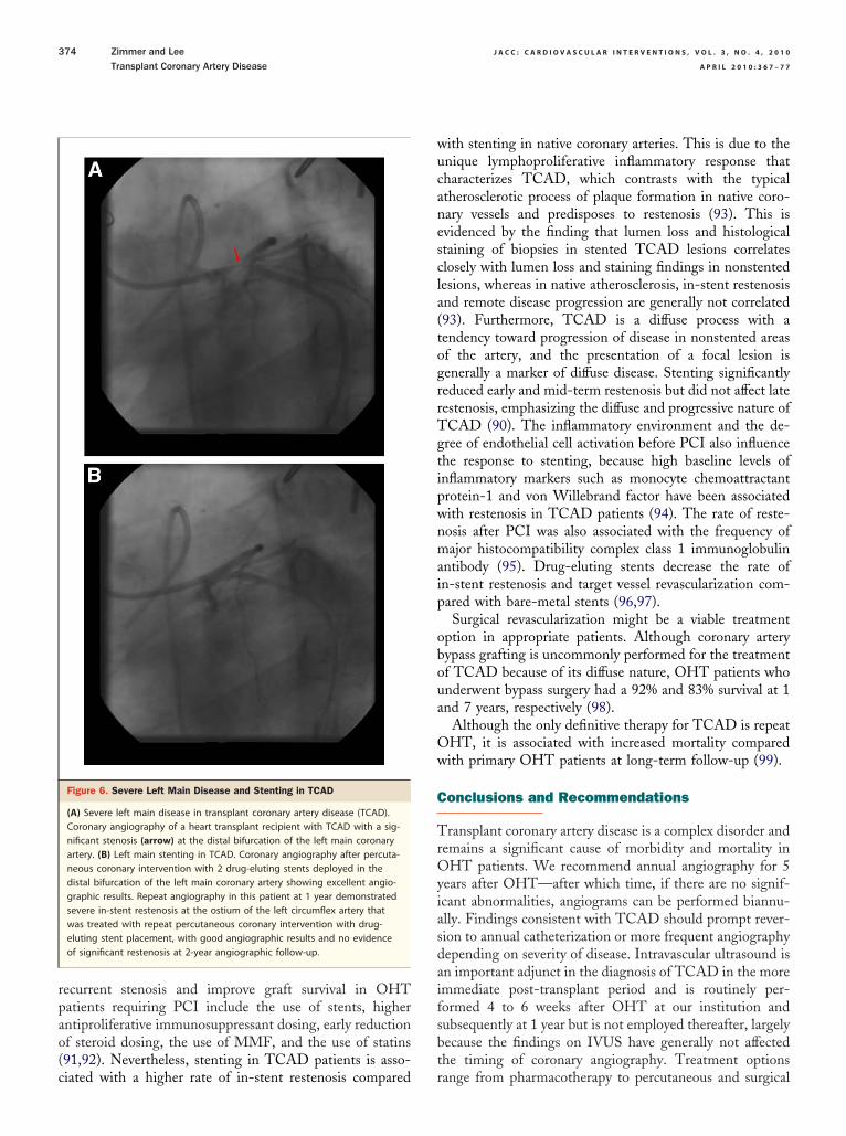
Figure 6. Severe Left Main Disease and Stenting in TCAD
(A) Severe left main disease in transplant coronary artery disease (TCAD).Coronary angiography of a heart transplant recipient with TCAD with a sig-nificant stenosis (arrow) at the distal bifurcation of the left main coronaryartery. (B) Left main stenting in TCAD. Coronary angiography after percuta-neous coronary intervention with 2 drug-eluting stents deployed in thedistal bifurcation of the left main coronary artery showing excellent angio-graphic results. Repeat angiography in this patient at 1 year demonstratedsevere in-stent restenosis at the ostium of the left circumflex artery thatwas treated with repeat percutaneous coronary intervention with drug-eluting stent placement, with good angiographic results and no evidenceof significant restenosis at 2-year angiographic follow-up.
iated with a higher rate of in-stent restenosis compared r
ith stenting in native coronary arteries. This is due to thenique lymphoproliferative inflammatory response thatharacterizes TCAD, which contrasts with the typicaltherosclerotic process of plaque formation in native coro-ary vessels and predisposes to restenosis (93). This isvidenced by the finding that lumen loss and histologicaltaining of biopsies in stented TCAD lesions correlateslosely with lumen loss and staining findings in nonstentedesions, whereas in native atherosclerosis, in-stent restenosisnd remote disease progression are generally not correlated93). Furthermore, TCAD is a diffuse process with aendency toward progression of disease in nonstented areasf the artery, and the presentation of a focal lesion isenerally a marker of diffuse disease. Stenting significantlyeduced early and mid-term restenosis but did not affect lateestenosis, emphasizing the diffuse and progressive nature ofCAD (90). The inflammatory environment and the de-ree of endothelial cell activation before PCI also influencehe response to stenting, because high baseline levels ofnflammatory markers such as monocyte chemoattractantrotein-1 and von Willebrand factor have been associatedith restenosis in TCAD patients (94). The rate of reste-osis after PCI was also associated with the frequency ofajor histocompatibility complex class 1 immunoglobulin
ntibody (95). Drug-eluting stents decrease the rate ofn-stent restenosis and target vessel revascularization com-ared with bare-metal stents (96,97).Surgical revascularization might be a viable treatment
ption in appropriate patients. Although coronary arteryypass grafting is uncommonly performed for the treatmentf TCAD because of its diffuse nature, OHT patients whonderwent bypass surgery had a 92% and 83% survival at 1nd 7 years, respectively (98).
Although the only definitive therapy for TCAD is repeatHT, it is associated with increased mortality comparedith primary OHT patients at long-term follow-up (99).
onclusions and Recommendations
ransplant coronary artery disease is a complex disorder andemains a significant cause of morbidity and mortality inHT patients. We recommend annual angiography for 5
ears after OHT—after which time, if there are no signif-cant abnormalities, angiograms can be performed biannu-lly. Findings consistent with TCAD should prompt rever-ion to annual catheterization or more frequent angiographyepending on severity of disease. Intravascular ultrasound isn important adjunct in the diagnosis of TCAD in the moremmediate post-transplant period and is routinely per-ormed 4 to 6 weeks after OHT at our institution andubsequently at 1 year but is not employed thereafter, largelyecause the findings on IVUS have generally not affectedhe timing of coronary angiography. Treatment options
ange from pharmacotherapy to percutaneous and surgical
rspiapsmrwo
RUtC
R
1
1
1
1
1
1
1
1
1
1
2
2
2
2
2
2
2
2
2
2
3
3
3
3
3
3
J A C C : C A R D I O V A S C U L A R I N T E R V E N T I O N S , V O L . 3 , N O . 4 , 2 0 1 0 Zimmer and Lee
A P R I L 2 0 1 0 : 3 6 7 – 7 7 Transplant Coronary Artery Disease
375
evascularization to repeat OHT. Progression of TCADhould prompt reconsideration of the patient’s immunosup-ressive regimen, with addition or titration of agents,ncluding cyclosporine, tacrolimus, sirolimus, or steroids asble in light of the patient’s existing comorbidities. Theresence of diffuse, severe disease that has been unrespon-ive to pharmacotherapy and that is not amenable to PCIerits evaluation for possible repeat OHT. Prospective
andomized trials comparing different pharmacotherapies asell as revascularization strategies are needed to identify theptimal therapy for patients who develop TCAD.
eprint requests and correspondence: Dr. Michael S. Lee,CLA Medical Center, Adult Cardiac Catheterization Labora-
ory, 10833 Le Conte Avenue, Room A2-237 CHS, Los Angeles,alifornia 90095-171715. E-mail: [email protected].
EFERENCES
1. Ross M, Kouretas P, Gamberg P, et al. Ten- and 20-year survivors ofpediatric orthotopic heart transplantation. J Heart Lung Transplant2006;25:261–70.
2. Taylor DO, Edwards LB, Boucek MM, et al. Registry of theInternational Society for Heart and Lung Transplantation: twenty-second official adult heart transplant report—2005. J Heart LungTransplant 2005;24:945–55.
3. Costanzo MR, Naftel DC, Pritzker MR, et al. Heart transplantcoronary artery disease detected by coronary angiography: a multi-institutional study of preoperative donor and recipient risk factors.Cardiac Transplant Research Database. J Heart Lung Transplant1998;17:744 –53.
4. Roussel JC, Baron O, Périgaud C, et al. Outcome of heart transplants15 to 20 years ago: graft survival, post-transplant morbidity, and riskfactors for mortality. J Heart Lung Transplant 2008;27:486–93.
5. Syeda B, Roedler S, Schukro C, Yahya N, Zuckermann A, Glogar D.Transplant coronary artery disease: incidence, progression and inter-ventional revascularization. Int J Cardiol 2005;104:269–74.
6. Haddad M, Pflugfelder PW, Guiraudon C, et al. Angiographic,pathologic, and clinical relationships in coronary artery disease incardiac allografts. J Heart Lung Transplant 2005;24:1218–25.
7. van Loosdregt J, van Oosterhout MF, Bruggink AH, et al. Thechemokine and chemokine receptor profile of infiltrating cells in thewall of arteries with cardiac allograft vasculopathy is indicative of amemory T-helper 1 response. Circulation 2006;114:1599–607.
8. Tanaka M, Zwierzchoniewska M, Mokhtari GK, et al. Progression ofalloresponse and tissue-specific immunity during graft coronary arterydisease. Am J Transplant 2005;5:1286–96.
9. Vasilescu ER, Ho EK, de la Torre L, et al. Anti-HLA antibodies inheart transplantation. Transpl Immunol 2004;12:177–83.
0. Raichlin E, Bae JH, Kushwaha SS, Lennon RJ, Prasad A, Rihal CS,Lerman A. Inflammatory burden of cardiac allograft coronary athero-sclerotic plaque is associated with early recurrent cellular rejection andpredicts a higher risk of vasculopathy progression. J Am Coll Cardiol2009;53:1279–86.
1. Rahmani M, Cruz RP, Granville DJ, McManus BM. Allograftvasculopathy versus atherosclerosis. Circ Res 2006;99:801–15.
2. Weis M, Cooke JP. Cardiac allograft vasculopathy and dysregulation ofthe NO synthase pathway, Arterioscler Thromb Vasc Biol 2003;23:567–75.
3. Ueland T, Gullestad L, Simonsen S, et al. Decreased endomyocardialRANKL expression in transplant coronary artery disease. Transplan-tation 2006;81:1467–70.
4. Wildhirt SM, Schulze C, Conrad N, Bauernschmitt R, Lange R, vonScheidt W. Persistently increased systemic, but not cardiac-specific,
adhesion molecule expression and coronary endothelial dysfunction inhuman cardiac allografts. J Thorac Cardiovasc Surg 2005;130:1175.
5. Tanaka M, Sydow K, Gunawan F, et al. Dimethylarginine dimethyl-aminohydrolase overexpression suppresses graft coronary artery disease.Circulation 2005;112:1549–56.
6. El-Hamamsy I, Stevens LM, Vanhoutte PM, Perrault LP. Injury ofthe coronary endothelium at implantation increases endothelial dys-function and intimal hyperplasia after heart transplantation. J HeartLung Transplant 2005;24:251–8.
7. Murata S, Miniati DN, Kon MH, et al. Superoxide dismutase mimeticm40401 reduces ischemia-reperfusion injury and graft coronary arterydisease in rodent cardiac allografts. Transplantation 2004;78:1166–71.
8. Tanaka M, Mokhtari GK, Terry RD, et al. Overexpression of humancopper/zinc superoxide dismutase (SOD1) suppresses ischemia-reperfusion injury and subsequent development of graft coronary arterydisease in murine cardiac grafts. Circulation 2004;110:II200–6.
9. Uehara S, Chase CM, Kitchens WH, et al. NK cells can triggerallograft vasculopathy: the role of hybrid resistance in solid organallografts. J Immunol 2005;175:3424–30.
0. Chu KE, Ho EK, de la Torre L, Vasilescu ER, Marboe CC. Therelationship of nodular endocardial infiltrates (Quilty lesions) to sur-vival, patient age, anti-HLA antibodies, and coronary artery diseasefollowing heart transplantation. Cardiovasc Pathol 2005;14:219–24.
1. Ternstrom L, Jeppsson A, Ricksten A, Nilsson F. Tumor necrosisfactor gene polymorphism and cardiac allograft vasculopathy. J HeartLung Transplant 2005;24:433–8.
2. Borish LC, Steinke JW. 2. Cytokines and chemokines. J Allergy ClinImmunol 2003;111:S460–75.
3. De Souza AI, Wait R, Mitchell AG, Banner NR, Dunn MJ, Rose ML.Heat shock protein 27 is associated with freedom from graft vascu-lopathy after human cardiac transplantation. Circ Res 2005;97:192–8.
4. Densem CG, Hutchinson IV, Yonan N, Brooks NH. Donor andrecipient-transforming growth factor-beta 1 polymorphism and cardiactransplant-related coronary artery disease. Transpl Immunol 2004;13:211–7.
5. Li H, Tanaka K, Oeser B, Kobashigawa JA, Tobis JM. Vascularremodelling after cardiac transplantation: a 3-year serial intravascularultrasound study. Eur Heart J 2006;27:1671–7.
6. Atkinson C, Southwood M, Pitman R, Phillpotts C, Wallwork J,Goddard M. Angiogenesis occurs within the intimal proliferation thatcharacterizes transplant coronary artery vasculopathy. J Heart LungTransplant 2005;24:551–8.
7. Holweg CT, Balk AH, Snaathorst J, et al. Intragraft heme oxygenase-1and coronary artery disease after heart transplantation. Transpl Immu-nol 2004;13:265–72.
8. Bader FM, Kfoury AG, Gilbert EM, et al. Percutaneous coronaryinterventions with stents in cardiac transplant recipients. J Heart LungTransplant 2006;25:298–301.
9. Bae JH, Rihal CS, Edwards BS, et al. Association of angiotensin-converting enzyme inhibitors and serum lipids with plaque regressionin cardiac allograft vasculopathy. Transplantation 2006;82:1108–11.
0. Kocık M, Malek I, Janek B, Zelızko M, Pirk J. Risk factors for thedevelopment of coronary artery disease of a grafted heart as detectedvery early after orthotopic heart transplantation. Transpl Int 2007;20:666–74.
1. Topkara VK, Dang NC, John R, et al. A decade experience of cardiacretransplantation in adult recipients. J Heart Lung Transplant 2005;24:1745–50.
2. Walker AH, Fildes JE, Leonard CT, Yonan N. The influence of donorage on transplant coronary artery disease and survival post hearttransplantation: is it safe to extend donor age? Transplant Proc2004;36:3139–41.
3. Botha P, Peaston R, White K, Forty J, Dark JH, Parry G. Smokingafter cardiac transplantation. Am J Transplant 2008;8:866–71.
4. Radovancevic B, Poindexter S, Birovljev S, et al. Risk factors fordevelopment of accelerated coronary artery disease in cardiac transplantrecipients. Eur J Cardiothorac Surg 1990;4:309–12.
5. Dobbels F, De Geest S, van Cleemput J, Droogne W, Vanhaecke J.Effect of late medication non-compliance on outcome after heart
transplantation: a 5-year follow-up. J Heart Lung Transplant 2004;23:1245–51.
3
3
3
3
4
4
4
4
4
4
4
4
4
4
5
5
5
5
5
5
5
5
5
5
6
6
6
6
6
6
6
6
6
6
7
7
7
7
7
7
7
J A C C : C A R D I O V A S C U L A R I N T E R V E N T I O N S , V O L . 3 , N O . 4 , 2 0 1 0
A P R I L 2 0 1 0 : 3 6 7 – 7 7
Zimmer and Lee
Transplant Coronary Artery Disease
376
6. Raichlin E, Edwards BS, Kremers WK, et al. Acute cellular rejectionand the subsequent development of allograft vasculopathy after cardiactransplantation. J Heart Lung Transplant 2009;28:320–7.
7. Kato T, Chan MC, Gao SZ, et al. Glucose intolerance, as reflectedby hemoglobin A1c level, is associated with the incidence andseverity of transplant coronary artery disease. J Am Coll Cardiol2004;43:1034 – 41.
8. Bozbas H, Altin C, Yildirir A, et al. Lipid profiles of patients with atransplanted heart before and after the operation. Transplant Proc2008;40:263–6.
9. Shirakawa I, Sata M, Saiura A, et al. Atorvastatin attenuatestransplant-associated coronary arteriosclerosis in a murine model ofcardiac transplantation. Biomed Pharmacother 2007;61:154–9.
0. Li H, Tanaka K, Anzai H, et al. Influence of pre-existing donoratherosclerosis on the development of cardiac allograft vasculopathyand outcomes in heart transplant recipients. J Am Coll Cardiol2006;47:2470–6.
1. Takayama H, Nathens AB, Merry H, et al. Is pre-transplant vasculardisease a risk factor for mortality and morbidity after heart transplan-tation? Eur J Cardiothorac Surg 2007;31:457–61.
2. Grattan MT, Moreno-Cabral CE, Starnes VA, Oyer PE, Stinson EB,Shumway NE. Cytomegalovirus infection is associated with cardiacallograft rejection and atherosclerosis. JAMA 1989;261:3561–6.
3. Hussain T, Burch M, Fenton MJ, et al. Positive pretransplantationcytomegalovirus serology is a risk factor for cardiac allograft vasculopa-thy in children. Circulation 2007;115:1798–805.
4. Shiba N, Chan MC, Kwok BW, Valantine HA, Robbins RC, HuntSA. Analysis of survivors more than 10 years after heart transplantationin the cyclosporine era: Stanford experience. J Heart Lung Transplant2004;23:155–64.
5. Price JF, Towbin JA, Dreyer WJ, et al. Symptom complex is associatedwith transplant coronary artery disease and sudden death/resuscitatedsudden death in pediatric heart transplant recipients. J Heart LungTransplant 2005;24:1798–803.
6. Kass M, Allan R, Haddad H. Diagnosis of graft coronary arterydisease. Curr Opin Cardiol 2007;22:139–45.
7. Gibson CM, Murphy SA, Rizzo MJ, et al., Thrombolysis In Myocar-dial Infarction (TIMI) Study Group. Relationship between TIMIframe count and clinical outcomes after thrombolytic administration.Circulation 1999:1945-50.
8. Baris N, Sipahi I, Kapadia SR, et al. Coronary angiography forfollow-up of heart transplant recipients: insights from TIMI framecount and TIMI myocardial perfusion grade. J Heart Lung Transplant2007;26:593–7.
9. Mintz GS, Nissen SE, Anderson WD, et al. American College ofCardiology clinical expert consensus document on standards for acqui-sition, measurement and reporting of intravascular ultrasound studies(IVUS): a report of the American College of Cardiology Task Force onClinical Expert Consensus Documents. J Am Coll Cardiol 2001;37:1478–92.
0. Tuzcu EM, Kapadia SR, Sachar R, et al. Intravascular ultrasoundevidence of angiographically silent progression in coronary atheroscle-rosis predicts long-term morbidity and mortality after cardiac trans-plantation. J Am Coll Cardiol 2005;45:1538–42.
1. Kobashigawa JA, Tobis JM, Starling RC, et al. Multicenter intravas-cular ultrasound validation study among heart transplant recipients:outcomes after five years. J Am Coll Cardiol 2005;45:1532–7.
2. König A, Kilian E, Sohn HY, et al. Assessment and characterization oftime-related differences in plaque composition by intravascularultrasound-derived radiofrequency analysis in heart transplant recipi-ents. J Heart Lung Transplant 2008;27:302–9.
3. Bogot NR, Durst R, Shaham D, Admon D. Cardiac CT of thetransplanted heart: indications, technique, appearance, and complica-tions. Radiographics 2007;27:1297–309.
4. Gregory SA, Ferencik M, Achenbach S, et al. Comparison of sixty-four-slice multidetector computed tomographic coronary angiographyto coronary angiography with intravascular ultrasound for the detectionof transplant vasculopathy. Am J Cardiol 2006;98:877–84.
5. Sigurdsson G, Carrascosa P, Yamani MH, et al. Detection of trans-
plant coronary artery disease using multidetector computed tomogra-phy with adaptative multisegment reconstruction. J Am Coll Cardiol2006;48:772–8.
6. Schepis T, Achenbach S, Weyand M, et al. Comparison of dual sourcecomputed tomography versus intravascular ultrasound for evaluation ofcoronary arteries at least one year after cardiac transplantation. Am JCardiol 2009;104:1351–6.
7. Iyengar S, Feldman DS, Cooke GE, Leier CV, Raman SV. Detectionof coronary artery disease in orthotopic heart transplant recipients with64-detector row computed tomography angiography. J Heart LungTransplant 2006;25:1363–6.
8. Elhendy A, Sozzi FB, van Domburg RT, et al. Accuracy of dobutaminetetrofosmin myocardial perfusion imaging for the noninvasive diagnosisof transplant coronary artery stenosis. J Heart Lung Transplant2000;19:360–6.
9. Rodrigues AC, Bacal F, Medeiros CC, et al. Noninvasive detection ofcoronary allograft vasculopathy by myocardial contrast echocardiogra-phy. J Am Soc Echocardiogr 2005;18:116–21.
0. Hognestad A, Endresen K, Wergeland R, et al. Plasma C-reactiveprotein as a marker of cardiac allograft vasculopathy in heart transplantrecipients. J Am Coll Cardiol 2003;42:477–82.
1. Fyfe AI, Rothenberg LS, DeBeer FC, Cantor RM, Rotter JI, Lusis AJ.Association between serum amyloid A proteins and coronary arterydisease: evidence from two distinct arteriosclerotic processes. Circula-tion 1997;96:2914–9.
2. Hilgendorff A, Kraemer U, Afsharian M, et al. Value of solubleadhesion molecules and plasma coagulation markers in assessingtransplant coronary artery disease in pediatric heart transplant recipi-ents. Pediatr Transplant 2006;10:434–40.
3. Mehra MR, Uber PA, Potluri S, Ventura HO, Scott RL, Park MH.Usefulness of an elevated B-type natriuretic peptide to predict allograftfailure, cardiac allograft vasculopathy, and survival after heart trans-plantation. Am J Cardiol 2004;94:454–8.
4. Crawford SE, Mavroudis C, Backer CL, et al. Captopril suppressespost-transplantation angiogenic activity in rat allograft coronary vessels.J Heart Lung Transplant 2004;23:666–73.
5. Yamamoto T, Sata M, Fukuda D, Takamoto S. The angiotensin IItype 1 receptor blocker valsartan attenuates graft vasculopathy. BasicRes Cardiol 2005;100:84–91.
6. Yousufuddin M, Cook DJ, Starling RC, et al. Angiotensin II receptorsfrom peritransplantation through first-year post-transplantation andthe risk of transplant coronary artery disease. J Am Coll Cardiol2004;43:1565–73.
7. Kobashigawa JA, Katznelson S, Laks H, et al. Effect of pravastatin onoutcomes after cardiac transplantation. N Engl J Med 1995;333:621–7.
8. Kobashigawa JA. Statins and cardiac allograft vasculopathy after hearttransplantation. Semin Vasc Med 2004;4:401–6.
9. Lijkwan MA, Cooke DT, Martens JM, et al. Cyclosporine treatmentof high dose and long duration reduces the severity of graft coronaryartery disease in rodent cardiac allografts. J Heart Lung Transplant2005;24:439–45.
0. Soukiasian HJ, Czer LS, Wang HM, et al. Inhibition of graft coronaryarteriosclerosis after heart transplantation. Am Surg 2004;70:833–40.
1. Lungu AO, Jin ZG, Yamawaki H, Tanimoto T, Wong C, Berk BC.Cyclosporin A inhibits flow-mediated activation of endothelial nitric-oxide synthase by altering cholesterol content in caveolae. J Biol Chem2004;279:48794–800.
2. Oriji GK, Keiser HR. Nitric oxide in cyclosporine A-induced hyper-tension: role of protein kinase C. Am J Hypertens 1999;12:1091–7.
3. Coffman TM, Carr DR, Yarger WE, Klotman PE. Evidence that renalprostaglandin and thromboxane production is stimulated in chroniccyclosporine nephrotoxicity. Transplantation 1987;43:282–5.
4. Rosenthal RA, Chukwuogo NA, Ocasio VH, Kahng KU. Cyclospo-rine inhibits endothelial cell prostacyclin production. J Surg Res1989;46:593–6.
5. Haug C, Duell T, Voisard R, et al. Cyclosporine A stimulatesendothelin release. J Cardiovasc Pharmacol 1995;26:S239–41.
6. Takeda Y, Miyamori I, Wu P, Yoneda T, Furukawa K, Takeda R.Effects of an endothelin receptor antagonist in rats with cyclosporine-
induced hypertension. Hypertension 1995;26:932–6.
7
7
7
8
8
8
8
8
8
8
8
8
8
9
9
9
9
9
9
9
9
9
9
J A C C : C A R D I O V A S C U L A R I N T E R V E N T I O N S , V O L . 3 , N O . 4 , 2 0 1 0 Zimmer and Lee
A P R I L 2 0 1 0 : 3 6 7 – 7 7 Transplant Coronary Artery Disease
377
7. Humiston D, Taylor D, Kfoury A, et al. Mycophenolate mofetilhistory and introduction into clinical heart transplantation. Cardiovas-cular Engineering 1997;2:198.
8. Kobashigawa J, Miller L, Renlund D, et al.; Mycophenolate MofetilInvestigators. A randomized active-controlled trial of mycophenolatemofetil in heart transplant recipients. Transplantation 1998;66:507–15.
9. Kobashigawa JA, Tobis JM, Mentzer RM, et al. Mycophenolatemofetil reduces intimal thickness by intravascular ultrasound after hearttransplant: reanalysis of the multicenter trial. Am J Transplant 2006;6:993–7.
0. Ikonen TS, Gummert JF, Serkova N, et al. Efficacies of sirolimus(rapamycin) and cyclosporine in allograft vascular disease in non-human primates trough levels of sirolimus correlate with inhibition ofprogression of arterial intimal thickening. Transplant Int 2000;13:S314–20.
1. Murphy GJ, Bicknell GR, Nicholson ML. Rapamycin inhibits vascularremodeling in an experimental model of allograft vasculopathy andattenuates associated changes in fibrosis-associated gene expression.J Heart Lung Transplant 2003;22:533–41.
2. Mehra MR, Uber PA. TOR inhibitors and cardiac allograft vasculopa-thy is inhibition of intimal thickening an adequate surrogate of benefit?J Heart Lung Transplant 2003;22:501–4.
3. Keogh A, Richardson M, Ruygrok P, et al. Sirolimus in de novo hearttransplant recipients reduces acute rejection and prevents coronaryartery disease at 2 years: a randomized clinical trial. Circulation2004;110:2694–700.
4. Lobach NE, Pollock-Barziv SM, West LJ, Dipchand AI. Sirolimusimmunosuppression in pediatric heart transplant recipients: a single-center experience. J Heart Lung Transplant 2005;24:184–9.
5. Hafizi S, Mordi VN, Andersson KM, Chester AH, Yacoub MH.Differential effects of rapamycin, cyclosporine A, and FK506 on humancoronary artery smooth muscle cell proliferation and signalling. VasculPharmacol 2004;41:167–76.
6. Raichlin E, Bae JH, Khalpey Z, et al. Conversion to sirolimus asprimary immunosuppression attenuates the progression of allograftvasculopathy after cardiac transplantation. Circulation 2007;116:2726 –33.
7. Raichlin E, Prasad A, Kremers WK, et al. Sirolimus as primaryimmunosuppression is associated with improved coronary vasomotor
function compared with calcineurin inhibitors in stable cardiac trans-plant recipients. Eur Heart J 2009;30:1356–63. K8. Kuppahally S, Al-Khaldi A, et al. Wound healing complications withde novo sirolimus versus mycophenolate mofetil-based regimen incardiac transplant recipients. Am J Transplant 2006;6:986–92.
9. Tanaka K, Li H, Curran PJ, et al. Usefulness and safety of percutane-ous coronary interventions for cardiac transplant vasculopathy. Am JCardiol 2006;97:1192–7.
0. Simpson L, Lee EK, Hott BJ, Vega DJ, Book WM. Long-term resultsof angioplasty vs stenting in cardiac transplant recipients with allograftvasculopathy. J Heart Lung Transplant 2005;24:1211–7.
1. Benza RL, Zoghbi GJ, Tallaj J, et al. Palliation of allograft vasculopa-thy with transluminal angioplasty: a decade of experience. J Am CollCardiol 2004;43:1973–81.
2. Wellnhofer E, Hiemann NE, Hug J, et al. A decade of percutaneouscoronary interventions in cardiac transplant recipients: a monocentricstudy in 160 patients. J Heart Lung Transplant 2008;27:17–25.
3. Jonas M, Fang JC, Wang JC, et al. In-stent restenosis and remotecoronary lesion progression are coupled in cardiac transplant vasculopa-thy but not in native coronary artery disease. J Am Coll Cardiol2006;48:453–61.
4. Hognestad A, Endresen K, Wergeland R, et al. Inflammatory responseand re-stenosis after percutaneous coronary intervention in hearttransplant recipients and patients with native atherosclerosis. J HeartLung Transplant 2005;24:1026–32.
5. McKay M, Pinney S, Gorwara S, et al. Anti-human leukocyte antigenantibodies are associated with restenosis after percutaneous coronaryintervention for cardiac allograft vasculopathy. Transplantation 2005;79:1581–7.
6. Lee MS, Kobashigawa JA, Tobis JM. Comparison of percutaneouscoronary intervention with bare-metal and drug-eluting stents forcardiac allograft vasculopathy. J Am Coll Cardiol Intv 2008;1;710–5.
7. Zakliczynski M, Lekston A, Osuch M, et al. Comparison of long-termresults of drug-eluting stent and bare metal stent implantation in hearttransplant recipients with coronary artery disease. Transplant Proc2007;39:2859–61.
8. Bhama JK, Nguyen DQ, Scolieri S, et al. Surgical revascularization forcardiac allograft vasculopathy: Is it still an option? J Thorac CardiovascSurg 2009;137:1488–92.
9. Mahle WT, Vincent RN, Kanter KR. Cardiac retransplantation inchildhood: analysis of data from the United Network for OrganSharing. J Thorac Cardiovasc Surg 2005;130:542–6.
ey Words: drug-eluting stent � heart transplantation.