Embed Size (px)
DESCRIPTION
CORONARY ARTERY DISEASE & ACUTE CORONARY SYNDROME. Anne Lucero ALTERED TISSUE PERFUSION. Definition -. CORONARY ARTERY DISEASE is a type of blood vessel disorder included under the category of atherosclerosis - PowerPoint PPT Presentation
Citation preview
CORONARY ARTERY DISEASE & ACUTE
CORONARY SYNDROME
Anne Lucero
ALTERED TISSUE PERFUSION
Definition -
• CORONARY ARTERY DISEASE is a type of blood vessel disorder included under the category of atherosclerosis
• ACUTE CORONARY SYNDROME spectrum of clinical syndromes representing degrees of coronary artery occlusion, including unstable angina, non-ST-segment Elevation MI, and ST-segment Elevation MI, or sudden death.
• ATHEROSCLEROSIS The accumulation of lipid containing materials found in the intima of the arteries, begins as soft deposits of fat and hardens with age
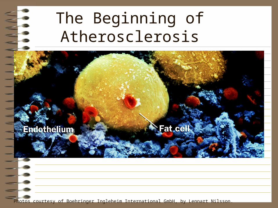
The Beginning of Atherosclerosis
Photos courtesy of Boehringer Ingleheim International GmbH, by Lennart Nilsson.
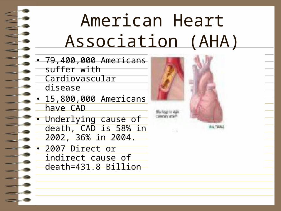
American Heart Association (AHA)
• 79,400,000 Americans suffer with Cardiovascular disease
• 15,800,000 Americans have CAD
• Underlying cause of death, CAD is 58% in 2002, 36% in 2004.
• 2007 Direct or indirect cause of death=431.8 Billion
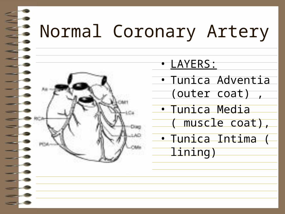
Normal Coronary Artery
• LAYERS: • Tunica Adventia
(outer coat) , • Tunica Media ( muscle
coat), • Tunica Intima ( lining)
Coronary Artery location and service
• Right coronary artery (RCA) descends to the right along the atrioventricular groove and turns posteriorly along the inferior margin of the right ventricle and diaphragmatic surface of the left ventricle. Supplies the Right atrium, right ventricle, posterior wall left ventricle and 90% of the AV node
.
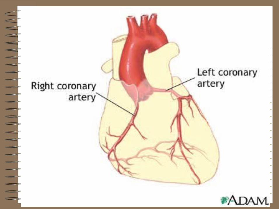
Coronary Artery, cont.
• Left coronary artery (LCA) has two branches, the left anterior descending (LAD) supplies the anterior portion of the LV and lower half of the LV, and a portion of the interventricular septum, terminates before reaching the posterior area. The left circumflex artery (LC) supplies the left atrium, high lateral wall LV
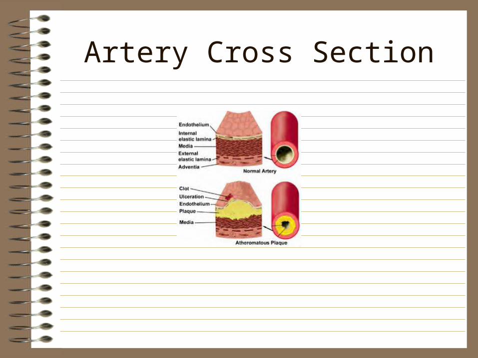
Artery Cross Section
Evolution to the Major Acute Coronary Syndromes
• Early Lipid formation,lipid deposits lead to narrowing of the artery lumen, with in the intimal wall
• Unstable plaque are not hemodynamically significant before rupture
• Inflammation in subendotheial area further weakens, predisposes plaque to rupture
• Speed of flow, turbulence, vessel anatomy contribute• Endothelium injury is site for fibrous plaque
thickening
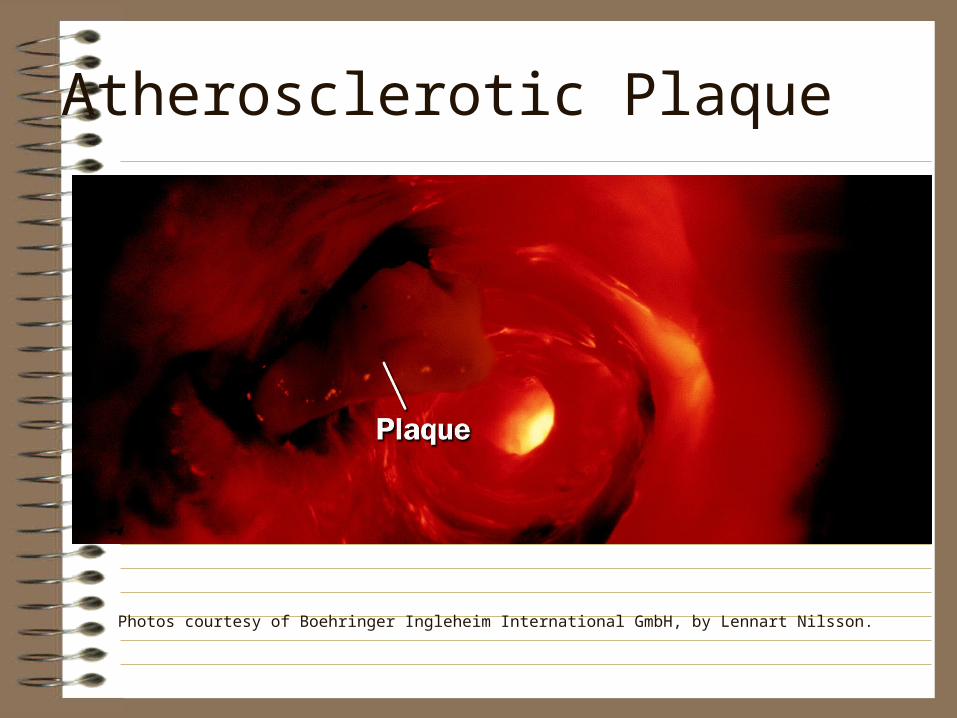
Atherosclerotic Plaque
Photos courtesy of Boehringer Ingleheim International GmbH, by Lennart Nilsson.
Pathophysiology cont
• Plaque Rupture, • Monolayer of platelets cover surface of rupture• Platelet adhesion
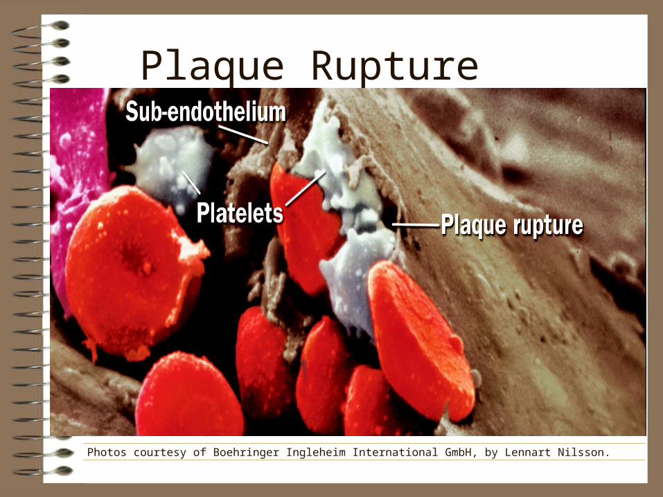
Plaque Rupture
Photos courtesy of Boehringer Ingleheim International GmbH, by Lennart Nilsson.
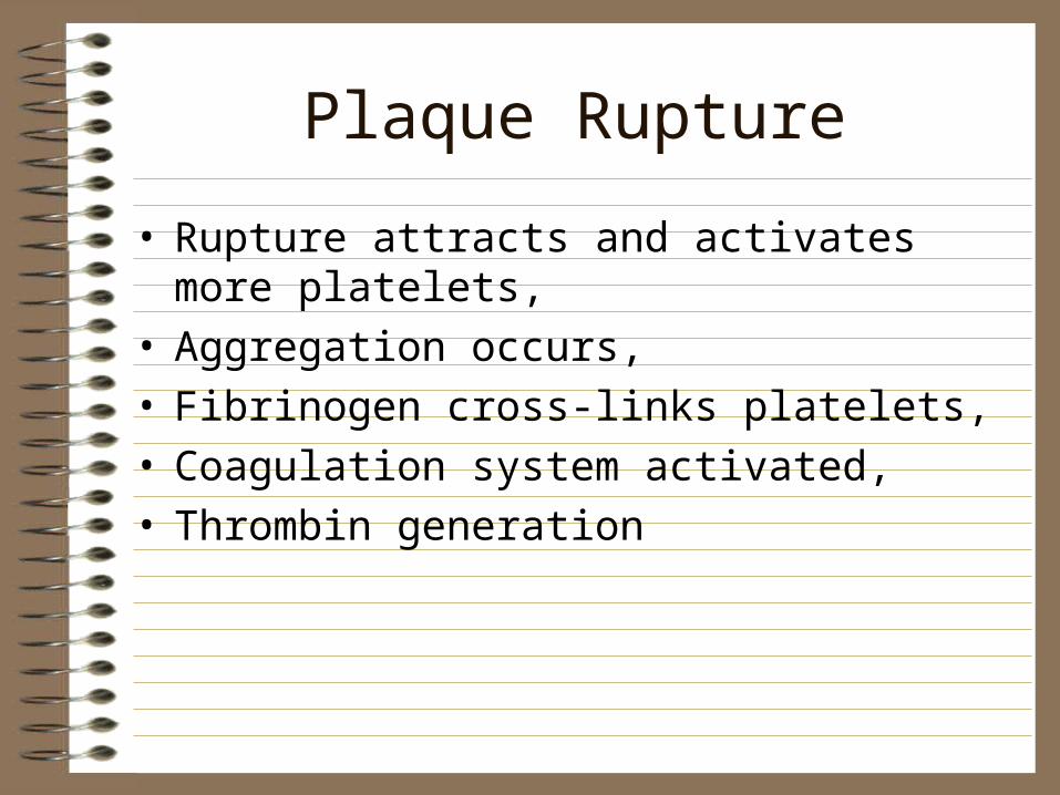
Plaque Rupture
• Rupture attracts and activates more platelets,• Aggregation occurs,• Fibrinogen cross-links platelets, • Coagulation system activated,• Thrombin generation

Platelet Activation
Photos courtesy of Boehringer Ingleheim International GmbH, by Lennart Nilsson.
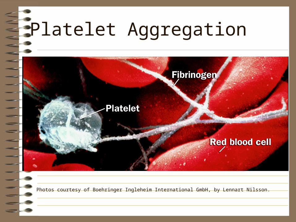
Platelet Aggregation
Photos courtesy of Boehringer Ingleheim International GmbH, by Lennart Nilsson.
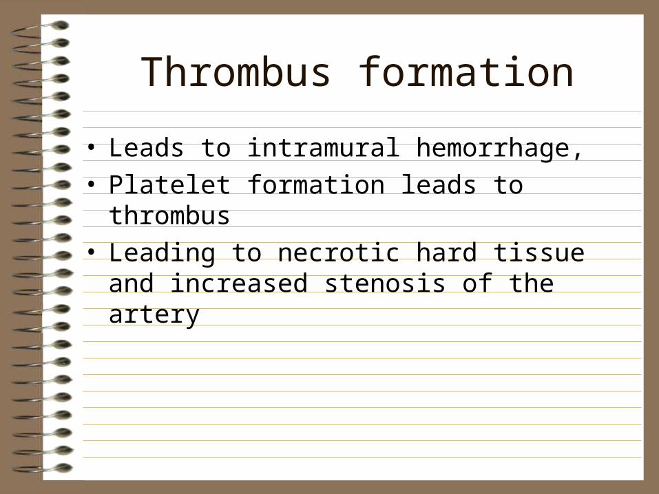
Thrombus formation
• Leads to intramural hemorrhage, • Platelet formation leads to thrombus• Leading to necrotic hard tissue and increased
stenosis of the artery
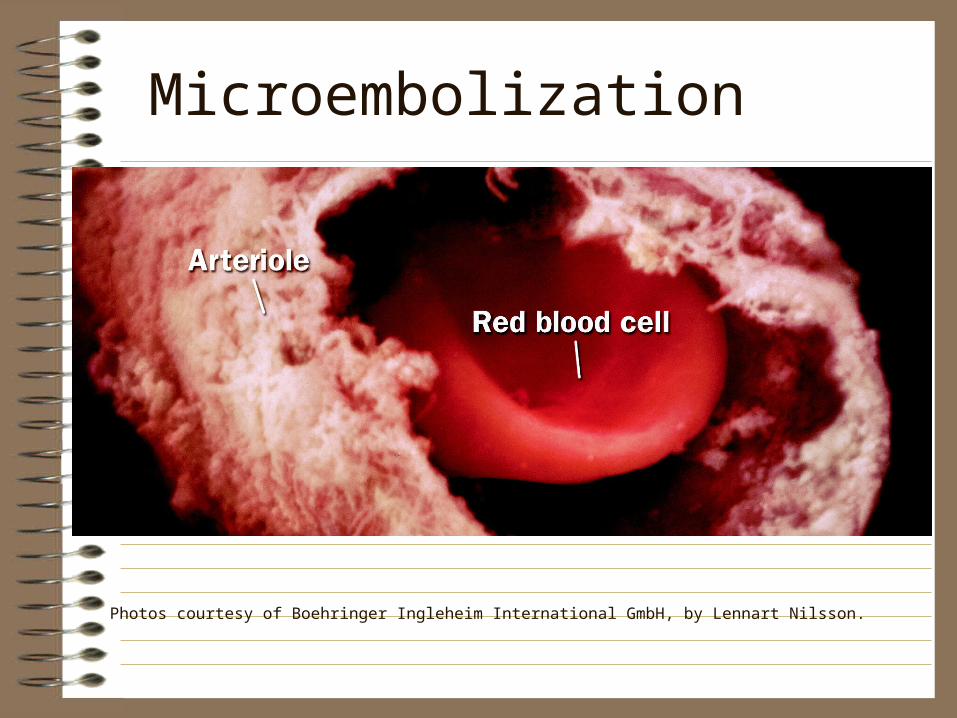
Microembolization
Photos courtesy of Boehringer Ingleheim International GmbH, by Lennart Nilsson.
Unstable Angina
• Partially occluding thrombus produces symptoms of ischemia
• Thrombus is platelet-rich,( tx with antiplatelet agents( asa),Glycoprotein Iib/IIIa inhibitors(RePro, Aggrastat) most effective
• Fibrinolytic tx is NOT effective, can cause paradoxical accelerate occlusion by release of clot bound thrombin, further activating platelets
• Intermittently occlusive thrombus may cause Myocardial necrosis, producing a non- elevated ST-segment MI
Occlusive Thrombus
• Clot enlarges, microemboli from distal thrombus lodge in coronary microvasculature, small elevations in cardiac troponin, high risk for MI
• If occludes flow for prolonged period,ST-segment elevation MI occurs, clot rich in Thrombin
• Prompt fibrinolysis( TPA, Activase, TNKase) or PTCA, may limit size, increase survival
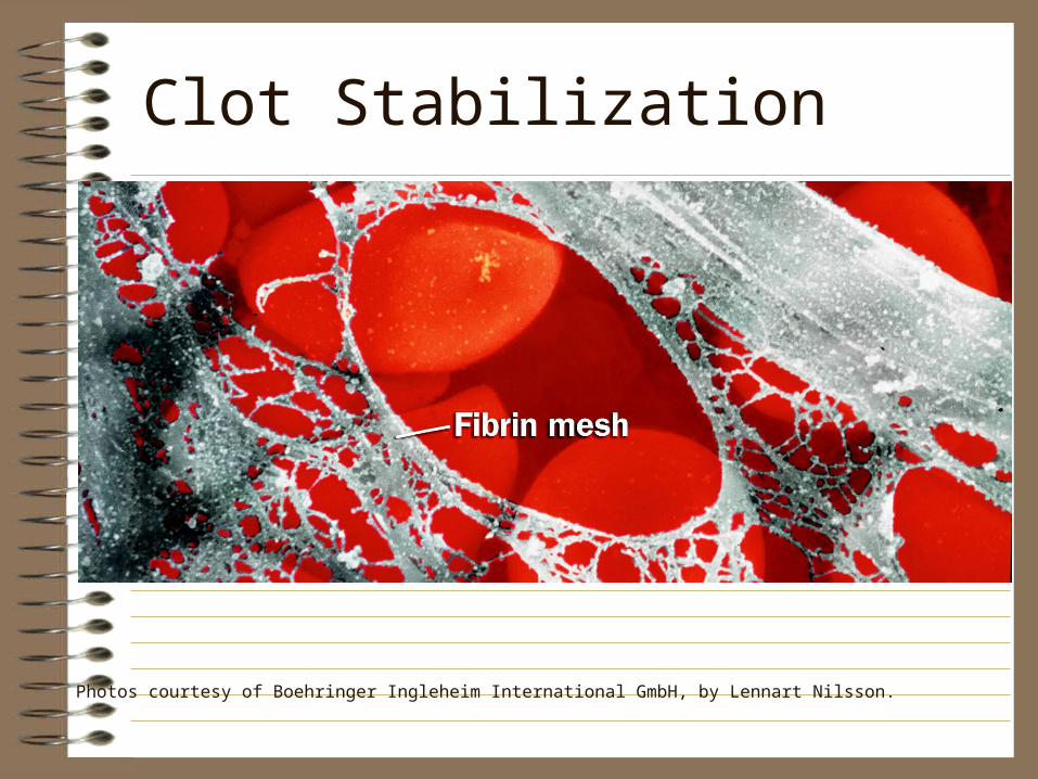
Clot Stabilization
Photos courtesy of Boehringer Ingleheim International GmbH, by Lennart Nilsson.
Risk Factors
• Characteristics or conditions that are statistically associated with high incidence of CAD, ACS
• Two types are: Unmodifiable and Modifiable

MODIFIABLE RISK FACTOR
Unmodifiable / Modifiable
• gender
• age
• family history
• race
• hypertension
• elevated serum lipids
• nutrition/ homocysteine level
• smoking
• obesity
• physical activity
Contributing Factors
• Diabetes
• Life style
• Personal stress

CASE STUDY
• MRS. JOHNSON, 52 years old, Father died of Cancer at 72, Mother has Hypertension and recently had cataract surgery. No siblings, has two daughters in good health.
• Visits Physicians office with a complaint of chest discomfort, 5’7” 190#, B/P 156/78, HR 95, RR 22, works out with a trainer 2X week, not a smoker.

Clinical Manifestations
• Angina : literally from the Greek, “Strangling of the breast bone”
• Physiology for Angina: CAD causes narrow lumen, less O2 carried to the myocardium, anaerobic metabolism stimulates nerve endings, transmits PAIN to cardiac nerves, Ischemia.
Angina Types/ Patterns
• Stable angina- brought on by exercise, relieved at rest
• Unstable angina-occurs with little effort, often stress induced
• Prinzmetal’s- chest discomfort at rest
• Nocturnal angina- only at night
• Angina decubitus- only laying down
• Intractable angina- refractory to medication, chronic
Chest Pain
• TIME-1-15 min duration, >30 min=MI
• QUALITY- stab, dull, burning
• QUANTIFY- scale of 1-10
• LOCATION-chest, jaw, groin,
• PRECIPITATING FACTORS- activity, sleep, family
• RELIEF FACTORS- rest, NTG, O2, MS
Diagnostics
• EKG• Labs-CPK-mb, Troponin, CRP(C-reactive
protein), Chem panel, pt/ptt, CBC,• History and Physical• CXR• Stress Test• Echocardiography• Coronary Angiography

UNFOLDING CASE
• MRS Johnson now has had an EKG in the office, it is abnormal
• Further studies are indicated and she is sent to the Hospital ER
• What would you want to know at this point?
Treatment Options
• Health Promotion
• Nutrition
• Pharmacology: Morphine, Oxygen, Nitroglycerin, ASA (MONA) Beta blockers, Calcium Channel, Antiplatelet aggregation agents, Glycoprotein IIb/IIIa Inhibitors
Nursing Diagnosis
• PAIN : related to Ischemia
• ANXIETY : related to diagnosis awareness
• DECREASED CARDIAC OUTPUT : related to myocardial ischemia
• ACTIVITY INTOLERENCE : related to myocardial ischemia
References• [http://www.acc.org] American College of
Cardiology• [http://www.americanheart.org/cpr]
American Heart Association• Understanding the Pathophysiology of
Acute Coronary Syndrome, A Crusade Educational tool. Duke Clinical Research Institute, Durham, NC 27705