Embed Size (px)
Citation preview

ORIGINAL RESEARCHpublished: 24 July 2015
doi: 10.3389/fnana.2015.00095
Transmitter inputs to differentmotoneuron subgroups in theoculomotor and trochlear nucleus inmonkeyChristina Zeeh 1, Michael J. Mustari 2, Bernhard J. M. Hess 3 and Anja K. E. Horn 1*
1 Institute of Anatomy and Cell Biology, Department I, Ludwig-Maximilians University, Munich, Germany, 2 WashingtonNational Primate Research Center and Department of Ophthalmology, University of Washington, Seattle, WA, USA,3 Vestibulo-Oculomotor Laboratory Zürich, Department of Neurology, University Hospital, Zürich, Switzerland
Edited by:Ricardo Insausti,
University of Castilla-La Mancha,Spain
Reviewed by:José M. Delgado-García,
University Pablo de Olavide,de Seville, SpainZoltan Rusznak,
Neuroscience Research Australia,Australia
*Correspondence:Anja K. E. Horn,
Institute of Anatomy and Cell Biology,Department I, Ludwig-MaximiliansUniversity, Pettenkoferstraße 11,
D-80336 Munich, [email protected]
Received: 07 May 2015Accepted: 06 July 2015Published: 24 July 2015
Citation:Zeeh C, Mustari MJ, Hess BJM andHorn AKE (2015) Transmitter inputs
to different motoneuron subgroups inthe oculomotor and trochlear nucleus
in monkey.Front. Neuroanat. 9:95.
doi: 10.3389/fnana.2015.00095
In all vertebrates the eyes are moved by six pairs of extraocular muscles enablinghorizontal, vertical and rotatory movements. Recent work showed that each extraocularmuscle is controlled by two motoneuronal groups: (1) Motoneurons of singly-innervatedmuscle fibers (SIF) that lie within the boundaries of motonuclei mediating a fast musclecontraction; and (2) motoneurons of multiply-innervated muscle fibers (MIF) in theperiphery of motonuclei mediating a tonic muscle contraction. Currently only limiteddata about the transmitter inputs to the SIF and MIF motoneurons are available. Here weperformed a quantitative study on the transmitter inputs to SIF and MIF motoneuronsof individual muscles in the oculomotor and trochlear nucleus in monkey. Pre-labeledmotoneurons were immunostained for GABA, glutamate decarboxylase, GABA-Areceptor, glycine transporter 2, glycine receptor 1, and vesicular glutamate transporters1 and 2. The main findings were: (1) the inhibitory control of SIF motoneurons forhorizontal and vertical eye movements differs. Unlike in previous primate studies aconsiderable GABAergic input was found to all SIF motoneuronal groups, whereasa glycinergic input was confined to motoneurons of the medial rectus (MR) musclemediating horizontal eye movements and to those of the levator palpebrae (LP) muscleelevating the upper eyelid. Whereas SIF and MIF motoneurons of individual eye musclesdo not differ numerically in their GABAergic, glycinergic and vGlut2 input, vGlut1containing terminals densely covered the supraoculomotor area (SOA) targeting MR MIFmotoneurons. It is reasonable to assume that the vGlut1 input affects the near responsesystem in the SOA, which houses the preganglionic neurons mediating pupillaryconstriction and accommodation and the MR MIF motoneurones involved in vergence.
Keywords: Glycine, GABA, vGlut, C-group, extraocular muscles
Abbreviations: AD, averaged density; AMPA receptors, α-amino-3-hydroxy-5-methyl-4-isoxazolepropionic acid receptor;ATD, ascending tract of Deiters; CCN, central caudal nucleus; ChAT, choline acetyltransferase; CMRF, centralmesencephalic reticular formation; CR, calretinin; CTB, Cholera toxin subunit B; EWpg, preganglionic Edinger-Westphalnucleus; GABA, gamma-aminobutyric acid; GABA-A, GABA-A receptor; GAD, glutamate decarboxylase; GlyR, glycinereceptor; GlyT, glycine transporter; INC, interstitial nucleus of Cajal; INT, internuclear neurons; IO, inferior obliquemuscle; IPSP, inhibitory postsynaptic potential; IR, inferior rectus muscle; LP, levator palpebrae muscle; LR, lateralrectus muscle; LVN, lateral vestibular nucleus; MIF, multiply-innervated muscles fibers; MLF, medial longitudinalfasciculus; MR, medial rectus muscle; MVN, medial vestibular nucleus; MVNm, MVN magnocellular part; MVNp,MVN parvocellular part; nIII, oculomotor nucleus; nIV, trochlear nucleus; NMDA, N-methyl-D-aspartate; nVI,abducens nucleus; PPH, prepositus nucleus; RIMLF, rostral interstitial nucleus of the medial longitudinal fasciculus;SIF, singly-innervated muscles fibers; SO, superior oblique muscle; SOA, supraoculomotor area; SR, superior rectusmuscle; SVN, superior vestibular nucleus; SVNm, SVN magnocellular part; TBS, Tris buffered saline; vGlut, vesicularglutamate transporter; VOR, vestibulo-ocular reflex; WGA-HRP, wheat germ agglutinin conjugated to horseradishperoxidase.
Frontiers in Neuroanatomy | www.frontiersin.org 1 July 2015 | Volume 9 | Article 95

Zeeh et al. Transmitter inputs to oculomotor nuclei
Introduction
The vertebrate eye is rotated by six extraocular muscles:four recti (superior, inferior, medial and lateral recti muscles)and two oblique muscles (superior and inferior oblique). Themuscles are innervated by motoneurons lying in the tegmentumof the brainstem. Motoneurons of the oculomotor nucleus(nIII) innervate the ipsilateral medial rectus (MR), inferiorrectus (IR), inferior oblique (IO) and contralateral superiorrectus (SR) muscles. Motoneurons of the trochlear nucleus(nIV) control the contralateral superior oblique muscle (SO),and motoneurons of the abducens nucleus (nVI) activate theipsilateral lateral rectus (LR) muscle (Büttner-Ennever, 2006).The levator palpebrae (LP) motoneurons lie in a separate clusterat the midline in caudal nIII termed the central caudal nucleus(CCN; Porter et al., 1989).
Each eye muscle has a highly complex morphology andconsists of at least six different muscle fiber types, whichcan be divided into two main categories. Firstly, there areslowly contracting (non-twitch) muscle fibers innervated bymultiple ‘‘en grappe’’ endplates that are distributed along thewhole muscle fiber (multiply-innervated fibers, MIF). Secondly,there are fast contracting (twitch) muscle fibers innervatedby one single ‘‘en plaque’’ ending in the middle third of themuscle fiber (singly-innervated fibers, SIF; Chiarandini andStefani, 1979; Lynch et al., 1994; for review: Spencer andPorter, 2006). Tract-tracing experiments in monkey and ratrevealed that the MIF and SIF motoneurons of all eye musclesform anatomically separated populations. SIF motoneurons liewithin the boundaries of the classical motonuclei (nIII, nIV,nVI), whereas the MIF motoneurons appear in subgroupsin the periphery of the motonuclei (Büttner-Ennever et al.,2001; Eberhorn et al., 2005). Thereby, in monkey the MIFmotoneurons of the MR and IR are situated together in theC-group at the dorsomedial border of nIII. Those of IOand SR are located midline within the S-group sandwichedbetween the two oculomotor nuclei. The MIF motoneurons ofthe SO form a dorsal cap of nIV, and those of the LR arearranged as a shell around the medial and ventral aspect ofnVI (Büttner-Ennever et al., 2001). Recent studies in monkeyrevealed that neurons within these peripheral cell groups alsogive rise to the palisade endings located at the myotendinousjunctions of MIFs (Lienbacher et al., 2011; Zimmermann et al.,2011).
Experiments injecting retrograde transsynaptic tracers intomonkey eye muscles revealed that SIF and MIF motoneuronsreceive inputs from different premotor neurons subservingdifferent functions. Whereas SIF motoneurons are targeted bypremotor afferents involved in the generation of eye movements,e.g., saccadic burst neurons, secondary vestibulo-ocular neurons,the peripheral MIFmotoneurons are targeted mainly by afferentsfrom premotor sources involved in gaze holding (Wasicky et al.,2004; Ugolini et al., 2006).
Significant progress has been made in the histochemicalcharacterization of premotor inputs to motoneurons ofindividual extraocular eye muscles (for review: McElligottand Spencer, 2000; Horn, 2006; Sekirnjak and du Lac, 2006).
These inputs differ in several points, one of them being theselective association of the calcium-binding protein calretinin(CR) with nerve endings targeting motoneurons involvedin upgaze (Zeeh et al., 2013). Monkey studies with differentmethodical approaches suggest that GABA is the majorinhibitory neurotransmitter of premotor neurons involvedin vertical eye movements, whereas glycine acts as inhibitorytransmitter of premotor neurons mediating horizontal eyemovements (Spencer et al., 1989, 1992; Spencer and Baker,1992). So far, few attempts have been made to study differingtransmitter-related inputs to MIF vs. SIF motoneurons (Yinget al., 2008). In the present study we investigated the presenceof glycinergic, GABAergic and glutamatergic inputs to SIF andMIF motoneurons of nIII and nIV in monkey. Preliminaryresults have been reported in abstract form (Schulze et al.,2009).
Materials and Methods
The tracer injections were undertaken either at the Departmentof Neurology at the University Hospital in Zürich (case 2) orat the National Primate Research Center at the University ofWashington in Seattle (case 1). All experimental proceduresconformed to the state and university regulations for laboratoryanimal care, including the Guide Principles of LaboratoryAnimal Care (NIH 8th edition, revised 2011) and they wereapproved by animal care officers and the institutional AnimalCare and Use Committees. The surgical procedures for tracer-injections into the extraocular muscle were described in detail ina previous report (Büttner-Ennever et al., 2001). All experimentalcases are listed in Table 1.
To identify the MR MIF motoneurons prior to glutamatedecarboxylase (GAD) or vesicular glutamate transporters (vGlut)immunostaining, two macaque monkeys (case 1, case 2) receiveda tracer injection of cholera toxin subunit B (CTB) into the MRof the left eye. Eachmonkey was therefore sedated with Ketamine(Ketalar 1–2 mg/kg) and kept in a surgical plane of anesthesiausing Isoflurane inhalation. Under sterile conditions, the MRof the left eye was exposed by retracting the eye lid and bymaking a conjunctival incision. Volumes of 5 µl (case 1) and3 µl (case 2) of CTB (1% in aqua bidest) were injected intothe myotendinous junctions of the left MR. For post-operativetreatment the monkeys received antibiotics and analgesics.
After a survival time of 4 days, the monkeys were euthanizedwith an overdose of sodium-pentobarbital (80 mg/kg bodyweight, Merial, Halbergmoos, Germany). Then, the animals weretranscardially perfused with 0.9% saline followed by either 4%paraformaldehyde in 0.1 M phosphate buffer or a mixture of 1%paraformaldehyde and 2.5% glutaraldehyde (for GABA staining)in 0.1 M phosphate buffer. Paraformaldehyde fixed brain tissueof five additional monkeys (case 3, case 4, case 5, case 6, case9) and glutaraldehyde fixed brain tissue of two monkeys (case7, case 8), all from other projects without eye muscle injections,were used for immunohistochemical staining of transmitter-related proteins only. The brains were removed from the skulland immersed in 10% sucrose in 0.1 M phosphate buffer andtransferred to 30% sucrose for frozen sectioning. Alternatively,
Frontiers in Neuroanatomy | www.frontiersin.org 2 July 2015 | Volume 9 | Article 95

Zeeh et al. Transmitter inputs to oculomotor nuclei
one 4% paraformaldehyde-fixed brain was embedded inparaffin.
Frozen sections of the brainstems were cut at 40 µm in thetransverse stereotaxic plane using a cryostat (MICROM HM560) and collected free-floating in cold 0.1 M phosphate buffer(pH 7.4). The paraffin block was cut at 10 µm using a slidingmicrotome (Leica, SM 2000 R) and mounted on superfrost slides(Thermo Scientific, Menzel-Gläser Superfrost Plus). A case froma previous study was used to demonstrate the location of MR SIFand MIF motoneurons (case 10) in Figure 1 (Büttner-Enneveret al., 2001).
Immmunocytochemical LabelingImmunohistochemistry was performed on cryo-sections(free-floating) or on paraffin-sections (on slide) applyingthe antibodies directed against the following antigens:GABA (mAB93), glycine transporter 2 (GlyT2), glycinereceptor 1 (GlyR1). On selected sections the motoneuronswere identified with the cholinergic marker anti-cholineacetyltransferase (ChAT) and combined with immunostainingfor either: (1) GABA-A receptor (GABA-A); (2) glutamatedecarboxylase (GAD); (3) vesicular glutamate transporter 1(vGlut1); or (4) vesicular glutamate transporter 2 (vGlut2).
FIGURE 1 | Coronal section through the oculomotor nucleus (nIII) of amonkey, who had received an injection with wheat germagglutinin-horseradish peroxidase (WGA-HRP) into the medial rectus(MR) muscle of the right eye. Retrogradely labeled neurons are found atthree locations: ventral in the A-group, dorsolateral in the B-group and in theperiphery dorsomedial to nIII in the C-group.Within the C-group motoneuronsof multiply-innervated muscle fibers (MIF) in the MR are located in the medialpart. Scale bar = 500 µm.
An overview of all antibodies with dilutions is given inTable 2.
Antisera
Cholera Toxin Subunit B (CTB)The polyclonal goat anti-choleragenoid antibody (703, LOT10327A4A, List Laboratories Inc., Campbell, CA, USA) wasused to detect the tracer CTB (103B, List) provided by thesame manufacturer. This tracing and detection method has beensuccessfully applied in numerous previous studies (e.g., Büttner-Ennever et al., 2001).
GABA93 MAbAmonoclonal antibody against GABA (GABA93MAb) was usedfor the detection of GABA. The specificity of GABA 93 MAb hasbeen published previously (Holstein et al., 2004).
GABA-A Receptor (GABA-A)For the detection of GABA-A receptors, we used a monoclonalantibody directed against the beta-chain of the GABA-A receptor(MAB341; formerly Roche 1381458, LOT 0612047758, CloneBD17, Chemicon now part of Milipore, Billerica, MA, USA;Bedford et al., 2001). This antibody is purified from GABAbenzodiazepine receptor from bovine cortex.
Glutamate Decarboxylase (GAD)Alternatively, GABAergic terminals were detected with amouse monoclonal antibody against the GABA-synthetizingenzyme GAD (GAD65/67 GC3108, batch number Z05507, clone1111, Biotrend, Cologne, Germany) or the rabbit polyclonalantibody against glutamate decarboxylase 65&67 (AB1511, LOTNG17374444, Millipore, Billerica, MA, USA). This antibody isderived from a synthetic peptide from the carboxy-terminus aspredicted from the cloned rat GlyT2.
Glycine Receptor (GlyR)A mouse monoclonal antibody against the glycine-receptor wasused to detect its localization (146 111, clone mAb2b (GlyR2b),Synaptic Systems, Goettingen, Germany). This antibody mAb2bspecifically binds to the N-terminus of the alpha-1-subunit of theglycine receptor (Lorenzo et al., 2006).
Vesicular Glutamate Transporters (vGluts)Two different types of vGluts were detected in the study: vGlut1and vGlut2.
For vGlut1 rabbit polyclonal antibodies were used (1350303,Synaptic Systems, Goettingen, Germany) that were generatedagainst fusion proteins containing glutathione-S-transferase andcarboxy-terminal and vGlut1 specific peptides (Bellocchio et al.,1998; Takamori et al., 2000). For the immunolabeling of vGlut2,a rabbit polyclonal antibody was used (8135402, SynapticSystems, Goettingen, Germany). This antibody was developedagainst fusion proteins containing glutathione-S-transferase andfragments from the carboxy-terminus of rat vGlut2 (Fremeauet al., 2001; Takamori et al., 2001).
Frontiers in Neuroanatomy | www.frontiersin.org 3 July 2015 | Volume 9 | Article 95

Zeeh et al. Transmitter inputs to oculomotor nuclei
Choline Acetyltransferase (ChAT)Cholinergic motoneurons were detected with a polyclonalantibody against ChAT raised in goat (AB144P, LOT LV1583390,Millipore, Billerica, MA, USA). The antibody is directed againstthe whole enzyme isolated from human placenta, which isidentical to the brain enzyme (Bruce et al., 1985).
ControlsControls for each primary antibody were carried out by theomission of primary antibodies, which in each case led tounstained sections.
Deparaffination ProcedureParaffin embedded sections were dewaxed in three changesof xylene for 5, 15 and 30 min, respectively. Sections wererehydrated in decreasing concentration of alcohol and thenrinsed in distilled water for 10 min. For antigen demaskingthe sections were reacted in 0.01M sodium citrate buffer (pH8.5–9) at +80◦C in a waterbath for 15 min. Then, sections incitrate buffer were allowed to cool down to room temperature for15 min, rinsed shortly in distilled water and transferred to Trisbuffered saline (TBS; pH 7.6) for subsequent immunostaining(Jiao et al., 1999).
Visualization of the TracerTo localize the tracer, brainstem sections wereimmunohistochemically stained with a polyclonal goat antibodyagainst CTB (1:20,000; List Biological laboratories, 703) asdescribed previously (Eberhorn et al., 2006). The antigenic siteswere visualized with a reaction in 0.025% diaminobenzidine and0.015% H2O2 in 0.1 M TBS (pH 7.6) for 10 min.
Combined Immunoperoxidase Labeling forTracer and Different MarkersIn selected frozen sections combined immunoperoxidaselabeling was used to simultaneously detect the tracer CTB andeither GAD or vGlut1. All sections were washed in 0.1 M TBS(pH 7.4) and treated with 1% H2O2, in 0.1 M TBS for 30 minto suppress endogenous peroxidase activity. The sections wereblocked with 5% normal horse serum in 0.1 M TBS, pH 7.4,containing 0.3% Triton X-100 (Sigma, St. Louis, MO, USA)for 1 h, and subsequently processed with either rabbit anti-vGlut1 (1:3000, Synaptic Systems, 135003) or mouse anti-GAD(1:4000, Biotrend GC 3108) in TBS with 5% normal horseserum and 0.3% Triton X-100 for 48 h at room temperature.After several buffer washes in 0.1 M TBS, the sections wereincubated in biotinylated horse anti-rabbit (for vGlut1 1:200;Vector laboratories, Burlingame, CA, USA) or biotinylated horseanti-mouse (for GAD 1:200; Vector laboratories, Burlingame,CA, USA) in 0.1 M TBS (pH 7.4) containing 2% bovine serumalbumin for 1 h at room temperature. Following three bufferwashes, all sections were incubated in ExtrAvidin-peroxidase(avidin conjugated horseradish peroxidase, 1:1000; Sigma, St.Louis, MO, USA) for 1 h at room temperature. After two rinsesin 0.1 M TBS, pH 7.4, and one rinse in 0.05 M TBS, pH 7.6, theantigenic sites were visualized by a reaction in 0.025% DAB,0.2% ammonium nickel sulfate (Riedl-De Haën; Germany) and
0.015% H2O2 in 0.05 M TBS (pH 7.6) for 10 min, which yieldeda black reaction-product. For the detection of CTB the sectionswere immunocytochemically treated with anti-CTB (1:20,000,List Biological Laboratories, 703) and visualized with a reactionin 0.025% diaminobenzidine and 0.015% H2O2 in 0.1 M TBS(pH 7.6) for 10 min which yielded a brown-reaction productas described above. After washing, the sections were mounted,air-dried, dehydrated in alcohol and cover-slipped with DPX(Sigma, St. Louis, MO, USA).
Combined Immunofluorescence Labeling forTracer and Different MarkersSelected frozen sections were immunostained for thesimultaneous detection of CTB and GAD or vGlut1. Aftera pretreatment with 5% normal donkey serum in 0.3% TritonX-100 (Sigma, St. Louis, MO, USA) in 0.1 M TBS (pH 7.4) atroom temperature for 1 h sections were incubated in a cocktailcontaining goat anti-CTB (1:5000, List Biological Laboratories,703) and either rabbit anti-GAD65/67 (1:500, Millipore, AB1511)or rabbit anti-vGlut1 (1:1000, Synaptic Systems, 135303) in 5%normal donkey serum with 0.3% Triton X-100 in 0.1 M TBS (pH7.4) at 4◦C for 48 h. After three washes in TBS, sections weretreated with a cocktail containing Cy3-tagged donkey anti-rabbit(1:200, Dianova, Jackson Immuno Research, Baltimore, MA,USA) and Alexa-488 tagged donkey anti-goat (1:200; MolecularProbes, OR, USA) in 0.1 M TBS (pH 7.4) and 2% bovine serumalbumin for 2 h at room temperature. After several bufferrinses free-floating frozen sections were mounted on glass slidesand dried at room temperature. Sections were cover-slippedwith GEL/MOUNT permanent aqueous mounting medium(Biomeda, CA, USA) and stored in the dark at 4◦C.
Single Immunoperoxidase Labeling forTransmitter and Transmitter Related ProteinsFrozen or paraffin sections were immunocytochemically treatedwith antibodies against one of the following antigens: GABA(93MAb), glycine transporter 2 (GlyT2), glycine receptor (GlyR)or vesicular glutamate transporter 2 (vGlut2). All sections werewashed in 0.1 M TBS (pH 7.4) and then pretreated with 1%H2O2 in 0.1 M TBS for 30 min and thoroughly washed. Thesections were then blocked with either 5% normal horse serum(for GABA or GlyR) or 5% normal rabbit serum (for GlyT2)or 5% normal goat serum (for vGlut2) in 0.1 M TBS, pH 7.4containing 0.3% Triton X-100 (Sigma, St. Louis, MO, USA) for1 h. This was followed by an incubation in either mouse anti-GABA (1:3000, Holstein), mouse anti-GlyR1 (1:1000, SynapticSystems 146 111) in TBS with 5% normal horse serum and 0.3%Triton X-100 or sheep anti-GlyT2 (1:5000, Millipore AB1771)in TBS with 5% normal rabbit serum and 0.3% Triton X-100or rabbit anti-vGlut2 (1:500, Synaptic Systems 135402) in TBSwith 5% normal goat serum and 0.3% Triton X-100 at roomtemperature for 48 h. After several buffer washes in 0.1 M TBSthe sections were incubated in either biotinylated horse anti-mouse IgG (1:200; Vector laboratories, Burlingame, CA, USA; forGABA or GlyR) or biotinylated rabbit anti-sheep (1:200; Vectorlaboratories, Burlingame, CA, USA; for GlyT2) or biotinylatedgoat anti-rabbit (1:200; Vector laboratories, Burlingame, CA,
Frontiers in Neuroanatomy | www.frontiersin.org 4 July 2015 | Volume 9 | Article 95

Zeeh et al. Transmitter inputs to oculomotor nuclei
USA; for vGlut2) in TBS containing 2% bovine serum albuminat room temperature for 1 h. Antigenic sites were detectedafter incubation in ExtrAvidin-peroxidase (avidin conjugatedhorseradish peroxidase, 1:1000; Sigma, St. Louis, MO, USA) andsubsequent reaction in 0.025% diaminobenzidine and 0.015%H2O2 in 0.05 M TBS (pH 7.6) for 10 min to yield a brownreaction product (see above). For vGlut2 the antigenic siteswere visualized with a reaction in 0.025% diaminobenzidine,0.2% ammonium nickel sulfate (Riedl-De Haën; Germany) and0.015% H2O2 in 0.05 M Tris-buffer (pH 7.6) for 10 mintoyield a black reaction-product. After washing, the sections weremounted, air-dried, dehydrated in alcohol and cover-slippedwith DPX (Sigma, St. Louis, MO, USA).
Combined Immunoperoxidase Labeling for ChATand Different MarkersIn selected frozen and paraffin sections, combinedimmunoperoxidase labeling served to simultaneously detectChAT and either GABA-A, GAD, vGlut1 or vGlut2.
Therefore the sections were washed in 0.1 M TBS (pH 7.4)and then pretreated with 1% H2O2 in 0.1 M TBS for 30 min.After washing, the sections were blocked with 5% normal horseserum in 0.1 M TBS, pH 7.4, containing 0.3% Triton X-100(Sigma, St. Louis, MO, USA) for 1 h and subsequently processedwith mouse antibodies against either GABA-A receptor (1:1000,Chemicon, now Millipore, MAB341) or GAD (1:4000, Biotrend,Cologne, Germany GC3108) or with rabbit antibodies againsteither vGlut1 (1:3000, Synaptic Systems, 135303) or vGlut2(1:500, Synaptic Systems, 135402) in TBS with 5% normal horseserum and 0.3%Triton X-100 at room temperature for 48 h. Afterseveral buffer washes in 0.1 M TBS, the sections were incubatedin biotinylated horse anti-mouse IgG (1:200; Vector laboratories,Burlingame, CA, USA; for GABA-A receptor and GAD) orbiotinylated horse anti-rabbit IgG (1: 200; Vector laboratories,Burlingame, CA, USA; for vGlut1 and vGlut2) in TBS containing2% bovine serum albumin for 1 h at room temperature. Afterseveral buffer washes and an 1 h incubation in ExtrAvidin-peroxidase (avidin conjugated horseradish peroxidase, 1:1000;Sigma, St. Louis, MO, USA) at room temperature antigenic siteswere detected with 0.025% diaminobenzidine, 0.2% ammoniumnickel sulfate (Riedl-De Haën; Germany) and 0.015% H2O2 in0.05M TBS (pH 7.6) for 10 min to yield a black reaction-product.
For the subsequent detection of ChAT, sections werethoroughly washed and incubated in 1% H2O2 in 0.1 M TBSfor 30 min to block residual peroxidase activity. Then, thesections were incubated in 5% normal horse serum in 0.1 MTBS, pH 7.4, containing 0.3% Triton X-100 (Sigma, St. Louis,MO, USA) for 1 h, and treated with goat anti-ChAT (1:100,Millipore AB144P) in 0.1 M TBS, pH 7.4, containing 0.3%Triton X-100 for 48 h at room temperature. After washingin 0.1 M TBS, the sections were incubated in biotinylatedhorse anti-goat IgG (1:200; Vector laboratories, Burlingame, CA,USA) in TBS containing 5% bovine serum albumin for 1 h atroom temperature. The antigen binding sides were detected byincubating sections in ExtrAvidin-peroxidase (avidin conjugatedhorseradish peroxidase, 1:1000; Sigma, St. Louis, MO, USA)and a subsequent reaction with 0.025% diaminobenzidine and
0.015%H2O2 in 0.05 M TBS (pH 7.6) for 10 min to yield a brownstaining. After washing, the sections were mounted, air-dried,dehydrated in alcohol and cover-slipped with DPX (Sigma, St.Louis, MO, USA).
Analysis of Stained SectionsThe slides were examined and analyzed with a Leica microscopeDMRB (Bensheim, Germany). Photographs were takenwith a digital camera (Pixera Pro 600 ES; Klughammer,MarktIndersdorf, Germany) mounted on the microscope. Theimages were captured on a computer with Pixera Viewfindersoftware (Klughammer) and processed with Photoshop 7.0(Adobe Systems, Mountain View, CA, USA). In each completeimage the sharpness, contrast, and brightness were adjustedusing the unsharp mask and levels adjustment tool of Photoshopuntil the appearance of the labeling seen through the microscopewas achieved. The images were arranged and labeled withCorelDraw (version 11.0; Corel Corporation).
The dual immunofluorescence staining of selected sectionswas imaged with a Leica TCS SP5 laser-scanning confocalfluorescence microscope (Leica, Heidelberg, Germany). Imageswere taken with a 63× oil objective at a resolution ofapproximately 310 nm per pixel. Dual-channel imaging ofAlexa 488 and Cy3 fluorescence was sequentially recordedat 488 nm excitation/525–550 nm emission and 564 nmexcitation/555–620 nm emission. Z-series were collected at0.31 µm optical sections taken through the section. Imagestacks were processed using ImageJ (public domain, Java-basedimage processing program developed at the National Institutesof Health).
Puncta Counts and Cell PerimeterMeasurementsThe GAD-, and GlyT2- input to ChAT-positive motoneurons ofnIII and nIV was quantified by counting immunoreactive punctaalong the measured length of the contour of a motoneuronwith Image J as described previously (Che Ngwa et al., 2014).Only those GAD-positive/Gly-positive puncta were counted,that were in the same focal plane as the attached somataand no space was seen between them suggestive for directsynaptic inputs. The analysis of each chosen group wasperformed on 10 µm paraffin sections. Frozen sections fromtwo additional cases were used as a visual control for theseresults. At least 22 cells in each motoneuron group wereanalyzed.
For the quantitative analysis of GAD inputs to MR MIFmotoneurons within the C-group the respective motoneuronshad been pre-labeled by a tracer injection into the MR(see ‘‘Tracer Injection Case’’ Section). In that case theimmunocytochemical detection of the tracer was combinedwith immunolabeling for GAD. IR MIF motoneurons wereidentified on the basis of their location within the C-group. TheMR MIF motoneurons lie more medially, whereas the IR-MIFmotoneurons lie closer to the dorsomedial border of nIII (Tanget al., 2015).
The ratio of the number of puncta per µm of celloutline was calculated with Excel software (Microsoft 2010).
Frontiers in Neuroanatomy | www.frontiersin.org 5 July 2015 | Volume 9 | Article 95
Zeeh et al. Transmitter inputs to oculomotor nuclei
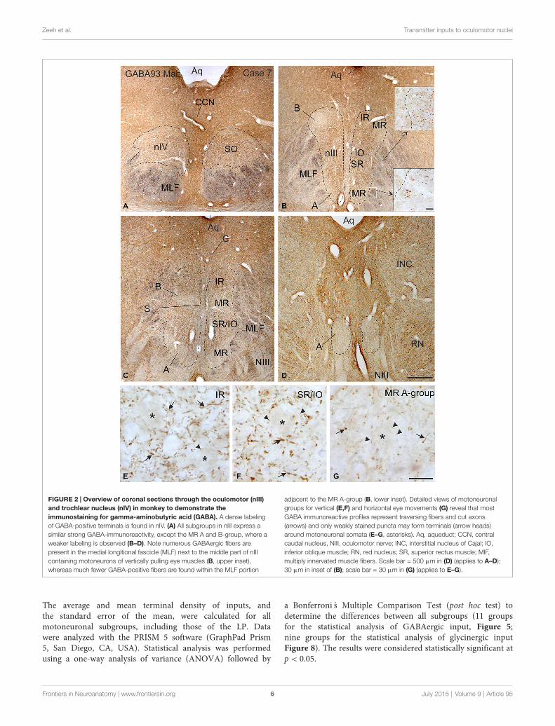
FIGURE 2 | Overview of coronal sections through the oculomotor (nIII)and trochlear nucleus (nIV) in monkey to demonstrate theimmunostaining for gamma-aminobutyric acid (GABA). A dense labelingof GABA-positive terminals is found in nIV. (A) All subgroups in nIII express asimilar strong GABA-immunoreactivity, except the MR A and B-group, where aweaker labeling is observed (B–D). Note numerous GABAergic fibers arepresent in the medial longitional fascicle (MLF) next to the middle part of nIIIcontaining motoneurons of vertically pulling eye muscles (B, upper inset),whereas much fewer GABA-positive fibers are found within the MLF portion
adjacent to the MR A-group (B, lower inset). Detailed views of motoneuronalgroups for vertical (E,F) and horizontal eye movements (G) reveal that mostGABA immunoreactive profiles represent traversing fibers and cut axons(arrows) and only weakly stained puncta may form terminals (arrow heads)around motoneuronal somata (E–G, asterisks). Aq, aqueduct; CCN, centralcaudal nucleus, NIII, oculomotor nerve; INC, interstitial nucleus of Cajal; IO,inferior oblique muscle; RN, red nucleus; SR, superior rectus muscle; MIF,multiply innervated muscle fibers. Scale bar = 500 µm in (D) (applies to A–D);30 µm in inset of (B); scale bar = 30 µm in (G) (applies to E–G).
The average and mean terminal density of inputs, andthe standard error of the mean, were calculated for allmotoneuronal subgroups, including those of the LP. Datawere analyzed with the PRISM 5 software (GraphPad Prism5, San Diego, CA, USA). Statistical analysis was performedusing a one-way analysis of variance (ANOVA) followed by
a Bonferroni s Multiple Comparison Test (post hoc test) todetermine the differences between all subgroups (11 groupsfor the statistical analysis of GABAergic input, Figure 5;nine groups for the statistical analysis of glycinergic inputFigure 8). The results were considered statistically significant atp < 0.05.
Frontiers in Neuroanatomy | www.frontiersin.org 6 July 2015 | Volume 9 | Article 95

Zeeh et al. Transmitter inputs to oculomotor nuclei
FIGURE 3 | Detailed view of coronal paraffin sections of theoculomotor (nIII) and trochlear nucleus (nIV) in the midbrain stained forglutamate decarboxylase (GAD) in black and choline acetyltransferase(ChAT) in brown (A–F). Numerous GAD-positive puncta outline most of themotoneuronal somata (asterisks) of the superior oblique (SO) (A) the levatorpalpebrae muscle (LP) (B) and the subgroup containing superior rectus (SR)and IO muscles. (D) In the MR subgroups fewer GAD-positive puncta areattached to the somata (C, asterisk), but are found in the neuropile contactingcut dendrites. A considerable number of GAD-positive puncta is found aroundMIF motoneurons in the C-group and S-group (E,F, asterisks indicatemotoneurons; arrows). Detailed views of confocal images in (G,H) show tracerlabeled MR MIF motoneurons (green) in the C-group that are in closeassociation with GAD-positive (red) puncta suggestive for direct synapticinputs (arrowheads). MIF, multiply innervated muscle fibers. Scale bar = 25 µmin (F) (applies to A–F); Scale bar = 25 µm in (G); Scale bar = 25 µm in (H).
Results
Tracer Injection CaseMedial Rectus MuscleInjection into the MR resulted in selective labeling of threemotoneuron subgroups as described earlier and shown inFigure 1 (Büttner-Ennever et al., 2001). The A-group lies in the
ventral and ventrolateral part of nIII and extends throughout thewhole nIII except the most caudal part. The B-group forms acircular cell group located dorsolaterally in the caudal half of nIII.The peripheral C-group dorsomedial to the nuclear boundariesof nIII consists of MIF motoneurons and extends throughout thewhole rostrocaudal length of the nIII (Figure 1).
GABAergic InputSIF MotoneuronsImmunolabeling for different GABAergic markers resulted in astrong GABA- and GAD-expression within the motoneuronalsubgroups of SR, IO and IR in nIII (Figures 2B,C, 3D). Similarily,the SO motoneurons in nIV and the LP motoneurons inCCN expressed strong immunoreactivity for GABA and GAD(Figures 2A, 3A,B). Visual inspection of all sections revealed aweaker GABA immunoreactivity in the MR subgroups, e.g., theventral A-group and dorsolateral B-group, which are consideredto be the SIF MR motoneurons (Figures 2B,C,D; Büttner-Ennever et al., 2001; Eberhorn et al., 2005). The weakerimmunolabeling in MR subgroups was not so evident insections immunostained for GAD (Figure 3C). The detailedviews in Figure 2 demonstrate a strong GABA-expressionin axons travelling within the medial longitudinal fascicle(MLF; Figures 2A–C, insets in Figure 2B) and within themotoneuronal subgroups of eye muscles mediating vertical(Figures 2E,F, arrows) and horizontal gaze (Figure 2G, arrows).The rather weak GABA-immunoreactivity in presumed nerveendings around motoneurons (Figures 2E–G, asterisks) maybe one reason for the differences seen in the GAD andGABA-staining pattern (Figures 2E–G, arrowheads). Since GADimmunoreactivity was strongly expressed in nerve endings(Ottersen and Storm-Mathisen, 1984), thin paraffin sectionsstained for GAD were used for the quantitative analysis ofGABAergic input to motoneurons (Figure 3). The countingrevealed a similarly dense GAD-positive puncta supply aroundthe somata of presumed SIF motoneurons for SR/IO, IR, SO andLP in CCN with an averaged density (AD) of 0.08 puncta/µm(Table 3; Figures 3A,B,D, 5). SIF motoneurons of MR werecontacted by fewer GAD-positive puncta, with an AD of 0.05puncta/µm for the A-group and 0.06 puncta/µm for the B-group(Table 3; Figures 3C, 5). Immunostaining for the GABA-Areceptor reflected that of GAD and GABA (Figure 4) with aweaker expression within the MR subgroups (Figure 4, compareC, F to G).
MIF MotoneuronsThe close inspection of presumed non-twitch MIF motoneuronsrevealed the following picture. A high density of GAD-immunoreactive puncta and a strong immunostaining forGABA-A receptor was present in the C-group containing MRand IRMIFmotoneurons (Figures 3E, 4D). This observation wasclarified by the analysis of tracer-labeled MR MIF motoneuronsfor GAD-immunoreactivity. Numerous GAD-positive profileswere in close proximity to tracer-labeled MIF motoneuronssuggesting synaptic contacts (Figures 3G,H, arrowheads).Similarily, numerous GAD-positive puncta were found aroundcholinergic neurons in the S-group, which represent MIF
Frontiers in Neuroanatomy | www.frontiersin.org 7 July 2015 | Volume 9 | Article 95

Zeeh et al. Transmitter inputs to oculomotor nuclei
TABLE 1 | An overview of injection, fixation and immunohistochemistry details for each case.
Case Injection Fixation Sections Immunhistochemistry
1 3 µl CTB, MR 4% paraformaldehyde Frozen CTB, CTB + GAD, CTB + vGlut12 5 µl CTB, MR 4% paraformaldehyde Frozen CTB, CTB + GAD3 4% paraformaldehyde Paraffin GAD, vGlut1 or vGlut2 + ChAT4 4% paraformaldehyde Frozen GABA-A +ChAT, GlyT25 4% paraformaldehyde Frozen GlyR6 4% paraformaldehyde Frozen GlyT27 1% paraformaldehyde 2, 5% glutaraldehyde Frozen GABA8 4% paraformaldehyde 0, 3% glutaraldehyde Frozen GABA9 4% paraformaldehyde Frozen vGlut210 WGA-HRP 4% paraformaldehyde Frozen
CTB, cholera toxin subunit B; GAD, glutamate decarboxylase; vGlut, vesicular glutamate transporter; ChAT, choline acetyltransferase; GABA, gamma-aminobutyric acid,
GABA-A, GABA receptor; GlyT, glycine transporter; GlyR, glycine receptor; MR, medial rectus muscle; WGA-HRP, wheat germ agglutinin conjugated to horseradish
peroxidase.
motoneurons of SR and IO (Figure 3F arrows), and in thedorsal cap of nIV containing MIF motoneurons of SO (notshown).
The quantitative analysis for theMIFmotoneurons resembledthe visual impression and revealed a strong supply of GAD-positive puncta for the S-group, the C-group (both 0.09puncta/µm) and for the SOMIF motoneurons (0.08 puncta/µm;Figure 5). The analysis of the GAD input to tracer-labeled MRmotoneurons in the C-group revealed an AD of 0.07 puncta/µmfor the MR MIF motoneurons and 0.1 puncta/µm for IR MIFmotoneurons.
To determine the differences between the differentmotoneuronal groups within nIII and nIV, 11 subgroupswere compared to each other (see Figure 5). Accordingto ANOVA and the subsequent Bonferronis MultipleComparison Test a significant difference was determinedbetween following subgroups (Figure 5): IR MIF motoneuronsreceived a significantly higher GAD-positive supply comparedto motoneurons of the A- and B-group. Motoneurons of the C-and S-group were contacted by significantly more GAD-positive
puncta compared to MR SIF motoneurons of the A-group. Formore details see Figure 5.
Immunohistochemical Localization ofGlycine
Glycine Transporter 2 (GlyT2) andGlycine-Receptor 1 (GlyR1)SIF MotoneuronsThe strongest expression of glycine markers was foundwithin the CCN. No differences in location and intensity inimmunostaining were noted between GlyT2 and GlyR1 withinthe CCN, where the somata and proximal dendrites of LPmotoneurons were completely outlined by immunoreactivepuncta (Figures 6A,D, 7A,B). This was confirmed by thequantitative analysis of GlyT2 input revealing an AD of 0.15puncta/µm (Figure 8). A strong GlyT2 expression was alsofound in the MR A- and B-group (Figures 6B,C,F,H). Inthe subgroups containing motoneurons of the vertical pullingeye muscles only few GlyT2-positive traversing fibers and
TABLE 2 | An overview of the primary antibodies and dilutions used for immunolabeling.
Antibody Host Antigen Manufacturer Cat. No. Dilution
GABA-A Mouse GABA-A receptor, beta-chain Chemicon, now Millipore, Billerica, USA MAB341 1:1000GABA93 MAb Mouse GABA-glutaraldehyde-BSA
conjugateHolstein et al. (2004) Holstein G, Mt. Sinai, Hospital,
New York1:3000
GAD Mouse Glutamate decarboxylase Biotrend, Cologne, Germany GC3108 1:4000GAD65/67 Rabbit Glutamate decarboxylase Millipore; Billerica, USA AB1511 1:500
(fluorescence)
GlyT2 Sheep Glycine transporter 2 (neuronal) Millipore; Billerica, USA AB1771 1:5000GlyR Mouse Glycine receptor alpha-1-subunit Synaptic systems, Goettingen, Germany 146 111 1:1000ChAT Goat Choline acetyltransferase
human placental enzymeMillipore, Billerica, USA AB144P 1:100
CTB Goat Choleragenoid List Biological Laboratories, Campbell,California
703 1:20,0001:5000(fluorescence)
vGlut1 Rabbit Vesicular glutamate transporter 1 Synaptic systems, Goettingen, Germany 135303 1:30001:1000(fluorescence)
vGlut2 Rabbit Vesicular glutamate transporter 2 Synaptic systems, Goettingen, Germany 135402 1:500
Frontiers in Neuroanatomy | www.frontiersin.org 8 July 2015 | Volume 9 | Article 95
Zeeh et al. Transmitter inputs to oculomotor nuclei
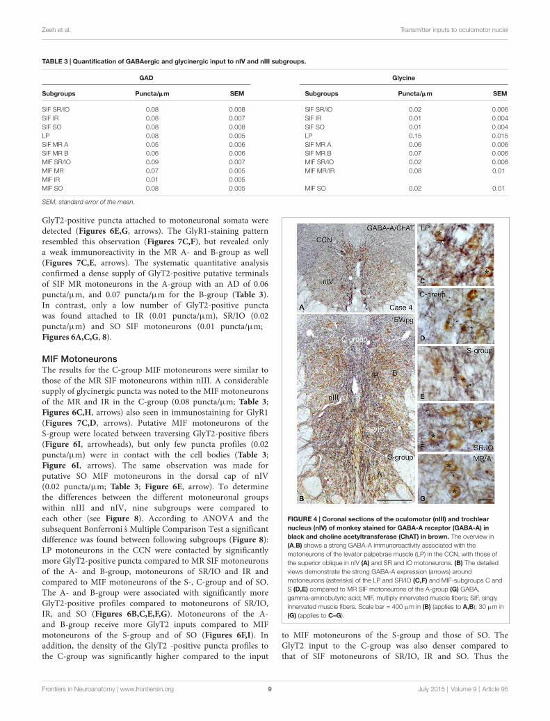
TABLE 3 | Quantification of GABAergic and glycinergic input to nIV and nIII subgroups.
GAD Glycine
Subgroups Puncta/µµµm SEM Subgroups Puncta/µµµm SEM
SIF SR/IO 0.08 0.008 SIF SR/IO 0.02 0.006SIF IR 0.08 0.007 SIF IR 0.01 0.004SIF SO 0.08 0.008 SIF SO 0.01 0.004LP 0.08 0.005 LP 0.15 0.015SIF MR A 0.05 0.006 SIF MR A 0.06 0.006SIF MR B 0.06 0.006 SIF MR B 0.07 0.006MIF SR/IO 0.09 0.007 MIF SR/IO 0.02 0.008MIF MR 0.07 0.005 MIF MR/IR 0.08 0.01MIF IR 0.01 0.005MIF SO 0.08 0.005 MIF SO 0.02 0.01
SEM, standard error of the mean.
GlyT2-positive puncta attached to motoneuronal somata weredetected (Figures 6E,G, arrows). The GlyR1-staining patternresembled this observation (Figures 7C,F), but revealed onlya weak immunoreactivity in the MR A- and B-group as well(Figures 7C,E, arrows). The systematic quantitative analysisconfirmed a dense supply of GlyT2-positive putative terminalsof SIF MR motoneurons in the A-group with an AD of 0.06puncta/µm, and 0.07 puncta/µm for the B-group (Table 3).In contrast, only a low number of GlyT2-positive punctawas found attached to IR (0.01 puncta/µm), SR/IO (0.02puncta/µm) and SO SIF motoneurons (0.01 puncta/µm;Figures 6A,C,G, 8).
MIF MotoneuronsThe results for the C-group MIF motoneurons were similar tothose of the MR SIF motoneurons within nIII. A considerablesupply of glycinergic puncta was noted to the MIF motoneuronsof the MR and IR in the C-group (0.08 puncta/µm; Table 3;Figures 6C,H, arrows) also seen in immunostaining for GlyR1(Figures 7C,D, arrows). Putative MIF motoneurons of theS-group were located between traversing GlyT2-positive fibers(Figure 6I, arrowheads), but only few puncta profiles (0.02puncta/µm) were in contact with the cell bodies (Table 3;Figure 6I, arrows). The same observation was made forputative SO MIF motoneurons in the dorsal cap of nIV(0.02 puncta/µm; Table 3; Figure 6E, arrow). To determinethe differences between the different motoneuronal groupswithin nIII and nIV, nine subgroups were compared toeach other (see Figure 8). According to ANOVA and thesubsequent Bonferroni s Multiple Comparison Test a significantdifference was found between following subgroups (Figure 8):LP motoneurons in the CCN were contacted by significantlymore GlyT2-positive puncta compared to MR SIF motoneuronsof the A- and B-group, motoneurons of SR/IO and IR andcompared to MIF motoneurons of the S-, C-group and of SO.The A- and B-group were associated with significantly moreGlyT2-positive profiles compared to motoneurons of SR/IO,IR, and SO (Figures 6B,C,E,F,G). Motoneurons of the A-and B-group receive more GlyT2 inputs compared to MIFmotoneurons of the S-group and of SO (Figures 6F,I). Inaddition, the density of the GlyT2 -positive puncta profiles tothe C-group was significantly higher compared to the input
FIGURE 4 | Coronal sections of the oculomotor (nIII) and trochlearnucleus (nIV) of monkey stained for GABA-A receptor (GABA-A) inblack and choline acetyltransferase (ChAT) in brown. The overview in(A,B) shows a strong GABA-A immunoreactivity associated with themotoneurons of the levator palpebrae muscle (LP) in the CCN, with those ofthe superior oblique in nIV (A) and SR and IO motoneurons. (B) The detailedviews demonstrate the strong GABA-A expression (arrows) aroundmotoneurons (asterisks) of the LP and SR/IO (C,F) and MIF-subgroups C andS (D,E) compared to MR SIF motoneurons of the A-group (G) GABA,gamma-aminobutyric acid; MIF, multiply innervated muscle fibers; SIF, singlyinnervated muscle fibers. Scale bar = 400 µm in (B) (applies to A,B); 30 µm in(G) (applies to C–G).
to MIF motoneurons of the S-group and those of SO. TheGlyT2 input to the C-group was also denser compared tothat of SIF motoneurons of SR/IO, IR and SO. Thus the
Frontiers in Neuroanatomy | www.frontiersin.org 9 July 2015 | Volume 9 | Article 95

Zeeh et al. Transmitter inputs to oculomotor nuclei
FIGURE 5 | Histogram demonstrating the quantitative analysis ofglutamate decarboxylase (GAD) input to motoneurons of oculomotor(nIII) and trochlear nucleus (nIV). The mean terminal density of stainedpuncta and the standard error of the mean were calculated for all motoneuralsubgroups. Note a similar strong input to almost all motoneuronal subgroupsin nIII and nIV. Only the motoneurons of MR (A- and B-group) receive a weakersupply by GAD-positive terminals. The table shows the results of the ANOVAand following Bonferronis Multiple Comparison Test.
density of GlyT2-positive puncta around SIF motoneurons forhorizontal eye movements was significantly higher comparedto those for vertical eye movements. For more details seeFigure 8.
Immunohistochemical Localization of vGlutImmunolabeling for vGlut revealed that the nIII and nIV werecompletely devoid of vGlut1-positive terminals and neurons,except for a weak puncta labeling along the midline betweenboth nIII (Figures 9A–C). A dense cluster of vGlut1-positiveterminals was seen dorsolateral to nIII (Figure 9C). Thesupraoculomotor area (SOA) above nIII and the perioculomotorregion around nIII contain fewer vGlut1 positive puncta(Figure 9B). At close inspection it was obvious that aconsiderable number of vGlut1-positive puncta was attachedto putative MR MIF motoneurons in the medial C-group, butnot IR MIF motoneurons (Figures 9C,F, arrows; Tang et al.,2015). Unlike for vGlut1, an even dense supply of vGlut2-positive puncta was found within nIII covering the somata of allmotoneuronal subgroups (Figure 10A). Detailed views revealeda similar dense supply of vGlut2-positive puncta to MIF andSIF motoneurons as shown here for the C-group and SR/IOsubgroup (Figures 10B,C).
Discussion
The present work in part confirms previous studies on thediffering inhibitory input to motoneurons subserving horizontaland vertical eye movements, respectively. It extends these
FIGURE 6 | Overviews of coronal sections through the monkeytrochlear (nIV) and oculomotor nucleus (nIII) immunostained forglycine transporter 2 (GlyT2). A dense glycinergic input is found to levatorpalpebrae motoneurons (LP) in the CCN (A,D) and to (A) and (B) groups ofthe MR muscle (B,C,F) compared to a few GlyT2-positive fibers and puncta(arrow) scattered within nIV (A,E) and the superior rectus/inferior oblique(SR/IO) motoneuronal group (B,C,G). (H) Shows a detailed view of theC-group (left side) with a MIF MR motoneuron (asterisk) covered withnumerous GlyT2-positive puncta (arrows) next to a MIF inferior rectus (IR)motoneuron (asterisk) associated with only few GlyT2-positive puncta(arrows). Similarily, neurons in the S-group (asterisks) are associated withGlyT2-positive puncta and fibers (I, arrow). Aq, aquaeduct; IR, inferior rectus;SO, superior oblique. Scale bar = 500 µm in (C) (applies to A–C); Scale bar =30 µm in (I) (applies to D–I).
findings by a quantitative analysis of the differing transmitterinputs to MIF vs. SIF motoneurons in monkey. Additionally,we showed for the first time that vGlut1-positive terminals areonly associated with MIF neurons. Although direct synaptic
Frontiers in Neuroanatomy | www.frontiersin.org 10 July 2015 | Volume 9 | Article 95

Zeeh et al. Transmitter inputs to oculomotor nuclei
FIGURE 7 | Overviews and detailed views of immunoperoxidaselabeling for glycine receptor 1 (GlyR1) in coronal sections of theoculomotor nucleus (nIII) in monkey. The strongest GlyR1 expression ispresent in the CCN as puncta profiles (A,B, stars and arrows). Unlike themotoneurons of the superior rectus and inferior oblique subgroup (SR/IO; F,asterisk) the MR SIF motoneurons (E, asterisks) show few GlyR1-positivepuncta (E, arrows). The MIF motoneurons in the C-group (C, lines; D, asterisk)are associated with numerous GlyR1-positive puncta (D, arrows). MIF, multiplyinnervated muscle fibers; SIF, singly innervated muscle fibers. Scale bar =200 µm in (A) (applies to A,C); Scale bar = 30 µm in (F) (applies to B,D–F).
contacts were not proven by EM studies in the presentwork, the close proximity between motoneurons and nerveendings suggest synaptic inputs. The results are discussedagainst the background of the current knowledge on premotorsources targeting MIF and/or SIF motoneurons and theirtransmitters.
GABAergic Input to nIII and nIVWith the application of GABA and GAD antibodies ourresults confirm previous studies in monkey demonstrating thatGABAergic neuronal profiles are predominantly associatedwith motoneurons subserving vertical eye movements(Figures 2–5; Spencer et al., 1992). Furthermore, thepreferential presence of GABA-A around motoneurons ofvertically pulling eye muscles is in line with the observationthat postsynaptic inhibitory postsynaptic potentials (IPSPs)evoked by electrical stimulation of the labyrinth in rabbitare blocked by the GABA-A antagonist picrotoxin (Ito et al.,1970).
The strong GABAergic input to CCN may arise fromthe principal trigeminal nucleus, whose electrical stimulationin cat evoked IPSPs in LP motoneurons (May et al.,2012) possibly interrupting the tonic activity of LP to
FIGURE 8 | Histogram demonstrating the quantitative analysis of theglycine transporter 2 (GlyT2) input to motoneurons in the trochlear(nIV), oculomotor (nIII) and CCN. The glycinergic input to all motoneuralgroups was quantified by counting immunoreactive puncta along themeasured length of the contour of a motoneuron. The mean terminal densityof inputs and the standard error of the mean were calculated for allmotoneural subgroups. The strongest supply by GlyT2-positive puncta is seento levator palpebrae motoneurons (LP), and fewer puncta are found aroundMR motoneurons of the A-, B- and C-group. The density of GlyT2-positivepuncta around motoneurons for horizontal eye movements (MR) wassignificant stronger compared to those for vertical eye movements. The addedtable shows the results of the ANOVA and following Bonferronis MultipleComparison Test.
enable the orbicularis oculi muscle to contract during blinks(Evinger and Manning, 1993).
The reports of a GABAergic input to MR motoneuronsare most controversial for different species, but may dependon differences in the methods and applied antibodies. Inaccordance with the present results a moderate supply ofGABAergic terminals was noted in the MR subdivisions inmonkey and cat using immunohistochemistry in frozen sections(Spencer et al., 1989; Spencer and Baker, 1992). However,studies applying postembedding GABA staining in semithinsections did not detect a significant difference of GABAergicinput to tracer-labeled MR motoneurons compared to the othersubdivisions in nIII, in cat and rabbit (de la Cruz et al.,1992; Wentzel et al., 1996), similar to the quantification ofGAD-positive inputs in thin paraffin sections in the presentstudy. In human, the number of GAD-positive profiles withinthe putative MR subgroups even exceeds that of motoneurongroups involved in vertical gaze. This may indicate anevolvement of inputs related to vergence, which is particularlyprominent in human (see below; Che Ngwa et al., 2014). The
Frontiers in Neuroanatomy | www.frontiersin.org 11 July 2015 | Volume 9 | Article 95

Zeeh et al. Transmitter inputs to oculomotor nuclei
FIGURE 9 | Overviews of coronal paraffin sections through the monkeytrochlear (nIV) and oculomotor nucleus (nIII) immunostained for thesimultaneous detection of vesicular glutamate transporter1 (vGlut1) inblack and choline acetyltransferase (ChAT) in brown (A–C). Note that SIFmotoneuron subgroups in nIV (A,D) and nIII (B,C,E) are devoid ofvGlut1-positive neuronal structures. Detailed views confirm the complete lack ofvGlut1-positive puncta in nIV (D) and nIII with the B-group as examples (E).
Note that numerous vGlut1-positive puncta are attached only to MIFmotoneurons in the C-group—and there confined to the MR motoneurons(F, arrows), but not present at IR motoneurons (F, asterisk). MIF motoneurons inthe S-group are associated with few vGlut1-positive puncta (G, arrows). EWpg;preganglionic Edinger-Westphal nucleus; MIF, multiply innervated muscle fibers;SIF, singly innervated muscle fibers. Scale bar = 500 µm in (C) (applies to A–C),Scale bar = 30 µm in (G) (applies to D–G).
finding of a considerable GABAergic input to the C- and S-groups confirms previous observations in monkey (Ying et al.,2008).
Glycinergic Input to nIIIThe considerable supply of GlyT2-positive nerve endings toMR subdivisions A and B resembled the labeling pattern ofglycine-positive afferents in nIII of previous reports in monkey(Figures 6–8; Spencer and Baker, 1992; Poyatos et al., 1997). Thesimilar distribution pattern of GlyT2-positive nerve endings inhuman nIII served there to identify the homolog MR subgroups(Che Ngwa et al., 2014). In cat, glycinergic terminals werefound in all motoneuron subgroups except the MR subdivisions(Spencer and Baker, 1992). In rabbit, a glycinergic input wasnoted to all subdivisions in the nIII including the MR region(Wentzel et al., 1996), but may colocalize with GABA (Wentzelet al., 1993). Based on current knowledge aboutMIFmotoneuron
organization the glycinergic terminals around the midline arenow considered to target the IO and SRMIFmotoneurons withinthe S-group (Büttner-Ennever et al., 2001; Wasicky et al., 2004),rather than the SR/IO SIF motoneurons (Spencer and Baker,1992; Spencer et al., 1992).The previously described associationof GlyT2 with LP motoneurons in the CCN was confirmedand is in line with a strong expression of glycine receptor 1seen here. The saccadic omnipause neurons were shown as onepossible glycinergic source to CCN (Horn and Büttner-Ennever,2008).
Functionally, glycine is similar to GABA as it increaseschloride conductance and evokes, therefore, IPSPs.Consequently, the likelihood that the postsynaptic cell reachesthe threshold for firing an action potential reduces. Thecolocalization of glycine and GABA in afferent inputs to MRmotoneurons may indicate a co-release of both transmitters(Wentzel et al., 1993). As shown for abducens motoneurons it
Frontiers in Neuroanatomy | www.frontiersin.org 12 July 2015 | Volume 9 | Article 95

Zeeh et al. Transmitter inputs to oculomotor nuclei
FIGURE 10 | Coronal section of the oculomotor nucleus (nIII) inmonkey showing the expression of vesicular glutamate transporter 2(vGlut2). (A) No differences were noted in the vGlut2 puncta labeling indifferent motoneuronal subgroups. The detailed views in (B,C) demonstratethe similar dense input of vGlut2-positive puncta (black) around cholinergicmotoneurons (brown, asterisks) in the C-group and around motoneurons ofthe vertically pulling superior rectus/ inferior oblique (SR/IO) muscles (B,C,arrows). EWpg, preganglionic Edinger-Westphal nucleus. Scale bar = 250 µmin (A, 30 µm in (C) (applies to B,C).
is possible that GABA-A and glycine receptors are distributeddifferently at somatal or dendritic membranes (Lorenzo et al.,2007), which may play a role in tuning the IPSPs (Russier et al.,2002).
Glycine can also serve as co-agonist with glutamate atpostsynaptic N-methyl-D-aspartate (NMDA) receptors (Johnsonand Ascher, 1987). It is an open question as to whether theglycinergic input to the MR subdivisions in nIII, which showeda weak GlyR expression, serve as an inhibitory transmitter, oras a co-agonist of excitatory glutamatergic afferents, e.g., frominternuclear neurons (INT) in nVI or ventral lateral vestibularnucleus (LVN; see below; Nguyen and Spencer, 1999).
Glutamatergic Input to nIII and nIVThis is the first description of the expression pattern of vGlutassociated with eye muscle motoneurons. VGluts selectivelypackage glutamate into synaptic vesicles and mediate glutamatetransport and therefore are used as markers for glutamatergicneuronal profiles (Takamori et al., 2000; Fremeau et al., 2001,2004; Zhou et al., 2007; for review: El Mestikawy et al., 2011).Whereas SIF and MIF motoneurons in nIII and nIV receive adense vGlut2-positive supply, a specific vGlut1-positive inputwas found only to MIF motoneurons, mainly those of MR(Figure 9F).
Transient responses of glutamate transmission are mediatedthrough ionotropic NMDA and non-NMDA (AMPA andkainate) receptors, whereas more persistent responses aremediated by metabotropic G-protein coupled receptors
(Dingledine et al., 1999). Thereby AMPA receptors conveythe fast component of postsynaptic responses, whereas NMDAreceptors mediate long lasting slower postsynaptic responses.Both, NMDA-receptors and AMPA receptors (GluR4 subunit),are only expressed in SIF, but not in MIF motoneuronsin monkey (Ying et al., 2008). This may indicate that SIFmotoneurons participate in the fast and slow components ofthe postsynaptic response to glutamate. This is in line with invitro studies on rat oculomotor neurons showing that smallermotoneurons with low-recruitment threshold currents havehigher input resistances and exhibit tonic firing—as assumed forMIF motoneurons and whose firing pattern remains essentiallyunmodified by glutamate application (Torres-Torrelo et al.,2012). The phasic-tonic firing of larger motoneurons—such asSIF motoneurons—with lower input resistances and with highrecruitment threshold currents, is strengthened by glutamateand could provide strong muscle contractions for (saccadic) eyemovements (Torres-Torrelo et al., 2012).
Premotor Sources and their Association withTransmittersSecondary vestibulo-ocular neurons (only SIF)A well-established input to nIII and nIV arises from thevestibular nuclei subserving the vertical angular vestibulo-ocularreflex (VOR; for review: Büttner-Ennever and Gerrits, 2004;Straka and Dieringer, 2004; Highstein and Holstein, 2006;Goldberg et al., 2012). Primary afferents from the anterior andposterior canals activate secondary vestibular neurons in themagnocellular parts of the medial vestibular nucleus (MVNm)and superior vestibular nucleus (SVNm), which in turn sendcontralateral excitatory and ipsilateral inhibitory projections tothe respective motoneurons of agonists and antagonists in nIIIand nIV (Graf and Ezure, 1986; Iwamoto et al., 1990; Graf et al.,1997; Goldberg et al., 2012).
Extracellular tracer injections into SVNm or MVNm andsingle cell reconstructions of identified up and down position-vestibular-pause neurons indicated that secondary vestibularneurons target only SIF motoneurons, but not MIFmotoneuronsin the S- or C-group (McCrea et al., 1987; Wasicky et al., 2004).This is in line with the lack of transneuronally labeled secondaryvestibular neurons in SVNm or MVNm after the injection ofrabies virus into the myotendinous junction of eye muscles, fromwhere only MIF (not SIF) motoneurons were retrogradely filled(Ugolini et al., 2006).
Glutamate and aspartateGlutamate and/or aspartate are widely accepted as the majorexcitatory neurotransmitter of the secondary vestibular neurons(Demêmes and Raymond, 1982; for review: McElligott andSpencer, 2000) and may reflect at least one portion of thevGlut2-positive input to nIII and nIV. This is in line withthe presence of numerous neurons expressing vGlut2 mRNA,but only few expressing weak vGlut1 mRNA signals in ratvestibular nuclei (Hisano et al., 2002; Zhang et al., 2011). Theexcitatory glutamatergic second-order vestibular inputs ontoabducens neurons act through AMPA receptors (Straka andDieringer, 1993). This pattern may apply to SIF motoneurons
Frontiers in Neuroanatomy | www.frontiersin.org 13 July 2015 | Volume 9 | Article 95
Zeeh et al. Transmitter inputs to oculomotor nuclei
in nIII and nIV, as indicated by their high expression of AMPAreceptors (GluR1, 2, 3 and 4) in human and monkey (Williamset al., 1996; Ying et al., 2008). Both, glutamate and NMDA,produce a depolarization of NMDA receptors primarily locatedat dendrites, but are not associated with the excitatory second-order vestibular input to oculomotor motoneurons (Durand andGueritaud, 1990).
Another source of glutamatergic input to nIII is conveyedby the ascending tract of Deiters (ATD; Nguyen and Spencer,1999; Büttner-Ennever and Gerrits, 2004; Holstein, 2012). Itoriginates from secondary vestibular neurons in the ventralMVN and the ventral LVN that receive excitatory inputsfrom the ipsilateral labyrinth, and target MR A and Bsubgroups in the ipsilateral nIII (not C-group; McCrea et al.,1987). The ATD carries head velocity signals, which aremodulated by utricular inputs and the viewing distance ofvisual targets to generate disconjugate vergence eye movements(Reisine et al., 1981; Chen-Huang and McCrea, 1998; Angelaki,2004). Postembedding immunostaining indicated that ATDafferents may use glutamate as a transmitter, whereas theadditional aspartate-labeling was attributed to the metabolicpool (Nguyen and Spencer, 1999). This glutamate/aspartateprojection targets primarily somata and proximal dendritesof MR motoneurons via synapses with asymmetric densitiesand spheroidal vesicles, the classical features of excitatorysynapses (Nguyen and Spencer, 1999). These projections maywell be included in vGlut2-positive afferents of the presentstudy.
GABAElectrophysiological and pharmacological studies have identifiedGABA as a major inhibitory transmitter of vertical secondaryvestibulo-ocular neurons in different species (for review:McElligott and Spencer, 2000; Straka and Dieringer, 2004;Sekirnjak and du Lac, 2006). These inhibitory projections arisein the SVN and MVN, and target the ipsilateral SO and IR,or SR and IO motoneurons (Holstein, 2012), predominantly attheir somata and proximal dendrites (Spencer and Baker, 1992;Wentzel et al., 1995). A fraction of the GABAergic fibers inthe MLF seen in the present study may represent the inhibitoryconnections of the vertical VOR (see also Spencer et al., 1989; forreview: Goldberg et al., 2012).
Non-secondary vestibulo-ocular connections (SIF andMIF)Additional projections to nIII and nIV arise from non-secondaryvestibular neurons, which are not directly activated by primaryafferents from the semicircular canals (Goldberg et al., 2012).They include the dorsal y-group, which receives disynapticinputs from vertical canal afferents (Blazquez et al., 2000),and projects to SIF and MIF motoneurons of SR and IOin the contralateral nIII and to IR and SO motoneuronson the ipsilateral side (Carpenter and Cowie, 1985; Wasickyet al., 2004). Electrical stimulation studies suggest that they-group may be part of cerebellar pathways for verticalsmooth-pursuit eye movements (Chubb and Fuchs, 1982). Atleast a subpopulation of non-secondary vestibular neurons
from the parvocellular MVN (MVNp) and the dorsal y-group, which target only motoneurons of upward movingeye muscles, contains calretinin (CR; Ahlfeld et al., 2011;Zeeh et al., 2013). These CR terminals were found to beexcitatory (Zeeh et al., 2013) and may contribute to thevGlut-positive input to SIF and MIF neurons seen here(Figures 9, 10).
Abducens internuclear neurons (only SIF)Another excitatory input to MR neurons arises from abducensINT, which carry a head-velocity and head position signal(burst, and burst-tonic) providing the neuroanatomicalbasis for conjugate horizontal eye movements (for review:Highstein and Holstein, 2006). In cat the tracer-labeledsynaptic endings of abducens INTs within the contralateralMR subgroup were shown to express immunoreactivityfor glutamate and aspartate (Nguyen and Spencer, 1999).The differing spatial location of glutamatergic afferentsfrom INTs and the ATD indicates that the more proximallocation of ATD synaptic input onto MR neurons may reducethe threshold for activation by the more distally locatedglutamatergic input from INTs during conjugate horizontaleye movements (Delgado-Garcia et al., 1986; Nguyen andSpencer, 1999). This may be used to reduce the synaptic delayfrom INT input to MR to ensure conjugacy of horizontal eyemovements.
Since tracer injections into nVI result in afferent labelingof all MR subgroups including the MIF motoneurons in theC-group (Wasicky et al., 2004), a strong glutaminergic inputvia this pathway must be anticipated (Nguyen and Spencer,1999). The more distal INT terminals on MR motoneuronsare thought to act through NMDA and non-NMDA (AMPA)receptors at the same postsynaptic site (Brodin and Shupliakov,1994). This arrangement is consistent with the known somato-dendritic distribution of NMDA and non-NMDA receptorson both, second-order vestibular (Cochran et al., 1987) andother extraocular motoneurons (Durand et al., 1987; Durandand Gueritaud, 1990; Durand, 1991; Straka and Dieringer,1993).
RIMLF and INC (SIF, SIF and MIF)Another monosynaptic input to SIF motoneurons of verticallypulling eye muscles originates from burst neurons in the rostralinterstitial nucleus of the medial longitudinal fasciculus (RIMLF)and the interstitial nucleus of Cajal (INC) encoding verticaland torsional saccades (Moschovakis et al., 1991a,b; Horn andBüttner-Ennever, 1998; Kokkoroyannis et al., 1996; for review:Horn, 2006). Tracer-labeled afferents fromRIMLF to nIII expressglutamate and aspartate (Spencer and Wang, 1996). Both aminoacids act on NMDA receptors, and in addition, glutamate acts onnon-NMDA receptors and may mediate different componentsof the postsynaptic response, and could thereby contribute tovGlut2 inputs.
GABAergic premotor neurons in the dorsomedial part ofthe RIMLF in cat (Spencer and Wang, 1996) and in INCin monkey (Horn et al., 2003) may monosynaptically inhibitthe motoneurons of anatogonistic eye muscles during up or
Frontiers in Neuroanatomy | www.frontiersin.org 14 July 2015 | Volume 9 | Article 95
Zeeh et al. Transmitter inputs to oculomotor nuclei
downward saccades as shown by intracellular recording studiesin cat (Sugiuchi et al., 2013). Since lesions of INC result invertical gaze-holding deficits with a head-tilt (Büttner et al.,2002) the INC is considered to function as velocity-to-positionintegrator of vertical eye movements (for review: Fukushimaand Kaneko, 1995). This function may be provided by premotorburst-tonic and tonic neurons that receive a burst signal fromRIMLF and project monosynaptically to motoneurons of verticalpulling eye muscles to transmit eye position signals (Dalezioset al., 1998; Horn and Büttner-Ennever, 1998; Sugiuchi et al.,2013). It is reasonable to assume that these premotor fibers targetSIF and MIF motoneurons, as indicated from tract-tracing aftersmall biocytin injections into INC in monkey (Kokkoroyanniset al., 1996). The differing projections from RIMLF and INCto SIF and MIF motoneurons conform to the concept that SIFmotoneurons are driven only from burst neurons in RIMLFand INC to generate the eye movement, whereas the burst-tonicand tonic input from INC targets also MIF motoneurons forgaze holding. Taken together it can be reasoned that premotorexcitatory burst and burst-tonic neurons in RIMLF and INCprovide a glutamatergic input to the motoneurons of vertical eyemovers, and thereby may form a portion of the vGlut2-positiveinput to nIII and nIV.
Prepositus nucleusA strong projection from the prepositus nucleus (PPH) tothe ipsilateral MR subgroup has been demonstrated (Bakeret al., 1977; McCrea and Baker, 1985; Belknap and McCrea,1988; for review: McCrea and Horn, 2006). Correlation ofthe neural activity of antidromically activated PPH neurons,with the resultant ipsilateral eye movements and contralateralhead movements, suggest an inhibitory action of this projection(Delgado-García et al., 1989), although excitatory projections tonIVmay also be present (Baker et al., 1977). Since no GABAergicprojection from PPH to nIII has been found in monkey(Carpenter et al., 1992), the inhibition may be transmitted viaglycine, as is the case for the abducens nucleus (Spencer et al.,1989). Whether the GlyT2-positive input to MR motoneuronsin this study may represent inhibitory projections from the PPHremains to be studied (Figures 6–8).
Inputs to only MIF motoneurons: Premotor sources andassociation with transmittersThe most selective transmitter-related input was found fromvGlut1-positive afferents to MR MIF motoneurons includingtheir dendrites, which reach up into the supraoculomotorarea (SOA) approaching the preganglionic neurons in thepreganglionic Edinger-Westphal nucleus (EWpg) controllingpupillary constriction and lens accommodation for the nearresponse (Tang et al., 2015; for review: McDougal and Gamlin,2015). Thereby the SOA is a well-suited target for premotorinputs controlling the near response as suggested by theabundance of synaptic contacts at the distal dendrites of MRMIF motoneurons compared to only few synapses targetingtheir somata and proximal dendrites (Erichsen et al., 2014).One source may arise from ‘‘near response neurons’’ in theSOA that increase their activity during convergence and can
be antidromically activated from MR subgroups (Judge andCumming, 1986; Mays et al., 1986; Zhang et al., 1991, 1992).
In monkey, a selective premotor input only to MIFmotoneurons was first described from the pretectum (Gamlinand Clarke, 1995; Büttner-Ennever et al., 1996; Wasicky et al.,2004). This includes the nucleus of the optic tract, whichprojects specifically to MIF motoneurons of nIII and nIV, andthe olivary pretectal nucleus, which targets primarily pupil-related preganglionic neurons in the rostral EWpg via excitatorysynapses (Gamlin and Clarke, 1995; Büttner-Ennever et al., 1996;Wasicky et al., 2004; Sun and May, 2014a,b). Another possiblesource is the central mesencephalic reticular formation (CMRF),which is associated with horizontal and vertical conjugate eyemovements (Waitzman et al., 1996; Wang et al., 2013). Recenttracer studies in monkey demonstrated a strong projection frompremotor neurons in the CMRF to the SOA including the C-group and the EWpg (Bohlen et al., 2015). This projection isbilateral and, if excitatory, may participate in the control ofvergence and the near triad (Bohlen et al., 2015). Whetherglutamatergic neurons in the SOA, in the pretectal nuclei or theCMRF give rise to the selective vGlut1 input to the somata ordendrites of MIF motoneurons, remains to be studied (Fujiyamaet al., 2003).
Conclusion
In conclusion the exclusive vGlut1 input to MIF motoneuronsand the higher density of GABA/glycinergic inputs to MR MIFmotoneurons in the C-group compared to SIF motoneuronswithin nIII confirm the concept that SIF and MIF motoneuronsreceive different inputs from premotor areas involved in differentfunctions: SIF motoneurons in generating eye movements,MIF motoneurons in gaze holding including vergence in thenear response (Wasicky et al., 2004; Büttner-Ennever, 2006;Ugolini et al., 2006). MIF neuron groups were shown tocontain also the cell bodies of palisade endings inserting at themyotendinous junction of extraocular muscles (Lienbacher et al.,2011; Zimmermann et al., 2011). But up to date it is not clear,whether they form a separate population of presumed sensoryneurons or are part of motoneurons giving rise to the multipleinnervation and palisade endings at non-twitch muscle fibers(Lienbacher and Horn, 2012).
Although all SIF motoneurons are involved in similar tasksexhibiting similar firing behavior during eye movments, thefunctional significance of differences in the chemical propertiesof premotor inputs—as seen for the specific expression ofcalretinin in excitatory premotor pathways for upgaze—isunclear (Zeeh et al., 2013). Transmitter inputs may not onlyconvey a specific postsynaptic reponse, but may modulatethe excitability of the motoneurons, for example by openingchloride channels conveyed by GABA and glycine (Lorenzoet al., 2007). Recent in vivo studies in rat demonstrated thatthe firing properties of motoneurons in nIII (tonic and phasicdischarge) as function of recruitment threshold current and cellsize can be modified by glutamatergic input (Torres-Torreloet al., 2012). Based on their findings from in vitro studiesof rat nIII motoneurons superfused with GABA, the authors
Frontiers in Neuroanatomy | www.frontiersin.org 15 July 2015 | Volume 9 | Article 95

Zeeh et al. Transmitter inputs to oculomotor nuclei
propose that motoneuron firing rates are essentially driven bytransient neurotransmission of different transmitters. Therebythis transient mechanism could act as a modulation systemrefining the output of the motoneurons (Torres-Torrelo et al.,2014).
Funding
Supported by Deutsche Forschungsgemeinschaft DFG HO1639/4–4, ‘‘Graduiertenförderung nach dem BayEFG’’, the Swiss
National Science Foundation; Grant number: 31–47287.96; theBetty and David Koetser Foundation for Brain Research (to BH)and National Institutes of Health EY013308; ORIP-0D010425;Research to Prevent Blindness.
Acknowledgments
We thank Dr. Gay R. Holstein (Mount Sinai School of Medicine)for providing the GABA93 MAb antibody used in this study aswell as Ahmed Messoudi for excellent technical assistance.
References
Ahlfeld, J., Mustari, M., and Horn, A. K. E. (2011). Sources of calretinin inputs tomotoneurons of extraocular muscles involved in upgaze. Ann. N. Y. Acad. Sci.1233, 91–99. doi: 10.1111/j.1749-6632.2011.06168.x
Angelaki, D. E. (2004). Eyes on target: What neurons must do for thevestibuloocular reflex during linear motion. J. Neurophysiol. 92, 20–35. doi: 10.1152/jn.00047.2004
Baker, R., Berthoz, A., and Delgado-García, J. (1977). Monosynaptic excitationof trochlear motoneurons following electrical stimulation of the prepositushypoglossi nucleus. Brain Res. 121, 157–161. doi: 10.1016/0006-8993(77)90445-0
Bedford, F. K., Kittler, J. T., Muller, E., Thomas, P., Uren, J. M., Merlo, D.,et al. (2001). GABA A receptor cell surface number and subunit stability areregulated by the ubiquitin-like protein Plic-1.Nat. Neurosci. 4, 908–916. doi: 10.1038/nn0901-908
Belknap, D. B., and McCrea, R. A. (1988). Anatomical connections of theprepositus and abducens nuclei in the squirrel monkey. J. Comp. Neurol. 268,13–28. doi: 10.1002/cne.902680103
Bellocchio, E. E., Hu, H., Pohorille, A., Chan, J., Pickel, V. M., and Edwards, R. H.(1998). The localization of the brain-specific inorganic phosphate transportersuggests a specific presynaptic role in glutamatergic transmission. J. Neurosci.18, 8648–8659.
Blazquez, P., Partsalis, A., Gerrits, N. M., and Highstein, S. M. (2000). Input ofanterior and posterior semicircular canal interneurons encoding head-velocityto the dorsal Y group of the vestibular nuclei. J. Neurophysiol. 83, 2891–2904.
Bohlen, M. O., Warren, S., andMay, P. J. (2015). A central mesencephalic reticularformation projection to the supraoculomotor area in macaque monkeys. BrainStruct. Funct. doi: 10.1007/s00429-015-1039-2 [Epub ahead of print].
Brodin, L., and Shupliakov, O. (1994). Functional diversity of central glutamatesynapses--pre- and post-synaptic mechanisms. Acta Physiol. Scand. 150, 1–10.doi: 10.1111/j.1748-1716.1994.tb09653.x
Bruce, G., Wainer, B. H., and Hersh, L. B. (1985). Immunoaffinity purification ofhuman choline acetyltransferase: comparison of the brain and placentalenzymes. J. Neurochem. 45, 611–620. doi: 10.1111/j.1471-4159.1985.tb04030.x
Büttner, U., Brandt, T., and Helmchen, C. (2002). The direction of nystagmusis important for the diagnosis of central paroxysmal positioningnystgmus (cPPV). Neuroophthal. 21, 97–104. doi: 10.1076/noph.21.2.97.3919.
Büttner-Ennever, J. A., Cohen, B., Horn, A. K. E., and Reisine, H. (1996).Pretectal projections to the oculomotor complex of the monkey and their rolein eye movements. J. Comp. Neurol. 366, 348–359. doi: 10.1002/(sici)1096-9861(19960304)366:2<348::aid-cne12>3.3.co;2-e
Büttner-Ennever, J. A., and Gerrits, N. M. (2004). ‘‘Vestibular System,’’ in Thehuman nervous system, eds Paxinos, G., and Mai, J. K. (Amsterdam: ElsevierAcademic Press), 479–510.
Büttner-Ennever, J. A., Horn, A. K. E., Scherberger, H., and D’ascanio, P.(2001). Motoneurons of twitch and nontwitch extraocular muscle fibers in theabducens, trochlear and oculomotor nuclei of monkeys. J. Comp. Neurol. 438,318–335. doi: 10.1002/cne.1318
Büttner-Ennever, J. A. (2006). The extraocular motor nuclei: organization andfunctional neuroanatomy. Prog. Brain Res. 151, 95–125. doi: 10.1016/s0079-6123(05)51004-5
Carpenter, M. B., and Cowie, R. J. (1985). Connections and oculomotorprojections of the superior vestibular nucleus and cell group ’y’. Brain Res. 336,265–287. doi: 10.1016/0006-8993(85)90653-5
Carpenter, M. B., Periera, A. B., and Guha, N. (1992). Immunocytochemistry ofoculomotor afferents in the squirrel monkey (Saimiri Sciureus). J. Hirnforsch.33, 151–167.
Che Ngwa, E., Zeeh, C., Messoudi, A., Büttner-Ennever, J. A., and Horn,A. K. E. (2014). Delineation of motoneuron subgroups supplying individualeye muscles in the human oculomotor nucleus. Front. Neuroanat. 8:2. doi: 10.3389/fnana.2014.00002
Chen-Huang, C., and McCrea, R. A. (1998). Viewing distance related sensoryprocessing in the ascending tract of deiters vestibulo-ocular reflex pathway.J. Vestib. Res. 8, 175–184. doi: 10.1016/S0957-4271(97)00001-3
Chiarandini, D. J., and Stefani, E. (1979). Electrophysiological identification oftwo types of fibres in rat extraocular muscles. J. Physiol. 290, 453–465. doi: 10.1113/jphysiol.1979.sp012783
Chubb, M. C., and Fuchs, A. F. (1982). Contribution of y group of vestibularnuclei and dentate nucleus of cerebellum to generation of vertical smooth eyemovements. J. Neurophysiol. 48, 75–99.
Cochran, S. L., Kasik, P., and Precht, W. (1987). Pharmacological aspects ofexcitatory synaptic transmission to second-order vestibular neurons in the frog.Synapse 1, 102–123. doi: 10.1002/syn.890010114
Dalezios, Y., Scudder, C. A., Highstein, S. M., and Moschovakis, A. K.(1998). Anatomy and physiology of the primate interstitial nucleus ofCajal. II. Discharge pattern of single efferent fibers. J. Neurophysiol. 80,3100–3111.
de la Cruz, R. R., Pastor, A. M., Martinez-Guijarro, F. J., López-García, C., andDelgado-García, J. M. (1992). Role of GABA in the extraocular motor nucleiof the cat: a postembedding immunocytochemical study. Neuroscience 51,911–929. doi: 10.1016/0306-4522(92)90529-b
Delgado-Garcia, J. M., del Pozo, F., and Baker, R. (1986). Behavior of neurons inthe abducens nucleus of the alert cat. II. Internuclear neurons.Neuroscience 17,953–973. doi: 10.1016/0306-4522(86)90073-4
Delgado-García, J. M., Vidal, P. P., Gómez, C. M., and Berthoz, A. (1989).A neurophysiological study of prepositus hypoglossi neurons projecting tooculomotor and preoculomotor nuclei in the alert cat. Neuroscience 29,291–307. doi: 10.1016/0306-4522(89)90058-4
Demêmes, D., and Raymond, J. L. (1982). Radioautographic identification ofglutamic acid labeled nerve endings in the cat oculomotor nucleus. Brain Res.231, 433–437. doi: 10.1016/0006-8993(82)90379-1
Dingledine, R., Borges, K., Bowie, D., and Traynelis, S. F. (1999). The glutamatereceptor ion channels. Pharmacol. Rev. 51, 7–61.
Durand, J., Engberg, I., and Tyc-Dumont, S. (1987). L-glutamate and N-methyl-D-asparatate actions on membrane potential and conductance of cat abducensmotoneurones. Neurosci. Lett. 79, 295–300. doi: 10.1016/0304-3940(87)90447-2
Durand, J., and Gueritaud, J. P. (1990). Excitatory amino acid actions onmembrane potential and conductance of brainstem motoneurons, in Amino-Acids, Chemistry, Biology andMedicine, eds Lubec, G. and Rosenthal L. (Escom:Leiden), 255–262.
Durand, J. (1991). NMDA actions on rat abducens motoneurons. Eur. J. Neurosci.3, 621–633. doi: 10.1111/j.1460-9568.1991.tb00848.x
Eberhorn, A. C., Ardenelau, P., Büttner-Ennever, J. A., and Horn, A. K. E. (2005).Histochemical differences betweenmotoneurons supplyingmultiply and singly
Frontiers in Neuroanatomy | www.frontiersin.org 16 July 2015 | Volume 9 | Article 95

Zeeh et al. Transmitter inputs to oculomotor nuclei
innervated extraocular muscle fibers. J. Comp. Neurol. 491, 352–366. doi: 10.1002/cne.20715
Eberhorn, A. C., Büttner-Ennever, J. A., and Horn, A. K. E. (2006). Identificationof motoneurons innervating multiply- or singly-innervated extraocular musclefibres in the rat. Neuroscience 137, 891–903. doi: 10.1016/j.neuroscience.2005.10.038
El Mestikawy, S., Wallén-Mackenzie, Å., Fortin, G. M., Descarries, L., andTrudeau, L.-E. (2011). From glutamate co-release to vesicular synergy: vesicularglutamate transporters. Nat. Rev. Neurosci. 12, 204–216. doi: 10.1038/nrn2969
Erichsen, J. T., Wright, N. F., andMay, P. J. (2014). Morphology and ultrastructureof medial rectus subgroup motoneurons in the macaque monkey. J. Comp.Neurol. 522, 626–641. doi: 10.1002/cne.23437
Evinger, C., and Manning, K. A. (1993). Pattern of extraocular muscle activationduring reflex blinking. Exp. Brain Res. 92, 502–506. doi: 10.1007/bf00229039
Fremeau, R. T., Jr., Troyer, M. D., Pahner, I., Nygaard, G. O., Tran, C. H., Reimer,R. J., et al. (2001). The expression of vesicular glutamate transporters definestwo classes of excitatory synapse. Neuron 31, 247–260. doi: 10.1016/s0896-6273(01)00344-0
Fremeau, R. T., Jr., Voglmaier, S., Seal, R. P., and Edward, R. H. (2004). VGlutsdefine subsets of excitatory neurons and suggest a novel role for glutamate.Trends Neurosci. 27, 98–103. doi: 10.1016/j.tins.2003.11.005
Fujiyama, F., Hioki, H., Tomioka, R., Taki, K., Tamamaki, N., Nomura, S., et al.(2003). Changes of immunocytochemical localization of vesicular glutamatetransporters in the rat visual system after the retinofugal denervation. J. Comp.Neurol. 465, 234–249. doi: 10.1002/cne.10848
Fukushima, K., and Kaneko, C. R. (1995). Vestibular integrators in theoculomotor system. Neurosci. Res. 22, 249–258. doi: 10.1016/0168-0102(95)00904-8
Gamlin, P. D., and Clarke, R. J. (1995). The pupillary light reflex pathway of theprimate. J. Am. Optom. Assoc. 66, 415–418.
Graf, W., and Ezure, K. (1986). Morphology of vertical canal related secondorder vestibular neurons in the cat. Exp. Brain Res. 63, 35–48. doi: 10.1007/bf00235644
Graf, W., Spencer, R., Baker, H., and Baker, R. (1997). Excitatory and inhibitoryvestibular pathways to the extraocular motor nuclei in goldfish. J. Neurophysiol.77, 2765–2779.
Goldberg, J. M., Wilson, V., Cullen, K., Angelaki, D., Broussard, D. M., Büttner-Ennever, J. A., et al. (2012). The Vestibular System: a Sixth Sense. New York:Oxford University Press.
Highstein, S. M., and Holstein, G. R. (2006). The anatomy of the vestibularnuclei. Prog. Brain Res. 151, 157–203. doi: 10.1016/S0079-6123(05)51006-9
Hisano, S., Sawada, K., Kawano, M., Kanemoto, M., Xiong, G., Mogi,K., et al. (2002). Expression of inorganic phosphate/vesicular glutamatetransporters (BNPI/VGLUT1 and DNPI/VGLUT2) in the cerebellum andprecerebellar nuclei of the rat. Mol. Brain Res. 107, 23–31. doi: 10.1016/s0169-328x(02)00442-4
Holstein, G. R., Martinelli, G. P., Henderson, S. C., Friedrich, V. L. J. Jr., Rabbitt,R. D., and Highstein, S. M. (2004). Gamma-aminobutyric acid is presentin a spatially discrete subpopulation of hair cells in the crista ampullarisof the toadfish, Opsanus tau. J. Comp. Neurol. 471, 1–10. doi: 10.1002/cne.11025
Holstein, G. R. (2012). ‘‘The vestibular system,’’ in The Human Nervous System,eds Mai, J. K. and Paxinos, G. 3rd Edn. (Amsterdam, Boston: Elsevier),1239–1269.
Horn, A. K. E., and Büttner-Ennever, J. A. (1998). Premotor neurons forvertical eye-movements in the rostral mesencephalon of monkey and man: thehistological identification by parvalbumin immunostaining. J. Comp. Neurol.392, 413–427. doi: 10.1002/(sici)1096-9861(19980323)392:4<413::aid-cne1>3.3.co;2-s
Horn, A. K. E., and Büttner-Ennever, J. A. (2008). Brainstem circuits controllinglid-eye coordination in monkey. Prog. Brain Res. 171, 87–95. doi: 10.1016/s0079-6123(08)00612-2
Horn, A. K. E., Helmchen, C., and Wahle, P. (2003). GABAergic neurons inthe rostral mesencephalon of the Macaque monkey that control vertical eyemovements. Ann. N. Y. Acad. Sci. 1004, 19–28. doi: 10.1196/annals.1303.003
Horn, A. K. E. (2006). The reticular formation. Prog. Brain Res. 151, 127–155.doi: 10.1016/S0079-6123(05)51005-7
Ito, M., Highstein, S. M., and Tsuchiya, T. (1970). The postsynaptic inhibitionof rabbit oculomotor neurones by secondary vestibular impulses and itsblockage by picrotoxin. Brain Res. 17, 520–523. doi: 10.1016/0006-8993(70)90260-x
Iwamoto, Y., Kitama, T., and Yoshida, K. (1990). Vertical eye movement-relatedsecondary vestibular neurons ascending in medial longitudinal fasciculus incat. II. Direct connections with extraocular motoneurons. J. Neurophysiol. 63,918–935.
Judge, S. J., and Cumming, B. G. (1986). Neurons in the monkey midbrainwith activity related to vergence eye movement and accommodation.J. Neurophysiol. 55, 915–930.
Jiao, Y., Sun, Z., Lee, T., Fusco, F. R., Kimble, T. D., and Meade, C. A. (1999).A simple and sensitive antigen retrieval method for free-floating and slide-mounted tissue sections. J. Neurosci. Methods 93, 149–162. doi: 10.1016/s0165-0270(99)00142-9
Johnson, J. W., and Ascher, P. (1987). Glycine potentiates the NMDA responsein cultured mouse brain neurons. Nature 325, 529–531. doi: 10.1038/325529a0
Kokkoroyannis, T., Scudder, C. A., Balaban, C. D., Highstein, S. M., andMoschovakis, A. K. (1996). Anatomy and physiology of the primate interstitialnucleus of Cajal I. Efferent projections. J. Neurophysiol. 75, 725–739.
Lienbacher, K., and Horn, A. E. (2012). Palisade endings and proprioception inextraocular muscles: a comparison with skeletal muscles. Biol. Cybern. 106,643–655. doi: 10.1007/s00422-012-0519-1
Lienbacher, K., Mustari, M., Ying, H. S., Büttner-Ennever, J. A., and Horn, A. K. E.(2011). Do palisade endings in extraocular muscles arise from neurons in themotor nuclei? Invest. Ophthalmol. Vis. Sci. 52, 2510–2519. doi: 10.1167/iovs.10-6008
Lorenzo, L. E., Barbe, A., Portalier, P., Fritschy, J. M., and Bras, H. (2006).Differential expression of GABAA and glycine receptors in ALS-resistant vs.ALS-vulnerable motoneurons: possible implications for selective vulnerabilityof motoneurons. Eur. J. Neurosci. 23, 3161–3170. doi: 10.1111/j.1460-9568.2006.04863.x
Lorenzo, L. E., Russier, M., Barbe, A., Fritschy, J. M., and Bras, H.(2007). Differential organization of γ-aminobutyric acid type A andglycine receptors in the somatic and dendritic compartments of ratabducens motoneurons. J. Comp. Neurol. 504, 112–126. doi: 10.1002/cne.21442
Lynch, G. S., Frueh, B. R., and Williams, D. A. (1994). Contractile properties ofsingle skinned fibers from the extraocular muscles, the levator and superiorrectus, of the rabbit. J. Physiol. 475, 337–346. doi: 10.1113/jphysiol.1994.sp020074
Mays, L. E., Porter, J. D., Gamlin, P. D., and Tello, C. A. (1986). Neural control ofvergence eye movements: neurons encoding vergence velocity. J. Neurophysiol.56, 1007–1021.
May, P. J., Vidal, P. P., Baker, H., and Baker, R. (2012). Physiological andanatomical evidence for an inhibitory trigemino-oculomotor pathway in thecat. J. Comp. Neurol. 520, 2218–2240. doi: 10.1002/cne.23039
McCrea, R. A., and Baker, R. (1985). Anatomical connections of the nucleusprepositus of the cat. J. Comp. Neurol. 237, 377–407. doi: 10.1002/cne.902370308
McCrea, R. A., and Horn, A. K. (2006). Nucleus prepositus. Prog. Brain Res. 151,205–230. doi: 10.1016/s0079-6123(05)51007-0
McCrea, R. A., Strassman, E., May, E., and Highstein, S. M. (1987). Anatomicaland physiological characteristics of vestibular neuronsmediating the horizontalvestibulo-ocular reflex of the squirrel monkey. J. Comp. Neurol. 264, 547–570.doi: 10.1002/cne.902640408
McDougal, D. H., and Gamlin, P. D. (2015). Autonomic control of the eye. Compr.Physiol. 5, 439–473. doi: 10.1002/cphy.c140014
McElligott, J., and Spencer, R. (2000). ‘‘Neuropharmacological aspects of thevestibulo-ocular reflex,’’ in Neurochemistry of the vestibular system, eds Beitz,A. J. and Anderson, J. H. (London: CRC Press. P), 199–222.
Moschovakis, A. K., Scudder, C. A., and Highstein, S. M. (1991a). The structureof the primate oculomotor burst generator. I. Medium-lead burst neurons withupward on-directions. J. Neurophysiol. 65, 203–217.
Moschovakis, A. K., Scudder, C. A., Highstein, S. M., and Warren, J. D. (1991b).The structure of the primate oculomotor burst generator. II. Medium-leadburst neurons with downward on-directions. J. Neurophysiol. 65, 218–229.
Frontiers in Neuroanatomy | www.frontiersin.org 17 July 2015 | Volume 9 | Article 95

Zeeh et al. Transmitter inputs to oculomotor nuclei
Nguyen, L. T., and Spencer, R. F. (1999). Abducens internuclear andascending tract of Deiters inputs to medial rectus motoneurons in the catoculomotor nucleus: Neurotransmitters. J. Comp. Neurol. 411, 73–86. doi: 10.1002/(sici)1096-9861(19990816)411:1<73::aid-cne6>3.0.co;2-7
Ottersen, O. P., and Storm-Mathisen, J. (1984). Glutamate- and GABA-containing neurons in the mouse and rat brain, as demonstrated with anew immunocytochemical technique. J. Comp. Neurol. 229, 374–392. doi: 10.1002/cne.902290308
Porter, J. D., Burns, L. A., andMay, P. J. (1989). Morphological substrate for eyelidmovements: innervation and structure of primate levator palpebrae superiorisand orbicularis oculi muscles. J. Comp. Neurol. 287, 64–81. doi: 10.1002/cne.902870106
Poyatos, I., Ponce, J., Aragón, C., Giménez, C., and Zafra, F. (1997). Theglycine transporter GlyT2 is a reliable marker for glycine-immunoreactiveneurons. Mol. Brain Res. 49, 63–70. doi: 10.1016/s0169-328x(97)00124-1
Reisine, H., Strassman, A., and Highstein, S. M. (1981). Eye position and headvelocity signals are conveyed to medial rectus motoneurons in the alert catby the ascending tract of Deiters’. Brain Res. 211, 153–157. doi: 10.1016/0006-8993(81)90075-5
Russier, M., Kopysova, I. L., Ankri, N., Ferrand, N., and Debanne, D. (2002).GABA and glycine co-release optimizes functional inhibition in rat brainstemmotoneurons in vitro. J. Physiol. 541, 123–137. doi: 10.1113/jphysiol.2001.016063
Schulze, C., Mustari, M. J., Holstein, G. R., and Horn, A. K. E. (2009). Transmitterinputs to different motoneuron subgroups in the oculomotor and trochlearnucleus in monkey. Soc. Neurosci. Abstr. 356.9.
Sekirnjak, C., and du Lac, S. (2006). Physiological and anatomicalproperties of mouse medial vestibular nucleus neurons projecting tothe oculomotor nucleus. J. Neurophysiol. 95, 3012–3023. doi: 10.1152/jn.00796.2005
Spencer, R. F., and Baker, R. (1992). GABA and glycine as inhibitoryneurotransmitters in the vestibulo-ocular reflex. Ann. N. Y. Acad. Sci. 656,602–611. doi: 10.1111/j.1749-6632.1992.tb25239.x
Spencer, R. F., and Porter, J. D. (2006). Biological organization of theextraocular muscles. Prog. Brain Res. 151, 43–80. doi: 10.1016/s0079-6123(05)51002-1
Spencer, R. F., Wang, S. F., and Baker, R. (1992). The pathways and functionsof GABA in the oculomotor system. Prog. Brain Res. 90, 307–331. doi: 10.1016/S0079-6123(08)63620-1
Spencer, R. F., and Wang, S. F. (1996). Immunohistochemical localization ofneurotransmitters utilized by neurons in the rostral interstitial nucleus ofthe medial longitudinal fasciculus (riMLF) that project to the oculomotorand trochlear nuclei in the cat. J. Comp. Neurol. 366, 134–148. doi: 10.1002/(sici)1096-9861(19960226)366:1<134::aid-cne9>3.0.co;2-4
Spencer, R. F., Wenthold, R. J., and Baker, R. (1989). Evidence for glycine as aninhibitory neurotransmitter of vestibular, reticular and prepositus hypoglossineurons that project to the cat abducens nucleus. J. Neurosci. 9, 2718–2736.
Straka, H., and Dieringer, N. (1993). Electrophysiological and pharmacologicalcharacterization of vestibular inputs to identified frog abducens motoneuronsand internuclear neurons in vitro. Eur. J. Neurosci. 5, 251–260. doi: 10.1111/j.1460-9568.1993.tb00491.x
Straka, H., and Dieringer, N. (2004). Basic organization principles of the VOR:lessons from frogs. Prog. Neurobiol. 73, 259–309. doi: 10.1016/j.pneurobio.2004.05.003
Sugiuchi, Y., Takahashi, M., and Shinoda, Y. (2013). Input-output organizationof inhibitory neurons in the interstitial nucleus of Cajal projecting to thecontralateral trochlear and oculomotor nucleus. J. Neurophysiol. 110, 640–657.doi: 10.1152/jn.01045.2012
Sun, W., and May, P. J. (2014a). Central pupillary light reflex circuits in the cat:I. The olivary pretectal nucleus. J. Comp. Neurol. 522, 3960–3977. doi: 10.1002/cne.23602
Sun, W., and May, P. J. (2014b). Central pupillary light reflex circuits in the cat: II.Morphology, ultrastructure and inputs of preganglionic motoneurons. J. Comp.Neurol. 522, 3978–4002. doi: 10.1002/cne.23601
Takamori, S., Rhee, J. S., Rosenmund, C., and Jahn, R. (2000). Identification ofa vesicular glutamate transporter that defines a glutamatergic phenotype inneurons. Nature 407, 189–194. doi: 10.1038/35025070
Takamori, S., Rhee, J. S., Rosemund, C., and Jahn, R. (2001). Identificationof differentiation-associated brain-specific phosphate transporter as a secondvesicular glutamate transporter (vGlut2). J. Neurosci. 21:RC182 (1–6).
Tang, X., Büttner-Ennever, J. A., Mustari, M. J., and Horn, A. K. E. (2015). Internalorganization of the medial rectus and inferior rectus neurons in the C-group ofthe oculomotor nucleus in monkey. J. Comp. Neurol. 523, 1809–1823. doi: 10.1002/cne.23760
Torres-Torrelo, J., Rodríguez-Rosell, D., Nunez-Abades, P., Carrascal, L., andTorres, B. (2012). Glutamate modulates the firing rate in oculomotor nucleusmotoneurons as a function of the recruitment threshold current. J. Physiol. 590,3113–3127. doi: 10.1113/jphysiol.2011.226985
Torres-Torrelo, J., Torres, B., and Carrascal, L. (2014). Modulation of the input-output function by GABAA receptor-mediated currents in rat oculomotornucleus motoneurons. J. Physiol. 592, 5047–5064. doi: 10.1113/jphysiol.2014.276576
Ugolini, G., Klam, F., Doldan Dans, M., Dubayle, D., Brandi, A.-M., Büttner-Ennever, J. A., et al. (2006). Horizontal eye movement networks in primatesas revealed by retrograde transneuronal transfer of rabies virus: Differencesin monosynaptic input to ‘‘slow’’ and ‘‘fast’’ abducens motoneurons. J. Comp.Neurol. 498, 762–785. doi: 10.1002/cne.21092
Waitzman, D. M., Silakov, V. L., and Cohen, B. (1996). Central mesencephalicreticular formation (cMRF) neurons discharging before and during eyemovements. J. Neurophysiol. 75, 1546–1572.
Wang, N., Perkins, E., Zhou, L., Warren, S., and May, P. J. (2013). Anatomicalevidence that the superior colliculus controls saccades through centralmesencephalic reticular formation gating of omnipause neuron activity.J. Neurosci. 33, 16285–16296. doi: 10.1523/jneurosci.2726-11.2013
Wasicky, R., Horn, A. K. E., and Büttner-Ennever, J. A. (2004). Twitch and non-twitch motoneuron subgroups of the medial rectus muscle in the oculomotornucleus of monkeys receive different afferent projections. J. Comp. Neurol. 479,117–129. doi: 10.1002/cne.20296
Wentzel, P. R., De zeeuw, C. I., Holstege, J. C., and Gerrits, N. M. (1993).Colocalization of GABA and Glycine in the rabbit oculomotor nucleus.Neurosci. Lett. 164, 25–29. doi: 10.1016/0304-3940(93)90848-f
Wentzel, P. R., De Zeeuw, C. I., Holstege, J. C., and Gerrits, N. M. (1995).Inhibitory synaptic inputs to the oculomotor nucleus from vestibulo-ocular-reflex-related nuclei in the rabbit.Neuroscience 65, 161–174. doi: 10.1016/0306-4522(94)00471-g
Wentzel, P. R., Gerrits, N. M., and De Zeeuw, C. I. (1996). GABAergic andglycinergic inputs to the rabbit oculomotor nucleus with special emphasis onthe medial rectus subdivision. Brain Res. 707, 314–319. doi: 10.1016/0006-8993(95)01389-x
Williams, T. L., Ince, P. G., Oakley, A. E., and Shaw, P. J. (1996). Animmunocytochemical study of the distribution of AMPA selective glutamatereceptor subunits in the normal human motor system. Neuroscience 74,185–198. doi: 10.1016/0306-4522(96)00117-0
Ying, H. S., Fackelmann, K., Messoudi, A., Tang, X. F., Büttner-Ennever,J. A., and Horn, A. K. (2008). Neuronal signalling expression profiles ofmotoneurons supplying multiply or singly innervated extraocular musclefibres in monkey. Prog. Brain Res. 171, 13–16. doi: 10.1016/s0079-6123(08)00602-x
Zeeh, C., Hess, B. J., and Horn, A. K. E. (2013). Calretinin inputs are confinedto motoneurons for upward eye movements in monkey. J. Comp. Neurol. 521,3154–3166. doi: 10.1002/cne.23337
Zhang, Y., Gamlin, P. D. R., and Mays, L. E. (1991). Antidromic identification ofmidbrain near response cells projecting to the oculomotor nucleus. Exp. BrainRes. 84, 525–528. doi: 10.1007/bf00230964
Zhang, Y., Mays, L. E., and Gamlin, P. D. (1992). Characteristics of nearresponse cells projecting to the oculomotor nucleus. J. Neurophysiol. 67,944–960.
Zhang, F. X., Pang, Y. W., Zhang, M. M., Zhang, T., Dong, Y. L., Lai, C. H., et al.(2011). Expression of vesicular glutamate transporters in peripheral vestibularstructures and vestibular nuclear complex of rat. Neuroscience 173, 179–189.doi: 10.1016/j.neuroscience.2010.11.013
Zhou, J., Nannapaneni, N., and Shore, S. (2007). Vesicular glutamate transporters1 and 2 are differentially associated with auditory nerve and spinal trigeminalinputs to the cochlear nucleus. J. Comp. Neurol. 500, 777–787. doi: 10.1002/cne.21208
Frontiers in Neuroanatomy | www.frontiersin.org 18 July 2015 | Volume 9 | Article 95
Zeeh et al. Transmitter inputs to oculomotor nuclei
Zimmermann, L., May, P. J., Pastor, A. M., Streicher, J., and Blumer, R. (2011).Evidence that the extraocular motor nuclei innervate monkey palisade endings.Neurosci. Lett. 489, 89–93. doi: 10.1016/j.neulet.2010.11.072
Conflict of Interest Statement: The authors declare that the research wasconducted in the absence of any commercial or financial relationships that couldbe construed as a potential conflict of interest.
Copyright © 2015 Zeeh, Mustari, Hess and Horn. This is an open-access articledistributed under the terms of the Creative Commons Attribution License (CCBY). The use, distribution and reproduction in other forums is permitted, providedthe original author(s) or licensor are credited and that the original publicationin this journal is cited, in accordance with accepted academic practice. Nouse, distribution or reproduction is permitted which does not comply with theseterms.
Frontiers in Neuroanatomy | www.frontiersin.org 19 July 2015 | Volume 9 | Article 95


![Subgroups of direct products of two right-angled Artin groups · papar,[4], was the contrast between nitely presented and nitely generated subgroups. Finitely generated subgroups](https://img.dokumen.tips/doc/110x75/5f06640a7e708231d417c234/subgroups-of-direct-products-of-two-right-angled-artin-groups-papar4-was-the.jpg)