Embed Size (px)
Citation preview

ORIGINAL RESEARCH ARTICLEpublished: 12 February 2014
doi: 10.3389/fnana.2014.00002
Delineation of motoneuron subgroups supplying individualeye muscles in the human oculomotor nucleusEmmanuel Che Ngwa1†, Christina Zeeh1,2 , Ahmed Messoudi 1, Jean A. Büttner-Ennever 1
and Anja K. E. Horn1,2*
1 Oculomotor Group, Institute of Anatomy and Cell Biology, Department I, Ludwig-Maximilians-University of Munich, Munich, Germany2 German Center for Vertigo and Balance Disorders, Ludwig-Maximilians-University of Munich, Munich, Germany
Edited by:
Paul J. May, University of MississippiMedical Center, USA
Reviewed by:
Joan S. Baizer, University of Buffalo,USAFiorenzo Conti, Università Politecnicadelle Marche, ItalyL. Craig Evinger, Stony BrookUniversity, USA
*Correspondence:
Anja K. E. Horn, Oculomotor Group,Institute of Anatomy and Cell Biology,Department I, Ludwig-Maximilians-University of Munich,Pettenkoferstrasse 11, D-80336Munich, Germanye-mail: [email protected]†Present address:
Emmanuel Che Ngwa, MedizinischeKlinik I, Klinikum Fulda AG,Pacelliallee 4, 36043 Fulda, Germany
The oculomotor nucleus (nIII) contains the motoneurons of medial, inferior, and superiorrecti (MR, IR, and SR), inferior oblique (IO), and levator palpebrae (LP) muscles. Thedelineation of motoneuron subgroups for each muscle is well-known in monkey, but notin human. We studied the transmitter inputs to human nIII and the trochlear nucleus(nIV), which innervates the superior oblique muscle (SO), to outline individual motoneuronsubgroups. Parallel series of sections from human brainstems were immunostainedfor different markers: choline acetyltransferase combined with glutamate decarboxylase(GAD), calretinin (CR) or glycine receptor. The cytoarchitecture was visualized with cresylviolet, Gallyas staining and expression of non-phosphorylated neurofilaments. Apartfrom nIV, seven subgroups were delineated in nIII: the central caudal nucleus (CCN), adorsolateral (DL), dorsomedial (DM), central (CEN), and ventral (VEN) group, the nucleusof Perlia (NP) and the non-preganglionic centrally projecting Edinger–Westphal nucleus(EWcp). DL, VEN, NP, and EWcp were characterized by a strong supply of GAD-positiveterminals, in contrast to DM, CEN, and nIV. CR-positive terminals and fibers were confinedto CCN, CEN, and NP. Based on location and histochemistry of the motoneuron subgroupsin monkey, CEN is considered as the SR and IO motoneurons, DL and VEN as the B- andA-group of MR motoneurons, respectively, and DM as IR motoneurons. A good correlationbetween monkey and man is seen for the CR input, which labels only motoneurons of eyemuscles participating in upgaze (SR, IO, and LP).The CCN contained LP motoneurons, andnIV those of SO. This study provides a map of the individual subgroups of motoneuronsin human nIII for the first time, and suggests that NP may contain upgaze motoneurons.Surprisingly, a strong GABAergic input to human MR motoneurons was discovered, whichis not seen in monkey and may indicate a functional oculomotor specialization.
Keywords: central caudal nucleus, nucleus of Perlia, extraocular muscles, motoneurons, calretinin, glycine, GABA,
eye movements
INTRODUCTIONEye movements are essential for vision, because they direct thefovea to a visual target, and stabilize gaze during locomotion to
Abbreviations: nIII, oculomotor nucleus; nIV, trochlear nucleus; nVI, abducensnucleus; CCN, central caudal nucleus; CEN, central group; ChAT, choline acetyl-transferase; CMRF, central mesencephalic reticular formation; CR, calretinin; DL,dorsolateral group; DM, dorsomedial group; DR, dorsal raphe nucleus; EAP,extravidin-peroxidase; EW, Edinger–Westphal nucleus; EWcp, centrally projectingEdinger–Westphal nucleus; EWpg, Edinger–Westphal nucleus containing pregan-glionic neurons; GABA, gamma-aminobutyric acid; GAD, glutamate decarboxylase;GlyR, glycine receptor; IC, inferior colliculus; INC, interstitial nucleus of Cajal; IO,inferior oblique muscle; IPN, interpeduncular nucleus; IR, inferior rectus muscle; LP,levator palpebrae muscle; LR, lateral rectus muscle; MGB, medial geniculate body;MIF, multiply-innervated non-twitch muscle fibers; ML, medial lemniscus; MLF,medial longitudinal fasciclus; MR, medial rectus muscle; NIII, oculomotor nerve;NP, nucleus of Perlia; NP-NF, non-phosphorylated neurofilaments; PAG, periaque-ductal gray; PB, phosphate buffer; PN, pontine nuclei; RIMLF, rostral interstitialnucleus of the medial longitudinal fasciculus; RN, red nucleus; SC, superior collicu-lus; SCP, superior cerebellar peduncle; SE of mean, standard error of the mean; SIF,singly-innervated twitch muscle fibers; SNc, substantia nigra pars compacta; SNr,substantia nigra pars reticulata; SO, superior oblique muscle; SOA, supraoculomotorarea; SR, superior rectus muscle; UCN, urocortin; VEN, ventral group.
compensate for head and body movements (Leigh and Zee, 2006;Horn and Leigh, 2011). The motor and premotor pathways for sev-eral eye movement types, e.g., saccades and the vestibulo-ocularreflex, are well studied in monkey, and they form the basis forassessing the homologous brain structures in humans, for exam-ple, in clinical cases of eye movement disorders (Horn and Leigh,2011; Kennard, 2011). However, different species have differentpatterns of eye movements, and different arrangements of theiroculomotor subgroups (for review: Büttner-Ennever, 2006). Inorder to analyze the clinical-anatomical studies involving horizon-tal and vertical, up- or downward eye movements, the knowledgeof the localization of the motoneurons of individual extraocu-lar muscles in human is essential. Despite the fact that efforts onthis topic have been undertaken since 1897 (Bernheimer, 1897)in human, the current map of individual motoneuronal groupsadopted in most textbooks is still that of the monkey (Warwick,1953a). In non-human primates, the oculomotor nucleus (nIII)and trochlear nucleus (nIV) lie in the mesencephalic tegmentumat the ventral border of the periaquaeductal gray beneath the
Frontiers in Neuroanatomy www.frontiersin.org February 2014 | Volume 8 | Article 2 | 1

Che Ngwa et al. Map of human oculomotor nucleus
aqueduct (for review: Büttner-Ennever, 2006). Since the classicalwork on the nIII in rhesus monkey by Warwick (1953a) usingdegeneration techniques, the topographic map has undergonesubstantial revisions in the primate using retrograde tract-tracingmethods (Büttner-Ennever and Akert, 1981; Porter et al., 1983;Büttner-Ennever et al., 2001; Büttner-Ennever, 2006). Neuronssupplying the ipsilateral medial rectus muscle (MR) are distributedinto three clusters within nIII: the ventral A-group extending intothe medial longitudinal fasciclus (MLF), the dorsolateral B-groupand the small C-group at the dorsomedial border of nIII (Büttner-Ennever and Akert, 1981). The motoneurons of the ipsilateralinferior rectus muscle (IR) are located dorsally at rostral levels ofthe nIII, and the motoneurons of the contralateral superior rec-tus muscle (SR) and ipsilateral inferior oblique muscle (IO) liepartly intermingled within the central nIII of one side (Spencerand Porter, 1981; Porter et al., 1983). The nIV contains only themotoneurons of the contralateral superior oblique muscle (SO;Porter et al., 1983). In primates, a separate midline nucleus at thetransition of nIII and nIV, the central caudal nucleus (CCN), con-tains the motoneurons of the levator palpebrae (LP) muscle, whichelevates the upper eyelid (Porter et al., 1989).
The Edinger–Westphal nucleus (EW) lies immediately dorsalto nIII. It is often included in the term “nIII complex,” although itdoes not contain motoneurons of extraocular muscles. However,recent work has shown that the EW contains different functionalcell groups, which must be clearly demarcated from each otherand from the nIII proper. In monkey, EW houses the preganglionic(pg) neurons of the ciliary ganglion, in accordance with traditionalbelief, and is now called EWpg (Horn et al., 2008; May et al., 2008).However, in human, the cytoarchitectural EW represents a cellgroup of non-pg centrally projecting (cp) neurons that containurocortin (UCN), and it is therefore now termed EWcp (Hornet al., 2008; Kozicz et al., 2011; Büttner-Ennever and Horn, 2014).
Transmitter content can also distinguish between oculomo-tor subgroups. Previous studies of transmitter content in catand monkey have shown that the motoneurons of horizon-tally moving eye muscles are controlled by glycinergic inputs,whereas those of vertically moving eye muscles by GABAer-gic afferents (Spencer et al., 1989, 1992; Spencer and Baker,1992). In addition, more recent reports revealed that onlythe motoneurons of muscles involved in upgaze, including theLP, are selectively targeted by calretinin (CR)-positive afferents(Ahlfeld et al., 2011; Zeeh et al., 2013); this finding proved very
useful in the present study for the recognition of IO and SRmotoneurons.
In the experiments reported here, we identified the motoneu-ron groups of individual eye muscles in human. This was basedpartly on a comparison with the localization of motoneuronsderived from tract-tracing experiments in monkey, and partlyon the cytoarchitecture and differential histochemical inputs tomotoneuron subgroups revealed by immunocytochemical stain-ing for non-phosphorylated neurofilaments (NP-NF) glutamatedecarboxylase (GAD), CR, glycine receptor (GlyR) in humanmidbrain sections. These groups have also been clearly sepa-rated from the EW and the nucleus of Perlia (NP) subgroups,and present a new map of the human oculomotor subgroups.A preliminary version of the map has been published previously(Büttner-Ennever and Horn, 2014).
MATERIALS AND METHODSANTISERACholine acetyltransferaseCholinergic motoneurons were detected with a polyclonal cholineacetyltransferase (ChAT) antibody raised in goat (AB144P, Chemi-con) against the whole enzyme isolated from human placenta,which is identical to the brain enzyme (Bruce et al., 1985; Table 1).In immunoblots, this antibody recognizes a 68–70 kDa protein.The appearance and distribution of ChAT-positive neurons withthis antibody in the present study is identical to the data of previousreports (Ichikawa and Shimizu, 1998).
Non-phosphorylated neurofilamentsNon-phosphorylated neurofilaments were detected using a mousemonoclonal antibody (IgG1), supplied as a high titer mouse ascitesfluid (Table 1). The antibody was raised against homogenizedhypothalami recovered from Fischer 344 rats (Sternberger et al.,1982). It reacts with a non-phosphorylated epitope in neurofil-ament H and is abolished when the epitope is phosphorylated(clone 02-135; SMI32, Sternberger Monoclonals Inc., Lutherville,MD, USA; Sternberger and Sternberger, 1983). This antibody visu-alizes two bands (200 and 180 kDa) in conventional immunoblots(Goldstein et al., 1987).
Glutamic acid decarboxylaseGABAergic terminals were detected with a monoclonal anti-body against the GABA-synthetizing enzyme glutamic acid
Table 1 | Sources and dilutions of primary antibodies.
Antigen Antibody Host Antibody source Dilution
Choline acetyltransferase (ChAT) Polyclonal anti-ChAT Goat Chemicon, Temecula, CA, USA, AB144P 1:100
Calretinin (CR) Polyclonal anti-CR Rabbit SWant, Bellinzona, Switzerland, 7669/3H 1:2500
Urocortin 1 (UCN) Polyclonal anti-UCN Rabbit Sigma, St. Louis, USA, U4757 1:8000
GAD Monoclonal anti-GAD Mouse Biotrend, GC3108 1:4000
α and β subunits of glycine receptor (GlyR) Monoclonal anti-GlyR
(clone mAb4a)
Mouse Synaptic Systems, Göttingen, Germany, 146
011
1:300
Non-phosphorylated neurofilaments (NP-NF) Monoclonal anti-NP-NF Mouse Sternberger, Lutherville, MD, USA, SMI-32P 1:5000
Frontiers in Neuroanatomy www.frontiersin.org February 2014 | Volume 8 | Article 2 | 2

Che Ngwa et al. Map of human oculomotor nucleus
decarboxylase (GAD; GAD65/67 GC3108, batch number Z05507,clone 1111, Biotrend, Cologne, Germany; Table 1). Two molecularforms of GAD – GAD65 and GAD67 – are known from differentspecies. There is 65% amino acid sequence homology between thetwo isoforms. Whereas GAD67 is a cytoplasmic protein consistingof 594 amino acid residues, GAD65 is an amphiphilic and mem-brane anchored protein consisting of 585 amino acid residues. Theantibody GC 3108 recognizes a linear epitope at the C-terminusof rat GAD, common to both isoforms. The hybridoma secretingthe antibody to GAD65/67 was generated by fusion of splenocytesfrom a mouse immunized with fragments of recombinant humanGAD65 fused to glutathione-S-transferase (Ziegler et al., 1996).
Glycine receptorThe GlyR is a ligand gated Cl− channel, mediating synaptic inhi-bition in various brain regions. It is a pentamer consisting of α
and β subunits. In this study, a monoclonal mouse antibody, clonemAb4a (Cat. No. 146 011, Synaptic Systems, Göttingen, Germany),was used, which recognizes the α and β subunits of the GlyR(Table 1). This antibody results in stronger labeling comparedto antibodies directed against the α subunit only (Pfeiffer et al.,1984; Waldvogel et al., 2010). The GlyR is present in postsynapticstructures and intracellular sites involved in protein synthesis andtransport shown by electron microscopy studies, which explainsthe diffuse immunostaining of neuronal somata and punctatelabeling along the membranes of neurons (Triller et al., 1985;Baer et al., 2009).
CalretininA rabbit polyclonal CR antibody (7699/3H, LOT 18299, Swant,Bellinzona, Switzerland) was used to detect CR-containing neu-ronal profiles (Table 1). CR is a calcium-binding protein ofthe EF-hand family, related to calbindin D-28k and calmod-ulin, with a widespread distribution within the brain in differentspecies (Andressen et al., 1993; Baizer and Baker, 2006; Baizer andBroussard, 2010). The CR antiserum is produced in rabbits byimmunization with recombinant human CR containing a 6-histag at the N-terminal.
UrocortinFor the identification of UCN-containing neurons a polyclonalantibody (Sigma, U-4757; Sigma, St. Louis, USA) was used. It wasraised in rabbit using a synthetic peptide corresponding to the C-terminus of human UCN (amino acids 25–40 with N-terminallyadded lysine), conjugated to keyhole limpet hemocyanin (KLH)as immunogen. The antibody does not cross-react with human orrat corticotrophin releasing factor or human adrenocorticotropichormone (Bachtell et al., 2003).
HUMAN TISSUEThe brainstems from seven postmortem human cases (case 1 –frozen; cases 2–6 – paraffin embedded) were obtained 24–72 hafter death from bodies donated to the Anatomical Institute ofthe Ludwig-Maximilians-University in accordance with the ethi-cal regulations of the University, and through the Reference Centerfor Neurodegenerative Disorders of the Ludwig-Maximilians-University with written consent from next of kin, who confirmedthe wishes at time of death. All procedures were approved by the
Table 2 | Human post-mortem cases used in the study.
Case Age Gender Post-mortem
delay (hour)
Fixation
duration (day)
Cutting
1 90 Female 24 2 Frozen
2 69 Male 24 2 Paraffin
3 57 Female 24 6 Paraffin
4 67 Male 24 10 Paraffin
5 75 Male 72 10 Paraffin
6 54 Female 24 8 Paraffin
Local Research Ethics Committees. The study is in accordance withthe ethical standards laid down in the 1964 Declaration of Helsinki.The age of the donators ranged from 54 to 90 years, and there is nohistory of neurological disease (Table 2). The tissue was immersedeither in 4% paraformaldehyde in 0.1 M phosphate buffer (PB), pH7.4, or in 10% formalin for 7 days. Five brainstems were embeddedin paraffin, and from each case serial sections of 5, 10, and 20 μmthickness were cut. Sections of 20 μm thickness were used forNissl- and Gallyas fiber staining, 5 and 10 μm thick sections wereimmunostained “on-slide” after deparaffination and rehydratingin distilled water. For freeze cutting, one brainstem (case 1) wasequilibrated in increasing concentrations of sucrose in 0.1 M PBand cut at 40 μm using a cryostat. Every sixth frozen section(240 μm interval) was defatted, rehydrated, then stained with0.5% cresyl violet for 5 min. In neighboring sections, the myelinwas stained with silver using the physical developing method ofGallyas (Gallyas, 1979). The nomenclature and abbreviations forhuman brainstem structures are in accordance with the revisednew edition of Olszewski and Baxter’s “cytoarchitecture of thehuman brainstem” (Büttner-Ennever and Horn, 2014).
Single immunostaining for NP-NF, GAD, CR, UCNParallel series of adjacent frozen sections (40 μm) were processed“free-floating,” whereas the paraffin sections (10 μm) were pro-cessed “on-slide” after deparaffination in three changes of xyleneand rehydration in decreasing concentration of alcohol (100,96, 90, and 70%) and a final rinse in distilled water. In addi-tion, for the paraffin sections of formalin-fixed tissue an antigenretrieval procedure preceded the protocol for immunostaining:after deparaffinizing, the sections were incubated in 0.01 Msodium citrate buffer (pH 8.5) in a water bath at 80◦C for 15 min,and then for another 15 min at room temperature, before beingrinsed and started with the immunostaining protocol (Jiao et al.,1999).
After a short rinse in double distilled water and 0.1 M PB, pH7.4, the sections were treated with 3% H2O2 and 10% methanolfor 15 min to eliminate endogenous peroxidase activity and werewashed extensively with 0.1 M Tris-buffered saline (TBS; pH 7.4).To block non-specific binding sites, the sections were then incu-bated with either 2% normal horse (for NP-NF, GAD, GlyR) or 2%normal goat serum (for CR, UCN) in 0.3% Triton-X 100 in 0.1 MTBS for 1 h at room temperature. Parallel 2 mm spaced series ofneighboring sections were subsequently treated either with mouse
Frontiers in Neuroanatomy www.frontiersin.org February 2014 | Volume 8 | Article 2 | 3

Che Ngwa et al. Map of human oculomotor nucleus
anti-NP-NF (1:5000; Sternberger) or mouse anti-GAD (1:4000,Biotrend) or mouse anti-GlyR (1:300, Synaptic Systems) or rabbitanti-CR (1:2500, Swant) or rabbit anti-UCN (1:8000; Sigma) for2 days at 4◦C. After washing in 0.1 M TBS, the sections were incu-bated either in biotinylated horse anti-mouse IgG (1:200; VectorLaboratories) or biotinylated goat anti-rabbit IgG (1:200; Vec-tor Laboratories) at room temperature for 1 h, followed by threewashes in 0.1 M TBS. Then, sections were incubated in extravidin-peroxidase (EAP; 1:1000; Sigma) for 1 h at room temperature.After two rinses in 0.1 M TBS, and one rinse in 0.05 M Tris-buffer(TB), pH 8, the EAP complex indicating the antigenic sites wasvisualized by a reaction in 0.05% diaminobenzidine (DAB) and0.01% H2O2 in 0.05 M TB for 10 min. After several rinses in TBS,“free floating” sections were mounted, air-dried, dehydrated inincreasing concentrations of alcohol and xylene, and coverslippedin DePex mounting medium (Serva, Heidelberg, Germany).
Combined immunoperoxidase labeling for ChAT and GADIn selected paraffin sections, combined immunoperoxidase label-ing was used to simultaneously detect ChAT and GAD.
After deparaffination and rehydration, the sections werewashed in 0.1 M TBS (pH 7.4), treated with 1% H2O2 in TBSfor 30 min, were rinsed again, and preincubated with 2% normalrabbit serum in 0.3% Triton-X 100 in TBS for 1 h at room temper-ature. The sections were then treated with goat anti-ChAT (1:100;Chemicon, AB144P) in TBS with 2% rabbit serum and 0.3% TritonX-100 for 48 h at room temperature. After three washes in 0.1 MTBS, the sections were incubated in biotinylated rabbit anti-goatIgG (1:200, Vector Laboratories) in TBS containing 2% bovineserum albumin for 1 h at room temperature. After three washesin 0.1 M TBS, the sections were treated with EAP (1:1000; Sigma)for 1 h. Then, two rinses with 0.1 M TBS were followed by onewash with 0.05 M TB, pH 8, and the reaction with 0.025% DAB,0.4% ammonium nickel sulfate, and 0.015% H2O2 in 0.05 M TB,pH 8, for 10 min. This results in a black staining of ChAT-positivestructures. After a thorough washing and blocking of residual per-oxidase activity with 1% H2O2 in 0.1 M TBS, the sections wereincubated in 2% normal horse serum in 0.3% Triton-X-100 in0.1 M TBS for 1 h at room temperature before being transferredto mouse anti-GAD (1:4000; Biotrend, GC 3108) in 2% normalhorse serum and 0.3% Triton-X-100 in TBS for 24 h at room tem-perature. After washing in 0.1 M TBS, the sections were incubatedin biotinylated horse anti-mouse IgG (1:200; Vector Laboratories,Burlingame, CA, USA) in TBS containing 2% bovine serum albu-min for 1 h at room temperature. The antigen binding site wasdetected by incubating sections in EAP (1:1000; Sigma, St. Louis,MO, USA) for 1 h and a subsequent reaction with 0.025% DAB and0.015% H2O2 in 0.05 M TB (pH 7.6) for 10 min to yield a brownstaining of GAD-positive profiles. After washing, the sections wereair-dried, dehydrated in alcohol, and coverslipped with DePexmounting medium (Sigma, St. Louis, MO, USA).
ANALYSIS OF STAINED SECTIONSThe slides were examined with a light microscope Leica DMRB(Bensheim, Germany). Brightfield photographs were taken witha digital camera (Pixera Pro 600 ES, Klughammer, Markt Inder-sdorf, Germany, or Microfire (Optronics, USA) mounted on the
microscope. The images were captured on a computer with PixeraViewfinder software (Klughammer, Markt Indersdorf) or Pic-ture frame 2.2 (Optronics, USA) and processed with Photoshop7.0 software (Adobe Systems, Mountain View, CA, USA). Thesharpness, contrast, and brightness were adjusted to reflect theappearance of the labeling seen through the microscope. The pic-tures were arranged and labeled with drawing software (Coreldraw11.0; COREL).
QUANTIFICATION OF CR AND GAD INPUTSThe CR and GAD inputs to all motoneuronal groups in nIIIand nIV were quantified by counting immunoreactive punctaalong the measured length of the contour of a motoneuron withImage J (public domain, Java-based image processing programdeveloped at the National Institutes of Health). The values weretransferred in a spreadsheet table for calculation of the statis-tics (Microsoft Excel, 2010). The analysis of each chosen groupwas performed on sections from two different cases. In onefocus plane, the immunoreactive puncta along the outlines ofat least 35 cells in each subgroup were counted. SimultaneousChAT-immunolabeling was used to identify the motoneurons.Immunoreactive puncta were considered to contact a motoneu-ron, when its soma and the CR or GAD-positive terminal were inthe same focal plane, and no space was seen between them. Theratio of the number of terminals per micrometer of cell outline wascalculated with Excel software (Microsoft 2010). Then, the averageand mean terminal density of inputs and the standard error of themean were calculated for all motoneuronal subgroups, includingthose of the LP.
Data were analyzed with the PRISM 5 software (GraphPadPrism 5, San Diego, CA, USA). Statistical analysis was per-formed using a one-way analysis of variance (ANOVA). p Valuesbelow 0.0001 were considered statistically significant. Two groupsof downgaze motoneurons were identified: those that receiveCR-input and those that do not. In addition, those groups ofdowngaze motoneurons receiving CR-input were separately ana-lyzed and compared with the CR-input of upgaze motoneurons,using the Bonferroni’s multiple comparison test. p Values below0.05 were considered statistically significant.
RESULTSThe cytoarchitecture of the nIV and nIII complex was visualizedwith Nissl- and Gallyas fiber staining, which revealed eight separatecell groups. All these cell groups differed in their staining patternfor the transmitter-related markers GAD, GlyR, and the calciumbinding protein CR. These findings are described in detail in thefollowing sections beginning with caudal levels.
TROCHLEAR NUCLEUSWith Nissl- and immunohistochemical staining for NP-NFs, thenIV can be delineated within the mesencephalic tegmentum. Atthe level of the inferior colliculus (IC), the nIV is clearly outlined asa round nucleus embedded in the fibers of the MLF (Figures 1A,B;corresponds to plate 32 in Olszewski and Baxter’s work, 2nd edi-tion, 1982, and 3rd edition by Büttner-Ennever and Horn, 2014).The NP-NF-staining reveals that the dendrites of the motoneuronsare interwoven within nIV (Figure 1C with inset), and that they
Frontiers in Neuroanatomy www.frontiersin.org February 2014 | Volume 8 | Article 2 | 4

Che Ngwa et al. Map of human oculomotor nucleus
FIGURE 1 |Transverse sections of the human trochlear nucleus (nIV)
demonstrating the cytoarchitecture in cresyl violet (A), Gallyas fiber
staining (B), and immunostaining for non-phosphorylated
neurofilaments (NP-NF) (C). The nIV is devoid of calretinin (CR) expressingneurons and fiber profiles (D,G),and it does not express immunoreactivity forthe glycine receptor (GlyR) (E,H). The nIV shows a modest supply byGABAergic punctate profiles revealed with antibodies against glutamate
decarboxylase (GAD) (F). Panels (A–C, F) show neighboring 40 μm frozensections of one case, panels (D,E) show neighboring 10 μm paraffin sectionsof another case. Panels (G–I) are detailed views from (D–F). A line drawingthe midbrain section at this level is given at the bottom. DR, dorsal raphenucleus; IC, inferior colliculus; ML, medial lemniscus; MLF, medial longitudinalfascicle; PAG, periaqueductal gray; PN, pontine nuclei; SCP, superiorcerebellar peduncle. Scale bar: (A–F) 500 μm; (G–I,C) inset 30 μm.
are confined to the nucleus at the medial and dorsal aspects. Thedendrites extend from the nuclear boundaries at the lateral andventral aspects and intermingle between the fibers of the MLF.The axons travel medial to the MLF (Figure 1C, arrows, inset). Asreported by others, two completely separate divisions of the nIVare apparent in the caudo-rostral direction (not shown; Pearson,1943; Büttner-Ennever and Horn, 2014).
No CR-positive neurons or puncta were found within theboundaries of nIV (Figures 1D,G). The same observationwas made for GlyR-immunostaining (Figures 1E,H). GAD-immunostaining did not reveal any labeled somata within nIV,but numerous labeled puncta were detected around cholinergicmotoneurons, many of them most likely representing synapticterminals (Figures 1F,I, arrows).
Frontiers in Neuroanatomy www.frontiersin.org February 2014 | Volume 8 | Article 2 | 5

Che Ngwa et al. Map of human oculomotor nucleus
CENTRAL CAUDAL NUCLEUS AND CAUDAL OCULOMOTOR NUCLEUSThe caudal end of nIII appears as a V-shaped nucleus with theCCN dorsally embedded in the V-opening shown on a planeapproximately 2 mm further rostral to nIV (Figures 2A–C; cor-responds to plate 34 in Büttner-Ennever and Horn, 2014). At thisplane, a small group of densely packed neurons adjacent to thedorsal rim of nIII becomes apparent in Nissl-stained sections.This cell group consists of UCN-positive neurons (Ryabinin et al.,2005; Horn et al., 2008) and has recently been termed EWcp(Figure 2A; Kozicz et al., 2011). As shown earlier, the EWcp doesnot express NP-NF-immunoreactivity (Figure 2C, arrow; Hornet al., 2008). Within the main nIII, four subgroups can be delin-eated at this level: a ventral (VEN) group outlined dorsomediallyby traversing fibers shown by Gallyas fiber staining (Figure 2B),and a central (CEN) group dorsal to it (Figures 2A,C). A lat-eral (LAT) group is apparent as cell islands between the rootletsof the third nerve (NIII), separated from the main nucleus bythe traversing fibers of the MLF (Figures 2A–C). A dorsolateralgroup (DL) appears as a relatively isolated circular subnucleus,most apparent in Gallyas staining and NP-NF-immunostaining(Figures 2B,C).
A strong supply by CR-positive fibers and nerve endings wasevident in the CEN group, thereby highlighting it selectivelyfrom CR-negative DL and DM groups (Figures 2D and 3D;Figures 4A,D). A considerable supply was also found around theLP motoneurons in the CCN (Figures 2D,G). Immunostainingfor the GlyR revealed a strong signal in the CCN (Figure 2E).At high magnification, the GlyR-immunostaining appears as dif-fuse staining of the neuronal somata and punctate labeling alongthe neuronal membrane surface of somata and dendrites of LPmotoneurons (Figure 2H). Within the nIII, the DL and VEN sub-groups were highlighted by their strong GlyR-immunostaining(Figure 2E). As in nIV, a strong supply by GAD-immunopositivepuncta was evident in CCN and in all subgroups of the caudal nIII(Figures 2F,I). The DL and VEN subgroups were outlined by theirrelatively stronger abundance of GAD-positive punctate labelingcompared to other subgroups (Figure 2F).
MID nIII, NUCLEUS OF PERLIAAt planes through the nIII 2 mm further rostral (corresponding toplate 36, Büttner-Ennever and Horn, 2014), the medial portion ofthe EWcp appears between the dorsal parts of nIII (Figures 3A–F).The NP-NF-negative EWcp is embedded in dorsoventrally trav-eling fibers (Figures 3B,C). At the midline of this level, anunpaired cell group is separated from the main nIII by dorsoven-trally traversing fibers. This nucleus is called the nucleus of Perlia(NP) (Figures 3A–C; Perlia, 1889). Between EWcp and the DLgroup, an additional dorsomedial group (DM) appears at thislevel (Figures 3A–F).
Calretinin-immunostaining revealed only a few scattered smallCR-positive neurons in nIII, mainly at the dorsomedial and medialborder between both nIII. A group of CR-positive neurons ispresent in the dorsal, medial, and ventral perioculomotor region,in part covering the EWcp (Figures 3D and 4M). The careful anal-ysis of neighboring 5 μm thick paraffin sections, stained either forCR or UCN (Figures 3G–I), revealed that both populations donot overlap to any great extent. Only few UCN-positive neurons
in EWcp express CR-immunoreactivity (Figures 3H,I, arrows).The CR-positive neurons in EWcp may form the origin of at leastone portion of the dorsoventrally running fibers that embracethe NP and separate it from the lateral nIII (Figure 3D, arrows,insert). As for CCN and CEN, a considerable supply of CR-positiveaxonal profiles was found around neurons in NP (Figures 3D and4G,J).
Whereas CEN, NP, and EWcp were largely devoid of GlyR-positive neuronal profiles (Figures 3E and 4H,K,N), the DMexpressed some GlyR-immunoreactivity in addition to DL andVEN (Figures 3E and 4B,E). The GlyR-labeling of DM most prob-ably represents dendrites of the adjacent motoneurons of LP andthe DL group, which are strongly labeled (Figures 4B,E). The closeinspection of sections stained for ChAT and GAD revealed that inall nIII subgroups the somata and proximal dendrites of the cholin-ergic motoneurons were associated with GAD-immunoreactiveprofiles. A similar strong GAD-input was found in the DL,CEN, VEN subgroups, as in the NP (Figures 4C,I,L). The DM,LAT showed the weakest supply from GAD-positive puncta, thenon-cholinergic neurons in EWcp the strongest (Figures 4F,O).
ROSTRAL nIIIAnother 2 mm further forward, at the rostral end of nIII, the DL isthe only remaining subgroup. It is bordered by the EWcp, whichforms a large cell group dorsally and a small extension ventrally(Figure 5A; corresponding to plate 38, Büttner-Ennever and Horn,2014). Interestingly at this level fibers arising from the nIII ofboth sides intermingle intensely with each other, apparent fromGallyas staining and NP-NF-immunostaining (Figures 5B,C,E,F,arrows). GAD-positive puncta covered the DL and EWcp densely(Figure 5D).
QUANTITATIVE ANALYSIS OF GAD AND CR-POSITIVE INPUTSA summarized view of the histochemical properties is given inFigure 6. For verification of the impression received from visualinspection, the GAD- and CR-positive inputs were quantifiedby counting immunoreactive puncta along the outlines of theperimeter of somata and proximal dendrites in all subgroups ofnIII and nIV. The quantitative analysis of GAD-positive punctaconfirmed the visual impression, and revealed that the strongestGABAergic input was found to the somata of EWcp neurons(Figures 6C,E,G,I,K and 7A; see also Figure 4O) with an averageddensity of 0.183 puncta/μm (see Table 3). Similar strong GAD-input was found to the motoneurons in nIV, CEN, DL, VEN, NP,and CCN (Table 3; Figures 6A,C,E,G,I,K and 7A). The weakestsupply was found to involve motoneurons in the DM and LATsubgroups (Table 3; Figures 6A,C,E,G,I,K and 7A). The one-wayANOVA revealed a significant difference of the mean values withp < 0.001 (Figure 7A). GlyR-immunostaining was only foundin CCN and the DL and VEN subgroups in nIII, all with a sim-ilar intensity (Figures 6C,E,G,I,K). All motoneurons includingneurons of NP expressed ChAT- and NP-NF-immunostaining,the neurons in EWcp contain UCN, as already shown previously(Horn et al., 2008; Figures 6B,D,F,H,J,L).
As is apparent from visual inspection of the immunocy-tochemical staining, the strongest CR-input is found aroundneurons of the NP, and around motoneurons in CEN (Table 3;
Frontiers in Neuroanatomy www.frontiersin.org February 2014 | Volume 8 | Article 2 | 6

Che Ngwa et al. Map of human oculomotor nucleus
FIGURE 2 |Transverse sections through the caudal plane of the
oculomotor nucleus (nIII). Cresyl violet (A), Gallyas fiber staining (B), andimmunostaining for non-phosphorylated neurofilaments (NP-NF) (C) revealseveral subnuclei of the oculomotor nucleus complex that exhibit differentstaining patterns for calretinin (CR) (D), glycine receptor (GlyR) (E), andglutamate decarboxylase (GAD) (F). The central caudal nucleus (CCN)appears as a separate nucleus embedded in the medially descending fibers(A–C). The CCN is high-lighted by its GlyR expression (E) and shows amoderate supply by CR- and GAD-positive profiles (D,F). A dorsolateralgroup (DL) of nIII is separated by encircling fibers (A–C). DL is devoid ofCR-positive profiles (D), but rich in GlyR- and GAD-positive profiles (E,F). Asimilar pattern is seen for the ventral group (VEN) and lateral group (LAT),which forms an island of cells within the medial longitudinal fascicle (MLF)
(A–F). A central group (CEN) is high-lighted by its strong expression of CR(D), but shows less staining for GAD (F) and almost none for GlyR (E).Panels (A–C,F) show neighboring 40 μm frozen sections of one case,(D,E) neighboring 10 μm paraffin sections of another case. Panels (G–I) aredetailed views from (D–F). Asterisks label corresponding blood vessels inneighboring frozen sections of one case (A–C,F), stars label those inneighboring paraffin sections from a different case (D,E). Detailed views oflevator palpebrae (LP) motoneurons in CCN are shown for CR (G), GlyR (H),and GAD-immunoreactivity (I). A line drawing at the bottom shows themidbrain section at this level. IPN, interpeduncular nucleus; ML, mediallemniscus; PAG, periaqueductal gray; SC, superior colliculus; SCP, superiorcerebellar peduncle; scale bars: (A–F) 500 μm;(G–I) 30 μm.
Frontiers in Neuroanatomy www.frontiersin.org February 2014 | Volume 8 | Article 2 | 7

Che Ngwa et al. Map of human oculomotor nucleus
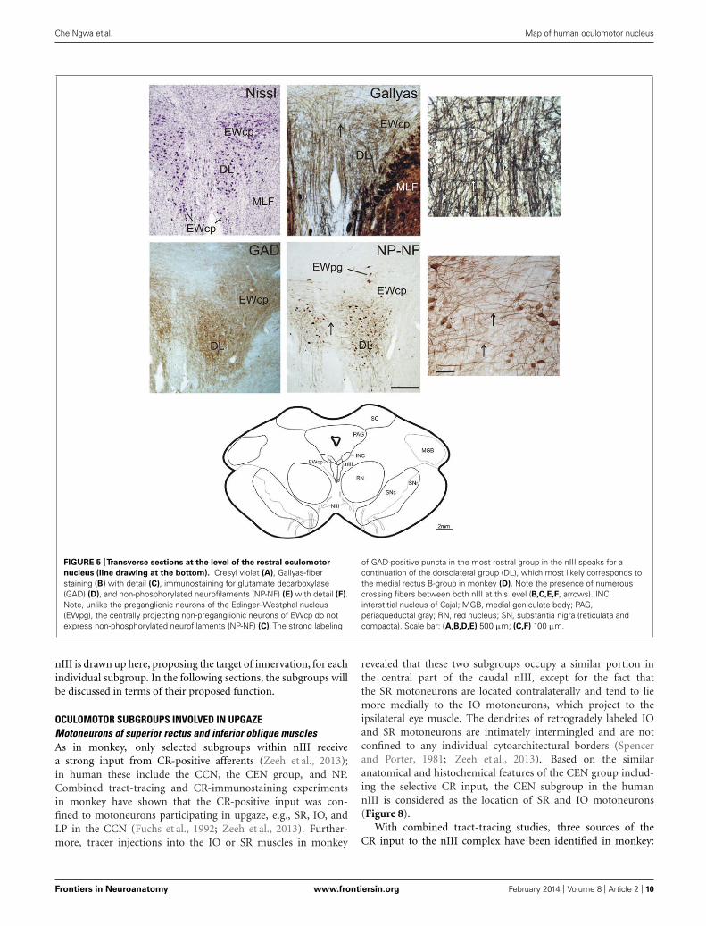
FIGURE 3 |Transverse sections at the level of the mid oculomotor
nucleus (line drawing at the bottom). Cresyl violet (A), fiber staining (B),and immunostaining for non-phosphorylated neurofilaments (NP-NF) (C)
reveal six subnuclei of the nIII complex at this level that exhibit differentstaining patterns for calretinin (CR) (D), glycine receptor (GlyR) (E), andglutamate decarboxylase (GAD) (F). The nucleus of Perlia (NP) forms anelongated midline cell group separated from the main nucleus bydorsoventrally traversing fibers (A,B), some expressing CR-immunoreactivity(D, arrows, inset). At the dorsomedial border of nIII, a compact cell group
forms the centrally projecting non-preganglionic part of the Edinger–Westphalnucleus (EWcp), which contain urocortin (UCN)-positive and some scatteredCR-positive neurons (D,G). The EWcp does not contain NP-NF (C), is devoidof GlyR (E), but receives a strong GAD input (F). High power magnification oftwo adjacent 5 μm paraffin sections immunostained for CR and UCN revealonly few UCN-positive neurons expressing CR (H,I, arrows). Correspondingblood vessels are indicated by asterisks. PAG, periaqueductal gray; RN, rednucleus; SC, superior colliculus; SN, substantia nigra (reticulata andcompacta). Scale bar: (A–F) 500 μm; (G) 100 μm; (H,I) 50 μm.
Frontiers in Neuroanatomy www.frontiersin.org February 2014 | Volume 8 | Article 2 | 8

Che Ngwa et al. Map of human oculomotor nucleus
FIGURE 4 | High-power photographs of the staining pattern with
different markers in the oculomotor nucleus complex. Numerouscalretinin (CR)-positive profiles are found in association with cell bodies onlyin CEN and NP (G,J), but not in DL, DM (A,D). CR-staining of somata is foundin EWcp (M; see also Figure 3). Strong labeling for glycine receptors (GlyR) isfound only in the dorsolateral group (DL) (B), and with some traversing fibersin the dorsomedial group (DM) (E), but none in CEN, NP and EWcp (H,K,N).
Different density of glutamate decarboxylase (GAD)-positive puncta is seenaround choline acetyltransferase (ChAT)-immunoreactive motoneurons in thedorsolateral (DL) (C), dorsomedial (DM) (F), central groups (CEN) (I), and thenucleus of Perlia (NP) (L). The strongest supply by GAD-positive punctateprofiles is present around ChAT-negative non-preganglionic centrallyprojecting neurons in the Edinger–Westphal nucleus (EWcp) (O). Scalebar (A–O) 30 μm.
Figures 6D,F,H,J and 7B). Furthermore, a high density of CR-positive puncta was noticed in the CCN with 0.051 puncta/μm(Table 3; Figures 6D and 7B). In contrast, only a fewmotoneurons in all other motoneuronal subgroups were asso-ciated with CR-positive profiles at an average density of around0.01 puncta/μm (Table 3; Figure 7B). A comparative anal-ysis revealed that the density of CR-positive puncta aroundmotoneurons for upgaze in CCN and the CEN subgroup wassignificantly stronger than those around motoneurons for down-or horizontal gaze (p < 0.001). Even those down- and horizontal
gaze motoneurons receiving some CR-input were contactedby significantly less CR-positive puncta, when separately ana-lyzed and compared with the CR-input of upgaze motoneurons(Bonferroni’s multiple comparison test; p < 0.05; not shown).
DISCUSSIONIn this study of the histochemical characteristics of the humannIII and nIV, eight cell groups were distinguished from each other.From these results, and those of a previous study on non-humanprimates (Zeeh et al., 2013), a map of the subgroups of the human
Frontiers in Neuroanatomy www.frontiersin.org February 2014 | Volume 8 | Article 2 | 9
Che Ngwa et al. Map of human oculomotor nucleus
FIGURE 5 |Transverse sections at the level of the rostral oculomotor
nucleus (line drawing at the bottom). Cresyl violet (A), Gallyas-fiberstaining (B) with detail (C), immunostaining for glutamate decarboxylase(GAD) (D), and non-phosphorylated neurofilaments (NP-NF) (E) with detail (F).Note, unlike the preganglionic neurons of the Edinger–Westphal nucleus(EWpg), the centrally projecting non-preganglionic neurons of EWcp do notexpress non-phosphorylated neurofilaments (NP-NF) (C). The strong labeling
of GAD-positive puncta in the most rostral group in the nIII speaks for acontinuation of the dorsolateral group (DL), which most likely corresponds tothe medial rectus B-group in monkey (D). Note the presence of numerouscrossing fibers between both nIII at this level (B,C,E,F, arrows). INC,interstitial nucleus of Cajal; MGB, medial geniculate body; PAG,periaqueductal gray; RN, red nucleus; SN, substantia nigra (reticulata andcompacta). Scale bar: (A,B,D,E) 500 μm; (C,F) 100 μm.
nIII is drawn up here, proposing the target of innervation, for eachindividual subgroup. In the following sections, the subgroups willbe discussed in terms of their proposed function.
OCULOMOTOR SUBGROUPS INVOLVED IN UPGAZEMotoneurons of superior rectus and inferior oblique musclesAs in monkey, only selected subgroups within nIII receivea strong input from CR-positive afferents (Zeeh et al., 2013);in human these include the CCN, the CEN group, and NP.Combined tract-tracing and CR-immunostaining experimentsin monkey have shown that the CR-positive input was con-fined to motoneurons participating in upgaze, e.g., SR, IO, andLP in the CCN (Fuchs et al., 1992; Zeeh et al., 2013). Further-more, tracer injections into the IO or SR muscles in monkey
revealed that these two subgroups occupy a similar portion inthe central part of the caudal nIII, except for the fact thatthe SR motoneurons are located contralaterally and tend to liemore medially to the IO motoneurons, which project to theipsilateral eye muscle. The dendrites of retrogradely labeled IOand SR motoneurons are intimately intermingled and are notconfined to any individual cytoarchitectural borders (Spencerand Porter, 1981; Zeeh et al., 2013). Based on the similaranatomical and histochemical features of the CEN group includ-ing the selective CR input, the CEN subgroup in the humannIII is considered as the location of SR and IO motoneurons(Figure 8).
With combined tract-tracing studies, three sources of theCR input to the nIII complex have been identified in monkey:
Frontiers in Neuroanatomy www.frontiersin.org February 2014 | Volume 8 | Article 2 | 10
Che Ngwa et al. Map of human oculomotor nucleus
FIGURE 6 |Two parallel series of drawings of transversal sections
through the trochlear (nIV) and oculomotor nucleus (nIII) complex
arranged from caudal to rostral. Panels (A,B) correspond to the level ofFigure 1, (C,D) to the level of Figure 2, (G,H) to the level of Figure 3, and(K,L) to the level of Figure 5. Panels (E,F and I,J) correspond to additionalintermediate levels. On the left, the density of glutamate decarboxylase(GAD) positive puncta and the intensity of immunostaining for the glycinereceptor (GlyR) in the different subgroups is indicated by blue hatching onthe left and green hatching on the right, respectively. The right columnshows the presence of neurons expressing immunoreactivity for urocortin(UCN, black), non-phosphorylated neurofilaments (NP-NF; gray) andcalretinin (CR; red filled circles). The density of CR-positive profiles isindicated by different grades of red hatching.
the rostral interstitial nucleus of the medial longitudinal fascicle(RIMLF), the interstitial nucleus of Cajal (INC) and the y-group of the vestibular nuclei (Ahlfeld et al., 2011). The RIMLFcontains premotor neurons of different types; some exhibit a high-frequency burst for upward saccades, others for downward sac-cades, and they are all intermingled with each other (Büttner et al.,1977; Horn and Büttner-Ennever, 1998). Considering their tar-gets, the CR-positive population probably represents the premotor
FIGURE 7 | Histogram of the quantitative analysis of GABAergic and
calretinin (CR) input to motoneurons in the oculomotor and
trochlear nucleus. The values are given in Table 3. (A) The GABA-inputwas quantified by counting glutamate decarboxylase-positive (GAD)puncta along the measured length of the contour of a given neuron. Themean terminal density of input and the standard error of the mean valueswere calculated for each subgroup. The one-way analysis of variancerevealed a significant difference (p < 0.001). The strongest input is seento non-preganglionic centrally projecting neurons in the Edinger–Westphalnucleus (EWcp; compare to Figure 4O), whereas the neurons of all othersubgroups did not show major differences. (B) The strongest CR input isseen to neurons of the nucleus of Perlia (NP), the central group inoculomotor nucleus (CEN), and the central caudal nucleus (CCN). Thenumber of counted CR-positive puncta associated with putative upgazemotoneurons is significantly higher compared to those aroundhorizontal – and downgaze motoneurons (***p < 0.001).
burst neurons for upward saccades (Ahlfeld et al., 2011). TheCR-input from INC to upgaze motoneurons may derive from pre-motor burst-tonic neurons involved in integration of the velocitysignal from RIMLF into the eye-position signal, required for gazestabilization after a saccade (Fukushima et al., 1992). The lack ofGAD in CR-immunopositive neuronal profiles in monkey nIII asrevealed by double-immunofluorescence and confocal scanning,indicated that the CR input is excitatory (Zeeh et al., 2013). CR-positive projections from the y-group to SR and IO motoneuronsmay provide the excitatory drive during smooth pursuit eye move-ments (Partsalis et al., 1995). The functional significance of theselective CR presence in upgaze pathways remains unclear, ithas been discussed in previous publications (Ahlfeld et al., 2011;Zeeh et al., 2013).
Frontiers in Neuroanatomy www.frontiersin.org February 2014 | Volume 8 | Article 2 | 11

Che Ngwa et al. Map of human oculomotor nucleus
Table 3 | Quantification of calretinin and GABAergic input to nIV and
nIII subgroups.
CR GAD
Subgroup Puncta/μm SE of mean Puncta/μm SE of mean
nIV 0.008 0.002 0.091 0.006
CEN 0.102 0.006 0.067 0.006
DM 0.012 0.004 0.052 0.005
DL 0.008 0.005 0.08 0.006
VEN 0.011 0.003 0.063 0.006
LAT 0.013 0.004 0.041 0.005
NP 0.103 0.008 0.069 0.008
CCN 0.051 0.006 0.063 0.005
EWcp 0 0 0.183 0.016
Central caudal nucleusPanegrossi (1898), was the first to describe the CCN in human.Originally he had termed the nucleus on the midline, situatedbetween the oculomotor nuclei at caudal levels, as nucleus poste-rior dorso-centralis. He found this nucleus as a constant featurein human, and noted it also in monkey, dog, and cat. Sincethis nucleus degenerated after removal of the bulbus in cat, hedesignated it as part of the nIII (review: Warwick, 1953a,b).Similarly, Tsuchida (1906) described a central medial nucleusbetween the main cell columns of caudal nIII, but he did notrelate it to Panegrossi’s findings. He called this medial nucleusthe caudal central nucleus (Tsuchida, 1906). In spite of the factthat Tsuchida (1906) designated it probably to the dorsal raphenucleus, his term was later adopted for the midline nucleus con-taining LP motoneurons. Based on removal of individual eyemuscles in monkey, Warwick was the first to show that the CCNcontains the LP motoneurons (Warwick, 1953b). This was laterconfirmed with tract-tracing methods, also showing that the LPmotoneurons of both eyes are intermingled within the CCN, witha slight predominance for a contralateral representation (Porteret al., 1989). There are conflicting reports as to whether some LPmotoneurons innervate the muscles of both sides (Sekiya et al.,1992; Van der Werf et al., 1997), or whether LP populations arecompletely separated for each eye (Porter et al., 1989). As in mon-key, the CCN in human forms an unpaired nucleus dorsal tothe caudal end of nIII (Schmidtke and Büttner-Ennever, 1992;Horn and Adamcyzk, 2011; Büttner-Ennever and Horn, 2014).Furthermore, the present study revealed, that in addition to asignificant CR-input, there is a strong input from GABAergicand glycinergic afferents to LP motoneurons, as found in mon-key (Horn and Büttner-Ennever, 2008; Zeeh et al., 2013). Onepossible source of direct or indirect inhibitory GABAergic affer-ents is the nucleus of the posterior commissure, since lesions ofthis area result in lid retraction (Schmidtke and Büttner-Ennever,1992; Averbuch-Heller, 1997). A further direct inhibitory connec-tion was shown from pontine neurons at the rostral and ventralborder of the principal trigeminal nucleus to LP motoneurons inthe CCN, which presumably provide the inhibition during blinks
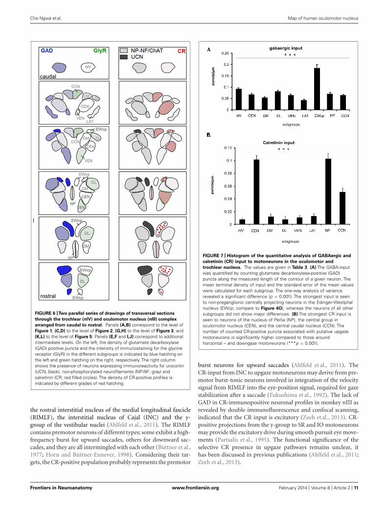
FIGURE 8 | Proposed map of the motoneurons for individual
extraocular muscles in human shown at four representative planes
from caudal to rostral. The right half shows corresponding sections inNissl staining to demonstrate the cytoarchitecture. The central caudalnucleus at most caudal planes contains the motoneurons of the levatorpalpebrae muscle (LP) (A). The medial rectus muscles (MR) is representedin two groups, the dorsolateral B- and the ventral A-group (A,B). The centralgroup represents the motoneurons of the inferior oblique (IO) and superiorrectus (SR) muscle (A,B). The nucleus of Perlia (NP) is separated from themain nucleus, but may contain SR motoneurons as well (B,C). Thedorsomedial group corresponds to the inferior rectus motoneurons (IR)(A–C). The centrally projecting neurons of the Edinger–Westphal nucleus(EWcp) appear as a single lateral group on caudal levels dorsal to nIII (A),adjoined by a medial group further rostrally (B), which both merge to asingle dorsal group (C). Another ventral extension of the EWcp appears onrostral levels (D). Note that the preganglionic neurons in the EWpg do notform a compact nucleus (B). Scale bar: (A–D) 500 μm.
Frontiers in Neuroanatomy www.frontiersin.org February 2014 | Volume 8 | Article 2 | 12

Che Ngwa et al. Map of human oculomotor nucleus
(May et al., 2012). The glycinergic input to LP motoneurons mayoriginate from saccadic omnipause neurons, as indicated by tract-tracing studies in monkey (Horn and Büttner-Ennever, 2008). Thefunction of this connection is not clear, yet, but may contributeto pathways involved in blink-saccade interaction (Leigh and Zee,2006).
Premotor neurons in the medial RIMLF in cat, and in theM-group in monkey, which target LP motoneurons, representa further possible CR source (Horn et al., 2000; Chen and May,2002). Furthermore, a monosynaptic excitatory connection fromINC to LP motoneurons has been described in cat (Chen andMay, 2007). This projection may originate from the same premo-tor neurons in INC, which target SR and IO motoneurons, therebycoupling vertical eye and lid movements not only during saccades,but also during gaze holding, to provide a larger, and freer upperfield of vision.
Nucleus of PerliaThe NP was described by Perlia (1889) and originally consideredas a cell group participating in the control of convergence, but upto now, without any proof (Warwick, 1955). In spite of severalreferences describing the presence of the NP in non-human pri-mates as a labeled midline group, after tracer injections into theciliary ganglion (Burde, 1983, 1988; Burde and Williams, 1989;Ishikawa et al., 1990), its existence is still questioned in thesespecies. The tracer labeled neurons are more likely to representmotoneurons of multiply-innervated muscles fibers (MIF) of theIO and SR, due to superficial contamination of the muscles asdiscussed previously (Büttner-Ennever et al., 2001; Horn et al.,2008). In fact, the morphology of the neurons of NP and theirhistochemical properties, e.g., expression of ChAT, cytochromeoxidase, NP-NF, and chondroitin sulfate proteoglycans, sug-gest that they may present motoneurons of singly-innervatedtwitch muscle fibers (SIF; Eberhorn et al., 2005; Horn et al.,2008). The present study demonstrated a CR input to NP andthereby indicates a role in upgaze, which supports the hypoth-esis that NP may represent SR twitch motoneurons that areseparated from the main subgroup in nIII by dorsoventrally trav-eling nerve fibers (Horn et al., 2008; Büttner-Ennever and Horn,2014).
OCULOMOTOR SUPGROUPS INVOLVED IN DOWNGAZEMotoneurons of superior oblique and inferior rectus musclesIn addition to the SO motoneurons in nIV, the IR motoneuronsin nIII participate in downward eye movements (Leigh and Zee,2006). In monkey, the IR motoneurons lie within the rostral halfof nIII appearing medial to the B group of the MR motoneu-rons. At the rostral end of nIII they form the dorsal part ofnIII (Evinger, 1988; Büttner-Ennever, 2006). Unlike motoneu-rons of horizontal moving eye muscles, a strong GABAergicinput was found to the motoneurons of all vertically pulling eyemuscles in monkey, including those for downgaze (Spencer andBaker, 1992). One well known source for GABAergic afferentsto the vertically pulling eye muscles arises from the secondaryvestibulo-ocular neurons in the superior vestibular nuclei (de laCruz et al., 1992; Wentzel et al., 1996; Highstein and Holstein,2006). Electrophysiological and pharmacological studies have
shown that stimulation of the vestibular nerve results in inhibitorypostsynaptic potentials in the ipsilateral nIV and nIII, which areblocked after administration of GABA antagonists (Obata andHighstein, 1970). Similarly, a lesion of the MLF results in adrastic decrease of GABA in nIII and nIV in cat (Precht et al.,1973).
Another source for GABAergic afferent input to nIII and nIVis the INC. In monkey, tracer injections into nIV or rostralnIII resulted in retrograde labeling of medium-sized GABAergicneurons in the contralateral INC (Horn et al., 2003). This is inline with the recordings of monosynaptic inhibitory postsynap-tic potentials in nIV and nIII after INC stimulation (Schwindtet al., 1974). Recent findings in cat confirm these results, andre-emphasize that premotor inhibitory neurons in INC may rep-resent inhibitory burst neurons of the vertical saccadic system(Sugiuchi et al., 2013).
OCULOMOTOR SUBGROUPS INVOLVED IN HORIZONTAL GAZEAside from the CCN, the VEN, LAT, and DL subgroups in nIIIreceive a strong glycinergic input, as indicated by the relativelyselective presence of GlyRs. In cat and monkey, glycinergic affer-ents were found to be associated specifically with motoneuronsinvolved in horizontal eye movements, i.e., MR in nIII and lat-eral rectus muscle (LR) in the abducens nucleus (nVI). This isin contrast to the high concentration of GABAergic input tomotoneurons for vertical eye movements in nIII and nIV (Spenceret al., 1989; Spencer and Baker, 1992). Tract-tracing experimentsin monkey had shown that the MR is represented in two separatedgroups within nIII (Büttner-Ennever and Akert, 1981; Porter et al.,1983): the A-group occupying the ventral part of nIII and extend-ing through its whole rostro-caudal extents, and the B-groupforming a well separated DL group at caudal nIII levels (Büttner-Ennever and Akert, 1981). In addition at caudal levels, the MRpopulation reaches as finger-like extensions into the fibers of theMLF, partly in conjunction with the A-group, partly forming com-pletely separated islands. Based on the similar cytoarchitecturalfeatures and the selective glycinergic inputs, we consider the VENgroup in human nIII as the homolog to the MR “A-group” inmonkey, including the extensions of LAT into the surroundingMLF. Accordingly, the DL group is considered to be the homologof the MR “B-group” (Figure 8): it has the same circular con-tour as in monkey, and a similar separation from the neighboringsubgroups, with no motoneuronal dendrites extending beyondits boundaries. Interestingly, at the rostral nIII pole the dendritesof presumed MR motoneurons reach across the midline to theircontralateral counterparts. Whether this is only the consequenceof the disappearance of the NP at this level, or whether it hasa functional background in the collection of common afferentinputs for controlling vergence, remains unclear.
The inhibitory control of horizontal gaze by glycinergic affer-ents that is seen for LR and MR motoneurons in cat and monkeyis found to be preserved in the human as well (Spencer et al.,1989, 1992; Spencer and Baker, 1992). With anatomical, record-ing and pharmacological methods, the inhibitory nature of theglycinergic projection from the prepositus nucleus to the nVIhas been demonstrated in the cat (Spencer et al., 1989). Up todate, the source of the glycinergic input to MR motoneurons in
Frontiers in Neuroanatomy www.frontiersin.org February 2014 | Volume 8 | Article 2 | 13

Che Ngwa et al. Map of human oculomotor nucleus
nIII is unknown. Although strychnine-sensitive GlyRs are knownto mediate synaptic inhibition by activating chloride channels(Dutertre et al., 2012), glycine can also contribute to excitatorytransmission by serving as an allosteric modulator for the gluta-mate N-methyl-D-aspartate receptor (Johnson and Ascher, 1987).Therefore, it is possible that the presence of GlyR seen in the MRsubgroups in primates is associated with the glutamatergic inputsfrom the ipsilateral lateral vestibular nuclei via the ascending tractof Deiters (Nguyen and Spencer, 1999), which may contribute toviewing distance related gain changes of the vestibulo-ocular reflex(Snyder and King, 1992; Chen-Huang and McCrea, 1998).
However, in contrast to cat and monkey, in human all pre-sumed MR subgroups receive an additional strong supply fromGABAergic afferents, which even exceeds that of the motoneu-rons for vertical gaze. This finding is in line with observationsfrom the human nVI, which also receives a strong GABAergic –in addition to a strong glycinergic – input. This observation is,surprisingly, not the same as in monkey, where only a moderateGABAergic input is observed (Spencer and Baker, 1992; Waldvo-gel et al., 2010). Thereby, the GABAergic inputs provided the leastuseful marker to delineate the motoneuronal subgroups in humannIII, but at the same time they revealed an interesting and unusualneuroanatomical difference between monkey and man, which isseldom observed.
Although the GABA-immunoreactivity in the cat nVI is rela-tively weak, both motoneurons and internuclear neurons get someGABAergic input (de la Cruz et al., 1989). In cat 20% of retro-gradely labeled small internuclear neurons in and around the nIIIexpressed GABA-immunoreactivity (de la Cruz et al., 1992) andmay be one source for the relatively weak GABAergic input tomotoneurons and internuclear neurons in nVI (de la Cruz et al.,1992). In cat, tracer-labeled MR motoneurons receive a similarstrong supply from glycinergic and GABAergic afferents (de laCruz et al., 1992).
Up to date, it is generally accepted that horizontal conjugateeye movements are mediated through the nVI, which containsmotoneurons and internuclear neurons. The motoneurons inner-vate the ipsilateral LR, the internuclear neurons activate thecontralateral MR motoneurons in nIII via the MLF (for review:Horn and Leigh, 2011). A separate “extra-MLF” vergence pathwayinvolving premotor neurons in the supraoculomotor area (SOA)with pure vergence signals (not conjugate eye movements) pro-vides the command to move the eyes at equal magnitudes, butin opposite direction for alignment of gaze between targets atdifferent depths (Mays, 1984). At the same location in the SOA,divergence neurons have been identified, which showed decreasedfiring rates with increasing vergence angles (Mays, 1984; Judge andCumming, 1986). Direct inputs from the SOA to MR motoneu-rons have been demonstrated (Zhang et al., 1991), and they wereshown to be related either to pure vergence or accommodation,or to both (Zhang et al., 1992). Theoretically, divergent eye move-ments require the activation of LR motoneurons and inhibition ofMR motoneurons, which could be mediated through inhibitionfrom GABAergic neurons in the SOA.
Another direct premotor input to motoneurons of the horizon-tal system was indicated from the central mesencephalic reticularformation (CMRF) after retrograde transsynaptic labeling studies
in monkey applying rabies virus injections into LR (Ugolini et al.,2006; Büttner-Ennever, 2008). The CMRF is closely intercon-nected with the superior colliculus and the paramedian pontinereticular formation including the saccadic omnipause neurons(Cohen and Büttner-Ennever, 1984; Chen and May, 2000; Wanget al., 2013) and has been found to be correlated with horizontaland vertical saccades (Waitzman et al., 2000a,b, 2002). Preliminarydata applying small biotin dextran injections into the rostrome-dial part of the CMRF in monkey revealed monosynaptic inputsto all MR motoneuron subgroups on both sides and pg neu-rons in the EWpg, indicating a role in vergence and the neartriad, at least of this CMRF region (May et al., 2011; Horn et al.,2012). The accompanying ultrastructural analysis revealed thatmany of the tracer labeled terminals contacting MR motoneu-rons have features in accordance with inhibitory synapses, someof them expressing GABA-immunolabeling (May et al., 2011).To what extent the GABA-negative afferent terminals may rep-resent glycinergic afferents remains to be studied. Based on themonkey data, the strong GABAergic input seen here in thehuman nIII may derive at least in part from the adjacent CMRFand/or SOA.
GENERAL ORGANIZATION IN OCULOMOTOR NUCLEUS: PRIMATEThe first anatomical description of the nIII is given by Stilling(1846). The partition into a dorsal and ventral portion, and thepresence of numerous decussating axons was first described byvon Gudden (1881; for review: Warwick, 1953a). A very precisedescription of the cytoarchitecture of the nIII was provided byPerlia on fetal human brain, which included the lateral and medialportion of the classical EW and the NP, which he originally hadtermed “Centralkern” (Perlia, 1889). Based on observations madeafter the removal of extraocular muscles in various species, dif-ferent variations of an nIII map had been proposed (reviewedby Warwick, 1953a). The elaborate work of Warwick, who plot-ted the neurons undergoing chromatolysis after the resection ofindividual extraocular muscles in monkey, provided a map of theprimate nIII, which was widely accepted and used as basis forthe human nIII in many textbook illustrations (Warwick, 1953a).The organization of the motoneuronal groups shows a sequencefrom rostral to caudal of IR, MR, IO, SR, and LP motoneurons.The newly developed tract-tracing method basically confirmedthe proposed arrangement of motoneuronal groups of individ-ual muscles in the nIII of monkey, but it revealed for the firsttime the presence of two motoneuron groups for the MR, the ven-tral A-group and the DL circular B-group (Büttner-Ennever andAkert, 1981; Porter et al., 1983; Büttner-Ennever, 2006). This two-fold representation of the MR within the nIII is most evident inprimates and its function remains unclear (Augustine et al., 1981;Sun and May, 1993; Büttner-Ennever, 2006). So far no differencesin histochemistry or afferent inputs have been found betweenthe A- and B-group (Spencer et al., 1992; Wasicky et al., 2004;Erichsen et al., 2014).
It has been known for a long time that extraocular musclesexhibit a complex architecture consisting of a global and orbitallayer. At least six different types of muscle fibers can be identi-fied, which can be divided into two main categories of SIF andmultiply-innervated non-twitch muscle fibers (MIF; for review:
Frontiers in Neuroanatomy www.frontiersin.org February 2014 | Volume 8 | Article 2 | 14
Che Ngwa et al. Map of human oculomotor nucleus
Spencer and Porter, 2006). Tract-tracing experiments in mon-key revealed that the motoneurons of MIFs are located in theperiphery of the motonuclei. For muscles innervated from thenIII the MIF motoneurons of IR and MR are located in theC-group DM to nIII, and those of IO and SR in the S-groupbetween the both nIII (Büttner-Ennever et al., 2001). Based ontheir different histochemical properties, SIF motoneurons wereidentified within nIII and putative MIF motoneurons have beenidentified around the medial aspects of nIII, also in human(Eberhorn et al., 2005, 2006). However, in the human nIII, theMIF motoneurons could not be allocated to specific extraocularmuscles, yet (Horn et al., 2008). Therefore, the proposed map ofthe human nIII applies only to the SIF motoneurons within nIII,and has yet to be extended in future studies by the location of MIFmotoneurons.
The exact knowledge of the location of the subgroups inner-vating individual eye muscles in human provides an importantbasis to localize lesions more accurately in MRI scans and relate itto clinical findings. Furthermore, the present work on transmitterinputs to individual eye muscle subgroups will form the basis forpostmortem studies of afferent inputs to nIII in cases with knowneye-movement deficits
AUTHOR CONTRIBUTIONSAcquisition of data and analysis was performed by Emmanuel CheNgwa, Christina Zeeh, and Ahmed Messoudi. Conception of thework was done by Anja K. E. Horn and Jean A. Büttner-Ennever.All authors contributed to the interpretation of data, preparationof the figures, and writing of the manuscript and approved thefinal version.
ACKNOWLEDGMENTSThis study is part of the medical doctoral thesis of EmmanuelChe Ngwa. The results are published with permission ofthe Medical Faculty of the Ludwig-Maximilians-University ofMunich. We are very grateful to Christine Glombik and LaureDjaleu for their excellent technical assistance. Supported byDeutsche Forschungsgemeinschaft DFG HO 1639/4-3, BMBF(IFB-01EO0901, Brain-Net-01GI0505).
REFERENCESAhlfeld, J., Mustari, M., and Horn, A. K. E. (2011). Sources of calretinin inputs to
motoneurons of extraocular muscles involved in upgaze. Ann. N. Y. Acad. Sci.1233, 91–99. doi: 10.1111/j.1749-6632.2011.06168.x
Andressen, C., Blümcke, I., and Celio, M. R. (1993). Calcium-binding pro-teins – selective markers of nerve cells. Cell Tissue Res. 271, 181–208. doi:10.1007/BF00318606
Augustine, J. R., Deschamps, E. G., and Ferguson, J. G. J. (1981). Functional organi-zation of the oculomotor nucleus in the baboon. Am. J. Anat. 161, 393–403. doi:10.1002/aja.1001610405
Averbuch-Heller, L. (1997). Neurology of the eyelids. Curr. Opin. Ophthalmol. 8,27–34. doi: 10.1097/00055735-199712000-00005
Bachtell, R. K., Weitemier, A. Z., Galvan-Rosas, A., Tsivkovskaia, N. O., Risinger, F.O., Phillips, T. J., et al. (2003). The Edinger–Westphal-lateral septum urocortinpathway and its relation to alcohol-induced hypothermia. J. Neurosci. 23, 2477–2487.
Baer, K., Waldvogel, H. J., Faull, R. L. M., and Rees, M. I. (2009). Local-ization of glycine receptors in the human forebrain, brainstem, and cervicalspinal cord: an immunohistochemical review. Front. Mol. Neurosci. 2:25. doi:10.3389/neuro.02.025.2009
Baizer, J. S., and Baker, J. F. (2006). Immunoreactivity for calretinin and calbindinin the vestibular nuclear complex of the monkey. Exp. Brain Res. 172, 103–113.doi: 10.1007/s00221-005-0318-1
Baizer, J. S., and Broussard, D. M. (2010). Expression of calcium-binding proteinsand nNOS in the human vestibular and precerebellar brainstem. J. Comp. Neurol.518, 872–895. doi: 10.1002/cne.22250
Bernheimer, S. (1897). Experimentelle Studien zur Kenntniss der Innervationder inneren und äusseren vom Oculomotorius versorgten Muskeln des Auges.Albrecht Von Graefes Arch. Ophthalmol. 44, 481–525. doi: 10.1007/BF02017581
Bruce, G., Wainer, B. H., and Hersh, L. B. (1985). Immunoaffinity purification ofhuman choline acetyltransferase: comparison of the brain and placental enzymes.J. Neurochem. 45, 611–620. doi: 10.1111/j.1471-4159.1985.tb04030.x
Burde, R. M. (1983). The visceral nuclei of the oculomotor complex. Trans. Am.Ophthalmol. Soc. 81, 532–548.
Burde, R. M. (1988). Direct parasympathetic pathway to the eye: revisited. BrainRes. 463, 158–162. doi: 10.1016/0006-8993(88)90540-9
Burde, R. M., and Williams, F. (1989). Parasympathetic nuclei. Brain Res. 498,371–375. doi: 10.1016/0006-8993(89)91119-0
Büttner, U., Büttner-Ennever, J. A., and Henn, V. (1977). Vertical eye movementrelated unit activity in the rostral mesencephalic reticular formation of the alertmonkey. Brain Res. 130, 239–252. doi: 10.1016/0006-8993(77)90273-6
Büttner-Ennever, J., and Horn, A. (eds). (2014). Olszewski and Baxter’s Cytoarchi-tecture of the Human Brainstem, 3rd Edn. Basel: Karger.
Büttner-Ennever, J. A. (2006). The extraocular motor nuclei: organization andfunctional neuroanatomy. Prog. Brain Res. 151, 95–125. doi: 10.1016/S0079-6123(05)51004-5
Büttner-Ennever, J. A. (2008). Mapping the oculomotor system. Prog. Brain Res.171, 3–11. doi: 10.1016/S0079-6123(08)00601-8
Büttner-Ennever, J. A., and Akert, K. (1981). Medial rectus subgroups of the ocu-lomotor nucleus and their abducens internuclear input in the monkey. J. Comp.Neurol. 197, 17–27. doi: 10.1002/cne.901970103
Büttner-Ennever, J. A., Horn, A. K. E., Scherberger, H., and D’Ascanio, P. (2001).Motoneurons of twitch and nontwitch extraocular muscle fibers in the abducens,trochlear, and oculomotor nuclei of monkeys. J. Comp. Neurol. 438, 318–335. doi:10.1002/cne.1318
Chen, B., and May, P. J. (2000). The feedback circuit connecting the superior col-liculus and central mesencephalic reticular formation: a direct morphologicaldemonstration. Exp. Brain Res. 131, 10–21. doi: 10.1007/s002219900280
Chen, B., and May, P. J. (2002). Premotor circuits controlling eyelid movements inconjunction with vertical saccades in the cat: I. The rostral interstitial nucleusof the medial longitudinal fasciculus. J. Comp. Neurol. 450, 183–202. doi:10.1002/cne.10313
Chen, B., and May, P. J. (2007). Premotor circuits controlling eyelid movements inconjunction with vertical saccades in the cat: II. Interstitial nucleus of Cajal. J.Comp. Neurol. 500, 676–692. doi: 10.1002/cne.21203
Chen-Huang, C., and McCrea, R. A. (1998). Viewing distance related sensory pro-cessing in the ascending tract of deiters vestibulo-ocular reflex pathway. J. Vest.Res. 8, 175–184. doi: 10.1016/S0957-4271(97)00001-3
Cohen, B., and Büttner-Ennever, J. A. (1984). Projections from the superior col-liculus to a region of the central mesencephalic reticular formation (cMRF)associated with horizontal saccadic eye movements. Exp. Brain Res. 57, 167–176.doi: 10.1007/BF00231143
de la Cruz, R. R., Escudero, M., and Delgado-García, J. M. (1989). Behaviour ofmedial rectus motoneurons in the alert cat. Eur. J. Neurosci. 1, 288–295. doi:10.1111/j.1460-9568.1989.tb00796.x
de la Cruz, R. R., Pastor, A. M., Martínez-Guijarro, F. J., López-García, C., andDelgado-García, J. M. (1992). Role of GABA in the extraocular motor nuclei ofthe cat: a postembedding immunocytochemical study. Neuroscience 51, 911–929.doi: 10.1016/0306-4522(92)90529-B
Dutertre, S., Becker, C.-M., and Betz, H. (2012). Inhibitory glycine receptors: anupdate. Biol. Chem. 287, 40216–40223. doi: 10.1074/jbc.R112.408229
Eberhorn, A. C., Ardelenanu, P., Büttner-Ennever, J. A., and Horn, A. K. E.(2005). Histochemical differences between motoneurons supplying multiply andsingly innervated extraocular muscle fibers. J. Comp. Neurol. 491, 352–366. doi:10.1002/cne.20715
Eberhorn, A. C., Büttner-Ennever, J. A., and Horn, A. K. E. (2006).Identification of motoneurons innervating multiply- or singly-innervated
Frontiers in Neuroanatomy www.frontiersin.org February 2014 | Volume 8 | Article 2 | 15

Che Ngwa et al. Map of human oculomotor nucleus
extraocular muscle fibres in the rat. Neuroscience 137, 891–903. doi:10.1016/j.neuroscience.2005.10.038
Erichsen, J. T., Wright, N. F., and May, P. J. (2014). The morphology and ultrastruc-ture of medial rectus subgroup motoneurons in the macaque monkey. J. Comp.Neurol. 522, 626–641. doi: 10.1002/cne.23437
Evinger, C. (1988). Extraocular motor nuclei: location, morphology and afferents.Rev. Oculomot. Res. 2, 81–117.
Fuchs, A. F., Becker, W., Ling, L., Langer, T. P., and Kaneko, C. R. (1992). Dischargepatterns of levator palpebrae superioris motoneurons during vertical lid and eyemovements in the monkey. J. Neurophysiol. 68, 233–243.
Fukushima, K., Kaneko, C. R., and Fuchs, A. F. (1992). The neuronal substrateof integration in the oculomotor system. Prog. Neurobiol. 39, 609–639. doi:10.1016/0301-0082(92)90016-8
Gallyas, F. (1979). Silver staining of myelin by means of physical development.Neurol. Res. 1, 203–209.
Goldstein, M. E., Sternberger, N. H., and Sternberger, L. A. (1987). Phosphorylationprotects neurofilaments against proteolysis. J. Neuroimmunol. 14, 149–160. doi:10.1016/0165-5728(87)90049-X
Highstein, S. M., and Holstein, G. R. (2006). The anatomy of the vestibular nuclei.Prog. Brain Res. 151, 157–203. doi: 10.1016/S0079-6123(05)51006-9
Horn, A. K., and Büttner-Ennever, J. A. (2008). Brainstem circuits controlling lid-eye coordination in monkey. Prog. Brain Res. 171, 87–95. doi: 10.1016/S0079-6123(08)00612-2
Horn, A. K., Eberhorn, A., Härtig, W., Ardelenanu, P., Messoudi, A., andBüttner-Ennever, J. A. (2008). Perioculomotor cell groups in monkey and mandefined by their histochemical and functional properties: reappraisal of theEdinger–Westphal nucleus. J. Comp. Neurol. 507, 1317–1335. doi: 10.1002/cne.21598
Horn, A. K., and Leigh, R. J. (2011). The anatomy and physiology of the ocularmotor system. Handb. Clin. Neurol. 102, 21–69. doi: 10.1016/B978-0-444-52903-9.00008-X
Horn, A. K. E., and Adamcyzk, C. (2011). “Reticular formation - eye movements,gaze and blinks,” in Human Nervous System, 3rd Edn, eds G. Paxinos and J. K.Mai (San Diego: Academic Press), 328–366.
Horn, A. K. E., Bohlen, M. O., Warren, S., and May, P. J. (2012). Evidence for thecentral mesencephalic reticular formation playing a role in the near triad. Soc.Neurosci. Abstr. 373.12.
Horn, A. K. E., and Büttner-Ennever, J. A. (1998). Premotor neurons for verticaleye-movements in the rostral mesencephalon of monkey and man: the histo-logical identification by parvalbumin immunostaining. J. Comp. Neurol. 392,413–427. doi: 10.1002/(SICI)1096-9861(19980323)392:4<413::AID-CNE1>3.0.CO;2-3
Horn, A. K. E., Büttner-Ennever, J. A., Gayde, M., and Messoudi, A. (2000).Neuroanatomical identification of mesencephalic premotor neurons coor-dinating eyelid with upgaze in the monkey and man. J. Comp. Neurol.420, 19–34. doi: 10.1002/(SICI)1096-9861(20000424)420:1<19::AID-CNE2>3.0.CO;2-D
Horn, A. K. E., Helmchen, C., and Wahle, P. (2003). GABAergic neurons in the rostralmesencephalon of the Macaque monkey that control vertical eye movements.Ann. N. Y. Acad. Sci. 1004, 19–28. doi: 10.1196/annals.1303.003
Ichikawa, T., and Shimizu, T. (1998). Organization of choline acetyltransferase-containing structures in the cranial nerve motor nuclei and spinal cord of themonkey. Brain Res. 779, 96–103. doi: 10.1016/S0006-8993(97)01090-1
Ishikawa, S., Sekiya, H., and Kondo, Y. (1990). The center for controlling the nearreflex in the midbrain of the monkey: a double labelling study. Brain Res. 519,217–222. doi: 10.1016/0006-8993(90)90080-U
Jiao, Y., Sun, Z., Lee, T., Fusco, F. R., Kimble, T. D., Meade, C. A., et al.(1999). A simple and sensitive antigen retrieval method for free-floatingand slide-mounted tissue sections. J. Neurosci. Methods 93, 149–162. doi:10.1016/S0165-0270(99)00142-9
Johnson, J. W., and Ascher, P. (1987). Glycine potentiates the NMDA response incultured mouse brain neurons. Nature 325, 529–531. doi: 10.1038/325529a0
Judge, S. J., and Cumming, B. G. (1986). Neurons in the monkey midbrain withactivity related to vergence eye movement and accommodation. J. Neurophysiol.55, 915–930.
Kennard, C. (2011). Disorders of higher gaze control. Handb. Clin. Neurol. 102,379–402. doi: 10.1016/B978-0-444-52903-9.00020-0
Kozicz, T., Bittencourt, J. C., May, P. J., Reiner, A., Gamlin, P. D. R., Palkovits,M., et al. (2011). The Edinger–Westphal nucleus: a historical, structural, andfunctional perspective on a dichotomous terminology. J. Comp. Neurol. 519,1413–1434. doi: 10.1002/cne.22580
Leigh, R. J., and Zee, D. S. (2006). The Neurology of Eye Movements. New York:Oxford University Press.
May, P. J., Horn, A. K. E., Mustari, M. J., and Warren, S. (2011). Central mes-encephalic reticular formation projections onto oculomotor motoneurons. Soc.Neurosci. Abstr. 699.01
May, P. J., Reiner, A. J., and Ryabinin, A. E. (2008). Comparison of the distributions ofurocortin-containing and cholinergic neurons in the perioculomotor midbrainof the cat and macaque. J. Comp. Neurol. 507, 1300–1316. doi: 10.1002/cne.21514
May, P. J., Vidal, P.-P., Baker, H., and Baker, R. (2012). Physiological and anatomicalevidence for an inhibitory trigemino-oculomotor pathway in the cat. J. Comp.Neurol. 520, 2218–2240. doi: 10.1002/cne.23039
Mays, L. E. (1984). Neural control of vergence eye movements: convergence anddivergence neurons in midbrain. J. Neurophysiol. 51, 1091–1108.
Nguyen, L. T., and Spencer, R. F. (1999). Abducens internuclear and ascending tractof Deiters inputs to medial rectus motoneurons in the cat oculomotor nucleus:neurotransmitters. J. Comp. Neurol. 411, 73–86. doi: 10.1002/(SICI)1096-9861(19990816)411:1<73::AID-CNE6>3.0.CO;2-7
Obata, K., and Highstein, S. M. (1970). Blocking by picrotoxin of both vestibularinhibition and GABA action on rabbit oculomotor neurones. Brain Res. 18, 538–541. doi: 10.1016/0006-8993(70)90136-8
Olszewski, J., and Baxter, D. (1982). Cytoarchitecture of the Human Brain Stem, 2ndEdn. Basel: Karger.
Panegrossi, G. (1898). Contributo allo studio anatomo-fisiologico dei centri deinerve oculomotori dell’uomo. Ric. Lab. Anat. norm. Univ. Roma 6, 103–155.
Partsalis, A. M., Zhang,Y., and Highstein, S. M. (1995). Dorsal Y group in the squirrelmonkey. I. Neuronal responses during rapid and long-term modifications of thevertical VOR. J. Neurophysiol. 73, 615–631.
Pearson, A. A. (1943). Trochlear nerve in human fetuses. J. Comp. Neurol. 78, 29–43.doi: 10.1002/cne.900780103
Perlia, D. (1889). Die Anatomie des Oculomotoriuscentrums beim Menschen.Albrecht von Graefes Arch. Ophthalmol. 35, 287–308. doi: 10.1007/BF01695201
Pfeiffer, F., Simler, R., Grenningloh, G., and Betz, H. (1984). Monoclonal antibodiesand peptide mapping reveal structural similarities between the subunits of theglycine receptor of rat spinal cord. Proc. Natl. Acad. Sci. U.S.A. 81, 7224–7227.doi: 10.1073/pnas.81.22.7224
Porter, J. D., Burns, L. A., and May, P. J. (1989). Morphological substratefor eyelid movements: innervation and structure of primate levator palpebraesuperioris and orbicularis oculi muscles. J. Comp. Neurol. 287, 64–81. doi:10.1002/cne.902870106
Porter, J. D., Guthrie, B. L., and Sparks, D. L. (1983). Innervation of monkeyextraocular muscles: localization of sensory and motor neurons by retrogradetransport of horseradish peroxidase. J. Comp. Neurol. 218, 208–219. doi:10.1002/cne.902180208
Precht, W., Baker, R., and Okada, Y. (1973). Evidence for GABA as the synaptictransmitter of the inhibitory vestibulo-ocular pathway. Exp. Brain Res. 18, 415–428. doi: 10.1007/BF00239109
Ryabinin, A. E., Tsivkovskaia, N. O., and Ryabinin, S. A. (2005). Urocortin1-containing neurons in the human Edinger–Westphal nucleus. Neuroscience 134,1317–1323. doi: 10.1016/j.neuroscience.2005.05.042
Schmidtke, K., and Büttner-Ennever, J. A. (1992). Nervous control of eyelid func-tion – a review of clinical, experimental and pathological data. Brain 115, 227–247.doi: 10.1093/brain/115.1.227
Schwindt, P. C., Precht, W., and Richter, A. (1974). Monosynaptic excitatory andinhibitory pathways from medial midbrain nuclei to trochlear motoneurons. Exp.Brain Res. 20, 223–238. doi: 10.1007/BF00238314
Sekiya, H., Kojima, Y., Hiramoto, D., Mukuno, K., and Ishikawa, S. (1992). Bilateralinnervation of the musculus levator palpebrae superioris by single motoneu-rons in the monkey. Neurosci. Lett. 146, 10–12. doi: 10.1016/0304-3940(92)90159-5
Snyder, L. H., and King, W. M. (1992). Effect of viewing distance and location ofthe axis of head rotation on the monkey’s vestibuloocular reflex. I. Eye movementresponses. J. Neurophysiol. 67, 861–874.
Frontiers in Neuroanatomy www.frontiersin.org February 2014 | Volume 8 | Article 2 | 16

Che Ngwa et al. Map of human oculomotor nucleus
Spencer, R. F., and Baker, R. (1992). GABA and glycine as inhibitory neurotrans-mitters in the vestibulo-ocular reflex. Ann. N. Y. Acad. Sci. 656, 602–611. doi:10.1111/j.1749-6632.1992.tb25239.x
Spencer, R. F., and Porter, J. D. (1981). Innervation and structure of extraocularmuscles in the monkey in comparison to those of the cat. J. Comp. Neurol. 198,649–665. doi: 10.1002/cne.901980407
Spencer, R. F., and Porter, J. D. (2006). Biological organization of the extraocularmuscles. Prog. Brain Res. 151, 43–80. doi: 10.1016/S0079-6123(05)51002-1
Spencer, R. F., Wang, S. F., and Baker, R. (1992). The pathways and func-tions of GABA in the oculomotor system. Prog. Brain Res. 90, 307–331. doi:10.1016/S0079-6123(08)63620-1
Spencer, R. F., Wenthold, R. J., and Baker, R. (1989). Evidence for glycine as aninhibitory neurotransmitter of vestibular, reticular, and prepositus hypoglossineurons that project to the cat abducens nucleus. J. Neurosci. 9, 2718–2736.
Sternberger, L. A., Harwell, L. W., and Sternberger, N. H. (1982). Neurotype:regional individuality in rat brain detected by immunocytochemistry withmonoclonal antibodies. Proc. Natl. Acad. Sci. U.S.A. 79, 1326–1330. doi:10.1073/pnas.79.4.1326
Sternberger, L. A., and Sternberger, N. H. (1983). Monoclonal antibodies distinguishphorphorylated and non-phosphorylated forms of neurofilaments in situ. Proc.Natl. Acad. Sci. U.S.A. 80, 6126–6130. doi: 10.1073/pnas.80.19.6126
Stilling, B. (1846). Disquisitiones de structure et functionibus cerebri. Jenae: sumt.F. Maukii. T. 1, 36.
Sugiuchi, Y., Takahashi, M., and Shinoda, Y. (2013). Input-output organizationof inhibitory neurons in the interstitial nucleus of Cajal projecting to the con-tralateral trochlear and oculomotor nucleus. J. Neurophysiol. 110, 640–657. doi:10.1152/jn.01045.2012
Sun, W. S., and May, P. J. (1993). Organization of the extraocular and preganglionicmotoneurons supplying the orbit in the lesser galago. Anat. Rec. 237, 89–103. doi:10.1002/ar.1092370109
Triller, A., Cluzeaud, F., Pfeiffer, F., Betz, H., and Korn, H. (1985). Distribution ofglycine receptors at central synapses: an immunoelectron microscopy study. J.Cell Biol. 101, 683–688. doi: 10.1083/jcb.101.2.683
Tsuchida, U. (1906). Ueber die Ursprungskerne der Augenbeweguns-Nerven imMittel- und Zwischenhirn. Arb. Hirnanat. Inst. Zürich 1–2, 1–205.
Ugolini, G., Klam, F., Doldan Dans, M., Dubayle, D., Brandi, A.-M., Büttner-Ennever, J. A., et al. (2006). Horizontal eye movement networks in primatesas revealed by retrograde transneuronal transfer of rabies virus: differences inmonosynaptic input to“slow”and“fast”abducens motoneurons. J. Comp. Neurol.498, 762–785. doi: 10.1002/cne.21092
Van der Werf, F., Aramideh, M., Ongerboer De Visser, B. W., Baljet, B., Speelman,J. D., and Otto, J. A. (1997). A retrograde double fluorescent tracing study of thelevator palpebrae superioris muscle in the cynomolgus monkey. Exp. Brain Res.113, 174–179. doi: 10.1007/BF02454155
von Gudden, B. (1881). Über den Tractus peduncularis transversus. Arch. Psychiatr.Nervenkr. 2, 364–366.
Waitzman, D. M., Pathmanathan, J., Presnell, R., Ayers, A. S., and Depalma-Bowles,S. (2002). Contribution of the superior colliculus and the mesencephalic reticularformation to gaze control. Ann. N. Y. Acad. Sci. 956, 111–129. doi: 10.1111/j.1749-6632.2002.tb02813.x
Waitzman, D. M., Silakov, V. L., Depalma-Bowles, S., and Ayers, A. S. (2000a). Effectsof reversible inactivation of the primate mesencephalic reticular formation. I.Hypermetric goal-directed saccades. J. Neurophysiol. 83, 2260–2284.
Waitzman, D. M., Silakov, V. L., Depalma-Bowles, S., and Ayers, A. S. (2000b). Effectsof reversible inactivation of the primate mesencephalic reticular formation. II.Hypometric vertical saccades. J. Neurophysiol. 83, 2285–2299.
Waldvogel, H. J., Baer, K., Eady, E., Allen, K. L., Gilbert, R. T., Mohler, H., et al.(2010). Differential localization of γ-aminobutyric acid type a and glycine recep-tor subunits and gephyrin in the human pons, medulla oblongata and uppermostcervical segment of the spinal cord: an immunohistochemical study. J. Comp.Neurol. 518, 305–328. doi: 10.1002/cne.22212
Wang, N., Perkins, E., Zhou, L., Warren, S., and May, P. J. (2013). Anatomicalevidence that the superior colliculus controls saccades through central mesen-cephalic reticular formation gating of omnipause neuron activity. J. Neurosci. 33,16285–16296. doi: 10.1523/JNEUROSCI.2726-11.2013
Warwick, R. (1953a). Representation of the extraocular muscles in the oculomo-tor nuclei of the monkey. J. Comp. Neurol. 98, 449–495. doi: 10.1002/cne.900980305
Warwick, B. (1953b). The identity of the posterior dorso-central nucleus ofPanegrossi. J. Comp. Neurol. 99, 599–611. doi: 10.1002/cne.900990307
Warwick, R. (1955). The so-called nucleus of convergence. Brain 78, 92–114. doi:10.1093/brain/78.1.92
Wasicky, R., Horn, A. K. E., and Büttner-Ennever, J. A. (2004). Twitch and non-twitch motoneuron subgroups of the medial rectus muscle in the oculomotornucleus of monkeys receive different afferent projections. J. Comp. Neurol. 479,117–129. doi: 10.1002/cne.20296
Wentzel, P. R., Gerrits, N. M., and Dezeeuw, C. I. (1996). GABAergic and glycin-ergic inputs to the rabbit oculomotor nucleus with special emphasis on themedial rectus subdivision. Brain Res. 707, 314–319. doi: 10.1016/0006-8993(95)01389-X
Zeeh, C., Hess, B. J., and Horn, A. K. E. (2013). Calretinin inputs are confinedto motoneurons for upward eye movements in monkey. J. Comp. Neurol. 521,3154–3166. doi: 10.1002/cne.23337
Zhang, Y., Gamlin, P. D. R., and Mays, L. E. (1991). Antidromic identification ofmidbrain near response cells projecting to the oculomotor nucleus. Exp. BrainRes. 84, 525–528. doi: 10.1007/BF00230964
Zhang, Y., Mays, L. E., and Gamlin, P. D. R. (1992). Characteristics of nearresponse cells projecting to the oculomotor nucleus. J. Neurophysiol. 67,944–960.
Ziegler, B., Augstein, A., Schröder, D., Mauch, L., Hahmann, J., Schlosser, M., et al.(1996). Glutamate decarboxylase (GAD) is not detectable on the surface of ratislet cells examined by cytofluorometry and complement-dependent antibody-mediated cytotoxicity of monoclonal GAD antibodies. Horm. Metab. Res. 28,11–15. doi: 10.1055/s-2007-979121
Conflict of Interest Statement: The authors declare that the research was conductedin the absence of any commercial or financial relationships that could be construedas a potential conflict of interest.
Received: 16 November 2013; accepted: 14 January 2014; published online: 12 February2014.Citation: Che Ngwa E, Zeeh C, Messoudi A, Büttner-Ennever JA and Horn AKE (2014)Delineation of motoneuron subgroups supplying individual eye muscles in the humanoculomotor nucleus. Front. Neuroanat. 8:2. doi: 10.3389/fnana.2014.00002This article was submitted to the journal Frontiers in Neuroanatomy.Copyright © 2014 Che Ngwa, Zeeh, Messoudi, Büttner-Ennever and Horn. This is anopen-access article distributed under the terms of the Creative Commons AttributionLicense (CC BY). The use, distribution or reproduction in other forums is permit-
ted, provided the original author(s) or licensor are credited and that the originalpublication in this journal is cited, in accordance with accepted academic practice.No use, distribution or reproduction is permitted which does not comply with theseterms.
Frontiers in Neuroanatomy www.frontiersin.org February 2014 | Volume 8 | Article 2 | 17