Embed Size (px)
Citation preview

Transcriptomics, DNA Damage and DNA
Damage Response in Space
Honglu Wu
NASA Johnson Space Center
Houston, Texas, USA
https://ntrs.nasa.gov/search.jsp?R=20170001947 2018-06-28T17:22:21+00:00Z
Space Radiation Risks
– Carcinogenesis (morbidity and mortality risk)
– Acute and Late Central Nervous System (CNS) risks
immediate or late functional changes
– Chronic & Degenerative Tissue Risks
cataracts, heart-disease, etc.
– Acute Radiation Risks – sickness or death

NASA Space Radiation Laboratory at BNL
Unrejoined chromosome breaks after
low- and high-LET radiation exposure in
human fibroblast cells. Wu et al. 2002

Experimental design
• Exposure pre-flight
• Exposure during flight
• Exposure post-flight
Effects
• Synergistic - enhanced
• Additive - same
• Antagonistic - reduced
Combined effects of radiation and spaceflight factors
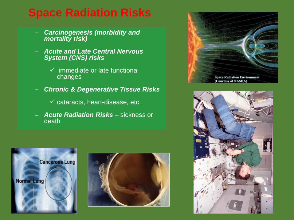
Chromosome aberration frequencies in pre- and post-flight astronaut lymphocytes irradiated in vitro with low-LET radiation (Wu et al. Phys. Med. 2001)
Nission: STS-103
Duration: 8 days
Blood draw schedule:
10 days before launch, JSC, kept at 4 C for 1
day before exposure
0 days after landing, KSC, kept at 4 C and
received next day. Kept at 4 C before exposure
14 days after landing, KSC, kept at 4 C for 1 day
before exposure
Irradiation: Whole blood was irradiated to
gamma rays
Procedure: Whole blood was stimulated to grow
with PHA in growth medium and chromosomes
were collected following standard procedures.
Chromosome analysis: Chromosomes #1 and
#5 were painted.

Wu et al. 2001
•Post-flight blood was collected
3 days after landing
•Samples were exposed to X-
rays
Greco et al. Adv. Space Res. 2003
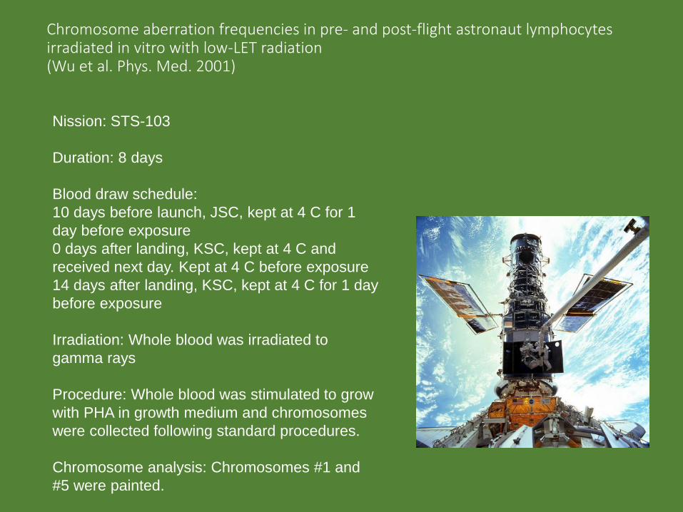
Chromosome aberrations in lymphocytes induced by beta particle exposure in flight(Bender et al. Rad. Res. 1967, 1968)
Chromosome deletion
0
0.05
0.1
0.15
0.2
0.25
0 0.5 1 1.5 2
Dose (Gy)
Fre
qu
en
cy
Ground
Flight
Rings and dicentics
0
0.05
0.1
0.15
0 0.5 1 1.5 2
Dose (Gy)
Fre
qu
en
cy
Ground
Flight
Chromosome deletion
0
0.1
0.2
0.3
0.4
0 1 2 3
Dose (Gy)
Fre
qu
en
cy
Ground
Flight
Rings and dicentrics
0
0.05
0.1
0.15
0.2
0.25
0.3
0.35
0 1 2 3
Dose (Gy)
Fre
qu
en
cy
Ground
Flight
Gemini-3 Gemini-11
Mission Duration 4 hr 52 min 4 day 1 hr 56 min
Temperature Ambient Refrigerated-ambient
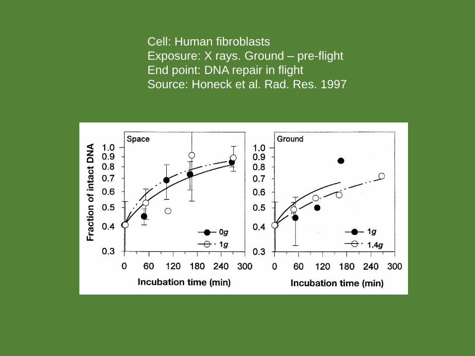
Cell: Human fibroblasts
Exposure: X rays. Ground – pre-flight
End point: DNA repair in flight
Source: Honeck et al. Rad. Res. 1997
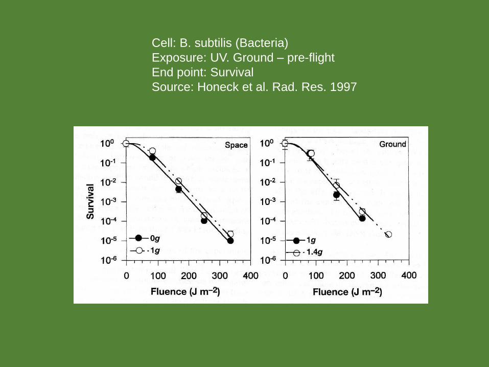
Cell: B. subtilis (Bacteria)
Exposure: UV. Ground – pre-flight
End point: Survival
Source: Honeck et al. Rad. Res. 1997
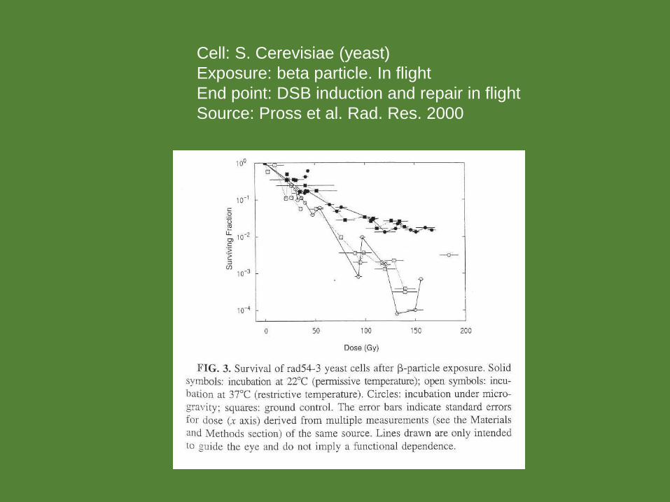
Cell: S. Cerevisiae (yeast)
Exposure: beta particle. In flight
End point: DSB induction and repair in flight
Source: Pross et al. Rad. Res. 2000

Chromosome aberrations in astronauts’ lymphocytes from direct exposure to space radiation
RBE for CA as a function of LET showing a similar trend as the quality factor
66
68
70
72
74
76
78
80
82
Bio dose equivalent (mGy-Eq) Effective dose (mSv)
Chart Title


Aim #1. Investigate changes of miRNA and RNA expression in G1 human fibroblast cells in space.
Aim #2. Investigate cellular responses to bleomycin-induced DNA damage in G1 human fibroblast cells in space.
Aim #3. Detect the DNA damage in cells from direct exposure to space radiation.
Micro-7 Project Objectives

Cell culture and flight hardware
Human fibroblast cells
BioCell from BioServe BioServe’s CGBA incubator
Confluent human fibroblast cells were cultured in BioCells. The cells were kept in CGBA on ISS at 37 C.
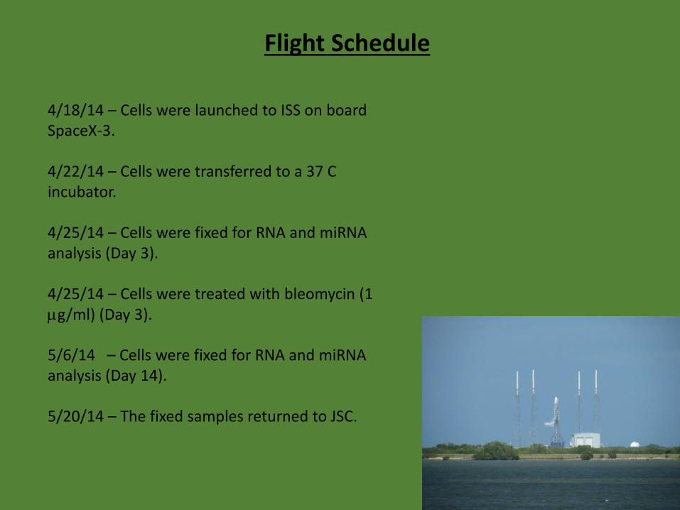
4/18/14 – Cells were launched to ISS on board SpaceX-3.
4/22/14 – Cells were transferred to a 37 C incubator.
4/25/14 – Cells were fixed for RNA and miRNA analysis (Day 3).
4/25/14 – Cells were treated with bleomycin (1 mg/ml) (Day 3).
5/6/14 – Cells were fixed for RNA and miRNA analysis (Day 14).
5/20/14 – The fixed samples returned to JSC.
Flight Schedule
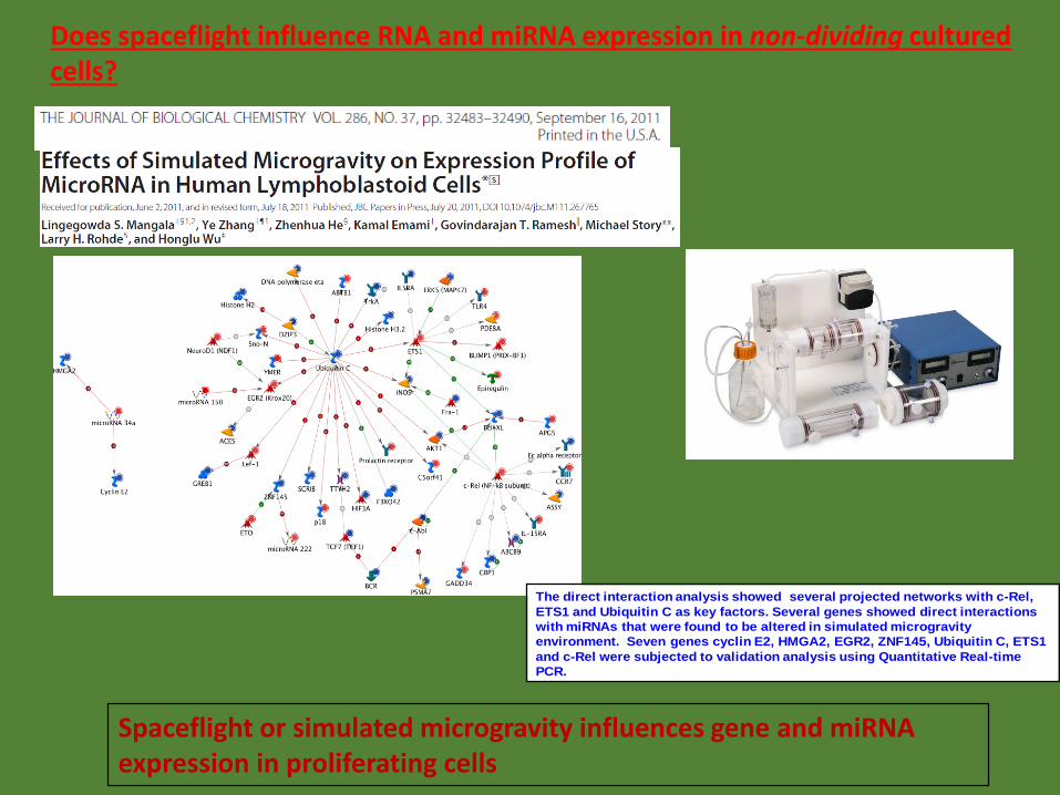
The direct interaction analysis showed several projected networks with c-Rel,
ETS1 and Ubiquitin C as key factors. Several genes showed direct interactions
with miRNAs that were found to be altered in simulated microgravity
environment. Seven genes cyclin E2, HMGA2, EGR2, ZNF145, Ubiquitin C, ETS1
and c-Rel were subjected to validation analysis using Quantitative Real-time
PCR.
Spaceflight or simulated microgravity influences gene and miRNA expression in proliferating cells
Does spaceflight influence RNA and miRNA expression in non-dividing cultured cells?

Microarray Results – Day 3 and Day 14
Number of genes having significant expression changes in the flight samples in comparison to the ground controls on Day 3 and Day 14

Microarray Results – Day 3 and Day 14 (Continued)
• The Day 3 data indicated activation of NFkB and other growth related pathways involving HGF and VEGF in the flown cells.
• The results are consistent with faster cell proliferation of the cells in space as measured by the percentage of ki-67 positive cells.
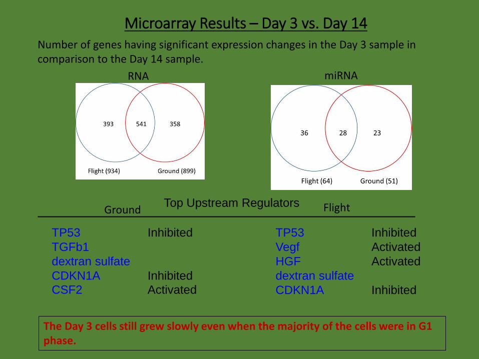
Number of genes having significant expression changes in the Day 3 sample in comparison to the Day 14 sample.
The Day 3 cells still grew slowly even when the majority of the cells were in G1 phase.
Microarray Results – Day 3 vs. Day 14
TP53 Inhibited
Vegf Activated
HGF Activated
dextran sulfate
CDKN1A Inhibited
FlightGround
TP53 Inhibited
TGFb1
dextran sulfate
CDKN1A Inhibited
CSF2 Activated
Top Upstream Regulators
RNA miRNA

No significant changes in the cytoskeleton between ground and flown cells
Ground Flight
Cells were stained with a-tubulin antibodies

Summary 1
• On Day 3, both the flown and ground cells were still proliferating slowly even though they were confluent, as measured by the expression of ki-67 positive cells, and the cells in space grew slightly faster.
• Gene and miRNA expression data for Day 3 indicated activation of NFkB and other growth related pathways involving HGF and VEGF in the flown cells.
• On Day 14 when the cells were mostly non-dividing, the gene and miRNA expression profiles between the flight and ground samples were indistinguishable.
• Comparison of gene and miRNA expressions in the Day 3 versus Day 14 samples revealed that most of the changes observed on Day 3 were related to cell growth for both the flown and ground cells.

Do microgravity and other spaceflight factors affect cellular response to DNA damages (by space radiation)?
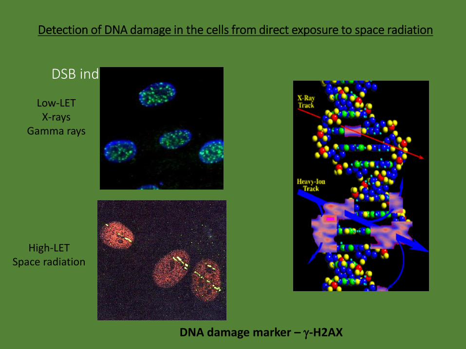
DSB induction
Low-LETX-rays
Gamma rays
High-LETSpace radiation
DNA damage marker – g-H2AX
Detection of DNA damage in the cells from direct exposure to space radiation

Distribution of g-H2AX foci size
A small fraction of g-H2AX foci are large and display a non-spherical track structure.

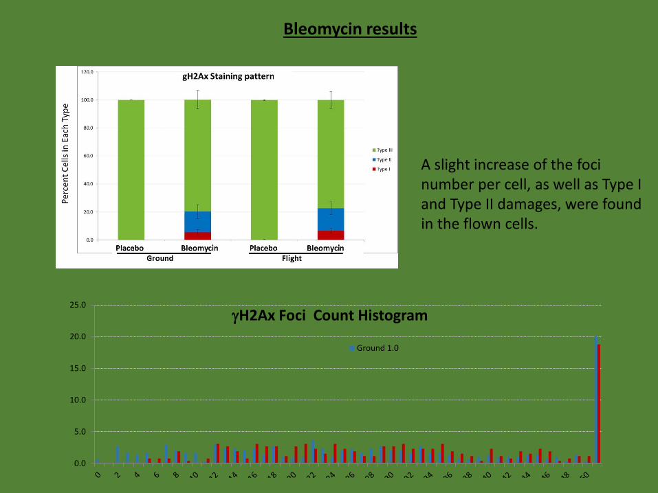
Quantification of bleomycin-induced damages with g-H2AX immunofluorescence staining patterns and foci counts
0.0 0.1 1.0 10.0Bleomycin concentration (mg/ml)
Perc
ent
Cel
ls in
Eac
h T
ype
TYPE I
TYPE II
TYPE III
Cellular response to bleomycin-induced DNA damage
0.0
5.0
10.0
15.0
20.0
25.0
gH2Ax Foci Count Histogram
Ground 1.0
Bleomycin results
A slight increase of the foci number per cell, as well as Type I and Type II damages, were found in the flown cells.

Expression of genes involved in DNA damage signaling
No difference in the expression of DNA damage response genes was found between the flight and ground samples.
PCRarray DNA Damage SignalingGround Flight
BBC3 ↑ BBC3 ↑CDKN1A ↑ CDKN1A ↑
PCNA ↑ PCNA ↑PPM1D ↑ PPM1D ↑

Summary 2
• Images of the 3-dimensional g-H2AX foci were captured with a laser confocal microscope. Quantitative analysis revealed a small fraction of foci that were larger and displayed a track pattern in the flight samples in comparison to the ground control.
• Damage in the DNA from bleomycin treatment was measured by the phosphorylation of a histone protein H2AX (g-H2AX), which showed slightly more foci in the cells on ISS than in the ground control. The difference was likely caused by the slightly faster growth of the cells in space.
• Although a number of genes, including CDKN1A and PCNA, were significantly altered in the cells after bleomycin treatment, no significant differences in the expression profiles of DNA damage response genes were found between the flight and ground samples.

• In true non-dividing human fibroblast cells, microgravity in space has little effect on the gene and miRNA expression. Gene and miRNA expression changes were observed in cells that were confluent, but still proliferating slowly. The faster growth in the flown cells was associated with the activation of NFkB pathways which triggers the expression of several growth factors and the suppression of the cell cycle checkpoint.
• The difference in g-H2AX formation in response to bleomycin-induced DNA damage between flight and ground was due to the faster growth rate of the cells in space, but spaceflight did not affect the response of the DNA damage response genes to bleomycintreatment.
Conclusions
Acknowledgement
NASA Johnson Space CenterYe ZhangTao LuJohn JeevarajanSamrawit YeshitlaAlan Feiveson Michael Wong
BioServe Space TechnologiesLouis StodieckStefanie CountrymanJonathan BenoShankini Doraisingam
NASA Ames Research CenterFathi KarouiaKevin Sato
Kennedy Space CenterAshleigh D. RugglesSatyanand Narayan
UT Southwestern Medical CenterMichael Story