Embed Size (px)
Citation preview

Journal of Zhejiang University-SCIENCE B (Biomedicine & Biotechnology) 2021 22(12):1034-1044www.jzus.zju.edu.cn; www.springer.com/journal/11585E-mail: [email protected]
ERα promotes transcription of tumor suppressor gene ApoA-I byestablishing H3K27ac-enriched chromatin microenvironment inbreast cancer cells
Bingjie WANG1, Yinghui SHEN1, Tianyu LIU2, Li TAN1*
1Center for Medical Research and Innovation, Shanghai Pudong Hospital, Fudan University Pudong Medical Center, and Shanghai KeyLaboratory of Medical Epigenetics, Institutes of Biomedical Sciences, Fudan University, Shanghai 200032, China2Colorectal Cancer Center, Department of General Surgery, Zhongshan Hospital, Fudan University, Shanghai 200032, China
Abstract: Apolipoprotein A-I (ApoA-I), the main protein component of high-density lipoprotein (HDL), plays a pivotal role inreverse cholesterol transport (RCT). Previous studies indicated a reduction of serum ApoA-I levels in various types of cancer,suggesting ApoA-I as a potential cancer biomarker. Herein, ectopically overexpressed ApoA-I in MDA-MB-231 breast cancercells was observed to have antitumor effects, inhibiting cell proliferation and migration. Subsequent studies on the mechanismof expression regulation revealed that estradiol (E2)/estrogen receptor α (ERα) signaling activates ApoA-I gene transcription inbreast cancer cells. Mechanistically, our ChIP-seq data showed that ERα directly binds to the estrogen response element(ERE) site within the ApoA-I gene and establishes an acetylation of histone 3 lysine 27 (H3K27ac)-enriched chromatinmicroenvironment. Conversely, Fulvestrant (ICI 182780) treatment blocked ERα binding to ERE within the ApoA-I gene anddownregulated the H3K27ac level on the ApoA-I gene. Treatment with p300 inhibitor also significantly decreased the ApoA-Imessenger RNA (mRNA) level in MCF7 cells. Furthermore, the analysis of data from The Cancer Genome Atlas (TCGA)revealed a positive correlation between ERα and ApoA-I expression in breast cancer tissues. Taken together, our study not onlyrevealed the antitumor potential of ApoA-I at the cellular level, but also found that ERα promotes the transcription of ApoA-Igene through direct genomic effects, and p300 may act as a co-activator of ERα in this process.
Key words: Apolipoprotein A-I (ApoA-I); Estrogen receptor α (ERα); Acetylation of histone 3 lysine 27 (H3K27ac); p300;Breast cancer
1 Introduction
According to the 2020 World Health Organization(WHO) report, the number of new breast cancer casessurpassed that of lung cancer for the first time, whichmeans that breast cancer had become the world’smost common cancer. Based on molecular characteristics,breast cancers are divided into hormone receptor (estrogenreceptor (ER)/progesterone receptor (PR))-positive/Erb-B2 receptor tyrosine kinase 2 (ERBB2)-negative(70% of patients), ERBB2-positive (15%–20%), andtriple-negative subtypes (15%) (Waks and Winer, 2019).
Estrogen signaling modulates a variety of physi‐ological processes through the direct or indirect regu‐lation of target gene transcription (Vrtačnik et al.,2014). The abnormal overactivation of ERs may leadto the development of breast cancer, and the specificunderlying mechanisms include: (1) amplification ofER coactivators (Anzick et al., 1997); (2) inhibitionof ER corepressors (Mussi et al., 2006); (3) overex‐pressed bridge proteins promoting the recruitment ofcoactivators by ERs (Zwijsen et al., 1998; McMahonet al., 1999); (4) the mutations of ER enabling it tobe activated at a lower concentration of estrogen(Herynk and Fuqua, 2004); and (5) ER localizationon the plasma membrane and the subsequent signal‐ing cascade activation (Levin and Pietras, 2008).Breast cancer patients with ERα-positive tumors aretreated with endocrine therapy; however, most ofthem will develop endocrine resistance (Hanker et al.,
Research Articlehttps://doi.org/10.1631/jzus.B2100393
* Li TAN, [email protected]
Li TAN, https://orcid.org/0000-0001-5961-0450
Received Apr. 27, 2021; Revision accepted July 14, 2021;Crosschecked Nov. 15, 2021
© Zhejiang University Press 2021

J Zhejiang Univ-Sci B (Biomed & Biotechnol) 2021 22(12):1034-1044 |
2020). This prompts us to conduct a deeper investi‐gation about the complex role of ERα in breast cancer.It has been reported that ERα can inhibit pro‐grammed death-ligand 1 (PD-L1) expression in breastcancer cells (Liu et al., 2018), which suggests thedouble-edged sword role of ERα. Investigations ofERα regulation of cancer-related genes will contributeto the clinical treatment of breast cancer.
Apolipoprotein A-I (ApoA-I) is the main pro‐tein component of high-density lipoprotein (HDL)(Shao and Heinecke, 2018). In addition to its well-known function in reverse cholesterol transport (RCT)and protective effect against cardiovascular diseases,ApoA-I is involved in inflammatory and immuneresponses (Gordon et al., 2011). Many recent studieshave discovered a reduction of serum ApoA-I invarious types of cancer (Georgila et al., 2019). Thedecrease of serum ApoA-I level has been relatedto the progression and metastasis of breast cancer(Gonçalves et al., 2006). Besides, a positive correl‑ation between the level of serum ApoA-I secreted intumor tissue fluid and chemotherapy sensitivity wasobserved in breast cancer (Cortesi et al., 2009;Zhang et al., 2016). ApoA-I contains ten consecutivehelical domains, which are essential for the biophysicalproperties of proteins that spontaneously dissolvelipids in an aqueous environment (Li et al., 1988).Based on the characteristics of these amphiphilichelical sequences, researchers have synthesized variouspeptides that do not share any sequence homology,but all mimic the function of ApoA-I (Reddy et al.,2014). The ApoA-I mimetic peptide D-4F has beenproved effective in reducing proliferative responseinduced by oxidized low-density lipoprotein (oxLDL)in human breast adenocarcinoma cells (Cedó et al.,2016).
Although numerous studies have shown thatApoA-I is a potential biomarker with antitumoreffects, the regulatory mechanism of its expressionin breast cancer remains unclear. Exploring the regu‐lation of ApoA-I expression will help us to bettermanipulate its level and function, which may contrib‐ute to prevention and treatment strategies for breastcancer. Herein, we explored the biological effect ofApoA-I on breast cancer cells and uncovered the effectof estradiol (E2)/ERα signaling on ApoA-I transcrip‐tion in breast cancer.
2 Materials and methods
2.1 Vector construction and cell transfection
The human breast cancer cell lines MCF7 andMDA-MB-231 were purchased from the ChineseAcademy of Sciences Cell Bank (Shanghai, China).The human breast cancer cell line BT549 was obtainedfrom Dr. Sulin LIU (Fudan University Cancer Hospital,Shanghai, China). The 293T cell line was kindlyprovided by Dr. Degui CHEN (Shanghai Institutionof Biochemistry and Cell Biology, Chinese Academyof Sciences, Shanghai, China). All cell lines used inthis study were cultured in Dulbecco’s modified Eagle’smedium (DMEM) high-glucose medium (HyClone,Marlborough, MA, USA) supplemented with 10%(volume fraction) fetal bovine serum (FBS; BI, Israel)and 1% (volume fraction) penicillin/streptomycin(Gibco, Carlsbad, CA, USA). The cells were placedin a humidified incubator at 37 °C under 5% CO2
atmosphere. The following compounds were used inthe corresponding experiments: 500 nmol/L ICI 182780treatment for 7 d (Fulvestrant, Sigma-Aldrich, Darm‐stadt, Germany); 1 nmol/L E2 treatment for 2 d (Sigma-Aldrich) after 5 d of hormone deprivation; 20 μmol/Lhistone acetyltransferase inhibitor II (Selleck, Houston,Texas, USA).
2.2 Cell culture and treatment procedure
The coding sequences (CDSs) of ApoA-I andERα amplified from the complementary DNAs (cDNAs)of MCF7 cells were separately inserted into a pLenti6.2/v5/TEV vector (Clonetech, Waltham, MA, USA). Therecombinant plasmids and packaging plasmids weretransfected into 293T cells using LipofectamineTM
2000 Transfection Reagent (Invitrogen, Carlsbad, CA,USA) according to the manufacturer’s protocol. After48 h of transfection, the cell supernatant was collectedand filtered. Virus-containing supernatant containing0.1% (volume fraction) polybrene (Sigma-Aldrich)was added to 1.5×10⁶ MDA-MB-231 cells seeded ina six-well plate. After 48 h of infection, cells werescreened using 10 μg/mL Blasticidin S HCl (Gibco).
2.3 qRT-PCR analysis
Total RNA was extracted from breast cancercells using the TRIzol® reagent (Ambion, Life Tech‐nologies, Carlsbad, CA, USA) and quantified by aNanoDrop 2000 spectrophotometer (Thermo Scientific,
1035
| J Zhejiang Univ-Sci B (Biomed & Biotechnol) 2021 22(12):1034-1044
Waltham, MA, USA). Total RNA was reverse-transcribedinto cDNA using a PrimescriptTM RT Reagent Kit withgenomic DNA (gDNA) eraser (TaKaRa, Kusatsu, Japan)according to the manufacturer’s protocol. The cDNAwas subjected to quantitative real-time polymerase chainreaction (qRT-PCR) amplification by using the SYBRGreen Master Mix (Roche, Basel, Switzerland) andperforming in triplicates on a Roche LightCycler 480II system. Table 1 shows the primer sequences used inthis study.
2.4 Western blot
Cells were harvested and lysed using sodiumdodecyl sulfate (SDS) lysis buffer containing proteaseinhibitors. Equal amounts of proteins were separatedby SDS-polyacrylamide gel electrophoresis (PAGE)and transferred onto nitrocellulose membrane using awet-transfer system (Bio-Rad, Hercules, CA, USA).After 1 h of blocking, the membranes were placed inbovine serum albumin (BSA) solution at 4 ℃ overnightwith the following primary antibodies: anti-β-actin(Proteintech, Rosemont, IL, USA; 66009-1-Ig), anti-vinculin (Cell Signaling Technology, Boston, MA,USA; 13901), anti-ApoA-I (Abcam, Cambridge, UK;ab52945), anti-ERα (Cell Signaling Technology; 8644),and anti-H3K27ac (Active Motif, Shanghai, China; 39133).After washing with Tris-buffered saline (TBS)/Tweenbuffer, the appropriate secondary antibodies (L3012:horseradish peroxidase (HRP)-conjugated goat anti-rabbit immunoglobulin G (IgG) secondary antibody;L3032: HRP-conjugated goat anti-mouse IgG secondaryantibody) were used for blocking at room temperature
for 1 h. The expression levels of proteins were detectedby the ChemiDoc Touch Imaging System (Bio-Rad).
2.5 CCK-8 and transwell assay
The cell counting kit-8 (CCK-8) experiments werecarried out with 2000 cells in accordance with thekit instructions (Enhanced Cell Counting Kit-8;Beyotime, Shanghai, China). Specifically, 2000 cellswere planted in each well of the 96-well plate. After0, 1, 2, 3, and 4 d, the medium was removed, and 100 μLof medium containing 10 µL CCK-8 solution was addedto react in the cell incubator for 2 h. The absorbancewas subsequently measured at 450 nm.
For the transwell assay, 5×10⁴ cells were placedon the upper layer of a cell culture insert with a cellpermeable membrane (Millicell Hanging Cell CultureInserts, 8.0 μm polyethylene terephthalate (PET);Millipore, Massachusetts, USA), and DMEM high-glucose medium containing 20% (volume fraction) FBS(BI) and 1% (volume fraction) penicillin/streptomycin(Gibco) was placed below the membrane. Followingan 8-h incubation period, the cells that had migratedthrough themembrane were stained and counted.
2.6 ChIP-seq and ChIP-qPCR
This experiment was performed as previouslydescribed (Kong et al., 2016). In particular, the mono‐layer of cultured cells was fixed with 1% (volumefraction) formaldehyde for 8 min, which process wasterminated by 2.5 mol/L glycine solution for 5 min.After washing with phosphate-buffered saline (PBS),cells were harvested and lysed using a cocktail containinghigh-salt and lysis buffer (50 mmol/L 4-(2-hydroxyerhyl)piperazine-1-erhanesulfonic acid (HEPES; pH 7.5),500 mmol/L NaCl, 1 mmol/L ethylenediamine tetraaceticacid (EDTA), 0.001 g/mL Na-deoxycholate, 1% (volumefraction) Triton X-100, 0.001 g/mL SDS) (Roche).Next, the cell lysate was sonicated for 30 min (30 son, 40 s off, 95% amplify) using a Bioruptor® sonicator(Diagenode, Belgium). The 10% (volume fraction)supernatant was taken out as the control group. The 2 μLof ERα or H3K27ac antibody (Active Motif) was addedto the remaining sonicated chromatin for rotatingovernight at 4 ℃. Then, 15 μL protein A/G magneticbeads (Invitrogen) were added to bind to the antibodyat 4 °C for 2 h. The beads were washed according tothe following method: thrice with high-salt and lysisbuffer, twice with low-salt buffer (10 mmol/L Tris-Cl
Table 1 Primer sequences of target gens in this study
Target gene
GAPDH
ApoA-I
GREB1
ERα
ApoA-I_ChIP-qPCR
Primer sequence (5'→3')
F: CTGACTTCAACAGCGACACC
R: GTGGTCCAGGGGTCTTACTC
F: TGAGGCTCTCAAGGAGAACG
R: CCTCACTGGGTGTTGAGCTT
F: GACCAGCTTCTGATCACCCC
R: TACCTAAAGCCGATGGTCGC
F: CCTCCTCATCCTCTCCCACA
R: ATGCGATGAAGTAGAGCCCG
F: GCCTTCAAACTGGGACACAT
R: CATTTCTGGCAGCAAGATGA
GAPDH: glyceraldehyde-3-phosphate dehydrogenase; ApoA-I: apoli‐poprotein A-I; GREB1: growth regulation by estrogen in breast cancer 1;ERα: estrogen receptor α; F: forward; R: reverse.
1036

J Zhejiang Univ-Sci B (Biomed & Biotechnol) 2021 22(12):1034-1044 |
(pH 8.0), 250 mmol/L LiCl, 1 mmol/L EDTA, 0.001g/mLNa-deoxycholate, and 0.5% (volume fraction) NP-40),and once with TE buffer (2 mmol/L EDTA, 10 mmol/LTris-Cl (pH 8.0)). The “beads” and the control groupwere resuspended in 100 μL elution and de-crosslinkbuffer (50 mmol/L Tris-Cl (pH 8.0), 10 mmol/LEDTA, and 0.01g/mL SDS) and incubated at 65 ℃on the oscillator for 6 h. Chromatin was eluted by 60 μLof DNA elution buffer (10 mmol/L Tris-Cl, pH 8.0)containing 20 μg of proteinase A for 2 h at 37 ℃ and20 μg of protease K for 1 h at 55 ℃ . The DNA waspurified using a MinElute® PCR purification kit (Qiagen,Valencia, CA, USA). A ChIP-seq library was constructedusing KAPA HyperPrep Kits (KAPA, Boston, MA,USA) according to the manufacture’s protocol. Sequenc‐ing was completed by Basepair Biotechnology Co.,Ltd. (Suzhou, China). The ChIP-qPCR experimentwas performed as described above.
Our own raw and processed nucleic acid sequenc‐ing data (ERα and H3K27ac ChIP-seq) were depositedin the National Center for Biotechnology Information(NCBI) Gene Expression Omnibus (GEO) (https://www.ncbi.nlm.nih.gov/geo) under the accession numberGSE136673.
2.7 TCGA data analysis
The data analysis was carried out by the methoddescribed in the previous article published by our group(Liu et al., 2018). In short, The Cancer Genome Atlas(TCGA) data were downloaded from http://www.cbioportal.org. The expression of ERα in tumor sampleswas determined based on clinical data.
2.8 Statistical analysis
The results derived from three independentexperiments were presented as mean±standard devia‐tion (SD). The significance of the data was analyzedwith GraphPad Prism 8.2.1 by a two-tailed unpairedt-test. The difference was considered significant whenP<0.05.
3 Results
3.1 Proliferation and migration of MDA-MB-231cells inhibited by ectopic overexpression of ApoA-I
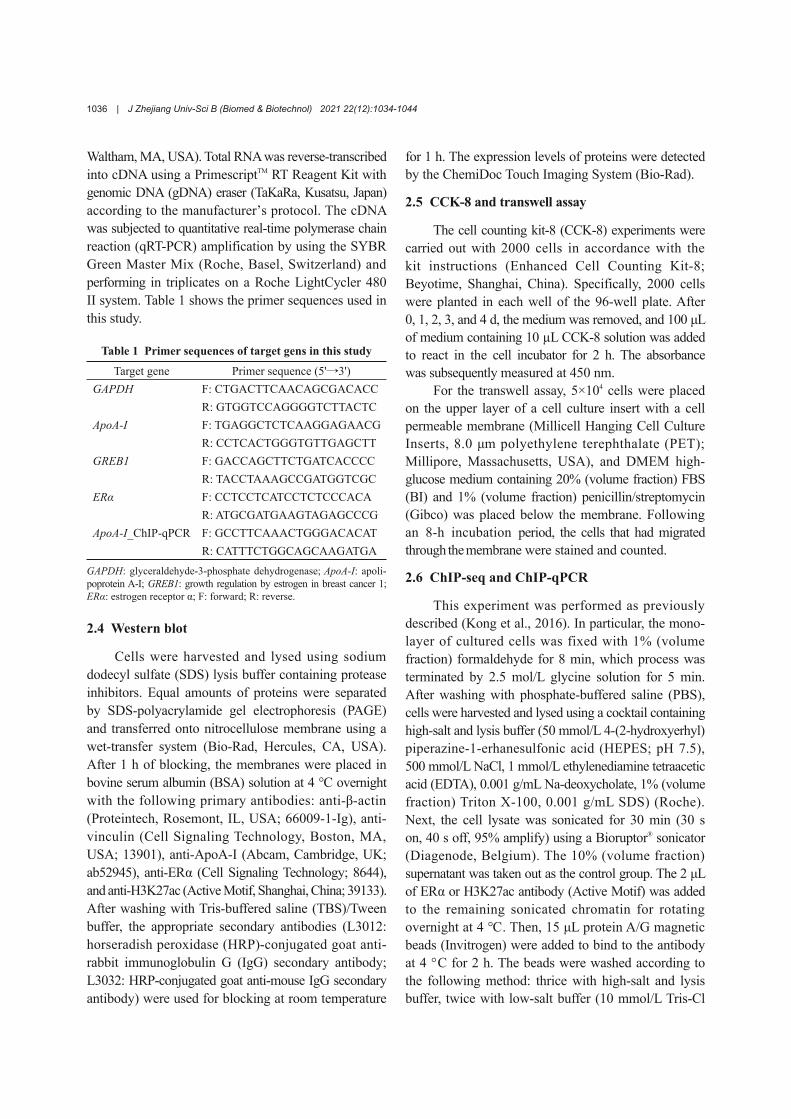
In order to investigate the effect of ApoA-I onthe biological behavior of breast cancer cells, we first
determined the messenger RNA (mRNA) levels ofApoA-I in three breast cancer cell lines (Fig. 1a).Since the mRNA level of ApoA-I in MDA-MB-231cells is the lowest, we constructed an ectopic ApoA-I-overexpression system in MDA-MB-231 cells (Fig. 1b).Then, we performed CCK-8 and transwell experimentsto detect the effect of ApoA-I overexpression onMDA-MB-231 cells. The CCK-8 test result showedthat the proliferation rate of ApoA-I-overexpressedMDA-MB-231 cells was significantly lower than thatof the control group (Fig. 1c). Furthermore, ApoA-Iinhibited the migration of MDA-MB-231 cells (Figs. 1dand 1e). This result indicated that ApoA-I may be atumor suppressor that can inhibit the proliferationand migration of breast cancer cells.
3.2 ApoA-I transcription induced by E2/ERαsignaling in breast cancer cells
In order to explore whether E2/ERα signalingmodulates ApoA-I transcription, we exposed ERα-positive MCF7 cells to estrogen antagonist ICI 182780to abolish the ERα functions. Accompanied by a decreaseof ERα (Fig. 2a), the mRNA levels of growth regulationby estrogen in breast cancer 1 (GREB1; the classicERα target gene) and ApoA-I in MCF7 cells weresignificantly downregulated by ICI 182780 (Fig. 2b).
Given that the ER inhibitor downregulated theApoA-I mRNA level in MCF7 cells, we speculatedthat the activation of ERα signaling would promotethe transcription of ApoA-I. We performed hormonedeprivation on ER-positive MCF7 cells, and then addedan equal volume of ethanol (EtOH, vehicle control) orE2. Our results showed that the transcription of bothGREB1 and ApoA-I was dramatically upregulated byE2 (Fig. 2c). Compared with ER-positive MCF7 cells,triple-negative BT549 and MDA-MB-231 cells hadlower ApoA-I mRNA levels (Fig. 1a). We hypothe‐sized that ectopic expression of ERα would also activateApoA-I transcription in ER-negative breast cancer cells.As expected, the western blotting and qRT-qPCR datashowed that the restoration of ERα expression inMDA-MB-231 cells upregulated the mRNA level ofApoA-I (Figs. 2d and 2e).
3.3 ERα directly binds to last exon of ApoA-I gene
In order to examine whether ERα has a directrole in regulating ApoA-I transcription, ChIP-seq wasconducted using anti-ERα antibodies. We observed that
1037
| J Zhejiang Univ-Sci B (Biomed & Biotechnol) 2021 22(12):1034-1044
ERα was enriched in the last exon of the ApoA-I genein both MCF7 and ERα-overexpressed MDA-MB-231cells (Fig. 3a). Notably, when challenging hormone-depleted MCF7 cells with E2, the ChIP signal intensityof ERα on the ApoA-I gene was more significant thanthat of hormone-depleted MCF7 cells treated with EtOH(Fig. 3a). In addition, by analyzing the ERα-enrichedregion, we found that an estrogen response element(ERE) was located right in the center of the ERα peak(Fig. 3b).
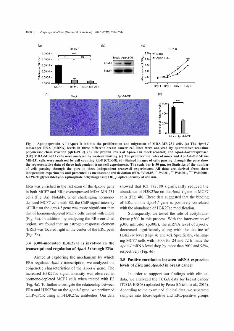
3.4 p300-mediated H3K27ac is involved in thetranscriptional regulation of ApoA-I through ERα
Aimed at exploring the mechanism by whichERα regulates ApoA-I transcription, we analyzed theepigenetic characteristics of the ApoA-I gene. Theincreased H3K27ac signal intensity was observed inhormone-depleted MCF7 cells when treated with E2(Fig. 4a). To further investigate the relationship betweenERα and H3K27ac on the ApoA-I gene, we performedChIP-qPCR using anti-H3K27ac antibodies. Our data
showed that ICI 182780 significantly reduced theabundance of H3K27ac on the ApoA-I gene in MCF7cells (Fig. 4b). These data suggested that the bindingof ERα on the ApoA-I gene is positively correlatedwith the abundance of H3K27ac modification.
Subsequently, we tested the role of acetyltrans‐ferase p300 in this process. With the intervention ofp300 inhibitor (p300i), the mRNA level of ApoA-Idecreased significantly along with the decline ofH3K27ac level (Figs. 4c and 4d). Specifically, challeng‐ing MCF7 cells with p300i for 24 and 72 h made theApoA-I mRNA level drop by more than 90% and 98%,respectively (Fig. 4d).
3.5 Positive correlation between mRNA expressionlevels of ERα and ApoA-I in breast cancer
In order to support our findings with clinicaldata, we analyzed the TCGA data for breast cancer(TCGA-BRCA) uploaded by Perou (Ciriello et al., 2015).According to the examined clinical data, we separatedsamples into ERα-negative and ERα-positive groups
Fig. 1 Apolipoprotein A-I (ApoA-I) inhibits the proliferation and migration of MDA-MB-231 cells. (a) The ApoA-Imessenger RNA (mRNA) levels in three different breast cancer cell lines were analyzed by quantitative real-timepolymerase chain reaction (qRT-PCR). (b) The protein levels of ApoA-I in mock (control) and ApoA-I-overexpressed(OE) MDA-MB-231 cells were analyzed by western blotting. (c) The proliferation rates of mock and ApoA-I-OE MDA-MB-231 cells were analyzed by cell counting kit-8 (CCK-8). (d) Stained images of cells passing through the pore showthe representative data of three independent transwell experiments. The scale bar is 50 μm. (e) Statistics of the numberof cells passing through the pore in three independent transwell experiments. All data are derived from threeindependent experiments and presented as mean±standard deviation (SD). ns P≥0.05, ** P<0.01, *** P<0.001, **** P<0.0001.GAPDH: glyceraldehyde-3-phosphate dehydrogenase; OD450: optical density at 450 nm.
1038

J Zhejiang Univ-Sci B (Biomed & Biotechnol) 2021 22(12):1034-1044 |
(Fig. 5a). The average mRNA level of ApoA-I in theERα-positive group was significantly higher than that inthe ERα-negative group (Fig. 5b). Moreover, the regres‐sion analysis revealed a positive correlation between themRNA expression levels of ERα and ApoA-I (Fig. 5c).
4 Discussion
Numerous studies have suggested the downregu‐lation of serum and tissue ApoA-I in various cancer types(Zamanian-Daryoush and Didonato, 2015). Therefore,
it is of high importance to understand the role of thisphenomenon and the associated regulatory mechanism.In this study, we demonstrated that ApoA-I can inhibitthe proliferation and migration of breast cancer cells.Moreover, ERα could directly bind to the last exon ofApoA-I gene to active its transcription, and p300 mightbe a co-activator of ERα by establishing an H3K27ac-enriched chromatin microenvironment in this process.
The ApoA-I mimetic peptide D-4F has been provento inhibit the proliferation of human breast cancercells (Peng et al., 2017). We found that the directoverexpression of human ApoA-I protein arrived at
Fig. 2 Estradiol (E2)/estrogen receptor α (ERα) signaling induces apolipoprotein A-I (ApoA-I) transcription in breastcancer cells. (a) The ERα protein levels in dimethyl sulfoxide (DMSO) or ICI 182780-treated MCF7 cells were analyzedby western blotting. (b) Messenger RNA (mRNA) levels of growth regulation by estrogen in breast cancer 1 (GREB1)and ApoA-I in DMSO or ICI 182780-treated MCF7 cells were analyzed by quantitative real-time polymerase chainreaction (qRT-PCR). (c) GREB1 and ApoA-I mRNA levels in hormone-depleted MCF7 cells treated with ethanol (EtOH)or E2 were analyzed by qRT-PCR. (d) The expression levels of ERα in mock (control) and ERα-overexpressed (OE) MDA-MB-231 cells were analyzed by western blotting. (e) The ApoA-I mRNA levels in mock and ERα-OE MDA-MB-231 cellswere analyzed by qRT-PCR. All data are derived from three independent experiments and presented as mean±standarddeviation (SD). ** P<0.01, *** P<0.001, **** P<0.0001. GAPDH: glyceraldehyde-3-phosphate dehydrogenase.
1039

| J Zhejiang Univ-Sci B (Biomed & Biotechnol) 2021 22(12):1034-1044
the same result. The antitumor effect of ApoA-I maybe achieved through a cellular autonomic mechanism,or its impact on immune responses (Gordon et al.,2011), or both, which remains an elusive topic. In thecellular autonomic mechanism, ApoA-I can inducechanges in the expression of cancer-related genes,thus producing anti-cancer effects. For example, ApoA-Iinduces the downregulation of vascular endothelialgrowth factor (VEGF) and hypoxia-inducible factor(HIF)-1α expression, thereby inhibiting the prolifera‐tion and migration of ovarian cancer cells (Gao et al.,2011, 2012). In colorectal adenocarcinoma cells, ApoA-Iinhibits the activity of cancer cells by downregulatingcyclooxygenase-2 (COX-2) expression (Aguirre-Portoléset al., 2018). In our in vitro model, the anti-cancer effectismorelikelytobeachievedthroughthecellularautonomicmechanism due to the absence of an immune system.Nonetheless, the relevant specific mechanism remainsto be further studied. Although most researches haveindicated that ApoA-I levels are negatively correlatedwith the occurrence and progression of various types
of cancer, there are also reports showing a positivecorrelation (Chen et al., 2013; Martin et al., 2015; Shiet al., 2018; Zografos et al., 2019). It is not clearwhether the positive correlation between ApoA-I levelsand cancer parameters as reported by a number of studiesreflects the cancer-promoting effects of ApoA-I inthese special cases.
Many transcription factors implicated in theregulation of ApoA-I promotor have been identified,including peroxisome proliferator-activated receptor γ(PPARγ), hepatocyte nuclear factor 4 (HNF4), liverreceptor homologue-1 (LRH1), and ApoA-I regulatoryprotein 1 (ARP1)/nuclear receptor subfamily 2 groupF member 2 (NR2F2) (Kardassis et al., 2014). Besides,an endogenously expressed long noncoding antisensetranscript, ApoA1-AS, can also modulate ApoA-Itranscription through recruiting histone methylationenzymes (Halley et al., 2014). Herein, for the firsttime, ERα was demonstrated to be an activator forApoA-I gene transcription in breast cancer cells: weobserved a positive correlation between the expression
Fig. 3 Estrogen receptor α (ERα) directly binds to last exon of apolipoprotein A-I (ApoA-I) gene. (a) UCSC (Universityof California, Santa Cruz) Genome Brower tracks for ApoA-I from ChIP-seq analysis showing representative data ofthree independent experiments; (b) Schematic diagram of estrogen response element (ERE) sequence and its location onApoA-I gene. ERE is marked in red. EtOH: ethanol; E2: estradiol; ERα-OE: ERα-overexpressed.
1040
J Zhejiang Univ-Sci B (Biomed & Biotechnol) 2021 22(12):1034-1044 |
of ERα and ApoA-I mRNAs, in both breast cancercells cultured in vitro and breast tumor samples.
Estrogen can exert its function through bothgenomic and non-genomic signaling. Our ChIP-seqdata displayed an ERα enrichment signal within theApoA-I gene, suggesting that ERα promoted thetranscription of ApoA-I gene through genomic signaling.Genomic signaling can be divided into direct and indirecttypes. In direct genomic signaling, dimerized ERαbinds directly to specific DNA sequences known asEREs (Klinge, 2001). By analyzing the sequence ofERα enrichment locus on the ApoA-I gene, we foundthe sequence AGGTCACGCTGTCCC that has provedto be an ERE (Bourdeau et al., 2004). ERα seems tomodulate ApoA-I transcription through direct genomicsignaling, while this is suggested by the experimentthrough the process focusing on the influence of EREsequence mutation.
Recruiting various co-regulators to form an ERcomplex is the intrinsic mechanism of estrogen signal‐ing (Heldring et al., 2007). The activity of ERs dependson co-regulators within this complex. We found thatH3K27ac abundance on the ApoA-I gene is tightlydependent on ERα. Moreover, the inhibitor of acetyl‐transferase p300 could suppress ApoA-I mRNA expres‐sion. We speculate that histone acetyltransferase p300may be a co-activator of ERα by making the chromatinregion containing ApoA-I more accessible to transcrip‐tional factors. A recent study revealed that Ajuba, theLIM protein in MCF7 and T47D breast cancer cells,could recruit deleted in breast cancer 1 (DBC1) andcAMP-regulated enhancer-binding protein (CREB)-binding protein (CBP)/p300 to enhance the regulatoryactivity of ERα on target genes by acetylating ERα(Xu et al., 2019). We hypothesize that p300 maycombine with ERα to enhance the transcriptional
Fig. 4 p300-mediated acetylation of histone 3 lysine 27 (H3K27ac) is involved in the transcriptional regulation ofapolipoprotein A-I (ApoA-I) by estrogen receptor α (ERα). (a) UCSC (University of California, Santa Cruz) GenomeBrower tracks for ApoA-I from ChIP-seq analysis showing representative data of two independent experiments.Hormone-depleted MCF7 cells were treated with EtOH (MCF7_EtOH) or E2 (MCF7_E2) for 24 h. (b) ChIP-qPCRanalysis of H3K27ac abundance on ApoA-I gene in MCF7 cells treated with dimethyl sulfoxide (DMSO) or ICI 182780for 7 d. (c) Western blot analysis of the H3K27ac level in MCF7 cells treated with DMSO or p300 inhibitor (p300i) for 24and 72 h. (d) Quantitative real-time polymerase chain reaction (qRT-PCR) analysis of ApoA-I messenger RNA (mRNA)levels in MCF7 cells treated with DMSO or p300i for 24 and 72 h. All data are derived from three independentexperiments and presented as mean±standard deviation (SD). ns P≥0.05, ** P<0.01, **** P<0.0001. EtOH: ethanol; E2:estradiol; GAPDH: glyceraldehyde-3-phosphate dehydrogenase.
1041

| J Zhejiang Univ-Sci B (Biomed & Biotechnol) 2021 22(12):1034-1044
activity of ERα on ApoA-I, which process requiresfurther verification.
5 Conclusions
This study revealed the tumor suppressive roleof ApoA-I in breast cancer cells and identified ERα asa new activator of ApoA-I gene transcription throughdirectly binding to the last exon of the ApoA-I gene.This is the site where an ERE sequence is found, andp300 may serve as a co-activator of ERα in activatingApoA-I expression by establishing an H3K27ac-enrichedchromatin microenvironment. Our study demonstratedthe mechanism of ERα promoting ApoA-I gene tran‐scription in breast cancer cells, providing furthersupporting evidence of the double-edged nature of theE2/ERα signaling in tumorigenesis. Moreover, giventhe positive regulatory effect of ERα on ApoA-I expres‐sion and the tumor suppressive role of ApoA-I in varioustypes of cancer, such as melanoma (Zamanian-Daryoushet al., 2013), breast cancer (Cedó et al., 2016), coloncancer (Gkouskou et al., 2016), and pancreatic cancer
(Peng et al., 2017), it is considered that the adminis‐tration of ApoA-I mimetic peptides may enhance theefficacy of systemic therapy for luminal breast cancer.
AcknowledgmentsThis work was supported by the National Natural
Science Foundation of China (Nos. 81672785, 31871291, and82073113 to Li TAN) and the National Key R&D Project ofChina (No. 2016YFA0101800 to Li TAN). Li TAN was alsosupported by the Innovative Research Team of High-level LocalUniversity in Shanghai. We thank Dr. Xuguo ZHU (Institutesof Biomedical Sciences, Fudan University, Shanghai, China) forconstructing the ERα-overexpressed MDA-MB-231 cells, Dr.Ruitu LV (Department of Chemistry, University of Chicago,Chicago, IL, USA) for analyzing the ChIP-seq data, and Prof.Yujiang Geno SHI (Division of Endocrinology, Diabetes andHypertension, Brigham and Women’s Hospital, HarvardMedical School, Boston, USA) for his valuable comments.
Author contributionsBingjie WANG performed the experimental research and
TCGA data analysis, wrote and edited the manuscript. YinghuiSHEN performed the ChIP-seq experiment. Tianyu LIU contrib‐uted to the data analysis, writing and editing of the manuscript.Li TAN contributed to the study design and editing of the
Fig. 5 Positive correlations between messenger RNA (mRNA) expression levels of estrogen receptor α (ERα) andapolipoprotein A-I (ApoA-I) in breast cancer. (a, b) The Cancer Genome Atlas (TCGA) data were analyzed for ERα (a) andApoA-I (b) mRNA expression levels in ERα-negative (ERα−) and ERα-positive (ERα+) breast cancer tissues. (c) Regressionanalysis of the correlation between ERα and ApoA-I mRNA levels in breast cancer tissues. * P<0.05, **** P<0.0001. TCGA-BRCA:TCGA data for breast cancer.
1042

J Zhejiang Univ-Sci B (Biomed & Biotechnol) 2021 22(12):1034-1044 |
manuscript. All authors have read and approved the final manu‐script, and therefore, have full access to all the data in the studyand take responsibility for the integrity and security of the data.
Compliance with ethics guidelinesBingjie WANG, Yinghui SHEN, Tianyu LIU, and Li
TAN declare that they have no conflict of interest.This article does not contain any studies with human or
animal subjects performed by any of the authors.
ReferencesAguirre-Portolés C, Feliu J, Reglero G, et al., 2018. ABCA1
overexpression worsens colorectal cancer prognosis byfacilitating tumour growth and caveolin-1-dependentinvasiveness, and these effects can be ameliorated using theBET inhibitor apabetalone. Mol Oncol, 12(10):1735-1752.https://doi.org/10.1002/1878-0261.12367
Anzick SL, Kononen J, Walker RL, et al., 1997. A1B1, a steroidreceptor coactivator amplified in breast and ovarian cancer.Science, 277(5328):965-968.https://doi.org/10.1126/science.277.5328.965
Bourdeau V, Deschênes J, Métivier R, et al., 2004. Genome-wideidentification of high-affinity estrogen response elementsin human and mouse. Mol Endocrinol, 18(6):1411-1427.https://doi.org/10.1210/me.2003-0441
Cedó L, García-León A, Baila-Rueda L, et al., 2016. ApoA-Imimetic administration, but not increased apoA-I-containingHDL, inhibits tumour growth in a mouse model of inheritedbreast cancer. Sci Rep, 6:36387.https://doi.org/10.1038/srep36387
Chen CL, Lin TS, Tsai CH, et al., 2013. Identification ofpotential bladder cancer markers in urine by abundant-protein depletion coupled with quantitative proteomics.J Proteomics, 85:28-43.https://doi.org/10.1016/j.jprot. 2013.04.024
Ciriello G, Gatza ML, Beck AH, et al., 2015. Comprehensivemolecular portraits of invasive lobular breast cancer.Cell, 163(2):506-519.https://doi.org/10.1016/j.cell. 2015.09.033
Cortesi L, Barchetti A, de Matteis E, et al., 2009. Identificationof protein clusters predictive of response to chemotherapyin breast cancer patients. J Proteome Res, 8(11):4916-4933.https://doi.org/10.1021/pr900239h
Gao F, Vasquez SX, Su F, et al., 2011. L-5F, an apolipoproteinA-I mimetic, inhibits tumor angiogenesis by suppressingVEGF/basic FGF signaling pathways. Integr Biol, 3(4):479-489.https://doi.org/10.1039/c0ib00147c
Gao F, Chattopadhyay A, Navab M, et al., 2012. ApolipoproteinA-I mimetic peptides inhibit expression and activity ofhypoxia-inducible factor-1α in human ovarian cancer celllines and a mouse ovarian cancer model. J PharmacolExp Ther, 342(2):255-262.https://doi.org/10.1124/jpet. 112.191544
Georgila K, Vyrla D, Drakos E, 2019. Apolipoprotein A-I(ApoA-I), immunity, inflammation and cancer. Cancers,
11(8):1097.https://doi.org/10.3390/cancers11081097
Gkouskou KK, Ioannou M, Pavlopoulos GA, et al., 2016.Apolipoprotein A-I inhibits experimental colitis and colitis-propelled carcinogenesis. Oncogene, 35(19):2496-2505.https://doi.org/10.1038/onc.2015.307
Gonçalves A, Esterni B, Bertucci F, et al., 2006. Postoperativeserum proteomic profiles may predict metastatic relapsein high-risk primary breast cancer patients receivingadjuvant chemotherapy. Oncogene, 25(7):981-989.https://doi.org/10.1038/sj.onc.1209131
Gordon SM, Hofmann S, Askew DS, et al., 2011. High densitylipoprotein: it’s not just about lipid transport anymore.Trends Endocrinol Metab, 22(1):9-15.https://doi.org/10.1016/j.tem.2010.10.001
Halley P, Kadakkuzha BM, Faghihi MA, et al., 2014. Regulationof the apolipoprotein gene cluster by a long noncodingRNA. Cell Rep, 6(1):222-230.https://doi.org/10.1016/j.celrep.2013.12.015
Hanker AB, Sudhan DR, Arteaga CL, 2020. Overcomingendocrine resistance in breast cancer. Cancer Cell, 37(4):496-513.https://doi.org/10.1016/j.ccell.2020.03.009
Heldring N, Pike A, Andersson S, et al., 2007. Estrogen receptors:how do they signal and what are their targets. PhysiolRev, 87(3):905-931.https://doi.org/10.1152/physrev.00026.2006
Herynk MH, Fuqua SAW, 2004. Estrogen receptor mutationsin human disease. Endocr Rev, 25(6):869-898.https://doi.org/10.1210/ER.2003-0010
Kardassis D, Mosialou I, Kanaki M, et al., 2014. Metabolismof HDL and its regulation. Curr Med Chem, 21(25):2864-2880.https://doi.org/10.2174/0929867321666140303153430
Klinge CM, 2001. Estrogen receptor interaction with estrogenresponse elements. Nucleic Acids Res, 29(14):2905-2919.https://doi.org/10.1093/nar/29.14.2905
Kong LC, Tan L, Lv RT, et al., 2016. A primary role of TETproteins in establishment and maintenance of De Novobivalency at CpG islands. Nucleic Acids Res, 44(18):8682-8692.https://doi.org/10.1093/nar/gkw529
Levin ER, Pietras RJ, 2008. Estrogen receptors outside the nucleusin breast cancer. Breast Cancer Res Treat, 108(3):351-361.https://doi.org/10.1007/s10549-007-9618-4
Li WH, Tanimura M, Luo CC, et al., 1988. The apolipoproteinmultigene family: biosynthesis, structure, structure‒functionrelationships, and evolution. J Lipid Res, 29(3):245-271.https://doi.org/10.1016/S0022-2275(20)38532-1
Liu L, Shen YH, Zhu XG, et al., 2018. ERα is a negativeregulator of PD-L1 gene transcription in breast cancer.Biochem Biophys Res Commun, 505(1):157-161.https://doi.org/10.1016/j.bbrc.2018.09.005
Martin LJ, Melnichouk O, Huszti E, et al., 2015. Serum lipids,lipoproteins, and risk of breast cancer: a nested case-controlstudy using multiple time points. J Nat Cancer Inst, 107(5):djv032.https://doi.org/10.1093/jnci/djv032
1043

| J Zhejiang Univ-Sci B (Biomed & Biotechnol) 2021 22(12):1034-1044
McMahon C, Suthiphongchai T, DiRenzo J, et al., 1999. P/CAFassociates with cyclin D1 and potentiates its activationof the estrogen receptor. Proc Natl Acad Sci USA, 96(10):5382-5387.https://doi.org/10.1073/pnas.96.10.5382
Mussi P, Liao L, Park SE, et al., 2006. Haploinsufficiency ofthe corepressor of estrogen receptor activity (REA) enhancesestrogen receptor function in the mammary gland. ProcNatl Acad Sci USA, 103(45):16716-16721.https://doi.org/10.1073/pnas.0607768103
Peng MY, Zhang Q, Cheng YN, et al., 2017. ApolipoproteinA-I mimetic peptide 4F suppresses tumor-associatedmacrophages and pancreatic cancer progression. Oncotarget,8(59):99693-99706.https://doi.org/10.18632/oncotarget.21157
Reddy ST, Navab M, Anantharamaiah GM, et al., 2014.Apolipoprotein A-I mimetics. Curr Opin Lipidol, 25(4):304-308.https://doi.org/10.1097/MOL.0000000000000092
Shao BH, Heinecke JW, 2018. Quantifying HDL proteinsby mass spectrometry: how many proteins are there andwhat are their functions? Expert Rev Proteomics, 15(1):31-40.https://doi.org/10.1080/14789450.2018.1402680
Shi FY, Wu H, Qu K, et al., 2018. Identification of serumproteins AHSG, FGA and APOA-I as diagnostic biomarkersfor gastric cancer. Clin Proteomics, 15:18.https://doi.org/10.1186/s12014-018-9194-0
Vrtačnik P, Ostanek B, Mencej-Bedrač S, et al., 2014. Themany faces of estrogen signaling. Biochem Med, 24(3):329-342.https://doi.org/10.11613/BM.2014.035
Waks AG, Winer EP, 2019. Breast cancer treatment: a review.JAMA, 321(3):288-300.https://doi.org/10.1001/jama.2018.19323
Xu BH, Li Q, Chen N, et al., 2019. The LIM protein Ajubarecruits DBC1 and CBP/p300 to acetylate ERα andenhances ERα target gene expression in breast cancercells. Nucleic Acids Res, 47(5):2322-2335.https://doi.org/10.1093/nar/gky1306
Zamanian-Daryoush M, Didonato JA, 2015. ApolipoproteinA-I and cancer. Front Pharmacol, 6:265.https://doi.org/10.3389/fphar.2015.00265
Zamanian-Daryoush M, Lindner D, Tallant TC, et al., 2013.The cardioprotective protein apolipoprotein A1 promotespotent anti-tumorigenic effects. J Biol Chem, 288(29):21237-21252.https://doi.org/10.1074/jbc.M113.468967
Zhang JW, Cai Y, Hu HB, et al., 2016. Nomogram basingpre-treatment parameters predicting early response for locallyadvanced rectal cancer with neoadjuvant chemotherapyalone: a subgroup efficacy analysis of FOWARC study.Oncotarget, 7(4):5053-5062.https://doi.org/10.18632/oncotarget.6469
Zografos E, Anagnostopoulos AK, Papadopoulou A, et al.,2019. Serum proteomic signatures of male breast cancer.Cancer Genomics Proteomics, 16(2):129-137.https://doi.org/10.21873/cgp.20118
Zwijsen RML, Buckle RS, Hijmans EM, et al., 1998. Ligand-independent recruitment of steroid receptor coactivatorsto estrogen receptor by cyclin D1. Genes Dev, 12(22):3488-3498.https://doi.org/10.1101/gad.12.22.3488
1044