Embed Size (px)
Citation preview

TRACING THE CRIMINAL
Part eight:Acidoresistant (acid-fast) criminals
Institute for microbiology presents

Intro: Spittoons in fight with TBIn Czechoslovakia between World War I and World War II the a society was formed with personal engagement of president Masaryk and his „League against TB“. It showed a big effort in fight against this disease. The part of this was education for people not to spit to the floor, but to use spittoons.
www.bikupan.se

Survey of individual parts
Clinical characteristics of acid-fast bacteria
Special properties of acid fast-bacteria
Diagnostics of acid-fast bacteria

Clinical characteristics of acid-fast bacteria

Story One• Johny did know already for many
years that he is HIV positive. He knew pretty well that he is more vulnerable than other people and that each infection can get him more quickly than other people.
• Nevertheless he was surprised that he started to cough recently. His doctors tried various variants, but after roentgen, PCR examination and culture examination came to conclusion that it is a miliary (granular) form of tuberculosis.
http://cs.wikipedia.org/wiki/Tuberkul%C3%B3za

The criminal was…
• Mycobacterium tuberculosis, although TB may be caused by Mycobacterium bovis, too.
• Interesting for this microbe: it lives inside cells. This is also related with the fact that antibody response is weak in tuberculosis (so neither antigen nor antibodies are detected) and cell immunity is very important (in vaccination, too).
• As in HIV infection just cell immunity is damaged, TB is one of opportune infections.
http://www.genomeindia.org

Mycobacterium inside a cell
http://www.nature.com/nrm/journal/v2/n8/fig_tab/nrm0801_569a_F3.html

Tuberculosis• At the first contact with the infection is formed primary
complex. It is a focus (usually localized in lungs) and corresponding regional lymph node.
• During the next infection post-primary TB is formed. It is worse. Usually a granulomatous formation is formed, later it subdues caseification („becoming cheese-like“) and then it is not enlarged anymore. Paradoxically, majority of damage in the organism is caused by the host organism reaction (late hypersensitivity – in the matter of fact, a specific type of an allergy)
• After years the original focus may re-activate, mostly in old age, at immunodeficiency, or ethanol abuse. Such person may be very dangerous for his/her environment.

Tuberculosis
http://www.stockmedicalart.com
www.tusalud.com.mx

Pulmonary form of TB is not the only one
sitemaker.umich.edu (2×)

TB worldwide
http://gamapserver.who.int/mapLibrary/Files/Maps/Global_TB_incidence_2011.png

Once more TBhttp://www.lung.ca
http://www.cbc.ca

Brain tuberculoma http://pathology.mc.duke.edu

More special facts about TB bacilli• Their cell wall is highly hydrophobic, it contains
mycolic acids. They nearly do not Gram stain, special staining methods are needed.
• It grows slowly, its generation period is long, so special media are needed
• They are very resistant to disinfectants. It is impossible to use „A“ class disinfectants against common bacteria, you need „T“ (against TB), eventually „M“ (against atypical mycobacteria)
• They are also resistant to antimicrobial drugs.

Cell wall of mycobacteria
www.primer.ru
http://oregonstate.edu

Story two• Mr Hassan lived in desert part of Sudan, where
wars and unrests were very common.• Recently even friends that still did neither run
away nor die started to dislike contacts with mr. Hasan. Mutilation of face of mr. Hassan was a clear mark, that mr. Hassan suffers the disease still too common in this part of world.
• Good luck that Hassan met members of a non-governement organisation, that where sure about diagnose. Using dapson it was possible to help Mr. Hassan.

Causative agent is• Mycobacterium leprae, a microbe even stranger
than TB mycobacterium• The causative agent was found in 1873 by the
norvegian doctor Gerhard Henrik Armauer Hansen, therefore Mycobacterium leprae is also known as Hansen‘s bacillus.
• Its generation period is much longer than in TB mycobacteria. In vitro culture was succesfull only recently and it has taken the whole year

Mycobacterium leprae• The disease is often asymptomatic and is not very
contagious. An exception is the cutaneus form with ulcers that is contagious. Sometimes a destructive form occurs, which attacks Schwann cells and macrophages in peripherial nerves. It disfigures the face and also other parts of the body
• Basic treatment of leprosy is not expensive, nevertheless in countries where leprosy is endemic it is still too much
• Therefore leprosy still requires help of foundations, non-government and charitative groups

Leprosy• Long incubation period – 2, 3, 7 … yrs• Tuberculoid leprosy
– Depigmented spots on skin, swollen peripheral nerves
– Active cellular immunity– Spontaneous consolidation
• Lepromatous leprosy– Insufficient cellular immunity– Confluent and swollen skin lesions, eyebrows fall
out, nose is in depression, fingers are lost, people become blind
http://abicko.avcr.cz/archiv/2002/10/obsah/9-gallery.html
http://blanicti-zoldneri.tym.cz/clanky/lepra.html

Leprosy
www2.bc.cc.ca.us (2×)

Story three• Mr. Piranha was a furious aquarist. Last month
he had a problem: he had to use his left hand only to do anything inside the fish tank, as he had an ulcus on his right hand.
• After examination, his case was closed as so named fish tank granuloma, common in aquarists. A similar disease in swimmers is called swimming pool granuloma.
• Causative agents are…

Atypic mycobacteria• Besides M. tuberculosis and M. leprae there
exist also plenty of other mycobacteria. Some of them,
e. g. Mycobacterium marinum, are so named atypical mycobacteria, sometimes causing wound infections and other problems.
• Some mycobacteria are non-pathogenous and they are normal part of human microflora, e. g. M. smegmatis.
http://www.microbeworld.org/component/jlibrary/?view=article&id=545

Infection of M. marinum
http://www.nejm.org/doi/full/10.1056/ENEJMicm000083
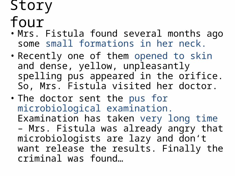
Story four• Mrs. Fistula found several months ago some
small formations in her neck.• Recently one of them opened to skin and
dense, yellow, unpleasantly spelling pus appeared in the orifice. So, Mrs. Fistula visited her doctor.
• The doctor sent the pus for microbiological examination. Examination has taken very long time – Mrs. Fistula was already angry that microbiologists are lazy and don‘t want release the results. Finally the criminal was found…

…it was actinomycosis• So the criminal was Actinomyces sp.• Actinomycetes are filamentous bacteria, in fact Gram-
positive, but they do not Gram stain very well, because their cell wall is hydrophobic and contains many mycolic acids.
• Actinomycetes are commonly found in oral cavity of healthy persons. From here they might commonly get to soft tissues of neck, face or thorax. They are anaerobic bacteria (or at least they grow the best at anaerobic conditions).
• Similar to actinomycetes are nocardiae, but they are strictly aerobic. Otherwise they are similar in many ways.
http://en.wikipedia.org

Actinomycosis
pathmicro.med.sc.edu (3×)

Nocardiosis
http://www.asm.org
pathmicro.med.sc.edu

Nocardia pneumonia with septicaemia
A previously well 57-year-old man … 3-day history of severe dyspnoea. We conclude that N. asteroides infection can present as a fulminant community-acquired pneumonia with bacteraemia in the absence of immunosuppression or coexistent infection.(From the article related to the picture)

Brain nocardiose on CT
http://www.appliedradiology.com

Special properties of acid-fast bacteria

Acidoresistance + alkaliresistance• Acids and alkali act only to hydrofilic components,
communication to water environment. In mycobacteria this is not fulfilled.
• So acids and alkali have weak effect only to them.• Acids are also not able to decolorize them, when
someway it was possible to stain them.• Majority of dyes is hydrophilic, too, and so
mycobacteria stain poorly, usually it is necessary to stain them at hot temperature, to stain them at all.
• Nocardia and Actinomycetes, unlike mycobacteria, are only partially acidoresistant. So, we stain them by Gram, but we have to know, that they stain poorly and inconstantly.

Consequences for clinical doctors
• Clinical doctor, sending sample (sputum, urine, pus or anything) „for bacteriological culture“, cannot hope in getting reference of eventual TB infection.
• To get info about TB, it is necessary to send sample separately and to mark it so that it should be examined for TB (TB-culture or TB-PCR). If so, the laboratory can perform the needed procedures.

Diagnostics of acid-fast bacteria

How to search for criminals• Microscopy: We use Ziehl-Neelsen stain and fluorescence
stain.• Culture: We use special media, and before the culture the
specimen should be treated by a hydroxide. The aim is to kill other bacteria, that would be more successful as they grow more quickly. Alkaliresistant mycobacteria survive that easily.
• Automatic culture: Various types of culture automats are used: they are able to detect culture positiveness much sooner than classic culture.
• Biochemical differentiation is possible in specialized laboratories.
• Animal experiment: guinea pig is used sometimes.• PCR diagnostics is more and more important.

Mycobacterium tuberculosis
http://www.health.qld.gov.au
Photo O. Z.

Ziehl-Neelsen staining• In step 1 we stain by carbolfuchsin (Gabbet) in hot until
steam rises. Without heating mycobacteria could not be stained, except use of more concentrated carbolfuchsin.
• In step 2 we decolorize (approx. 15 s) by „acid alcohol“, what is mixture of alcohol with a mineral acid, most commonly HCl. After that we rinse the slide with water.
• In step 3 we counterstain the background, so everything decolorized in Step 2. We counterstain by methylene blue. approx. 30 s (it would be also possible to use malachit green) and we rinse the slide with water, we dry it and we observe it with immersion objective.
• Result: red acidoresistant rods on blue or green background.

Ziehl-Neelsen stain
www.spjc.edu

Ziehl-Neelsen stain
http://es.wikipedia.org/wiki/Archivo:Mycobacterium_tuberculosis_Ziehl-Neelsen_stain_02.jpg

Cryptosporidiahttp://www.scientificdevice.com/intl_product_pages/icryptosporidium_stains.htm
It is interesting, that Ziehl-Neelsen staining may be used also for a group of parasites, so called intestinal coccidia (cryptosporidia and cyclospores)

Culture of mycobacteria• Hydroxide should be used before culture.• We use liquid Šula or Banić media and egg Ogawa or
Löwenstein-Jenssen media. Egg media are solid because of egg white coagulation, they do not contain agar.
• Even solid media are in test tube and closed firmly. This is not only because personnel would be endangered, but also as media would dry.
• Results are read after 1 (check for contamination), 3, 6 and for sure after 9 weeks of culture. (Positive results are mostly found after 6 weeks of culture.)

Appearance of mycobaterial colonies
http://www.stockmedicalart.com/

To liquid Šula medium
• Even positive test tube is clear by first view, as the growth of mycobacteria is visible only at the bottom („blue mess“, as student J. H. called it )

Tests of antituberculotic susceptibility (not antibiotic!)• Antituberculotics are strange chemicals, different
from antibiotics (with exceptions).• Always we combine 3 or 4 of them: resistances
appear quickly, and some have only intra- of only extracellular effect.
• We cannot use diffusion disk tests.• Antituberculotics are added directly into culture
media, growth control is added.• Growth present mycobacteria resistant.• Growth absent mycobacteria susceptible.

Survey of commonly used antituberculotics
Antituberculotic Abbrev.
Isoniazid H, INHEthambutol ERifampicin RPyrazinamid ZStreptomycin S, STM
Tuberculous liver of an experimental guinea pig
Courtessy of dr. Jana Svobodová and dr. Lev Mezenský

PCR for TBPCR is a method used in TB diagnostics more than diagnostics of other bacteria. The reason is that it makes the diagnostics much faster and the risk of environmental contamination is not so serious.
• 1, 2, 3, 4 = patients No. 1, 2, 3, 4
• 5 = positive control 6 = negative control
• 7 = ladder (to measure position of a band)
• upper row (c) = sample strip, lower row (b) = IC
Institute for microbiology

PCR kit for TB diagnostics
http://www.cinnagen.com

Indirect diagnostics of tuberculosis
• The most important type of immunity in TB is cell-mediated immunity.
• Formation of antibodies occurs, but measurable levels of antibodies are present only in some cases. So positive finding of anti-TB antibodies is a sign of infection, but negative finding has very low information value.
• Cell-mediated immunity may be tested– by skin test (tuberculin test), especially after vaccination– by INF-gamma release test (reaction of patient cells to
antigen exposition is tested).

Skin test (Mantoux)
• It is used for checking of vaccination effect, but also for proof of an eventual latent infection.
• The complete living patient is needed for the test, so it is not a laboratory test. Test is performed by dermatovenerology or other specialized departments. Recently they are replaced by next type tests
• The tests are positive in case of activation of cell-mediated immunity; in the matter of fact, it is a specific type of delayed allergy.

Test of interferon gamma release (Quantiferon© TB-GOLD)• A modern way of checking the cell-mediated
immunity is examination of induced interferon gamma release; in practice, the only really used test is Quantiferon TB-GOLD, that is why only this test would be mentioned later.
• It was proven that in TB, including latent TB, tuberculosis antigens activate T-lymphocytes and they produce big amounts of interferon gamma.
• Similarly those T-lymphocytes may be activated non-specifically by mitogen, that is why mitogen is used as a positive control.

Quantiferon – three test tubes• We need non-clotted (heparinized) blood to three test tubes
(we need lymphocytes!).• First test-tube contains the mitogen (MIT) – here, in normal
circumstances, always stimulation of IFN-gamma should be observed.
• Second test-tube contains TB antigens (TB) – here IFN-gamma formation stimulation should be observed in TB infection only.
• Third test-tube does not contain anything (NIL) – here we should (normally) never see IFN-gamma stimulation.

Quantiferon – results
• Interferon concentration is measured by ELISA• As positive we consider a result, where T-lymphocytes
react to stimulation of mycobacterium antigen, but in test-tube with „nothing“ the INF-gamma is not formed.
• As negative we consider a result, where T-lymphocytes react to mitogen stimulation, but they do not react to mycobacterial antigen stimulation.
• Unsure result is seen (1) if T-lymphocytes are not activated by the mitogen or (2) IFN-gamma is formed even in the test-tube where no stimulator was present.

Results – example*
NIL [IU/ml]
TB minus NIL [IU/ml]
MIT minus NIL [IU/ml]
Final test interpretation
Presence of M. tuberculosis infection
≤ 8,0
< 0,35 ≥ 0,5Negative
Not likely≥ 0,35and < 25% of NIL value
≥ 0,5
≥ 0,35 and ≥ 25% of NIL value
Any value
Positive Likely
< 0,35 < 0,5
UnsureCannot by determined
≥ 0,35 and < 25% of NIL value
< 0,5
> 8,0 Any value Any value
*the result may be different in subtypes of the test

Nocardia and Actinomyces microscopy• These bacteria are Gram staining, although they
staining poorly and they are very pleomorph.• Both of them are typical by their branched filaments,
staining Gram-positive, although some parts of the filament may stain Gram-negative or they might remain unstained at all.
• Sometimes, short (coccoid) forms may also occur in microscopy.

Actinomyces israelii pathmicro.med.sc.edu

Nocardia asteroideswww2.mf.uni-lj.si
http://www.nocardia.it

Nocardia and Actinomyces culture
• Although both genera are similar in many properties, one is different: Nocardia is strictly aerobic, while Actinomyces grows in anaerobic conditions.
http://filebox.vt.edu

Antibiotic susceptibility of Actinomyces and Nocardia• Unlike mycobacteria, in nocardiae and
actinomycetes antibiotic susceptibility can be tested using diffusion disc test. We have to know, that they grow slowly and badly.
• For nocardiosis we use co-trimoxazol for therapy, eventually ampicillin or macrolides.
• In actinomycosis we use penicillin, eventually doxycyklin and more antibiotics.

Lepromin test in leprosy diagnostics• There is an animal. Its name is nine banded
armadillo.• It is necessary for production of lepromin.• This substance is used in lepromin test, the
equivalent of tuberculin test for TB.
http://www.1-costaricalink.com/costa_rica_fauna/nine_banded_armadillo.htm

The End
Logo of a TB congress
http://www.niaid.nih.gov