Embed Size (px)
Citation preview

www.elsevier.com/locate/yexpr
Experimental Parasitology 117 (2007) 124–132
Toxocara canis: Molecular cloning, characterization, expressionand comparison of the kinetics of cDNA-derived arginine kinase q
Susiji Wickramasinghe a, Kouji Uda b, Mitsuru Nagataki a, Lalani Yatawara a,R.P.V.J. Rajapakse c, Yoshiya Watanabe a,d, Tomohiko Suzuki b, Takeshi Agatsuma a,*
a Department of Environmental Health Sciences, Kochi Medical School, Oko, Nankoku City, Kochi 783-8505, Japanb Laboratory of Biochemistry, Faculty of Science, Kochi University, Kochi 780-8520, Japan
c Department of Veterinary Pathobiology, Faculty of Veterinary Medicine and Animal Science, University of Peradeniya, Peradeniya, Sri Lankad Research and Development Division, Life Science Center, Sophy Incorporation, Agawa, Kochi 781-1522, Japan
Received 26 November 2006; received in revised form 26 March 2007; accepted 27 March 2007Available online 4 April 2007
Abstract
Arginine kinase (AK) is a member of a highly conserved family of phosphagen kinases. We determined the cDNA sequence of Tox-
ocara canis AK, cloned it in pMAL plasmid and expressed it in Escherichia coli as a fusion protein with maltose-binding protein. Theprotein has a theoretical molecular mass of 45,376 Da and an estimated isoelectric point (pI) of 8.38. Alignment of the cDNA-derivedamino acid sequence of T. canis AK with other phosphagen kinase sequences showed high amino acid identity with other nematode AKs,and phylogenetic analysis placed it as a distinct branch within a nematode AK cluster. Analysis of the N-terminus sequence of T. canis
AK revealed the presence of a signal targeting peptide presumably targeting this protein to cytosol or endoplasmic reticulum (ER). T.
canis AK showed high activity for L-arginine. The kinetic constants (Km = 0.12 mM, Kcat = 29.18, and Kd = 0.23 mM) and Vmax
(43.76 lmol Pi/min/mg protein) of T. canis recombinant-AK were determined for the forward reaction. It also exhibited a synergismfor substrate binding ðKArg
d =KArgm ¼ 1:96Þ. Comparison of Kcat=KArg
m values in various arginine kinases indicates that T. canis AK hasa high catalytic efficiency (248.19 s�1 mM�1). The present study contains the first description of arginine kinase in a zoonotic nematode.The determination of T. canis AK and its phosphagen biosynthetic pathway, which is completely different from those in mammalian hosttissues, suggests this enzyme as a possible novel chemotherapy target for VLM syndrome in humans.� 2007 Published by Elsevier Inc.
Index Descriptors and Abbreviations: Toxocara canis; Arginine kinase; Phosphagen kinase; Signal peptide; Kinetic constants; GS region
1. Introduction
Phosphagens are phosphorylated guanidine compoundsthat are linked to ATP by way of a reversible reaction cat-alyzed by phosphagen (guanidino) kinases (phospha-gen + MgADP + H+
M guanidine acceptor + MgATP).The traditional and conventional view is that phosphagens
0014-4894/$ - see front matter � 2007 Published by Elsevier Inc.
doi:10.1016/j.exppara.2007.03.015
q The nucleotide sequence of Toxocara canis AK reported in this study isavailable in the DDBJ/EMBL/GenBank databases under AccessionNumber EF015466. The EC Number of arginine kinase is 2.7.3.3.
* Corresponding author. Fax: +81 88 880 2535.E-mail address: [email protected] (T. Agatsuma).
function as ATP buffers, permitting maintenance of highATP values during periods in which there is disequilibriumof ATP supply and demand (Kammermeier, 1987, 1993).Phosphagens also play a number of other roles, includingregulation of glycogenolysis, proton buffering, and intracel-lular energy transport (Ellington, 2001).
Eight phosphagens have been identified thus far; allhave the characteristic guanidino group but differ dramat-ically in terms of other chemical features. These com-pounds range from arginine phosphate (AP), anunmodified amino acid, to thalassemine phosphate (ThP),which is a highly modified guanidino compound. AP andThP, as well as the other phosphagens, creatine phosphate

S. Wickramasinghe et al. / Experimental Parasitology 117 (2007) 124–132 125
(CP), glycocyamine phosphate (GP), taurocyamine phos-phate (TP), hypotaurocyamine phosphate (HTP), lombri-cine phosphate (LP), and opheline phosphate (OP), canbe classified on the basis of similarities in terms of chemis-try and biosynthetic pathway (Robin, 1974).
In vertebrates, phosphocreatine is the sole phosphagen,and the corresponding phosphagen kinase is creatinekinase (CK). In invertebrates, at least seven unique phos-phagens and corresponding kinases are present in additionto phosphocreatine (Thoai, 1968; Watts, 1968; Morrison,1973; McLeish and Kenyon, 2005). Arthropod and molluscspecies contain only arginine kinase (AK) phosphagenkinases (PK) (Uda et al., 2005). Nematode PKs are thoughtto be arginine kinase. Each phosphagen has its own corre-sponding phosphagen kinase: arginine kinase (AK), tha-lassemine kinase (ThK), creatine kinase (CK),glycocyamine kinase (GK), taurocyamine kinase (TK),hypotaurocyamine kinase (HTK), lombricine kinase(LK), and opheline kinase (OK). CK, GK, and AK are rig-idly specific for their respective substrates creatine/CP, gly-cocyamine/GP, and arginine/AP (Thoai, 1968). Incontrast, TK, HTK, LK, OK, and ThK are broadly spe-cific and display varying degrees of activity when presentedwith taurocyamine/TP, hypotaurocyamine/HTP, lombri-cine/LP, opheline/OP, and/or thalassemine/ThP (Thoai,1968; Thoai et al., 1972).
Toxocara canis is a common round worm of dogs. Lar-val toxocariasis is a serious condition and a global zoonoticdisease (Glickman and Magnawal, 1993). Humans becomeinfected with T. canis through ingestion of infective eggs inthe environment. Organs commonly affected are brain,liver, lung (visceral larva migrans—VLM), and eye (ocularlarva migrans—OLM) where infection can cause perma-nent visual, neurologic, or other tissue damage (Kazacos,2000). Recently, an outbreak of VLM (spinal) due to T.
canis infection has been reported in southern Kyushu,Japan (Umehara et al., 2006).
In the present study we report molecular cloning, char-acterization, expression, kinetic constants (Km, Kcat, andKd), and Vmax of T. canis recombinant arginine kinaseand its phylogenetic relationship with other known phos-phagen kinases.
2. Materials and methods
2.1. cDNA amplification
The eggs of T. canis were collected from an infected dogin Sri Lanka. Total RNA was isolated from infective larvae(L2) by the acid guanidinium thiocyanate–phenol–chloro-form extraction method (Chomczynski and Sacchi, 1987).mRNA was purified from total RNA using poly(A)+ isola-tion kit (Nippon Gene, Tokyo, Japan). Single-strandedcDNA was synthesized with Ready-To-Go You-PrimeFirst-Strand Beads (Amersham Pharmacia Biotech, NJ,USA) with a lock-docking oligo-dT primer (Borson et al.,1992).
2.2. PCR amplification of Toxocara canis AK
The 3 0-half of the cDNA sequence of T. canis AK wassearched from NEMBASE2 (http://www.nematodes.org/nematodeESTs/nembase.html).
A poly(G)+ tail was added to the 3 0 end of the T. caniscDNA pool with terminal deoxynucleotidyl transferase(Promega, WI, USA). The 5 0-half of the cDNA of T. canis
AK was amplified using the oligo-dC primer (5 0-GAATTC18-3 0) and the PK-ToxocaraR2 primer (5 0-CATCGG CTT CAT TGA TCT GGA G-3 0) designed fromthe sequence of 3 0-half (Suzuki and Furukohri, 1994).PCRs were done in a total volume of 50 ll. Each reactionmixture contained cDNA, 10 pmol of each primer, 4 ll of2.5 mM of Ex Taq dNTPs, 1 U of Ex Taq� polymerase,5 ll of 10· Ex Taq� buffer (TaKaRa, Japan). PCR condi-tions were as follows. Initial denaturation at 94 �C for2 min, followed by 35 cycles of 94 �C for 30 s, annealingat 50 �C for 35 s, and extension at 72 �C for 2 min and afinal extension at 72 �C for 4 min. PCR was carried outin a My Cycler� Thermal Cycler (BioRad, USA). PCRproducts were purified by QIA quick PCR purification col-umn (QIAGEN, GmbH, Hilden, Germany).
2.3. T-vector cloning and sequence determination of
Toxocara canis AK
Purified product (300 bp) was ligated into pGEM� T-vector (Promega, USA). Ligated product was transformedinto Escherichia coli JM109 cells. Positive clones wereobtained and plasmid DNA extraction was performedusing the alkaline SDS method. Nucleotide sequences weredetermined with an ABI PRISM 3100-Avant DNA sequen-cer using a BigDye Terminators v3.1 Cycle Sequencing Kit(Applied Bio-systems, CA, USA) with two T-vector-spe-cific primers (T7 and SP6).
2.4. KOD PCR amplification
KOD PCR amplification was done to increase PCRspecificity when producing PCR products to be cloned.PCRs were performed in total volume of 50 ll. The reac-tion mixture contained cDNA, 10 pmol of TcanisPK5 for-ward primer (5 0-AAGAATTCATGGCATTTCTCAAGAACCAG-3 0), 10 pmol of TcanisPK3PstI reverse primer(TTCTGCAGCTATTTTTGTTGTTCCTCCAG), 1 U ofKOD Plus DNA polymerase (TOYOBO CO., LTD.,Japan), 5 ll of 10· of KOD Plus buffer, 5 ll of 2 mMKOD dNTPs, and 4 ll of 25 mM MgSO4. The amplifiedproducts were purified using QIA quick PCR purificationcolumns (QIAGEN, GmbH, Hilden, Germany).
2.5. An A-tailing procedure for blunt-ended PCR fragments
An A-tail was added to the 3 0 end of the purified KODPCR fragments (blunt-ended). A-tailing was done in a totalvolume of 30 ll. The reaction mixture contained purified

Fig. 1. SDS–polyacrylamide gel electrophoresis (PAGE) of recombinantToxocara canis AK expressed as fusion protein with MBP at variousstages of the expression and purification process.
126 S. Wickramasinghe et al. / Experimental Parasitology 117 (2007) 124–132
KOD PCR product, 15 U of Gene Taq DNA polymerase(Wako Nippon Gene), 3 ll of 10· Gene Taq buffer and1.2 ll of 5 mM dATP. This mixture was incubated at70 �C for 30 min. A-tailing product was purified, sub-cloned, and sequenced as described above.
2.6. pMAL cloning and expression of Toxocara canis AKused for enzymatic assay
The open-reading frame (ORF) of T. canis AK wascloned into the BamH1/SalI site of pMAL-c2 (New Eng-land Biolabs, MA, USA). The maltose binding protein(MBP)-T. canis AK fusion protein was expressed inE. coli TB1 cells by induction with 1 mM IPTG at 25 �Cfor 24 h. The cells were resuspended in 5· TE buffer, son-icated, and the soluble protein was extracted. Recombi-nant fusion protein was purified by affinitychromatography using amylose resin (New England Bio-labs, MA, USA). The purity of the expressed enzymewas verified by SDS–PAGE and the purified enzyme wasplaced on ice until determination of enzymatic activitywithin 12 h.
2.7. Enzyme assays
Enzyme activity was measured (UV/Visible Spectro-photometer 4300 Pro, Amersham, Biosciences) with anNADH-linked assay at 25 �C (Morrison and James,1965) and determined for the forward reaction (phospha-gen synthesis) (Fujimoto et al., 2005). The reaction mix-ture (total 1.0 ml) contained 0.65 ml of 100 mM Tris–HCl (pH 8), 0.05 ml of 750 mM KCl, 0.05 ml of250 mM Mg-acetate, 0.05 ml of 25 mM phosphoenolpyr-uvate made up in 100 mM imidazole/HCl (pH 7),0.05 ml of 5 mM NADH made up in Tris–HCl (pH 8),0.05 ml of pyruvate kinase/lactate dehydrogenase mixturemade up in 100 mM imidazole/HCl (pH 7), 0.05 ml of anappropriate concentration of ATP made up in 100 mMimidazole/HCl (pH 7), and 0.05 ml of recombinantenzyme. The reaction was started by adding 0.05 ml ofan appropriate concentration of guanidine substrate madeup in 100 mM Tris–HCl (pH 8). The initial velocity valueswere obtained by varying the concentration of guanidinesubstrate under the fixed concentrations of the ATP. Pro-tein concentration was estimated from the absorbance at280 nm (0.77 AU at 280 nm in a 1 cm cuvette correspondsto 1 mg protein/ml). The KArg
m value was determined fromthe enzyme reaction using seven or eight different sub-strate concentrations of L-arginine around the roughKArg
m value. To determine the Kd value, the above reactionswere done at four different concentrations of ATP(10 mM, 7 mM, 5 mM, and 3 mM). To estimate kineticconstants (Km and Kcat), a Lineweaver–Burk plot wasmade and fitted by the least-square method in MicrosoftExcel. The kinetics of phosphagen kinase can be explainedas a random-order, rapid-equilibrium kinetic mechanism(Morrison and James, 1965), and the Kd, the dissociation
constant, was obtained graphically as described previously(Suzuki et al., 1997).
2.8. Sequence alignments, phylogenetic analysis, and protein
modeling
Multiple sequence alignments were done using the pro-grams CLUSTAL W, GENETYXMAX (ver. 6.0), andCLUSTAL W program available on the DDBJ homepage(http://www.ddbj.nig.ac.jp/).
Phylogenetic analysis was performed using the distancemethod in MEGA (ver. 3.1). For distance analyses, theKimura 2-parameter model was used to construct the dis-tance matrix and the tree was inferred from this using theneighbour-joining (NJ) approach. Bootstrap re-sampling(1000 replicates; seed = 64,238) was performed to assessthe degree of support for groupings on the tree. AccessionNumbers of other amino acid sequences used in the presentstudy are given in Table 1 (supplementary data).
A model of the 3D structure of T. canis AK was con-structed using the internet-based homology-modeling ser-ver SWISS-MODEL (http://swissmodel.expasy.org). The‘‘First Approach Mode’’ was used with default settings.The obtained model was visualized using the SWISS-PdbViewer program (http://www.expasy.org/spdbv/).
3. Results and discussion
3.1. 3D structure and SDS–PAGE of Toxocara canis AK
The 3D structure of T. canis AK was predicted bySWISS MODEL and visualized. The purified T. canis
AK was verified by SDS–PAGE (Fig. 1), which showed amolecular mass of approximately 85 kDa (Recombinant-T. canis AK + MBP).

S. Wickramasinghe et al. / Experimental Parasitology 117 (2007) 124–132 127
3.2. cDNA sequence determination of Toxocara canis AK
and the phylogenetic relationship with other phosphagen
kinases
The full-length (1300 bp) T. canis cDNA was success-fully amplified by PCR and comprises of a 42 bp of 5 0
untranslated region (UTR), 1203 bp of ORF coding for a400-amino acid residue protein, and 55 bp of 3 0 UTR.
Fig. 2. Amino acid sequence alignment of Toxocara canis AK with the represavailable from the home page of DDBJ (http://www.ddbj.nig.ac.jp). GS regionused in this alignment are given in Supplementary Table 1. Black blocks repres80% of the PKs. This figure was prepared with GeneDoc (http://www.psc.edu
The protein has a theoretical molecular mass of45,376 Da and an estimated isoelectric point (pI) of 8.38(http://br.expasy.org/tools/pi_tool.html). The cDNAsequence is available through the GenBank under Acces-sion No. EF015466.
An alignment of the deduced amino acid sequence of T.canis AK with representatives of other phosphagen kinases(Neanthes GK, Homo sapiens CK, Eisenia LK, Siphonoso-
entatives of other phosphagen kinases made with CLUSTAL W programis shown in a box. Accession numbers of reference amino acid sequences
ent the residues conserved in all PKs and gray blocks residues conserved in/biomed/genedoc/).

128 S. Wickramasinghe et al. / Experimental Parasitology 117 (2007) 124–132
ma HTK, Riftia TK, Schistosoma and Fasciola PK, Dro-
sophila, Limulus, Liolophura, Nautilus, Trypanosoma, C.
elegans, and Ascaris AK) is shown in Fig. 2. T. canis AKhas 89% sequence identity with Ascaris suum AK, 61% withC. elegans (Nematoda), 62% with Trypanosoma cruzi AK(Protozoa), 61% with Limulus polyphemus AK (Arthrop-oda), 53% with Liolophura japonica AK (Mollusca), 37–44% with Schistosoma and Fasciola (Trematode) PKs,and only 35% with GK, LK, TK, and CK (see Table 1,supplementary data). Therefore, it is evident that T. canis
AK amino acid sequence is more similar to nematodeAKs than to other PKs.
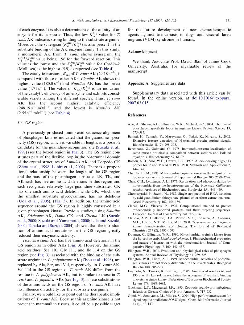
The results of phylogenetic trees constructed from theamino acid sequences of AKs and other PKs using theneighbour-joining (NJ) method (MEGA ver 3.1) clearlydemonstrated the presence of two major groups: a CKgroup (CKs, GKs, LKs, and TKs) and an AK group asdescribed by other researchers (Uda et al., 2005). Further-more, our results indicated that the AK group is dividedinto two sub groups. The first sub-group comprises thearthropoda, protozoan, and nematode AKs. The secondsub-group comprises the molluscan AKs and trematodePKs. T. canis AK is a member of the nematode AK clusterforming a branch with C. elegans (Fig. 3).
3.3. Prediction of a signal peptide and protein localizationsites of Toxocara canis AK
The presence of cytoplasmic and mitochondrial CKs incells is well established in vertebrate and invertebrate ani-
Fig. 3. A phylogenetic tree constructed by neighbour-joining method (1000 bover. 3.1 program. The sequence of Toxocara canis AK is boxed. Bootstrap vachanges inferred as having occurred along each branch. Accession numberSupplementary Table 1.
mals (Ellington, 2001; Wyss et al., 1992). The existence oftrue mitochondrial AKs remains controversial but recentstudy has shown the presence of true mitochondrial AKsin Drosophila AK2 and Caenorhabditis AK4 (Uda et al.,2006). T. canis AK has a long N-terminal extension of20–30 amino acid residues, compared to the other PKs(see Fig. 3). We suspected that this extension might corre-spond to a signal peptide. Therefore, we analyzed the N-terminus sequence of T. canis AK using seven independentmethods to find the possible, signal targeting peptide(Table 2). All but one of six signal peptide prediction pro-grams identified such peptide. The majority of these pro-grams showed the cleavage site of this signal peptide tobe the 23rd position of the N-terminal extension (H2N-Met-Ala-Phe-Leu-Lys-Asn-Gln-Lys-Ala-Ile-Phe-Gly-Ser-Leu-Leu-Ala-Val-Ala-Gly-Gly-Ala-Thr-Ala-). Usually, N-terminus signal peptides target the mitochondrial matrix,endoplasmic reticulum, and peroxisome while C-terminussignal peptides target the peroxisomes. Further, using theN-terminus sequence and six different programs, we pre-dicted protein localization sites of T. canis AK. Three pro-grams judged T. canis AK to be cytoplasmic, two judged itto be associated with endoplasmic reticulum (ER) and onesuggested it is part of a secretory pathway (see Table 2).These results strongly suggest that T. canis AK has a sig-nal peptide presumably targeting this protein to cytosol orER. However, to confirm these predictions we are contin-uing our research to produce anti-sera for recombinant T.
canis AK and determine cellular localization of T. canis
AK by fluorescent antibody techniques.
otstrap) for 17 amino acid sequences of phosphagen kinases using MEGAlues are shown at the branching point. Scale bar indicates the number ofs of reference amino acid sequences used in this analysis are shown in

Table 3Recombinant-Toxocara canis arginine kinase (AK) activity for theforward reaction
Substrate Substrateconcentration(mM)
Substratefinalconcentration[S] (mM)
Absorbance(D340/min)
PKactivity(lmol/min/mgprotein)
Blank(control) 0 0.00000 0.0021 0.060L-Arginine 200 9.52381 1.4187 40.339D-Arginine 200 9.52381 0.0219 0.623Creatine 50 2.38095 0.0006 0.017Glycocyamine 50 2.38095 0.0014 0.040Taurocyamine 50 2.38095 0.0010 0.028
Protein concentration of Toxocara canis AK was estimated (absorbance1.92) at 280 nm with MBP. We used 50 ll of substrate, ATP (100 mM)and of diluted (10·) sample of Toxocara canis AK for each reaction. Forthe calculation of PK activity, we used 2 as state of fusion protein (Tox-
ocara canis AK + MBP).
Table 2The prediction of signal peptide and protein localization sites of Toxocara
canis AK by 11 methods
Program Presence ofsignal peptide
Cleavage site Localization site
MitoProta,l Yes 34SOSUIsignalb,l Yes 23SignalPc,l Yes 23–24TargetPd Yes 23 Secretory pathwayPSORTIIe Yes 23–24 CytoplasmicPrediSif,l Yes 23iPSORTg,l NoSubLoch,m CytoplasmicSoftberryi,m CytoplasmicWoLF PSORTj,m Endoplasmic reticulumPredotark,m Endoplasmic reticulum
a MitoProt (http://ihg.gsf.de/ihg/mitoprot.html) (Claros and Vincens,1996).
b SOSUIsignal (http://sosui.proteome.bio.tuat.ac.jp/sosuisignal/sosui-signal_submit.html) (Gomi et al., 2004).
c SignalP (http://www.cbs.dtu.dk/services/SignalP/) (Jannick et al.,2004).
d TargetP (http://www.cbs.dtu.dk/services/TargetP/) (Olof et al., 2000).e PSORTII (http://psort.ims.u-tokyo.ac.jp/) (Paul and Kenta, 1996).f PrediSi (http://www.predisi.de/) (Von Heijne, 1985).g iPSORT (http://biocaml.org/ipsort/iPSORT/) (Bannai et al., 2002).h SubLoc (http://www.bioinfo.tsinghua.edu.cn/SubLoc/eu_predict.htm)
(Hua and Sun, 2001).i Softberry (http://sun1.softberry.com/cgi-bin/programs/proloc/
protcompan.pl).j WoLF PSORT (http://wolfpsort.seq.cbrc.jp/) (Paul et al., 2006).
k Predotar (http://genoplante-info.infobiogen.fr/predotar/predo-tar.html) (Small et al., 2004).
l These programs predict signal peptide sites only.m These programs predict protein localization sites only.
S. Wickramasinghe et al. / Experimental Parasitology 117 (2007) 124–132 129
3.4. Enzyme activity
Arginine kinase catalyzes the reversible transphospho-rylation between adenosine diphosphate and phosphoargi-nine, which is involved in ATP buffering. Therefore, itplays a major role in metabolic events such as glycogenol-ysis, glycolysis, and oxidative phosphorylation in inverte-brates with an AP/AK system (Ellington, 2001). Researchhas shown that there is significant AK activity in mitochon-dria from the mid gut of tobacco horn worm (Chamberlin,1997), L. polyphemus (Doumen and Ellington, 1990), andfrom range of crustaceans (Ellington and Hines, 1991;Chen and Lehninger, 1973; Skorkowski et al., 1976; Hirdand McLean, 1983; Hird and Robin, 1985). It has recentlybeen shown that AK activity appears to be localized in theintermembrane compartment of cardiac mitochondria in L.
polyphemus and C. sapidus (Pineda and Ellington, 1998).Further studies showed that AK is found in actin-contain-ing regions of cray fish myofibrils (Benzonana and Gabbi-ani, 1978), the Z-line region of flight muscle and the Aband in tubular muscle in Drosophila (Lang et al., 1980),and on migrating neuronal growth cones in the grass hop-per (Wang et al., 1998). In the epimastigote of T. cruzi, AKactivity increased continuously (by seven fold) over the
course of the parasite growth curve and there was a corre-lation between growth rate, enzyme-specific activity andenzyme protein (Guillermo et al., 2001).
The enzyme activity of the recombinant T. canis PK wasmeasured with an NADH-linked assay, on the availableguanidine substrate of L-arginine, D-arginine, creatine, gly-cocyamine, and taurocyamine. T. canis AK showed strongactivity for L-arginine (v = 40.339 lmol Pi/min/mg proteinunder 9.5 mM L-arginine and 19 mM ATP) for the forwardreaction, very little activity for 9.5 mM D-arginine(v = 0.623) and absolutely no activity for creatine, glycocy-amine, and taurocyamine substrates examined at the finalsubstrate concentration of 2.38 mM (Table 3). Therefore,we determined the phosphagen kinase of T. canis to bean arginine kinase. Further, we measured the activity ofT. canis AK against a series of L-arginine concentrations(4 mM, 5 mM, 7 mM, 8 mM, 10 mM, 12 mM, 16 mM,and 20 mM), and obtained the Lineweaver–Burk plot todetermine the KArg
m and Vmax.
3.5. Comparison of kinetic constants and Vmax of Toxocara
canis AK with other AKs
The kinetic constants, KArgm , KArg
d , Kcat (catalytic rate con-stant), and Vmax of T. canis recombinant-AK, were deter-mined for the forward reaction. Although T. canis AKrecombinant enzyme has an MBP-tag at the N-terminus,this tag is not likely to affect the kinetic constants signifi-cantly (Uda et al., 2005). KArg
m , KArgd , and Vmax values for
recombinant T. canis AK were determined to be 0.12 ±0.003 mM, 0.23 ± 0.03 mM, and 43.76 ± 0.29 lmol Pi/min/mg protein, respectively. T. canis AK shows the lowestKArg
m and KArgd values, while molluscan AKs show the highest
KArgm (0.35–1.49 mM) and Kd
Arg (0.67–3.82 mM) values,respectively. The Vmax value is highest in Limulus (Arthrop-oda) and lowest in Nautilus (Mollusca) according to the dataavailable so far (see Table 4). Furthermore, the Km value ofdifferent enzymes is highly variable, being a specific property

Table 4Comparison of kinetic parameters for the forward reaction of arginine kinase (AK) from various sources
Source Enzyme state References KArgm
(mM)KArg
d
(mM)KArg
d =KArgm KATP
m
(mM)KATP
d
(mM)Kcat (l/s) Kcat=KArg
m Vmax (lmol Pi/min/mg protein)
NematodaToxocara canis Recombinant
(MBP tag)Present study 0.12 ± 0.003 0.23 ± 0.03 1.96 0.30 ± 0.04 0.60 ± 0.07 29.18 ± 0.19 248.19 43.76 ± 0.29
ArthropodaLimulus polyphemus Azzi et al. (2004) 0.17 ± 0.02 180.0 ± 24 0 1058.82 270
MolluscaCrassostrea gigas Recombinant
(MBP tag)Fujimoto et al. (2005) 0.35 ± 0.11 0.82 ± 0.37 2.34 ± 0.91 0.97 ± 0.25 2.26 ± 0.59 47.50 ± 2.05 136.00 71.30 ± 3.08
Corbicula japonica two-domain Recombinant(MBP tag)
Suzuki et al. (2003) 0.42 ± 0.14 2.49 ± 0.11 5.93 ± 0.19 0.46 ± 0.05 2.75 ± 0.27 73.30 ± 10.30 174.52 110.0 ± 15.40
Corbicula japonica domain 2 Recombinant(MBP tag)
Suzuki et al. (2003) 0.26 ± 0.09 0.67 ± 00.08 2.58 ± 0.89 0.97 ± 0.13 2.45 ± 0.05 44.70 ± 4.57 171.92 67.0 ± 6.86
Scapharca Recombinant(MBP tag)
a 1.49 ± 0.09 52.20 ± 1.63 35.03 78.30 ± 2.45
Octopus vulgaris Recombinant(MBP tag)
b 0.95 ± 0.03 3.78 ± 0.05 3.98 29.41 ± 0.72 30.96 44.12 ± 1.08
Nautilus pompillius Recombinant(MBP tag)
b 0.67 ± 0.11 2.26 ± 0.07 3.37 1 71 ± 0.11 2.55 2.57 ± 0.17
EchinodermataStichopus japonicus Recombinant
(MBP tag)
b 0.41 ± 0.06 1.25 ± 0.23 3.05 25.67 ± 0.26 62.61 38.50 ± l.89
ProtozoaTrypanasoma cruzi Recombinant (no tag) Claudio et al. (2000) 0.3 0.3
Note: Some parameters recalculated from the data of the references.a Unpublished data by Suzuki. Takeuchi, Fujimoto, Ryotani, and Uda.b Unpublished data by Suzuki, Inoue.
130S
.W
ickra
ma
sing
he
eta
l./
Ex
perim
enta
lP
ara
sitolo
gy
11
7(
20
07
)1
24
–1
32
S. Wickramasinghe et al. / Experimental Parasitology 117 (2007) 124–132 131
of each enzyme. It is also a determinant of the affinity of anenzyme for its substrate. Thus, the low KArg
m value for T.
canis AK indicates strong binding to the substrate arginine.Moreover, the synergism ðKArg
d =KArgm Þ is also present in the
substrate binding of the AK enzyme family. In this study,a monomeric AK from T. canis shows synergism, theKArg
d =KArgm value being 1.96 for the forward reaction. This
value is the lowest and the KArgd =KArg
m value for Corbicula
(Mollusca) is the highest (5.9) as reported (see Table 4).The catalytic constant, Kcat of T. canis AK (29.18 s�1), is
compared with those of other AKs. Limulus AK shows thehighest value (180.0 s�1) and Nautilus AK has the lowestvalue (1.71 s�1). The value of Kcat=KArg
m is an indicationof the catalytic efficiency of an enzyme and exhibits consid-erable variety among the different species. Thus, T. canis
AK has the second highest catalytic efficiency(248.19 s�1 mM�1) and the lowest is Nautilus AK(2.55 s�1 mM�1) (see Table 4).
3.6. GS region
A previously produced amino acid sequence alignmentof phosphagen kinases indicated that the guanidino speci-ficity (GS) region, which is variable in length, is a possiblecandidate for the guanidine-recognition site (Suzuki et al.,1997) (see the boxed region in Fig. 3). The GS region con-stitutes part of the flexible loop in the N-terminal domainof the crystal structures of Limulus AK and Torpedo CK(Zhou et al., 1998; Lahiri et al., 2002). There is a propor-tional relationship between the length of the GS regionand the mass of the phosphagen substrate. LK, TK, andAK each has five amino acid deletions in this region andeach recognizes relatively large guanidine substrates. CKhas one such amino acid deletion while GK, which usesthe smallest substrate glycocyamine, has no deletions(Uda et al., 2005), (Fig. 3). In addition, the amino acidsequence around the GS region is highly conserved in agiven phosphagen kinase. Previous studies, using Nautilus
AK, Stichopus AK, Danio CK, and Eisenia LK (Suzukiet al., 2000; Suzuki and Yamamoto, 2000; Uda and Suzuki,2004; Tanaka and Suzuki, 2004), showed that the introduc-tion of amino acid mutations in the GS region greatlyreduced their enzymatic activity.
Toxocara canis AK has five amino acid deletions in theGS region as in other AKs (Fig. 3). However, the aminoacid residues, Ser 110, Gly 111, and Ile 114 on the GSregion (see Fig. 3), associated with the binding of the sub-strate arginine in L. polyphemus AK (Zhou et al., 1998), arereplaced by Ala, Ser, and Val, respectively, in T. canis AK.Val 114 in the GS region of T. canis AK differs from theresidue in L. polyphemus AK, but is similar to those in T.
cruzi and L. japonica AKs (see Fig. 3). These substitutionsof the amino acids on the GS region of T. canis AK haveno influence on activity for the substrate L-arginine.
Finally, we would like to point out the therapeutic impli-cations of T. canis AK. Because this arginine kinase is notpresent in mammalian tissues, it could be a possible target
for the future development of new chemotherapeuticagents against toxocariasis in dogs and visceral larvamigrans (VLM) syndrome in humans.
Acknowledgment
We thank Associate Prof. David Blair of James CookUniversity, Australia, for invaluable review of themanuscript.
Appendix A. Supplementary data
Supplementary data associated with this article can befound, in the online version, at doi:10.1016/j.exppara.2007.03.015.
References
Azzi, A., Shawn, A.C., Ellington, W.R., Michael, S.C., 2004. The role ofphosphagen specificity loops in arginine kinase. Protein Science 13,575–585.
Bannai, H., Tamada, Y., Maruyama, O., Nakai, K., Miyano, S., 2002.Extensive feature detection of N-terminal protein sorting signals.Bioinformatics 18 (2), 298–305.
Benzonana, G., Gabbiani, G., 1978. Immunofluorescent localization ofsome muscle proteins: a comparison between sections and isolatedmyofibrils. Histochemistry 57, 61–76.
Borson, N.D., Salo, W.L., Drewes, L.R., 1992. A lock-docking oligo(dT)primer for 50 and 3 0 RACE PCR. PCR Methods and Applications 2,144–148.
Chamberlin, M., 1997. Mitochondrial arginine kinase in the midgut of thetobacco horn worm. Journal of Experimental Biology 200, 2789–2796.
Chen, C.H., Lehninger, A.L., 1973. Respiration and phosphorylation bymitochondria from the hepatopancreas of the blue crab Callinectes
sapidus. Archives of Biochemistry and Biophysics 154, 449–459.Chomczynski, P., Sacchi, N., 1987. Single-step method of RNA isolation
by acid guanidinium thiocyanate–phenol–chloroform extraction. Ana-lytical Biochemistry 162, 156–159.
Claros, M.G., Vincens, P., 1996. Computational method to predictmitochondrially imported proteins and their targeting sequences.European Journal of Biochemistry 241, 779–786.
Claudio, A.P., Guillermo, D.A., Paveto, M.C., Iribarren, A., Cabanas,M.L., Hector, N.T., Mirtha, M.F., 2000. Trypanosoma cruzi argininekinase characterization and cloning. The Journal of BiologicalChemistry 275 (2), 1495–1501.
Doumen, C., Ellington, W.R., 1990. Mitochondrial arginine kinase fromthe horseshoe crab, Limulus polyphemus. I. Physicochemical propertiesand nature of interaction with the mitochondrion. Journal of Com-parative Physiology. B 160, 449–457.
Ellington, W.R., 2001. Evolution and physiological roles of phosphagensystems. Annual Reviews of Physiology 63, 289–325.
Ellington, W.R., Hines, A.C., 1991. Mitochondrial activities of phospha-gen kinases are not widely distributed in the invertebrates. BiologicalBulletin 180, 505–507.
Fujimoto, N., Tanaka, K., Suzuki, T., 2005. Amino acid residues 62 and193 play the key role in regulating the synergism of substrate bindingin oyster arginine kinase. Federation of European Biochemical SocietyLetters 579, 1688–1692.
Glickman, L.T., Magnawal, J.F., 1993. Zoonotic roundworm infections.Infectious Disease Clinics of North America 7, 717–732.
Gomi, M., Sonoyama, M., Mitaku, S., 2004. High performance system forsignal peptide prediction: SOSUIsignal. Chem-Bio Informatics Journal4 (4), 142–147.

132 S. Wickramasinghe et al. / Experimental Parasitology 117 (2007) 124–132
Guillermo, D.A., Claudio, A.P., Maria, S.R., Cristina, P., Lusia, C.,Soledad, I., Nelia, M.G., De, B., Hector, N.T., Mirtha, M.F., 2001.Arginine kinase of the flagellate protozoa Trypanosoma cruzi; regula-tion of its expression and catalytic activity. Federation of EuropeanBiochemical Society Letters 498, 22–25.
Hird, F.J.R., McLean, R.M., 1983. Synthesis of phosphocreatine andphosphoarginine by mitochondria from various sources. ComparativeBiochemistry and Physiology 76B, 41–46.
Hird, F.J.R., Robin, Y., 1985. Studies on phosphagen synthesis bymitochondrial preparations. Comparative Biochemistry and Physiol-ogy 80B, 517–520.
Hua, S.J., Sun, Z.R., 2001. Support vector machine approach for proteinsubcellular localization prediction. Bioinformatics 17, 721–728.
Jannick, D.B., Henrik, N., Gunnar von, H., Søren, B., 2004. Improvedprediction of signal peptides-SignalP 3.0. Journal of Molecular Biology340, 783–795.
Kammermeier, H., 1987. Why do cells need phosphocreatine and aphosphocreatine shuttle? Journal of Molecular and Cell Cardiology 19,115–118.
Kammermeier, H., 1993. Meaning of energetic parameters. Basic Researchin Cardiology 88, 380–384.
Kazacos, K.R., 2000. Protecting children from helminthic zoonosis.Contemporary Pediatrics 17 (3 Suppl), 1–24.
Lahiri, S.D., Wang, P.F., Babbitt, P.C., McLeish, M.J., Kenyon, G.L.,Allen, K.N., 2002. The 21 A structure of Torpedo californica creatinekinase complexed with the ADP–Mg2+–NO3
� creatine transition-stateanalogue complex. Biochemistry 41, 13861–13867.
Lang, A.B., Wyss, C., Eppenberger, H.M., 1980. Localization of argininekinase in muscle fibers of Drosophila melanogaster. Journal of MuscleResearch and Cell Motility 1, 147–161.
McLeish, M., Kenyon, G., 2005. Relating structure to mechanism increatine kinase. Critical Review, Biochemistry and Molecular Biology40, 1–20.
Morrison, J.F., 1973. Arginine kinase and other invertebrate guanidinokinases. In: Boyer, P.C. (Ed.), The Enzymes. Academic Press, NewYork, pp. 457–486.
Morrison, J.F., James, E., 1965. The mechanism of the reaction catalyzedby adenosine triphosphate-creatine phospho-transferase. BiochemistryJournal 97, 37–52.
Olof, E., Henrik, N., Søren, B., Gunnar von, H., 2000. Predicting sub-cellular localization of proteins based on their N-terminal amino acidsequence. Journal of Molecular Biology 300, 1005–1016.
Paul, H., Kenta, N., 1996. A probabilistic classification system forpredicting the cellular localization sites of proteins. Intelligent Systemsfor Molecular Biology 4, 109–115.
Paul, H., Keun-Joon, P., Takeshi, O., Kenta, N. 2006. Protein subcellularlocalization prediction with WoLF PSORT. In. Proceedings of theFourth Annual Asia Pacific Bioinformatics Conference APBC06,Taipei, Taiwan, pp. 39–48.
Pineda, A.O., Ellington, W.R., 1998. Immunogold transmission electronmicroscopy studies of arginine kinase localization in arthropodmitochondria. Journal of Experimental Zoology 281, 73–79.
Robin, Y., 1974. Phosphagens and molecular evolution in worms.BioSystems 6, 49–56.
Skorkowski, E.F., Aleksandrowicz, Z., Wrzolkowa, T., Swierczyski, J.,1976. Isolation and some properties of mitochondria from theabdomen of the crayfish Orconectes limosus. Comparative Biochem-istry and Physiology 80B, 517–520.
Small, I., Peeters, N., Legeai, F., Lurin, C., 2004. Predotar: a tool forrapidly screening proteomes for N-terminal targeting sequences.Proteomics 4 (6), 1581–1590.
Suzuki, T., Furukohri, T., 1994. Evolution of phosphagen kinase. Primarystructure of glycocyamine kinase and arginine kinase from inverte-brates. Journal of Molecular Biology 237, 353–357.
Suzuki, T., Yamamoto, Y., 2000. Gene structure of two-domainarginine kinases from Anthopleura japonicus and Pseudocardium
sachalinensis. Comparative Biochemistry and Physiology 127, 513–518.
Suzuki, T., Kawasaki, Y., Furukohri, T., Ellington, W.R., 1997. Evolu-tion of phosphagen kinase VI. Isolation, characterization and cDNAderived amino acid sequence of lombricine kinase from the earthwormEisenia foetida and identification of a possible candidate for theguanidine substrate recognition site. Biochimica et Biophysica Acta1343, 152–159.
Suzuki, T., Yamamoto, Y., Umekawa, M., 2000. Stichopus japonicus
arginine kinase: gene structure and unique substrate recognitionsystem. Biochemistry Journal 351, 579–585.
Suzuki, T., Tomoyuki, T., Uda, K., 2003. Kinetic properties andstructural characteristics of an unusual two-domain arginine kinaseof the clam Corbicula japonica. Federation of European BiochemicalSociety Letters 533, 95–98.
Tanaka, K., Suzuki, T., 2004. Role of amino-acid residue 95 in substratespecificity of phosphagen kinases. Federation of European Biochem-ical Society Letters 573, 78–82.
Thoai, V.N., 1968. Homologous phosphagen phosphokinases. In: vanThoai, N., Roche, J. (Eds.), Homologous Enzymes and BiochemicalEvolution. Gordon and Breach, NY, pp. 199–229.
Thoai, V.N., Robin, Y., Guillou, Y., 1972. A new phosphagen N 0-phosphoryl-guanidinoethylphospho-O-(a-N,N0-dimethyl)serine (phos-phothalassemine). Biochemistry 11, 3890–3895.
Uda, K., Suzuki, T., 2004. Role of amino acid residues on the GS regionof Stichopus arginine kinase and Danio creatine kinase. Protein Journal23, 53–64.
Uda, K., Iwai, A., Suzuki, T., 2005. Hypotaurocyamine kinase evolvedfrom a gene for arginine kinase. Federation of European BiochemicalSociety Letters 579, 6756–6762.
Uda, K., Fujimoto, N., Akiyama, Y., Mizuta, K., Tanaka, K.,Ellington, W.R., Suzuki, T., 2006. Evolution of the arginine kinasegene family. Comparative Biochemistry and Physiology Part D 1(2006), 209–218.
Umehara, F., Ookatsu, H., Hayasi, D., Uchida, A., Douchi, Y.,Kawabata, H., Goto, R., Hashiguchi, A., Matsuura, E., Okubo, R.,Higuchi, I., Arimura, K., Nawa, Y., Osame, M., 2006. MRI studies ofspinal visceral larva migrans syndrome. Journal of the NeurologicalSciences 49 (Issue 1), 7–12.
Von Heijne, G., 1985. Signal sequences. The limits of variation. Journal ofMolecular Biology 184, 99–105.
Wang, Y.-M.E., Esbensen, P., Bentley, D., 1998. Arginine kinaseexpression and localization in growth cone migration. Journal ofNeuroscience 18, 987–998.
Watts, D.C., 1968. The origin and evolution of phosphagen phospho-transferases. In: van Thoai, N., Roche, J. (Eds.), HomologousEnzymes and Biochemical Evolution. Gordon and Breach, NY, pp.279–296.
Wyss, M., Smeitink, J., Wevers, R.A., Wallimann, T., 1992. Mitochon-drial creatine kinase: a key enzyme of aerobic energy metabolism.Biochimica et Biophysica Acta 1102, 119–166.
Zhou, G., Somasundaram, T., Blanc, E., Parthasarathy, G.,Ellington, W.R., Chapman, M.S., 1998. Transition state structureof arginine kinase: implications for catalysis of bimolecularreactions. Proceedings of National Academy of Science USA 95,8449–8454.