Embed Size (px)
Citation preview

1521-0103/356/2/424–433$25.00 http://dx.doi.org/10.1124/jpet.115.227488THE JOURNAL OF PHARMACOLOGY AND EXPERIMENTAL THERAPEUTICS J Pharmacol Exp Ther 356:424–433, February 2016Copyright ª 2016 by The American Society for Pharmacology and Experimental Therapeutics
Tonically Active cAMP-Dependent Signaling in the VentrolateralMedulla Regulates Sympathetic and Cardiac Vagal Outflows
Vikram J. Tallapragada, Cara M. Hildreth, Peter G.R. Burke, Darryl A. Raley,Sarah F. Hassan, Simon McMullan, and Ann K. GoodchildDept Biomedical Sciences, Faculty of Medicine, Macquarie University, Sydney, NSW, Australia
Received July 20, 2015; accepted November 13, 2015
ABSTRACTThe ventrolateral medulla contains presympathetic and vagal pre-ganglionic neurons that control vasomotor and cardiac vagal tone,respectively. G protein-coupled receptors influence the activity ofthese neurons. Gas activates adenylyl cyclases, which drive cyclicadenosine monophosphate (cAMP)–dependent targets: proteinkinase A (PKA), the exchange protein activated by cAMP (EPAC),and hyperpolarization-activated cyclic nucleotide–gated (HCN)channels. The aim was to determine the cardiovascular effects ofactivating and inhibiting these targets at presympathetic andcardiac vagal preganglionic neurons. Urethane-anesthetized ratswere instrumented to measure splanchnic sympathetic nerveactivity (sSNA), arterial pressure (AP), heart rate (HR), as well asbaroreceptor and somatosympathetic reflex function, or werespinally transected and instrumented to measure HR, AP, andcardiac baroreflex function. All drugs were injected bilaterally. In the
rostral ventrolateral medulla (RVLM), Sp-cAMPs and 8-Br-cAMP,which activate PKA, as well as 8-pCPT, which activates EPAC,increased sSNA, AP, and HR. Sp-cAMPs also facilitated thereflexes tested. Sp-cAMPs also increased cardiac vagal drive andfacilitated cardiac baroreflex sensitivity. Blockadeof PKA, usingRp-cAMPs or H-89 in the RVLM, increased sSNA, AP, and HR andincreased HR when cardiac vagal preganglionic neurons weretargeted. Brefeldin A, which inhibits EPAC, and ZD7288, whichinhibits HCN channels, each alone had no effect. Cumulative,sequential blockade of all three inhibitors resulted in sympathoinhi-bition. The major findings indicate that Gas-linked receptors in theventral medulla can be recruited to drive both sympathetic andparasympathetic outflows and that tonically active PKA-dependentsignaling contributes to the maintenance of both sympatheticvasomotor and cardiac vagal tone.
IntroductionKey centers for the autonomic control of vasomotor tone and
heart rate are located in the ventrolateral medulla oblongata.Presympathetic neurons of the rostral ventrolateral medulla(RVLM) regulate the activity of sympathetic preganglionicneurons of the spinal cord, predominantly those controllingvasomotor tone (Dampney, 1994; Pilowsky and Goodchild,2002; Guyenet, 2006). Cardiac vagal preganglionic neuronsare localized primarily in the nucleus ambiguus and innervatecardiac ganglia to control heart rate (Wang et al., 2001). Thetonic activity of both of these neuronal populations in theventrolateral medulla, as now accepted, is dependent onsynaptic drive resulting from the sum of excitatory andinhibitory input (Wang et al., 2001; Lipski et al., 2002;Guyenet, 2006). Blockade of ionotropic glutamate receptors
in both regions, however, fails to decrease sympatheticvasomotor tone or increase heart rate (HR), respectively,despite the fact that blockade of GABA-A receptors in theseregions has clear directionally opposite responses (Dampneyet al., 2003; Hildreth and Goodchild, 2010). Inputs arisingfrommultiple brain sites are encoded by a plethora of not onlyionotropic but also G protein-coupled receptors (GPCR) pre-sent in the region (Lovick, 1985; Dampney, 1994; Bowmanet al., 2013). Those GPCRs that can be recruited to drive ortonically modulate these two neuronal populations have notbeen clearly identified.The multitude of GPCRs are linked to heterotrimeric G
proteins, whose a subunits signal via three major intracellu-lar proteins: adenylyl cyclase, phospholipase C-b, and Rho(Brown and Sihra, 2008). Despite this convergence, theexpression and functions of G protein-related signaling mol-ecules in controlling cardiovascular autonomic functionsmediated by the ventrolateral medulla are poorly understood.We have previously demonstrated that mRNA for all Ga
proteins are expressed in the ventrolateral medulla, withGas most abundant (Parker et al., 2012). Gas mRNA is present
This work was supported by the National Health and Medical ResearchCouncil [APP1028183, APP1030301], the Australian Research Council[DP120100920], and the Hillcrest Foundation [FR2013/1308, FR2014/0781].Dr. Darryl Raley died before the completion of this study.
dx.doi.org/10.1124/jpet.115.227488.
ABBREVIATIONS: 8-Br-cAMP, 8-bromoadenosine 39,59-cyclic monophosphate; 8-pCPT, 8-pCPT-29-O-Me-cAMP; AP, arterial pressure; BFA,brefeldin A; BRS, baroreflex sensitivity; cAMP, cyclic adenosine monophosphate; EPAC, exchange protein activated by cAMP; GPCR, G protein-coupled receptors; H-89, N-[2-[[3-(4-bromophenyl)-2-propenyl]amino]ethyl]-5-isoquinolinesulfonamide dihydrochloride; HCN, hyperpolarization-activated cyclic nucleotide–gated; HR, heart rate; MAP, mean arterial pressure; PBS, phosphate-buffered saline; PE, phenylephrine; PKA, proteinkinase A; Rp-cAMPs, Rp-diastereomer of adenosine 39, 59-cyclic monophosphorothioate; RVLM, rostral ventrolateral medulla; SNP, sodiumnitroprusside; Sp-cAMP, Sp-diastereomer of adenosine 39, 59-cyclic monophosphorothioate; sSNA, splanchnic sympathetic nerve activity;ZD-7288, 4-ethylphenylamino-1,2-dimethyl-6-methylaminopyrimidinium chloride.
424
at ASPE
T Journals on A
pril 5, 2019jpet.aspetjournals.org
Dow
nloaded from
in all adrenergic C1 neurons, an important cardiovascularsubpopulation within the region. Gas proteins couple toadenylyl cyclases that catalyze the conversion of ATP to cyclicadenosine monophosphate (cAMP). cAMP in turn can activatethree downstream targets: cAMP-dependent protein kinase A(PKA), exchange proteins activated by cAMP (EPAC), andhyperpolarization-activated cyclic nucleotide–gated (HCN)channels (Beavo and Brunton, 2002; Bos, 2003; Holz et al.,2006). Cardiovascular autonomic functions regulated bycAMP in ventrolateral medulla are the focus of this study.The objective is to determine whether Gas-linked receptors
can drive and/or tonically modulate outputs from the ventralmedulla. Specifically the aims are to determine: the effects of1) activating or 2) inhibiting cAMP-dependent effectors onsplanchnic sympathetic outflow, blood pressure, heart rate,and baroreceptor and somatosympathetic reflex functionsmediated by RVLM presympathetic and cardiac vagal path-ways originating in the ventrolateral medulla.
Materials and MethodsAll experiments were approved by the Macquarie University
Animal Ethics Committee (Protocol Number 2009-019) and conductedin accordance with the Australian Code of Practice for the Care andUse of Animals for Scientific Purposes.
Surgical Preparation
A total of 47 male Sprague-Dawley rats (350–450 g) were used. Ratswere anesthetized with urethane (1.2–1.3 g/kg, i.p.) and depth ofanesthesia was assessed every 30–40minutes bymonitoringwithdrawal,respiratory, or blood pressure responses to firm pinch of the hind paw.Additional doses of urethane (20–30mg, i.v.) were given as required. Coretemperaturewasmaintainedbetween36.5°Cand37.0°Cwitha feedback-controlled heating blanket (Harvard Apparatus, Holliston, MA).
Both femoral veins and the right femoral arterywere cannulated forthe administration of drugs and fluids and for the measurement ofarterial blood pressure, respectively. Heart rate was derived from theR-wave of the electrocardiogram (ECG) obtained from leads attachedto both forepaws and one hind limb. A tracheotomy was performed topermit artificial ventilation. Rats were secured in a stereotaxic frame.
Procedures Specific for Assessment of RVLM VasomotorFunction. Rats were vagotomized. The left greater splanchnic nervewas dissected via a retroperitoneal approach and cut at the distal endto permit recording of efferent nerve activity. The sciatic nerve wasisolated, cut at the distal end, and stimulated to drive the somato-sympathetic reflex response. Rats were paralyzed with pancuroniumbromide (0.4 mg given as a 0.2-ml bolus, i.v., then an infusion of 20%pancuronium in 0.5% glucose in saline at 1.5 ml/h) and artificiallyventilated with oxygen-enriched room air. End-tidal CO2 was moni-tored and blood gases measured regularly; ventilation was adjusted tomaintain PaCO2 and pH within a physiologic range (PaCO2 40 63 mmHg; pH 7.35–7.45). The dorsal medullary surface was exposedby occipital craniotomy and nerves were mounted on bipolar silverwire electrodes and covered in paraffin oil.
The RVLM was mapped on both sides by pneumatic microinjectionof glutamate, as previously described (Burke et al., 2008). Pressor siteswere located 1.8–2.2 mm rostral and 1.6–2.0 mm lateral to thecalamus scriptorius and between 3.3–3.8 mm ventral to the brainstemsurface. A site was considered to be within the RVLM if a 50-nlmicroinjection of 100 mM glutamate caused a rise in blood pressure$35 mmHg.
Procedures Specific for Assessment of Cardiac Vagal Func-tion. Rats were spinally transected between cervical segments 7 and8 and cardioinhibitory regions of both sides of the brainwere identifiedby glutamate microinjection (50 nl, 100 mM), as described previously
(Hildreth and Goodchild, 2010). Sites in and around the nucleusambiguus that produced bradycardic responses greater than 50 bpmwere selected for injection of drugs.
Experimental Protocols
In the RVLM, the cumulative dose response evoked by drugs wasperformed in initial studies to determine effective doses to be used.Bilateral injections of 50 nl per side of each drug were made withincreasing doses. Only one drug and vehicle was used in each animal.
Each drug was then assessed using the same protocol. Following acontrol period of recording, bilateralmicroinjections of 50 nl per side ofthe test drug (or vehicle) were made into the selected sites. Supra-maximal somatosympathetic (2–15 V sciatic nerve stimulation, 50 �0.1-millisecond pulses at 1 Hz) and baroreceptor reflexes [sequentialinjection of sodium nitroprusside (SNP; 10 mg in 0.4 ml saline) andphenylephrine (PE; 10 mg in 0.4 ml saline) via two different femoralvenous cannulae, as previously described (Burke et al., 2008)] wereactivated before and every 5–10 minutes after drug injection for up to1 hour. Only one drug was tested in each animal with the exception ofone study in which the three inhibitors were sequentially injected.Parameters measured were sSNA, arterial pressure (AP), HR, andsympathetic baroreflex and somatosympathetic reflex functions.Cardiac baroreflex function was not measured in these animals.
For experiments investigating cardiac vagal pathways, two 100-nlinjections of the vehicle followed by the test drug were made on eachside, approximately 600 mm apart, to effectively target cardiac vagalpreganglionic neurons [as described previously (Hildreth and Good-child, 2010)]. Injection of PE (10 mg/kg) permitted calculation of heartrate baroreflex sensitivity (BRS) before and after vehicle and druginjections; effects on HR were also monitored.
At the conclusion of recordings, injection sites were marked with50–100 nl of ink/dye and the animalwas euthanized (0.8ml of 3MKCl,i.v.). Brainstems were removed, drop-fixed in 4% formaldehyde over-night, and cryopreserved until histologic processing. Coronal sections(100 mm) were cut on a vibratome and injection sites verified.
Data Acquisition and Analysis
Neurograms were amplified (gain: 10,000; CWE Incorporated,Ardmore, PA), bandpass filtered (0.1–2 kHz), sampled at 3 kHz(1401 Power mkII; CED, Cambridge, UK), and rectified and smoothedwith a 2-second time constant (Spike 2; CED). For RVLM microinjec-tions, bilateral injections of phosphate-buffered saline (PBS) precededall drug microinjections. Peak changes in mean arterial pressure(MAP), heart rate (HR), and splanchnic sympathetic nerve activity(sSNA) were measured with respect to control data measured over120 seconds 5 minutes prior to drug/PBS injection. For time-courseanalysis 120-second blocks of data were averaged every 10 minutes.sSNA activity was normalized with respect to background noisepostmortem (0%) and baseline activity prior to vehicle injection(100%). sSNA response to sciatic nerve stimulation was analyzedusing peristimulus waveform averaging (McMullan et al., 2008);baroreceptor-function curves were generated as previouslydescribed (Burke et al., 2008). Analyses for baroreceptor- andsomatosympathetic-reflex function were conducted 5–20 minutespost–target drug injection. For CVPN microinjection, peak changesin HR and BRS were calculated as described previously (Hildreth andGoodchild, 2010).
Analysis was conducted using GraphPad Prism (v 5.0). All valuesare expressed as mean plus or minus standard error. One-wayanalysis of variance (ANOVA) or paired or unpaired Student’s t testwas used to analyze drug effects on baseline and reflex parameters.P , 0.05 was considered significant.
Drugs
The following drugs were used in this study; L-glutamatedisodium salt, Sp-cAMPs (Sp-diastereomer of adenosine 39, 59-cyclic
cAMP Signaling in the Ventrolateral Medulla 425
at ASPE
T Journals on A
pril 5, 2019jpet.aspetjournals.org
Dow
nloaded from

monophosphorothioate), 8-Br-cAMP (8-bromoadenosine 39,59-cyclicmonophosphate, Rp-cAMPs (Rp-diastereomer of adenosine 39, 59-cyclic monophosphorothioate), H-89 (N-[2-[[3-(4-bromophenyl)-2-propenyl]amino]ethyl]-5-isoquinolinesulfonamide dihydrochloride),8-pCPT (8-pCPT-29-O-Me-cAMP), BFA (brefeldin A), ZD-7288, PE,and SNP and all were obtained from Sigma-Aldrich (St. Louis, MO).Pancuronium bromide was obtained from AstraZeneca Australia(North Ryde, NSW, Australia). Drugs were dissolved in PBS(10mM, pH 7.4) with the exception of BFA, which was first solubilizedin ethanol before dilution in PBS. Urethane, PE, and SNP wereprepared in 0.9% NaCl.
ResultsActivating cAMP-Dependent Pathways in the RVLM
Cell-permeable drugs that activate downstream effectorsPKA, EPAC, and HCN channels were microinjected into theRVLM to determine the effects of cAMP stimulation oncardiovascular tone and reflex function.cAMP Analogs in the RVLM: Effects on Baseline
Parameters. Bilateral cumulative microinjection of twocAMP analogs, Sp-cAMPs (0.5, 1.5, and 5 nmol, n 5 4) and8-Br-cAMP (1 and 10 nmol, n5 4) increased sSNA (Sp-cAMPs:F(3, 11) 5 5.9, p 5 0.011; Br-cAMP: F(2, 6) 5 60.8, p 5 0.0001)and MAP (Sp-cAMPs: F(3, 11) 5 8.4, p 5 0.0035; Br-cAMP:F(2, 6) 5 20.7, p 5 0.002) in a dose-dependent manner (Fig. 1).Effects were rapid and prolonged. Five-nanomolar Sp-cAMPwas selected for detailed investigation.Microinjection of Sp-cAMPs (5 nmol, n5 6) evoked increases
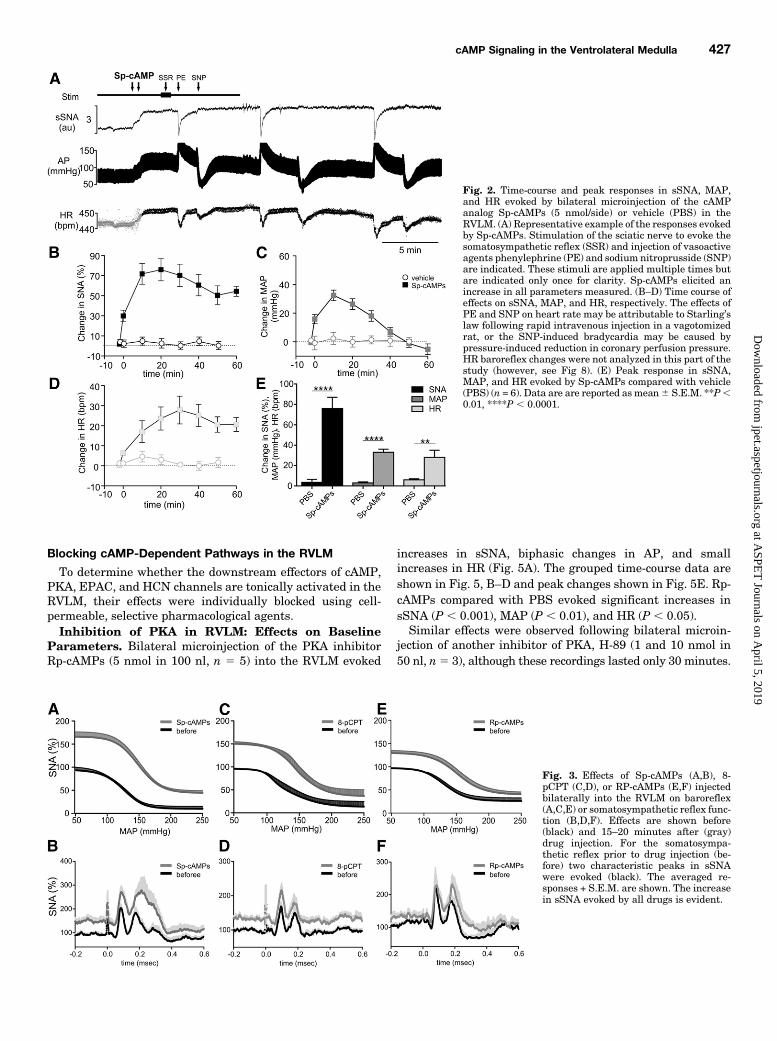
in sSNA, HR, and MAP that were maximal at 10–20 minutes,whereas vehicle had little effect (Fig. 2, A–D). sSNA and HRremained elevated for the remainder of the experiment (.1hour), whereas MAP recovered within 40 minutes (Fig. 2,A–D). Sp-cAMPs (5 nmol, n 5 6) evoked significant peakincreases in sSNA,MAP, andHR compared with control (PBS,n 5 6) (P , 0.01 for all parameters) (Fig. 2E).Sp-cAMP in the RVLM: Effect on Sympathetic
Reflexes. The effect of Sp-cAMPs in the RVLM was tested
on reflexes that are dependent on the GABAergic [baroreflex(Schreihofer and Guyenet, 2002) or glutamatergic (somato-sympathetic reflex) (Kiely andGordon, 1993)] synapses withinthe RVLM.Microinjection of Sp-cAMPs significantly increased the
upper plateau and maximum gain of the sympathetic barore-flex function curve compared with control (PBS) (Fig. 3A andTable 1). The data were acquired from experiments repre-sented in Fig. 2. Importantly, cardiac baroreflex changes arenot reported following drug injection into the RVLM (however,see Fig. 8).Intermittent stimulation of the sciatic nerve resulted in a
characteristic two-phase excitatory response in sSNA. Thetotal AUC (22.8 6 4.9 versus 13.6 6 3.1 au, Sp-cAMPs versusPBS, P5 0.0083) but not the amplitude of the two peaks [89.6621.2% (peak 1) and 87.7 6 29.8% (peak 2) versus 120 619.6% and 103.6 6 28.8% sSNA; Sp-cAMPs versus PBS n.s.]was significantly affected by Sp-cAMPs compared with PBS(Fig. 3B). The effect of Sp-cAMPs was to increase the width ofthe second peak, particularly at more delayed latencies.8-pCPT in RVLM: Effects on Baseline Parameters.
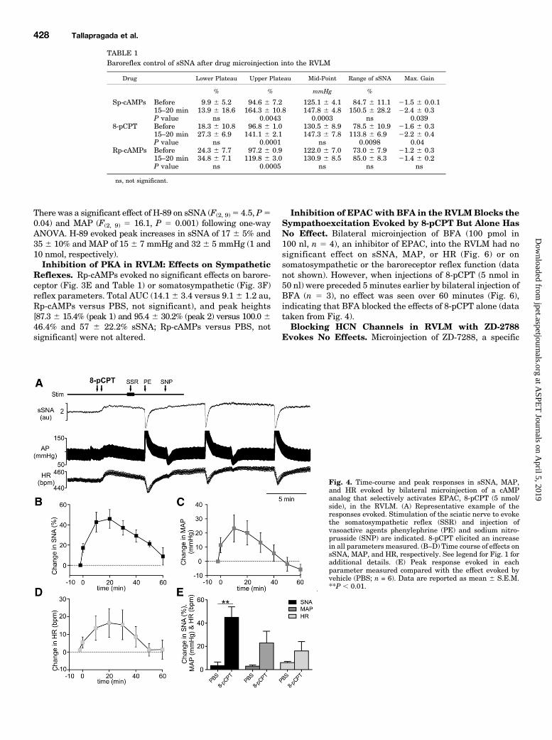
Bilateral microinjection into the RVLM of 8-pCPT (5 nmol in50 nl, n 5 6), a cAMP analog that selectively activates EPAC(Vliem et al., 2008) increased sSNA, AP, andHR (Fig. 4A). Thegrouped time-course data are shown in Fig. 4, B–D, and thepeak responses compared with bilateral PBS injection areshown in Fig. 4E. 8-pCPT significantly increased sSNA (p ,0.01). Increases in MAP and HR were not statisticallysignificantly different.8-pCPT in RVLM: Effects on Sympathetic Re-
flexes. 8-pCPT significantly increased the upper plateauand the maximum gain of the sympathetic baroreflex (Fig.3C and Table 1). In contrast, 8-pCPT evoked no significanteffect on somatosympathetic reflex parameters (Fig. 3D): totalAUC (8.7 6 2.6 versus 6.3 6 1.4 au, 8-pCPT versus PBS n.s.)and peak heights (66.96 21.9% (peak 1) and 39.23 6 23.9(peak 2) versus 64.1 6 9.7% and 44.3 6 18.1 sSNA; 8-pCPTversus PBS, not significant).
Fig. 1. The change in sympathetic nerve activity (SNA) and mean arterial pressure (MAP) evoked by increasing doses of analogs of cAMPmicroinjectedbilaterally and cumulatively in the RVLM. (A) Change in sSNA elicited by vehicle (PBS), Sp-cAMPs (0.5, 1.5, 5 nmol, n = 4), and 8-Br-cAMP (1 and10 nmol) (n = 3). (B) Change in MAP evoked by the same agents (n = 4 for both). Data are are reported as mean6 S.E.M.; *P, 0.05, **P, 0.01, ****P,0.0001.
426 Tallapragada et al.
at ASPE
T Journals on A
pril 5, 2019jpet.aspetjournals.org
Dow
nloaded from
Blocking cAMP-Dependent Pathways in the RVLM
To determine whether the downstream effectors of cAMP,PKA, EPAC, and HCN channels are tonically activated in theRVLM, their effects were individually blocked using cell-permeable, selective pharmacological agents.Inhibition of PKA in RVLM: Effects on Baseline
Parameters. Bilateral microinjection of the PKA inhibitorRp-cAMPs (5 nmol in 100 nl, n 5 5) into the RVLM evoked
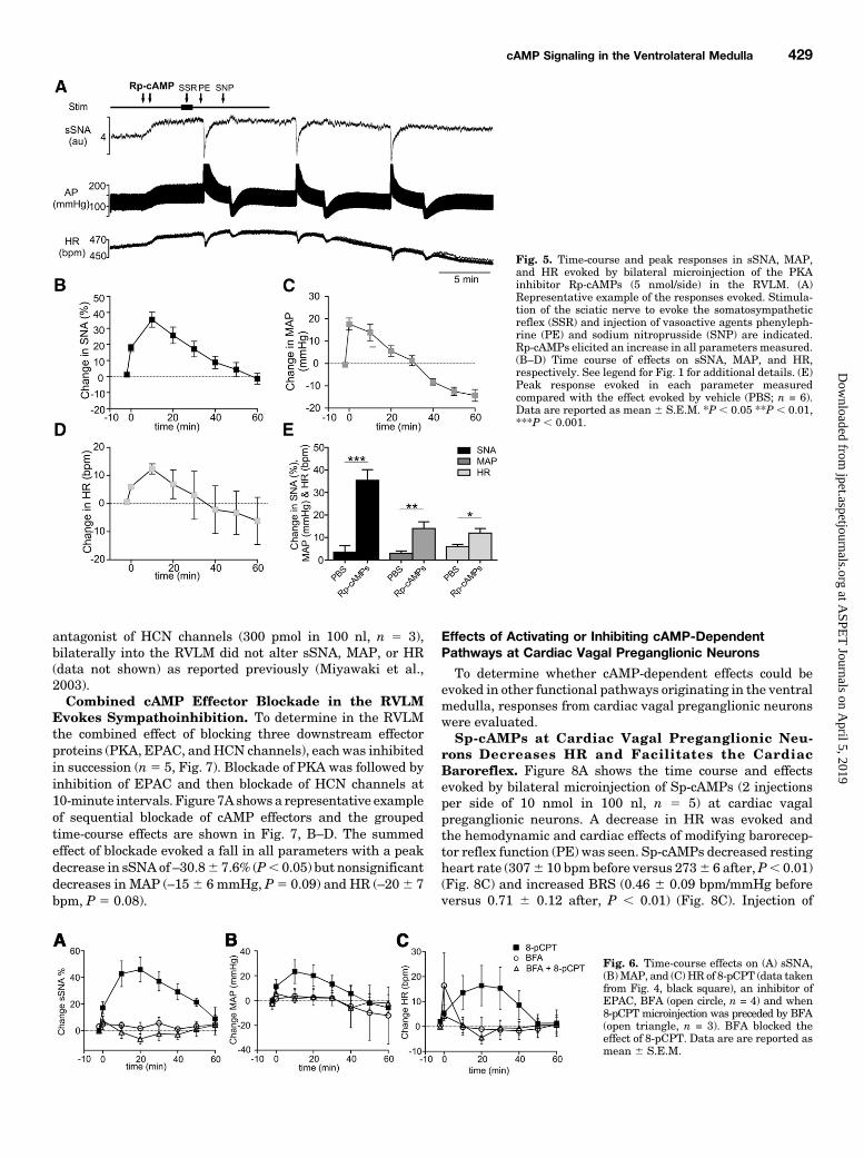
increases in sSNA, biphasic changes in AP, and smallincreases in HR (Fig. 5A). The grouped time-course data areshown in Fig. 5, B–D and peak changes shown in Fig. 5E. Rp-cAMPs compared with PBS evoked significant increases insSNA (P , 0.001), MAP (P , 0.01), and HR (P , 0.05).Similar effects were observed following bilateral microin-
jection of another inhibitor of PKA, H-89 (1 and 10 nmol in50 nl, n5 3), although these recordings lasted only 30 minutes.
Fig. 2. Time-course and peak responses in sSNA, MAP,and HR evoked by bilateral microinjection of the cAMPanalog Sp-cAMPs (5 nmol/side) or vehicle (PBS) in theRVLM. (A) Representative example of the responses evokedby Sp-cAMPs. Stimulation of the sciatic nerve to evoke thesomatosympathetic reflex (SSR) and injection of vasoactiveagents phenylephrine (PE) and sodium nitroprusside (SNP)are indicated. These stimuli are applied multiple times butare indicated only once for clarity. Sp-cAMPs elicited anincrease in all parameters measured. (B–D) Time course ofeffects on sSNA, MAP, and HR, respectively. The effects ofPE and SNP on heart rate may be attributable to Starling’slaw following rapid intravenous injection in a vagotomizedrat, or the SNP-induced bradycardia may be caused bypressure-induced reduction in coronary perfusion pressure.HR baroreflex changes were not analyzed in this part of thestudy (however, see Fig 8). (E) Peak response in sSNA,MAP, and HR evoked by Sp-cAMPs compared with vehicle(PBS) (n = 6). Data are are reported asmean6 S.E.M. **P,0.01, ****P , 0.0001.
Fig. 3. Effects of Sp-cAMPs (A,B), 8-pCPT (C,D), or RP-cAMPs (E,F) injectedbilaterally into the RVLM on baroreflex(A,C,E) or somatosympathetic reflex func-tion (B,D,F). Effects are shown before(black) and 15–20 minutes after (gray)drug injection. For the somatosympa-thetic reflex prior to drug injection (be-fore) two characteristic peaks in sSNAwere evoked (black). The averaged re-sponses + S.E.M. are shown. The increasein sSNA evoked by all drugs is evident.
cAMP Signaling in the Ventrolateral Medulla 427
at ASPE
T Journals on A
pril 5, 2019jpet.aspetjournals.org
Dow
nloaded from
There was a significant effect of H-89 on sSNA (F(2, 9)5 4.5, P50.04) and MAP (F(2, 9) 5 16.1, P 5 0.001) following one-wayANOVA. H-89 evoked peak increases in sSNA of 17 6 5% and35 6 10% and MAP of 15 6 7 mmHg and 32 6 5 mmHg (1 and10 nmol, respectively).Inhibition of PKA in RVLM: Effects on Sympathetic
Reflexes. Rp-cAMPs evoked no significant effects on barore-ceptor (Fig. 3E and Table 1) or somatosympathetic (Fig. 3F)reflex parameters. Total AUC (14.1 6 3.4 versus 9.1 6 1.2 au,Rp-cAMPs versus PBS, not significant), and peak heights[87.3 6 15.4% (peak 1) and 95.4 6 30.2% (peak 2) versus 100.0 646.4% and 57 6 22.2% sSNA; Rp-cAMPs versus PBS, notsignificant] were not altered.
Inhibition of EPACwith BFA in the RVLMBlocks theSympathoexcitation Evoked by 8-pCPT But Alone HasNo Effect. Bilateral microinjection of BFA (100 pmol in100 nl, n 5 4), an inhibitor of EPAC, into the RVLM had nosignificant effect on sSNA, MAP, or HR (Fig. 6) or onsomatosympathetic or the baroreceptor reflex function (datanot shown). However, when injections of 8-pCPT (5 nmol in50 nl) were preceded 5 minutes earlier by bilateral injection ofBFA (n 5 3), no effect was seen over 60 minutes (Fig. 6),indicating that BFA blocked the effects of 8-pCPT alone (datataken from Fig. 4).Blocking HCN Channels in RVLM with ZD-2788
Evokes No Effects. Microinjection of ZD-7288, a specific
TABLE 1Baroreflex control of sSNA after drug microinjection into the RVLM
Drug Lower Plateau Upper Plateau Mid-Point Range of sSNA Max. Gain
% % mmHg %
Sp-cAMPs Before 9.9 6 5.2 94.6 6 7.2 125.1 6 4.1 84.7 6 11.1 21.5 6 0.0.115–20 min 13.9 6 18.6 164.3 6 10.8 147.8 6 4.8 150.5 6 28.2 22.4 6 0.3P value ns 0.0043 0.0003 ns 0.039
8-pCPT Before 18.3 6 10.8 96.8 6 1.0 130.5 6 8.9 78.5 6 10.9 21.6 6 0.315–20 min 27.3 6 6.9 141.1 6 2.1 147.3 6 7.8 113.8 6 6.9 22.2 6 0.4P value ns 0.0001 ns 0.0098 0.04
Rp-cAMPs Before 24.3 6 7.7 97.2 6 0.9 122.0 6 7.0 73.0 6 7.9 21.2 6 0.315–20 min 34.8 6 7.1 119.8 6 3.0 130.9 6 8.5 85.0 6 8.3 21.4 6 0.2P value ns 0.0005 ns ns ns
ns, not significant.
Fig. 4. Time-course and peak responses in sSNA, MAP,and HR evoked by bilateral microinjection of a cAMPanalog that selectively activates EPAC, 8-pCPT (5 nmol/side), in the RVLM. (A) Representative example of theresponses evoked. Stimulation of the sciatic nerve to evokethe somatosympathetic reflex (SSR) and injection ofvasoactive agents phenylephrine (PE) and sodium nitro-prusside (SNP) are indicated. 8-pCPT elicited an increasein all parameters measured. (B–D) Time course of effects onsSNA, MAP, and HR, respectively. See legend for Fig. 1 foradditional details. (E) Peak response evoked in eachparameter measured compared with the effect evoked byvehicle (PBS; n = 6). Data are reported as mean 6 S.E.M.**P , 0.01.
428 Tallapragada et al.
at ASPE
T Journals on A
pril 5, 2019jpet.aspetjournals.org
Dow
nloaded from
antagonist of HCN channels (300 pmol in 100 nl, n 5 3),bilaterally into the RVLM did not alter sSNA, MAP, or HR(data not shown) as reported previously (Miyawaki et al.,2003).Combined cAMP Effector Blockade in the RVLM
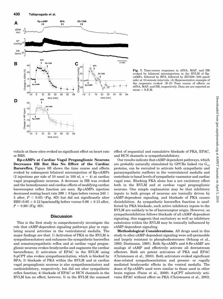
Evokes Sympathoinhibition. To determine in the RVLMthe combined effect of blocking three downstream effectorproteins (PKA, EPAC, andHCN channels), each was inhibitedin succession (n5 5, Fig. 7). Blockade of PKA was followed byinhibition of EPAC and then blockade of HCN channels at10-minute intervals. Figure 7Ashows a representative exampleof sequential blockade of cAMP effectors and the groupedtime-course effects are shown in Fig. 7, B–D. The summedeffect of blockade evoked a fall in all parameters with a peakdecrease in sSNA of –30.86 7.6% (P, 0.05) but nonsignificantdecreases in MAP (–156 6 mmHg, P5 0.09) and HR (–206 7bpm, P 5 0.08).
Effects of Activating or Inhibiting cAMP-DependentPathways at Cardiac Vagal Preganglionic Neurons
To determine whether cAMP-dependent effects could beevoked in other functional pathways originating in the ventralmedulla, responses from cardiac vagal preganglionic neuronswere evaluated.Sp-cAMPs at Cardiac Vagal Preganglionic Neu-
rons Decreases HR and Facilitates the CardiacBaroreflex. Figure 8A shows the time course and effectsevoked by bilateral microinjection of Sp-cAMPs (2 injectionsper side of 10 nmol in 100 nl, n 5 5) at cardiac vagalpreganglionic neurons. A decrease in HR was evoked andthe hemodynamic and cardiac effects of modifying barorecep-tor reflex function (PE) was seen. Sp-cAMPs decreased restingheart rate (3076 10 bpmbefore versus 2736 6 after,P, 0.01)(Fig. 8C) and increased BRS (0.46 6 0.09 bpm/mmHg beforeversus 0.71 6 0.12 after, P , 0.01) (Fig. 8C). Injection of
Fig. 5. Time-course and peak responses in sSNA, MAP,and HR evoked by bilateral microinjection of the PKAinhibitor Rp-cAMPs (5 nmol/side) in the RVLM. (A)Representative example of the responses evoked. Stimula-tion of the sciatic nerve to evoke the somatosympatheticreflex (SSR) and injection of vasoactive agents phenyleph-rine (PE) and sodium nitroprusside (SNP) are indicated.Rp-cAMPs elicited an increase in all parameters measured.(B–D) Time course of effects on sSNA, MAP, and HR,respectively. See legend for Fig. 1 for additional details. (E)Peak response evoked in each parameter measuredcompared with the effect evoked by vehicle (PBS; n = 6).Data are reported as mean 6 S.E.M. *P , 0.05 **P , 0.01,***P , 0.001.
Fig. 6. Time-course effects on (A) sSNA,(B)MAP, and (C) HR of 8-pCPT (data takenfrom Fig. 4, black square), an inhibitor ofEPAC, BFA (open circle, n = 4) and when8-pCPT microinjection was preceded by BFA(open triangle, n = 3). BFA blocked theeffect of 8-pCPT. Data are are reported asmean 6 S.E.M.
cAMP Signaling in the Ventrolateral Medulla 429
at ASPE
T Journals on A
pril 5, 2019jpet.aspetjournals.org
Dow
nloaded from
vehicle at these sites evoked no significant effect on heart rateor BRS.Rp-cAMPs at Cardiac Vagal Preganglionic Neurons
Decreases HR But Has No Effect of the CardiacBaroreflex. Figure 8B shows the time course and effectsevoked by subsequent bilateral microinjection of Rp-cAMPs(2 injections per side of 10 nmol in 100 nl, n 5 4) at cardiacvagal preganglionic neurons. A decrease in HR was evokedand the hemodynamic and cardiac effects of modifying cardiacbaroreceptor reflex function are seen. Rp-cAMPs injectiondecreased resting heart rate 2996 8 bpm before versus 24065 after, P , 0.05) (Fig. 8D) but did not significantly alterBRS (0.65 6 0.12 bpm/mmHg before versus 0.66 6 0.13 after,P 5 0.98) (Fig. 8D).
DiscussionThis is the first study to comprehensively investigate the
role that cAMP-dependent signaling pathways play in regu-lating neural activities in the ventrolateral medulla. Themajor findings are that: 1) Activation of PKA in the RVLM issympathoexcitatory and enhances the sympathetic baroreflexand somatosympathetic reflex and at cardiac vagal pregan-glionic neurons evokes bradycardia and augments the cardiacbaroreflexes; 2) activation of EPAC in the RVLM using8-pCPT also evokes sympathoexcitation, which is blocked byBFA; 3) blockade of PKA within the RVLM and at cardiacvagal preganglionic neurons is also sympathoexcitatory andcardioinhibitory, respectively, but did not alter sympatheticreflex function; 4) blockade of EPAC or HCN channels in theRVLM has no effect; however, 5) in the RVLM the summed
effect of sequential and cumulative blockade of PKA, EPAC,and HCN channels is sympathoinhibitory.Our results indicate that cAMP-dependent pathways, which
are probably naturally stimulated by GPCRs linked via Gas
proteins, can be recruited to activate both sympathetic andparasympathetic outflows in the ventrolateral medulla andcontribute to basal levels of sympathetic vasomotor and cardiacvagal tone. Blocking PKA alone has a net excitatory effectboth in the RVLM and at cardiac vagal preganglionicneurons. One simple explanation may be that inhibitoryinputs to both groups of neurons are tonically driven bycAMP-dependent signaling, and blockade of PKA causesdisinhibition. As sympathetic baroreflex function is unaf-fected by PKA blockade, such active inhibitory inputs to theRVLM are unlikely to be of baroreceptor origin. However, assympathoinhibition follows blockade of all cAMP-dependentsignaling, this suggests that excitatory as well as inhibitorysubstrates within the RVLM may be tonically influenced bycAMP-dependent signaling.Methodological Considerations. All drugs used in this
study to alter cAMP-dependent signaling were cell-permeableand largely resistant to phosphodiesterases (Schaap et al.,1993; Dostmann, 1995). Both Sp-cAMPs and 8-Br-cAMP areanalogs of cAMP and effectively activate all downstreameffectors. Both are potent activators of PKA and EPAC(Christensen et al., 2003). Both activators evoked significantdose-related sympathoexcitation and pressor- or vagallymediated bradycardic effects in the ventral medulla. Thedoses of Sp-cAMPs used were similar to those used in otherbrain regions (Paine et al., 2009). 8-pCPT selectively acti-vates EPAC without effect on PKA (Christensen et al., 2003;
Fig. 7. Time-course responses in sSNA, MAP, and HRevoked by bilateral microinjection in the RVLM of Rp-cAMPs, followed by BFA, followed by ZD7288 (300 pmol/side) at 10-minute intervals. (A) Representative example ofthe responses evoked. (B–D) Time course of effects onsSNA, MAP, and HR, respectively. Data are are reported asmean 6 S.E.M.
430 Tallapragada et al.
at ASPE
T Journals on A
pril 5, 2019jpet.aspetjournals.org
Dow
nloaded from

Brown et al., 2014), although it may have some nonspecific/EPAC-independent effects, at least as identified in platelets(Herfindal et al., 2013). Nevertheless, the effects of 8-pCPTwere similar to those evoked by Sp-cAMPs and were blockedby prior treatment with BFA, which alone had no effect asdescribed elsewhere (Zhong and Zucker, 2005). ZD7288 is acommonly used selective blocker of HCN channels (Harris andConstanti, 1995), although some effect on sodium channelshas been suggested (Wu et al., 2012).Rp-cAMPs, which inhibits PKA, has little effect on EPAC
(Christensen et al., 2003; Brown et al., 2014) or onH-89, whichalso inhibits PKA-evoked similar dose-dependent effects,although H-89 actions could also occur via other kinases(Lochner and Moolman, 2006). The pressor effect evoked byH-89 in the RVLM confirm what has been previously noted(Xu and Krukoff, 2006). ZD 7288, which blocks HCN channelsbut is ineffective alone in the RVLM, as described previously(Miyawaki et al., 2003), contributed to inhibitory effects whenpreceded by other drugs. Nevertheless, as in most pharmaco-logical studies of this type, it is possible that the effects evokedby the drugs used may not be attributable to the substratestargeted.Heart rate, sympathetic, and blood pressure responses
evoked by drug injection in vagotomized spinal-cord intactanimals are interpreted as sympathetically mediated, albeitmodified by competing baroreflex pathways. The splanchnicnerve innervates functionally diverse targets, including gut
vasculature, gastrointestinal muscles, and adrenal gland, andcannot therefore be interpreted as a purely vasomotor output.Conversely, data from spinally transected animals are inter-preted as consequences of direct drug effects on cardiac vagalmotor circuits, as described previously (Hildreth and Goodchild,2010), as all sympathetic outputs were disrupted also, thusproviding conditions of maximal baroreflex unloading.Drug interaction with medullary interneurons presynaptic
to sympathetic/parasympathetic outputs are probable. Wehave previously shown select effects on respiratory functionwithin subregions of the ventrolateral medulla (Burke et al.,2013), and it is possible that changes in respiratory-sympatheticcoupling contribute to the effects seen here.Sites of cAMP Activation in the Ventral Medulla.
Activation of cAMP-dependent pathways in the RVLM evokedsympathoexcitation and a pressor effect and bradycardia atcardiac vagal preganglionic neurons. This is in keeping withour finding that the Gas subunit mRNA is abundant in theRVLM (Parker et al., 2012) and consistent with a postsynapticsite of action, as suggested previously in neonatal RVLMbrainslice preparations, in which 8-Br-cAMP and the adenylylcyclase activator forskolin increased the firing rate of RVLM“pacemaker” neurons in the presence of tetrodotoxin (Sun andGuyenet, 1990). Activation of Gas-linked receptors in theRVLM using pituitary adenylate cyclase–activating peptideevokes sympathoexcitation and pressor responses, althoughreflex functions were unaffected (Farnham et al., 2012). On
Fig. 8. Effects on HR and cardiac baroreflex sensitivity ofdual bilateral microinjections of Sp-cAMPs (each injection,10 nmol) and Rp-cAMPs (each injection, 10 nmol) in theventral medulla targeting cardiac vagal preganglionicneurons in spinally transected animals. (A) Representativeexample of the effects of Sp-cAMPs on HR and also theeffect of the vasoactive agent phenylephrine used todetermine BRS. (B) Representative example of the effectsof Rp-cAMPs on HR. (C) Grouped data of the effects of SP-cAMPs on resting HR and BRS and (D) grouped data of theeffects of Rp-cAMPs on resting HR and BRS. Data arereported as mean 6 S.E.M. *P , 0.05 **P , 0.01.
cAMP Signaling in the Ventrolateral Medulla 431
at ASPE
T Journals on A
pril 5, 2019jpet.aspetjournals.org
Dow
nloaded from
the other hand, cardiac vagal nerve activity is increased bysystemic adenosine (da Silva et al., 2012) or by activation ofb-adrenergic receptors, specifically b1, which reduces GABAer-gic and glycinergic (as well as glutamatergic) conductances atcardiac vagal preganglionic neurons (Bateman et al., 2012).Recently, b1 and b2 receptors have been identified on putativepresympathetic RVLM neurons, and their selective activationevoked depolarization and hyperpolarization, respectively(Oshima et al., 2014). These data suggest that cAMP-dependentsignaling can be elicited by catecholamine release in theventrolateral medulla.Injections of PKA and EPAC activators enhanced both
sympathetic and cardiac baroreflex functions. This could beexplained by the activation of cAMP in presympatheticneurons and/or in inhibitory inputs and in cardiac vagalpreganglionic neurons and/or in excitatory inputs, respec-tively. The effect, at least of PKA, on the somatosympatheticreflex [mediated by glutamatergic synapses in the RVLM(Kiely and Gordon, 1993)] could indicate modulation ofglutamatergic inputs or postsynaptic effects, particularly asfacilitation appeared more prominent at slowly conductingpossibly catecholaminergic cells in the region.Tonically Active PKA-Dependent Signaling in the
Ventral Medulla. Blockade of PKA, using both Rp-cAMPsand H-89, evoked sympathoexcitation and vagally mediatedbradycardia, indicating tonically active PKA-dependent sig-naling in the ventrolateral medulla. Although a pressor effectinitially accompanied the sympathoexcitation elicited by bothagents, at later time points a depressor response, which wasnot accompanied by splanchnic sympathoinhibition, wasevoked by Rp-cAMPs. This biphasic effect may indicate thatsplanchnic sympathetic drive is counteracted by other effec-tors, such as inhibition of excitatory input supplying othersympathetic vasomotor outflows. Nevertheless the effects onMAP suggest that vasomotor pathways are affected as well asboth sympathetic and parasympathetic pathways controllingheart rate. As the tonic activity of RVLM neurons supplyingvasomotor tone is dependent on the balance of tonic excitatoryand inhibitory input, we speculate that the early net excit-atory action of Rp-cAMPs could be mediated by effects atinhibitory inputs; however, as the sympathetic baroreflex(mediated by inhibitory presynaptic input) was unaffected,actions at other functional inhibitory inputs would be in-dicated. GABA-A receptor blockade at both the RVLM andcardiac vagal preganglionic neurons indicate significant levelsof tonic inhibitory input to neurons controlling vasomotor(Schreihofer and Guyenet, 2002) and cardiac (Hildreth andGoodchild, 2010) functions. Furthermore, there is someprecedent for PKA-dependent disinhibition, as modulation ofglycinergic release occurs in spinal cord (Katsurabayashiet al., 2004). It is possible that blocking PKAmay redistributethe active pool of cAMP to other effectors; however, at least inthe RVLM, blocking either EPAC or HCN channels alone hadlittle effect. There is little evidence supporting the idea oftonically active peptides in the RVLM (Burke et al., 2008;Pilowsky et al., 2008; Farnham et al., 2012). Nevertheless thefindings here suggest that a neurotransmitter acting via Gas-linked receptor/s is active in the ventrolateral medulla. Onepossibility is a catecholamine acting at b receptors where, atleast in the neonatal RVLM, b2-receptor blockade depolarizedneurons (Oshima et al., 2014). However, an alternative expla-nation could be that such a receptor is constitutively active
(Milligan, 2003; Costa and Cotecchia, 2005) and candidatesthat are Gas-linked in the RVLM include the H2 andmelanocortin 3/4 receptors (Granata and Reis, 1987; Kawabeet al., 2006). When activated in the RVLM, only the histamine2 receptor causes sympathoinhibition, probably via excitationof an inhibitory input (Granata and Reis, 1987).Although blockade of PKA in RVLM evoked sympathoexci-
tation, blockade of other cAMP effectors each had no effect.Nevertheless combined blockade resulted in sympathoinhibi-tion, suggesting actions at both inhibitory and excitatorysynaptic sites. It should be noted that the cAMP effectorsare restricted to spatially separated microdomains within cellbodies and terminals in the ventralmedulla (Karpen andRich,2004; Calebiro and Maiellaro, 2014), so sequential block-ade may have disturbed the balance within intracellularcompartments.
ConclusionsOur data show that cAMP-dependent pathways, signaling
via PKA and EPAC, can be recruited in the ventrolateralmedulla to evoke excitation in sympathetic circuitry control-ling the heart, vasculature, and baroreflex, as well as excita-tion of the cardiac vagus and circuitry controlling the cardiacbaroreflex. Importantly, the results indicate that PKA-dependent pathways are tonically active in a region control-ling the basal level of sympathetic and cardiac vagal tones.These effects are in contrast to the effects of blockingexcitatory ionotropic receptors in the RVLM or at cardiacvagal preganglionic neurons, which do not alter the level ofsympathetic activity or HR, respectively (Dampney et al.,2003; Hildreth and Goodchild, 2010). Thus GPCRs utilizingGas proteins in the ventrolateral medulla contribute to settingthe level of sympathetic tone including sympathetic vasomo-tor as well as cardiac vagal tone.
Authorship Contributions
Participated in research design: Goodchild, Hildreth, Tallapragada.Conducted experiments: Tallapragada, Hildreth, Raley, Burke.Performed data analysis: Tallapragada, Hildreth, Burke, Hassan.Wrote or contributed to the writing of the manuscript: Goodchild,
Tallapragada, Hildreth, Hassan, Burke, McMullan.
References
Bateman RJ, Boychuk CR, Philbin KE, and Mendelowitz D (2012) b adrenergic re-ceptor modulation of neurotransmission to cardiac vagal neurons in the nucleusambiguus. Neuroscience 210:58–66.
Beavo JA and Brunton LL (2002) Cyclic nucleotide research – still expanding afterhalf a century. Nat Rev Mol Cell Biol 3:710–718.
Bos JL (2003) Epac: a new cAMP target and new avenues in cAMP research. Nat RevMol Cell Biol 4:733–738.
Bowman BR, Kumar NN, Hassan SF, McMullan S, and Goodchild AK (2013) Brainsources of inhibitory input to the rat rostral ventrolateral medulla. J Comp Neurol521:213–232.
Brown DA and Sihra TS (2008) Presynaptic signaling by heterotrimeric G-proteins.Handbook Exp Pharmacol 184:207–260.
Brown LM, Rogers KE, McCammon JA, and Insel PA (2014) Identification and val-idation of modulators of exchange protein activated by cAMP (Epac) activity:structure-function implications for Epac activation and inhibition. J Biol Chem289:8217–8230.
Burke PG, Li Q, Costin ML, McMullan S, Pilowsky PM, and Goodchild AK (2008)Somatostatin 2A receptor-expressing presympathetic neurons in the rostral ven-trolateral medulla maintain blood pressure. Hypertension 52:1127–1133.
Burke PG, Sousa LO, Tallapragada VJ, and Goodchild AK (2013) Inhibition of pro-tein kinase A activity depresses phrenic drive and glycinergic signalling, but notrhythmogenesis in anaesthetized rat. Eur J Neurosci 38:2260–2270.
Calebiro D and Maiellaro I (2014) cAMP signaling microdomains and their obser-vation by optical methods. Front Cell Neurosci 8:350.
Christensen AE, Selheim F, de Rooij J, Dremier S, Schwede F, Dao KK, Martinez A,Maenhaut C, Bos JL, and Genieser HG, et al. (2003) cAMP analog mapping ofEpac1 and cAMP kinase. Discriminating analogs demonstrate that Epac and
432 Tallapragada et al.
at ASPE
T Journals on A
pril 5, 2019jpet.aspetjournals.org
Dow
nloaded from

cAMP kinase act synergistically to promote PC-12 cell neurite extension. J BiolChem 278:35394–35402.
Costa T and Cotecchia S (2005) Historical review: Negative efficacy and the con-stitutive activity of G-protein-coupled receptors. Trends Pharmacol Sci 26:618–624.
Dampney RA (1994) The subretrofacial vasomotor nucleus: anatomical, chemical andpharmacological properties and role in cardiovascular regulation. Prog Neurobiol42:197–227.
Dampney RA, Horiuchi J, Tagawa T, Fontes MA, Potts PD, and Polson JW (2003)Medullary and supramedullary mechanisms regulating sympathetic vasomotortone. Acta Physiol Scand 177:209–218.
da Silva VJ, Gnecchi-Ruscone T, Bellina V, Oliveira M, Maciel L, de Carvalho AC,Salgado HC, Bergamaschi CM, Tobaldini E, and Porta A, et al. (2012) Acuteadenosine increases cardiac vagal and reduces sympathetic efferent nerve activi-ties in rats. Exp Physiol 97:719–729.
Dostmann WR (1995) (RP)-cAMPS inhibits the cAMP-dependent protein kinase byblocking the cAMP-induced conformational transition. FEBS Lett 375:231–234.
Farnham MM, Lung MS, Tallapragada VJ, and Pilowsky PM (2012) PACAP causesPAC1/VPAC2 receptor mediated hypertension and sympathoexcitation in normaland hypertensive rats. Am J Physiol Heart Circ Physiol 303:H910–H917.
Granata AR and Reis DJ (1987) Hypotension and bradycardia elicited by histamineinto the C1 area of the rostral ventrolateral medulla. Eur J Pharmacol 136:157–162.
Guyenet PG (2006) The sympathetic control of blood pressure. Nat Rev Neurosci 7:335–346.
Harris NC and Constanti A (1995) Mechanism of block by ZD 7288 of thehyperpolarization-activated inward rectifying current in guinea pig substantianigra neurons in vitro. J Neurophysiol 74:2366–2378.
Herfindal L, Nygaard G, Kopperud R, Krakstad C, Døskeland SO, and Selheim F(2013) Off-target effect of the Epac agonist 8-pCPT-29-O-Me-cAMP on P2Y12 re-ceptors in blood platelets. Biochem Biophys Res Commun 437:603–608.
Hildreth CM and Goodchild AK (2010) Role of ionotropic GABA, glutamate andglycine receptors in the tonic and reflex control of cardiac vagal outflow in the rat.BMC Neurosci 11:128.
Holz GG, Kang G, Harbeck M, Roe MW, and Chepurny OG (2006) Cell physiology ofcAMP sensor Epac. J Physiol 577:5–15.
Karpen JW and Rich TC (2004) Resolution of cAMP signals in three-dimensionalmicrodomains using novel, real-time sensors. Proc West Pharmacol Soc 47:1–5.
Katsurabayashi S, Kubota H, Moorhouse AJ, and Akaike N (2004) Differentialmodulation of evoked and spontaneous glycine release from rat spinal cord glyci-nergic terminals by the cyclic AMP/protein kinase A transduction cascade. JNeurochem 91:657–666.
Kawabe T, Chitravanshi VC, Kawabe K, and Sapru HN (2006) Cardiovascular effectsof adrenocorticotropin microinjections into the rostral ventrolateral medullarypressor area of the rat. Brain Res 1102:117–126.
Kiely JM and Gordon FJ (1993) Non-NMDA receptors in the rostral ventrolateralmedulla mediate somatosympathetic pressor responses. J Auton Nerv Syst 43:231–239.
Lipski J, Lin J, Teo MY, and van Wyk M (2002) The network vs. pacemaker theory ofthe activity of RVL presympathetic neurons–a comparison with another putativepacemaker system. Auton Neurosci 98:85–89.
Lochner A and Moolman JA (2006) The many faces of H89: a review. CardiovascDrug Rev 24:261–274.
Lovick TA (1985) Projections from the diencephalon and mesencephalon to nucleusparagigantocellularis lateralis in the cat. Neuroscience 14:853–861.
McMullan S, Pathmanandavel K, Pilowsky PM, and Goodchild AK (2008) Somaticnerve stimulation evokes qualitatively different somatosympathetic responses inthe cervical and splanchnic sympathetic nerves in the rat. Brain Res 1217:139–147.
Milligan G (2003) Constitutive activity and inverse agonists of G protein-coupledreceptors: a current perspective. Mol Pharmacol 64:1271–1276.
Miyawaki T, Goodchild AK, and Pilowsky PM (2003) Maintenance of sympathetictone by a nickel chloride-sensitive mechanism in the rostral ventrolateral medullaof the adult rat. Neuroscience 116:455–464.
Oshima N, Onimaru H, Yamamoto K, Takechi H, Nishida Y, Oda T,, and KumagaiH (2014) Expression and functions of beta and beta-adrenergic receptors on thebulbospinal neurons in the rostral ventrolateral medulla. Hypertens Res 37:976–983.
Paine TA, Neve RL, , and Carlezon WA, Jr. (2009) Attention deficits and hyperac-tivity following inhibition of cAMP-dependent protein kinase within the medialprefrontal cortex of rats. Neuropsychopharmacology 34:2143–2155.
Parker LM, Tallapragada VJ, Kumar NN, and Goodchild AK (2012) Distribution andlocalisation of Ga proteins in the rostral ventrolateral medulla of normotensive andhypertensive rats: focus on catecholaminergic neurons. Neuroscience 218:20–34.
Pilowsky PM, Abbott SB, Burke PG, Farnham MM, Hildreth CM, Kumar NN, Li Q,Lonergan T, McMullan S, and Spirovski D, et al. (2008) Metabotropic neuro-transmission and integration of sympathetic nerve activity by the rostral ventro-lateral medulla in the rat. Clin Exp Pharmacol Physiol 35:508–511.
Pilowsky PM and Goodchild AK (2002) Baroreceptor reflex pathways and neuro-transmitters: 10 years on. J Hypertens 20:1675–1688.
Schaap P, van Ments-Cohen M, Soede RD, Brandt R, Firtel RA, Dostmann W,Genieser HG, Jastorff B, and van Haastert PJ (1993) Cell-permeable non-hydrolyzable cAMP derivatives as tools for analysis of signaling pathways con-trolling gene regulation in Dictyostelium. J Biol Chem 268:6323–6331.
Schreihofer AM and Guyenet PG (2002) The baroreflex and beyond: control of sym-pathetic vasomotor tone by GABAergic neurons in the ventrolateral medulla. ClinExp Pharmacol Physiol 29:514–521.
Sun MK and Guyenet PG (1990) Excitation of rostral medullary pacemaker neuronswith putative sympathoexcitatory function by cyclic AMP and beta-adrenoceptoragonists ‘in vitro’. Brain Res 511:30–40.
Vliem MJ, Ponsioen B, Schwede F, Pannekoek WJ, Riedl J, Kooistra MR, Jalink K,Genieser HG, Bos JL, and Rehmann H (2008) 8-pCPT-29-O-Me-cAMP-AM: animproved Epac-selective cAMP analogue. ChemBioChem 9:2052–2054.
Wang J, Irnaten M, Neff RA, Venkatesan P, Evans C, Loewy AD, Mettenleiter TC,and Mendelowitz D (2001) Synaptic and neurotransmitter activation of cardiacvagal neurons in the nucleus ambiguus. Ann N Y Acad Sci 940:237–246.
Wu X, Liao L, Liu X, Luo F, Yang T, and Li C (2012) Is ZD7288 a selective blocker ofhyperpolarization-activated cyclic nucleotide-gated channel currents? Channels(Austin) 6:438–442.
Xu Y and Krukoff TL (2006) Adrenomedullin in the rostral ventrolateral medullainhibits baroreflex control of heart rate: a role for protein kinase A. Br J Pharmacol148:70–77.
Zhong N and Zucker RS (2005) cAMP acts on exchange protein activated by cAMP/cAMP-regulated guanine nucleotide exchange protein to regulate transmitter re-lease at the crayfish neuromuscular junction. J Neurosci 25:208–214.
Address correspondence to: Dr. Ann K Goodchild, Dept BiomedicalSciences, Faculty of Medicine, 2 Technology Place, Macquarie University,2109, Sydney, NSW, Australia. E-mail: [email protected]
cAMP Signaling in the Ventrolateral Medulla 433
at ASPE
T Journals on A
pril 5, 2019jpet.aspetjournals.org
Dow
nloaded from
![INDEX [jpet.aspetjournals.org]jpet.aspetjournals.org/content/jpet/279/3/local/back-matter.pdf1996 Index 1591 Angiotensin IIreceptors, atpha-2 adreno-ceptor interactions, 795 Angiotensin](https://img.dokumen.tips/doc/110x75/5f668da720e0797faa000f58/index-jpet-jpet-1996-index-1591-angiotensin-iireceptors-atpha-2-adreno-ceptor.jpg)


![INDEX [jpet.aspetjournals.org]jpet.aspetjournals.org/content/jpet/220/3/local/back...703 INDEX Volume 220, January-March, 1982 Abernethy, DR.,seeGreenblatt, D.J.,120 Acetaminophen-induced](https://img.dokumen.tips/doc/110x75/5d3598fd88c993f1228bd8ae/index-jpet-jpet-index-volume-220-january-march-1982-abernethy-drseegreenblatt.jpg)

![INDEX [jpet.aspetjournals.org]jpet.aspetjournals.org/content/jpet/215/3/local/back-matter.pdf · synaptic neuromuscular ef-fects(rats),53 Caldwell, R.W.,seeCook, L.S., 198 Calcium](https://img.dokumen.tips/doc/110x75/6048ac2eb6e98b514e6c33d6/index-jpet-jpet-synaptic-neuromuscular-ef-fectsrats53-caldwell-rwseecook.jpg)
![INDEX [jpet.aspetjournals.org]jpet.aspetjournals.org/content/jpet/214/3/local/back...738 Index Vol.214 tory andexcitatory responses to neurotransmitters (sea hare), 161 Barry, B.K.,see](https://img.dokumen.tips/doc/110x75/5f64cf4036391b5a5d722ff5/index-jpet-jpet-738-index-vol214-tory-andexcitatory-responses-to-neurotransmitters.jpg)
![INDEX [jpet.aspetjournals.org]jpet.aspetjournals.org/content/jpet/182/3/local/back...cat, effects of2,5-dimethoxy-4-methylamphet-amine on, 145 rat, assay fortryptamine in,363 rats,](https://img.dokumen.tips/doc/110x75/5f64cf4036391b5a5d722ff6/index-jpet-jpet-cat-effects-of25-dimethoxy-4-methylamphet-amine-on-145.jpg)
![New INDEX [jpet.aspetjournals.org]jpet.aspetjournals.org/content/jpet/187/3/local/back... · 2005. 12. 3. · hycanthone effects onspermatogonial cells, de-oxyribonucleic acidsynthesis](https://img.dokumen.tips/doc/110x75/6067c6518625ed3f66076f25/new-index-jpet-jpet-2005-12-3-hycanthone-effects-onspermatogonial-cells.jpg)


![INDEX [jpet.aspetjournals.org]jpet.aspetjournals.org/content/jpet/228/3/local/back-matter.pdf · 811 INDEX Volume228,January-March, 1984 Acara,M.,seeRennick, B.,387 Acetaminophen,](https://img.dokumen.tips/doc/110x75/5d54da8888c993de0a8b9929/index-jpet-jpet-811-index-volume228january-march-1984-acaramseerennick.jpg)
![INDEX [jpet.aspetjournals.org]jpet.aspetjournals.org/content/jpet/174/3/local/back-matter.pdf · pressin(ADH)effectsof,541 1,1-Dimethyl-4-phenylpiperazinium, effecton ... Guanethidine,blockadeofDMAE-inducedpo](https://img.dokumen.tips/doc/110x75/5ab70e0a7f8b9a156d8e7acc/index-jpet-jpet-adheffectsof541-11-dimethyl-4-phenylpiperazinium-effecton.jpg)
![INDEX [jpet.aspetjournals.org]jpet.aspetjournals.org/content/jpet/230/3/local/back-matter.pdf · histrionicotoxin effects (frogs), 619 ... distribution kinetics ana- ... and myocardium](https://img.dokumen.tips/doc/110x75/5b7ac0067f8b9ae1328d73ab/index-jpet-jpet-histrionicotoxin-effects-frogs-619-distribution-kinetics.jpg)
![INDEX [jpet.aspetjournals.org]jpet.aspetjournals.org/content/jpet/234/3/local/back...effect, 708 Blockade, reticuloendothelial, enzyme-al-bumin conjugates, chronic adininis-tration](https://img.dokumen.tips/doc/110x75/60757ab7f966210d5e51d2f2/index-jpet-jpet-effect-708-blockade-reticuloendothelial-enzyme-al-bumin.jpg)
![INDEX [jpet.aspetjournals.org]jpet.aspetjournals.org/content/jpet/219/3/local/back... · 2005-12-24 · hypothyroidism (rats), 231 apha-1 andalpha-2, pre-and postsynaptic, selectivity](https://img.dokumen.tips/doc/110x75/5e79a7ee3dd9680cf7440526/index-jpet-jpet-2005-12-24-hypothyroidism-rats-231-apha-1-andalpha-2-pre-and.jpg)

![New INDEX [jpet.aspetjournals.org]jpet.aspetjournals.org/content/jpet/252/3/local/back... · 2006. 2. 15. · 1388 Index Vol.252 taglandins, cerebral and mesenteric arteries (dogs,](https://img.dokumen.tips/doc/110x75/606ca57a2c3b6b406f164e65/new-index-jpet-jpet-2006-2-15-1388-index-vol252-taglandins-cerebral-and.jpg)


![INDEX [jpet.aspetjournals.org]](https://img.dokumen.tips/doc/110x75/629818f027424e7e5e6aa348/index-jpet-.jpg)


