Embed Size (px)
Citation preview
Today’s Lecture Summary:Today’s Lecture Summary:
MicroscopesMicroscopes
A Tour of the Cell A Tour of the Cell

Reading Assignment:Reading Assignment:
Chapter 4Chapter 4

Learning Outcomes:
By the end of today’s lecture, you should be able to:
• Recognize the different kinds of images seen through different microscopes
• Identify the major parts and functions of prokaryotic and eukaryotic cells

How we study cells:How we study cells:Light microscope (LM)Light microscope (LM)Dissecting MicroscopeDissecting MicroscopeTransmission electron microscope (TEM)Transmission electron microscope (TEM)Scanning electron microscope (SEM)Scanning electron microscope (SEM)

Magnification: - the ratio of an object’s image to its real size (e.g. 40X, 100X)
Resolving power: - the measure of the clarity of an image- the minimum distance two points can be separated and still be distinguished as being separate

Light/Compound Light/Compound Microscope:Microscope:
- uses light to magnify uses light to magnify an objectan object
- gives a 2-D imagegives a 2-D image
- the different images - the different images are based on the are based on the various staining various staining techniques usedtechniques used

Dissecting Microscope:
- uses light to magnify an object
- gives a 3-D image

Electron microscopes:Electron microscopes: specimen preparation kills the cellsspecimen preparation kills the cells have a higher resolution than LMhave a higher resolution than LMTEM:TEM: SEM:SEM:2-D images2-D images 3-D images3-D images

1 cm = 1/100m
1mm = 1/1000m
1μm = 1 micrometer = 1 millionth of a meter
1nm = 1 nanometer = 1 billionth of a meter
Light and electron microscopes have different magnification and resolving powers.

Learning Outcomes:
By the end of today’s lecture, you should be able to:
• Recognize the different kinds of images seen through different microscopes
• Identify the major parts and functions of prokaryotic and eukaryotic cells
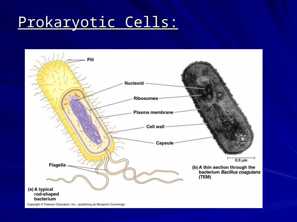
Prokaryotic Cells:Prokaryotic Cells:

Examples of prokaryotes:
Bacillus polymxa :
Escherichia coli :

There are limits to cell size:
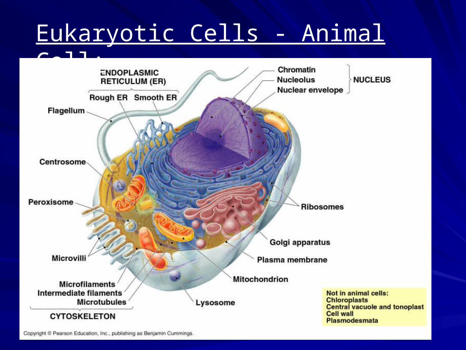
Eukaryotic Cells - Animal Cell:

Eukaryotic Cells - Plant Cell:

Isolation of Cell Organelles:Isolation of Cell Organelles:
cell fractionationcell fractionation
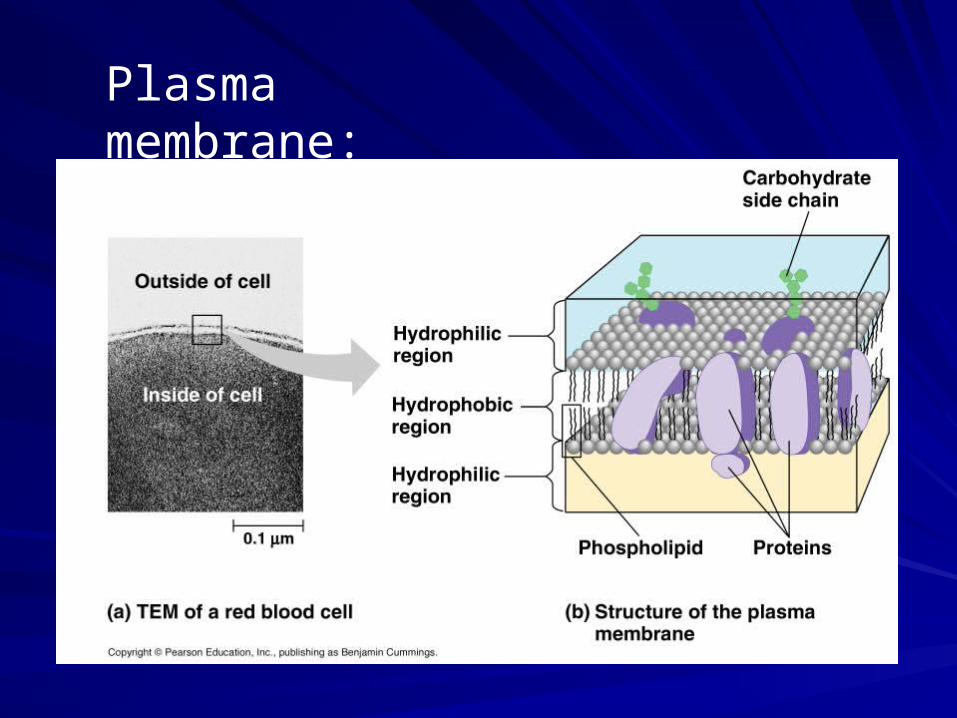
Plasma membrane:

Nucleus and nucleolus:

Ribosomes: function in protein synthesis

Endoplasmic reticulum:
- Smooth ER
- Rough ER

Golgi apparatus:

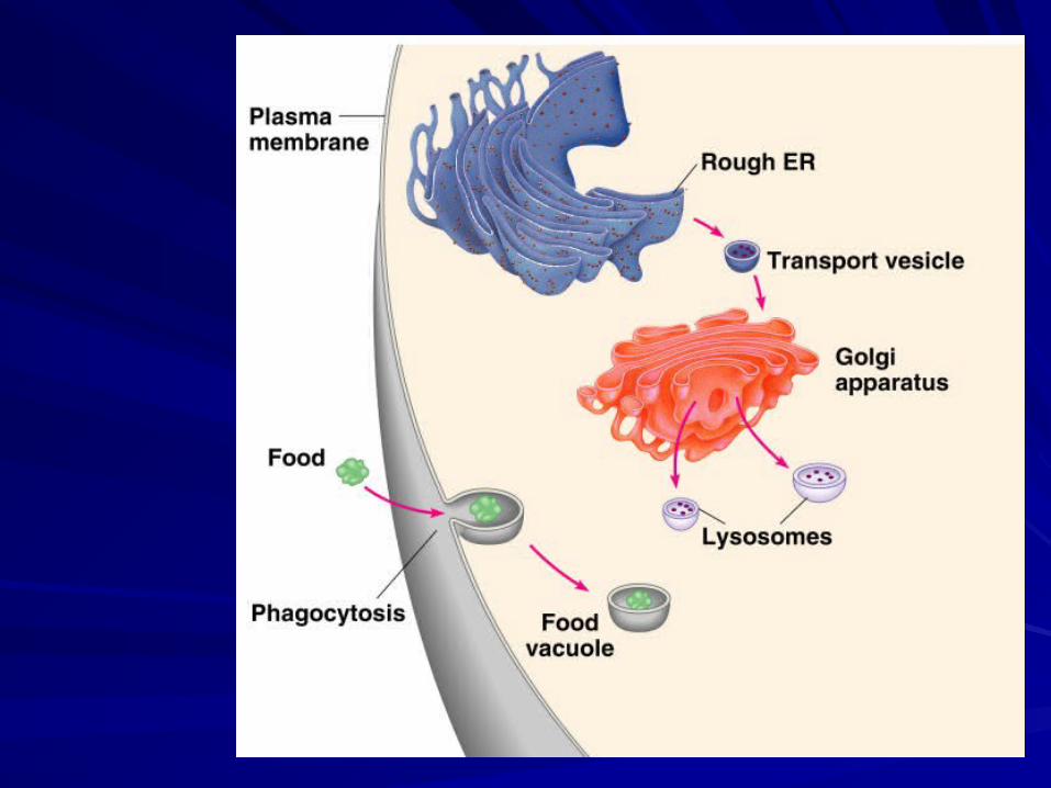
Lysosome formation:


Lysosomes (TEM):

Macrophage destroying bacteria:

Overview of the endomembrane system:

Mitochondrion:

Cytoskeleton : functions in structural support and cell movement

Cytoskeleton in muscle cells:

Central vacuole:
- found in plant cells

Chloroplast:

Cilia and flagella are found more often in animal cells:
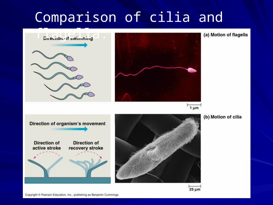
Comparison of cilia and flagella:

Cell walls are found in plant & fungal cells:

Learning Outcomes:
By the end of today’s lecture, you should be able to:
• Recognize the different kinds of images seen through different microscopes
• Identify the major parts and functions of prokaryotic and eukaryotic cells

Check for understanding…

Animal Cell: Plant Cell: