Embed Size (px)
Citation preview
RESEARCH Open Access
To gate or not to gate - dosimetricevaluation comparing Gated vs. ITV-basedmethodologies in stereotactic ablativebody radiotherapy (SABR) treatment oflung cancerJoshua Kim* , Qixue Wu, Bo Zhao, Ning Wen, Munther Ajlouni, Benjamin Movsas and Indrin J. Chetty
Abstract
Background: To compare retrospectively generated gated plans to conventional internal target volume(ITV)-based plans and to evaluate whether gated radiotherapy provides clinically relevant dosimetricimprovements to organs-at-risk (OARs).
Methods: Evaluation was performed of 150 stereotactic ablative radiotherapy treatment plans delivered to128 early-stage (T1-T3 (<5 cm)) NSCLC patients. To generate gated plans, original ITV-based plans werere-optimized and re-calculated on the end-exhale phase and using gated planning target volumes (PTV).Gated and ITV-based plans were produced for 3 × 18 Gy and 4 × 12 Gy fractionation regimens. Dosedifferences between gated and ITV-based plans were analyzed as a function of both three-dimensionalmotion and tumor volume. OARs were analyzed using RTOG and AAPM dose constraints.
Results: Differences between gated and ITV-based plans for all OAR indices were largest for the 3 × 18 Gy regimen.For this regimen, MLD differences calculated by subtracting the gated values from the ITV-based values (ITV vs. Gated)were 0.10 ± 0.56 Gy for peripheral island (N = 57), 0.16 ± 0.64 Gy for peripheral lung-wall seated (N = 57), and 0.10 ± 0.64 Gy for central tumors (N = 36). Variations in V20 were similarly low, with the greatest differences occurring inperipheral tumors (0.20 ± 1.17 %). Additionally, average differences (in 2Gy-equivalence) between ITV and gated lungindices fell well below clinical tolerance values for all fractionation regimens, with no clinically meaningful differencesobserved from the 4 × 12 Gy regimen and rarely for the 3 × 18 Gy regimen (<2 % of cases). Dosimetric differencesbetween gated and ITV-based methods did generally increase with increasing tumor motion and decreasing tumorvolume. Dose to ribs and bronchial tree were slightly higher in gated plans compared to ITV-based plans and slightlylower for esophagus, heart, spinal cord, and trachea.
Conclusions: Analysis of 150 SABR-based lung cancer treatment plans did not show a substantial benefit for thegating regimen when compared to ITV-based treatment plans. Small benefits were observed only for the largest tumormotion (exceeding 2 cm) and the high dose treatment regimen (3 × 18 Gy), though these benefits did not appear tobe clinically relevant.
Keywords: SABR, Gating, ITV-based planning, Treatment planning
* Correspondence: [email protected] of Radiation Oncology, Henry Ford Health System, 2799 W.Grand Blvd, Detroit, MI 48202, USA
© 2016 The Author(s). Open Access This article is distributed under the terms of the Creative Commons Attribution 4.0International License (http://creativecommons.org/licenses/by/4.0/), which permits unrestricted use, distribution, andreproduction in any medium, provided you give appropriate credit to the original author(s) and the source, provide a link tothe Creative Commons license, and indicate if changes were made. The Creative Commons Public Domain Dedication waiver(http://creativecommons.org/publicdomain/zero/1.0/) applies to the data made available in this article, unless otherwise stated.
Kim et al. Radiation Oncology (2016) 11:125 DOI 10.1186/s13014-016-0699-2
BackgroundStereotactic ablative radiotherapy (SABR), also calledstereotactic body radiation therapy, is a radiotherapytreatment method for delivering high dose in fewfractions (~1–5) [1]. It is essential that the high dosesused in SABR treatments be highly conformal to thetumor volume and delivered with high accuracy. Forlung cancer, SABR has been used as the primary treat-ment in prospective trials for medically inoperable, earlystage non-small-cell lung cancers (NSCLC) [2] and hasprovided much greater local tumor control after threeyears relative to conventionally fractionated radiotherapy(~90 % [3, 4] vs. <55 % [5]) leading to a higher overall3-year survival rate (~56 % [4] vs. 20–40 % [5, 6]).Due to respiratory motion of the tumor, immobilization
and motion management for lung cancer is important foraccurately defining and treating the tumor. Theimmobilization devices used are somewhat dependent onthe motion management strategy used. They come inmany forms including cradles that hold a polyurethanefoam that conforms to the patient’s body as it cures [7],cushions that conform to the patient’s contours and be-come rigid as air is evacuated (e.g. BodyFIX®) [7, 8], andabdominal compression plates that constrict abdominalmotion [9]. Several types of strategies have been intro-duced to mitigate the effects of motion. These techniquesinclude: motion encompassing methods, respiratory gat-ing, breath-hold control, forced compression approaches,and tumor-tracking [10]. Many of these methods employfour-dimensional computed tomography (4DCT), wherethe patients’ respiratory waveforms are monitored duringvery low pitch helical CT acquisitions [11–13]. Thesewaveforms are then used to retrospectively sort theprojections into a predetermined number of phases of thebreathing cycle, and the binned projections are used toreconstruct a CT of the patient at each phase. These phaseimages can be further used to generate derived imagessuch as the average, maximum intensity, and minimumintensity image sets. One type of motion-encompassingmethod involves the use of 4DCT phase and derived im-ages to define the full range of motion during respirationand to include that full range within an internal targetvolume (ITV) [14] (or alternatively using a mid-ventilationapproach with a MidV volume [15]), which is thenexpanded to the planning target volume (PTV) for treat-ment planning. Treatment planning is then typicallyperformed on the average CT. Respiratory gated radiother-apy involves monitoring an internal [16] (e.g. implantedfiducial markers) or external [17] (e.g. abdominal surfacemotion) surrogate for the tumor. The beam is activatedonly when the surrogate lies within some predefined gatingwindow on the waveform. This enables the use of thereconstructed image of only a single phase of the waveformand the use of smaller treatment planning margins that are
expected to allow for reduced dose delivered to surround-ing organs-at-risk (OARs). In tumor tracking modalities,real-time image guidance is employed to dynamicallymodify the beam delivery so that the radiation deliveredtracks with the motion of the target. This can be accom-plished through moving the linac itself through use of arobotic arm [18] or using the multi-leaf collimators (MLCs)to track with the tumor [19]. While the previous methodsallow for the patient to breath normally, breath-holdmethods seek to limit the tumor motion by only treatingduring the time patients are holding their breath [20–22].Forced compression methods mechanically limit the rangeof motion by using a plate to physically compress theabdomen during delivery [23]. Patients with inoperablelung tumors already suffer from compromised breathingand often suffer from comorbidities, and, therefore, only asubset are able to hold their breath for sufficiently longtimes for breath-hold methods. Similarly, some patients arenot able to handle forced compression of their abdomen,and accurate repositioning of the abdominal compressionplate is sometimes difficult.The American Association of Physicists in Medicine
(AAPM) Task Group 76 [10] recommends use of a5 mm motion threshold above which motion manage-ment is needed. In this study, respiratory gated treat-ment plans were retrospectively generated for a largecohort of SABR treatment plans (n = 150) that had beenplanned and treated using a motion encompassingmethod for managing tumor motion based on the use ofan internal target volume (ITV), which is the standardmethod employed in our clinic. Clinically relevantdosimetric indices between gated and ITV-based planswere compared to evaluate benefits of gating withparticular regard to normal lung tissue sparing.
MethodsPatientsRetrospective analysis was performed for 150 SABRplans that were each generated for individual tumorsand were used to treat 128 medically inoperable NSCLCpatients between 2010 and 2014. Patients with multipletumors presented either with synchronous or metachro-nous primary tumors or both. All tumors were early-stage (T1-T3 (<5 cm)) to match criteria outlined in thereport of Radiation Therapy Oncology Group (RTOG)0915 [24]. Of 128 patients, 57 were male and 71 werefemale, and median patient age was 73 years (range:32–96). Patients were immobilized using the BlueBAGBodyFIX® (Elekta, Stockholm, Sweden) immobilizationsystem. Median tumor volume was 10.9 cm3 (range:0.3–75.6 cm3). The distribution of tumors, sortedaccording to position relative to the proximal bron-chial tree and according to lobe, is given in Table 1.Thirty-six tumors were located centrally, defined as
Kim et al. Radiation Oncology (2016) 11:125 Page 2 of 11
being within 2 cm of the proximal bronchial tree [24]or mediastinal structures, and 114 lesions werelocated peripherally. Peripheral lesions were subdi-vided into “island” (n = 57) or lung wall-seated tumors(n = 57) using definitions given by Altman et al. [25].Island tumors are enclosed by lung parenchyma, andlung wall-seated tumors abut the lung wall. Tumorswere fairly evenly split between upper (n = 75) andlower (n = 65) lobes with 10 tumors located in theright middle lobe.
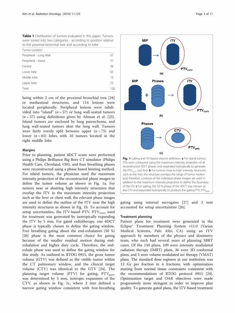
MarginsPrior to planning, patient 4DCT scans were performedusing a Philips Brilliance Big Bore CT simulator (PhilipsHealth Care, Cleveland, OH), and four breathing phaseswere reconstructed using a phase-based binning method.For island tumors, the physician used the maximumintensity projection of the reconstructed phase images todefine the tumor volume as shown in Fig. 1a. Fortumors near or abutting high intensity structures thatoverlap the ITV in the maximum intensity projectionsuch as the liver or chest wall, the relevant phase imagesare used to define the outline of the ITV near the highintensity structures as shown in Fig. 1b. To account forsetup uncertainties, the ITV-based PTV, PTVconv, usedfor treatment was generated by isotropically expandingthe ITV by 5 mm. For gated radiotherapy, one 4DCTphase is typically chosen to define the gating window.Free breathing gating about the end-exhalation (50 %)[26] phase is the most common choice for gatingbecause of the smaller residual motion during end-exhalation and higher duty cycle. Therefore, the end-exhale phase was used to define the gating window forthis study. As outlined in RTOG 0915, the gross tumorvolume (GTV) was defined as the visible tumor withinthe CT pulmonary window, and the clinical targetvolume (CTV) was identical to the GTV [24]. Theplanning target volume (PTV) for gating, PTVgate,was determined by 5 mm, isotropic expansion of theCTV, as shown in Fig. 1c, where 2 mm defined anarrow gating window consistent with free-breathing
gating using internal surrogates [27] and 3 mmaccounted for setup uncertainties [26].
Treatment planningPatient plans for treatment were generated in theEclipse® Treatment Planning System v11.0 (VarianMedical Systems, Palo Alto, CA) using an ITVapproach by members of the physics and dosimetryteam, who each had several years of planning SBRTcases. Of the 150 plans, 109 were intensity modulatedradiation therapy (IMRT) plans, 36 were 3D conformalplans, and 5 were volume modulated arc therapy (VMAT)plans. The standard dose regimen at our institution was12 Gy per fraction in 4 fractions, with optimizationstarting from normal tissue constraints consistent withthe recommendations of RTOG protocol 0915 [24].Optimization target and OAR objectives were madeprogressively more stringent in order to improve planquality. To generate gated plans, the ITV-based treatment
Table 1 Distribution of tumors evaluated in this paper. Tumorswere sorted into two categories - according to position relativeto the proximal bronchial tree and according to lobe
Tumor Location
Peripheral - Lung Wall 57
Peripheral - Island 57
Central 36
Lower lobe 65
Middle lobe 10
Upper lobe 75
Total 150
Fig. 1 Gating and ITV-based volume definition. a For island tumors,ITVs were contoured using the maximum intensity projection of allreconstructed 4DCT phases and expanded isotropically to generatethe PTVconv (red line). b For tumors close to high intensity structuressuch as the liver, the structure overlaps the range of tumor motionand, therefore, contours of the individual phase images are used inaddition to the maximum intensity projection to define the boundaryof the ITV. c For gating, the 50 % phase of the 4DCT was chosen asthe CTV and expanded isotropically to produce the gating PTV, PTVgate
Kim et al. Radiation Oncology (2016) 11:125 Page 3 of 11
plan was copied, and the isocenter for the new gatingplane was moved to the geometric center of the PTVgate
contour as determined in Eclipse. Treatment fields werethen realigned to the new isocenter position. Optimizationof the new PTVgate plan was performed beginning withconstraints derived from RTOG 0915 with more stringentlung constraints introduced later that were considered tobe appropriate due to the use of the gating window. Aswith the conventional plans, the OAR and PTV objectivesfor the gated plans were made more stringent in a waythat was unique to each patient in order to minimizeOAR dose while maintaining plan quality. Dose was thenrecalculated based on the optimized fluence. The end-exhalation phase of the 4DCT was used as the treatmentplanning CT. Another commonly used high dose fraction-ation regimen explored in this study was 3 fractions of18 Gy per fraction. To evaluate the 3 × 18 Gy schedule,original clinical treatment plans were modified withrespect to fraction number and dose delivered perfraction. Modified plans were re-optimized using scaledtarget volume objectives and OAR constraints derivedfrom the AAPM Task Group 101 [28] report withoptimization objectives that were made more stringentjust as with the 4 × 12 Gy schedule. A single physicist withexperience in generating SBRT plans was responsible forboth optimizing modified conventional plans and generat-ing the gated plans. To establish plan quality independentof the planner, all plans were required to achieve standard-ized constraints with respect to plan conformity, targetcoverage, dose heterogeneity, and dose to OARs.
Evaluation metricsThe main focus of the evaluation is lung toxicity. Ana-lysis for lung toxicity will primarily be using mean lungdose (MLD) and lung volume percentage receiving dosesof >20 Gy (V20). MLD [29] and V20 [24] have beenshown to correlate well with incidence of radiation
pneumonitis in conventional fractionation regimens.Trends between the total tumor motion (defined as themagnitude of the three-dimensional difference inposition between the tumor’s center of mass at end-inhalation (0 % phase) and end-exhalation (50 % phase))and the calculated difference between gated and ITV-based plans were investigated. For each methodology(ITV vs. Gated), lung was defined to be the total lungminus the PTV used for each method.Additionally, we wanted to determine how the use of
gating-based margins would affect doses calculated forother OARs. For each plan, only those OARs that weredetermined by the physician to be proximate enough tothe tumor to receive significant dose were contouredand then included in the study (e.g. heart was contouredfor central lesions, ribs for peripheral tumors). For thispatient population, the most commonly contoured non-lung OARs were spinal cord (n = 133), heart (n = 80),esophagus (n = 70), and ribs (n = 53). Additionally, thebronchial tree and trachea were contoured and analyzedfor thirteen centrally located tumors. Delivered doses aswell as doses calculated using gating margins for bothregimens were compared to recommended values ofRTOG 0915 and AAPM TG101.For all metrics, a two-tailed paired t-test was used
to evaluate the significance of the results from usingthe two planning methodologies on the same patientpopulation. A difference was considered to be statisti-cally significant if the p-value was ≤0.05.
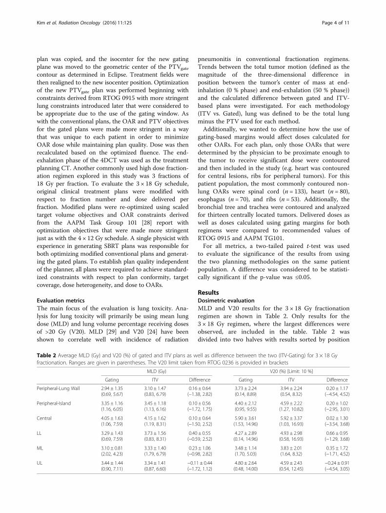
ResultsDosimetric evaluationMLD and V20 results for the 3 × 18 Gy fractionationregimen are shown in Table 2. Only results for the3 × 18 Gy regimen, where the largest differences wereobserved, are included in the table. Table 2 wasdivided into two halves with results sorted by position
Table 2 Average MLD (Gy) and V20 (%) of gated and ITV plans as well as difference between the two (ITV-Gating) for 3 × 18 Gyfractionation. Ranges are given in parentheses. The V20 limit taken from RTOG 0236 is provided in brackets
MLD (Gy) V20 (%) [Limit: 10 %]
Gating ITV Difference Gating ITV Difference
Peripheral-Lung Wall 2.94 ± 1.35(0.69, 5.67)
3.10 ± 1.47(0.83, 6.79)
0.16 ± 0.64(−1.38, 2.82)
3.73 ± 2.24(0.14, 8.89)
3.94 ± 2.24(0.54, 8.32)
0.20 ± 1.17(−4.54, 4.52)
Peripheral-Island 3.35 ± 1.16(1.16, 6.05)
3.45 ± 1.18(1.13, 6.16)
0.10 ± 0.56(−1.72, 1.75)
4.40 ± 2.12(0.95, 9.55)
4.59 ± 2.22(1.27, 10.82)
0.20 ± 1.02(−2.95, 3.01)
Central 4.05 ± 1.63(1.06, 7.59)
4.15 ± 1.62(1.19, 8.31)
0.10 ± 0.64(−1.50, 2.52)
5.90 ± 3.61(1.53, 14.96)
5.92 ± 3.37(1.03, 16.93)
0.02 ± 1.30(−3.54, 3.68)
LL 3.29 ± 1.43(0.69, 7.59)
3.73 ± 1.56(0.83, 8.31)
0.40 ± 0.55(−0.59, 2.52)
4.27 ± 2.89(0.14, 14.96)
4.93 ± 2.98(0.58, 16.93)
0.66 ± 0.95(−1.29, 3.68)
ML 3.10 ± 0.81(2.02, 4.23)
3.33 ± 1.40(1.79, 6.79)
0.23 ± 1.06(−0.98, 2.82)
3.48 ± 1.14(1.70, 5.03)
3.83 ± 2.01(1.64, 8.32)
0.35 ± 1.72(−1.71, 4.52)
UL 3.44 ± 1.44(0.90, 7.11)
3.34 ± 1.41(0.87, 6.60)
−0.11 ± 0.44(−1.72, 1.12)
4.80 ± 2.64(0.48, 14.00)
4.59 ± 2.43(0.54, 12.45)
−0.24 ± 0.91(−4.54, 3.05)
Kim et al. Radiation Oncology (2016) 11:125 Page 4 of 11
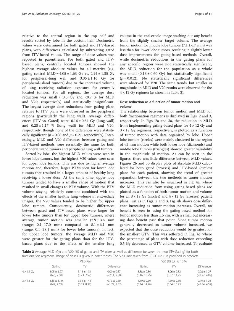
relative to the central region in the top half andresults sorted by lobe in the bottom half. Dosimetricvalues were determined for both gated and ITV-basedplans, with differences calculated by subtracting gatedfrom ITV-based values. The range of dose values wasreported in parentheses. For both gated and ITV-based plans, centrally located tumors showed thehighest average absolute values for all metrics (e.g.gating central MLD = 4.05 ± 1.63 Gy vs. 2.94 ± 1.35 Gyfor peripheral-lung wall and 3.35 ± 1.16 Gy forperipheral-island tumors) due to the increased volumeof lung receiving radiation exposure for centrallylocated tumors. For all regions, the average dosereduction was small (<0.5 Gy and <0.7 % for MLDand V20, respectively) and statistically insignificant.The largest average dose reductions from gating plansrelative to ITV plans were observed in the peripheralregions (particularly the lung wall). Average differ-ences (ITV vs. Gated) were: 0.16 ± 0.64 Gy (lung wall)and 0.20 ± 1.17 % (lung wall) for MLD and V20,respectively, though none of the differences were statisti-cally significant (p = 0.08 and p = 0.21, respectively). Inter-estingly, MLD and V20 differences between gating andITV-based methods were essentially the same for bothperipheral island tumors and peripheral lung wall tumors.Sorted by lobe, the highest MLD values were seen in
lower lobe tumors, but the highest V20 values were seenfor upper lobe tumors. This was due to higher averagemotion and, therefore, larger PTVs seen for lower lobetumors that resulted in a larger amount of healthy lungreceiving a lower dose. At the same time, upper lobetumors tended to have a smaller range of motion thatresulted in small changes to PTV volume. With the PTVvolume staying relatively constant combined with theeffects of the smaller healthy lung volume in end-exhaleimages, the V20 values tended to be higher for upperlobe tumors. Consequently, dosimetric differencesbetween gated and ITV-based plans were larger forlower lobe tumors than for upper lobe tumors, whereaverage tumor motion was smaller (2.9 ± 3.4 mm(range: 0.1–17.0 mm) compared to 8.1 ± 6.1 mm(range: 0.1–28.1 mm) for lower lobe tumors). In fact,for upper lobe tumors, the average MLD and V20were greater for the gating plans than for the ITV-based plans due to the effect of the smaller lung
volume in the end-exhale image washing out any benefitfrom the slightly smaller target volume. The averagetumor motion for middle lobe tumors (7.1 ± 6.7 mm) wasless than for lower lobe tumors, resulting in slightly lowerdose improvements for gating-based methods. Overall,while dosimetric reductions in the gating plans forany specific region were not statistically significant,the MLD reduction for the population as a wholewas small (0.13 ± 0.60 Gy) but statistically significant(p = 0.012). No statistically significant differenceswere observed for V20. The same trends, but smaller inmagnitude, in MLD and V20 results were observed for the4 × 12 Gy regimen (as shown in Table 3).
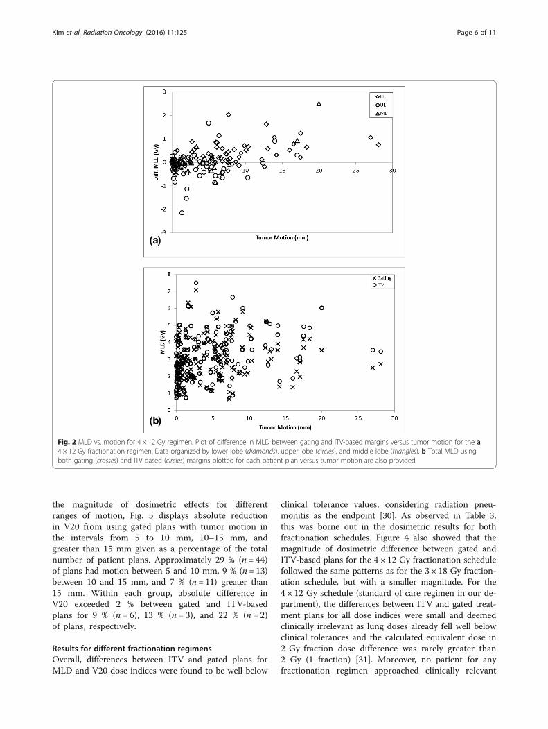
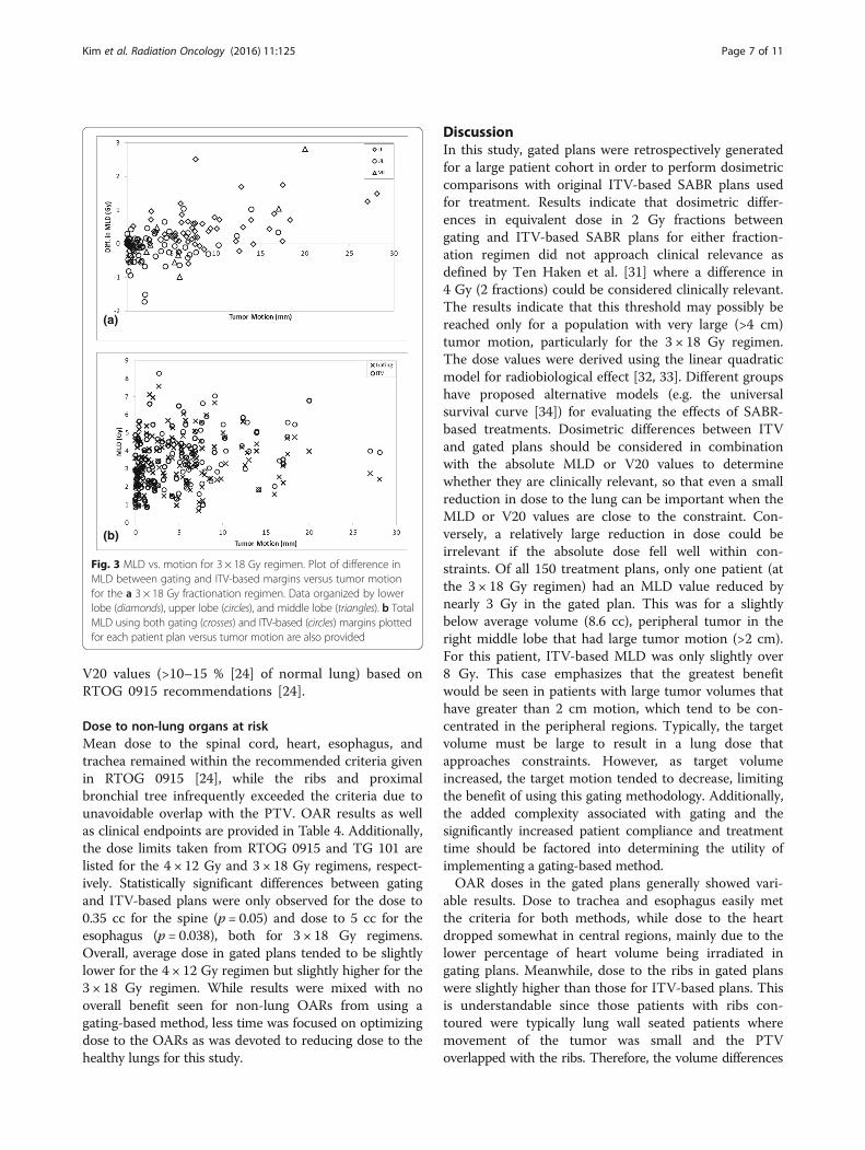
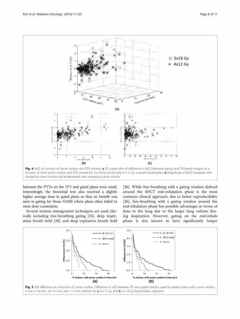
Dose reduction as a function of tumor motion andvolumeThe relationship between tumor motion and MLD forboth fractionation regimens is displayed in Figs. 2 and 3,respectively. In Figs. 2a and 3a, the reduction in MLDfrom implementing gating-based plans for 4 × 12 Gy and3 × 18 Gy regimens, respectively, is plotted as a functionof tumor motion with data organized by lobe. Upperlobe tumors (circles) were mainly clustered in the regionof <5 mm motion while both lower lobe (diamonds) andmiddle lobe tumors (triangles) showed greater variabilityin the magnitude of motion. As can be seen in thefigures, there was little difference between MLD values.Figures 2b and 3b display plots of absolute MLD calcu-lated for both gated (crosses) and ITV-based (circles)plans for each patient, showing the trend of greaterseparation between the two methods as tumor motionincreases. This can also be visualized in Fig. 4a, wherethe MLD reduction from using gating-based plans areplotted as a function of both tumor motion and volumefor all 3 × 18 Gy (circles) and 4 × 12 Gy (crosses) patientplans. Just as in Figs. 2 and 3, Fig. 4b shows dose differ-ence increasing as tumor motion increases. Overall, nobenefit is seen in using the gating-based method fortumor motion less than 1.5 cm, with a small but increas-ing dose benefit past that point. Since tumor motiongenerally decreased as tumor volume increased, weexpected that the dose reduction would be greatest forthe smallest GTV. This was reflected in Fig. 4c wherethe percentage of plans with dose reduction exceeding0.5 Gy decreased as GTV volume increased. To evaluate
Table 3 Average MLD (Gy) and V20 (%) of gated and ITV plans as well as difference between the two (ITV-Gating) for bothfractionation regimens. Range of doses is given in parentheses. The V20 limit taken from RTOG 0236 is provided in brackets
MLD (Gy) V20 (%) [Limit: 10 %]
Gating ITV Difference Gating ITV Difference
4 × 12 Gy 3.05 ± 1.27(0.65, 7.08)
3.16 ± 1.34(0.73, 7.52)
0.09 ± 0.57(−2.14, 2.50)
3.88 ± 2.39(0.46, 13.75)
3.96 ± 2.32(0.37, 14.75)
0.08 ± 1.07(−3.27, 4.09)
3 × 18 Gy 3.35 ± 1.40(0.69, 7.59)
3.51 ± 1.48(0.83, 8.31)
0.13 ± 0.60(−1.72, 2.82)
4.49 ± 2.69(0.14, 14.96)
4.69 ± 2.66(0.54, 16.93)
0.19 ± 1.08(−3.54, 4.52)
Kim et al. Radiation Oncology (2016) 11:125 Page 5 of 11
the magnitude of dosimetric effects for differentranges of motion, Fig. 5 displays absolute reductionin V20 from using gated plans with tumor motion inthe intervals from 5 to 10 mm, 10–15 mm, andgreater than 15 mm given as a percentage of the totalnumber of patient plans. Approximately 29 % (n = 44)of plans had motion between 5 and 10 mm, 9 % (n = 13)between 10 and 15 mm, and 7 % (n = 11) greater than15 mm. Within each group, absolute difference inV20 exceeded 2 % between gated and ITV-basedplans for 9 % (n = 6), 13 % (n = 3), and 22 % (n = 2)of plans, respectively.
Results for different fractionation regimensOverall, differences between ITV and gated plans forMLD and V20 dose indices were found to be well below
clinical tolerance values, considering radiation pneu-monitis as the endpoint [30]. As observed in Table 3,this was borne out in the dosimetric results for bothfractionation schedules. Figure 4 also showed that themagnitude of dosimetric difference between gated andITV-based plans for the 4 × 12 Gy fractionation schedulefollowed the same patterns as for the 3 × 18 Gy fraction-ation schedule, but with a smaller magnitude. For the4 × 12 Gy schedule (standard of care regimen in our de-partment), the differences between ITV and gated treat-ment plans for all dose indices were small and deemedclinically irrelevant as lung doses already fell well belowclinical tolerances and the calculated equivalent dose in2 Gy fraction dose difference was rarely greater than2 Gy (1 fraction) [31]. Moreover, no patient for anyfractionation regimen approached clinically relevant
(a)
(b)
Fig. 2 MLD vs. motion for 4 × 12 Gy regimen. Plot of difference in MLD between gating and ITV-based margins versus tumor motion for the a4 × 12 Gy fractionation regimen. Data organized by lower lobe (diamonds), upper lobe (circles), and middle lobe (triangles). b Total MLD usingboth gating (crosses) and ITV-based (circles) margins plotted for each patient plan versus tumor motion are also provided
Kim et al. Radiation Oncology (2016) 11:125 Page 6 of 11
V20 values (>10–15 % [24] of normal lung) based onRTOG 0915 recommendations [24].
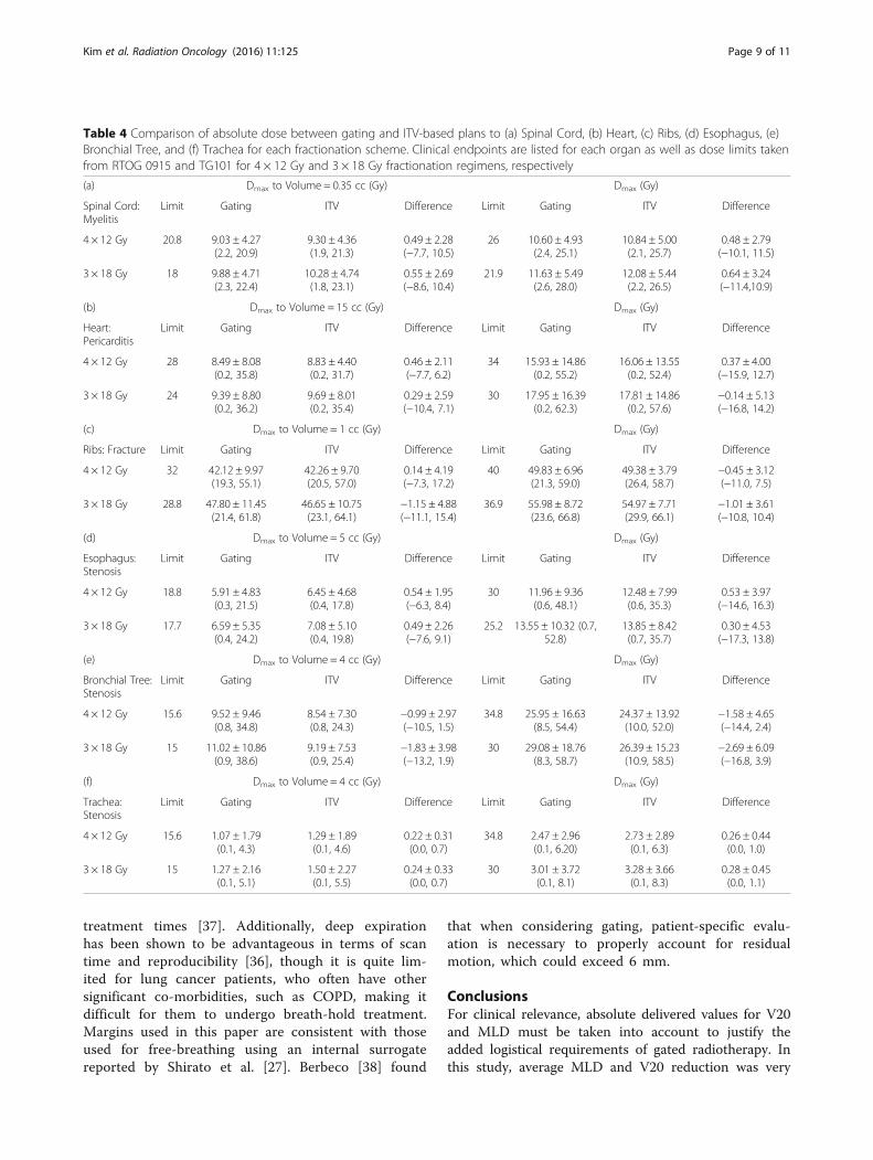
Dose to non-lung organs at riskMean dose to the spinal cord, heart, esophagus, andtrachea remained within the recommended criteria givenin RTOG 0915 [24], while the ribs and proximalbronchial tree infrequently exceeded the criteria due tounavoidable overlap with the PTV. OAR results as wellas clinical endpoints are provided in Table 4. Additionally,the dose limits taken from RTOG 0915 and TG 101 arelisted for the 4 × 12 Gy and 3 × 18 Gy regimens, respect-ively. Statistically significant differences between gatingand ITV-based plans were only observed for the dose to0.35 cc for the spine (p = 0.05) and dose to 5 cc for theesophagus (p = 0.038), both for 3 × 18 Gy regimens.Overall, average dose in gated plans tended to be slightlylower for the 4 × 12 Gy regimen but slightly higher for the3 × 18 Gy regimen. While results were mixed with nooverall benefit seen for non-lung OARs from using agating-based method, less time was focused on optimizingdose to the OARs as was devoted to reducing dose to thehealthy lungs for this study.
DiscussionIn this study, gated plans were retrospectively generatedfor a large patient cohort in order to perform dosimetriccomparisons with original ITV-based SABR plans usedfor treatment. Results indicate that dosimetric differ-ences in equivalent dose in 2 Gy fractions betweengating and ITV-based SABR plans for either fraction-ation regimen did not approach clinical relevance asdefined by Ten Haken et al. [31] where a difference in4 Gy (2 fractions) could be considered clinically relevant.The results indicate that this threshold may possibly bereached only for a population with very large (>4 cm)tumor motion, particularly for the 3 × 18 Gy regimen.The dose values were derived using the linear quadraticmodel for radiobiological effect [32, 33]. Different groupshave proposed alternative models (e.g. the universalsurvival curve [34]) for evaluating the effects of SABR-based treatments. Dosimetric differences between ITVand gated plans should be considered in combinationwith the absolute MLD or V20 values to determinewhether they are clinically relevant, so that even a smallreduction in dose to the lung can be important when theMLD or V20 values are close to the constraint. Con-versely, a relatively large reduction in dose could beirrelevant if the absolute dose fell well within con-straints. Of all 150 treatment plans, only one patient (atthe 3 × 18 Gy regimen) had an MLD value reduced bynearly 3 Gy in the gated plan. This was for a slightlybelow average volume (8.6 cc), peripheral tumor in theright middle lobe that had large tumor motion (>2 cm).For this patient, ITV-based MLD was only slightly over8 Gy. This case emphasizes that the greatest benefitwould be seen in patients with large tumor volumes thathave greater than 2 cm motion, which tend to be con-centrated in the peripheral regions. Typically, the targetvolume must be large to result in a lung dose thatapproaches constraints. However, as target volumeincreased, the target motion tended to decrease, limitingthe benefit of using this gating methodology. Additionally,the added complexity associated with gating and thesignificantly increased patient compliance and treatmenttime should be factored into determining the utility ofimplementing a gating-based method.OAR doses in the gated plans generally showed vari-
able results. Dose to trachea and esophagus easily metthe criteria for both methods, while dose to the heartdropped somewhat in central regions, mainly due to thelower percentage of heart volume being irradiated ingating plans. Meanwhile, dose to the ribs in gated planswere slightly higher than those for ITV-based plans. Thisis understandable since those patients with ribs con-toured were typically lung wall seated patients wheremovement of the tumor was small and the PTVoverlapped with the ribs. Therefore, the volume differences
(a)
(b)
Fig. 3 MLD vs. motion for 3 × 18 Gy regimen. Plot of difference inMLD between gating and ITV-based margins versus tumor motionfor the a 3 × 18 Gy fractionation regimen. Data organized by lowerlobe (diamonds), upper lobe (circles), and middle lobe (triangles). b TotalMLD using both gating (crosses) and ITV-based (circles) margins plottedfor each patient plan versus tumor motion are also provided
Kim et al. Radiation Oncology (2016) 11:125 Page 7 of 11
between the PTVs on the ITV and gated plans were small.Interestingly, the bronchial tree also received a slightlyhigher average dose in gated plans so that no benefit wasseen in gating for those OARS where plans often failed tomeet dose constraints.Several motion management techniques are used clin-
ically including free-breathing gating [35], deep inspir-ation breath hold [20], and deep expiration breath hold
[36]. While free-breathing with a gating window definedaround the 4DCT end-exhalation phase is the mostcommon clinical approach, due to better reproducibility[26], free-breathing with a gating window around theend-inhalation phase has possible advantages in terms ofdose to the lung due to the larger lung volume dur-ing inspiration. However, gating on the end-inhalephase is also known to have significantly longer
Fig. 4 MLD as function of tumor motion and GTV volume. a 3D scatter plot of difference in MLD between gating and ITV-based margins as afunction of both tumor motion and GTV volume for 3 × 18 Gy (circles) and 4 × 12 Gy (crosses) fractionation. b Magnitude of MLD increased withincreasing tumor motion and c decreased with increasing tumor volume
(a) (b)
Fig. 5 V20 difference as a function of tumor motion. Difference in V20 between ITV and gated margins used for patient plans with tumor motionin the 5–10 mm, 10–15 mm, and >15 mm intervals for a 4 × 12 Gy and b 3 × 18 Gy fractionation regimens
Kim et al. Radiation Oncology (2016) 11:125 Page 8 of 11
treatment times [37]. Additionally, deep expirationhas been shown to be advantageous in terms of scantime and reproducibility [36], though it is quite lim-ited for lung cancer patients, who often have othersignificant co-morbidities, such as COPD, making itdifficult for them to undergo breath-hold treatment.Margins used in this paper are consistent with thoseused for free-breathing using an internal surrogatereported by Shirato et al. [27]. Berbeco [38] found
that when considering gating, patient-specific evalu-ation is necessary to properly account for residualmotion, which could exceed 6 mm.
ConclusionsFor clinical relevance, absolute delivered values for V20and MLD must be taken into account to justify theadded logistical requirements of gated radiotherapy. Inthis study, average MLD and V20 reduction was very
Table 4 Comparison of absolute dose between gating and ITV-based plans to (a) Spinal Cord, (b) Heart, (c) Ribs, (d) Esophagus, (e)Bronchial Tree, and (f) Trachea for each fractionation scheme. Clinical endpoints are listed for each organ as well as dose limits takenfrom RTOG 0915 and TG101 for 4 × 12 Gy and 3 × 18 Gy fractionation regimens, respectively
(a) Dmax to Volume = 0.35 cc (Gy) Dmax (Gy)
Spinal Cord:Myelitis
Limit Gating ITV Difference Limit Gating ITV Difference
4 × 12 Gy 20.8 9.03 ± 4.27(2.2, 20.9)
9.30 ± 4.36(1.9, 21.3)
0.49 ± 2.28(−7.7, 10.5)
26 10.60 ± 4.93(2.4, 25.1)
10.84 ± 5.00(2.1, 25.7)
0.48 ± 2.79(−10.1, 11.5)
3 × 18 Gy 18 9.88 ± 4.71(2.3, 22.4)
10.28 ± 4.74(1.8, 23.1)
0.55 ± 2.69(−8.6, 10.4)
21.9 11.63 ± 5.49(2.6, 28.0)
12.08 ± 5.44(2.2, 26.5)
0.64 ± 3.24(−11.4,10.9)
(b) Dmax to Volume = 15 cc (Gy) Dmax (Gy)
Heart:Pericarditis
Limit Gating ITV Difference Limit Gating ITV Difference
4 × 12 Gy 28 8.49 ± 8.08(0.2, 35.8)
8.83 ± 4.40(0.2, 31.7)
0.46 ± 2.11(−7.7, 6.2)
34 15.93 ± 14.86(0.2, 55.2)
16.06 ± 13.55(0.2, 52.4)
0.37 ± 4.00(−15.9, 12.7)
3 × 18 Gy 24 9.39 ± 8.80(0.2, 36.2)
9.69 ± 8.01(0.2, 35.4)
0.29 ± 2.59(−10.4, 7.1)
30 17.95 ± 16.39(0.2, 62.3)
17.81 ± 14.86(0.2, 57.6)
−0.14 ± 5.13(−16.8, 14.2)
(c) Dmax to Volume = 1 cc (Gy) Dmax (Gy)
Ribs: Fracture Limit Gating ITV Difference Limit Gating ITV Difference
4 × 12 Gy 32 42.12 ± 9.97(19.3, 55.1)
42.26 ± 9.70(20.5, 57.0)
0.14 ± 4.19(−7.3, 17.2)
40 49.83 ± 6.96(21.3, 59.0)
49.38 ± 3.79(26.4, 58.7)
−0.45 ± 3.12(−11.0, 7.5)
3 × 18 Gy 28.8 47.80 ± 11.45(21.4, 61.8)
46.65 ± 10.75(23.1, 64.1)
−1.15 ± 4.88(−11.1, 15.4)
36.9 55.98 ± 8.72(23.6, 66.8)
54.97 ± 7.71(29.9, 66.1)
−1.01 ± 3.61(−10.8, 10.4)
(d) Dmax to Volume = 5 cc (Gy) Dmax (Gy)
Esophagus:Stenosis
Limit Gating ITV Difference Limit Gating ITV Difference
4 × 12 Gy 18.8 5.91 ± 4.83(0.3, 21.5)
6.45 ± 4.68(0.4, 17.8)
0.54 ± 1.95(−6.3, 8.4)
30 11.96 ± 9.36(0.6, 48.1)
12.48 ± 7.99(0.6, 35.3)
0.53 ± 3.97(−14.6, 16.3)
3 × 18 Gy 17.7 6.59 ± 5.35(0.4, 24.2)
7.08 ± 5.10(0.4, 19.8)
0.49 ± 2.26(−7.6, 9.1)
25.2 13.55 ± 10.32 (0.7,52.8)
13.85 ± 8.42(0.7, 35.7)
0.30 ± 4.53(−17.3, 13.8)
(e) Dmax to Volume = 4 cc (Gy) Dmax (Gy)
Bronchial Tree:Stenosis
Limit Gating ITV Difference Limit Gating ITV Difference
4 × 12 Gy 15.6 9.52 ± 9.46(0.8, 34.8)
8.54 ± 7.30(0.8, 24.3)
−0.99 ± 2.97(−10.5, 1.5)
34.8 25.95 ± 16.63(8.5, 54.4)
24.37 ± 13.92(10.0, 52.0)
−1.58 ± 4.65(−14.4, 2.4)
3 × 18 Gy 15 11.02 ± 10.86(0.9, 38.6)
9.19 ± 7.53(0.9, 25.4)
−1.83 ± 3.98(−13.2, 1.9)
30 29.08 ± 18.76(8.3, 58.7)
26.39 ± 15.23(10.9, 58.5)
−2.69 ± 6.09(−16.8, 3.9)
(f) Dmax to Volume = 4 cc (Gy) Dmax (Gy)
Trachea:Stenosis
Limit Gating ITV Difference Limit Gating ITV Difference
4 × 12 Gy 15.6 1.07 ± 1.79(0.1, 4.3)
1.29 ± 1.89(0.1, 4.6)
0.22 ± 0.31(0.0, 0.7)
34.8 2.47 ± 2.96(0.1, 6.20)
2.73 ± 2.89(0.1, 6.3)
0.26 ± 0.44(0.0, 1.0)
3 × 18 Gy 15 1.27 ± 2.16(0.1, 5.1)
1.50 ± 2.27(0.1, 5.5)
0.24 ± 0.33(0.0, 0.7)
30 3.01 ± 3.72(0.1, 8.1)
3.28 ± 3.66(0.1, 8.3)
0.28 ± 0.45(0.0, 1.1)
Kim et al. Radiation Oncology (2016) 11:125 Page 9 of 11
small (~0.20 Gy and ~0.3 %, respectively) with only afew ITV-based plans where MLD reduction exceededeven 2 Gy. Few of these patients had total MLD or V20values that approached dosimetric constraints. Differ-ences were even smaller for the 4 × 12 Gy regimen. OARresults did not show meaningful improvement in gatedplans. This is especially true for those sites (ribs andbronchial tree) most likely to exceed dose constraintswhere average dose to those sites increased compared toITV-based plans. For tumors with motion of 2 cm orless, which is the range of tumors observed in this study,no significant dose reduction was observed by imple-menting a gating-based method.
AbbreviationsAAPM: American Association of Physicists in Medicine; CTV: Clinical targetvolume; GTV: Gross tumor volume; IMRT: Intensity modulated radiationtherapy; ITV: Internal target volume; MLD: Mean lung dose; NSCLC: Nonsmall cell lung cancer; OAR: Organ at risk; PTV: Planning target volume;PTVgate: Planning target volume for gating; RTOG: Radiation TherapyOncology Group; SABR: Stereotactic ablative body radiotherapy;V20: Percent volume receiving 20 Gy; VMAT: Volume modulated arctherapy
AcknowledgementsNot applicable.
FundingResearch partially supported by Varian Medical Systems, Palo Alto, CA. VarianMedical Systems did not play a role in the design or execution of the studyor writing of the manuscript.
Availability of data and materialsThe data in this study is not shared since it contains information obtainedpartially under the funding of a private industry grant and due to liabilityrelated to sharing information regarding patient treatments.
Authors’ contributionsJK, NW, and IJC were involved in the conceptual design of the project. JKand QW were active in the modification of treatment plans, contouring ofgating targets and modification of OAR contours for gating plans,generation and optimization of gating plans, and analyzing data. MA and BMwere involved in patient care, OAR and target contouring, and plan approval.BZ was active in developing methods to be used for sorting, processing, andanalyzing data. All authors contributed to writing, editing, and giving finalapproval to the manuscript.
Competing interestsResearch partially supported by Varian Medical Systems, Palo Alto, CA.
Consent for publicationNot applicable.
Ethics approval and consent to participateAll data was collected and analyzed as part of an institutional review boardapproved study.
Received: 29 June 2016 Accepted: 10 September 2016
References1. Papiez L, Timmerman R, DesRosiers C, Randall M. Extracranial stereotactic
radioablation: physical principles. Acta Oncol. 2003;42:882–94.2. Timmerman RD, Kavanagh BD, Cho LC, Papiez L, Xing L. Stereotactic body
radiation therapy in multiple organ sites. J Clin Oncol. 2007;25:947–52.
3. Timmerman R, Paulus R, Galvin J, Michalski J, Straube W, et al. Stereotacticbody radiation therapy for inoperable early stage lung cancer. JAMA.2010;303:1070–6.
4. Zhang J, Yang F, Li B, Li H, Liu J, et al. Which is the optimal biologicallyeffective dose of stereotactic body radiotherapy for Stage I non-small-celllung cancer? A meta-analysis. Int J Radiat Oncol Biol Phys. 2011;81:e305–16.
5. Kaskowitz L, Graham MV, Emami B, Halverson KJ, Rush C. Radiation therapyalone for stage I non-small cell lung cancer. Int J Radiat Oncol Biol Phys.1993;27:517–23.
6. Gauden S, Ramsay J, Tripcony L. The curative treatment by radiotherapyalone of stage I non-small cell carcinoma of the lung. Chest.1995;108:1278–82.
7. Daly ME, Perks JR, Chen AM. Patterns-of-care for thoracic sterotactic bodyradiotherapy among practicing radiation oncologists in the United States.J Thorac Oncol. 2013;8:202–7.
8. Sio TT, Jensen AR, Miller RC, Santos LE F de l, Hallemeier CL, et al. Influenceof patient’s physiologic factors and immobilization choice with stereotacticbody radiotherapy for upper lung tumors. J Appl Clin Med Phys.2014;15:4931.
9. Rasheed A, Jabbour SK, Rosenberg S, Patel A, Goyal S, et al. Motion andvolumetric change as demonstrated by 4DCT: The effects of abdominalcompression on the GTV, lungs, and heart in lung cancer patients. PractRadiat Oncol. 2015. epub ahead of print; S1879-8500(15)00454-3.
10. Keall PJ, Mageras GS, Balter JM, Emery RS, Forster KM, et al. Themanagement of respiratory motion in radiation oncology report of AAPMTask Group 76. Med Phys. 2006;33:3874–900.
11. Vedam SS, Keall PJ, Kini VR, Mostafavi H, Shukla HP, et al. Acquiring a four-dimensional computed tomography dataset using an external respiratorysignal. Phys Med Biol. 2003;48:45–62.
12. Ford EC, Mageras GS, Yorke E, Ling CC. Respiration-correlated spiral CT: Amethod of measuring respiratory-induced anatomic motion for radiationtreatment planning. Med Phys. 2003;30:88–97.
13. Keall P. 4-dimensional computed tomography imaging and treatmentplanning. Semin Radiat Oncol. 2004;14:81–90.
14. Ge H, Cai H, Kelsey CR, Yin FF. Quantification and minimization ofuncertainties of internal target volume for stereotactic body radiationtherapy of lung cancer. Int J Radiat Oncol Biol Phys. 2013;85:438–43.
15. Peulen H, Belderbos J, Rossi M, Sonke JJ. Mid-ventilation based PTV marginsin Stereotactic Body Radiotherapy (SBRT): a clinical evaluation. RadiotherOncol. 2014;110:511–6.
16. Imura M, Yamazaki K, Shirato H, Onimaru R, Fujino M, et al. Insertion andfixation of fiducial markers for setup and tracking of lung tumors inradiotherapy. Int J Radiat Oncol Biol Phys. 2005;63:1442–7.
17. Jiang SB. Technical aspects of image-guided respiration-gated radiationtherapy. Med Dosim. 2006;31:141–51.
18. Schweikard A, Shiomi H, Adler J. Respiration tracking in radiosurgery. MedPhys. 2004;31:2738–41.
19. Keall PJ, Kini V, Vedam SS, Mohan R. Motion adaptive x-ray therapy: Afeasibility study. Phys Med Biol. 2001;46:1–10.
20. Hanley J, Debois MM, Mah D, Mageras GS, Raben A, et al. Deep inspirationbreath-hold technique for lung tumors: The potential value of targetimmobilization and reduced lung density in dose escalation. Int J RadiatOncol Biol Phys. 1999;45:603–11.
21. Stromberg JS, Sharpe MB, Kim LH, Kini VR, Jaffray DA, et al. Active breathingcontrol (ABC) for Hodgkin’s disease: reduction in normal tissue irradiationwith deep inspiration and implications for treatment. Int J Radiat Oncol BiolPhys. 2000;48:797–806.
22. Kim DJ, Murray BR, Halperin R, Roa WH. Held-breath self-gating techniquefor radiotherapy of non-small-cell lung cancer: a feasibility study. Int J RadiatOncol Biol Phys. 2001;49:43–9.
23. Lax I, Blomgren H, Naslund I, Svanstrom R. Stereotactic radiotherapy ofmalignancies in the abdomen, Methodological aspects. Acta Oncol.1994;33:677–83.
24. Videtic GM, Hu C, Singh A, Chang JY, Parker W, et al. Radiation therapyoncology group (RTOG) protocol 0915: a randomized phase 2 studycomparing 2 stereotactic body radiation therapy (SBRT) schedules formedically inoperable patients with Stage I peripheral non-small cell lungcancer. Int J Radiat Oncol Biol Phys. 2013;87:S3.
25. Altman MB, Jin JY, Kim S, Wen N, Liu D, et al. Practical methods forimproving dose distributions in Monte Carlo-based IMRT planning of lungwall-seated tumors treated with SBRT. J Appl Clin Med Phys. 2012;13:4007.
Kim et al. Radiation Oncology (2016) 11:125 Page 10 of 11
26. Zhao B, Yang Y, Li T, Li X, Heron DE, et al. Image-guided respiratory-gatedlung stereotactic body radiotherapy: which target definition is optimal?Med Phys. 2009;36:2248–57.
27. Shirato H, Shimizu S, Kitamura K, Nishioka T, Kagei K, et al. Four-dimensionaltreatment planning and fluoroscopic real-time tumor tracking radiotherapyfor moving tumor. Int J Radiat Oncol Biol Phys. 2000;48:435–42.
28. Bendedict SH, Yenice KM, Followill D, Galvin JM, Hinson W, et al.Stereotactic body radiation therapy: The report of AAPM Task Group 101.Med Phys. 2010;37:4078–101.
29. Wang W, Xu Y, Schipper M, Matuszak MM, Ritter T, et al. Effect of normallung definition on lung dosimetry and lung toxicity prediction in radiationtherapy treatment planning. Int J Radiat Oncol Biol Phys. 2013;86:956–63.
30. Semenenko VA, Li XA. Lyman-Kutcher-Burman NTCP model parameters forradiation pneumonitis and xerostomia based on combined analysis ofpublished clinical data. Phys Med Biol. 2008;53:737–55.
31. Ten Haken RK, Balter JM, Marsh LH, Robertson JM, Lawrence TS. Potentialbenefits of eliminating planning target volume expansions for patientbreathing in the treatment of liver tumors. Int J Radiat Oncol Biol Phys.1997;38:613–7.
32. Fowler JF. The linear-quadratic formula and progress in fractionatedradiotherapy. Br J Radiol. 1989;62:679–94.
33. Brenner DJ. The linear-quadratic model is an appropriate methodology fordetermining isoeffective doses at large doses per fraction. Semin RadiatOncol. 2008;18:234–9.
34. Park C, Papiez L, Zhang S, Story M, Timmerman RD. Universal survival curveand single fraction equivalent dose: useful tools in understanding potencyof ablative radiotherapy. Int J Radiat Oncol Biol Phys. 2008;70:847–52.
35. Rao M, Wu J, Cao D, Wong T, Mehta V, et al. Dosimetric impact of breathingmotion in lung stereotactic body radiotherapy treatment using intensitymodulated radiotherapy and volumetric modulated arc therapy [corrected].Int J Radiat Oncol Biol Phys. 2012;83:e251–6.
36. Berson AM, Emery R, Rodriquez L, Richards GM, Ng T, et al. Clinicalexperience using respiratory gated radiation therapy: comparison of free-breathing and breath-hold techniques. Int J Radiat Oncol Biol Phys.2004;60:419–26.
37. Saito T, Sakamoto T, Oya N. Comparison of gating around end-expirationand end-inspiration in radiotherapy for lung cancer. Radiother Oncol.2009;93:430–5.
38. Berbeco RI, Nishioka S, Shirato H, Chen GT, Jiang SB. Residual motion oflung tumours in gated radiotherapy with external respiratory surrogates.Phys Med Biol. 2005;50:3655–67.
• We accept pre-submission inquiries
• Our selector tool helps you to find the most relevant journal
• We provide round the clock customer support
• Convenient online submission
• Thorough peer review
• Inclusion in PubMed and all major indexing services
• Maximum visibility for your research
Submit your manuscript atwww.biomedcentral.com/submit
Submit your next manuscript to BioMed Central and we will help you at every step:
Kim et al. Radiation Oncology (2016) 11:125 Page 11 of 11