Embed Size (px)
Citation preview
1
A DISSERTATION ON
“TO EVALUATE EFFICACY OF LOCAL AMIKACIN
THERAPY AS AN ADJUVANT TO PARENTRAL
ANTIBIOTICS IN CONTROL OF SURGICAL SITE
INFECTION COMPARED TO PARENTRAL ANTIBIOTIC
ALONE IN A TERTIARY CARE CENTRE”
Dissertation submitted to
THE TAMIL NADU Dr.M.G.R.MEDICAL UNIVERISTY
CHENNAI
with partial fulfilment of the regulations
for the Award of the degree
M.S. [General Surgery]
Branch – I
DEPARTMENT OF GENERAL SURGERY,
STANLEY MEDICAL COLLEGE ,
CHENNAI.
APRIL-2018
2
CERTIFICATE
This is to certify that the dissertation entitled “TO EVALUATE
EFFICACY OF LOCAL AMIKACIN THERAPY AS AN
ADJUVANT TO PARENTRAL ANTIBIOTICS IN CONTROL
OF SURGICAL SITE INFECTION COMPARED TO
PARENTRAL ANTIBIOTIC ALONE IN A TERTIARY CARE
CENTRE” is a bonafide original work of Dr.M. GNANA SEZHIAN , in
partial fulfilment of the requirements for M.S.Branch–I (General Surgery)
Examination of the Tamil Nadu Dr. M.G.R. Medical University to be held in
APRIL 2018 under my guidance and supervision in 2017-18.
Prof.Dr.A.K.RAJENDRAN, M.S.,D.Ortho.
Professor of General Surgery,
Guide and supervisor,
Stanley Medical College & Hospital
Chennai – 600001
Dr. PONNAMBALA NAMASIVAYAN, M.D, DNB
Dean
Stanley Medical College and Hospital,
Chennai-600001
3
DECLARATION
I, Dr. M . GNANA SEZHIAN solemnly declare that dissertation titled, “TO
EVALUATE EFFICACY OF LOCAL AMIKACIN THERAPY AS
AN ADJUVANT TO PARENTRAL ANTIBIOTICS IN CONTROL
OF SURGICAL SITE INFECTION COMPARED TO
PARENTRAL ANTIBIOTIC ALONE IN A TERTIARY CARE
CENTRE” is a bonafide work done by me at Govt. Stanley Medical College &
Hospital during 2015-2018 under the guidance and supervision of my Unit Chief.
Prof.DR.A.K.RAJENDRAN, M.S., D.Ortho Professor of Surgery. The
dissertation is submitted to Tamil Nadu Dr. M.G.R. Medical University, towards
partial fulfilment of requirement for the award of M.S. Degree (Branch – I) in
General Surgery, Examination to be held in April 2018.
Place : Chennai.
Date :
(Dr. M. GNANA SEZHIAN)
4
5
CERTIFICATE - II
This is to certify that this dissertation work titled “TO EVALUATE
EFFICACY OF LOCAL AMIKACIN THERAPY AS AN ADJUVANT TO
PARENTRAL ANTIBIOTICS IN CONTROL OF SURGICAL SITE
INFECTION COMPARED TO PARENTRAL ANTIBIOTIC ALONE IN
A TERTIARY CARE CENTRE”
of the candidate Dr. M. GNANA SEZHIAN
with the registration number – 221511055
for the award of Master of Surgery degree in the branch of General Surgery.
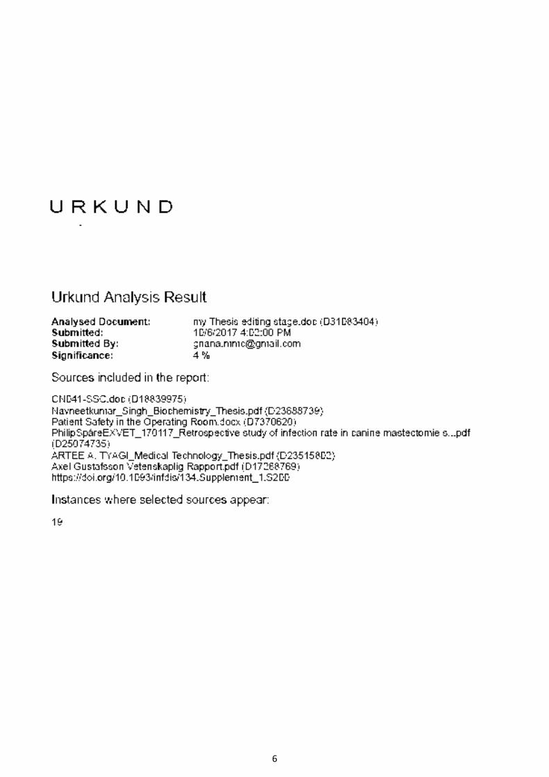
I personally verified the urkund.com website for the purpose of
plagiarism check. I found that the uploaded thesis file contains from the
introduction to conclusion pages and result shows four (4) percentage of
plagiarism in the dissertation.
Chennai – 600 001 Guide & Supervisor
Date - Prof.Dr.A.K.RAJENDRAN M..S.,D.Ortho
Head of the Department and Professor of
General Surgery,
Stanley Medical College & Hospital
Chennai – 600001
6

7
ACKNOWLEDGEMENT
I express my heartful gratitude to Prof. Dr. PONNAMBALA
NAMASIVAYAN, M.D.,DNB., Dean, Stanley Medical College and
Hospital, Chennai-1 for permitting me to do this study and use the
resources of the college.
I am profoundly indebted to Prof. Dr. A.K. RAJENDRAN M.S.,
D.Ortho., Professor of General Surgery, Head of the Department, Stanley
Medical College and hospital, for his constant and valuable support, who
has also been my Guide and Supervisor for this dissertation
I express my deepest sense of thankfulness to my Assistant
Professors Dr. C. ARUN BABU, M.S., Dr. VIJAYALAKSHMI M.S.,
Dr. JEYALAKSHMI M.S., Stanley Medical College and Hospital for
their valuable inputs and constant encouragement without which this
dissertation could not have been materialised.
I consider it a privilege to have done this study under the
supervision of my beloved former professor and head of the department
8
Prof.Dr.D.NAGARAJAN M.S. who has been a source of constant
inspiration and encouragement to accomplish this work.
I am also immensely grateful to my seniors and juniors
Dr.Chandrasekaran.K M.S., Dr.Niyas Ahamed M.S.,
Dr.Vijayalakshmi M.S., Dr.Rajachandrasekar M.S, Dr. Somanath
Sharma M.S., Dr.G.P.Kumaran M.S., Dr. Vinoth Kumar.M,
Dr.Srilaxmi, Dr.Vinoth, Dr. Monica, Dr.Lakshmanamoorthy for their
constant support and inputs throughout the study period. I Would like to
thank my colleague Dr.Arun Raja Thangavel MD for his valuable
suggestion for this dissertation. I am Grateful to my parents , siblings and
my friends especially Dr.Sathish Dev, Dr,Devi Prasadh , Dr. Arunvathani
and Dr.E. Suganya for their present help, and guidance.
I am extremely thankful to all the Members of the
INSTITUTIONAL ETHICAL COMMITTEE for giving approval for
my study. I also thank all the patients who were part of the study and my
Professional colleagues for their support and criticisms.
(Dr. M. GNANA SEZHIAN)
9
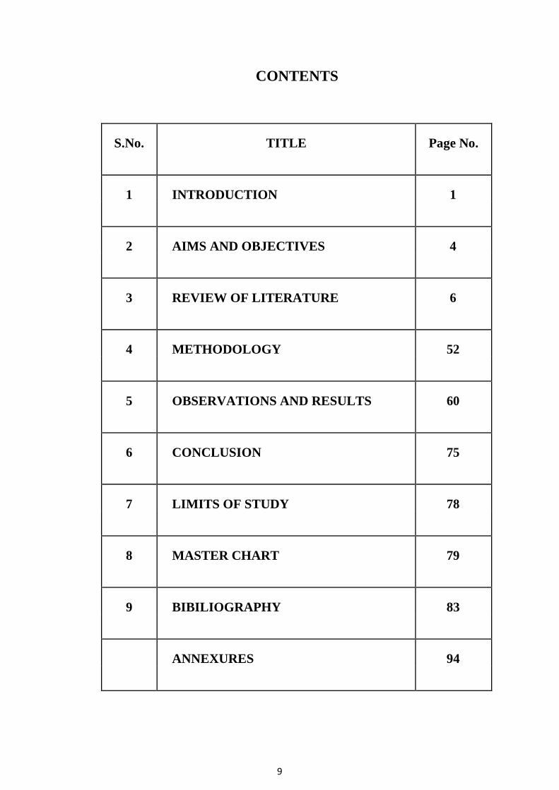
CONTENTS
S.No. TITLE Page No.
1 INTRODUCTION 1
2 AIMS AND OBJECTIVES 4
3 REVIEW OF LITERATURE 6
4 METHODOLOGY 52
5 OBSERVATIONS AND RESULTS 60
6 CONCLUSION 75
7 LIMITS OF STUDY 78
8 MASTER CHART 79
9 BIBILIOGRAPHY 83
ANNEXURES 94

1
INTRODUCTION
2
INTRODUCTION
Infections that occur in the wound created by an invasive surgical
procedure are generally referred to as surgical site infections (SSIs). SSIs are
one of the most important causes of healthcare-associated infections (HCAIs). A
prevalence survey undertaken in 2006 suggested that approximately 8% of
patients in hospital in the UK have an HCAI. SSIs accounted for 14% of these
infections and nearly 5% of patients who had undergone a surgical procedure
were found to have developed an SSI.(1)
However, prevalence studies tend to underestimate SSI because many of
these infections occur after the patient has been discharged from hospital.
SSIs are associated with considerable morbidity and it has been reported that
over one-third of postoperative deaths are related, at least in part, to SSI.(2)
In patients undergoing laparotomy with contaminated and dirty wounds
the infection rate is 20% to 30% and 30% to 40% respectively.(3),(4)
SSIs leads to severe morbidity in the operated patient in the form of costs
of treatment and prolonged hospital stay and the need for redo surgery in some
cases. Most infection occur from the skin and superficial microbes and various
methods can be used to tackle this condition by using this matter of fact.

3
Several preventive steps are followed and recommended by most of the
surgical research teams and the use of local antibiotic over the wound site as an
attempt to prevent the surgical site infection is one of them. A cost effective and
adequately sufficient method is being studied to prevent surgical site infection
through this method.

4
AIMS AND OBJECTIVES
5
AIMS AND OBJECTIVES
An prospective case control study
1. To analyse the effects of local antibiotic (Amikacin) therapy at the
surgical site along with systemic antibiotic therapy in an attempt to
prevent surgical site infections in contaminated and dirty surgical wounds
as compared to that of systemic antibiotics alone.
2. Grading the SSIs in both the groups and study the effects of local
antibiotic in reducing the incidence/severity of SSIs at the end of first
and second week of the post operative period

6
REVIEW OF LITERATURE
7
REVIEW OF LITERATURE
Defining surgical site infection
Postoperative wound infections, also known as surgical site infections
(SSIs), complicates many surgical patients. As defined by the Centers for
Disease Control and Prevention (CDC), these infections typically occur within
30 days of an operation at the site or part of the body where the surgery took
place, or within a year if an implant is left in place and the infection is thought
to be secondary to surgery.(5–7)
SSI is now the most common and most costly
hospital acquired infection.(31-33)
Since skin is normally colonised by a range of microorganisms that could
cause infection, defining an SSI requires evidence of clinical signs and
symptoms of infection rather than microbiological evidence alone. SSIs
frequently only affect the superficial tissues, but some more serious infections
affect the deeper tissues or other parts of the body manipulated during the
procedure.
The majority of SSIs occurs most often between the 5th and 10th
postoperative days. However, where a prosthetic implant is used, SSIs affecting
the deeper tissues may occur several months after the operation.
8
Although the outcome measure for SSI used by many studies is based on
tandard definitions such as those described by the Centers for Disease Control
and Prevention (CDC)(8)
or the Surgical Site Infection Surveillance Service,(9)
other valid measures based on clinical signs and symptoms have been described
such as the Southampton(10)
and ASEPSIS(11)
methods.
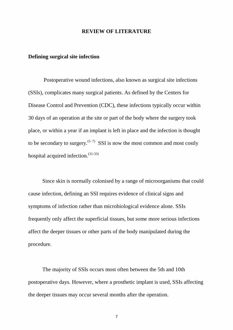
Figure 1 – different types of surgical site infections, three levels (87)
The CDC definition(12)
describes three levels of SSI:
9
• superficial incisional, affecting the skin and subcutaneous tissue. These
infections may be indicated by localised (Celsian) signs such as redness, pain,
heat or swelling at the site of the insicion or by the drainage of pus.
• deep incisional, affecting the fascial and muscle layers. These infections may
be indicated by the presence of pus or an abcess, fever with tenderness of the
wound, or a separation of the edges of the incision exposing the deeper tissues.
• organ or space infection, which involves any part of the anatomy other than
the incision that is opened or manipulated during the surgical procedure, for
example joint or peritoneum.
These infections may be indicated by the drainage of pus or the formation
of an abscess detected by histopathological or radiological examination or
during re-operation.
In addition, there may also be microbiological evidence of wound
infection from cultures obtained aseptically from wound fluid or tissue.
However, since skin sites are normally colonized by a variety of organisms,
positive wound cultures in the absence of clinical signs are rarely indicative of
SSI. Some studies report infections that affect any part of the incision, whereas
other studies focus only on those that affect the deeper tissues as these may be
considered to be more important and their definition less subjective. Variation
introduced by the definition of SSIs and the methods used to detect them need
10
to be taken account when combining or comparing evidence from different
studies. This variation has been an important limiting factor in reviewing
evidence for this guideline.
Surveillance for surgical site infection
Surveillance of SSI provides data that can both inform and influence
practice to minimise the risk of SSI, as well as communicate more clearly the
risks of infection to patients.(13)
Surveillance was first recognised as an
important tool in reducing rates of infection in the 1980s.(14)
The Study on the
Efficacy of Nosocomial Infection Control (SENIC) showed that surveillance
and infection control programmes that included the collection, analysis and
feedback of data on infection rates to surgeons were associated with significant
reductions in rates of SSI.(15)
Since then, many national surveillance systems have been established
and have reported reductions in rates of SSI in association with surveillance,
feedback of data to clinicians and benchmarking of rates of SSI.(9–12)
Consumer
demand for information about the performance of healthcare providers has also
led to compulsory public reporting of data on HCAIs, including SSIs.
National surveillance systems, such as the Surgical Site Infection
Surveillance System in England and similar schemes in Wales and Northern
11
Ireland, provide standardised surveillance methods that enable hospitals to
benchmark their rates of SSI.
Such benchmarking can be a powerful driver for change but requires
participating hospitals to use uniform methods of finding and defining cases of
SSI that are likely to reliably identify a large proportion of the infections, and a
reliable approach to analysing rates of SSI that takes account of variation in risk
associated with different procedures and risk factors in the patients undergoing
surgery. Most national surveillance systems target surveillance towards defined
groups of patients undergoing similar operative procedures, following each case
up to identify those that develop an SSI, although the sensitivity of case-finding
will be influenced by the methods employed.(16)
This enables rates of SSI to be calculated using the number of procedures
as the denominator. Feedback of rates to individual surgical teams and
comparisons with the benchmark rate are essential components of effective
surveillance.(15)
The risk index developed by the CDC in the USA, which takes
account of the underlying illness of the patient, the duration of the operation and
the wound classification of the procedure, is commonly used to adjust rates of
SSI and improve the validity of comparisons where case-mix may vary over
time or between centers.(17)

12
However, comparisons between different surveillance systems is
complicated because of variation in both the methods of surveillance and the
application and interpretation of case definitions.(18)
Since some SSIs may take
many days to develop, evidence of infection may not become apparent until
after the patient has been discharged from hospital.
Surveillance focused on detecting SSI during the inpatient stay is thus
likely to underestimate the true rate of SSI, a problem that is exacerbated by the
increasing trend towards shorter lengths of postoperative hospital stay and
day surgery.(19)
Therefore, systems that enable cases of SSI to be identified after
discharge from hospital enhance the value of surveillance. However, there are a
number of practical difficulties in reliably identifying SSI in community
settings and methods that systematically and accurately identify SSI are
required if valid comparisons of rates are to be made.(20)
It is important to note that no such centralized system to report SSIs
exists in our nation as of now, and it should be considered in the future to create
a system to detect analyse and audit this serious post operative nosocomial
complication of surgical patients
Risk factors
The risk of SSI is increased by factors that:

13
• increase the risk of endogenous contamination (for example, procedures
that involve parts of the body with a high concentration of normal flora such as
the bowel)
• increase the risk of exogenous contamination (for example, prolonged
operations that increase the length of time that tissues are exposed)
• diminish the efficacy of the general immune response (for example,
diabetes, malnutrition, or immunosuppressive therapy with radiotherapy,
chemotherapy or steroids) or local immune response (for example, foreign
bodies, damaged tissue or formation of a haematoma).
Randomised controlled trials, which require the assessment of
comparability between groups, have not been undertaken for risk factors.
While data on risk factors for SSI are available from observational studies
using regression analyses, factors that are significant in one type of surgery may
not be generalisable to other surgical procedures.
Age:
Five studies were identified.(9,21–24)
One prospective observational study
using logistic regression to analyse data collected from 142 medical centres
identified age as an independent risk factor for SSI.(21)
. Trained nurses gathered
data on inherent and operative risk factors for SSI in patients undergoing
14
general and vascular surgery. Of 163 624 patients who were included in the
study, 7035 developed SSI(17)
within 30 days of surgery.
Patients aged over 40 had a statistically significantly increased risk of
developing SSI compared with those under 40 years (OR 1.24, 95% CI 1.07 to
1.44). Another prospective observational study examined SSI in patients
undergoing total hip replacement, hemiarthroplasty or revision procedures as
part of SSI surveillance in England.10 [EL = 2+] Trained personnel collected
clinical and operative data throughout the duration of the hospital stay.
Detected cases of SSI were thus classified as occurring in the immediate
postoperative period.
Age over 75 was found to be a significant risk factor (compared with a
baseline of age under 65) when all types of hip replacement were considered
together (for age 75–79 years OR 1.56, 95% CI 1.16 to 2.10, for age ≥ 80 years
OR 1.66, 95% CI 1.24 to 2.21).
A retrospective observational study conducted in the USA included
patients who underwent general surgery with antibiotic prophylaxis at a
community hospital.(22) .
Demographic and clinical information was extracted
from the database including readmission up to 28 days post-surgery.
Regression techniques were used to identify independent risk factors for SSI
detected early (between 2 and 7 days postoperatively), necessitating
15
readmission or causing death. Age was found to be a statistically significant risk
factor for early SSI incidence (SSI incidence for each decade increase in age
OR 1.22, P < 0.01).
One large prospective study (n = 23 649 wounds) including children and
adults undergoing procedures on mostly clean wounds stratified results by age
group.(23)
Observations of SSI were made for 28 days postoperatively and a
broad trend of increasing SSI incidence with increasing age was reported.
A prospective cohort study of adult surgical patients (n = 144 485) from
11 hospitals reported an SSI incidence rate of 1.2%.24 [EL = 2+] A direct linear
trend of increasing risk of deep or organ space SSI from age 17 until age 65
(1.1% for each year of age, P < 0.002) was reported. However, for patients aged
over 65 the risk of SSI decreased by 1.2% for each extra year of life (P = 0.008)
Underlying illness
The American Society of Anesthesiologists’ (ASA) classification of
physical status score is used to assess a patient’s preoperative physical condition
and provides a simple measure of the severity of the underlying illness. Four
studies were identified that found ASA score to be an indicator of
SSI development.(9,17,21,24)
16
A prospective cohort study of adult surgical patients (n = 144 485) from
11 hospitals reported an SSI incidence rate of 1.2%.24 [EL = 2+] A statistically
significantly higher SSI incidence for those with an ASA score of 3 or greater
compared with those with an ASA score of 1 or 2 (OR 3.0, 95% CI 2.6 to 3.2)
was reported.
This effect was also demonstrated in a prospective observational study
examining SSI in patients undergoing total hip replacement, hemiarthroplasty or
revision procedures.(9)
. Cases of SSI occurring in the immediate postoperative
period were included.
Overall, the SSI incidence rate was 3.07% (n = 24 808 procedures, cases
of SSI = 761). Multivariate analysis showed ASA score of 3 or greater to be an
independent risk factor for SSI (OR 1.55, 95% CI 1.29 to 1.88).
A prospective observational study using logistic regression to analyse
data collected from patients undergoing general or vascular surgery in 142
medical centres also identified ASA score as an independent risk factor for
SSI.21 [EL = 2+] The SSI incidence rate was 4.3%. Compared with an ASA
score of 1, a score of 3 and a score of 4 or 5 were found to be statistically
significantly associated with SSI (OR 1.97, 95% CI 1.53 to 2.54 and OR 1.77,
95% CI 1.34 to 2.32, respectively).
17
In one retrospective observational study, analysis of data from the
National Nosocomial Infections Surveillance System (n = 84 691 operations)
found an overall SSI incidence of 2.8%.(17)
.The majority of patients (94%) were
undergoing clean or clean-contaminated surgery. The strength of association
between ASA score and SSI development risk was estimated (Goodman–
Kruskal Gstatistic = 0.34, standard error (SE) = 0.01) and stratification of
results by ASA score demonstrated that the rate of SSI increased by a factor of
4.7 as ASA score ranged between 1 (1.5 SSIs per 100 operations) to 5 (7.1 SSIs
per 100 operations).
In addition, there are some specific underlying diseases or conditions that
are independently associated with an increased risk of SSI. Surgical site
infection.A number of studies in cardiac, spinal, vascular and general surgery
and have shown that diabetes is strongly associated with an increased risk of
SSI.(21,23,25–29)
Studies report a two- to three-fold increase in risk of developing
an SSI in patients with diabetes. This may be related to altered cellular immune
function.
A prospective cohort study (with a parallel case–control analysis) of 1044
cardiothoracic surgery patients demonstrated evidence that the rate of SSI is
independently associated with postoperative hyperglycaemia (OR 2.02, 95% CI
1.21 to 3.37) and that the risk of SSI correlated with the degree of
hyperglycaemia during the postoperative period (for patients with postoperative
18
glucose levels of 200–249 mg/dl, 250–299 mg/dl and ≥ 300 mg/dl, SSI ORs
were 2.54, 2.97 and 3.32, respectively).(27)
One large prospective study of procedures on mostly clean wounds in
children and adults reported that malnourishment increased the incidence of SSI
from 1.8% to 16.6% (univariate analysis).(23)
Two studies were identified that
found low serum albumin to be an indicator of SSI development.(21,22)
In a large prospective cohort study of general and vascular surgery patients (n =
163 624 patients), multivariate analysis demonstrated that those with a low
preoperative serum albumin (≤ 3.5 g/dl) were more likely to develop SSI (OR
1.13, 95% CI 1.04 to 1.22), compared with those with normal serum albumin
levels.(21)
The results of a retrospective observational study of patients undergoing
general surgery with antibiotic prophylaxis (n = 9016) further suggested that
low serum albumin was associated with the development of SSI within the first
2–7 days postoperatively (OR 2.27, P < 0.01, per gram percent decrease).(22)
One study was identified that found treatments associated with anti-cancer
therapy to be indicators of SSI development.(21)
The prospective cohort of general and vascular surgery patients also
found that radiotherapy within 90 days prior to surgery (OR 1.37, 95% CI 1.08
19
to 1.74) and use of steroids (OR 1.39, 95% CI 1.18 to 1.63) independently
predicted development of SSI.(21)
Obesity
Adipose tissue is poorly vascularised and the consequent effect on
oxygenation of the tissues and functioning of the immune response is thought to
increase the risk of SSI. In addition, operations on patients who are obese can be
more complex and prolonged.(30)
The effect of obesity on the risk of SSI has
been investigated in cardiac and spinal surgery and in caesarean section. Studies
report ORs of between 2 and 7 for SSI in patients with a body mass index of 35
kg/m2 or more.(23,25–31)
Smoking
The wound healing process may be affected by the vasoconstrictive
effects and reduced oxygencarrying capacity of blood associated with smoking
cigarettes. Four studies were identified that investigated the association of
smoking with SSI development.(21,26,29,32)
One prospective observational study, using logistic regression to analyse
data collected from patients (n = 163 624) undergoing general and vascular
surgery in 142 medical centres, identified smoking as an independent risk factor
for SSI.21 [EL = 2+] Smokers had a statistically significantly greater risk of
developing SSI compared with non-smokers (OR 1.23, 95% CI 1.04 to 1.22).
20
A case–control study of adults undergoing cardiac surgery (n = 117)
examined risk factors for SSI.29 [EL = 2+] Statistically significantly more
patients who developed an SSI smoked compared with uninfected controls
(28.2% versus 14.1%) and, following logistic regression analysis,
smoking remained an independent risk factor for SSI (OR 3.27, 95% CI 1.04 to
10.20)
A prospective observational study investigated SSI in patients undergoing
breast reduction surgery.32 [EL = 2+] Participants (n = 87) were instructed to
stop smoking at least 4 weeks prior to surgery. Twenty-four patients developed
SSI, which occurred 8 days postoperatively on average. Statistically
significantly more smokers developed SSI than non-smokers (37.2% versus
18.2%, P < 0.05). Sixteen of 43 smokers developed SSI. Those who smoked
more cigarettes were more (19)
likely to develop SSI (estimated cigarettes
smoked mean 146 000 range 29 200–228 125 versus mean 10 950 range 9125–
54 750, P < 0.001) and those who had smoked for a longer time also
experienced statistically significantly more infections (mean pack years 20,
range 4–31 versus mean pack years 2, range 1–8, P < 0.001)
A retrospective observational study of cardiac surgery (n = 3008)
investigating risk factors for SSI, using logistic regression techniques, found
that smokers developed statistically significantly more sternal SSIs (OR 1.39,
95% CI 1.05 to 1.86) and deep sternal SSIs (OR 2.41, 95% CI 1.42 to 4.10) than

21
non-smokers and that peripheral vascular disease was also an independent risk
factor for the development of deep SSI (OR 2.11, 95% CI 1.09 to 4.09).(26)
A further prospective study of cardiac surgery patients reported 199 SSIs
occurring within 2345 included participants.(28)
. Multivariate analysis also
demonstrated that generalized peripheral vascular disease statistically
significantly increased the risk of SSI (OR 1.64, 95% CI 1.16 to 2.33).
Wound classification
The significance of the microbial flora normally colonising the operative
site in the subsequent risk of SSI has been recognised for many decades. The
wound classification developed by the
National Academy of Sciences in the 1960s distinguishes four levels of
risk, from clean, where the procedure involves a sterile body site, to dirty,
where the procedure involves a heavily contaminated site.
Three studies were identified that examined the association of wound
classification with SSI incidence.(16,21,24)
In a retrospective analysis of a large
infection surveillance data set, the SSI incidence rate per 100 operations was
2.1, 3.3, 6.4, 7.1 for clean, clean-contaminated, contaminated and dirty wound
classes, respectively.(17)

22
Figure 2 – per operative image during laprotomy done for pyoperitoneum,
showing pus from the abdominal cavity

23
Another study of general and vascular procedures reported that wound
class was an independent predictor of SSI (clean surgery SSI OR 1 , SSI ORs
for clean-contaminated, contaminated and dirty wound classes were 1.04, 1.7
and 1.5, respectively, P < 0.0001),(21)
while a third prospective study found that
SSI was statistically significantly increased in contaminated and
dirty wounds (wound class > 2 OR 2.3, 95% CI 2.0 to 2.7).(24)
Site and complexity of procedure
For many types of surgery there is evidence that the risk of SSI is
affected by the specific site of the operation.Complexity of the procedure is also
indicated as an SSI risk factor.
One study of general and vascular surgery estimated that there was a two-
to three-fold increased risk of SSI with increasing surgical complexity measured
as work relative value units.(21)
However, complex surgery is more often
distinguished by prolonged duration of the procedure. In studies of cardiac and
hip replacement surgery,(9)
there was a 1.5- to 1.75-fold increased risk of SSI
associated with longer duration of surgery.
While some of these patient characteristics, such as obesity,
hyperglycaemia, malnutrition and smoking, may be modified prior to surgery,
others, such as the complexity of the procedure and the underlying illness in the
patient, cannot.Mechanisms of accounting for variation in intrinsic
24
characteristics of patients or procedures that influence the risk of SSI are
important for surveillance systems in order to enable valid comparisons of rates
among surgeons, among hospitals, or across time. Early surveillance systems(23)
used the basic wound classification to adjust for risk of SSI but analyses of
large data sets on a range of operative procedures identified a few key risk
factors that were associated with an increased risk of SSI and that when used in
combination provided a better indicator of risk of SSI than the wound
classification.(21,25)
This National Nosocomial Infection Surveillance (NNIS) system risk
index is based on the presence of the following risk factors:
1. a patient with an ASA preoperative assessment score of 3, 4 or 5 (a
simple measure of the
severity of the patient’s underlying illness)
2. an operation classified as contaminated or dirty-infected
3. an operation lasting over T hours, where T depends on the operative
procedure being performed.(2,16)
The T time is the 75th percentile of the
distribution of operation time for a particular category of procedures rounded to
the nearest hour.(17)
. While this NNIS risk index does not measure all the factors
that contribute to the risk of developing an SSI, it does provide a practical way
of adjusting rates for the major patient and operative risk factors and it is used
to stratify rates of SSI by most national surveillance systems.
25
Other more complex risk stratification systems to predict the risk of SSI have
also been developed.(21,26)
Evidence statements on risk factors
Age
The age of the patient is a significant independent predictor of the risk of SSI
development generally and for early SSI development.
Moreover, in adults a direct linear trend of increasing risk of SSI until age 65
has been demonstrated.
For those aged over 65, an inverse linear trend of SSI risk was found, although
this finding may be subject to selection bias (i.e. only those who are fit enough
undergo surgery).
Underlying illness
Those patients with an ASA score of 3 or more have a severe systemic disease
and have been found to have a significantly higher risk of SSI.
Studies have repeatedly shown that diabetes is strongly associated with an
increased risk of SSI.
Malnutrition has been implicated as a risk factor for SSI
There is evidence from a prospective and a retrospective study that the risk of
SSI is increased in patients with a low serum albumin.
Radiotherapy and steroid use have both been linked to an increased risk of SSI.
Obesity

26
Studies have repeatedly shown that obesity is strongly associated with an
increased risk of SSI.
Smoking
Smoking, duration of smoking and number of cigarettes smoked are associated
with an increased risk of SSI.
Peripheral vascular disease has been demonstrated to increase SSI risk in a
prospective and a retrospective study.
Wound classification
There is consistent evidence that the risk of infection increases with level of
wound contamination.

27
Strategies to prevent surgical site infection:
Now that we have analysed the factors which are risk factors for
developing a surgical site infection let us go the various steps and
recommendations used to prevent this complication in surgical patients.
Recommendations are categorized as either (1) basic practices that should
be adopted by all acute care hospitals or (2) special approaches that can be
considered for use in locations and/or populations within hospitals when HAIs
are not controlled by use of basic practices.
Basic practices include recommendations where the potential to impact
HAI risk clearly outweighs the potential for undesirable effects. Special
approaches include recommendations where the intervention is
likely to reduce HAI risk but where there is concern about the risks for
undesirable outcomes resulting from the intervention, where the quality of
evidence is low, or where evidence supports the impact of the intervention in
select settings (eg, during outbreaks) or for select patient populations.
Hospitals can prioritize their efforts by initially focusing on
implementation of the prevention approaches listed as basic practices. If HAI
surveillance or other risk assessments suggest that there are ongoing .

28
opportunities for improvement, hospitals should then consider adopting some or
all of the prevention approaches listed as special approaches. These can
be implemented in specific locations or patient populations or can be
implemented hospital-wide, depending on outcome data, risk assessment, and/or
local requirements.
.
I. Basic practices for preventing SSI: recommended for all
acute care hospitals
1. Administer antimicrobial prophylaxis according to evidence- based
standards and guidelines. (34-36)
a. Begin administration within 1 hour before incision to maximize
tissue concentration.(37,38)
Administering agent closer than 1 hour is
effective, and some studies show superior efficacy for administration
between 0 and 30 minutes prior to incision compared with administration
between 30 and 60 minutes.(39,40)
. Two hours are allowed for the
administration of vancomycin and fluoroquinolones.
b. Select appropriate agents on the basis of the surgical procedure,
the most common pathogens causing SSIs for a specific procedure, and
published recommendations. (38)
c. Discontinue agent within 24 hours after surgery.
29
Although guidelines suggest stopping the antimicrobial agent within 24 hours of
surgery, there is no evidence that agents given after closure contribute
to efficacy, and they do contribute to increased resistance (41,42)
and the risk of
Clostridium difficile disease.(43)
d. Adjust dosing on the basis of patient weight; for example:
i. Use 30 mg/kg for pediatric patients, 2 g of cefazolin for patients
weighing 80 kg or more, and 3 g for patients weighing 120 kg or more.
ii. Vancomycin should be dosed at 15 mg/kg. Gentamicin should be
dosed at 5 mg/kg for adult patients and 2.5 mg/kg for pediatric patients.
(a) For morbidly obese patients receiving gentamicin, the
weight used for dose calculation should be the ideal weight plus
40% of the excess weight.
e. Redose prophylactic antimicrobial agents for long procedures
and in cases with excessive blood loss during the procedure.
i. Prophylactic antimicrobials should be redosed at intervals of 2 half-
lives (measured from time the preoperative dose was administered) in cases that
exceed this time.
f. Use a combination of parenteral antimicrobial agents and oral
antimicrobials to reduce the risk of SSI following colorectal
procedures.(44-51)
30
i. The additional SSI reduction achieved with mechanical bowel
preparation has not been studied, but the data supporting use of oral
antimicrobials have all been generated in combination with mechanical bowel
preparation.
ii. Mechanical bowel preparation without oral antimicrobials does not
decrease the risk of SSI.
2. Do not remove hair at the operative site unless the presence of hair will
interfere with the operation. Do not use razors (53)
a. If hair removal is necessary, remove hair outside the operating
room using clippers or a depilatory agent.
3. Control blood glucose during the immediate postoperative period for cardiac
surgery patients and noncardiac surgery patients (54-57)
a. Maintain postoperative blood glucose of 180 mg/dL or lower.
i. The recommendation of maintaining postoperative blood glucose less
than 200 mg/dL at 6 AM on postoperative days 1 and 2 is being replaced. In
2014, this measure will be revised in the SCIP to assess glucose control (180
mg/dL or lower) in cardiac surgery patients in the time frame of 18-24 hours
after anesthesia end time. Several societies, experts , and the National Quality
Forum support this new recommendation.(58,59)
b. Intensive postoperative glucose control (targeting levels less
than 110 mg/dL) has not been shown to reduce the risk of SSI and may
31
actually lead to higher rates of adverse outcomes, including stroke and
death.(60)
4. Maintain normothermia (temperature of 35.5°C or more) during the
perioperative period .
a. Even mild degrees of hypothermia can increase SSI rates.
Hypothermia may directly impair neutrophil function or impair it
indirectly by triggering subcutaneous vasoconstriction and subsequent
tissue hypoxia In addition, hypothermia may increase blood loss, leading
to wound hematomas or need for transfusion, both of which can increase
rates of SSI.(61)
b. Randomized controlled trials have shown the benefits of both
preoperative and intraoperative warming to reduce SSI rates and to
reduce intraoperative blood loss.(62-64)
5. Optimize tissue oxygenation by administering supplemental oxygen during
and immediately following surgical procedures involving mechanical
ventilation
a. Supplemental oxygen is most effective when combined with
additional strategies to improve tissue oxygenation, including
maintenance of ormothermia and appropriate volume replacement. The
available evidence is in patients undergoing surgery with general
anesthesia using mechanical ventilation.
32
i. Seven randomized clinical trials have been published
comparing 80% with 30%-35% Fi02 in patients undergoing general anesthesia
with intraoperative mechanical ventilation and postoperative oxygen delivered
for 2-6 hours via a nonrebreathing mask.
ii. Three trials in patients undergoing elective colorectal
resection and 1 each in open appendectomy and total gastrectomy with
esophagojejunal anastomosis reported an approximate 40% decrease in the rate
of SSI. Three of the studies reported protocols that included maintenance of
perioperative normothermia and liberal fluid replacement. Two trials in mixed
surgical populations undergoing emergency or elective laparotomy for
gastrointestinal, gynecologic, or urologic procedures reported different results.
(a.) The large multicenter trial that restricted perioperative
fluid replacement reported no difference.
(b.) A follow-up study performed in this population noted
that patients undergoing cancer surgery who
received 80% Fi02 had higher rates of mortality
than patients undergoing cancer surgery who
received 30% Fi02.
(c.) The smaller trial without standardized protocols for
perioperative normothermia or volume
replacement reported an increase in SSIs.In this
study, the 80% Fi02 group had a significantly
33
higher proportion of patients with high body mass
index (more than 30), higher blood loss, more
crystalloid infused, and longer operations. This
group also had 5 patients who remained intubated
postoperatively (vs 1 in the 35 % group).
Postoperative intubation was predictive of SSI.
b. A meta-analysis of 5 of the above-referenced studies concluded
that perioperative supplemental oxygen led to a relative risk (RR)
reduction of 25% for SSI.
6. Use alcohol-containing preoperative skin preparatory agents if no
contraindication exists.
a. Alcohol is highly bactericidal and effective for preoperative skin
antisepsis but does not have persistent activity when used alone. Rapid,
persistent, and cumulative antisepsis can be achieved by combining
alcohol with chlorhexidine gluconate or an iodophor.
i. Alcohol is contraindicated for certain procedures, including procedures
in which the preparatory agent may pool or not dry (eg, involving hair)
due to fire risk. Alcohol may also be contraindicated for procedures
involving mucosa, cornea, or ear.
34
b. The most effective disinfectant to combine with alcohol is
unclear.
i. A recent trial of 849 patients undergoing cleancontaminated surgery
randomized patients to preoperative skin antisepsis with chlorhexidinealcohol
or povidone-iodine.(65)
The overall rate of SSI was significantiy lower in the
chlorhexidinealcohol group than in the povidone-iodine group (9.5% vs 16% [P
= .004]; RR, 0.59 [95% confidence interval (CI), 0.41-0.85]).
ii. In contrast, a single-center study compared povidone- iodine followed
by isopropyl alcohol versus chlorhexidine-alcohol versus iodine-alcohol using
a sequential implementation design.(66)
General surgical patients who received
skin antisepsis with iodine-alcohol had the lowest rates of SSI (3.9 per 100
procedures), compared with 6.4 per 100 procedures for patients who received
povidone-iodine followed by alcohol and 7.1 per 100 procedures for patients
who received chlorhexidine-alcohol.In the absence of alcohol, chlorhexidine
gluconate may have advantages over povidone-iodine, in eluding longer
residual activity and activity in the presence of blood or serum.(67)
iv. These disinfectants are not interchangeable. Follow the manufacturers'
instructions to ensure correct application.
7. Use impervious plastic wound protectors for gastrointestinal and biliary tract
surgery.
35
a. A wound protector is a plastic sheath that lines a wound and can
facilitate retraction of an incision during surgery without the need for
additional mechanical retractors.
b. A recent meta-analysis of 6 randomized clinical trials in 1,008
patients reported that use of a plastic wound protectors was associated
with a 45% decrease in SSIs.
i. There was a nonsignificant trend toward greater protective effect using
a dual-ring protector compared with a single-ring protector.
8. Use a checklist based on the World Health Organization (WHO) checklist to
ensure compliance with best practices to improve surgical patient safety (quality
of evidence:
a. The WHO checklist is a 19-item surgical safety checklist to
improve adherence with best practices.
b. A multicenter quasi-experimental study conducted in 8 countries
demonstrated that use of the WHO checklist led to lower rates of surgical
complications, including SSI and death.
c. These findings have been confirmed in subsequent single-center
and multicenter quasi-experimental studies.
9. Perform surveillance for SSI :

36
a. Identify high-risk, high-volume operative procedures to be
targeted for SSI surveillance on the basis of a risk assessment of patient
populations, operative procedures performed, and available SSI
surveillance data.
b. Identify, collect, store, and analyze data needed for the
surveillance program.
i. Develop a database for storing, managing, and accessing data collected
on SSIs.
ii. Implement a system for collecting data needed to identify SSIs. Data
are required from surgical and microbiological databases. Obtain the following
data from surgical databases: patient name, medical record number, date, type
of procedure, surgeons, anesthesiologists, incision time, wound class, ASA
score, closure time, and presence of an SSI. Ideally, these data are supplemented
with process data, including prophylactic agent and dose and time(s) of
administration of prophylactic agent. For patients diagnosed with an SSI,
necessary microbiological data include type of SSI, infecting organism and
antimicrobial susceptibilities, and date of infection. More detailed surgical
and patient information may be useful for some procedures, including use of
general anesthesia, emergency or trauma-related surgery, body mass
index, and diagnosis of diabetes.

37
Prepare periodic SSI reports (time frame will depend on hospital needs and
volume of targeted procedures).
iv. Collect denominator data on all patients undergoing targeted
procedures in order to calculate SSI rates for each type of procedure.
v. Identify trends (eg, in SSI rates and pathogens causing SSIs).
c. Use updated CDC NHSN definitions for SSI.
d. Perform indirect surveillance for targeted procedures.
e. Perform postoperative surveillance for 30 days; extend the
postoperative surveillance period to 90 days for certain procedure
categories.
i. Procedures that require 90-day surveillance are determined
by specific procedure codes.
f. Surveillance should be performed on patients readmitted to the
hospital.
i. If an SSI is diagnosed at your institution but the surgical procedure was
performed elsewhere, notify the hospital where the original procedure was
performed.
g. Develop a system for routine review and interpretation of SSI rates to
detect significant increases or outbreaks and to identify areas where additional
38
resources might be needed to improve SSI rates. If increased rates are identified,
determine the number of potentially preventable infections that occurred,
defined as the number of SSIs that occurred during a procedure in which less
than 100% of recommended practices and processes were completed.
II. Special approaches for preventing SSI
Standard infection control methods of outbreak investigation are recommended
for use in locations and/or populations within the hospital with unacceptably
high SSI rates despite implementation of the basic SSI prevention strategies
listed above.
1. Screen for S. aureus and decolonize surgical patients with an
antistaphylococcal agent in the preoperative setting for high-risk procedures,
including some orthopedic and cardiothoracic procedures.
a. Screening for S. aureus refers to the practice of attempting
to identify patients colonized with methicillin- susceptible S. aureus (MSSA)
and/or MRSA. Decolonization refers to the practice of treating patients
with known S. aureus colonization with antimicrobial and/or antiseptic agents
to eliminate S. aureus colonization.
i. There is no standardized approach to either screening or
decolonizing. Most clinicians attempt to decolonize surgical
39
patients with a combination of chlorhexidine gluconate applied to
the skin and nasal mupirocin.
b. A Cochrane review concluded that mupirocin alone may be
effective, particularly in certain groups, including orthopedic and
cardiothoracic patients.
Several nonrandomized trials corroborate this conclusion.
c. Clinical practice guidelines from the American Society of
Health-System Pharmacists recommend giving mupirocin intranasally to
all patients with documented S. aureus colonization for orthopedic
procedures, including total joint replacement and hip fracture repair, and
cardiac procedures.
d. Some trials demonstrate that preoperative screening for S.
aureus, coupled with intranasal mupirocin and chlorhexidine bathing is
effective in reducing SSI for some patients.
i. For example, a randomized, double-blind, placebocontrolled,
multicenter trial that evaluated rapid identification of S. aureus nasal carriers
followed by decolonization was associated with a greater than 2-fold reduction
in the risk for postoperative infection due to S. aureus and an almost 5-fold
reduction in risk for deep incisional SSI due to S. aureus.
(a) This study was performed in a setting with high baseline rates of SSI
and in the absence of MRSA.

40
e. In contrast, other trials have failed to demonstrate a benefit.
i. A prospective, interventional cohort study with a crossover design
involving 21,000 patients concluded that universal, rapid screening for MRSA
at admission coupled with decolonization of carriers did not reduce the rate of
SSI due to MRSA.
ii. A double-blind randomized controlled trial involving more than 4,000
patients showed that intranasal application of mupirocin, which was not coupled
with chlorhexidine bathing, did not significantly reduce the S. aureus SSI rate.
(a) In a secondary analysis of these data, the use of intranasal mupirocin
was associated with an overall decreased rate of nosocomial S. aureus
infections among the S. aureus carriers.
f. A recently published meta-analysis of 17 studies concluded that
decolonization strategies prevent grampositive SSIs, S. aureus SSIs, and
MRSA SSIs, although there was significant heterogeneity among the
trials.
g. Factors that impact the decision to implement screening for S.
aureus and decolonization include adherence to basic SSI prevention
strategies, baseline rate of SSI due to S. aureus, individual patient risk
factors for acquiring SSI due to S. aureus, availability of resources to
implement the protocol, and ability to follow- up on protocol parameters
(eg, laboratory results) and adherence.

41
h. Routine preoperative decolonization with mupirocin without
screening is not currently recommended.
i. Mupirocin resistance has been documented.
2. Perform antiseptic wound lavage .
a. Wound lavage is a common practice, although the solution used
for lavage differs among surgeons.(68)
b. Several groups have evaluated whether dilute povidone- iodine
lavage of the surgical wound can decrease the risk of SSI. A meta-
analysis published in
2010 evaluated 24 randomized controlled trials and concluded that lavage
with dilute povidone-iodine decreased the risk of SSI compared with
nonantiseptic lavage (RR, 0.64 [95% CI, 0.51-0.82]).
3. Perform an SSI risk assessment,
a. Convene a multidisciplinary team (eg, surgical leadership,
hospital administration, quality management services, and infection
control) to identify gaps, improve performance, measure compliance,
assess impact of interventions, and provide feedback.
42
b. Determine baseline SSI rates by surgical specialty, procedure,
and/or surgeon to better target your evaluation and interventions.
4. Observe and review operating room personnel and the environment of care in
the operating room.
a. Perform direct observation audits of operating room personnel to
assess operating room processes and practices to identify infection
control lapses, including but not limited to adherence to process measures
(antimicrobial prophylaxis choice, timing and duration protocols, hair
removal, etc), surgical hand antisepsis, patient skin preparation, operative
technique, surgical attire (wearing and/or laundering outside the operating
room), and level of operating room traffic. Perform remediation when
breaches of standards are identified.
i. Operating room personnel should include surgeons, surgical
technologists, anesthesiologists, circulating nurses, residents, medical students,
trainees, and device manufacturer representatives.
Review instrument processing and flash sterilization logs
ii. Review maintenance records for operating room heating, ventilation,
and air conditioning system, including results of temperature and relative
humidity
testing, and test for maintenance of positive air pressure in the operating
room(s).
43
5. Observe and review practices in the postanesthesia care unit, surgical
intensive care unit, and/or surgical ward .
a. Perform direct observation audits of hand hygiene practices
among all personnel with direct patient contact.
b. Evaluate wound care practices.
c. Perform direct observation audits of environmental cleaning
practices.
d. Provide feedback and review infection control measures with
staff in these postoperative care settings.
III. Approaches that should not be considered a routine part of SSI
prevention
1. Do not routinely use vancomycin for antimicrobial prophylaxis
(quality of evidence: n).
a. Vancomycin should not routinely be used for antimicrobial
prophylaxis, but it can be an appropriate agent for specific scenarios. Reserve
vancomycin for specific clinical circumstances, such as a proven outbreak of
SSI due to MRSA; high endemic rates of SSI due to MRSA; targeted high-risk
patients who are at increased risk for SSI due to MRSA (including
cardiothoracic surgical patients and elderly patients with diabetes); and high-
risk surgical procedures in which an implant is placed.

44
i. No definitions for high endemic rates of SSI due to MRSA have been
established.
ii. Studies of the efficacy of vancomycin prophylaxis were published
prior to the emergence of community- acquired MRSA.
b. Two meta-analyses of studies comparing glycopeptides to beta-
lactam antimicrobial prophylaxis concluded that there was no difference
in rates of SSI between the 2 antimicrobial prophylaxis regimens.
c. A meta-analysis of 6 studies concluded that prophylaxis with a
glycopeptide and a second agent was protective against SSI due to gram-
positive organisms compared with prophylaxis with a /3-lactam alone. Of
note, the 2 randomized controlled trials included in the metaanalysis
combined a glycopeptide with non-/3-lactam antibiotic(s). Thus, no study
has prospectively analyzed
the effect of providing both glycopeptides and (3-lactam antimicrobials for
preoperative antimicrobial prophylaxis. As vancomycin does not have activity
against gram-negative pathogens and appears to have less activity
against MSSA than /3-lactam agents, many experts recommend adding
vancomycin to standard antimicrobial prophylaxis for the specific clinical
circumstances described above.
2. Do not routinely delay surgery to provide parenteral nutrition.
45
a. Preoperative administration of total parenteral nutrition has not
been shown to reduce the risk of SSI in prospective randomized
controlled trials and may increase the risk of SSI.
b. Individual trials comparing enteral and parenteral perioperative
nutrition and "immunomodulating" diets containing arginine and/or
glutamine with "standard" control diets tend to have very small numbers
and fail to show significant differences. Two recent meta-analyses,
however, demonstrate reduction in postoperative infectious complication
in patients receiving enteral diets containing glutamine and/or arginine
administered either before or after the surgical procedure.
3. Do not routinely use antiseptic-impregnated sutures as a strategy to prevent
SSIs.
a. Human volunteer studies involving foreign bodies have
demonstrated that the presence of surgical sutures decreases the inoculum
required to cause an SSI from 106 to 102 organisms.
b. Some trials have shown that surgical wound closure with
triclosan-coated polygactin 910 antimicrobial sutures may decrease the
risk of SSI compared with standard sutures. For example, a recent
randomized controlled trial of 410 colorectal surgeries concluded that the

46
rate of SSI decreased more than 50% (9.3% in the control group vs 4.3%
among cases; P = .05).
c. In contrast, a recent systematic review and meta-analysis
evaluated 7 randomized clinical trials and concluded that neither rates of
SSI (odds ratio [OR], 0.77[95% CI, 0.4-1.51]; P = .45) nor rates of
wound dehiscence (OR, 1.07 [95% CI, 0.21-5.43]; P = .93) were
statistically different compared with controls.In addition, one small study
raised concern about higher rates of wound dehiscence while using these
sutures.
d. The impact of routine use of antiseptic-impregnated sutures on
development of resistance to antiseptics is unknown.
4. Do not routinely use antiseptic drapes as a strategy to prevent SSIs.
a. An incise drape is an adhesive film that covers the surgical
incision site to minimize bacterial wound contamination due to
endogenous flora. These drapes can be impregnated with antiseptic
chemicals, such as iodophors.
b. A 2007 Cochrane review of 5 trials concluded that nonantiseptic
incise drapes were associated with a higher risk of SSI compared with no

47
incise drape (RR, 1.23 [95% CI, 1.02-1.48] ), although this association
may have been caused by one specific study. Two trials (abdominal and
cardiac surgical patients) compared iodophor-impregnated drapes to no
drapes. While wound contamination was decreased in one trial, neither
trial demonstrated that iodophor-impregnated drapes decreased the rate of
SSI. A nonrandomized retrospective study similarly concluded that
impregnated drapes do not prevent SSIs after hernia repair.
IV. Unresolved issues
1. Preoperative bathing with chlorhexidine-containing products.
a. Preoperative bathing with agents such as chlorhexidine has been shown
to reduce bacterial colonization of the skin. Several studies have examined the
utility of preoperative showers, but none has definitively proven that they
decrease SSI risk.
i. To gain the maximum antiseptic effect of chlorhexidine, adequate levels of
CHG must be achieved and maintained on the skin. Typically, adequate
levels are achieved by allowing CHG to dry completely. New strategies for
preoperative bathing with chlorhexidine, such as preimpregnated cloths, have
shown promise, but data are currently insufficient to support this approach.
2. Use of gentamicin-collagen sponges.
48
a. Gentamicin-collagen sponges have been evaluated as a way to decrease SSI
among colorectal and cardiac surgical patients.
i. Colorectal surgical patients. Several single-center randomized trials have
demonstrated that gentamicin- collagen sponges decrease the risk of SSI
following colorectal procedures.(69-71)
The rate of SSI was higher with the
sponge, however, in a recent large, multicenter randomized clinical trial.
ii. Cardiothoracic surgical patients. Four randomized controlled trials have
evaluated the use of gentamicin- collagen sponges in cardiothoracic surgery
Three of these trials demonstrated a decrease in SSIs,(72-74)
and one
showed no difference. A recent meta-analysis combining these trials concluded
that the risk of deep sternal wound infection was significantly lower in patients
who received a gentamicin- collagen sponge than in patients who did not
(RR, 0.62 [95% CI, 0.39-0.97]) despite significant heterogeneity among the
trials.186
USE OF LOCAL ANTIBIOTIC OVER WOUND SITE:
Studies have shown the effectiveness of using various antiseptic solutions
in the skin preparation before making the skin incision, including use of
alcohol, betadine, chlorhexidine etc.(65)
and the use of antiseptic wound lavage
like dilute povidone iodine lavage (68)
.
49
The use of Amikacin Sulfate as local antibiotic agent in treating Urinary
tract infection as been shown in a Japanese study (76)
, in which amikacin was
used as bladder wash agent or renal pelvic lavage and vesical instillation.
There are studies to show the effectiveness of using antibiotic impregnated
implants or prosthesis to prevent the SSIs. Study by Katsuhiro Tofuku et al. (75)
showed the effectiveness of using vancomycin impregnated fibrin sealant for
the prevention of surgical site infection associated with spinal instrumentation.
Study by Joseph Huh Et Al.(77)
showed use of Sustained-release lipid
particle-encapsulated amikacin applied to contaminated PTFE grafts increased
survival and decreased postoperative graft infections. Adjunctive use of local,
delayed-release antibiotics in contaminated vascular beds may allow wider
clinical use of prosthetic grafts.
Amikacin also has local action at the wound site with nonspecific actions
like enhancing growth of granulation tissue (78)
. Study by Nandita Pal et.al. (79-82)
showed that the most common organisms isolated from the surgical site
infections included Staphylococcus aureus, E.Coli, Klebsiella, Pseudomonas etc
(82-84) and a majority of them showed sensitivity to Inj. Amikacin (used in
combination to other drugs like Cefaperazone Sulbactum or Piperazillin
Tazobactum) (80-84)
.

50
Based on these observations , the advantages of using Inj. Amikacin
which showed less systemic absorption from the local wound site and
sensitivity to a majority of the organisms isolated from the SSI sites and its once
daily dosage, we have chosen the use of Inj. Amikacin for this study.
The Inj.,Amikacin , dosage calculated based on weight of the patient and
applied on the local wound site in the subcutaneous place before the skin
closure. A Feeding tube (8 or 10 Fr) is placed as a subcutaneous DT and skin
closed. On POD 1 through POD 3, the same once daily dose of Amikacin is
injected through the feeding tube and the tube closed.Intentionally no suction
drainage is applied to the feeding tube, to prevent any additional advantage of
using suction DT in subcutaneous plane as shown by review of studies done by
B. Manzoor et al. (86)
, that there could be some preventive effect in using
subcutaneous DT in the development of SSIs.
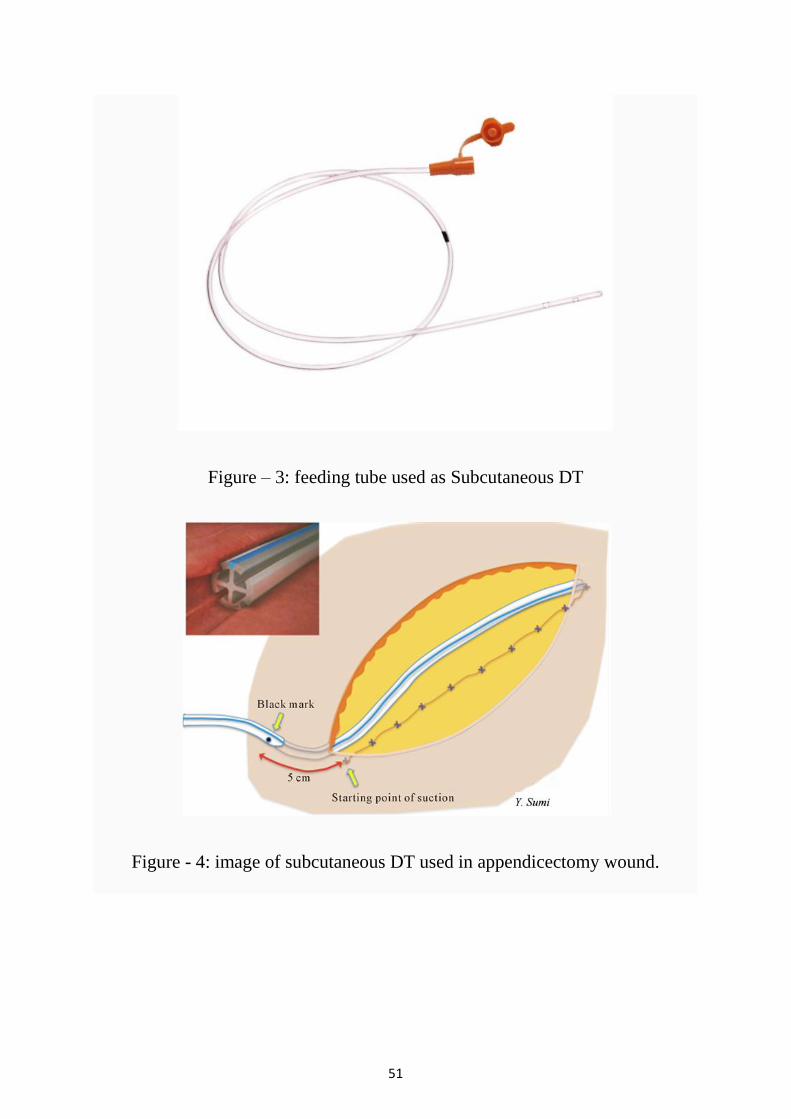
51
Figure – 3: feeding tube used as Subcutaneous DT
Figure - 4: image of subcutaneous DT used in appendicectomy wound.

52
METHODOLOGY
53
METHODOLOGY
1. Type of study: Prospective and Observational Study
2. Study approval: Prior to commencement of this study - Thesis &
Ethical Committee of Stanley Medical College and Hospital,
Chennai had approved the thesis protocol.
3. Place of study: Stanley Medical College and Hospital
4. Period of study: 10 months November 2016 to August 2017
5. Source of data: All cases of abdominal surgeries which falls under
contaminated (classIII) and dirty (class IV) wounds like emergency
laprotomies, open appendicectomies etc
6. Sample size: A total of 25 cases and 25 control
Study group (A): All elective and emergency surgeries of the
abdomen in which local antibiotic therapy was given
peroperatively & postoperatively along with systemic antibiotic
54
Control group (B): All cases of contaminated and dirty wounds
which are matched with the cases ,who received only systemic
antibiotics
7. Selection of patients:
All patients operated for abdominal surgeries, both elective and
emergency surgeries, which falls under class III (clean
contaminated) and class IV ( dirty)
a) Sampling method- Purposive.
b) Inclusion criteria-
All cases of abdominal surgeries which falls under contaminated
(classIII) and dirty (class IV) wounds like emergency laprotomies,
open appendicectomies etc
c) Exclusion criteria –
Extremes of age <18 yrs >70 yrs
Patients on immunosuppressants, chemo/radiotherapy, steroids other
serious pre-existing cardiovascular, pulmonary and immunological
disease.
Uncontrolled diabetic patients
Clean (Class I) and Clean contaminated (Class II) surgical wounds
55
8. Study procedure:
• Method of sampling was non-random, purposive.
• Ethical clearance will be obtained from the institute ethical
committee
• Written informed consent will be obtained from all patients before
subjecting them for the study
• All patients planned for abdominal surgeries were counseled and
the procedure explained in their local language
• All patients in the group were assigned as study and corresponding
matched control were selected
• The following parameters will be taken and observations will be
recorded and tabulated and analyzed to achieve the objective.
• The study group patients which included cases of abdominal
surgeries with class III and class IV type of wounds, peroperatively a
single adult dose of Inj.Amikacin was applied over the ‘subcutaneous
56
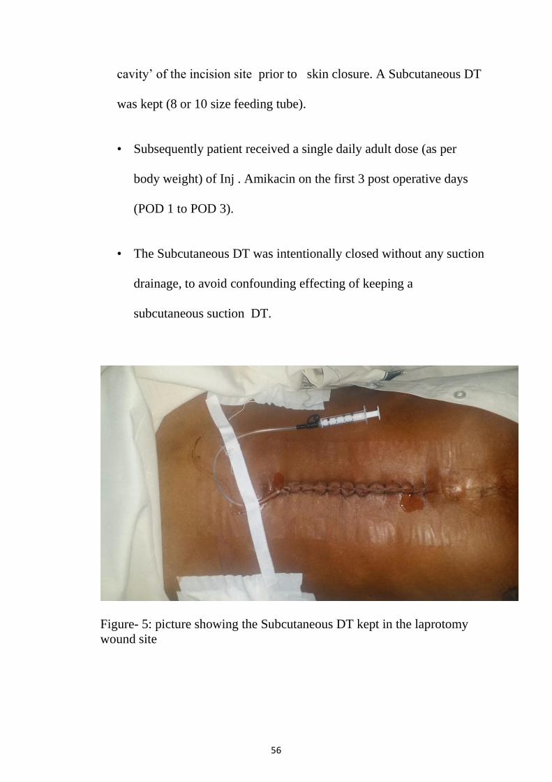
cavity’ of the incision site prior to skin closure. A Subcutaneous DT
was kept (8 or 10 size feeding tube).
• Subsequently patient received a single daily adult dose (as per
body weight) of Inj . Amikacin on the first 3 post operative days
(POD 1 to POD 3).
• The Subcutaneous DT was intentionally closed without any suction
drainage, to avoid confounding effecting of keeping a
subcutaneous suction DT.
Figure- 5: picture showing the Subcutaneous DT kept in the laprotomy
wound site
57
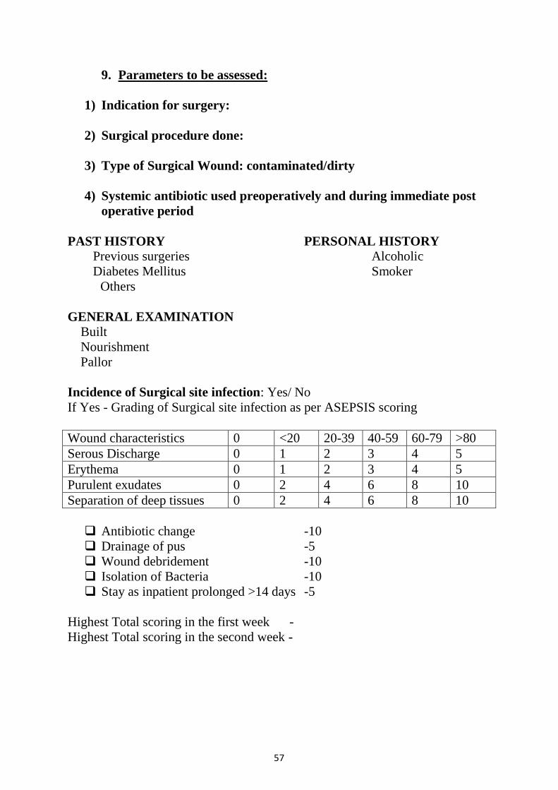
9. Parameters to be assessed:
1) Indication for surgery:
2) Surgical procedure done:
3) Type of Surgical Wound: contaminated/dirty
4) Systemic antibiotic used preoperatively and during immediate post
operative period
PAST HISTORY PERSONAL HISTORY
Previous surgeries Alcoholic
Diabetes Mellitus Smoker
Others
GENERAL EXAMINATION
Built
Nourishment
Pallor
Incidence of Surgical site infection: Yes/ No
If Yes - Grading of Surgical site infection as per ASEPSIS scoring
Wound characteristics 0 <20 20-39 40-59 60-79 >80
Serous Discharge 0 1 2 3 4 5
Erythema 0 1 2 3 4 5
Purulent exudates 0 2 4 6 8 10
Separation of deep tissues 0 2 4 6 8 10
Antibiotic change -10
Drainage of pus -5
Wound debridement -10
Isolation of Bacteria -10
Stay as inpatient prolonged >14 days -5
Highest Total scoring in the first week -
Highest Total scoring in the second week -
58
10. Data Analysis:
Statistical methods:
Diagnosis, total asepsis scoring, antibiotic changes at 1 week, stay as
Prolonged >14days, Systemic Antibiotic used were considered as outcome
variables. Case and control group were consider as primary explanatory
variable. Demographic age and gender were consider as other explanatory
variable.
Descriptive analysis:
Descriptive analysis was carried out by mean and standard deviation for
quantitative variables, frequency and proportion for categorical variables. Data
was also represented using appropriate diagrams like bar diagram, pie diagram
and box plots.
Quantitative outcome;
The association between categorical explanatory variables and
quantitative outcome was assessed by comparing the mean values. The mean
differences along with their 95% CI were presented. Independent sample t-
test.Association between quantitative explanatory and outcome variables was
assessed by calculating person correlation coefficient and the data was
represented in a scatter diagram.
59
Categorical outcome:
The association between explanatory variables and categorical outcomes
was assessed by cross tabulation and comparison of percentages. Chi square test
was used to test statistical significance.
P value < 0.05 was considered statistically significant. IBM SPSS version 22
was used for statistical analysis.(1)
1. Machines IB. IBM SPSS Statistics for Windows, Version 22.0. IBM
Corp Armonk, NY; 2013.

60
OBSERVATIONS AND RESULTS
61
OBSERVATIONS AND RESULTS
The study group receives Inj.Amikacin over the wound site before skin
closure and one 3 consecutive days after surgery. This is in addition to the usual
Intravenous antibiotic given for all cases of laprotomy surgery.
The subsequent development of surgical site infection in this study group is
compared to the control group which does not receive the additional local
wound site Inj.Amikacin.
The incidence of surgical site infection and the grading (based on
ASEPSIS grading) is done for the both groups for 2 weeks post operatively.
The second week monitoring is to assess if there is any residual effect of adding
Amikacin or any adverse effect due to its addition to the treatment regiment.

62
Table 1: Descriptive analysis of group in study population (N=50)
Group Frequency Percentage
Case 25 50.00%
Control 25 50.00%
Among the study population, 50% people were in case group and 50% people
were in control group. (table 1 & figure 1)
Figure 6: Bar chart of group distribution in study population (N=50)
63
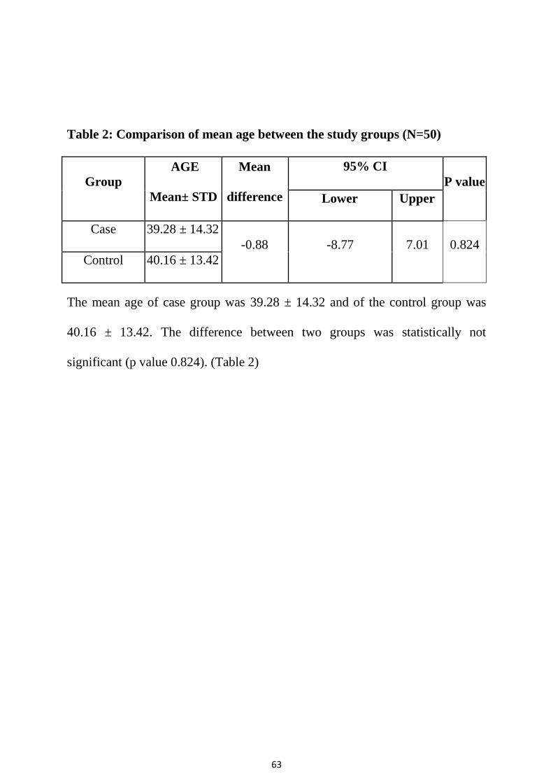
Table 2: Comparison of mean age between the study groups (N=50)
Group
AGE
Mean± STD
Mean
difference
95% CI
P value
Lower Upper
Case 39.28 ± 14.32
-0.88 -8.77 7.01 0.824
Control 40.16 ± 13.42
The mean age of case group was 39.28 ± 14.32 and of the control group was
40.16 ± 13.42. The difference between two groups was statistically not
significant (p value 0.824). (Table 2)

64
Table 3: Association of group with gender of study population (N=50)
Gender
Group
Chi square P-value
Case (N=25) Control(N=25)
Male 21 (84%) 20 (80%)
0.136 0.713
Female 4 (16%) 5 (20%)
Among the case group 21 (84%) were male and 4 (16%) were female. The
number of male and female participants was 20 (80%) and 5 (20%) in control
group. The differences gender proportion between the two groups was
statistically not significant (P value 0.713). (Table 3 & figure 2)
Figure 7: Bar chart of comparing gender composition of the two study
groups (N=50)
65
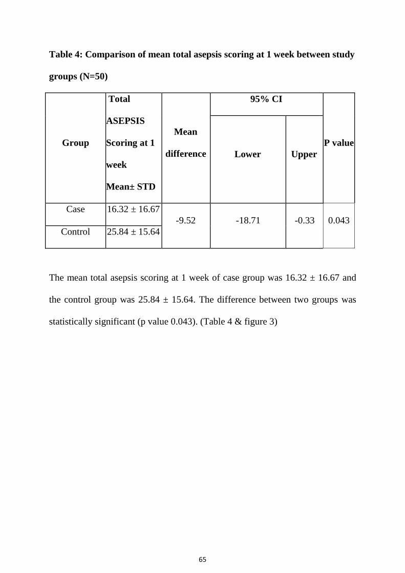
Table 4: Comparison of mean total asepsis scoring at 1 week between study
groups (N=50)
Group
Total
ASEPSIS
Scoring at 1
week
Mean± STD
Mean
difference
95% CI
P value
Lower Upper
Case 16.32 ± 16.67
-9.52 -18.71 -0.33 0.043
Control 25.84 ± 15.64
The mean total asepsis scoring at 1 week of case group was 16.32 ± 16.67 and
the control group was 25.84 ± 15.64. The difference between two groups was
statistically significant (p value 0.043). (Table 4 & figure 3)

66
Figure 8: Comparison of Total ASEPSIS scoring at 1 week between the two
groups (N=50)

67
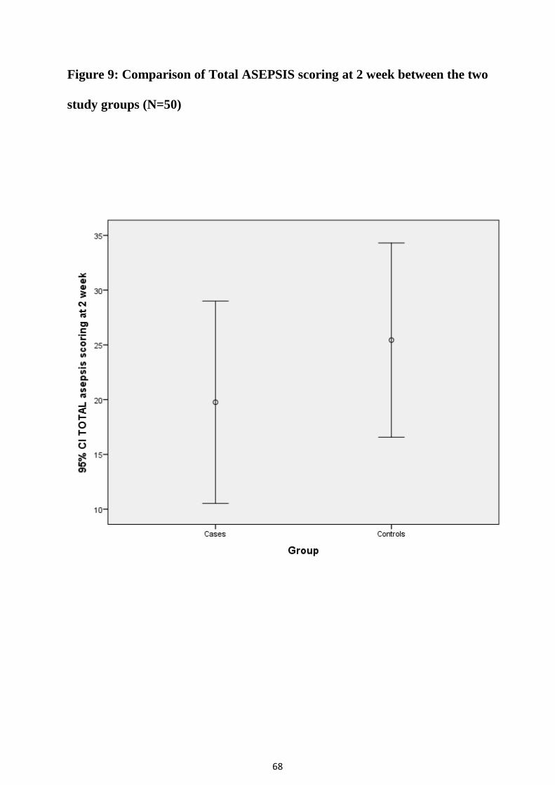
Table 5: Comparison of mean total ASEPSIS scoring at 2 week across the
two groups (N=50)
Group
Total
ASEPSIS
Scoring at 2
week
Mean± STD
Mean
difference
95% CI
P value
Lower Upper
Case 19.76 ± 22.38
-5.68 -18.15 6.79 0.365
Control 25.44 ± 21.48
The mean total asepsis scoring at 2 week of case group was 19.76 ± 22.38 and
the control group was 25.44 ± 21.48. The difference between two groups was
statistically not significant (p value 0.365). (Table 5 & figure 4)
68
Figure 9: Comparison of Total ASEPSIS scoring at 2 week between the two
study groups (N=50)
69
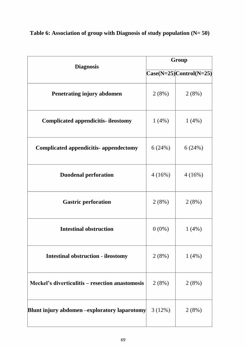
Table 6: Association of group with Diagnosis of study population (N= 50)
Diagnosis
Group
Case(N=25) Control(N=25)
Penetrating injury abdomen 2 (8%) 2 (8%)
Complicated appendicitis- ileostomy 1 (4%) 1 (4%)
Complicated appendicitis- appendectomy 6 (24%) 6 (24%)
Duodenal perforation 4 (16%) 4 (16%)
Gastric perforation 2 (8%) 2 (8%)
Intestinal obstruction 0 (0%) 1 (4%)
Intestinal obstruction - ileostomy 2 (8%) 1 (4%)
Meckel’s diverticulitis – resection anastomosis 2 (8%) 2 (8%)
Blunt injury abdomen –exploratory laparotomy 3 (12%) 2 (8%)
70
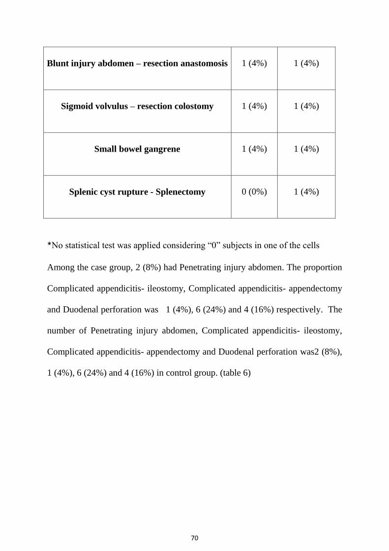
Blunt injury abdomen – resection anastomosis 1 (4%) 1 (4%)
Sigmoid volvulus – resection colostomy 1 (4%) 1 (4%)
Small bowel gangrene 1 (4%) 1 (4%)
Splenic cyst rupture - Splenectomy 0 (0%) 1 (4%)
*No statistical test was applied considering “0” subjects in one of the cells
Among the case group, 2 (8%) had Penetrating injury abdomen. The proportion
Complicated appendicitis- ileostomy, Complicated appendicitis- appendectomy
and Duodenal perforation was 1 (4%), 6 (24%) and 4 (16%) respectively. The
number of Penetrating injury abdomen, Complicated appendicitis- ileostomy,
Complicated appendicitis- appendectomy and Duodenal perforation was2 (8%),
1 (4%), 6 (24%) and 4 (16%) in control group. (table 6)
71
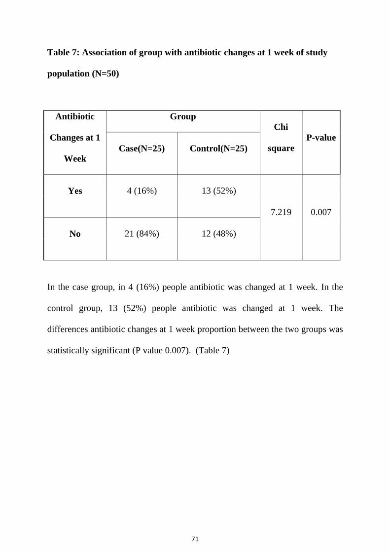
Table 7: Association of group with antibiotic changes at 1 week of study
population (N=50)
Antibiotic
Changes at 1
Week
Group
Chi
square
P-value
Case(N=25) Control(N=25)
Yes 4 (16%) 13 (52%)
7.219 0.007
No 21 (84%) 12 (48%)
In the case group, in 4 (16%) people antibiotic was changed at 1 week. In the
control group, 13 (52%) people antibiotic was changed at 1 week. The
differences antibiotic changes at 1 week proportion between the two groups was
statistically significant (P value 0.007). (Table 7)
72
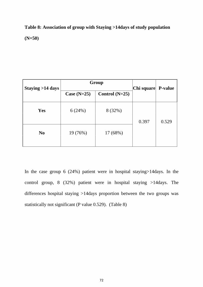
Table 8: Association of group with Staying >14days of study population
(N=50)
Staying >14 days
Group
Chi square P-value
Case (N=25) Control (N=25)
Yes 6 (24%) 8 (32%)
0.397 0.529
No 19 (76%) 17 (68%)
In the case group 6 (24%) patient were in hospital staying>14days. In the
control group, 8 (32%) patient were in hospital staying >14days. The
differences hospital staying >14days proportion between the two groups was
statistically not significant (P value 0.529). (Table 8)
73
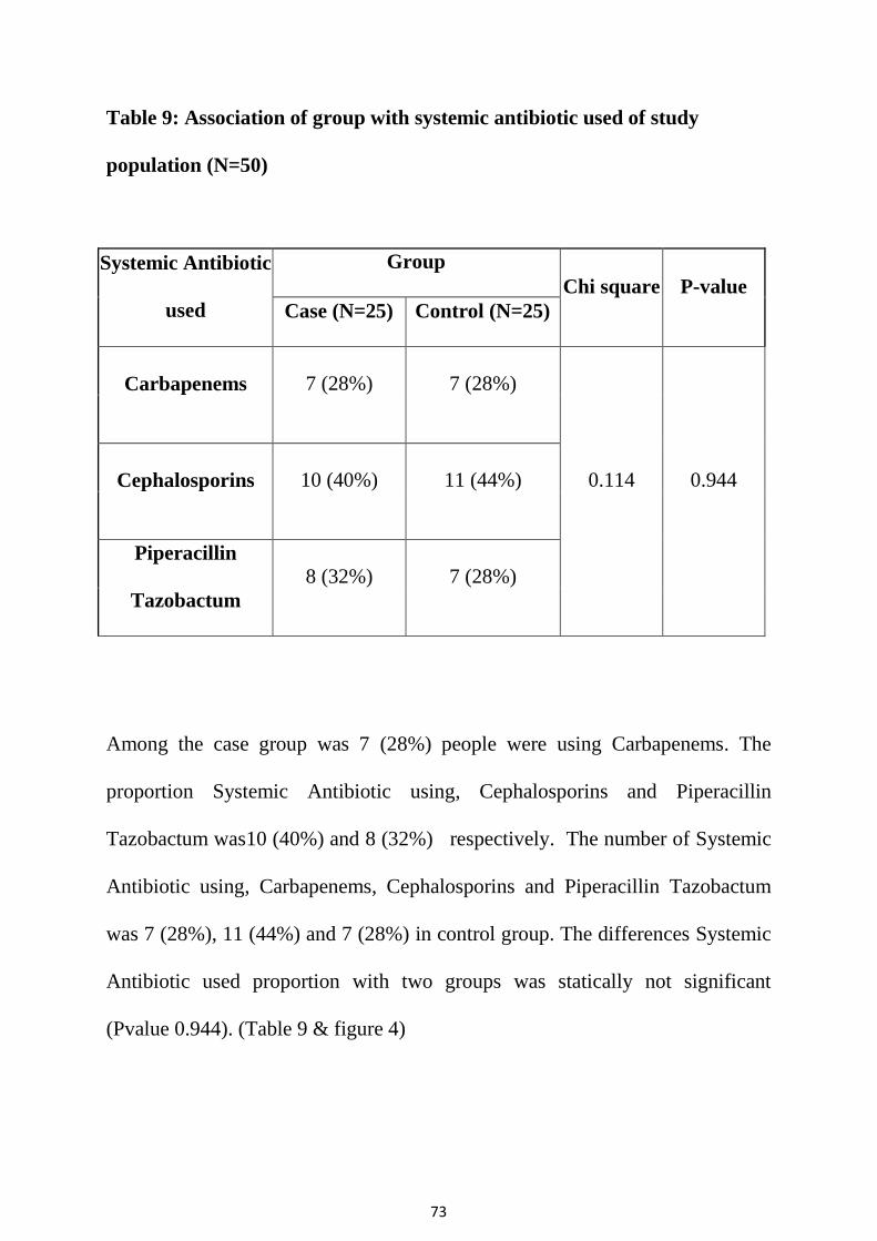
Table 9: Association of group with systemic antibiotic used of study
population (N=50)
Systemic Antibiotic
used
Group
Chi square P-value
Case (N=25) Control (N=25)
Carbapenems 7 (28%) 7 (28%)
0.114 0.944 Cephalosporins 10 (40%) 11 (44%)
Piperacillin
Tazobactum
8 (32%) 7 (28%)
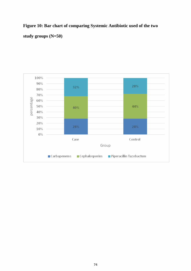
Among the case group was 7 (28%) people were using Carbapenems. The
proportion Systemic Antibiotic using, Cephalosporins and Piperacillin
Tazobactum was10 (40%) and 8 (32%) respectively. The number of Systemic
Antibiotic using, Carbapenems, Cephalosporins and Piperacillin Tazobactum
was 7 (28%), 11 (44%) and 7 (28%) in control group. The differences Systemic
Antibiotic used proportion with two groups was statically not significant
(Pvalue 0.944). (Table 9 & figure 4)
74
Figure 10: Bar chart of comparing Systemic Antibiotic used of the two
study groups (N=50)

75
CONCLUSION

76
CONCLUSION
This prospective, interventional and comparative study was
conducted among 50 purposively selected patients who underwent
abdominal surgeries categorized as dirty and contaminated wounds in the
department of General Surgery, Stanley Medical College and Hospital
from NOV-2016 TO AUG 2017.
The study was conducted to analyse the effectiveness of
using local antibiotic over the wound site to prevent surgical site
infections. The SSIs were graded using on of the standard methods of
grading ASEPSIS scoring system, which grades the SSIs from 0 to 70
assessing various parameters. The scoring was done for 1st and 2
nd week
after surgery.
The cases and controls were sufficiently matched against age, sex,
age, antibiotics used, the type of surgical diagnosis and treatment given,
the type of surgical wound. Differences found to be statistically
insignificant.
Subsequently the ASEPSIS scores at the end of 1st week of surgery
showed that the study group patients who received the Inj.Amikacin in
the local wound site showed significantly lesser grade of SSIs compared
to that of the control group. The ASEPSIS score at the end of 2nd
week of

77
surgery showed lesser grade of SSIs in the study group compared to the
control though it was statistically insignificant.
The probability of antibiotic change and duration of stay in the
patient was lesser in the study group though the later parameter was not
statistically significant.
Hence overall conclusion is that the patients who received local
wound site antibiotic (Inj.Amikacin) showed lesser grades of SSIs , more
so in the 1st week of surgery and lesser need for antibiotic change and
lesser duration of stay in hospital during the postoperative period
compared to the control group which only received the systemic
antibiotics.
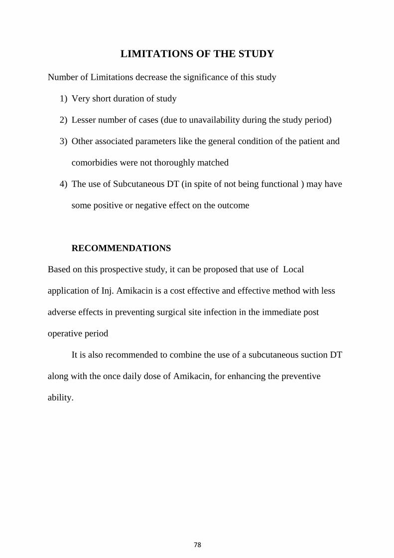
78
LIMITATIONS OF THE STUDY
Number of Limitations decrease the significance of this study
1) Very short duration of study
2) Lesser number of cases (due to unavailability during the study period)
3) Other associated parameters like the general condition of the patient and
comorbidies were not thoroughly matched
4) The use of Subcutaneous DT (in spite of not being functional ) may have
some positive or negative effect on the outcome
RECOMMENDATIONS
Based on this prospective study, it can be proposed that use of Local
application of Inj. Amikacin is a cost effective and effective method with less
adverse effects in preventing surgical site infection in the immediate post
operative period
It is also recommended to combine the use of a subcutaneous suction DT
along with the once daily dose of Amikacin, for enhancing the preventive
ability.
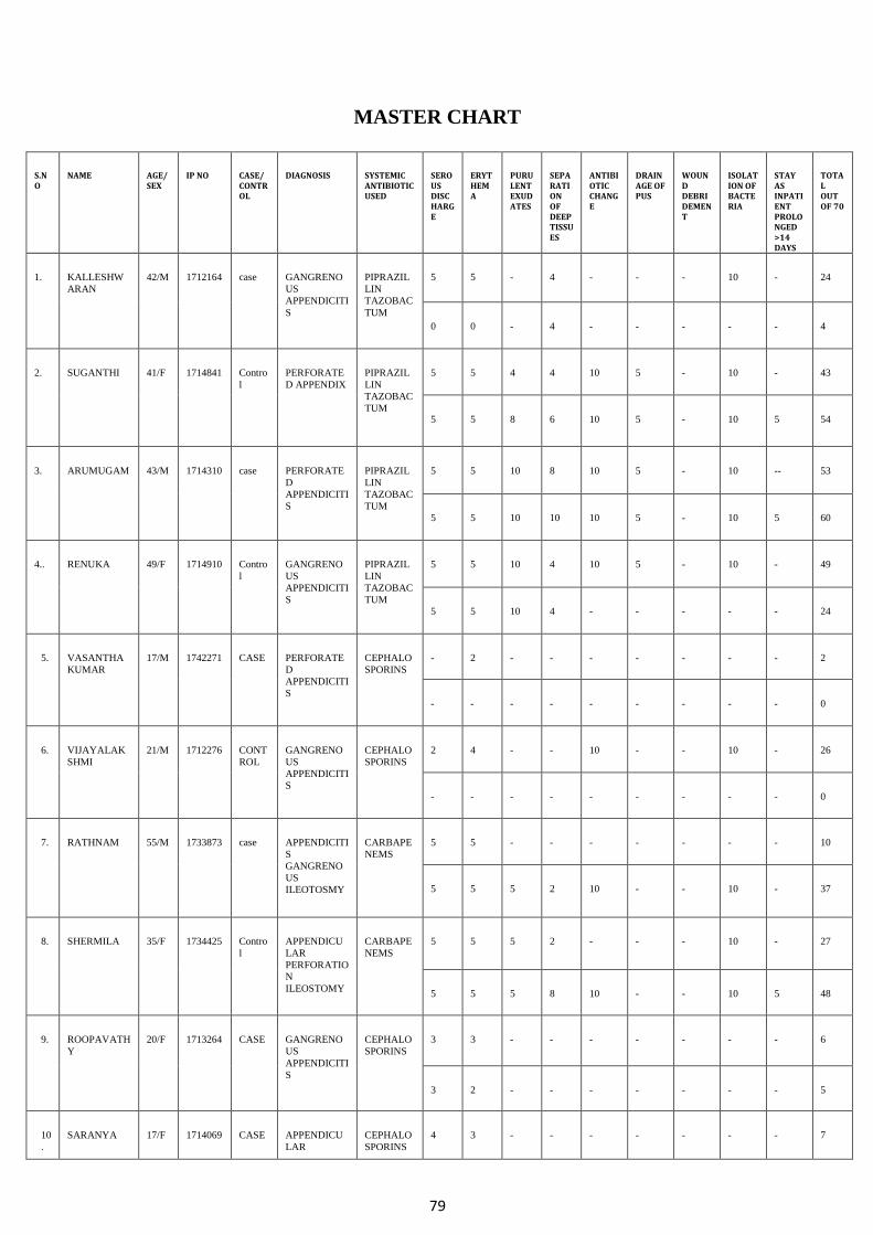
79
MASTER CHART
S.NO
NAME AGE/SEX
IP NO CASE/ CONTROL
DIAGNOSIS SYSTEMIC ANTIBIOTIC USED
SEROUS DISCHARGE
ERYTHEMA
PURULENT EXUDATES
SEPARATION OF DEEP TISSUES
ANTIBIOTIC CHANGE
DRAINAGE OF PUS
WOUND DEBRIDEMENT
ISOLATION OF BACTERIA
STAY AS INPATIENT PROLONGED >14 DAYS
TOTAL OUT OF 70
1. KALLESHW
ARAN
42/M 1712164 case GANGRENO
US
APPENDICITI
S
PIPRAZIL
LIN
TAZOBAC
TUM
5 5 - 4 - - - 10 - 24
0 0 - 4 - - - - - 4
2. SUGANTHI 41/F 1714841 Contro
l
PERFORATE
D APPENDIX
PIPRAZIL
LIN
TAZOBAC
TUM
5 5 4 4 10 5 - 10 - 43
5 5 8 6 10 5 - 10 5 54
3. ARUMUGAM 43/M 1714310 case PERFORATE
D
APPENDICITI
S
PIPRAZIL
LIN
TAZOBAC
TUM
5 5 10 8 10 5 - 10 -- 53
5 5 10 10 10 5 - 10 5 60
4.. RENUKA 49/F 1714910 Contro
l
GANGRENO
US
APPENDICITI
S
PIPRAZIL
LIN
TAZOBAC
TUM
5 5 10 4 10 5 - 10 - 49
5 5 10 4 - - - - - 24
5. VASANTHA
KUMAR
17/M 1742271 CASE PERFORATE
D
APPENDICITI
S
CEPHALO
SPORINS
- 2 - - - - - - - 2
- - - - - - - - - 0
6. VIJAYALAK
SHMI
21/M 1712276 CONT
ROL
GANGRENO
US
APPENDICITI
S
CEPHALO
SPORINS
2 4 - - 10 - - 10 - 26
- - - - - - - - - 0
7. RATHNAM 55/M 1733873 case APPENDICITI
S
GANGRENO
US
ILEOTOSMY
CARBAPE
NEMS
5 5 - - - - - - - 10
5 5 5 2 10 - - 10 - 37
8. SHERMILA 35/F 1734425 Contro
l
APPENDICU
LAR
PERFORATIO
N
ILEOSTOMY
CARBAPE
NEMS
5 5 5 2 - - - 10 - 27
5 5 5 8 10 - - 10 5 48
9. ROOPAVATH
Y
20/F 1713264 CASE GANGRENO
US
APPENDICITI
S
CEPHALO
SPORINS
3 3 - - - - - - - 6
3 2 - - - - - - - 5
10
.
SARANYA 17/F 1714069 CASE APPENDICU
LAR
CEPHALO
SPORINS
4 3 - - - - - - - 7
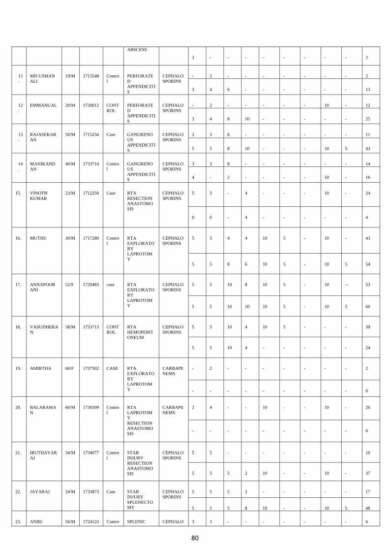
80
ABSCESS
2 - - - - - - - - 2
11
.
MD USMAN
ALI
19/M 1713548 Contro
l
PERFORATE
D
APPENDICITI
S
CEPHALO
SPORINS
- 2 - - - - - - - 2
3 4 6 - - - - - - 13
12
.
EMMANUAL 20/M 1720012 CONT
ROL
PERFORATE
D
APPENDICITI
S
CEPHALO
SPORINS
- 2 - - - - - 10 - 12
3 4 8 10 - - - - - 25
13
.
RAJASEKAR
AN
50/M 1715234 Case GANGRENO
US
APPENDICITI
S
CEPHALO
SPORINS
2 3 6 - - - - - - 11
5 5 8 10 - - - 10 5 43
14
.
MANIKAND
AN
40/M 1733714 Contro
l
GANGRENO
US
APPENDICITI
S
CEPHALO
SPORINS
3 3 8 - - - - - - 14
4 - 2 - - - - 10 - 16
15. VINOTH
KUMAR
23/M 1712250 Case RTA
RESECTION
ANASTOMO
SIS
CEPHALO
SPORINS
5 5 - 4 - - - 10 - 24
0 0 - 4 - - - - - 4
16. MUTHU 30/M 1717280 Contro
l
RTA
EXPLORATO
RY
LAPROTOM
Y
CEPHALO
SPORINS
5 5 4 4 10 5 - 10 - 43
5 5 8 6 10 5 - 10 5 54
17. ANNAPOOR
ANI
52/F 1720483 case RTA
EXPLORATO
RY
LAPROTOM
Y
CEPHALO
SPORINS
5 5 10 8 10 5 - 10 -- 53
5 5 10 10 10 5 - 10 5 60
18. VASUDHERA
N
38/M 1733713 CONT
ROL
RTA
HEMOPERIT
ONEUM
CEPHALO
SPORINS
5 5 10 4 10 5 - - - 39
5 5 10 4 - - - - - 24
19. AMIRTHA 60/F 1737502 CASE RTA
EXPLORATO
RY
LAPROTOM
Y
CARBAPE
NEMS
- 2 - - - - - - - 2
- - - - - - - - - 0
20. BALARAMA
N
60/M 1730509 Contro
l
RTA
LAPROTOM
Y
RESECTION
ANASTOMO
SIS
CARBAPE
NEMS
2 4 - - 10 - - 10 - 26
- - - - - - - - - 0
21. IRUTHAYAR
AJ
34/M 1734077 Contro
l
STAB
INJURY
RESECTION
ANASTOMO
SIS
CEPHALO
SPORINS
5 5 - - - - - - - 10
5 5 5 2 10 - - 10 - 37
22. JAYARAJ 24/M 1733873 Case STAB
INJURY
SPLENECTO
MY
CEPHALO
SPORINS
5 5 5 2 - - - - - 17
5 5 5 8 10 - - 10 5 48
23. ANBU 56/M 1724123 Contro SPLENIC CEPHALO 3 3 - - - - - - - 6
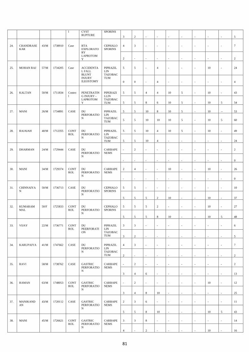
81
l CYST
RUPTURE
SPORINS
3 2 - - - - - - - 5
24. CHANDRASE
KAR
43/M 1738910 Case RTA
EXPLORATO
RY
LAPROTOM
Y
CEPHALO
SPORINS
4 3 - - - - - - - 7
2 - - - - - - - - 2
25. MOHAN RAJ 57/M 1734205 Case ACCIDENTA
L FALL
BLUNT
INJURY
ILEOSTOMY
PIPRAZIL
LIN
TAZOBAC
TUM
5 5 - 4 - - - 10 - 24
0 0 - 4 - - - - - 4
26. KALTAN 50/M 1711834 Contro
l
PENETRATIN
G INJURY -
LAPROTOM
Y
PIPERAZI
LLIN
TAZOBAC
TUM
5 5 4 4 10 5 - 10 - 43
5 5 8 6 10 5 - 10 5 54
27. MANI 26/M 1734891 CASE DU
PERFORATIO
N
PIPRAZIL
LIN
TAZOBAC
TUM
5 5 10 8 10 5 - 10 -- 53
5 5 10 10 10 5 - 10 5 60
28. RAJAIAH 40/M 1712355 CONT
ROL
DU
PERFORATIO
N
PIPRAZIL
LIN
TAZOBAC
TUM
5 5 10 4 10 5 - 10 - 49
5 5 10 4 - - - - - 24
29. DHARMAN 24/M 1729444 CASE DU
PERFORATIO
N
CARBAPE
NEMS
- 2 - - - - - - - 2
- - - - - - - - - 0
30. MANI 34/M 1729374 CONT
ROL
DU
PERFORATIO
N
CARBAPE
NEMS
2 4 - - 10 - - 10 - 26
- - - - - - - - - 0
31. CHINNAIYA
N
50/M 1736713 CASE DU
PERFORATIO
N
CEPHALO
SPORINS
5 5 - - - - - - - 10
5 5 5 2 10 - - 10 - 37
32. KUMARAM
MAL
50/F 1725833 CONT
ROL
DU
PERFORATIO
N
CEPHALO
SPORINS
5 5 5 2 - - - 10 - 27
5 5 5 8 10 - - 10 5 48
33. VIJAY 22/M 1736771 CONT
ROL
DU
PEERFORATI
ON
PIPRAZIL
LIN
TAZOBAC
TUM
3 3 - - - - - - - 6
3 2 - - - - - - - 5
34. KARUPAIYA 41/M 1747662 CASE DU
PERFORATIO
N
PIPRAZIL
LIN
TAZOBAC
TUM
4 3 - - - - - - - 7
2 - - - - - - - - 2
35. RAVI 58/M 1738762 CASE GASTRIC
PERFORATIO
N
CARBAPE
NEMS
- 2 - - - - - - - 2
3 4 6 - - - - - - 13
36. RAMAN 63/M 1748053 CONT
ROL
GASTRIC
PERFORATIO
N
CARBAPE
NEMS
- 2 - - - - - 10 - 12
3 4 8 10 - - - - - 25
37. MANIKAND
AN
43/M 1720112 CASE GASTRIC
PERFORATIO
N
CARBAPE
NEMS
2 3 6 - - - - - - 11
5 5 8 10 - - - 10 5 43
38. MANI 45/M 1726621 CONT
ROL
GASTRIC
PERFORATIO
N
CARBAPE
NEMS
3 3 8 - - - - - - 14
4 - 2 - - - - 10 - 16
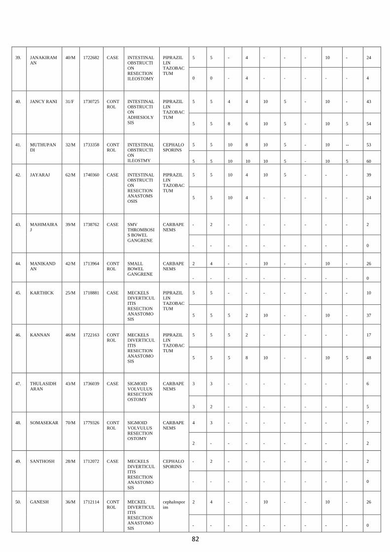
82
39. JANAKIRAM
AN
40/M 1722682 CASE INTESTINAL
OBSTRUCTI
ON
RESECTION
ILEOSTOMY
PIPRAZIL
LIN
TAZOBAC
TUM
5 5 - 4 - - - 10 - 24
0 0 - 4 - - - - - 4
40. JANCY RANI 31/F 1730725 CONT
ROL
INTESTINAL
OBSTRUCTI
ON
ADHESIOLY
SIS
PIPRAZIL
LIN
TAZOBAC
TUM
5 5 4 4 10 5 - 10 - 43
5 5 8 6 10 5 - 10 5 54
41. MUTHUPAN
DI
32/M 1733358 CONT
ROL
INTESTINAL
OBSTRUCTI
ON
ILEOSTMY
CEPHALO
SPORINS
5 5 10 8 10 5 - 10 -- 53
5 5 10 10 10 5 - 10 5 60
42. JAYARAJ 62/M 1740360 CASE INTESTINAL
OBSTRUCTI
ON
RESECTION
ANASTOMS
OSIS
PIPRAZIL
LIN
TAZOBAC
TUM
5 5 10 4 10 5 - - - 39
5 5 10 4 - - - - - 24
43. MAHIMAIRA
J
39/M 1738762 CASE SMV
THROMBOSI
S BOWEL
GANGRENE
CARBAPE
NEMS
- 2 - - - - - - - 2
- - - - - - - - - 0
44. MANIKAND
AN
42/M 1713964 CONT
ROL
SMALL
BOWEL
GANGRENE
CARBAPE
NEMS
2 4 - - 10 - - 10 - 26
- - - - - - - - - 0
45. KARTHICK 25/M 1718881 CASE MECKELS
DIVERTICUL
ITIS
RESECTION
ANASTOMO
SIS
PIPRAZIL
LIN
TAZOBAC
TUM
5 5 - - - - - - - 10
5 5 5 2 10 - - 10 - 37
46. KANNAN 46/M 1722163 CONT
ROL
MECKELS
DIVERTICUL
ITIS
RESECTION
ANASTOMO
SIS
PIPRAZIL
LIN
TAZOBAC
TUM
5 5 5 2 - - - - - 17
5 5 5 8 10 - - 10 5 48
47. THULASIDH
ARAN
43/M 1736039 CASE SIGMOID
VOLVULUS
RESECTION
OSTOMY
CARBAPE
NEMS
3 3 - - - - - - - 6
3 2 - - - - - - - 5
48. SOMASEKAR 70/M 1779326 CONT
ROL
SIGMOID
VOLVULUS
RESECTION
OSTOMY
CARBAPE
NEMS
4 3 - - - - - - - 7
2 - - - - - - - - 2
49. SANTHOSH 28/M 1712072 CASE MECKELS
DIVERTICUL
ITIS
RESECTION
ANASTOMO
SIS
CEPHALO
SPORINS
- 2 - - - - - - - 2
- - - - - - - - - 0
50. GANESH 36/M 1712114 CONT
ROL
MECKEL
DIVERTICUL
ITIS
RESECTION
ANASTOMO
SIS
cephalospor
ins
2 4 - - 10 - - 10 - 26
- - - - - - - - - 0

83
BIBLIOGRAPHY
(1) Smyth ET, McIlvenny G, Enstone JE, et al. Four Country Healthcare
Associated Infection Prevalence Survey 2006: overview of the results.
Journal of Hospital Infection 2008;69:230–48.
(2) Townsend CM. Sabiston Textbook of Surgery. In Courtney M. Townsend,
R, Daniel Beauchame, B. Mark Evers, Kenneth L. Mottox (eds). Surgical
infections and choice of antibiotics. 17th edition, Vol. 1, W.B. Saunders,€;
2005:257-283.
(3) Cruse PJ, Ford R. The epidemiology of wound infection- a 10 year
prospective study of 62939 wounds. Sug Clin N Am. 1980;60(1):27-40.
(4). Kulaylat MN, Dayton MT. Surgical complications. 19ed. Chapter 13. In:
Sabiston Text book of Surgery. Townsend Beauchamp, Evers Mattox, eds.
Philadelphia: Saunders Elsevier; 2012:286.
(5) Horan TC, Gaynes RP, Martone WJ, Jarvis WR, Emori TG. CDC definitions
of nosocomial surgical site infections, 1992: a modification of CDC definitions
of surgical wound infections. Infect Control Hosp Epidemiol. 1992;13(10):606–
608.
(6). Barie PS, Wilson SE. Impact of evolving epidemiology on treatments for
complicated skin and skin structure infections: the surgical perspective. J Am
Coll Surg. 2015;220(1):105–106.
(7) Horan TC, Andrus M, Dudeck MA. CDC/NHSN surveillance definition of
health care-associated infection and criteria for specific types of infections in
the acute care setting. Am J Infect Control. 2008;36(5): 309–332.
(8). Horan TC, Gaynes RP, Martone WJ, et al. CDC definitions of nosocomial
surgical site infections, 1992: a modification of CDC definitions of
surgical wound infections. Infection Control and Hospital Epidemiology
1992;13:606–8.
84
(9). Ridgeway S, Wilson J, Charlet A, et al. Infection of the surgical site after
arthroplasty of the hip. Journal of Bone and Joint Surgery – British
Volume 2005;87:844–50.
(10) Bailey IS, Karran SE, Toyn K, et al. Community surveillance of
complications after hernia surgery. British Medical Journal
1992;304:469–71.
(11). Wilson AP, Treasure T, Sturridge MF, et al. A scoring method (ASEPSIS)
for postoperative wound infections for use in clinical trials of
antibiotic prophylaxis. Lancet 1986;1:311–13.
(12) Health Protection Agency. Surveillance of Surgical Site Infection in
England: October 1997–September 2005. London: Health Protection
Agency; 2006.
(13) Department of Health CMO. Winning Ways: Working Together to Reduce
Healthcare Associated Infection in England. London: Department
of Health; 2003.
(14). Cruse PJ, Foord R. The epidemiology of wound infection. A 10-year
prospective study of 62,939 wounds. Surgical Clinics of North America
1980;60:27–40.
(15). Haley RW, Culver DH, White JW, et al. The efficacy of infection
surveillance and control programs in preventing nosocomial infections in
US hospitals. American Journal of Epidemiology 1985;121:182–205.
(16). Glenister HM, Taylor LJ, Cooke EM, Bartlett CLR. A Study of
Surveillance Methods for Detecting Hospital Infection. London: Public Health
Laboratory Service; 1992.
(17). Culver DH, Horan TC, Gaynes RP, et al. Surgical wound infection rates by
wound class, operative procedure, and patient risk index.
National Nosocomial Infections Surveillance System. American Journal of
Medicine 1991;91(3B):152S–7S.
85
(18). Wilson J, Ramboer I, Suetens C, et al. Hospitals in Europe Link for
Infection Control through Surveillance (HELICS). Inter-country comparison
of rates of surgical site infection – opportunities and limitations. Journal of
Hospital Infection 2007;65 Suppl 2:165–70.
(19). Mannien J, Wille JC, Snoeren RL, et al. Impact of postdischarge
surveillance on surgical site infection rates for several surgical procedures:
results from the nosocomial surveillance network in The Netherlands. Infection
Control and Hospital Epidemiology 2006;27:809–16.
(20). Whitby M, McLaws ML, Collopy B, et al. Post-discharge surveillance: can
patients reliably diagnose surgical wound infections? Journal of
Hospital Infection 2002;52:155–60.
(21). Neumayer L, Hosokawa P, Itani K, et al. Multivariable predictors of
postoperative surgical site infection after general and vascular surgery:
results from the patient safety in surgery study. Journal of the American College
of Surgeons 2007;204:1178–87.
(22). Scott JD, Forrest A, Feurstein S, et al. Factors associated with
postoperative infection. Infection Control and Hospital Epidemiology
2001;22:347–51.
(23). Cruse PJ, Foord R. A five-year prospective study of 23,649 surgical
wounds. Archives of Surgery 1973;107:206–10.
(24). Kaye KS, Schmit K, Pieper C, et al. The effect of increasing age on the
risk of surgical site infection. Journal of Infectious Diseases
2005;191:1056–62.
(25). Friedman ND, Sexton DJ, Connelly SM, et al. Risk factors for surgical site
infection complicating laminectomy. Infection Control and
Hospital Epidemiology 2007;28:1060–5.
(26). Ridderstolpe L, Gill H, Granfeldt H, et al. Superficial and deep sternal
wound complications: Incidence, risk factors and mortality. European
Journal of Cardio-Thoracic Surgery 2001;20:1168–75.
86
(27). Latham R, Lancaster AD, Covington JF, et al. The association of diabetes
and glucose control with surgical-site infections among cardiothoracic
surgery patients. Infection Control and Hospital Epidemiology 2001;22:607–12.
(28). Russo PL, Spelman DW. A new surgical-site infection risk index using
risk factors identified by multivariate analysis for patients undergoing
coronary artery bypass graft surgery. Infection Control and Hospital
Epidemiology 2002;23:372–6.
(29). Abboud CS, Wey SB, Baltar VT. Risk factors for mediastinitis after
cardiac surgery. Annals of Thoracic Surgery 2004;77:676–83.
(30). Olsen MA, Mayfield J, Lauryssen C, et al. Risk factors for surgical site
infection in spinal surgery. Journal of Neurosurgery
2003;98:149–55.
(31). Anderson DJ, Pyatt DG, Weber DJ, Rutala WA. Statewide costs
of health care-associated infections: estimates for acute care
hospitals in North Carolina. Am J Infect Control 2013;41(9):
764-768.
(32). Lewis SS, Moehring RW, Chen LF, Sexton DJ, Anderson DJ.
Assessing the relative burden of hospital-acquired infections in
a network of community hospitals. Infect Control Hosp Epidemiol
2013;34(11):1229-1230.
(33). Zimlichman E, Henderson D, Tamir O, et al. Health careassociated
infections: a meta-analysis of costs and financial
impact on the us health care system. JAMA Intern Med 2013;
173(22):2039-2046.
(34). Mangram AJ, Horan TC, Pearson ML, Silver LC, Jarvis WR;
Hospital Infection Control Practices Advisory Committee.
Guideline for prevention of surgical site infection, 1999. Infect
Control Hosp Epidemiol 1999;20(4):250-278.
87
(35) . Antimicrobial prophylaxis for surgery. Treat Guidel Med Lett
2009;7(82):47-52.
(36). Bratzler DW, Dellinger EP, Olsen KM, et al. Clinical practice
guidelines for antimicrobial prophylaxis in surgery. Am J Health
Syst Pharm 2013;70(3): 195-283.
(37). Bratzler DW, Houck PM. Antimicrobial prophylaxis for surgery:
an advisory statement from the National Surgical Infection
Prevention Project. Clin Infect Dis 2004;38(12):1706-1715.
(38). Bratzler DW, Hunt DR. The surgical infection prevention and
surgical care improvement projects: national initiatives to improve
outcomes for patients having surgery. Clin Infect Dis
2006;43(3):322-330.
(39) Steinberg JP, Braun BI, Hellinger WC, et al. Timing of antimicrobial
prophylaxis and the risk of surgical site infections:
results from the Trial to Reduce Antimicrobial Prophylaxis Errors.
Ann Surg 2009;250(1):10-16.
(40). van Kasteren ME, Mannien J, Ott A, Kullberg BJ, de Boer AS,
Gyssens IC. Antibiotic prophylaxis and the risk of surgical site
infections following total hip arthroplasty: timely administration
is the most important factor. Clin Infect Dis 2007;44(7):
921-927.
(41). Harbarth S, Samore MH, Lichtenberg D, Carmeli Y. Prolonged
antibiotic prophylaxis after cardiovascular surgery and its effect
on surgical site infections and antimicrobial resistance. Circulation
2000;101(25):2916-2921.
(42). McDonald M, Grabsch E, Marshall C, Forbes A. Single- versus
multiple-dose antimicrobial prophylaxis for major surgery: a
systematic review. Aust N ZJ Surg 1998;68(6):388-396.
88
(43). Coakley BA, Sussman ES, Wolfson TS, et al. Postoperative antibiotics
correlate with worse outcomes after appendectomy for
nonperforated appendicitis. J Am Coll Surg 2011;213(6):778-
783.
(44). Cannon JA, Altom LK, Deierhoi RJ, et al. Preoperative oral
antibiotics reduce surgical site infection following elective colorectal
resections. Dis Colon Rectum 2012;55(11):1160-1166.
(45). Englesbe MJ, Brooks L, Kubus J, et al. A statewide assessment
of surgical site infection following colectomy: the role of oral
antibiotics. Ann Surg 2010;252(3):514-519.
(46). Fry DE. Colon preparation and surgical site infection. Am J
Surg 2011;202(2):225-232.
(47). Hendren S, Fritze D, Banerjee M, et al. Antibiotic choice is
independently associated with risk of surgical site infection
after colectomy: a population-based cohort study. Ann Surg
2013;257(3):469-475.
(48). Lewis RT. Oral versus systemic antibiotic prophylaxis in elective
colon surgery: a randomized study and meta-analysis send a
message from the 1990s. Can J Surg 2002;45(3):173-180.
(49). Nelson RL, Glenny AM, Song F. Antimicrobial prophylaxis for
colorectal surgery. Cochrane Database Syst Rev 2009;(1):
CD001181.
(50). Deierhoi RJ, Dawes LG, Vick C, Itani KM, Hawn MT. Choice
of intravenous antibiotic prophylaxis for colorectal surgery
does matter. J Am Coll Surg 2013;217(5):763-769.
(51). Toneva GD, Deierhoi RJ, Morris M, et al. Oral antibiotic bowel
preparation reduces length of stay and readmissions after colorectal
surgery. ] Am Coll Surg 2013;216(4):756-762.
89
(52). Tanner J, Woodings D, Moncaster K. Preoperative hair removal
to reduce surgical site infection. Cochrane Database Syst Rev
2006;(3):CD004122.
(53). Dronge AS, Perkal MF, Kancir S, Concato J, Asian M, Rosenthal
RA. Long-term glycemic control and postoperative infectious
complications. Arch Surg 2006;141(4):375-380.
(54). Golden SH, Peart-Vigilance C, Kao WH, Brancati FL. Perioperative
glycemic control and the risk of infectious complications
in a cohort of adults with diabetes. Diabetes Care 1999;
22(9):1408-1414.
(55). Olsen MA, Nepple JJ, Riew KD, et al. Risk factors for surgical
site infection following orthopaedic spinal operations. / Bone
Joint Surg Am 2008;90(l):62-69.
(56). Kwon S, Thompson R, Dellinger P, Yanez D, Farrohki E, Flum
D. Importance of perioperative glycemic control in general
surgery: a report from the Surgical Care and Outcomes Assessment
Program. Ann Surg 2013;257(1):8-14.
(57). Umpierrez GE, Smiley D, Jacobs S, et al. Randomized study
of basal-bolus insulin therapy in the inpatient management of
patients with type 2 diabetes undergoing general surgery
(RABBIT 2 surgery). Diabetes Care 2011;34(2):256-261.
(58). Jacobi J, Bircher N, Krinsley J, et al. Guidelines for the use of
an insulin infusion for the management of hyperglycemia in
critically ill patients. Crit Care Med 2012;40(12):3251-3276.
(59). Umpierrez GE, Hellman R, Korytkowski MT, et al. Management
of hyperglycemia in hospitalized patients in non-critical
care setting: an endocrine society clinical practice guideline. /
Clin Endocrinol Metab 2012;97(l):16-38.
(60). Gandhi GY, Nuttall GA, Abel MD, et al. Intensive intraoperative
90
insulin therapy versus conventional glucose management
during cardiac surgery: a randomized trial. Ann Intern Med
2007;146(4):233-243.
(61). Sessler DI. Complications and treatment of mild hypothermia.
Anesthesiology 2001;95(2):531-543.
(62). Kurz A, Sessler DI, Lenhardt R; Study of Wound Infection and
Temperature Group. Perioperative normothermia to reduce the
incidence of surgical-wound infection and shorten hospitalization.
N Engl J Med 1996;334(19):1209-1215.
(63). Melling AC, Ali B, Scott EM, Leaper DJ. Effects of preoperative
warming on the incidence of wound infection after clean surgery:
a randomised controlled trial. Lancet 2001;358(9285):
876-880.
(64). Wong PF, Kumar S, Bohra A, Whetter D, Leaper DJ. Randomized
clinical trial of perioperative systemic warming in major
elective abdominal surgery. Br J Surg 2007;94(4):421-426.
(65). Darouiche RO, Wall MJ Jr, Itani KM, et al. Chlorhexidinealcohol
versus povidone-iodine for surgical-site antisepsis. N
Engl J Med 2010;362(l):18-26.
(66). Swenson BR, Hedrick TL, Metzger R, Bonatti H, Pruett TL,
Sawyer RG. Effects of preoperative skin preparation on postoperative
wound infection rates: a prospective study of 3 skin
preparation protocols. Infect Control Hosp Epidemiol 2009;
30(10):964-971.
(67). 119. Larson E. Guideline for use of topical antimicrobial agents. Am }
Infect Control 1988;16(6):253-266.
(68). Whiteside OJ, Tytherleigh MG, Thrush S, Farouk R, Galland
RB. Intra-operative peritoneal lavage—who does it and why?
Ann R Coll Surg Engl 2005;87(4):255-258.

91
(69). de Bruin AF, Gosselink MP, van der Harst E, Rutten HJ. Local
application of gentamicin collagen implants in the prophylaxis
of surgical site infections following gastrointestinal surgery: a
review of clinical experience. Tech Co/oprocto/2010;14(4):301-310.
(70). Guzman Valdivia Gomez G, Guerrero TS, Lluck MC, Delgado
FJ. Effectiveness of collagen-gentamicin implant for treatment
of "dirty" abdominal wounds. World J Surg 1999;23(2):123-126.
(71). Rutten HJ, Nijhuis PH. Prevention of wound infection in elective
colorectal surgery by local application of a gentamicincontaining
collagen sponge. Eur J Surg Suppl 1997;(578):31- 35.
(72). Eklund AM, Valtonen M, Werkkala KA. Prophylaxis of sternal
wound infections with gentamicin-collagen implant: randomSTRATEGIES
TO PREVENT SSI: 2014 UPDATE S87
ized controlled study in cardiac surgery. / Hosp Infect 2005;
59(2):108-112.
(73). Friberg O, Svedjeholm R, Soderquist B, Granfeldt H, Vikerfors
T, Kallman J. Local gentamicin reduces sternal wound infections
after cardiac surgery: a randomized controlled trial. Ann
Thorac Surg 2005;79(1):153-161.
(74). Schimmer C, Ozkur M, Sinha B, et al. Gentamicin-collagen
sponge reduces sternal wound complications after heart surgery:
a controlled, prospectively randomized, double-blind
study. / Thorac Cardiovasc Surg 2012;143(l):194-200.
(75). Tofuku, K., Koga, H., Yanase, M., & Komiya, S. (2012). The use of
antibiotic-impregnated fibrin sealant for the prevention of surgical site infection
associated with spinal instrumentation. European Spine Journal, 21(10), 2027-
2033.
92
(76). Wada, S., Nishio, S., Kishimoto, T., Maekawa, M., Yuki, K., Sasaki, S., &
Funai, K. (1985). Study of the local use of amikacin. Hinyokika kiyo. Acta
urologica Japonica, 31(5), 907-913.
(77). Joseph Huh M.D.,John C.Chen M.D.,Grace M.Furman Ph.D., Charles
Malki Bryan King Fernando Kafie M.D., Samuel Eric Wilson M.D.
(1998).Local Treatment of Prosthetic Vascular Graft Infection with
Multivesicular Liposome-Encapsulated Amikacin. Journal of Surgical Research
volume 74, issue 1. 54-58.
(78). P. Federspil, W. Schiitzle, E. Tiesler; Pharmacokinetics and Ototoxicity of
Gentamicin, Tobramycin, and Amikacin, The Journal of Infectious Diseases,
Volume 134, Issue Supplement_1, 1 August 1976, Pages S200–
S205, https://doi.org/10.1093/infdis/134.Supplement_1.S200
(79). Kownhar H, Shankar EM, Vignesh R. High isolation rate of
Staphylococcus aureus from surgical site infections in an Indian hospital. J
Antimicrob Chemother 2008;61(3):758–60.
(80). Ali SA, Tahir S., Memon SA, Noshad A, ShaikhJ. Pattern of pathogens
and their sensitivity isolated from superficial surgical site infections in a tertiary
care hospital. J Ayub Med Coll Abbottabad 2009;21(2):80–2.
(81). Giacometti A, Cirioni O, Schimizzi AM, Prete MSD, Barchiesi F, D'Errico
MM, Petrelli E, Scalise G. Epidemiology and Microbiology of Surgical Wound
Infections. J Clin Microbiol 2000;38(2):918–22.
(82). Yadav S, Yadav A, Sharma M, Chaudhary U. Prevalence and sensitivity
pattern of staphyloccus aureus in surgical wound infections. Int J Pharm Bio Sc
2010;1(3).
(83). Rajaduraipandi K, Mani KR, Panneerselvam K, Mani M, Bhaskar M,
Manikandan P. Prevalence And Antimicrobial Susceptibility Pattern Of
Methicillin Resistant Staphylococcus aureus: A Multicentre Study. Indian J Med
Microbiol 2006;24(1):34–8.
93
(84). Wei ZQ, Chen YG, Yu YS, Lu WX, Li LJ. Nosocomial spread of multi-
resistant Klebsiella pneumoniae containing a plasmid encoding multiple ß-
lactamases. J Med Microbiol 2005;54(9):885–8.
(85). Javiya VA, Ghatak SB, Patel KR, Patel JA. Antibiotic susceptibility
patterns of Pseudomonas aeruginosa at a tertiary care hospital in Gujarat, India.
Indian J Pharmacol 2008;40:230–4.
(86). Manzoor, B., Heywood, N., & Sharma, A. (2015). Review of
Subcutaneous Wound Drainage in Reducing Surgical Site Infections after
Laparotomy. Surgery research and practice, 2015.
(87).Horan TC, Gaynes RP, Martone WJ, Jarvis WR, Emori TG. CDC
definitions of nosocomial surgical site infections, 1992: a modification of CDC
definitions of surgical wound infections. Infect. Control Hosp.
Epidemiol. 13(10), 606–608 (1992).
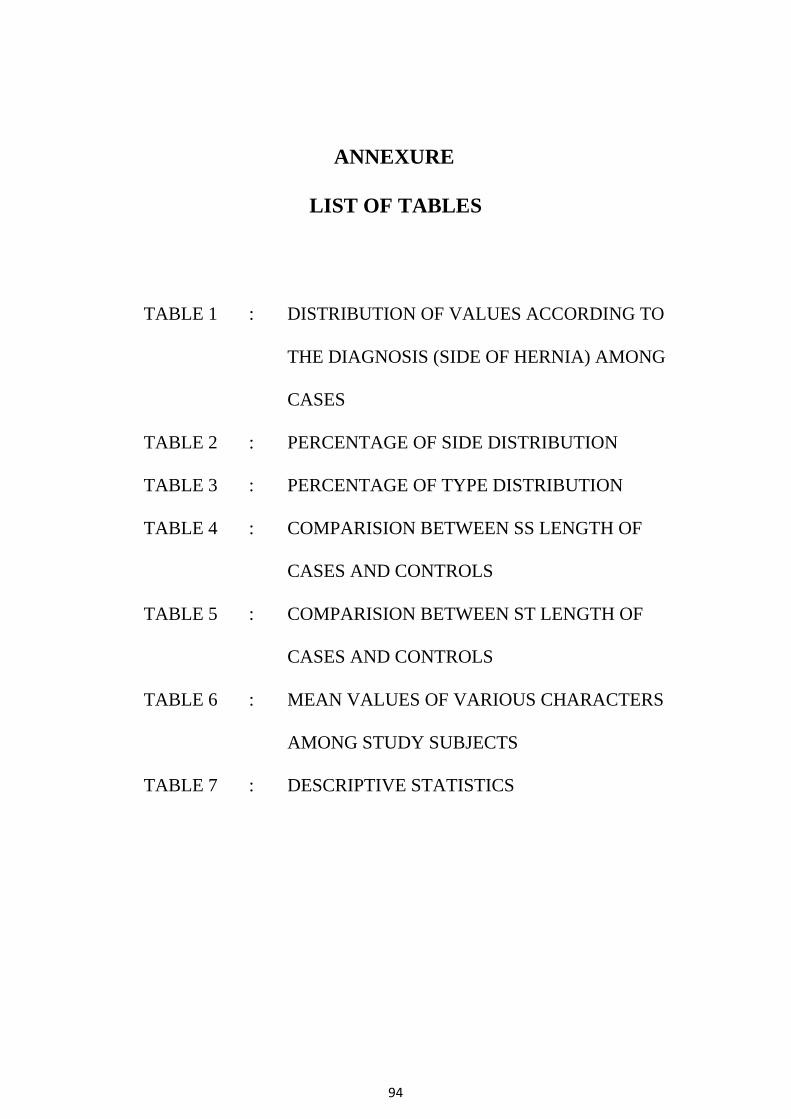
94
ANNEXURE
LIST OF TABLES
TABLE 1 : DISTRIBUTION OF VALUES ACCORDING TO
THE DIAGNOSIS (SIDE OF HERNIA) AMONG
CASES
TABLE 2 : PERCENTAGE OF SIDE DISTRIBUTION
TABLE 3 : PERCENTAGE OF TYPE DISTRIBUTION
TABLE 4 : COMPARISION BETWEEN SS LENGTH OF
CASES AND CONTROLS
TABLE 5 : COMPARISION BETWEEN ST LENGTH OF
CASES AND CONTROLS
TABLE 6 : MEAN VALUES OF VARIOUS CHARACTERS
AMONG STUDY SUBJECTS
TABLE 7 : DESCRIPTIVE STATISTICS
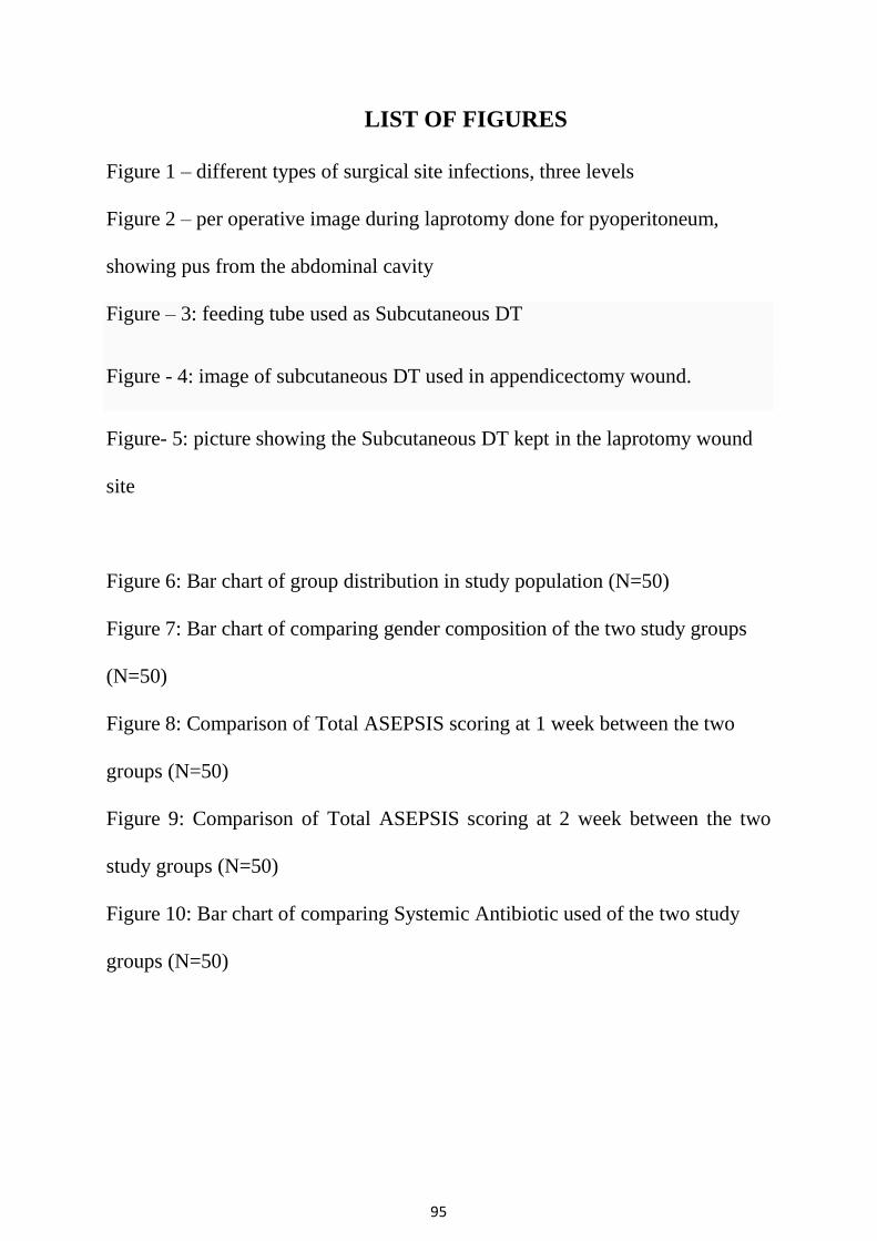
95
LIST OF FIGURES
Figure 1 – different types of surgical site infections, three levels
Figure 2 – per operative image during laprotomy done for pyoperitoneum,
showing pus from the abdominal cavity
Figure – 3: feeding tube used as Subcutaneous DT
Figure - 4: image of subcutaneous DT used in appendicectomy wound.
Figure- 5: picture showing the Subcutaneous DT kept in the laprotomy wound
site
Figure 6: Bar chart of group distribution in study population (N=50)
Figure 7: Bar chart of comparing gender composition of the two study groups
(N=50)
Figure 8: Comparison of Total ASEPSIS scoring at 1 week between the two
groups (N=50)
Figure 9: Comparison of Total ASEPSIS scoring at 2 week between the two
study groups (N=50)
Figure 10: Bar chart of comparing Systemic Antibiotic used of the two study
groups (N=50)
96
PROFORMA
“Evaluate Efficacy Of Local Amikacin Therapy
As An Adjuvant To Parentral Antibiotics In Control
Of Surgical Site Infection Compared To Parentral
Antibiotic Alone In A Tertiary Care Centre.”
. ,
:
, _____________________ .
.
,
.
,
.
.
,
, .
.
:
/
:
:
( , )
:
: (Dr.GNANA SEZHIAN)
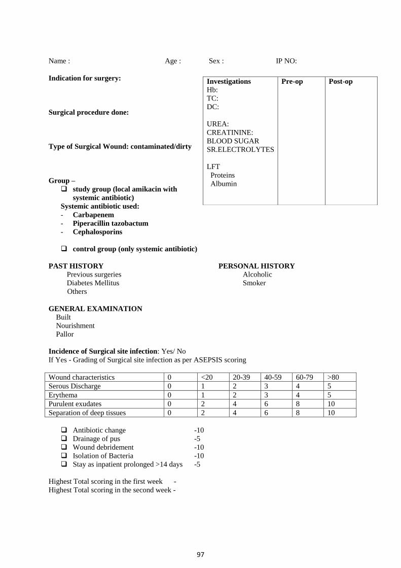
97
Name : Age : Sex : IP NO:
Indication for surgery:
Surgical procedure done:
Type of Surgical Wound: contaminated/dirty
Group –
study group (local amikacin with
systemic antibiotic)
Systemic antibiotic used:
- Carbapenem
- Piperacillin tazobactum
- Cephalosporins
control group (only systemic antibiotic)
PAST HISTORY PERSONAL HISTORY
Previous surgeries Alcoholic
Diabetes Mellitus Smoker
Others
GENERAL EXAMINATION
Built
Nourishment
Pallor
Incidence of Surgical site infection: Yes/ No
If Yes - Grading of Surgical site infection as per ASEPSIS scoring
Wound characteristics 0 <20 20-39 40-59 60-79 >80
Serous Discharge 0 1 2 3 4 5
Erythema 0 1 2 3 4 5
Purulent exudates 0 2 4 6 8 10
Separation of deep tissues 0 2 4 6 8 10
Antibiotic change -10
Drainage of pus -5
Wound debridement -10
Isolation of Bacteria -10
Stay as inpatient prolonged >14 days -5
Highest Total scoring in the first week -
Highest Total scoring in the second week -
Investigations Hb:
TC:
DC:
UREA:
CREATININE:
BLOOD SUGAR
SR.ELECTROLYTES
LFT
Proteins
Albumin
Pre-op Post-op