Embed Size (px)
Citation preview

Instructions for use
Title PRODUCTION OF NEONATAL HEMOLYTIC DISEASE IN NEWBORN PIGS BY ORAL ADMINISTRATIONOF ANTI-PORCINE ERYTHROCYTE OVINE SERUM
Author(s) KAGOTA, Katsumoto; ABE, Noboru; TOKORO, Kazunobu
Citation Japanese Journal of Veterinary Research, 30(3-4): 94-107
Issue Date 1982-12-28
DOI 10.14943/jjvr.30.3-4.94
Doc URL http://hdl.handle.net/2115/2266
Type bulletin
File Information KJ00002374068.pdf
Hokkaido University Collection of Scholarly and Academic Papers : HUSCAP
Jpn. J. Vet. Res., 30, 94-107 (1982)
PRODUCTION OF NEONATAL HEMOLYTIC DISEASE IN NEWBORN PIGS BY ORAL ADMINISTRATION
OF ANTI-PORCINE ERYTHROCYTE OVINE SERUM
Katsumoto KAGOT A, Noboru ABE*l
and Kazunobu TOKORO*l
Department of Veterinary Internal Aledicine Faculty of Veterinary lWedicine
Hokkaido University, Sapporo 060, Japan
(Received for publication, June 28, 1982)
The mInImUm time of separating piglets from their mothers in order to
prevent hemolytic disease in newborn pigs was investigated by examining the red blood cell count, packed blood cell volume, blood hemoglobin concentration and erythrocyte osmotic fragility of naturally suckled piglets
which had been given anti-porcine erythrocyte ovine serum orally at different times after birth. The erythrocyte osmotic fragility test proved to be the
most effective test to detect the hemolytic condition. Decrease in erythrocyte
resistance was observed only in the piglets given the serum within 18 hours of life. From this result, it was recommended that piglets should be fostered
by a non-sensitized sow other than their mother for 24 hours after birth to
prevent the occurrence of neonatal hemolytic disease.
INTRODUCTION
It is well known that colostral immunoglobulins are absorbed from the intestinal
mucosa during the early stage after birthD. The absorption of these large molecules
ceases within a certain period due to a mechanism known as "gut closure". Blood
group antibodies found in the colostrum are agents of hemolytic disease of neonatal
piglets and are among the large molecules influenced by gut closure. Therefore, the
onset of hemolytic disease is closely related to the absorption of antibodies in the maternal
colostrum before gut closure occurs.
The purpose of this study was to estimate the minimum time necessary to foster
piglets in order to prevent neonatal hemolytic disease. For this purpose we examined
the clinical and hematological changes and erythrocyte osmotic fragility of naturally
suckled piglets given anti-porcine erythrocyte ovine serum orally at different times after
birth.
Present address *1 Shintoku Animal Husbandry Experiment Station, Shintoku 081, Japan
Experimental newborne pigs' hemolytic disease 95
MATERIALS AND METHODS
Anti-porcine erythrocyte ovine serum was prepared as the blood group antibody
and then given intravenously or orally to newborn piglets at different times after birth.
Judgement of the absorption of the antibody was made in accordance with the ery
throcyte fragility test.
Preparation of blood group antibody
The blood was collected from apparent healthy swme aged 6 months old, washed
several times with saline, and then prepared for 50 % erythrocyte suspension with saline.
This specimen was used as the antigen for obtaining the antibody. Namely 50 ml of
erythrocyte suspension was inoculated intravenously a total of 5 times to 2 Corriedale
ewes at 5 day's intervals. Seven days after the final inoculation, whole blood was
collected and the serum was separated. The serum was kept at - 20°C until use. The
agglutinin titer of both ovine sera applied to the direct agglutination test with the swine
erythrocytes was the same, ie., 1: 256.
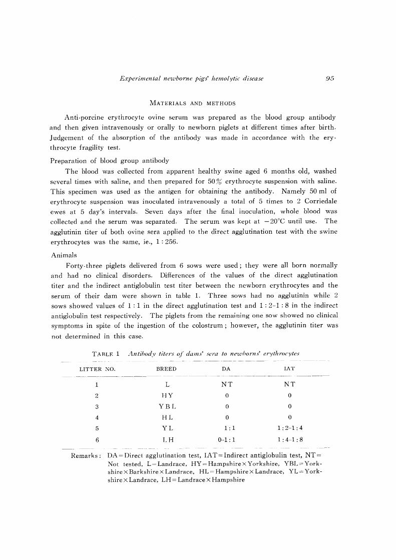
Animals
Forty-three piglets delivered from 6 sows were used; they were all born normally
and had no clinical disorders. Differences of the values of the direct agglutination
titer and the indirect antiglobulin test titer between the newborn erythrocytes and the
serum of their dam were shown in table 1. Three sows had no agglutinin while 2
sows showed values of 1 : 1 in the direct agglutination test and 1 : 2-1 : 8 in the indirect
antiglobulin test respectively. The piglets from the remaining one sow showed no clinical
symptoms in spite of the ingestion of the colostrum; however, the agglutinin titer was
not determined in this case.
TABLE 1 Antibody titers of dams' sera to ne'lohorns' erythrocytes
LITTER NO.
1
2
3
4
5
6
BREED
L
HY
YBL
HL
YL
LH
DA
NT
o o o 1: 1
0-1: 1
IAT
NT
o o o
1: 2-1: 4
1: 4-1: 8
Remarks: DA = Direct agglutination test, IA T = Indirect antiglobulin test, NT = Not tested, L= Landrace, HY = Hampshire X Yorkshire, YBL = Yorkshire X Barkshire X Landrace, HL = Hampshire X Landrace, YL= Yorkshire X Landrace, LH = Landrace X Hampshire
96 KAGOT A, K. et a1.
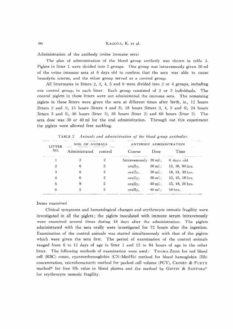
Administration of the antibody (ovine immune sera)
The plan of administration of the blood group antibody was shown in table 2.
Piglets in litter 1 were divided into 2 groups. One group was intravenously given 20 ml
of the ovine immune sera at 6 days old to confirm that the sera was able to cause
hemolytic icterus, and the other group served as a control group.
All littermates in litters 2, 3, 4, 5 and 6 were divided into 2 or 4 groups, including
one control group, in each litter. Each group consisted of 2 or 3 individuals. The
control piglets in these litters were not administered the immune sera. The remaining
piglets in these litters were given the sera at different times after birth, ie., 12 hours
(litters 2 and 4), 15 hours (litters 4 and 5), 18 hours (litters 3, 4, 5 and 6), 24 hours
(litters 3 and 5), 30 hours (litter 3), 36 hours (litter 2) and 60 hours (litter 2). The
sera dose was 30 or 40 ml for the oral administration. Through out this experimen t
the piglets were allowed free suckling.
TABLE 2 Animals and administration of the blood group antibodies
NOS. OF ANIMALS LITTER
NO. Administrated control
122
262
362
462
592
622
Items examined
ANTIBODY ADMINISTRATION --- ---- --- -
Course Dose Time -------
Intravenously 20 ml; 6 days old
orally, 30ml; 12, 36, 60 hrs.
orally, 30 ml; 18, 24, 30 hrs.
orally, 30ml; 12, 15, 18 hrs.
orally, 40ml; 15, 18, 24 hrs.
orally, 40ml; 18 hrs.
Clinical symptoms and hematological changes and erythrocyte osmotic fragility were
investigated in all the piglets; the piglets inoculated with immune serum intravenously were examined several times during 18 days after the administration. The piglets
administrated with the sera orally were investigated for 72 hours after the ingestion.
Examination of the control animals was started simultaneously with that of the piglets
which were given the sera first. The period of examination of the control animals
ranged from 6 to 12 days of age in litter 1 and 12 to 84 hours of age in the other
litter. The following methods of examination were used: THOMA-ZEISS for red blood
cell (RBC) count, cyanmethemoglobin (CN-MetHb) method for blood hemoglobin (Hb)
concentration, microhematocrit method for packed cell volume (PCV), CROSBY & FURTH
method4) for free Hb value in blood plasma and the method by GIFFIN & SANFORD71
for erythrocyte osmotic fragility.

E.rperimelltal Jle'wborlll' pigs' hClllolytic disease 97
RESULTS
Control animals
Hematological observations of the control animals were indicated in figure 1 (litters
2-6) and figure 2 (litter 1). The range of the RBC count and PCV and Hb concen
tration values were widely spread from 320-620 million/pI, 20-40;:'':; and 5-12 g/dl respec
tively (tab. 3). In the erythrocyte osmotic fragility test of the controls, the initial points
of hemolysis ranged between 0.64 and 0.76% of NaCl concentration, and that of the
complete hemolysis ranged between 0.38 and 0.52 % (tab. 3). General features of these
control animals were gradual decline in RBC count and PCV and Hb concentration,
and normal resistance of the erythrocytes to hypotonic saline.
FIGURE 1 Hematological obserl'ations of control piglets in litters 2-6
Litter No. 2 3 4 5 6
Hours offer birth 12 18 42 84 18 30 M /2 24 36 15 23 39 61 18 45
PCV ROC
~ (0/0) OU"/pl)
~-" ~ 600
~ 500 ~ 3D <;' I 400
- ... 0... __ -- 8=::::::8--------0 I g:=~:.-:.-:.·:::--"8:::::~ : 20
--0-------0
6 300
Hb(g/dO /2 ~ <==:: • • 10 :=::==----.. *'"~~ .... • 8 --------. 6
0.86 ~ 080 ....,
~S:
1111 II II <:<:;
0.70
I II t:l~
II
C":l ......
II II II II 11 -""
Jl II ~~ 0.60 ....... CCi ~ 0.50 ~
TABLE 3 Ranges of estimated '['alues of control piglets in litters 1-6
Hb ERYTHROCYTE
LITTER PERIOD RBC COUNT PCV CONCEN- FRAGILITY (NaCl%)
NO. EXAMINED TRATION Complete .. Initial
(1Q4j,al) (rc) (gjdl) hemolysis hemolysis
1 6-12 days 320-450 20-27 5-10 0.40-0.46 0.68-0.72
2 12-84 hrs. 400-600 20-40 8-12 0.44-0.50 0.64-0.72
3 18-60 hrs. 475-600 23-32 10-12 0.46-0.50 0.66-0.70
4 12-36 hrs. 400-520 25-30 9-10 0.42-0.52 0.64-0.66
5 15-61 hrs. 450-620 27-35 9-10 0.40-0.44 0.64-0.70
6 18-45 hrs. 470-600 24-33 10-12 0.38-0.44 0.72-0.76
98 KAGOT A, K. et al.
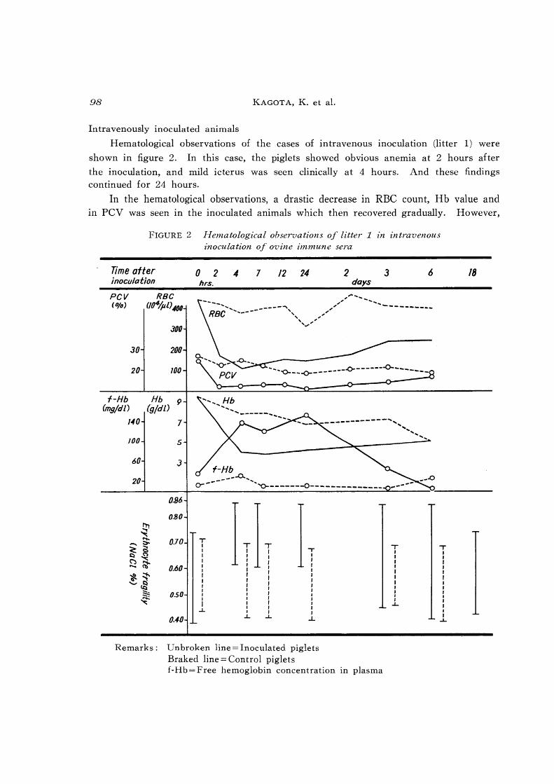
Intravenously inoculated animals
Hematological observations of the cases of intravenous inoculation (litter 1) were
shown in figure 2. In this case, the piglets showed obvious anemia at 2 hours after
the inoculation, and mild icterus was seen clinically at 4 hours. And these findings continued for 24 hours.
In the hematological observations, a drastic decrease in RBC count, Hb value and In pev was seen in the inoculated animals which then recovered gradually. However,
FIGURE 2 Hematological observations of litter 1 m intravenous inoculation of ovine immune sera
Time after 0 2 4 7 12 24 2 3 inoculation hrs. days
PCV R8C (0/0) (/g4/pl) 400
JOO
JO 200
20 100 -_ "'-______ -0-__ _
'~--~-------" ----f-Hb Hb 9
(mg/dL) (g/dO 140 7 -------------'" 100
60
20
5
J
0.86
0.80
0.70
0.60
0.50
0.40
f-Hb _------0... .............
~ ~-------o----------___ _
T I I I I I I I I I I I I
I ...L
T I I I I I I I I I I J I I I
.L
T I I I I I I I I I I I , I
...L
.,.. I J I I I , I I I I I I I
..L
Remarks: Unbroken line = Inoculated piglets Braked line = Control piglets f-Hb = Free hemoglobin concentration III plasma
T I I I I I I I r J I I
..L..
.... ", .....
T I , I , I I I I I I I I I I
..L..
18
Experimental l1elOborne pigs' hemolytic disease 99
the RBC count was still lower at 6 days after the inoculation compared to the controls.
On the other hand, free Hb concentration in the plasma was rapidly shifted up after
the inoculation of the immune serum, reaching a peak during 4-24 hours. At 6 days
later, it decreased to the normal value. The erythrocyte resistance became the weakest
at 4 hours after the inoculation. The value for complete hemolysis returned to the
same level of the control animals at 3 days after the injection, while the value for initial
hemolysis was not recovered to the normal one until 18 days after the inoculation. No
animal died during the experimental period.
Orally administrated animals
Hematological observations of the animals given the immune sera at 12 hours of
FIGURE 3 Hematological observations of piglets given ovine imnmne sera at 12 hours of life
Litter No. 2 4
Hours after 0 IS 24 30 48 72 0 6 12 18 24 48 administration
PCV RBG (%) (T04/p L)
500
30 400
9 I 300
0.. 0-'",
I 20 " I '0-----__ -0 6
Hb (g/di) 10
~ 8
6
0.86
II !!,1 0.80 ~ s:
...... ~ ~~ 0.70
~~ ::-t-..
0.60 ~~ -::'<Q ~ ~ 0.50
Icterus + + + - + *-ttt
100 KAGOT A, K. et a1.
life were shown in figure 3 (litters 2 and 4). In the administrated animals of this age,
a trend of decrease in the RBC count and PCV and Hb concentration was also noticed;
however, most of these values stayed within the ranges as were obtained from the esti
mation of the control animals (tab. 3). Only one exception, a piglet in litter 4, showed
a low value of PCV below 20% at 18 hours after the administration. Characteristic
features in these cases were a decrease in the resistance of the erythrocytes to hemo
lysis in hypotonic saline and the successive appearance of clinical icterus. Weakening of
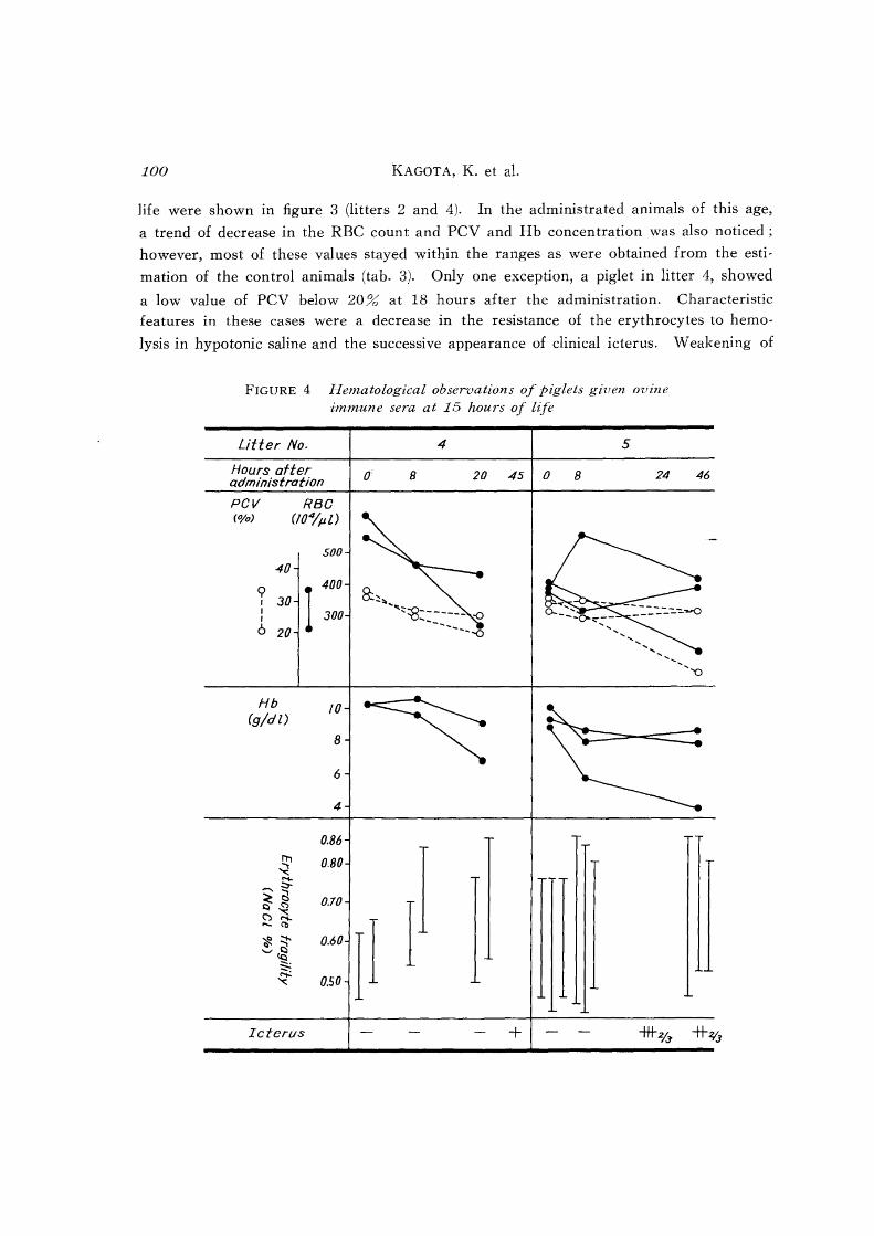
FIGURE 4 Hematological observations of piglets given ovine immune sera at 15 hours of life
Litter No.
Hours after administration
PGV RBG (0/0) (t04jpZ)
soo 40
<;>
1 400
I 30 I 300 I
6 20
Hb 10 (gldl)
8
6
4
0.86 n, ~ 0.80 -....... ::,-
~ ti 0.70 ~ ~ ~"I-..... (1)
~~ 0.60 ,-,<§ -. ~ ~ 0.50
Icterus
4 5
o 8 20 45 0 B 24
8::~"" .... ;:~------- .0 -,..,,- ....
----~
II + -ttt2/.3'
46
*~
E.rj)criJllcJltal JlCL,·horne pigs' hemolytic disease 101
the erythrocytes appeared within 24 hours after the ingestion of the sera and then
reached a peak at which the initial hemolysis occurred at 0.863~ of NaCl concentration
in all animals. An increase in hemolysis of the erythrocytes in physiological saline was
also noticed. The icterus appeared clinically at 30 hours (litter 2) and 18 hours (litter
4) after the administration and increased thereafter. Results of the erythrocyte fragility
test and clinical findings showed that all piglets administered at 12 hours of age
developed hemolytic condition.
The findings on the animals given the sera at 15 hours of age were shown in
figure 4 (litters 4 and 5). One of the 2 animals in litter 4 showed lower values in the
RBC count and PCV than those of the control animals (tab. 3) at 20 hours after the
administration, and its erythrocytes began to hemolyze at 0.86 Jb of NaCl concentration.
At 45 hours after the administration, all animals of this litter showed clinical icterus
Litter No. Hours after administration pcv RBC (%) Oll1Po
40
<;> 30 I
: 20 6
Hb (gjd!)
~ -.....: .-...9: ~g (")~ -q;-,*:-1--'-'t:I
tc:i :::::.:
~
6
0.86
0.80
0.70
0.60
0050
0.40
Icterus
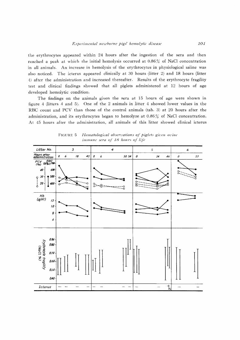
FIGURE 5 Hematological ohscrvations of piglets gh'l'll O'l'ille immune sera of 18 hours of lifi'
3 4 5
o 6 18 42 0 6 3054 o 24 46
0..., ..Q... o---'o-== .... _=::::.::-_-.g
....... _----.-----
nH II II n 11 I + ~
6
o 27
I
102 KAGOT A, K. et al.
and an increase of the hemo ytic color of erythrocytes III the physiological saline. In
litter 5, one of the 3 animals showed a marked decrease in RBC count, PCV and Hb
concentration. The point of initial hemolysis after the administration ranged between
0.80 and 0.86 %. Increase of the hemolytic color and clinical anemia with icterus were
observed in 2 animals at 46 hours after the ingestion. A decrease with time in ery
throcyte resistance was a common finding in all piglets of these groups in litters 4 and 5.
Results from the piglets administered the sera at 18 hours of life were shown in
figure 5 (litters 3, 4 and 5). The values of RBC count, PCV and Hb concentration
were kept within the estimated ranges of the control animals except for the PCV of
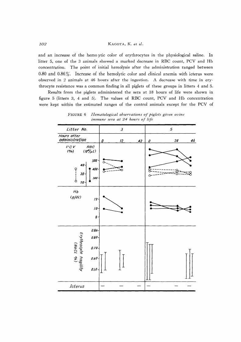
FIGURE 6 Hematological observations of piglets gi'ven ovzne immune sera at 24 hours of life
Litter No. 3 5
Hours after administration 0 12 42 0 24 46
PCV RBC (%) (Ir/pl)
500 40
1> ro I 30 I I 30() J
6 20
::::::--:~~~~--o o-------~--(j~---"':..~
Hb (Sid/)
/2
~ 10
8
0.86 I1'l ~ 0.80 S -...,
~g 0.70 ("')~
II ..... Cb
II ~~ 0.60 '-'~ -. :::::--~ 0.50 I
Icterus
Experimental ne7.vhorne pigs' hemolytic disease
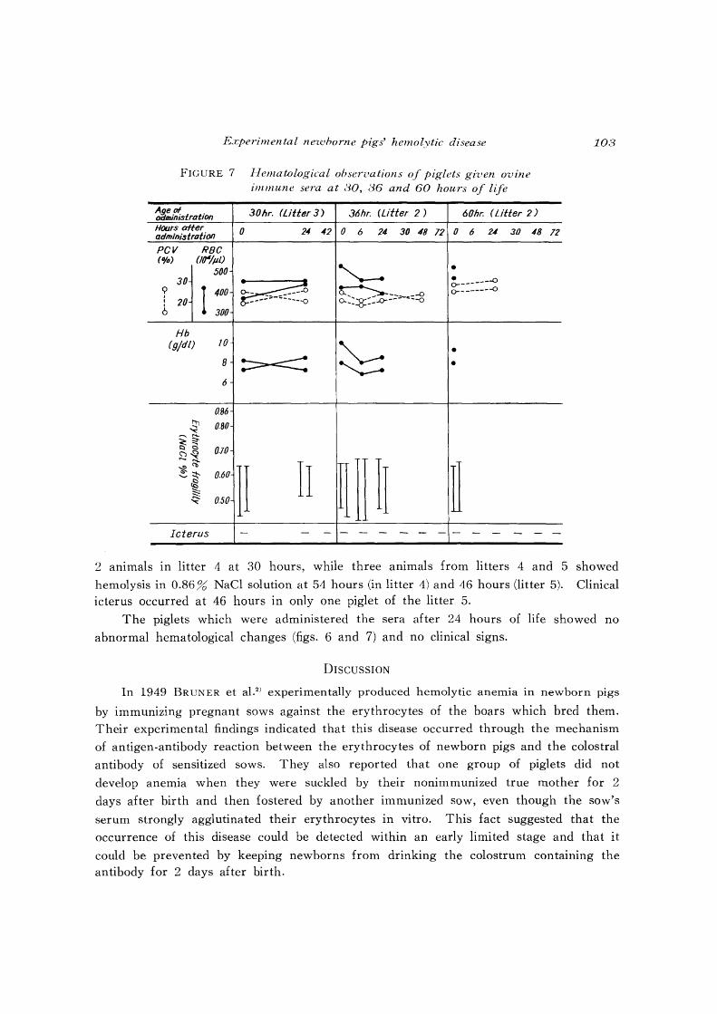
FIGURE 7 Hematological observations of piglets given ovine immune sera at 80, 36 and 60 hours of llfe
Age of oiJministration 30hr. (Utter 3) 36hr. (Litter 2 ) 60hr. (Lifter 2 )
Hours after 0 24 42 0 6 24- 30 48 12 0 6 24- 30 48 administration
PCV RBC (0/11) OfJ4/pl)
500 "-.- • 30
~ ~::::::~ ? I 400 ~~;-c~:S , 20 ====: 6
... ~ .--300
Hb (g/dl) 10
~ • 8 :::==-<::: • 6
086
-S' 0.80 '"' ..... ~~ 0.70 ~~
II ~[]11 ~ ~<b
II ~~ 0.60 ~ ~ q: 0.50
Icterus - - - - - - - - - - - - - -
108
72
-
2 animals in litter 4 at 30 hours, while three animals from litters 4 and 5 showed
hemolysis in 0.86 % NaCl solution at 54 hours (in litter 4) and 46 hours (litter 5). Clinical icterus occurred at 46 hours in only one piglet of the litter 5.
The piglets which were administered the sera after 24 hours of life showed no
abnormal hematological changes (figs. 6 and 7) and no clinical signs.
DISCUSSION
In 1949 BRUNER et aL2) experimentally produced hemolytic anemia in newborn pigs
by immunizing pregnant sows against the erythrocytes of the boars which bred them.
Their experimental findings indicated that this disease occurred through the mechanism
of antigen-antibody reaction between the erythrocytes of newborn pigs and the colostral
antibody of sensitized sows. They also reported that one group of piglets did not
develop anemia when they were suckled by their nonimmunized true mother for 2
days after birth and then fostered by another immunized sow, even though the sow's
serum strongly agglutinated their erythrocytes in vitro. This fact suggested that the
occurrence of this disease could be detected within an early limited stage and that it
could be prevented by keeping newborns from drinking the colostrum containing the antibody for 2 days after birth.
104 KAGOT A, K. et al.
On the other hand, many studies were carried out concerning the passive immunity
of pigs1•18 ,23). Some wokers established that the capacity to absorb antibodies from the
gut contents is lost at an early stage after birth due to a phenomenon called "gut
closure", which correlates with morphological changes such as formation of the terminal
web in the apical cytoplasm and disappearance of pinocytosis in the intestinal epithelial
cellsI2 ,13.lli.21,22J. There are many reports suggesting that cessation of the immunoglobulin
uptake by the small intestine occurs from 8 to 36 hours after birth in naturally suckled
newborn piglets8.14.17.19,2o.24J. However, retardation of gut closure by starvation or feeding
of small molecular substances such as glucose and amino acid mixtures has been achieved
by some scientists1o•16
,!7). In fact, PAYNE & MARSH (1962)17) reported a piglet which
showed absorption of antibody even at 106 hours of life when the animal was not
allowed to suck its dam's milk.
As described above, it is thought that protection against this disease may be achieved
by separating piglets from their mothers and not feeding them their mother's milk
until the gut closure is completed.
Since the time in which gut closure operates is influenced by the size, the form of
intaked matter and the time of ingestion are important factors. In this regard, several
studies were carried out to ascertain the minimum time during which piglets of iso
immunized sows should be deprived of maternal milk in order to survive in various
conditions. BAXTON et al. (1955)3) returned piglets to their mother at 12, 18 and 24
hours of age after artificial feeding and found that all animals showed a positive reaction
to the anti·globulin sensitization test and clinical signs of various degrees at 6 or 12
hours after drinking their mother's milk. They concluded that different pigs seemed
to display variations both in their capacity to absorb iso-antibody from the digestive
tract and in the period over which the absorption took place. HIMENO et al. (1968)10)
also removed piglets from their sow and gave them water only, artificial milk or cows
milk 6, 8, 9, and 10 times at the rate of once every hour and then returned them to
their mother. They found that the group fed 6 times developed anemic and icteric
conditions while the piglets fed 8, 9 and 10 times survived vigorously without any
clinical signs. They thus recommended the 10 times administration of artificial milk
for control of this disease to allow for the individual difference. As shown, the results
of the above experiments differed from each other; the differences can probably be
attributed to variations in the feeding conditions and the methods employed for detection
of the onset of the disease.
The method used in our experiment differed from BRUNER'S2) in the point of the
use of immunized ovine serum instead of immunized sows milk for easy obtaining of
high titer antibody against porcine erythrocyte. There has been no report on the
production of the hemolytic disease by direct administration of these heterologous anti
serum to newborn piglets. In one case of experiments with rabbits5•6), clinical symptoms
Experimental neluhorne pigs' hemolytic disease 10{)
of vanous degrees were observed following the administration of the serum Ie., 1 ml/kg
body weight caused death with shock syndrome. In our experiment, no shock syndrome
was observed, but clear hemolytic anemia appeared when 20 ml of the immunized serum
was inoculated intravenously and orally to the piglets. In spite of the smaller amount
of the serum compared with the natural dose of the colostral immunoglobulin,9) clear
hemolytic anemia was seen when intravenous inoculations of 20 ml of the immunized
serum were used. This fact revealed that the antiserum, which was employed in oral
administration, possessed an ability to cause clear hemolysis when it flowed into the
blood stream.
In the present experiment, RBC count, PCV and Hb concentration and erythrocyte
fragility were examined in addition to clinical changes to detect the onset of hemolytic
anemic condition. Among these items, RBC count and PCV and Hb concentration in
the control animals showed a trend of gradual decline with time. This drop might
have resulted from blood dilution after the intake of colostrum. Thus this trend might
account for the abnormal values of these items in hemolytic condition. In contrast with
this trend, erythrocyte resistance remained constant in the control animals. And as
compared to results of our previous studies1ll, we found some cases showing a weakened
erythrocyte resistance without clinical signs. Therefore, the result of the erythrocyte
osmotic fragility test was considered to be useful to detect the hemolytic condition.
Judging from the result of the erythrocyte osmotic fragility test, decrease of the ery
throcyte resistance occurred only in the animals given the ovine serum within 18 hours
of life. Since the administration of the serum at 18 hours of life induced a decrease
of the erythrocyte resistance in three animals of the nine piglets, this age was considered
to be a turning-point, from a sensitive to an insensitive phase, under this experimental
condition. In other words, this result suggests that removal of piglets from the im
munized mother for 24 hours is necessary to prevent the onset of the disease when
neonatal piglets are fostered by non-sensitized sows.
REFERENCES
1) BRAMBELL, F. \-V. R. (1957): The passive immunity of the young mammal Biol.
Re·l'., 33, 488~531
2) BRUNER, D. \V., BROWN, R. G., HULL, F. E. & KINKAID, A. S. (1949): Blood factors and baby pig anemia J. A.m. Vet. ivIed. A. ssoc. , 115, 94~96
3) BUXTON, J. c., BROOKSBANK, N. H. & COOMBS, R. R. A. (1955): Hemolytic disease of newborn pigs caused by maternal iso-immunization Rr. Vet . .I., 111, 463~473
4) CROSBY, W. H. & FURTH, F. W. (1956): A modification of benzidine method
for measurement of hemoglobin in plasma and urine Blood, 11, 380~383
5) DAMESHEK, W., SCHWARTZ, S. O. & GROSS, S. (1938): Hemolysins as the cause of clinical and experimental hemolytic anemias with particular references to the nature of spherocytosis and increased fragility A.m. J. Aled. Sci., 196, 769~792
106 KAGOT A, K. et al.
6) DAVIDSOHN, I., HERMONI, D. & HANAWALT, E. G. (1957): Experimental hemo
lytic anemia, a didactic tool in hematology and immunohematology Am. J. Clin.
Pathol., 27, 520-527
7) GIFFIN, H. Z. & SANFORD, A. H. (1918): Clinical observations concerning the
fragility of erythrocytes J. Lab. Clin. Aled., 4, 465-478
8) HARDY, R. N. (1969): The breakdown of [131 1J gammaglobulin in the digestive
tract of the newborn pig intestine J. Physiol. 205, 435-451
9) HIMENO, K., NAGANO, R., MORI, T., MOGI, K., ABE, T. & HOSODA, T. (1968):
Studies on the hemolytic disease of newborn pigs. V. changes in agglutination
titer of colostrum and serum collected from sow producing affected litters Jpn. J. Zootec. Sci., 39, 275-280 (in Japanese with English summary)
10) HIMENO, K., NAGANO, R., MORI, T., MOGI, K., ABE, T. & HOSODA, T. (1968):
Studies on the hemolytic disease of newborn pigs. VI. The prevention by oral administration before suckling form hemolytic disease Ibid., 39, 467-475 (in
Japanese with English summary)
11) KAGOTA, K., ABE, N. & TOKORO, K. (1982): Clinicohematological studies on
subclinical case of neonatal hemolytic disease in pigs Jpn, J. Vet. Res., 30, 79-93
12) KENWORTHY, R., STUBBS, J. M. & SYME, G. (1967): Ultrastructure of small
intestinal epithelium in wearned and unwearned pigs and pigs with post-wearning
diarrhea J. Pathol. Bac te rio I. , 93, 493-498
13) MATTISON, A. G. M. & KARLSSON, B. W. (1966): Electron microscopic and
immunochemical studies on the small intestine of newborn piglets Arch, Zool., 18, 575-581
14) MILLER, E. R., HARMON, B. G., ULLREY, D. E., SCHMIDT, D. A., LUECKE, R. W. & HOEFER, J. M. (1962): Antibody absorption, retention and production by the
baby pig J. Anim. Sci., 41, 236-252
15) MURATA, H. & NAMIOKA, S. (1977): The duration of colostral immunoglobulin
uptake by the epithelium of the small intestine of neonatal piglets J. Camp.
Pa tho I. , 87, 431-429
16) MURATA, H., Y AGUCHI, H. & NAMIOKA, S. (1979): Relationship between the
intestinal permeability to macromolecules and invation of septicemia-inducing
Escherichia coli neonatal piglets Infect. Immunol., 26, 339-347
17) PAYNE, L. C. & MARSH, C. L. (1962): Absorption of gamma-globulin by small
intestine Fed. Proc., 103, 751-756
18) PORTER, P. & ALLEN, W. D. (1972): Classes of immunoglobulin related to
immunity in the pig J. Am. Vet. Med .. A.ssoc., 160, 511-518
19) RAMIREZ, C. G., MILLER, E. R., ULLREY, D. E. & HOEFER, J. A. (1963): Swine
hematology from birth to maturity. III. Blood volume of the nursing pig J. Anim. Sci., 22, 1068-1080
20) SPEER, V. c., BROWN, A., QUINN, L. & CATRON, D. V. (1959): The cessation
of antibody absorption in young pig J. Immunol., 83, 632-634
21) STALEY, T. E., JONES, E. W. & MARSHAL, A. E. (1968): The jejunal absorptive
cell of the newborn pig. An electron microscopic study Anat. Rec., 161, 497-515
Experimental newborne pigs' hemolytic disease 107
22) STALEY, T. E., JONES, E. W. & CORLEY, L. D. (1969): The fine structure of
the duodenal absorptive cell in the newborn pig before and after feeding of
colostrum Am. J. Vet. Res., 30, 567-581
23) Y ABIKI, T. & NAMIOKA, S. (1976): Immunoglobulins in porcine umbilical cord
blood and maternal placenta Ibid., 37, 535-540
24) YOUNG, JR. G. A. & UNDERDAHL, N. R. (1949): Swine influenza as a possible
factor in suckling pig mortalites. II. Colostral transfer of hemoagglutinin inhi
bitors for swine influenza virus from dam to offspring Cornell. Vet., 39, 120-128