Embed Size (px)
Citation preview

Title: NON ENDOSCOPIC PREDICTORS OF VARICEAL BLEEDING IN PATIENTS WITH LIVER CIRRHOSIS
P.ARUN PRASATH
MD GENERAL MEDICINE
MMC,APRIL 2012

ROLE OF NON ENDOSCOPIC PREDICTORS OF VARICEAL
BLEEDING IN PATIENTS WITH LIVER CIRRHOSIS
ABSTRACT
Background: Esophageal varices develop as a consequence of portal
hypertension in patients with chronic liver disease and are present in
approximately 50% of patients with cirrhosis of the liver. Mortality rate
of an episode of esophageal varical bleeding is approximately 20% at six
weeks. Predicting the grade of varices by non-invasive methods at the
time of registration is likely to predict the need for prophylactic β
blockers or endoscopic variceal ligation in patients with cirrhosis and
portal hypertension. Therefore, the present study has been undertaken to
determine the appropriateness of the various clinical, biochemical and
imaging parameters in predicting the existence and also the grade of
esophageal varices in cirrhosis of the liver.
Methods: This was a prospective observational study which included 70
patients with liver cirrhosis who satisfied the inclusion and exclusion
criteria.All patients included in the study were subjected to detailed
history, clinical examination and blood investigations like liver function
tests, complete blood counts including thrombocytopaenia, renal function
tests, prothrombin time, Hepatitis B surface antigen, anti-HCV antibody.
Ultrasonography of the abdomen and Ascitic fluid analysis including
SAAG were done. And the patients were subjected to endoscopy.

Results: Of the seventy cases studied, presence of varices increases as
patients progress to decompensated liver disease (Child Pugh grade B &
C ). Decrease in platelet count below 100000/μL was found to be a
predictor of esophageal varices in patients with cirrhosis. Increase in
prothrombin time more than 25 seconds is associated with grade 2,3
varices. Value of serum ascitic albumin gradient (SAAG) more than
1.4g/dl is found to be a predictor for presence and large grade of
esophageal varices .Portal vein diameter more than 1.3cm is associated
with linear increase in grade of varices. Majority of patients with marked
hepatic encephalopathy had grade 3 varices. In patients with serum
albumin less than 3g/dl most of the patients had grade 3 varices.
Conclusion: A combination of these non invasive parameters in cirrhotic
patients like platelet count, portal vein diameter, SAAG, Prothrombin
time along with serum albumin, encephalopathy grade, Child pugh score
for screening esophageal varices can substantially reduce the cost of
health care and discomfort for patients as well as reduce burden on
endoscopy units.
Keywords: Portal hypertension Cirrhosis Liver Esophageal varices Platelet count Prothrombin time

1
INTRODUCTION
Esophageal varices develop as a consequence of portal
hypertension in patients with chronic liver disease and are present in
approximately 50% of patients with cirrhosis of the liver. The grade of
esophageal varices often correlates with the severity of liver disease.
While approximately 85% of individuals with Child Pugh C cirrhosis
have varices, they are present in only 45% those with Child-Pugh A
cirrhosis.[5]The rate of development of new varices and increase in
grades of varices is 8% per year; the former is largely predicted by a
hepatic venous pressure gradient (HVPG) exceeding 10 mm Hg[6,7]and
the latter by the presence of decompensated cirrhosis, alcohol etiology
and red wale signs[11].
Large size varices, the presence of red flag signs, severe liver
disease and portal pressure greater than 12 mm Hg[8,9] predict greater
risk of bleeding. Mortality rate of an episode of esophageal varical
bleeding is approximately 20% at six weeks.[10,11].
Predicting the grade of varices by non-invasive methods at the time
of registration is likely to predict the need for prophylactic β blockers or
endoscopic variceal ligation in patients with cirrhosis and portal
hypertension. Therefore, the present study has been undertaken to

2
determine the appropriateness of the various clinical, biochemical and
imaging parameters in predicting the existence and also the grade of
esophageal varices in cirrhosis of the liver.
Its prevalence varies from 20-30% in patients with cirrhosis[8].
After varices have developed, one-third of all patients die of bleeding
gastro-esophageal varices[13]. The risk of initial bleeding from varices is
25% to 35% within 2 years, with most first-bleeding episodes occurring
within one year after detection of varices[10]. The reported mortality from
the first episode of variceal bleeding in western studies ranges from 17%
to 57%[15] as compared to 5-10% mortality reported in our population
[16].
The Baveno III Consensus Conference on portal hypertension
recommended that when liver cirrhosis is diagnosed ,all cirrhotic patients
should be screened for the presence of esophageal varices.[14] Repeat
endoscopy is recommended at 1–2 years interval in patients with small
varices to evaluate the development or progression of varices and 2–3
years interval in patients without varices [19].However, this approach has
two major limitations.
Endoscopy is an invasive procedure and secondly the cost
effectiveness of endoscopy is also questionable[19] as only 9-36% patients
with cirrhosis are found to have varices on screening endoscopy. It may

3
be more cost-effective when only high risk patients are routinely screened
for the presence of varices so as to reduce the procedure cost and
increasing burden of endoscopy units. There are factors that predict risk
for first variceal hemorrhage[20]. Certain clinical, biochemical and
ultrasonographic parameters either singly or in combination have good
predictive power for non-invasively assessing the risk of bleeding from
varices. However, the factors that predict the presence of varices are not
as well defined. Identification of non-invasive predictors of esophageal
varices will help us to carry out upper gestrointestinal endoscopy in
selected group of patients thus avoiding unnecessary intervention and
expenses, at the same time not missing high risk patients with increased
chances of bleeding.

4
AIMS & OBJECTIVES
1. To identify non invasive parameters for prediction of esophageal
varices in newly diagnosed patients with cirrhosis, without
previous upper gastro intestinal bleed.
2. To assess the Predictive value of Portal vein diameter, Platelet
count, SAAG (Serum ascitic albumin gradient),in predicting
esophageal varices in cirrhotic patients.
3. To assess the usefulness of prothrombin time in predicting
esophageal varices in patients with cirrhosis.
4. To develop parameters for identifying candidates for upper gastro
intestinal endoscopic screening.

5
REVIEW OF LITERATURE
CIRRHOSIS
Definition
Cirrhosis is the end-stage manifestation of every chronic
progressive liver disease. It is a diffuse process characterized by loss of
hepatic parenchyma, formation of fibrous septa and structurally abnormal
regenerative nodules, resulting in the distortion of the normal architecture
and of gross vascular anatomy and microcirculation (21,22).
Epidemiology
Liver cirrhosis is a leading cause of death worldwide. It is the end
result of a long-lasting process, usually clinically silent and unnoticed by
the patient and the physician for years. In the past, up to 30–40% of cases
have been discovered at autopsy (23). Due to the widespread use of
imaging techniques, such as ultrasound and computed tomography it may
be assumed that currently most cirrhotic livers are discovered earlier.
Etiology Causes of liver cirrhosis
Infectious
Virus hepatitis B, C, D, Schistosomiasis

6
Autoimmune
Autoimmune hepatitis,Primary biliary cirrhosis, Autoimmune
cholangitis, Overlap syndromes.
Metabolic-toxic
Ethanol, Nonalcoholic fatty liver disease (insulin resistance;
metabolic syndrome),Indian childhood cirrhosis.
Drug-induced
CCl4, arsenic, methotrexate, isoniazid, amiodarone, a-
methyldopa.
Genetic–hereditary
Hereditary hemochromatosis, Wilson’s disease,a1-antitrypsin-
deficiency, Porphyria cutanea tarda, Glycogen storage diseases,
Galactosemia, Tyrosinemia, Urea cycle disturbances,
Abetalipoproteinemia, Cystic fibrosis.
Biliary
Secondary biliary cirrhosis (gallstones, strictures),Primary
sclerosing cholangitis, Ischemic cholangiopathy, Ductopenia, bile duct
atresia, Alagille's syndrome.

7
Vascular
Chronic right heart failure ,Constrictive pericarditis, Budd-Chiari
syndrome, Sinusoidal obstruction syndrome (venoocclusive
disease),Hereditary hemorrhagic telangiectasia (Osler-Rendu-Weber
disease).
Cryptogenic
Pathogenesis
The following pathophysiological mechanisms are important in the
development of liver cirrhosis
• Hepatocyte death with loss of hepatic parenchyma
• Fibrosis
• Changes in cell growth (hyperplasia, regeneration) and
• Vascular and circulatory alterations.
Cell Death
Chronic loss of hepatocytes is regarded as the primary stimulus and
perpetuating factor in the development of liver cirrhosis. In order for
cirrhosis to develop, liver cell loss must be sustained and long-lasting.

8
Liver injury may be mediated by immune mechanisms (for example
cytotoxic lymphocytes attacking virally infected hepatocytes),
inflammatory reactions (mediated by neutrophils and macrophages) or
toxic factors (for example via oxidative stress and calcium-mediated
cytotoxicity).
Fibrosis and Circulatory Disturbances
Fibrosis plays a crucial role in nodular transformation of the liver.
However, it represents just one facet of liver cirrhosis and must not be
equated with cirrhosis [24].Isolated fibrosis, even if extensive, does not
necessarily result in cirrhosis. Cirrhosis is more than just widespread liver
fibrosis. Further pathogenetic factors, such as liver cell loss and
circulatory disturbances, must supervene in order for cirrhosis to develop.
The development of liver cirrhosis is accompanied by a marked increase
in collagen content and by deposition of extracellular matrix, both
produced mainly by stellate cells, which are activated and transformed
into myofibroblasts. Progressive disease is characterized by increasing
fibrosis with fibrous tissue surrounding islands of hepatic parenchyma,
thus leading to the formation of pseudolobuli. Fibrous septa may form
bridges between portal tracts (portal-portal septa) and between portal
tracts and central veins (portal-central septa). These remodeling processes
are accompanied by hemodynamic alterations. Vascular channels within

9
the fibrous septa lead to the establishment of intrahepatic vascular shunts
between afferent (portal vein and hepatic artery) and efferent (hepatic
vein) vessels of the liver, which are significant for the development of the
sequelae of liver cirrhosis.
Disturbances in Hepatocyte Growth and Proliferation
The proliferation of hepatocytes in a cirrhotic liver is viewed as a
reactive regenerative process after cell loss. Regeneration, however, is
incomplete, since complete restoration of normal hepatic architecture
does not occur and the parenchymal defects are replenished by surrogate
tissue. Generally, with advancing cirrhosis and increasing Child-Pugh
stage the proliferation of hepatocytes decreases. Nodular transformation
in cirrhosis of biliary origin is not pronounced until the late stages of the
disease. In alcoholic cirrhosis, hepatocyte proliferation is inhibited which
possibly contributes to the micronodular aspect of alcoholic cirrhosis.
Pathology
A simple, reproducible and comprehensible, macroscopical
description of cirrhosis is its classification according to the size of
nodules, specifically
• Micronodular
• Macronodular, and

10
• Mixed forms.
Macroscopical Findings
Micronodular Cirrhosis
A liver cirrhosis in which nearly all nodules measure less than
3mm in size. Typical causes for micronodular cirrhotic transformation are
chronic alcohol abuse, bile duct obstruction, chronic venous outflow tract
obstruction, hereditary hemochromatosis, Indian childhood cirrhosis.
Macronodular Cirrhosis
Macronodular cirrhosis is characterized by nodules greater than 3
mm in size. Liver cirrhosis due to chronic viral hepatitis and autoimmune
hepatitis is macronodular. Typical end-stage macronodular cirrhosis is
small and hard (“shrunken liver”).
Mixed Forms
If the number of micronodules roughly equals that of
macronodules, a mixed form of cirrhosis is said to be present. During the
course of the disease micronodular cirrhosis may give way to the
macronodular form. Viral superinfections, autoimmune processes and
circulatory disturbances account for this transformation. Transformation
of macrondular to micronodular cirrhosis does not occur.

11
DIAGNOSIS
Clinical Manifestations
Physical findings in patients with liver cirrhosis
Ascites : Portal hypertension, Hypoalbuminemia
Hepatomegaly: Facultative; small liver in posthepatitic cirrhosis
Splenomegaly : Portal hypertension
Skin Changes
Glazing lips and tongue : papillary atrophy
Oral rhagades : Zinc deficiency
Spider angiomas : Central arteriole with radiating vessels
due to increased estrogen
“Banknote” skin : Skin atrophy due to zinc deficiency
Palmar erythema : ↑ estrogen
Dupuytren’s disease : Palmar fibromatosis; occurs
predominantly in alcoholics
Jaundice : Advanced hepatocellular failure
Purpura : Vascular fragility, thrombocytopenia
Scratch signs : Pruritus
Xanthelasma : Chronic biliary/cholestatic diseases
Caput medusa : portal hypertension

12
Nail Changes
White nails: Predominantly thumb and index finger
Clubbed fingers/hour glass nails : In hepatopulmonary syndrome
Endocrine Changes
Feminization in men, Abdominal baldness, ↓ Terminal hair in men,
Testicular atrophy.
Gynecomastia : Increased ratio of estrogen to free androgen due to
decreased testosterone production, and increased
peripheral conversion of testosterone to estradiol.
Amenorrhea
Foetor hepaticus : Intestinal methylmercaptans
Muscle atrophy : Cytokines; malnutrition
Parotid gland swelling: Malnutrition; predominantly in alcoholics.
Laboratory Findings
Aminotransferases : Viral cirrhosis: ALT > AST
Alcoholic cirrhosis: AST > ALT
Parameters of Cholestasis:Biliary cirrhosis: ↑ALP and gGT
Bilirubin: Serum level rises in advanced stage of cirrhosis
Choline esterase: Parameter of hepatocyte synthetic capacity. Serum
level decreases in advanced stage of cirrhosis.

13
Prothrombin time: Parameter of hepatocyte synthetic capacity.
Prolonged in advanced stage of cirrhosis
Albumin: Parameter of hepatocyte synthetic capacity. Serum
level decreases in advanced stage of cirrhosis
g-globulins Serum levels are increased with a broad based g-band
on serum electrophoresis in 80% of patients with
cirrhosis. g-globulins make up for 20–35% of all
proteins.
1)Autoimmune hepatitis: g-globulins increased in all
patients. g-globulins > 50% of total protein.
2)Primary biliary cirrhosis: ↑IgM
3)Alcoholic cirrhosis: ↑IgA
4)Viral cirrhosis: ↑IgG
Blood count :Mild normo- to macrocytic anemia. Leukopenia,
thrombocytopenia (hypersplenism)
Ammonia: Serum levels increased in advanced stage of cirrhosis.
Levels do not correlate with signs and symptoms of
hepatic encephalopathy
Branched-chainamino acids: Serum levels decreased in advanced stage
of cirrhosis
Aromatic aminoacids: Serum levels increased in advanced stage of
cirrhosis.

14
Imaging Techniques
Imaging techniques play a leading role in the diagnosis of early
stages of cirrhosis and in detecting focal (neoplastic) alterations in
cirrhotic livers. Sonography, CT-scanning and magnetic resonance
imaging are the prime imaging modalities in the diagnosis of cirrhosis.
Course and Prognosis
The natural history of cirrhosis is characterized by a “compensated
phase”, defined by the absence of complications, such as ascites, variceal
bleeding, encephalopathy and by preserved synthetic and excretory
functions(albumin ≥ 3.5 g/dL, INR ≤ 1.5, total bilirubin ≤ 1.5 mg/dL),
followed by a rapidly progressive phase marked by increasing portal
pressure and declining liver function, resulting in the development of
ascites, portal hypertensive gastrointestinal bleeding, encephalopathy and/
or jaundice. The development of any of these complications defines the
transition from a compensated to a “decompensated phase”. Transition
from a compensated to a decompensated stage occurs at a rate of 5–7%
per year. During a 10 year follow up of compensated viral cirrhosis, HCC
develops in 21–32% of cases, followed by ascites (19.5–23%), jaundice
(17%), upper gastrointestinal bleeding (4.5–6%), and encephalopathy (1–
2%) [25, 25, 26]. Survival of patients with compensated cirrhosis is
significantly longer than that of decompensated patients with median
15
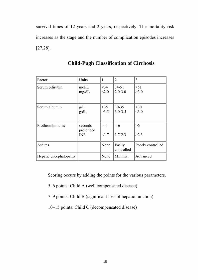
survival times of 12 years and 2 years, respectively. The mortality risk
increases as the stage and the number of complication episodes increases
[27,28].
Child-Pugh Classification of Cirrhosis
Factor Units 1 2 3
Serum bilirubin mol/L mg/dL
<34 <2.0
34-51 2.0-3.0
>51 >3.0
Serum albumin g/L g/dL
>35 >3.5
30-35 3.0-3.5
<30 <3.0
Prothrombin time seconds prolonged INR
0-4 <1.7
4-6 1.7-2.3
>6 >2.3
Ascites None Easily controlled
Poorly controlled
Hepatic encephalopathy None Minimal Advanced
Scoring occurs by adding the points for the various parameters.
5–6 points: Child A (well compensated disease)
7–9 points: Child B (significant loss of hepatic function)
10–15 points: Child C (decompensated disease)

16
PORTAL HYPERTENSION
Portal hypertension is defined as the elevation of the hepatic
venous pressure gradient (HVPG) to >5 mmHg. Portal hypertension is
caused by a combination of two simultaneously occurring hemodynamic
processes: (1) increased intrahepatic resistance to the passage of blood
flow through the liver due to cirrhosis and regenerative nodules, and (2)
increased splanchnic blood flow secondary to vasodilation within the
splanchnic vascular bed. Portal hypertension is directly responsible for
the two major complications of cirrhosis: variceal hemorrhage and
ascites. Varices are dilated, often tortuous veins. They occur most often in
the distal esophagus and in the gastric fundus in patients with portal
hypertension. Duodenal and rectal varices rarely occur and are of minor
clinical importance.
Epidemiology
Two thirds of all patients with liver cirrhosis with increasing portal
hypertension develop esophageal varices and a portal hypertensive
gastropathy . At the time of diagnosis of cirrhosis 60% of patients with
decompensated and 30% of those with compensated cirrhosis already
have varices. Approximately 10–15% of patients with esophageal varices
concomitantly also have gastric fundal varices

17
The causes of portal hypertension are usually subcategorized as
prehepatic, intrahepatic, and posthepatic.
Classification of Portal Hypertension
Prehepatic
Portal vein thrombosis
Splenic vein thrombosis
Massive splenomegaly (Banti's syndrome)
Hepatic
Presinusoidal
Schistosomiasis
Congenital hepatic fibrosis
Sinusoidal
Cirrhosis—many causes
Alcoholic hepatitis
Postsinusoidal
Hepatic sinusoidal obstruction (venoocclusive syndrome)
Posthepatic
Budd-Chiari syndrome
Inferior vena caval webs
Cardiac causes
Restrictive cardiomyopathy
Constrictive pericarditis
Severe congestive heart failure
.

18
Anatomy, Etiology and Pathophysiology
The veins of the esophageal wall consist of a subepithelial and a
submucous plexus. Both plexus communicate through perforating veins.
In the distal, precardiac esophagus the veins are mainly subepithelial.
Esophageal varices are fed by the gastric coronary veins and the short
gastric veins.
The variceal pressure depends on the pressure gradient between the
portal vein and the right atrium and varies with respiration. The mean
variceal pressure is 20–25 cm H2O. The most important pathogenetic
factor in the development and increase in size of gastroesophageal varices
is portal hypertension. The hepatic venous pressure gradient (HVPG),
determined by the difference between wedged and free hepatic venous
pressure, is a good estimate of portal pressure. Varices start developing
with HVPG values ≥10–12 mmHg [29].With increasing HVPG values
both the transmural variceal pressure and the variceal wall tension rise,
and the risk of bleeding increases [30].
Rarely esophagogastric varices may also develop in the absence of
portal hypertension. Thus, for example, obstruction of the superior vena
cava at the level of junction with the azygos vein, by increasing the
outflow resistance of the azygos vein may lead to the development of
isolated varices in the proximal esophagus (“downhill-varices”).

19
Mediastinal tumors, bronchial and esophageal cancer, goiter, fibrous
adhesions or inadvertent ligation of vessels during thyroid resection may
cause “downhill-varices”. Isolated fundal varices are usually due to an
isolated block in the splenic vein.
Diagnosis
The diagnosis of subepithelial varices is made endoscopically. The
dilated vessels protrude to a variable degree into the lumen, and their size
and the appearance of the vessel wall may be assessed endoscopically.
Both have prognostic significance. These aspects of the varices forms the
basis for classifying them into different grades . The size of the varix
must be graded.
Grade 1 (F1): the varices can be depressed by the endoscope.
Grade 2 (F2): the varices cannot be depressed by the endoscope.
Grade 3 (F3): the varices are confluent around the circumference of the
oesophagus.
Laboratory findings, such as thrombocytopenia (<90,000/mL),
splenomegaly, platelet count/spleen diameter ratio ,diameter of portal
vein (≥13 mm), lowered serum albumin and FibroTest have been
proposed as noninvasive predictors of the presence of esophageal varices
[30,31]. These parameters, however, especially when esophageal varices

20
are still small, are unreliable and do not substitute for endoscopy. In
subjects with liver cirrhosis the risk of having varices increases with
decreasing platelet counts,increasing bilirubin concentration in serum,
and rising INR.The probability of having medium or large varices at
platelet counts >150,000/mm3 has been reported to be negligible.
Course and Prognosis
In patients with cirrhosis, esophageal varices develop at a rate of
approximately 5–12% per year. If on initial endoscopy small (<5 mm)
varices are present, the rate of progression to large varices is
approximately 10–15% per year [33, 34].Varices may rupture and bleed.
Approximately one third to one half of all patients with esophageal
varices bleed at least once during their lifetime. Up to 25% of patients
with newly diagnosed and untreated esophageal varices will bleed within
the first 2 years after diagnosis.
The risk of hemorrhage primarily depends on variceal size. It is 7%
within 2 years in patients with small varices (diameter < 5 mm) and rises
to 30% in those with large varices (diameter > 5 mm). Acute variceal
bleeding always is a life threatening event and the risk of dying from the
first variceal hemorrhage is approximately 20% [35,36].Without
treatment, recurrent bleeding is the rule which in up to 20% of cases may
occur as a fulminant hemorrhage from fundal varices. Rebleeding occurs

21
within the first 6 weeks in 30% of cases and within 1 year after the first
bleed in 70% of patients. The earlier the recurrence, the higher the
mortality risk.
The MELD score (≥18) is a good predictor of short-term (6 weeks
to 3 months) mortality among cirrhotic patients at first episode of
bleeding from esopahgeal varices [37]. Measurement of the HVPG
obtained within 48 h of admission also may predict efficacy of treatment
and short-term prognosis. However, it is not universally available and
simple clinical variables, such as systolic blood pressure, Child-Pugh
score and etiology of cirrhosis may be used instead as accurate predictors
of short-term prognosis. Due to early and combined use of
pharmacological and endoscopic therapies, and short-term antibiotic
prophylaxis, in-hospital mortality of patients with cirrhosis and variceal
bleeding decreased continuously over the past two decades [38].
Predictors of Variceal Bleeding
Since variceal bleeding is associated with a high mortality risk, it is
important to define predictors and to assess the risk of bleeding in order
to establish effective prophylactic measures. The risk of variceal
hemorrhage depends on the severity of liver disease (MELD score; Child-
Pugh score) and rises with decreasing liver function. The size of varices,

22
variceal pressure and the appearance of the surface of the vessel wall are
important predictors of variceal bleeding.
Large vessels with high wall tension are more likely to bleed than
small ones [38,39,40]. The wall tension correlates directly with
transmural pressure and the diameter of a vessel and indirectly with wall
thickness. It increases with rising portal pressure, increasing vessel size
and decreasing wall thickness. Thus, not surprisingly, a large vessel with
a thin wall will exhibit a higher wall tension than a small vessel with a
thick wall and will therefore be more likely to rupture.
The surface appearance of the vessel wall may yield important
information regarding impending hemorrhage. Diffuse redness of the
vessel, red color signs, such as “cherry red spots” and “red wale marks”
(correspond to microtelangiectasia of the varix) and hemocystic spots
looking like blood blisters (>4 mm; saccular aneurysm projections), are
all thought to indicate a high risk for bleeding . A “white nipple sign” on
a varix represents a platelet-fibrin plug and indicates previous bleeding
but is not predictive of rebleeding [41].Every third patient with variceal
hemorrhage, however, does not present these endoscopic signs.
Thus, hemodynamic parameters are preferable in predicting the
risk of variceal bleeding. The level of HVPG is a reliable and
independent indicator for esophageal variceal bleeding. The normal value

23
of HVPG is 5 mmHg. Portal hypertension starts at a HVPG >5 mmHg,
but values of >10–12 mmHg are clinically significant. With a HVPG
<10–12 mmHg, varices do not develop, and preexisting varices do not
bleed. Once HVPG increases to >12–16 mmHg, the risk of bleeding is
high, but the degree of portal pressure elevation over 12 mmHg does not
correlate directly with bleeding.
Independent risk factors for esophageal variceal hemorrhage:
1)Variceal characteristics
• Size
• Wall tension
• Intravariceal pressure
• Red colour signs
2)Liver function (Child-Pugh-score; MELD score)
3)Continuing alcohol abuse
Prognostic significance of endoscopic and functional criteria for
variceal bleeding:
Endoscopic criteria Bleeding risk(%)
1)Red wale markings
Absent 19
Mild 33
Moderate 39
Severe 80
2)Variceal size
Small < 3 mm 18
Medium 3–5 mm 29
Large > 5 mm 49

24
Endoscopic criteria Bleeding risk(%)
3)Cherry-red-spots
Absent 23
Mild 32
Moderate 40
Severe 55
4)Liver function (Child-Pugh-Score)
Child A 17
Child B 31
Child C 39
Portal vein diameter
It had been reported that portal vein diameter was an independent
predictor for the presence of varices (42). However, few data are
available about the relationship between portal vein diameter and LEV.
Studies showed that portal vein diameter was the second most important
predictor for LEV in patients with a spleen width of ≤44.5 mm. However,
it did not play an important role in predicting LEV in patients with spleen
width of > 44.5 mm.
Prothrombin time
Prothrombin time is considered a marker of hepatocellular
dysfunction. As portal hypertension is a consequence, in part, of the
generalized vasodilation and the hyperdynamic splanchnic and systemic
circulatory state, the degree of hepatic function likely affects the

25
development of portal hypertension via humoral factors and, therefore,
the development of varices. Moreover, the degree of liver fibrosis is
related to liver function and fibrosis can directly affect portal
hypertension. It has been reported that serum fibrosis markers can detect
LEV with a high accuracy, though several studies showed prothrombin
time was associated with LEV on univariate analysis (43).
SAAG and its association
The development of the serum ascites-to-albumin gradient (SAAG)
has replaced the description of exudative or transudative fluid. When the
gradient between the serum albumin level and the ascitic fluid albumin
level is >1.1g/dL, the cause of the ascites is most likely due to portal
hypertension; this is usually in the setting of cirrhosis. When the gradient
is <1.1 g/dL, infectious or malignant causes of ascites should be
considered. When levels of ascitic fluid proteins are very low, patients are
at increased risk for developing SBP.
SAAG appears to retain its predictive value despite diuretics,
infusion of albumin, therapeutic paracentesis or infection in the ascitic
fluid. The finding of high SAAG denotes high chances of presence of
esophageal varices in patients due to cirrhosis.

26
Prophylaxis and Therapy
The management of patients with esophageal varices aims at three goals:
• Prevention of first variceal hemorrhage (primary bleeding prophylaxis)
• Treatment of acute variceal hemorrhage, and
• Prevention of recurrent variceal hemorrhage (secondary bleeding
prophylaxis)
The outcome of the patients critically depends on the success of
these measures. There is an increased array of therapeutic options
including pharmacological, endoscopic, mechanically compressing
(balloon tamponade), radiologic-invasive (TIPSS) and surgical
techniques which may be applied according to the clinical situation[44,
45].
Currently β-adrenergic blocking agents, nitrates, vasoconstrictors
(e.g. terlipressin) and growth hormone inhibitors, such as somatostatin
and octreotide are important. Endoscopic techniques encompass
sclerotherapy and band ligation, surgical procedures include the creation
of various portal-systemic shunts or the staple-gun transection of the
esophagus as a salvage procedure for active variceal bleeding after failure
of acute endoscopic therapy.

27
“Preprimary” Prophylaxis
“Preprimary” prophylaxis refers to the prevention of the
development of esophagogastric varices in patients with liver cirrhosis.
The best way to achieve this goal is to successfully treat the underlying
disease that leads to cirrhotic transformation. Beta-blockers are
ineffective in preventing the development of varices in patients with
cirrhosis [46]. For cirrhotic patients without varices, screening endoscopy
every 3 years, or sooner if liver function deteriorates, is recommended.
Primary Bleeding Prophylaxis
Because every episode of variceal hemorrhage is associated with a
high mortality rate, patients with cirrhosis and varices should be treated
before the first bleeding occurs. Primary prophylaxis refers to the
prevention of the first variceal hemorrhage and relies on measures like
• Lowering portal venous pressure, and
• Obliterating varices
The first goal can be achieved by pharmacotherapy, the latter by
endoscopic techniques.
Pharmacotherapy. Nonselective b-blockers reduce portal-venous
pressure by reducing cardiac output and splanchnic blood flow. In

28
addition, splanchnic vasoconstriction is enhanced by an uninhibited
activation of α-receptors. Nonselective β- adrenergic antagonists are the
mainstay of pharmacologic prevention of a first esophageal variceal
hemorrhage [47]. The individual dose of β-blockers must be determined
for each patient individually by adjusting the dose weekly with the goal
of reducing heart rate by 25% from the baseline value falling below a
rate of 55/min or a systolic blood pressure of 90 mmHg (adjust to the
maximal tolerated dose).
Treatment with β-blockers is lifelong. Only patients in whom β-
blockers lead to a durable decrease of HVPG < 12 mmHg or > 20% from
baseline benefit from treatment. Falls in HVPG > 20% are associated
with lower mortality [48]. In addition, reduction of HVPG also correlates
with a reduced risk of spontaneous bacterial peritonitis or bacteremia .
The β-blocker of choice is propranolol, 80–160 mg p.o. daily in 3–4
divided doses; alternatively nadolol 20–240 mg p.o. daily can be used.
Nadolol is less lipophilic than propranolol and does not cross the blood–
brain barrier. It is better tolerated and leads to less drug withdrawal (4%)
due to side effects compared with propranolol (up to 30%).The use of b-
blockers also seems warranted in patients, with fundal varices as fundal
and esophageal varices usually occur together.

29
Depending on Child-Pugh class, the number of patients needed to
treat in order to prevent one bleeding episode is 5–14. The primary
prophylactic effect of β-blockers seems to be more pronounced in
patients with large varices and a high Child-Pugh score, which means that
in order to achieve the same effect, fewer patients need to be treated.
Primary prophylaxis with propranolol is cost-effective, even if compared
with no therapy [49].Thus, despite their effectiveness in some patients,
HVPG does not fall < 12 mmHg or ≥20% from baseline in up to two
thirds of patients treated with β-blockers despite adequate β-blockade.
Possibly Doppler patterns of splanchnic hemodynamics can serve
as a non-invasive clue for the a prior identification of good and poor
responders to β-blockers. Cirrhotic patients who responded poorly to
nadolol, in contrast to good responders, showed a pronounced arterial
splanchnic vasodilatation at a baseline echo-color-Doppler study.
Therefore, in up to two thirds of patients treated with β-blockers and in
15–25% with contraindications to β-blocker therapy or those who cannot
tolerate the required doses because of untoward side effects, the question
as to an alternative pharmacologic primary prophylaxis arises.
Long-acting oral nitrates, isosorbide mono- or dinitrate, because of
their vasolidating effect lower both the systemic, splanchnic and portal
pressure. Combined with β-blockers, the drop in pressure is slightly more

30
pronounced than with sole β-blocker therapy. Monotherapy with nitrates
may impair renal function and worsen a pre-existing ascites. There are
even worries that nitrates may increase the mortality rate. Therefore,
nitrates should not be used as monotherapy in the prophylaxis of
esophageal variceal bleeding. Carvedilol and the long-acting somatostatin
analogue octreotide also reduce HVPG but both substances are not used
in the long-term prevention of esophageal hemorrhage.
Endoscopic Techniques. Endsocopic multiband ligation (EVL) of
esophageal varices is safe and effective. It reduces the rate of first
bleeding to 30–40% and the hemorrhage related mortality to 30% within
2 years. Patients with compensated Child-Pugh class A cirrhosis benefit
most from ligation. Most authors agree that in patients with high-risk
esophageal varices, EVL is more effective than propranolol for the
primary prevention of variceal bleeding [50]. However, there are also
data showing that in patients in whom propranolol lowers HVPG
effectively (<12 mmHg or a decrease of >20%), its efficacy is
comparable to ligation .
If quality of life is considered, then EVL is similarly cost-effective
as β-blockade .EVL is usually performed once every 2 weeks until
varices are eradicated. Recent data show that EVL yields good results
even if performed at bi-monthly intervals. Postbanding ulcers occur

31
regularly and usually are asymptomatic. Proton pump inhibitors may
reduce their size. Endoscopic obliteration of varices is followed by
lifelong treatment with β blockers. Endoscopic injection sclerotherapy is
inferior to EVL and should not be performed in primary prophylaxis of
esophageal varices.
Therapy of Acute Variceal Hemorrhage
Patients with acute variceal bleeding are managed in an intensive
care unit. The first goal always is to secure vital functions. In somnolent
patients, especially before performing the initial diagnostic endoscopy,
endotracheal intubation is strongly advised. Erythromycin infusion (250
mg) prior to endoscopy may improve stomach cleansing and quality of
endoscopic examination in these patients. Prior to all hemostatic
measures, stabilization of cardiocirculatory function is mandatory aiming
at a systolic blood pressure of approximately 100mmHg and a
haemoglobin value not more than 10 g/dL. Higher blood pressure and Hb
values lead to an increase in portal pressure with a higher risk of recurrent
bleeding.
Pharmacotherapy
In suspected acute variceal bleeding vasoactive drugs should be
started as soon as the diagnosis is made, even before diagnostic
endoscopy. Vasoactive drug therapy should be maintained for 2–5 days.

32
Vasopressin lowers portal pressure by inducing contraction
especially of the smooth muscle of splanchnic arterioles. However,
vasopressin also causes systemic vasoconstriction which may lead to
serious side effects, such as malignant cardiac arrhythmias, myocardial
infarction, intestinal ischemia, cerebrovascular ischemia and local tissue
necrosis.
Terlipressin, a synthetic analog of vasopressin, Compared to the
short acting vasopressin, the action of terlipressin is prolonged to 3–4 h
and, most importantly, it does not show the dreaded side effects of
vasopressinh Possibly the most important action of terlipressin is its
beneficial effect on renal function in patients with the hepatorenal
syndrome.
Somatostatin has an effect comparable to terlipressin. It reduces
splanchnic blood flow, has only few side effects (hyperglycemia) and is
well tolerated.Octreotide,a long acting analog of somatostatin, has
similar efficacy.
Patients with marked hepatic coagulopathy may benefit from fresh
frozen plasma.
Bacterial infections in cirrhotic patients are associated with failure
to control bleeding and represent an independent risk factor for recurrent

33
hemorrhage [52]. Antibiotic prophylaxis is an integral part of therapy for
patients presenting with variceal bleeding and should be instituted from
admission.
Fluoroquinolones (ofloxacin, ciprofoxacin, levofloxacin,
norfloxacin) or beta-lactams (amoxicillin-clavulanate, cephalosporins) are
effective and reduce the risk of bacterial infections by about 30% and
mortality risk by about 9% .Intravenous ceftriaxone (1 g qd) seems to be
more effective than oral norfloxacin (400 mg bid) in the prophylaxis of
bacterial infections in patients with advanced cirrhosis and hemorrhage .
Endoscopic Techniques. Endoscopic therapy has a success rate of
90%. It is the treatment of choice in patients with acute variceal
hemorrhage and should be performed immediately after initial diagnostic
endoscopy. Sclerotherapy and urgent band ligation are the endoscopic
techniques available to stop acute variceal hemorrhage. Variceal band
ligation is superior to sclerotherapy.
Balloon Tamponade. If endoscopic emergency treatment is not
readily available, if the bleeding is too rapid to permit endoscopy, or if
medical and endoscopic therapy fails to control bleeding (for example,
because of insufficient visualization for band ligation), balloon
tamponade may achieve a satisfactory compression of the
esophagogastric bleeding site in 80–90% of cases. The application of

34
balloon tamponade serves to gain time until definite hemostasis can be
achieved. Because pressure ulcers may develop rapidly, tubes must be
deblocked not later than 12 h after placement and then every 4–6 h for a
period of 10 min. Complete large volume paracentesis lowers
intravariceal pressure and improves respiratory function by lowering the
diaphragm.
TIPSS and Surgical Shunts:
If acute variceal bleeding is refractory to all of the above
measures, surgical or nonsurgical shunting of portal blood to the systemic
circulation is indicated as a salvage procedure. Both methods achieve
acute hemostatic success rates of 90–100%.
Prevention of Recurrent Variceal Hemorrhage
Without adequate secondary prophylaxis approximately two thirds
of patients rebleed within 6 weeks after the first variceal hemorrhage.
Prevention of recurrent variceal hemorrhage is mandatory and is
performed by combining band ligation with nonselective b-adrenergic
blocking agents. The additional administration of β -blockers in patients
in whom esophageal varices have been obliterated by banding further
reduces mortality rate from 18% to 7% . The results of combining β-

35
blockers (propranolol or nadolol) with long-acting nitrates (isosorbide
mononitrate) in secondary prophylaxis are controversial.
Shunt procedures should only be viewed as reserve techniques in
secondary prophylaxis. Their excellent effect on portal hypertension and
on lowering the rate of rebleeding is counterbalanced by the high
encephalopathy rate. Surgical shunts should only be considered in
recurrent bleeding in Child-Pugh class A patients.

36
MATERIALS AND METHODS
Study Design: Prospective Observational Study.
Study Population:
Patients admitted with complaints suggestive of liver cirrhosis in
Medical Ward, GGH, Chennai, were taken into Study.
Inclusion Criteria:
Patients with cirrhosis of liver without any past history of upper
(or) lower gastrointestinal bleed who were diagnosed based on history,
physical findings, biochemical parameters, sonography and endoscopic
methods.
Exclusion criteria;
1) Patients with history of upper or lower gastro intestinal bleed.
2) Patients on previous/current treatment with drugs for portal
hypertension.
3) Patients who had undergone procedures for esophageal varices like
banding , sclerotherapy injection (or) shunts.
4) Patients with esophageal varices due to extra hepatic cause.

37
Ethical Clearance: Obtained.
Informed Consent: Obtained from all patients.
Methodology
A total of 70 patients with liver cirrhosis were identified during the
period of February 2011 to December 2011, according to the above
criteria and were included in the study. All patients included in the study
were subjected to detailed history, clinical examination and blood
investigations.
History includes presence of jaundice, abdominal distension, pedal
edema,oliguria, haemetemesis, malena, features suggestive of
coagulopathy like gum bleed or hematuria included. Clinical examination
of the study population was focussed on the presence of jaundice,
,clubbing,dupuytrens contracture, loss of secondary sexual charecters,
anemia, gynaecomastia, parotid enlargement, spider naevi, palmar
erythema , testicular atrophy hepatic flap, splenomegaly and ascites, were
noted.

38
Laboratory Investigations:
All patients underwent biochemical tests, like liver function tests
(serum bilirubin, ALT, AST, ALP, serum albumin), complete blood
counts (haemoglobin, PCV, total and differential count,
thrombocytopaenia), renal function tests(blood urea, serum creatinine),
prothrombin time. Hepatitis B surface antigen and anti-HCV antibody
were also investigated in all blood samples. ultrasonography of the
abdomen was done to confirm the presence of cirrhosis and to find portal
vein diameter, ascites and presence of collaterals and Ascitic fluid
analysis including SAAG in patients with ascites.
Chest X-ray and ECG were taken for all patients. Upper GI
endoscopy was done in all patients to confirm the presence of varices and
also to grade them.
Statistical Analysis:
Statistical analysis was done with
1. SPSS software version 19
2. Microsoft Excel 2010.
Conflicts of interest: None

39
OBSERVATION & RESULTS
RELATION BETWEEN SEX AND GRADE OF VARICES
Sex * Endoscopy Cross tabulation(Table 1)
P=0.763
No significant gender difference in the distribution of grade of varices
was found in our study.
Sex
MaleFemale
Freq
uenc
y
30
25
20
15
10
5
0
Endoscopy
0
1
2
3
SEX Endoscopy-Grade of varices Total 0 1 2 3
F
No. of patients 0 7 4 3 14% within Sex 0 50.0 28.6 21.4 100.0% within Endoscopy 0 22.6 21.1 18.8 20.0
M
No. of patients 4 24 15 13 56% within Sex 7.1 42.9 26.8 23.2 100.0% within Endoscopy 100.0 77.4 78.9 81.3 80.0
Total
No. of patients 4 31 19 16 70% within Sex 5.7 44.3 27.1 22.9 100.0% within Endoscopy 100.0 100.0 100.0 100.0 100.0

40
RELATION BETWEEN AGE AND GRADE OF VARICES
Age Group in years * Endoscopy (Table 2)
Age Group in years
Endoscopy-Grade of varices Total
0 1 2 3 Upto 40
No. of patients 0 7 2 1 10% within Age Group in years 0 70.0 20.0 10.0 100.0
% within Endoscopy 0 22.6 10.5 6.3 14.3
41-50
No. of patients 1 15 11 5 32% within Age Group in years 3.1 46.9 34.4 15.6 100.0
% within Endoscopy 25.0 48.4 57.9 31.3 45.7
51-60
No. of patients 1 8 5 8 22% within Age Group in years 4.5 36.4 22.7 36.4 100.0
% within Endoscopy 25.0 25.8 26.3 50.0 31.4
Above 60
No. of patients 2 1 1 2 6% within Age Group in years 33.3 16.7 16.7 33.3 100.0
% within Endoscopy 50.0 3.2 5.3 12.5 8.6
Total
No. of patients 4 31 19 16 70% within Age Group in years 5.7 44.3 27.1 22.9 100.0
% within Endoscopy 100 100 100 100 100
Pearson Chi-Square-16.600, P value-.055. No significance in the
distribution of age and grade of varices was found in our study.

41
Age Group in years
Above 6051-6041-50Upto 40
Freq
uenc
y
16
14
12
10
8
6
4
2
0
Endoscopy
0
1
2
3

42
RELATIONSHIP BETWEEN PLATELET COUNT AND GRADE
OF VARICES
Platelet count * Endoscopy Crosstabulation (Table 3)
Platelet count/µL
Endoscopy-Grade of varices Total
P value 0 1 2 3
<= 100000
No. of patients 0 0 3 13 16 P <.001
% within Platelet Count 0 0 18.8 81.3 100.0
% within Endoscopy 0 0 15.8 81.3 22.9
100001-150000
No. of patients 0 9 10 3 22% within Platelet count 0 40.9 45.5 13.6 100.0
% within Endoscopy 0 29.0 52.6 18.8 31.4
150001-200000
No. of patients 0 17 5 0 22% within Platelet count 0 77.3 22.7 0 100.0
% within Endoscopy 0 54.8 26.3 0 31.4
> 200000
No. of patients 4 5 1 0 10% within Platelet count 40.0 50.0 10.0 0 100.0
% within Endoscopy 100 16.1 5.3 0 14.3
Total
No. of patients 4 31 19 16 70% within Platelet count 5.7 44.3 27.1 22.9 100.0
% within Endoscopy
100.0 100.0 100.0 100.0 100.0

43
Platelet count
> 200000150001-200000
100001-150000<= 100000
Freq
uenc
y
20
15
10
5
0
Endoscopy
0
1
2
3
Pearson Chi-Square-72.996,P value-<.001,significant
Out of the 70 patients,16 patients had platelet count less than 1 lakh
of which 13 patients had grade 3 varices, and 3 patients with grade 2
varices. And patients with platelet count above 2 lakhs none of them had
grade 3 varices and one patient with grade 2 varices. The above
observations suggested a strong association between a low platelet count
and large varices, and a significant `P' value.

44
RELATIONSHIP BETWEEN PROTHROMBIN TIME AND
GRADE OF VARICES
Prothrombin time * Endoscopy Cross tabulation(Table 4)
Prothrombin time(seconds)
Endoscopy- Grade of varices Total
P value
0 1 2 3 <= 15
No. of patients 4 0 0 0 4 P < .001
% within Prothrombin time
100 0 0 0 100.0
% within Endoscopy 100 0 0 0 5.7
16-25
No. of patients 0 26 1 0 27 % within Prothrombin time
0 96.3 3.7 0 100.0
% within Endoscopy 0 83.9 5.3 0 38.6
26-35
No. of patients 0 5 15 2 22 % within Prothrombin time
0 22.7 68.2 9.1 100.0
% within Endoscopy 0 16.1 78.9 12.
5 31.4
> 35
No. of patients 0 0 3 14 17 % within Prothrombin time
0 0 17.6 82.4 100.0
% within Endoscopy 0 0 15.8 87.
5 24.3
Total
No. of patients 4 31 19 16 70 % within Prothrombin time
5.7 44.3 27.1 22.9 100.0
% within Endoscopy 100 100.0 100. 10
0.0 100.0
Pearson Chi-Square-150.104 , P value < .001

45
34 out of 39 patients with Grade 2,3 varices had a prolonged
prothrombin time more than 25 seconds, while in patients with a
prothrombin time of less than 25 seconds majority had grade 1,0 varices.
A significant 'p' value was observed.
Prothrombin time
> 3526-3516-25<= 15
Freq
uenc
y
30
25
20
15
10
5
0
Endoscopy
0
1
2
3

46
RELATIONSHIP BETWEEN SERUM ALBUMIN AND GRADE
OF VARICES
Serum albumin * Endoscopy Cross tabulation(Table 5)
Serum albumin (g/dl)
Endoscopy- Grade of varices Total
P value
0 1 2 3 <= 3
No. of patients 0 0 2 15 17 P < .001
% within S.albumin 0 0 11.8 88.2 100.0
% within Endoscopy 0 0 10.5 93.8 24.3
3-3.5
No. of patients 0 1 15 1 17 % within S.albumin 0 5.9 88.2 5.9 100.0
% within Endoscopy 0 3.2 78.9 6.3 24.3
> 3.5
No. of patients 4 30 2 0 36 % within S.albumin 11.1 83.3 5.6 0 100.0
% within Endoscopy 100.0 96.8 10.5 0 51.4
Total
No. of patients 4 31 19 16 70 % within S.albumin 5.7 44.3 27.1 22.9 100.0
% within Endoscopy 100.0 100.0 100.
0100.
0 100.0
Pearson Chi-Square-102.562, P < .001
In our sample with 70 patients, 17 patients had their serum albumin
less than 3g/dl, who had grade 2,3 varices, in 36 patients the serum
albumin is more than 3.5 with no varices or grade one varices. Value of
serum albumin for patients showed inverse relationship with increasing
grade of varices. `P' value was significant.

47
Serum Albumin
> 3.53-3.5<= 3
Freq
uenc
y
40
30
20
10
0
Endoscopy
0
1
2
3

48
RELATIONSHIP BETWEEN ENCEPHALOPATHY AND
GRADE OF VARICES
Encephalopathy * Endoscopy Crosstabulation (Table 6)
Encephalopathy
Endoscopy- Grade of varices Total
P value
0 1 2 3 Marked
No. of patients 0 0 1 9 10
P< .001
% within Encephalopathy 0 0 10.0 90.0 100.0
% within Encephalopathy 0 0 5.3 56.3 14.3
Mild
No. of patients 0 2 15 7 24 % within Encephalopathy 0 8.3 62.5 29.2 100.0
% within Endoscopy 0 6.5 78.9 43.8 34.3
No
No. of patients 4 29 3 0 36 % within Encephalopathy 11.1 80.6 8.3 0 100.0
% within Endoscopy 100 93.5 15.8 0 51.4
Total
No. of patients 4 31 19 16 70 % within Encephalopathy 5.7 44.3 27.1 22.9 100.0
% within Endoscopy 100 100 100 100 100
Pearson Chi-Square-71.104, P < .001
Hepatic encephalopathy had a linear relation with grade of varices. In our
sample with 70 patients ,10 patients had marked hepatic encephalopathy,
90% had grade 3 varices, in 24 patients there is mild hepatic
encephalopathy with 2/3 rd of patients having grade 2 varices. `P' value
was significant.

49
Encephalopathy
NilMildMarked
Freq
uenc
y40
35
30
25
20
15
10
5
0
Endoscopy
0
1
2
3

50
RELATIONSHIP BETWEEN PORTAL VEIN DIAMETER AND
GRADE OF VARICES
Portal Vein diameter * Endoscopy Cross tabulation (Table 7) Portal Vein Diameter(cms)
Endoscopy- Grade of varices Total
P value
0 1 2 3 1.00
No. of patients 4 4 1 0 9 P< .001
% within Portal Vein diameter 44.4 44.4 11.1 0 100.0
% within Endoscopy 100.0 12.9 5.3 0 12.9
1.10
No. of patients 0 23 4 0 27 % within Portal Vein diameter 0 85.2 14.8 0 100.0
% within Endoscopy 0 74.2 21.1 0 38.6
1.20
No. of patients 0 4 6 2 12 % within Portal Vein diameter 0 33.3 50.0 16.7 100.0
% within Endoscopy 0 12.9 31.6 12.5 17.1
1.30
No. of patients 0 0 6 3 9 % within Portal Vein diameter 0 0 66.7 33.3 100.0
% within Endoscopy 0 0 31.6 18.8 12.9
1.40
No. of patients 0 0 1 4 5 % within Portal Vein diameter 0 0 20.0 80.0 100.0
% within Endoscopy 0 0 5.3 25.0 7.1
1.50
No. of patients 0 0 0 6 6 % within Portal Vein diameter 0 0 0 100.
0 100.0
% within Endoscopy 0 0 0 37.5 8.6
1.60
No. of patients 0 0 0 1 1
% within Portal 0 0 0 100. 100.0

51
Vein diameter 0
% within Endoscopy 0 0 0 6.3 1.4
12.00
No. of patients 0 0 1 0 1 % within Portal Vein diameter 0 0 100 .0 100.0
% within Endoscopy 0 0 5.3 .0 1.4
Total
No. of patients 4 31 19 16 70 % within Portal Vein diameter 5.7 44.3 27.1 22.9 100.0
% within Endoscopy 100.0 100.0 100 100 100.0
Pearson Chi-Square-95.6, P value- < .001
In patients with portal vein diameter <1.1cm none of them had Grade 2,3
varices. Large varices were seen patients with portal vein diameter
>1.4cm. The above observations suggested a strong association between a
larger portal vein diameter with large varices, and a significant `P' value.
Portal Vein diameter
1.601.501.401.301.201.101.00
Freq
uenc
y
30
20
10
0
Endoscopy
0
1
2
3

52
RELATIONSHIP BETWEEN SAAG(Serum Ascites Albumin
Gradient) AND GRADE OF VARICES
SAAG gradient * Endoscopy Crosstabulation (Table 8)
SAAG g/dl
Endoscopy – Grade of varices Total
P value
0 1 2 3 1.1
No. of patients 3 2 0 0 5 P < .001
% within Saag 60.0 40.0 0 0 100.0 % within Endoscopy 75.0 6.5 0 0 7.1
1.2
No. of patients 1 16 1 0 18 % within Saag 5.6 88.9 5.6 0 100.0 % within Endoscopy 25.0 51.6 5.3 0 25.7
1.3
No. of patients 0 11 5 0 16 % within Saag 0 68.8 31.3 0 100.0 % within Endoscopy 0 35.5 26.3 0 22.9
1.4
No. of patients 0 2 11 1 14 % within Saag 0 14.3 78.6 7.1 100.0 % within Endoscopy 0 6.5 57.9 6.3 20.0
1.5
No. of patients 0 0 1 6 7 % within Saag 0 0 14.3 85.7 100.0 % within Endoscopy 0 0 5.3 37.5 10.0
1.6
No. of patients 0 0 1 7 8 % within Saag 0 0 12.5 87.5 100.0 % within Endoscopy 0 0 5.3 43.8 11.4
No. of patients 0 0 0 2 2

53
1.8
% within Saag 0 0 0 100 100.0 % within Endoscopy 0 0 0 12.5 2.9
Total
No. of patients 4 31 19 16 70 % within Saag 5.7 44.3 27.1 22.9 100.0 % within Endoscopy 100.0 100.0 100.0 100 100.0
SAAG
1.81.61.51.41.31.21.1
Freq
uenc
y
20
18
16
14
12
10
8
6
4
2
0
Endoscopy
0
1
2
3
Pearson Chi-Square-111.265, P < .001.
When the Value of SAAG was between1.1 and 1.3 it was noted
that Grade 3 varices were absent. When the SAAG values increased
more than 1.3, there was considerable increase in grade 2,3varices.

54
RELATIONSHIP BETWEEN CTP SCORE AND GRADE OF
VARICES
CTP Score * Endoscopy Crosstabulation (Table 9)
CTP Score Endoscopy-Grade of varices Total
0 1 2 3
A
No. of patients 4 13 0 0 17
% within CTP Score
23.5 76.5 0 0 100.0
% within Endoscopy 100.0 41.9 0 0 24.3
B
No. of patients 0 18 17 1 36
% within CTP Score
0 50.0 47.2 2.8 100.0
% within Endoscopy 0 58.1 89.5 6.3 51.4
C
No. of patients 0 0 2 15 17
% within CTP Score
0 0 11.8 88.2 100.0
% within Endoscopy 0 0 10.5 93.8 24.3
Total
No. of patients 4 31 19 16 70
% within CTP Score
5.7 44.3 27 22.9 100.0
% within Endoscopy
100.0 100.0 100.0 100.0 100.0

55
Pearson Chi-Square-, P <77.71 . P<.001.
Child Pugh score had a linear relation with grade of varices. In our
sample with 70 patients, CTP score C found in 17 patients of which 15
patients had grade 3 varices. CTP score A found in 17 patients of which 4
patients did not had varices and 13 patients had grade 1 varices. P' value
was significant.
CTP Score
CBA
Freq
uenc
y
20
15
10
5
0
Endoscopy
0
1
2
3

56
DISCUSSION Our study sample consisted of 70 patients of whom 56 were male
and 14 were females. No significant gender difference in the distribution
of grade of varices was found in our study (Table 1). Distribution of
grade of varices was studied in various age groups and no significant
correlation was detected.(Table 2)
We studied the frequency of distribution of varices and found that
Grade I predominated (42%),while 6 % of the study population did not
have varices.
Our study could find significant association between
thrombocytopaenia and varices.(Table 3). An inverse relation between
thrombocytopaenia and grade of varices is noted, out of 70 patients 44
patients had platelet count less than 1.5lakhs,had grade 3,2 varices.
Especially thae number of grade 3 varices increases when the platelet
count was below 1 lakh. In patients with platelet count above 2 lakhs
grade 3 varices were not present and only one patient had grade 2
varices.
PLATELET COUNT IN OTHER STUDIES PREDICTING
OESOPHAGEAL VARICES
Garcia- Tsao et al.[53](180 patients), Pilette et al.[54](116 patients) and
K.Thomopoulos et al.[55](184 patients) reported a low platelet count to
57
be an independent risk factor for the presence of varices. Mohammad
Khuram et al.[56](200 patients) found esophageal varices in 146 with
121 having thrombocytopenia (94.5%).Chalasani et al found that of 346
patients, the presence of splenomegaly on physical examination (OR, 2.0;
95% CI, 1.1-3.8) and a platelet count less than 88103/μL (OR, 1.6; 95%
CI, 1.0-3.0) were independent risk factors for the presence of large
varices.
PROTHROMBIN TIME IN VARIOUS STUDIES PREDICTING
OESOPHAGEAL VARICES
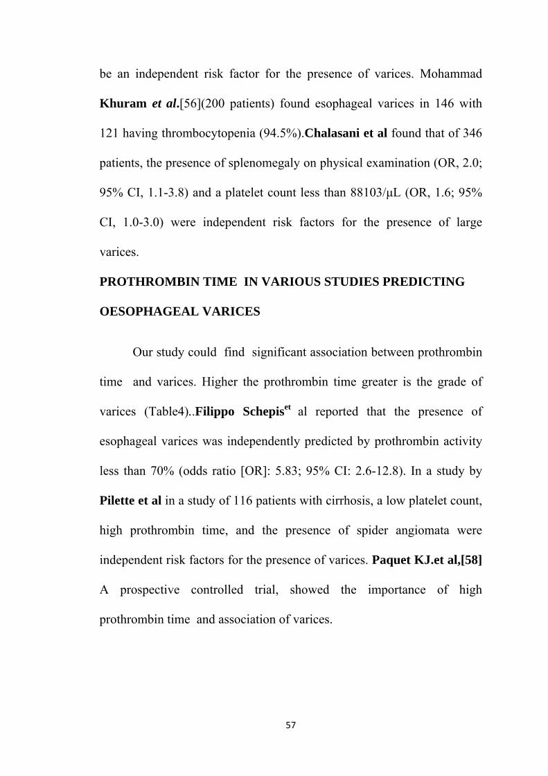
Our study could find significant association between prothrombin
time and varices. Higher the prothrombin time greater is the grade of
varices (Table4)..Filippo Schepiset al reported that the presence of
esophageal varices was independently predicted by prothrombin activity
less than 70% (odds ratio [OR]: 5.83; 95% CI: 2.6-12.8). In a study by
Pilette et al in a study of 116 patients with cirrhosis, a low platelet count,
high prothrombin time, and the presence of spider angiomata were
independent risk factors for the presence of varices. Paquet KJ.et al,[58]
A prospective controlled trial, showed the importance of high
prothrombin time and association of varices.

58
SERUM ALBUMIN AND OESOPHAGEAL VARICES
Our study could find significant association between serum
albumin and varices. Lower the serum albumin levels the greater is the
grade of varices ( Table 5). In our sample with 70 patients, 17 patients
had their serum albumin less than 3g/dl, had grade 2,3 varices. In a
logistic regression study by Garcia-Tsao et al of 180 patients, the
presence of spider angiomata, a low albumin level, and a low platelet
count were independent risk factors for the presence of varices.
HEPATIC ENCEPHALOPATHY AND OESOPHAGEAL
VARICES
Our study could find significant association between hepatic
encephalopathy and varices. Higher the encephalopathy grade greater is
the grade of varices ( Table 6).
PORTAL VEIN DIAMETER IN OTHER STUDIES PREDICTING
OESOPHAGEAL VARICES
Our study could find significant association between Portal vein
diameter and grade of varices. As the portal vein diameter increases the
grade of varices also increases (Table 7). Grade 2,3 varices are seen with
portal vein diameter more than 1.4cm.

59
Arulprakash Sarangapani et al, 2009, The study analyzed 106
patients with liver diseases. On multivariate analysis, independent
predictors for the presence of Large varices were, spleen size >13.8 mm,
portal vein >13 mm, splenic vein >11.5 mm.
Schepis et al [57] suggested that cirrhotic patients should be
screened by upper gastrointestinal endoscopy when prothrombin activity
was less than 70%, platelet count less than 100 × 109/L, and
ultrasonographic portal vein diameter greater than 13 mm are observed,
whereas those without any of these predictors should not undergo
endoscopy.
SAAG IN VARIOUS STUDIES PREDICTING OESOPHAGEAL
VARICES
Our study could find that, when the value of SAAG was
between1.1 and 1.3 it was noted that Grade 3 varices were absent. When
the SAAG values increased more than 1.3, there was considerable
increase in grade 2,3varices. Kajani et al. [59] reported that the portal
pressure correlated with the SAAG only in patients with PHT caused by
alcoholic liver disease.
The SAAG is able to define the presence or absence of PHT with
an accuracy of 96.7%. This test is accurate despite ascitic fluid infection,

60
diuresis, therapeutic paracentesis, albumin infusion, and etiology of liver
diseases. Gurubacharya et al the study included 32 patients with
ascites, demonstrated by ultrasonography, who had measurement of the
SAAG. All had upper gastrointestinal endoscopy with assessment of the
presence and size of EV. High SAAG was considered to be present when
SAAG was >=1.1 g/dl and Low SAAG when it measured < 1.1 g/dl. In a
study performed by Hoefs et al. (1983)[60], it was shown that an excellent
correlation exists between portal hypertension and SAAG .According to
Goyal et al (1989) Serum ascites albumin gradient, showed a strong
correlation to portal pressure, and was found to be the best diagnostic
index (with an overall accuracy of 97 per cent). Demyrel et al (2003)
study supports the observation that SAAG values increase in ascites due
to portal hypertension.
CTP IN VARIOUS STUDIES PREDICTING OESOPHAGEAL
VARICES
Our study could find that when the CTP score increases the
variceal grade also increases. Of the 17 patients with CTP score C,15
patients had grade 3 varices and similarly of the 17 patients with CTP
score A none of the patients had grade 2,3 varices. (Table 9).The P value
was significant.

61
In a study by Atif Zaman, et al. patients in CTP score B or C are
nearly 3 times more likely to have large varices on upper endoscopy than
are those in Child-Pugh score A. Cales et al, . In their study, multivariate
analysis revealed that initial size of varices and interval worsening of the
Child-Pugh score predicted the development of varices.

62
LIMITATIONS OF THE STUDY
• The major limitation of the study was the smaller number of
subjects.
• Being a tertiary care center, the proportion of patients with more
severe disease and multiple risk factors were getting admitted more
than those with milder disease.
• In our study female subjects were relatively less because of overall
lesser incidence of cirrhosis among female population.

63
SUMMARY AND CONCLUSION
We studied seventy patients to find out non endoscopic predictors
of esophageal varices in patients with cirrhosis who did not have previous
history of upper gastrointestinal bleed. Presence of varices increases as
patients progress to decompensated liver disease (Child Pugh grade B &
C ). Decrease in platelet count below 100000/μL was found to be a
predictor of esophageal varices in patients with cirrhosis. Increase in
prothrombin time more than 25 seconds is associated with grade 2,3
varices. Value of serum ascitic albumin gradient (SAAG) more than
1.4g/dl is found to be a predictor for presence and large grade of
esophageal varices .Portal vein diameter more than 1.3cm is associated
with linear increase in grade of varices. Majority of patients with marked
hepatic encephalopathy had grade 3 varices. In patients with serum
albumin less than 3g/dl most of the patients had grade 3 varices.
A combination of these non invasive parameters in cirrhotic
patients like platelet count, portal vein diameter, SAAG, Prothrombin
time along with serum albumin, encephalopathy grade, Child pugh score
for screening esophageal varices can substantially reduce the cost of
health care and discomfort for patients as well as reduce burden on
endoscopy units.

64
BIBILIOGRAPHY
1. Harrisons Principles of Internal Medicine 18th Edition.
2. Feldman: Sleisenger & Fordtran's Gastrointestinal and Liver
Disease, 8th edition.
3. Diseases of the Liver and Biliary System Sheila Sherlock,11th
edition.
4. Clinical hepatology: Principles and practice of hepatobiliary
diseases.
5. Pagliaro L, D’Amico G, Pasta L, Politi F, Vizzini G, Traina M, et
al.Portal hypertension in cirrhosis: Natural history. In: Bosch J,
Groszmann RJ, editors. Portal Hypertension. Pathophysiology and
Treatment.Oxford: Blackwell Scientific; 1994. pp. 72–92.
6. Groszmann RJ, Garcia-Tsao G, Bosch J, Grace ND, Burroughs
AK, Planas R, et al. Beta-blockers to prevent gastroesophageal
varices in patients with cirrhosis. N Engl J Med. 2005;353:2254–
61.
7. Merli M, Nicolini G, Angeloni S, Rinaldi V, De Santis A, Merkel
C, et al. Incidence and natural history of small esophageal varices
in cirrhotic patients. J Hepatol. 2003;38:266–72.

65
8. The North Italian Endoscopic Club for the Study and Treatment of
Esophageal Varices. Prediction of the first variceal hemorrhage in
patients with cirrhosis of the liver and esophageal varices. N Engl J
Med. 1988;319:983–9.
9. Armonis A, Patch D, Burroughs AK. Hepatic venous pressure
measurement: An old test as new prognostic marker in cirrhosis?
Hepatology. 1997;25:245, 8.
10. D’Amico G, De Franchis R. Upper digestive bleeding in cirrhosis.
Post-therapeutic outcome and prognostic indicators. Hepatology.
2003;38:599–612
11. Carbonell N, Pauwels A, Serfaty L, Fourdon O, Levy VG, Poupon
R. Improved survival after variceal bleeding in patients with
cirrhosis over the past two decades. Hepatology. 2004;40:652–9.
12. Wasty WH, Yousuf M, Mirza MR. Frequency of esophageal
varices among patients undergoing GI endoscopy. Pak J Med Sci
2005; 21:164-7.
13. Rigo GP, Merighi A, Chalen NJ, Mastronardi M, Codeluppi PL,
Ferrari A, et al. A prospective study of the ability of three
endoscopic classifications to predict hemorrhage from esophageal
varices. Gastrointest Endosc 1992; 38: 425-9.
14. The Northern Italian Endoscopic Club for the study and treatment
of esophageal varices. Prediction of the first variceal hemorrhage

66
in patients with cirrhosis of the liver and esophageal varices: a
prospective multicenter study. N Engl J Med 1988; 319: 983-9. 6.
Jensen DM. Endoscopic screening for varices in cirrhosis: findings,
implications, and outcomes. Gastroenterology 2002; 122: 1620–30.
15. Farooqi RJ, Farooqi JI, Rehman M, Ahmad H, Ahmad F, Gul S.
Outcome after injection sclerotherapy for esophageal variceal
bleeding in patients with liver cirrhosis and COPD. J Postgrad Med
Inst 2005; 19: 76-80.
16. Grace ND, Groszmann RJ, Garcia-Tsao G. Portal hypertension and
variceal bleeding: an AASLD single topic symposium. Hepatology
1998; 28: 868–80.
17. D’Amico G, Garcia-Tsao G, Calés P. Diagnosis of portal
hypertension: how and when. In: De Franchis R, (edi).
18. Proceedings of the third Baveno International Consensus
Workshop on definitions, methodology and therapeutic strategies.
Oxford: Blackwell Science 2001; 36–63. 10. D’Amico G, Pagliaro
L, Bosch J. The treatment of portal hypertension: a meta-analytic
review. Hepatology 1995; 22:332–54.
19. Brennan MRS, Targownik L, Gareth SD, Hetal AK, Ian MG.
Endoscopic screening for esophageal varices in cirrhosis: Is it ever
cost effective? Hepatology 2003; 37: 366-77.

67
20. Zoli M, Merkel C, Donatella M, Marchesini G, Gatta A, Pisi
E.Evaluation of a new endoscopic index to predict first bleeding
from the upper gastrointestinal tract in patients with cirrhosis.
Hepatology 1996; 24: 1047-52.
21. Anthony PP, Ishak KG, Nayak NC, et al (1977) The morphology
of cirrhosis: defi nition, nomenclature and classification.Bull
World Health Organ 55: 521–40.
22. Anthony PP, Ishak KG, Nayak NC, et al (1978) The morphology
of cirrhosis. Recommendations on definition, nomenclature,and
classification by a working group sponsored by the World Health
Organization. J Clin Pathol 31: 395–414.
23. Hällen J, Nord J (1965) Liver cirrhosis unsuspected during,life: a
series of 79 cases. J Chron Dis 17: 951–8.
24. Fifth Pan American Congress of Gastroenterology, Report of the
Board for Classification and Nomenclature of Cirrhosis of the
Liver (1956) Gastroenterology 31: 213–9.
25. Benvegnù L, Gios M, Boccato S, et al (2004) Natural history of of
major compensated viral cirrhosis: a prospective study on the
incidence and hierarchy complications. Gut 53:744–9.
26. Sangiovanni A, Prati GM, Fasani P, et al (2006) The natural
history of compensated cirrhosis due to hepatitis C virus: A 17-
year cohort study of 214 patients. Hepatology 43: 1303–10

68
27. D’Amico G, Garcia-Tsao G, Pagliaro L (2006) Natural history and
prognostic indicators of survival in cirrhosis: a systematic review
of 118 studies. J Hepatol 44: 217–31.
28. Huo TI, Lin HC, Lee FY, et al (2006) Occurrence of cirrhosis-
related complications is a time-dependent prognostic predictor
independent of baseline model for end-stage liver disease score.
Liver Int 26: 55–61.
29. Ginès P, Uriz J, Calahorra B, et al (2002) Transjugular intrahepatic
portosystemic shunting versus paracentesis plus albumin for
refractory ascites in cirrhosis. Gastroenterology 123: 1839–47.
30. Polio J, Groszmann RJ (1986) Hemodynamic factors involved in
the development and rupture of esophageal varices: a
pathophysiologic . approach to treatment. Semin Liver Dis 6: 318–
31.
31. Qamar AA, Grace ND, Groszmann RJ, et al (2008) Platelet count
is not a predictor of the presence or development of
gastroesophageal varices in cirrhosis. Hepatology 47: 153–9.
32. Thabut D, Trabut JB, Massard J, et al (2006) Non-invasive
diagnosis of large oesophageal varices with FibroTest in patients
with cirrhosis: a preliminary retrospective study.Liver Int 26: 271–
8.

69
33. De Franchis R (2004) Incidental esophageal varices.
Gastroenterology 126: 1860.
34. Boyer TD (1997) Natural history of portal hypertension. Clin Liver
Dis 1: 31–44.
35. De Franchis R (1988) Prediction of the first variceal hemorrhage in
patients with cirrhosis of the liver and esophageal varices – a
prospective multicenter study. N Engl J Med 319: 983–9.
36. Lay CS, Tsai YT, Teg CY, et al (1997) Endoscopic variceal
ligation in prophylaxis of fi rst variceal bleeding in cirrhotic
patients with high risk esophageal varices. Hepatology 25: 1346–
50.
37. Amitrano L, Guardascione MA, Bennato R, et al (2005) MELD
score and hepatocellular carcinoma identify patients at different
risk of short-term mortality among cirrhotics bleeding from
esophageal varices. J Hepatol 42: 820–5.
38. Carbonell N, Pauwels A, Serfaty L, et al (2004) Improved survival
after variceal bleeding in patients with cirrhosis over the past two
decades Hepatology 40: 652–9.
39. Chalasani N, Imperiale TF, Ismail A, et al (1999) Predictors of
large esophageal varices in patients with cirrhosis. AmJ
Gastroenterol 94: 3285 91.

70
40. North Italian Endoscopic Club for the study and treatment of
esophageal varices (1988) Prediction of the fi rst variceal
hemorrhage in patients with cirrhosis of the liver and esophageal
varices: a prospective multicenter study. N Engl J Med 319: 983–
9.
41. Siringo S, McCormick PA, Mistry P, et al (1991) Prognostic
significance of the white nipple sign in variceal bleeding
Gastrointest Endosc 37: 51–5
42. Schepis F, Camma C, Niceforo D, Magnano A, Pallio S,
Cinquegrani M, et al. Which patients with cirrhosis should undergo
endoscopic screening for esophageal varices detection?
Hepatology. 2001;33:3338.10.1053/ jhep.2001.21410
43. Ng FH, Wong SY, Loo CK, Lam KM, Lai CW, Cheng CS.
Prediction of oesophagogastric varices in patients with liver
cirrhosis. J Gastroenterol Hepatol. 1999;14:785–90. 10.1046/j.
1440‐1746.1999.01949.
44. D’Amico G, Pagliaro L, Bosch J (1999) Pharmacological treatment
of portal hypertension: an evidence-based approach. Semin Liver
Dis 19: 475–505.
45. De Franchis R (2000) Updating consensus in portal hypertension:
report of the Baveno III consensus workshop on definitions,

71
methodology and therapeutic strategies in portal hypertension. J
Hepatol 33: 846–52
46. Groszmann RJ, Garcia-Tsao G, Bosch J, et al (2005) Betablockers
to prevent gastroesophageal varices in patients with cirrhosis. N
Engl J Med 353: 2254–61.
47. Conn HO, Grace ND, Bosch J, et al (1991) Propranolol in the
prevention of the first hemorrhage from esophagogastric varices: a
multicenter randomized clinical trial. Hepatology 13: 902–12
48. D’Amico G, Pagliaro L, Bosch J (1999) Pharmacological treatment
of portal hypertension: an evidence-based approach. Semin Liver
Dis 19: 475–505
49. Teran JC, Imperiale TF, Mullen KD, et al (1997) Primary
prophylaxis of variceal bleeding in cirrhosis: a cost effectiveness
analysis. Gastroenterology 112: 473–82.
50. Tripathi D, Graham C, Hayes PC (2007) Variceal band ligation
versus beta-blockers for primary prevention of variceal bleeding: a
meta-analysis. Eur J Gastroenterol Hepatol 19: 835–45.
51. Garcia-Tsao G, Sanyal AJ, Grace ND, et al (2007) AASLD
Practice Guidelines. Prevention and Management of
gastroesophageal varices and variceal hemorrhage in cirrhosis.
Hepatology 46: 922–38.

72
52. Goulis J, Armonis A, Patch D, et al (1998) Bacterial infection is
independently associated with failure to control bleeding in
cirrhotic patients with gastrointestinal hemorrhage. Hepatology 27:
1207–12.
53. Garcia-Tsao G, Escorsell A, Zakko M. Predicting the presence of
significant portal hypertension and varices in compensated
cirrhotic patients. Hepatology. 1997;26:927–30.
54. Pilette C, Oberti F, Aube C. Non-invasive diagnosis of esophageal
varices in chronic liver disease. J Hepatol. 1999;31:867, 73.
55. Thomopoulos KC, Labropoulou KC, Mimidis KP, Katsakoulis EC,
large esophageal varices in patients with cirrhosis. Dig and Liver
Dis. 2003;35:473–8.
56. Khuram M, Khan NY, Arif M, Irshad MM, Hammatul Bushra K,
Hassan Z. Association of platelet count to splenic index ratio with
presence of esophageal varices in patients with hepatitis C virus
related compensated cirrhosis. Pak J Gastrenterol. 2006;20:37–42.
57. Schepis F, Camma C, Niceforo D, Magnano A, Pallio S,
Cinquegrani M et al. Which patients with cirrhosis should undergo
endoscopic screening for oesophageal varices detection?
Hepatology 2001: 33: 333-8.
73
58. Paquet KJ. Prophylactic endoscopic sclerosing treatment of
esophageal wall in varices: A prospective controlled trial.
Endoscopy. 1982;14:4–5.
59. Kajani, MA, Yoo, YK, Alexander, JA, et al. Serum-ascites albumin
gradients in nonalcoholic liver disease. Dig Dis Sci 1990;35: 33–
37.
60. Hoefs, JC, Jonas, GM. Diagnostic paracentesis. Adv Int Med
1992;37: 391–409.
61. Arch Intern Med. 2001;161:2564-2570
74
ANNEXURES PROFORMA
NON ENDOSCOPIC PREDICTORS OF OESOPHAGEAL
VARICES IN PATIENTS WITH LIVER CIRRHOSIS
1. Name
2. Age/Sex:
3. Address:
4. IP No.
5. Occupation:
6. Presenting illness
Blood vomitting
Black tarry stools
Abdominal distension
Pedal edema
Jaundice
Oliguria
Altered sleep pattern/ consciousness level
7. Past history
(DM/HT/Asthma/Seizures/Others/history of liver disease)

75
8. Personal history:
(Alcohol, Smoking, IV drug abuse/Exposure to STD, Blood
transfusion)
9. Clinical examination:
Pallor
Icterus
Clubbing
Cyanosis
Pedal Edema
Significant lymphadenopathy
KF ring
Signs of Liver cell failure
Vitals
Pulse rate
Blood Pressure
Respiratory rate
Temperature
Signs of bleeding
JVP
10. SYSTEM EXAMINATION
Abdomen: Ascites, liver span, splenomegaly
Respiratory system:

76
CVS Examination:
CNS Examination:
11. Investigations:
Blood Biochemistry
Complete Hemogram
TC, DC, Hb, Platelet count
Coagulation Profile
Blood sugar
RFT
Urea
Creatinine
Electrolytes
LFT
Total/Direct Bilirubin
SGPT
SGOT
SAP
Total protein, S.Albumin
Viral markers (HBsAg, Anti HCV)
Ascitic Fluid Analysis
Protein/Sugar
Cell Count

77
Cytology
Gramstain/AFB
Culture
SAAG ratio
Chest X-ray
ECG
USG abdomen
Portal vein size
Spleen size
Ascites
Child's Grading: A/B/C
UGI Endoscopy
Varices grade
12. Treatment
13. Outcome
Discharge
Death