Embed Size (px)
Citation preview
OPEN
ORIGINAL ARTICLE
TIP30 regulates lipid metabolism in hepatocellular carcinomaby regulating SREBP1 through the Akt/mTOR signalingpathwayF Yin1,7, G Sharen1,2,7, F Yuan3,7, Y Peng1, R Chen4, X Zhou5, H Wei1, B Li4, W Jing5 and J Zhao1,4,6
Lipid reprogramming has been considered as a crucial characteristic in hepatocellular carcinoma (HCC) initiation and progression.However, detailed molecular mechanisms have yet to be clearly defined. Here, we examined the effects of tumor suppressor TIP30 onthe regulation of HCC lipid metabolism. We found that decreased TIP30 expression leads to elevated fatty acid synthesis and enhancedlevels of lipogenic enzymes SCD and FASN in HCC cells. Moreover, SREBP1 is one of the key transcription factors regulating liver lipidmetabolism, and TIP30 deficiency significantly increased SREBP1 expression and nuclear accumulation. Small interfering RNAstargeting SREBP1 could reverse fatty acid synthesis induced by TIP30 deficiency. Furthermore, downregulating TIP30 activated theAkt/mTOR signaling pathway to upregulate SREBP1 expression, which promoted lipid metabolism by activating gene transcription oflipogenesis, including fasn and scd. We also showed that TIP30 deficiency-regulated lipid metabolism promoted proliferation of HCCcells. Clinically, our data revealed that TIP30 expression significantly correlated with SREBP1 in patients with HCC and that acombination of TIP30 and SREBP1 is a powerful predictor of HCC prognosis. Together, our data suggested a novel function of TIP30 inHCC progression and indicate that TIP30 regulation of SREBP1 may represent a novel target for HCC treatment.
Oncogenesis (2017) 6, e347; doi:10.1038/oncsis.2017.49; published online 12 June 2017
INTRODUCTIONThe morbidity and mortality of hepatocellular carcinoma (HCC)ranks top5 and top3 respectively among common malignanttumors worldwide. The poor outcomes of HCC patients are mainlydue to high recurrence of HCC after surgery and resistance tochemotherapy.1 Meanwhile, sorafenib was used as first linetargeted drugs in treating advanced HCC, which can only prolongsthe survival period of less than 3 months.2 Consequently, toelaborate the pathogenesis and progression of HCC and todevelop new therapeutic strategies seem extremely crucial.Major risk factors of HCC are viral hepatitis, exposure to
hepatotoxins and alcohol abuse, whereas recent clinic andepidemiology researches indicate nonalcoholic fatty liverdisease (NAFLD) increases HCC incidence.3 Recently, metabolicreprogramming, especially lipid metabolism alteration, isconsidered to be the initiating factor of tumor occurrenceand progression. Continuous de novo cholesterogenesis andlipogenesis are frequently activated in tumors for providing extralipids and lipid precursors during rapid cell proliferation.4 Severalkey enzymes have been identified to promote de novo lipidsynthesis, including fatty acid synthase (FASN), stearoyl-CoAdesaturase (SCD) and acetyl-CoA carboxylase (ACC).5 However,detailed mechanisms of abnormal lipid metabolism have not yetbeen comprehensively identified during HCC progression.TIP30, namely HTATIP2 or CC3, was firstly discovered in small cell
lung carcinoma, using differential analyses between highly
metastatic human variant cells and less metastatic classic cells.6
Subsequently, TIP30 was found to be downregulated in varioustumors and is considered to be a tumor suppressor due to itspro-apoptotic activity and its anti-metastatic and anti-angiogeniccapacities.7–11 Our previous research reported that TIP30-regulatedtumor metastasis and chemoresistance in various cancers.12–18
Moreover, we also found downregulated TIP30 induces epithelial–mesenchymal transition in HCC and pancreatic cancer.19,20
Considering the crucial role of lipid metabolic reprogrammingin cancer development, identifying new molecules and pathwaysthat are involved in this process is vital. Recently, TIP30 has beenpreliminarily revealed to affect fatty acid storage and oxidation inhepatocytes21 and we will extensively investigate the role of TIP30in lipid metabolism deregulation of HCC. We indicate decreasedTIP30 promotes lipid metabolism via Akt/mTOR/SREBP1 signalingand that the combination of TIP30 and SREBP1 is an effectivepredictor for HCC prognosis.
RESULTSTIP30 is a negative regulator of lipid metabolism in HCC cellsTo determine whether TIP30 regulates lipid metabolism of HCCcells, microarray analysis was firstly applied for comparing geneexpression profiles of HCC-LM3 infected with shNon or shTip30.837 genes were differentially expressed upon TIP30 knockdown(fold change ⩾ 2, Po0.05). Through GO and KEGG enrichment
1PLA General Hospital Cancer Center Key Lab, PLA Postgraduate School of Medicine, Beijing, China; 2Molecular Pathology Laboratory, College of Basic Medicine, Inner MongoliaMedical University, Hohhot, China; 3Department of Orthopedics, PLA General Hospital, Beijing, China; 4International Joint Cancer Institute, The Second Military Medical University,Shanghai, China; 5Changhai Hospital, The Second Military Medical University, Shanghai, China and 6Shanghai University Of Medicine & Health Sciences, Shanghai, China.Correspondence: Dr W Jing, Changhai Hospital, The Second Military Medical University, Changhai Road, Shanghai 200433, China.E-mail: [email protected] Professor J Zhao, International Joint Cancer Institute, The Second Military Medical University, 800 Xiangyin Road, Library Building 9-11th Floor, Shanghai 200433, China.E-mail: [email protected] authors contributed equally to this work.Received 24 October 2016; revised 18 March 2017; accepted 2 May 2017
Citation: Oncogenesis (2017) 6, e347; doi:10.1038/oncsis.2017.49
www.nature.com/oncsis
analysis, these genes were enriched in several biologicalpathways. The results indicated that genes that significantlycorrelated to TIP30 expression were involved in fatty acidmetabolism (Supplementary Figure 1A), which suggested theimportant role of TIP30 involved in lipid metabolism regulation.Furthermore, effects of TIP30 on cellular lipid levels were exploredin HCC-LM3, SMMC-7721 and HepG2 cell lines using the lipophilicdye BODIPY 493/503. We employed lentiviruses to knockdown oroverexpress TIP30 expression in HCC cell lines, and the lentiviralinfection efficiency is showed in Figure 1d. We observed thatsilencing TIP30 led to increased levels of neutral lipid staining inHCC-LM3 and SMMC-7721 cells, whereas the staining wassignificantly decreased in TIP30-overexpressed cells (Figure 1a).Similar results were obtained when the lipid accumulation induceroleate was added to the culture medium (SupplementaryFigure 1B). Consistently, intracellular triglyceride levels werefurther determined to support the negative effects of TIP30on lipid metabolism regulation (Figure 1b).To well explain the mechanisms of TIP30 regulating de novo
lipid synthesis, we analyzed lipogenesis-related enzymes(FASN, SCD, and ACC) levels of HCC cells with different TIP30expressions. Results demonstrated both the mRNA and proteinlevels of SCD and FASN notablely elevated in TIP30-depleted cells,whereas overexpression of TIP30 reduced SCD and FASN levelscomparing to control (Figures 1c and d). However, ACC expressionremained unchanged with TIP30 knockdown or overexpression(Figure 1c). As fatty acid oxidation (FAO) played an important rolein lipid metabolism reprogramming in several types of cancer, wealso analyzed the levels of critical FAO-related factors, includingCPT1A and ACOX1. However, TIP30 exerted no effects on thesetwo oxidative enzymes in HCC cells (Figure 1c). Taken together,our research indicates TIP30 can regulate de novo fatty acidsynthesis of HCC cells.
SREBP1 is essential for TIP30 deficiency-mediated lipogenesis-promoting effectsThe previous analysis showed that SREBPs (sterol regulatoryelement-binding proteins) were critical transcription factors thatcontrol lipogenesis and lipid uptake.22 SREBPs firstly located inendoplasmic reticulum membrane, which was considered as itsinactive precursors. Once the sterol levels drop, SREBPs transferredfrom endoplasmic reticulum to Golgi apparatus, where matureforms were released by proteases (site-1 and site-2). Thereafter,mature SREBPs entered nucleus to bind SRE-containing genepromoters to induce transcription.23 Emerging evidence indicatesthat SREBP1 is a crucial linkage of oncogenic signaling transduc-tion and cancer metabolism.24 To elucidate the molecularmechanisms of the TIP30 deficiency-mediated upregulationof lipogenic enzymes, cellular SREBP1 levels were examined.qRT–PCR and western blot analyses revealed that SREBP1mRNA levels were significantly increased after TIP30 knockdown(Figures 1c and 2a). Additionally, immunostaining showed nuclearaccumulation of SREBP1 with TIP30 deficiency (Figure 2b;Supplementary Figure 2B). Meanwhile, overexpression of TIP30leaded to adverse effects on SREBP1 levels in HCC cells (Figures 1cand 2a,b). SREBP1 expression was reduced in TIP30 knockdowncells which TIP30 are re-expressed in (Supplementary Figure 2A).We then used small interfering RNAs (siRNAs) targeting SREBP1 toexplore whether TIP30 deficiency upregulated lipogenic enzymeexpression through SREBP1. SREBP1 depletion of TIP30-deficientHCC-LM3 and SMMC-7721 cells leaded to decreased expression ofFASN and SCD (Figure 2c). Moreover, intracellular triglyceridelevels and the intensity of BODIPY staining were remarkablyreduced by siSREBP1 in TIP30-deficient HCC cells (Figure 2d;Supplementary Figure 2C). These results demonstrate that SREBP1is involved in TIP30 deficiency related lipogenesis-promotingeffects.
Downregulating TIP30 enhances SREBP1 expression by activatingthe Akt/mTOR signaling pathwayLoss of TIP30 can activate EGFR/AKT signaling of human lungadenocarcinoma and mammary cancer.25,26 We have previouslyconfirmed that TIP30 deficiency can activate AKT signaling in HCCand laryngeal carcinoma.16,20 Previous studies have indicatedactivation of AKT/mTOR signaling had critical effects on lipidmetabolism regulation.27,28 Here, we find downregulation of TIP30expression activates AKT and causes elevated mTOR phosphoryla-tion in HCC-LM3 cells, whereas up-regulating TIP30 expressionreduced AKT and mTOR phosphorylation levels (Figure 3a).Additionally, we found that blockade of AKT by its inhibitor,MK-2206, dramatically attenuated the TIP30 deficiency-inducedup-regulation of p-mTOR and SREBP1 levels (Figure 3b). Moreover,MK-2206 could reverse the upregulated FASN and SCD expressionin TIP30-deficient HCC-LM3 cells (Figure 3c). Intracellular triglycer-ide levels and the intensity of BODIPY staining were also reducedafter MK-2206 treatment (Figures 3d and e). These data showthat AKT/mTOR/SREBP1 signaling is required for TIP30 to regulatelipid metabolism in HCC cells.
Decreased TIP30 promotes the proliferation of HCC cells viaSREBP1-related lipid metabolism in vitro and in vivoGiven important effects of lipid metabolism on tumor progression,we examined whether TIP30-regulated HCC cell growth via SREBP1-mediated lipid metabolism. As shown in Figure 4a, depletion ofSREBP1 signaling with siRNA inhibited TIP30 deficiency-inducedenhanced HCC-LM3 and SMMC-7721 cell growth. Using colony-forming assays, we found that decreasing the TIP30 deficiency-induced HCC cell colony formation was also dependent on SREBP1(Figure 4b). To better understand the role of lipid metabolismreprogramming in TIP30-regulated HCC growth in vivo, xenograftstudies were performed. We established stable HCC-LM3 cell lineswith both TIP30 and SREBP1 deficiency and the corresponding cellswere subcutaneously injected into 4 weeks old BALB/c nude mice.Results showed decreased SREBP1 significantly abolished theaccelerated tumor growth of TIP30-deficient HCC-LM3 cells(Figures 4c and d). Interestingly, the levels of triglycerides wereincreased in the tumor tissues with reduced TIP30 expression,whereas silencing SREBP1 reversed this effect (Figure 4e), furthersupporting the conclusion that lipid metabolism deregulationcontributed to HCC cell growth. Meanwhile, immunohistochemicalstaining showed tumors originated from TIP30-silenced HCC-LM3cells exhibited increased SREBP1, SCD and FASN expression(Figure 4f), which was coincident with results obtained in HCC celllines in vitro. Taken together, our results suggest that TIP30 canmodulate SREBP1-related lipid metabolism, which contributes totumor growth in HCC.
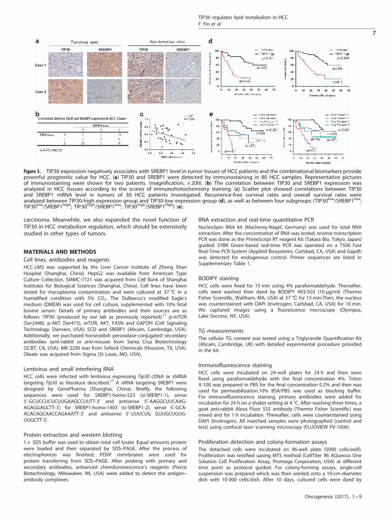
Decreased TIP30 is associated with elevated SREBP1 levels in HCCsamples, and combinational biomarkers provide powerfulprognostic value for HCC patientsTo better understand the correlation between TIP30 and SREBP1expression, immunohistochemical staining of 80 clinical HCCsamples was performed. SREBP1 protein levels in HCC tissuesnegatively correlated with TIP30 expression (r=− 0.473, Po0.001)(Figures 5a and b), suggesting that SREBP1 may be upregulated byTIP30 deficiency in HCC. We then measured TIP30 and SREBP1mRNA levels in 30 HCC tissue samples investigated to providefurther support for our research. Results showed the negativeassociation(r=− 0.37, P= 0.039) between TIP30 and SREBP1 mRNAlevels (Figure 5c). Using NCBI GEO databases to analyze the HCCsample array (GEO dataset accession GSE36376),29 we also foundthe negative correlation of TIP30 and SREBP1 in 240 HCC samples(r=− 0.24, Po0.001), showing the same tendency as our results(Supplementary Figure 3). Meanwhile, patients with low TIP30
TIP30 regulates lipid metabolism in HCCF Yin et al
2
Oncogenesis (2017), 1 – 9
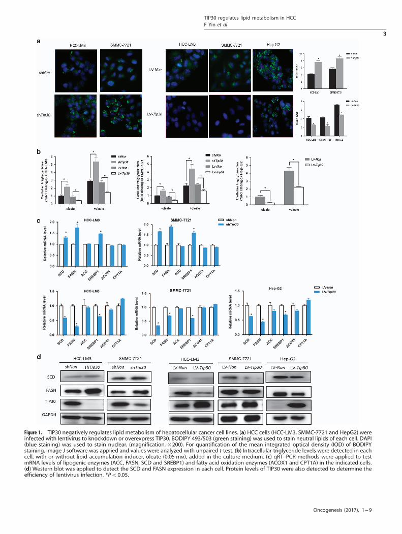
Figure 1. TIP30 negatively regulates lipid metabolism of hepatocellular cancer cell lines. (a) HCC cells (HCC-LM3, SMMC-7721 and HepG2) wereinfected with lentivirus to knockdown or overexpress TIP30. BODIPY 493/503 (green staining) was used to stain neutral lipids of each cell. DAPI(blue staining) was used to stain nuclear. (magnification, × 200). For quantification of the mean integrated optical density (IOD) of BODIPYstaining, Image J software was applied and values were analyzed with unpaired t-test. (b) Intracellular triglyceride levels were detected in eachcell, with or without lipid accumulation inducer, oleate (0.05 mM), added in the culture medium. (c) qRT–PCR methods were applied to testmRNA levels of lipogenic enzymes (ACC, FASN, SCD and SREBP1) and fatty acid oxidation enzymes (ACOX1 and CPT1A) in the indicated cells.(d) Western blot was applied to detect the SCD and FASN expression in each cell. Protein levels of TIP30 were also detected to determine theefficiency of lentivirus infection. *Po0.05.
TIP30 regulates lipid metabolism in HCCF Yin et al
3
Oncogenesis (2017), 1 – 9

levels and high SREBP1 levels exhibited the poorest recurrence-free survival (RFS) as well as overall survival (OS), indicating thatcombinational detection of the two molecules may have apowerful prognostic value (Figures 5d and e).
DISCUSSIONIncreasing evidences showed lipid metabolism was a key playerin tumor growth, metastasis and resistance to therapies. As themain metabolic organ, liver is crucial for carrying out lipidmetabolism, and aberrant activation of lipogenesis has beenconsidered as an oncogenic event in human HCC.30,31 In thepresent research, we evaluated whether TIP30 participates inabnormal lipid metabolism of HCC.
TIP30 was first discovered as a metastasis suppressor in 1997 andas a tumor suppressor in 2003.6,7 The tumor suppressor function ofTIP30 has been extensively demonstrated in various types of humantumors, including HCC. TIP30 exerts its tumor-suppressive role byinfluencing cell apoptosis, growth, metastasis and angiogenesis.32
Recently, TIP30 has also been confirmed to regulate the metabolicadaptation to glucose limitation of HeLa cells, which contributes totumor metastasis and aggressiveness.33 For the first time, our studydemonstrated that TIP30 is a negative regulator of lipid metabolismin HCC. We also demonstrated that decreased TIP30 may facilitatelipid metabolism through the AKT/mTOR/SREBP1 signaling pathwayto promote tumor growth in HCC.Normal tissues often utilize circulating lipids, while more than
90% of fatty acids are produced from de novo synthesis in tumors
Figure 2. SREBP1 is essential for TIP30 deficiency-mediated lipogenesis-promoting effects. (a) Western blot was performed for detectingcytosolic precursor (P) and nucleic mature (M) forms of SREBP1 in indicated HCC cells lysates. (b) Results of immunofluorescence analysis wereshowed in indicated HCC cells. Red staining represented SREBP1 protein. DAPI (blue staining) was used to stain nuclear. (magnification, × 200)For quantification of the mean integrated optical density (IOD) of BODIPY staining, Image J software was applied and values were analyzedwith unpaired t-test. (c) Indicated protein levels were detected in HCC-LM3-shTip30 and SMMC-7721-shTip30 cells transfected with si-SREBPl(si-SREBPl-1 or si-SREBPl-2) or si-NC using western blot. (d) Intracellular triglyceride levels were detected in cells as decreased in c. *Po0.05.
TIP30 regulates lipid metabolism in HCCF Yin et al
4
Oncogenesis (2017), 1 – 9

cells during their rapid growth and proliferation.34 Thus, severalkey lipogenic enzymes are activated to increase de novolipogenesis of cancer cells. SREBP1-regulated downstreamlipogenic enzymes (FASN and SCD), have been confirmed to beelevated in various tumors.35,36 Consistently, our results demon-strated that TIP30 deficiency could promote the lipid synthesisof HCC cells via the up-regulation of FASN and SCD. We alsofound that the mRNA levels of two oxidative enzymes (CPT1A andACOX1) were not affected by TIP30 in HCC cells. However, a recentreport preliminarily suggested that TIP30-regulated fattyacid oxidations in normal hepatocytes by evaluating the CO2
production of cells labeled with [14C] palmitate.21 Consideringthe different cell lines and research methods applied in thesedata, the role of TIP30 in fatty acid oxidations of HCC needsfurther evaluation from transcriptional and post-transcriptionalregulation.As the main regulator of hepatic lipogenesis, SREBP1 is highly
activated in cancers and activates the fatty acid pathway in humanHCC cell lines. Genetic or pharmacological inhibition of SREBP1resulted in cell growth arrest and decreased cell proliferation.24
Recent study reported that inhibition of de novo lipid biosynthesisby suppressing the SREBP pathway prevented HCC progression.37
Our present data also confirmed that decreased TIP30 couldpromote HCC cell growth via SREBP1-related lipid metabolismin vitro and in vivo, which was also responsible for elevated FASNand SCD expression induced by TIP30 deficiency.Several experimental models have revealed critical effects of Akt
on lipogenesis regulation. It has been recently found that livertumors induced by AKT/c-Met displayed increased lipogenesis andgenetic deletion of the main lipogenic enzyme, FASN, suppressedthe in vivo hepatocarcinogenesis driven by AKT and c-Metoncogenes.38,39 Another research reported that excessive activationof AKT in mice liver accelerated fatty acid synthesis as well as tumordevelopment.40 In human lung adenocarcinoma, breast tumorsand glioma, p-AKT and p-ERK1/2 were upregulated by TIP30deficiency.25,26,41 We previously revealed that loss of TIP30 activatedAKT/GSK-3β/β-catenin signaling, which was vital to growth,
chemoresistance and self-renewal of laryngeal carcinoma.16
Additionally, downregulation of TIP30 could activate AKT to regulatethe levels of epithelial–mesenchymal transition related transcriptionfactors in HCC-LM3 cells.20 mTOR activation by Akt contributes toregulation of de novo lipogenesis. Through up-regulating SREBP1transcription, processing and nucleic accumulation, mTOR signalingsenses nutrients for growth and accelerates de novo lipogenesis.42
In particular, Calvisi has reported that AKT-mTORC1 signaling-induced lipogenesis accelerated HCC development from transcrip-tional and post-transcriptional aspects, including downregulation ofFASN ubiquitination and interruption of SREBPs degradation.40
Consistently, in our study, SREBP1 was revealed to be upregulatedby TIP30 deficiency-mediated Akt/mTOR activation. Meanwhile,CD147 has been reported to form a complex with integrinβ toactivate PI3K/Akt pathway and then reprogram lipid metabolismthrough Akt/mTOR/SREBP1 signaling in HCC.43,44 As a cancer-associated biomarker for detection and an effective target fortreatment, CD147 also forms complexes with CD44 and EGFR toinduces EGFR downstream signaling (ERK, pSTAT3) in breast cancerand pancreatic cancer.45,46 Also, loss of TIP30 can improve EGFRactivity in various tumors and Tip30 knockout in primaryhepatocytes of mouse leads to trapping of EGF-EGFR complex,which contributes to prolonged EGFR signaling.25,26,41,47 Consider-ing both TIP30 and CD147 could regulate EGFR related signaling, itwould be interesting to figure out whether downregulated TIP30expression may synergistically act with increased CD147 expressionin HCC in future research.In addition, we confirmed that TIP30 expression was negatively
associated with SREBP1 expression in clinical HCC samples. TIP30is an important prognostic predictor for various cancers.19,48,49
Upregulated SREBP1 associated with a poor prognosis of HCCpatients.50 Remarkably, when the combined effects of TIP30and SREBP1 were evaluated, the sensitivity for survival analysis ofHCC patients was improved.In summary, we linked TIP30 to lipid metabolism through
SREBP1 in HCC, which revealed alternative mechanisms under-lying TIP30-induced growth regulation in hepatocellular
Figure 3. Downregulating TIP30 enhanced SREBP1 expression through activating Akt/mTOR signaling. (a) Western blotting showed p-AKT andp-mTOR levels of HCC-LM3 cells infected with shNon, shTip30, LV-Non and LV-Tip30, respectively. (b) HCC-LM3-shNon and HCC-LM3-shTip30were treated with AKT inhibitor MK-2206 (0.5 μM) for 24 h, indicated protein expressions were analyzed using western blot. (c) MK-2206(0.5 μM) was added in HCC-LM3-shTip30 cells. After 24 h of the treatment, mRNA levels of TIP30 and lipogenic enzymes were tested usingqRT–PCR. BODIPY 493/503 staining (magnification, × 200) (d) and intracellular triglyceride levels (e) were performed in cells as decreased in c.For quantification of the mean integrated optical density (IOD) of BODIPY staining, Image J software was applied and values were analyzedwith unpaired t-test. *Po0.05.
TIP30 regulates lipid metabolism in HCCF Yin et al
5
Oncogenesis (2017), 1 – 9
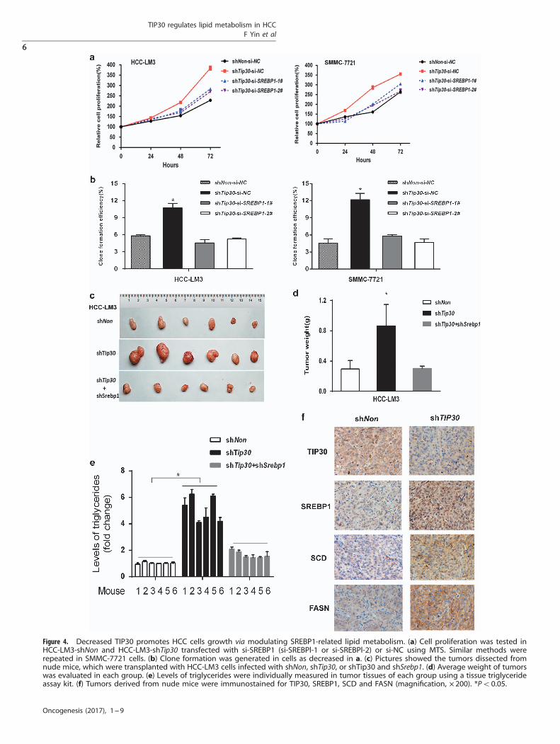
Figure 4. Decreased TIP30 promotes HCC cells growth via modulating SREBP1-related lipid metabolism. (a) Cell proliferation was tested inHCC-LM3-shNon and HCC-LM3-shTip30 transfected with si-SREBP1 (si-SREBPl-1 or si-SREBPl-2) or si-NC using MTS. Similar methods wererepeated in SMMC-7721 cells. (b) Clone formation was generated in cells as decreased in a. (c) Pictures showed the tumors dissected fromnude mice, which were transplanted with HCC-LM3 cells infected with shNon, shTip30, or shTip30 and shSrebp1. (d) Average weight of tumorswas evaluated in each group. (e) Levels of triglycerides were individually measured in tumor tissues of each group using a tissue triglycerideassay kit. (f) Tumors derived from nude mice were immunostained for TIP30, SREBP1, SCD and FASN (magnification, × 200). *Po0.05.
TIP30 regulates lipid metabolism in HCCF Yin et al
6
Oncogenesis (2017), 1 – 9
carcinoma. Meanwhile, we also expanded the novel function ofTIP30 in HCC metabolism regulation, which should be extensivelystudied in other types of tumors.
MATERIALS AND METHODSCell lines, antibodies and reagentsHCC-LM3 was supported by the Liver Cancer Institute of Zhong ShanHospital (Shanghai, China). HepG2 was available from American TypeCulture Collection. SMMC-7721 was acquired from Cell Bank of ShanghaiInstitutes for Biological Sciences (Shanghai, China). Cell lines have beentested for mycoplasma contamination and were cultured at 37 °C in ahumidified condition with 5% CO2. The Dulbecco’s modified Eagle’smedium (DMEM) was used for cell culture, supplemented with 10% fetalbovine serum. Details of primary antibodies and their sources are asfollows: TIP30 (produced by our lab as previously reported);17 p-mTOR(Ser2448), p-AKT (Ser473), mTOR, AKT, FASN and GAPDH (Cell SignalingTechnology, Danvers, USA); SCD and SREBP1 (Abcam, Cambridge, USA).Additionally, we purchased horseradish peroxidase-conjugated secondaryantibodies (anti-rabbit or anti-mouse) from Santa Cruz Biotechnology(SCBT, CA, USA). MK-2206 was from Selleck Chemicals (Houston, TX, USA).Oleate was acquired from Sigma (St Louis, MO, USA).
Lentivirus and small interfering RNAHCC cells were infected with lentivirus expressing Tip30 cDNA or shRNAtargeting Tip30 as literature described.17 A siRNA targeting SREBP1 weredesigned by GenePharma (Shanghai, China). Briefly, the followingsequences were used: for SREBP1-homo-523 (si-SREBP1-1), sense5′-GCUCCUCUCUUGAAGCCUUTT-3′ and antisense 5′-AAGGCUUCAAG-AGAGGAGCTT-3′; for SREBP1-homo-1403 (si-SREBP1-2), sense 5′-GCA-ACACAGCAACCAGAAATT-3′ and antisense 5′-UUUCUG GUUGCUGUG-UUGCTT-3′.
Protein extraction and western blotting1× SDS buffer was used to obtain total cell lysate. Equal amounts proteinwere loaded and then separated by SDS–PAGE. After the process ofelectrophoresis was finished, PDVF membranes were used forprotein transferring from SDS–PAGE. After probing with primary andsecondary antibodies, enhanced chemiluminescence’s reagents (PierceBiotechnology, Milwaukee, WI, USA) were added to detect the antigen–antibody complexes.
RNA extraction and real-time quantitative PCRNucleoSpin RNA kit (Macherey-Nagel, Germany) was used for total RNAextraction. After the concentration of RNA was tested, reverse transcriptionPCR was done as the PrimeScript RT reagent Kit (Takara Bio, Tokyo, Japan)guided. SYBR Green-based real-time PCR was operated on a 7500 FastReal-Time PCR System (Applied Biosystems, Carlsbad, CA, USA) and Gapdhwas detected for endogenous control. Primer sequences are listed inSupplementary Table 1.
BODIPY stainingHCC cells were fixed for 15 min using 4% paraformaldehyde. Thereafter,cells were washed then dyed by BODIPY 493/503 (10 μg/ml) (ThermoFisher Scientific, Waltham, MA, USA) at 37℃ for 15 min.Then, the nucleuswas counterstained with DAPI (Invitrogen, Carlsbad, CA, USA) for 10 min.We captured images using a fluorescence microscope (Olympus,Lake Success, NY, USA).
TG measurementsThe cellular TG content was tested using a Triglyceride Quantification Kit(Abcam, Cambridge, UK) with detailed experimental procedure providedin the kit.
Immunofluorescence stainingHCC cells were incubated on 24-well plates for 24 h and then werefixed using paraformaldehyde with the final concentration 4%. TritonX-100 was prepared in PBS for the final concentration 0.2% and then wasused for permeabilization.10% BSA/PBS was used as blocking buffer.For immunofluorescence staining, primary antibodies were added forincubation for 24 h on a shaker setting at 4 °C. After washing three times, agoat anti-rabbit Alexa Fluor 555 antibody (Thermo Fisher Scientific) wasmixed and for 1 h incubation. Thereafter, cells were counterstained usingDAPI (Invitrogen). All matched samples were photographed (control andtest) using confocal laser scanning microscopy (FLUOVIEW FV-1000).
Proliferation detection and colony-formation assaysThe detached cells were incubated on 96-well plate (5000 cells/well).Proliferation was testified uasing MTS method (CellTiter 96 AQueous OneSolution Cell Proliferation Assay, Promega Corporation, USA) at differenttime point as protocol guided. For colony-forming assays, single-cellsuspension was prepared which was then seeded onto a 10-cm-diameterdish with 10 000 cells/dish. After 10 days, cultured cells were dyed by
Figure 5. TIP30 expression negatively associates with SREBP1 level in tumor tissues of HCC patients and the combinational biomarkers providepowerful prognostic value for HCC. (a) TIP30 and SREBP1 were detected by immunostaining in 80 HCC samples. Representative picturesof immunostaining were shown for two patients. (magnification, × 200). (b) The correlation between TIP30 and SREBP1 expression wasanalyzed in HCC tissues according to the scores of immunohistochemistry staining. (c) Scatter plot showed correlations between TIP30and SREBP1 mRNA level in tumors of 30 HCC patients investigated. Recurrence-free survival rates and overall survival rates wereanalyzed between TIP30-high expression group and TIP30-low expression group (d), as well as between four subgroups (TIP30low/SREBP1low;TIP30low/SREBP1high; TIP30high/SREBP1low; TIP30high/SREBP1high) (e).
TIP30 regulates lipid metabolism in HCCF Yin et al
7
Oncogenesis (2017), 1 – 9

crystal violet for 15 min. Then, dye was washed out and we counted clonesthat were containing 450 cells. Clone formation efficiency was calculatedas clones to total cells seeded on the dish.
Microarray analysisHCC-LM3 cell line was infected with shTip30 or control lentivirus. After7 days, TRIzol (Thermo Fisher Scientific) was used to extract cellular RNA,which was then purified by an RNeasy kit (Qiagen, Hilden, Germany).NimbleGen Gene Expression Microarray was applied in microarray analysis.Axon GenePix 4000B microarray scanner was used for scanning and rawdata were extracted by NimbleScan software 2.5. Gene Ontology (GO) andKyoto Encyclopedia of Genes and Genomes (KEGG) enrichment analysis ofthe differentially expressed genes (fold change ⩾ 2, Po0.05) wereperformed for predicting biologic effects of TIP30. Such analysis wasachieved by the ClueGo plugin of Cytoscape (Software version 3.2.3,INSERM UMRS1138, Paris, France), which is a functional annotation way toevaluate over-representation of functional categories in interested geneticsets.51 Enrichment analysis was performed via functional annotation chartand annotation clustering options, which was limited to GO terms andKEGG pathways in ‘Biologic Process’ categories. Functional annotation wasdeemed significant with P-valueo0.05, using Fisher’s exact test.
Tumor xenograft mouse modelAnimal studies are authorized by medical ethics committee of PLA GeneralHospital. Male Balb/c nude mice (4 weeks old) are randomly allocated intothree groups (6 mice/group) and the number of mice is determinedaccording to prior experience of in vivo studies in our laboratory. Wesubcutaneously inject 5 × 10⁶ indicated cells into each mouse. Investigatorswere not blinded for the animal studies. During the experiment, mice weremonitored and euthanized for histopathology examination after cellinoculation for 28 days. Then, the tumor weight and their triglycerideslevels were measured.
Patients, immunohistochemistry and scoringSamples of 80 patients who had radical resection of HCC were collected from2003 to 2007 at Guangxi Cancer Hospital (Nanning, China). Radical surgerywas defined as previously reported.20 Informed consent authorized by EthicsCommittee of Guangxi Cancer Hospital was acquired from patients whenspecimen collection was performed. Supplementary Table 2 showedclinicopathological features of the above patients. All the patients weremonitored for recurrence every 1–6 months after the curative resectiondepending on the post-operative time. Immunohistochemistry of clinicalsamples were performed as previously reported.17,20 Two experiencedpathologists independently evaluated the staining scores. According to thestaining intensity and distribution, immunostaining scores were semiquanti-tatively estimated.17 Immunohistochemical scores of ⩽ 4 and scores of ⩾ 5were classified as low and high expression, respectively.
Statistical analysisWe repeated in vitro experiments in triplicate. SPSS software (version 16.0,Chicago, IL, USA) was used for statistical analysis. Pearson chi-square test andStudent’s t-test were applied for analysis of dichotomous variables andcontinuous variables, respectively. Correlations of two variables weredetermined using Spearman rank test. Survival analyses of investigatedpatients were achieved using Kaplan–Meier analysis with log-rank test. Whenmore than two data sets were analyzed, variance analysis was performed.The results are showed as the mean± s.e.m. The above statistical analyseswere all two-sided with Po0.05 deemed statistically significant.
CONFLICT OF INTERESTThe authors declare no conflict of interest.
ACKNOWLEDGEMENTSThis work is supported in part by grants from Ministry of Science and Technology ofChina “973” and “863” programs (2014AA021103, 2014AA020704), Beijing NovaProgram (Z171100001117112), National Nature Science Foundation of China(81472281, 81502129, 81402424, 81472719, 81670573), Shanghai Key Laboratory ofCell Engineering (14DZ2272300), Shanghai Leading Academic Discipline Project(B905).
REFERENCES1 El-Serag HB. Hepatocellular carcinoma. N Engl J Med 2011; 365: 1118–1127.2 Connell LC, Harding JJ, Abou-Alfa GK. Advanced hepatocellular cancer: the
current state of future research. Curr Treat Options Oncol 2016; 17: 43.3 Piscaglia F, Svegliati-Baroni G, Barchetti A, Pecorelli A, Marinelli S, Tiribelli C et al.
Clinical patterns of hepatocellular carcinoma in nonalcoholic fatty liver disease: amulticenter prospective study. Hepatology 2016; 63: 827–838.
4 Martinez-Outschoorn UE, Peiris-Pages M, Pestell RG, Sotgia F, Lisanti MP. Cancermetabolism: a therapeutic perspective. Nat Rev Clin Oncol 2016; 14: 113.
5 Rohrig F, Schulze A. The multifaceted roles of fatty acid synthesis in cancer.Nat Rev Cancer 2016; 16: 732–749.
6 Shtivelman E. A link between metastasis and resistance to apoptosis of variantsmall cell lung carcinoma. Oncogene 1997; 14: 2167–2173.
7 Ito M, Jiang C, Krumm K, Zhang X, Pecha J, Zhao J et al. TIP30 deficiency increasessusceptibility to tumorigenesis. Cancer Res 2003; 63: 8763–8767.
8 Shi M, Zhang X, Wang P, Zhang HW, Zhang BH, Wu MC. TIP30 regulates apoptosis-related genes in its apoptotic signal transduction pathway. World J Gastroenterol2005; 11: 221–227.
9 Zhang H, Zhang Y, Duan HO, Kirley SD, Lin SX, McDougal WS et al. TIP30 isassociated with progression and metastasis of prostate cancer. Int J Cancer 2008;123: 810–816.
10 Ouyang H, Gore J, Deitz S, Korc M. microRNA-10b enhances pancreatic cancer cellinvasion by suppressing TIP30 expression and promoting EGF and TGF-betaactions. Oncogene 2014; 33: 4664–4674.
11 NicAmhlaoibh R, Shtivelman E. Metastasis suppressor CC3 inhibits angiogenicproperties of tumor cells in vitro. Oncogene 2001; 20: 270–275.
12 Zhao J, Lu B, Xu H, Tong X, Wu G, Zhang X et al. Thirty-kilodalton Tat-interactingprotein suppresses tumor metastasis by inhibition of osteopontin transcription inhuman hepatocellular carcinoma. Hepatology 2008; 48: 265–275.
13 Zhao J, Ni H, Ma Y, Dong L, Dai J, Zhao F et al. TIP30/CC3 expression in breastcarcinoma: relation to metastasis, clinicopathologic parameters, and P53expression. Hum Pathol 2007; 38: 293–298.
14 Zhao J, Zhang X, Shi M, Xu H, Jin J, Ni H et al. TIP30 inhibits growth of HCC celllines and inhibits HCC xenografts in mice in combination with 5-FU. Hepatology2006; 44: 205–215.
15 Zhao J, Chen J, Lu B, Dong L, Wang H, Bi C et al. TIP30 induces apoptosis underoxidative stress through stabilization of p53 messenger RNA in human hepato-cellular carcinoma. Cancer Res 2008; 68: 4133–4141.
16 Zhu M, Yin F, Yang L, Chen S, Chen R, Zhou X et al. Contribution of TIP30 tochemoresistance in laryngeal carcinoma. Cell Death Dis 2014; 5: e1468.
17 Tong X, Li K, Luo Z, Lu B, Liu X, Wang T et al. Decreased TIP30expression promotes tumor metastasis in lung cancer. Am J Pathol 2009; 174:1931–1939.
18 Bu F, Liu X, Li J, Chen S, Tong X, Ma C et al. TGF-beta1 induces epigenetic silenceof TIP30 to promote tumor metastasis in esophageal carcinoma. Oncotarget 2015;6: 2120–2133.
19 Guo S, Jing W, Hu X, Zhou X, Liu L, Zhu M et al. Decreased TIP30 expressionpredicts poor prognosis in pancreatic cancer patients. Int J Cancer 2014; 134:1369–1378.
20 Zhu M, Yin F, Fan X, Jing W, Chen R, Liu L et al. Decreased TIP30 promotesSnail-mediated epithelial-mesenchymal transition and tumor-initiating propertiesin hepatocellular carcinoma. Oncogene 2015; 34: 1420–1431.
21 Liao BM, Raddatz K, Zhong L, Parker BL, Raftery MJ, Schmitz-Peiffer C. Proteomicanalysis of livers from fat-fed mice deficient in either PKCdelta or PKCepsilonidentifies Htatip2 as a regulator of lipid metabolism. Proteomics 2014; 14:2578–2587.
22 Horton JD. Sterol regulatory element-binding proteins: transcriptional activatorsof lipid synthesis. Biochem Soc Trans 2002; 30: 1091–1095.
23 Horton JD, Goldstein JL, Brown MS. SREBPs: activators of the complete programof cholesterol and fatty acid synthesis in the liver. J Clin Invest 2002; 109:1125–1131.
24 Guo D, Bell EH, Mischel P, Chakravarti A. Targeting SREBP-1-driven lipidmetabolism to treat cancer. Curr Pharm Design 2014; 20: 2619–2626.
25 Li A, Zhang C, Gao S, Chen F, Yang C, Luo R et al. TIP30 loss enhances cytoplasmicand nuclear EGFR signaling and promotes lung adenocarcinogenesis in mice.Oncogene 2013; 32: 2273–2281.
26 Zhang C, Mori M, Gao S, Li A, Hoshino I, Aupperlee MD et al. Tip30 deletion inMMTV-Neu mice leads to enhanced EGFR signaling and development of estrogenreceptor-positive and progesterone receptor-negative mammary tumors. CancerRes 2010; 70: 10224–10233.
27 Porstmann T, Santos CR, Griffiths B, Cully M, Wu M, Leevers S et al. SREBP activityis regulated by mTORC1 and contributes to Akt-dependent cell growth.Cell Metab 2008; 8: 224–236.
28 Bhat M, Sonenberg N, Gores GJ. The mTOR pathway in hepatic malignancies.Hepatology 2013; 58: 810–818.
TIP30 regulates lipid metabolism in HCCF Yin et al
8
Oncogenesis (2017), 1 – 9

29 Lim HY, Sohn I, Deng S, Lee J, Jung SH, Mao M et al. Prediction of disease-freesurvival in hepatocellular carcinoma by gene expression profiling. Ann Surg Oncol2013; 20: 3747–3753.
30 Cao D, Song X, Che L, Li X, Pilo MG, Vidili G et al. Both de novo synthetized andexogenous fatty acids support the growth of hepatocellular carcinoma cells. LiverInt 2016; 37: 80–89.
31 Yamashita T, Honda M, Takatori H, Nishino R, Minato H, Takamura H et al.Activation of lipogenic pathway correlates with cell proliferation and poorprognosis in hepatocellular carcinoma. J Hepatol 2009; 50: 100–110.
32 Yu X, Li Z, Wu WK. TIP30: a novel tumor-suppressor gene. Oncol Res 2014; 22:339–348.
33 Chen V, Shtivelman E. CC3/TIP30 regulates metabolic adaptation of tumor cells toglucose limitation. Cell Cycle 2010; 9: 4941–4953.
34 Currie E, Schulze A, Zechner R, Walther TC, Farese RV Jr. Cellular fatty acidmetabolism and cancer. Cell Metab 2013; 18: 153–161.
35 Menendez JA, Lupu R. Fatty acid synthase and the lipogenic phenotype in cancerpathogenesis. Nat Rev Cancer 2007; 7: 763–777.
36 Bansal S, Berk M, Alkhouri N, Partrick DA, Fung JJ, Feldstein A. Stearoyl-CoAdesaturase plays an important role in proliferation and chemoresistance inhuman hepatocellular carcinoma. J Surg Res 2014; 186: 29–38.
37 Li N, Zhou ZS, Shen Y, Xu J, Miao HH, Xiong Y et al. Inhibition of the SREBPpathway suppresses hepatocellular carcinoma through repressing inflammation.Hepatology 2017; 65: 1936–1947.
38 Hu J, Che L, Li L, Pilo MG, Cigliano A, Ribback S et al. Co-activation of AKTand c-Met triggers rapid hepatocellular carcinoma development via themTORC1/FASN pathway in mice. Sci Rep 2016; 6: 20484.
39 Li L, Pilo GM, Li X, Cigliano A, Latte G, Che L et al. Inactivation of fatty acidsynthase impairs hepatocarcinogenesis driven by AKT in mice and humans.J Hepatol 2016; 64: 333–341.
40 Calvisi DF, Wang C, Ho C, Ladu S, Lee SA, Mattu S et al. Increased lipogenesis,induced by AKT-mTORC1-RPS6 signaling, promotes development of humanhepatocellular carcinoma. Gastroenterology 2011; 140: 1071–1083.
41 Hu Y, Chen F, Liu F, Liu X, Huang N, Cai X et al. Overexpression of TIP30inhibits the growth and invasion of glioma cells. Mol Med Rep 2016; 13:605–612.
42 Bakan I, Laplante M. Connecting mTORC1 signaling to SREBP-1 activation. CurrOpin Lipidol 2012; 23: 226–234.
43 Li Y, Wu J, Song F, Tang J, Wang SJ, Yu XL et al. Extracellular membrane-proximaldomain of HAb18G/CD147 binds to metal ion-dependent adhesion site (MIDAS)
motif of integrin beta1 to modulate malignant properties of hepatoma cells. J BiolChem 2012; 287: 4759–4772.
44 Li J, Huang Q, Long X, Zhang J, Huang X, Aa J et al. CD147 reprograms fatty acidmetabolism in hepatocellular carcinoma cells through Akt/mTOR/SREBP1c andP38/PPARalpha pathways. J Hepatol 2015; 63: 1378–1389.
45 Li L, Tang W, Wu X, Karnak D, Meng X, Thompson R et al. HAb18G/CD147promotes pSTAT3-mediated pancreatic cancer development via CD44s. ClinCancer Res 2013; 19: 6703–6715.
46 Grass GD, Tolliver LB, Bratoeva M, Toole BP. CD147, CD44, and theepidermal growth factor receptor (EGFR) signaling pathway cooperate toregulate breast epithelial cell invasiveness. J Biol Chem 2013; 288:26089–26104.
47 Zhang C, Li A, Zhang X, Xiao H. A novel TIP30 protein complex regulatesEGF receptor signaling and endocytic degradation. J Biol Chem 2011; 286:9373–9381.
48 Lu B, Ma Y, Wu G, Tong X, Guo H, Liang A et al. Methylation of Tip30 promoter isassociated with poor prognosis in human hepatocellular carcinoma. Clin CancerRes 2008; 14: 7405–7412.
49 Kumtepe Y, Halici Z, Sengul O, Kunak CS, Bayir Y, Kilic N et al. High serumHTATIP2/TIP30 level in serous ovarian cancer as prognostic or diagnostic marker.Eur J Med Res 2013; 18: 18.
50 Li C, Yang W, Zhang J, Zheng X, Yao Y, Tu K et al. SREBP-1 has a prognostic roleand contributes to invasion and metastasis in human hepatocellular carcinoma.Int J Mol Sci 2014; 15: 7124–7138.
51 Bindea G, Mlecnik B, Hackl H, Charoentong P, Tosolini M, Kirilovsky A et al.ClueGO: a Cytoscape plug-in to decipher functionally grouped gene ontology andpathway annotation networks. Bioinformatics 2009; 25: 1091–1093.
Oncogenesis is an open-access journal published by Nature PublishingGroup. This work is licensed under a Creative Commons Attribution 4.0
International License. The images or other third partymaterial in this article are includedin the article’s Creative Commons license, unless indicated otherwise in the credit line; ifthe material is not included under the Creative Commons license, users will need toobtain permission from the license holder to reproduce the material. To view a copy ofthis license, visit http://creativecommons.org/licenses/by/4.0/
© The Author(s) 2017
Supplementary Information accompanies this paper on the Oncogenesis website (http://www.nature.com/oncsis)
TIP30 regulates lipid metabolism in HCCF Yin et al
9
Oncogenesis (2017), 1 – 9