Embed Size (px)
Citation preview

Review
Ticks feeding on humans: a review of records onhuman-biting Ixodoidea with special reference topathogen transmission
AGUSTIN ESTRADA-PENA1* and FRANS JONGEJAN2
1 Departamento de Parasitologıa, Facultad de Veterinaria, C/. Miguel Servet, 177, 50013-Zaragoza,Spain; 2 Department of Parasitology and Tropical Veterinary Medicine, Utrecht University, PO Box80165, 3508 TD Utrecht, The Netherlands
(Received 18 February 1999; accepted 3 May 1999)
Abstract. In this article, literature records of argasid and ixodid ticks feeding on humans worldwide areprovided in view of increased awareness of risks associated with tick bites. Ticks can cause paralyses,toxicoses, allergic reactions and are vectors of a broad range of viral, rickettsial, bacterial and protozoanpathogens. Approximately 12 argasid species (Argas and Ornithodos) are frequently found attached tohumans who intrude into tick-infested caves and burrows. Over 20 ixodid tick species are often found onhumans exposed to infested vegetation: four of these are Amblyomma species, 7 Dermacentor spp.,3 Haemaphysalis spp., 2 Hyalomma spp. and 6 Ixodes species. Personal protection methods, such asrepellents and acaricide-impregnated clothing are advised to minimize contact with infected ticks.Acaricidal control of ixodid ticks is impractical because of their wide distribution in forested areas, buthouses infested with soft ticks can be sprayed with acaricidal formulations. Attached ticks should beremoved without delay. The best way is to grasp the tick as close to the skin as possible with finetweezers and pull firmly and steadily without twisting. Finally, despite the fact that most people who arebitten destroy the offending tick in disgust, it is recommended that they preserve specimens in ethanol fortaxonomic identification and detection of pathogens by molecular methods.
Key words: ticks, Ixodidae, Argasidae, vectorial capacity, global distribution, tick-borne diseases,removal
Introduction
Ticks (Acari: Ixodoidea) are among the most important vectors of diseases affectingboth humans and animals (Sonenshine, 1991). Diseases transmitted by ticks tolivestock constitute a major factor which limits animal production in many tropicaland sub-tropical areas of the world and have been reviewed elswhere (Jongejan andUilenberg, 1994). In humans, ticks can cause severe toxic conditions such asparalyses and toxicoses, irritation and allergy, and their ability to transmit a greatvariety of infectious diseases is a major public health concern. In this article a
* To whom correspondence should be addressed at: Fax: 134–76–761612; e-mail:[email protected]
Experimental and Applied Acarology 23: 685–715, 1999.© 1999 Kluwer Academic Publishers. Printed in the Netherlands.

distinction is made between tick species which are frequently encountered, and thosethat are found occasionally on humans. Species commonly found on man are alsoimportant vectors of viral, rickettsial, bacterial and protozoan diseases.
The bites of soft ticks, notably Argas and Ornithodoros species, usually inhabitantsof nests and burrows of birds and rodents and human dwellings in rural areas, causeirritation, blisters, bruising and a more or less severe pruritus. Typical outbreaks ofargasids tick bites occur when people intrude into tick-infested caves, nests andburrows. Argas monolakensis, Ornithodoros coriaceus, Ornithodoros erraticus andOrnithodoros moubata are some of the major argasids reported to feed on humans.Ixodid ticks are encountered by humans while exposed to infested vegetation, usuallyin forested areas. Ticks frequently found on humans are Amblyomma americanumand A.hebraeum, Dermacentor andersoni and D. variabilis, Hyalomma anatolicumand H.marginatum, Haemaphysalis spinigera, and Ixodes scapularis, I. ricinus, I.persulcatus and I. holocyclus.
Ticks transmit a greater variety of pathogenic micro-organisms than any otherarthropod vector group. Some of the major diseases transmitted by argasids (mainlyOrnithodoros) are caused by Borrelia species, which induce relapsing fevers. Man-biting Ixodes ticks transmit viral infections causing European Tick-borne Encephalitisand the more severe Russian Spring Summer Encephalitis. Other viral infections,such as Crimean-Congo Hemorrhagic Fever transmitted by Hyalomma spp, occursporadically throughout vast areas of Africa, Asia and Europe, but can causemortality. Another potentially deadly flavivirus is transmitted by Haemaphysalisticks which causes Kyasanur Forest Disease in India, claiming many victimsannually. Bacterial diseases, such as Lyme disease caused by Borrelia burgdorferisensu lato, occur in the United States, Europe and Asia and are transmitted by Ixodesticks. The tick-borne rickettsiosis, Rocky Mountain spotted fever, a life-threateningbut treatable rickettsial disease widespread throughout the eastern United States, istransmitted by Dermacentor ticks. Another rickettsial infection, fievre boutonneuse,occurs mainly in Europe, caused by Rickettsia conorii. In southern Africa,Amblyomma ticks transmit Rickettsia africae, whereas Dermacentor reticulatustransmits Omsk Hemorrhagic Fever, caused by Rickettsia sibirica, in the formerSoviet Union. Two other rickettsioses, monocytic and granulocytic ehrlichiosis, causedby Ehrlichia chaffeensis and Ehrlichia phagocytophila group, respectively, were dis-covered relatively recently. E. chaffeensis is transmitted by Amblyomma americanum,whereas E. phagocytophila is transmitted by Ixodes scapularis in the USA, andIxodes ricinus in Europe. Protozoan diseases play a relatively minor role; Babesiadivergens and B. microti are both transmitted by Ixodes ticks in the USA and Europe,and there are possibly other species. In this article we bring together literaturerecords of human biting ixodoidea worldwide, with emphasis on ticks of medical andveterinary importance, geographical distribution, vectorial capacity, preventive meas-ures and removal of attached ticks.
686
Argasidae: the genus Argas
Argas monolakensis is an important argasid tick feeding on humans. It is the vectorof Mono Lake virus and occurs in California, USA (Schwan et al., 1992a). Argasmoreli and Argas neghmei have been implicated in human parasitism in Peru andChile, respectively, where humans and chickens cohabit (Kohls and Hoogstraal,1961; Keirans et al., 1979). Pruritus lasting for several weeks has been describedfollowing bites by A. cucumerinus (Clifford et al., 1978).
Authentic records of Argas persicus parasitizing man are rare. Among almost 300records of tick bites of humans in western Siberia, only two (in Omsk) wereattributed to A. persicus (Fedorov, 1968). The extensive review of argasids in theformer Soviet Union by Filippova (1966) does not mention A. persicus feeding onman. However, records of human dwellings infested with this species are quitenumerous. Although viral and rickettsial pathogens have been found in A. persicuscollected in nature, this does not mean that A. persicus is a competent vector.
Argas reflexus is another argasid tick found on humans, especially followingpigeon eradication from European buildings. When pigeons are removed from theattics of old houses, ticks appear seeking new hosts on curtains and windows (Romanet al., 1960). Furthermore, anaphylactic shock, caused by an unidentified toxin of A.reflexus, has been reported in Italy (Miadonna et al., 1982) and Poland (Grzywaczand Kuzmicki, 1975). When pigeons were driven from resting places infested byArgas polonicus in Krakow, Poland, frequent tick bites in humans resulted, withsymptoms such as erythema, fever, weakness and diarrhoea (Siuda et al., 1982).Argas vulgaris, a species closely related to A. polonicus, has been reported fromhumans in the former Soviet Union (Filippova, 1966).
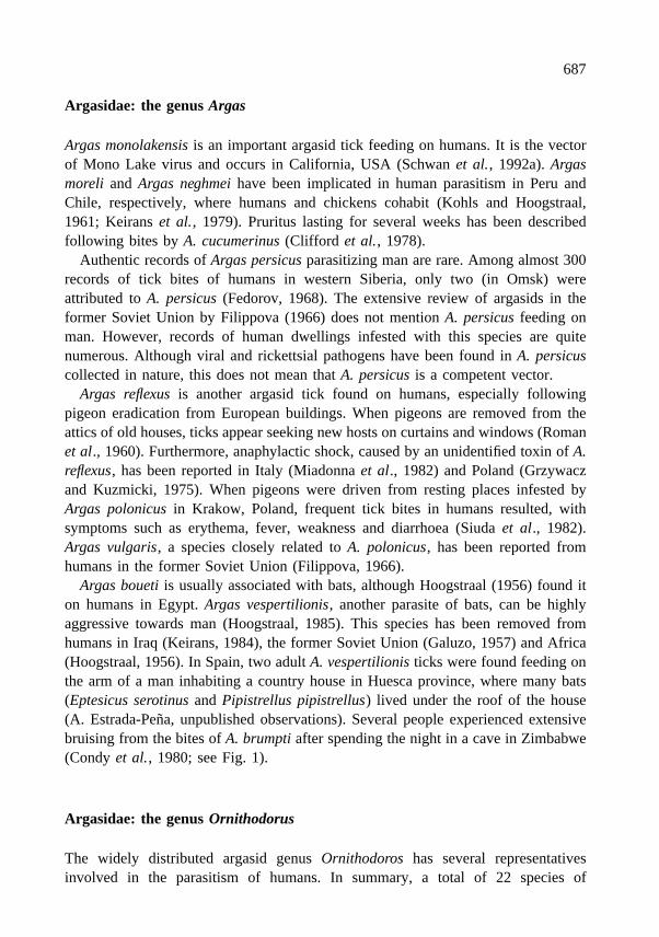
Argas boueti is usually associated with bats, although Hoogstraal (1956) found iton humans in Egypt. Argas vespertilionis, another parasite of bats, can be highlyaggressive towards man (Hoogstraal, 1985). This species has been removed fromhumans in Iraq (Keirans, 1984), the former Soviet Union (Galuzo, 1957) and Africa(Hoogstraal, 1956). In Spain, two adult A. vespertilionis ticks were found feeding onthe arm of a man inhabiting a country house in Huesca province, where many bats(Eptesicus serotinus and Pipistrellus pipistrellus) lived under the roof of the house(A. Estrada-Pena, unpublished observations). Several people experienced extensivebruising from the bites of A. brumpti after spending the night in a cave in Zimbabwe(Condy et al., 1980; see Fig. 1).
Argasidae: the genus Ornithodorus
The widely distributed argasid genus Ornithodoros has several representativesinvolved in the parasitism of humans. In summary, a total of 22 species of
687

Ornithodoros species have been reported on humans, and 12 species are foundfrequently (Table 1).
O. boliviensis and O. kelleyi are usually parasites of bats, and bite humans onlywhen they enter caves or bat-infested houses (Hoogstraal, 1985). New Worldrepresentatives of the genus are well known ectoparasites of man, and act as vectors
Figure 1. Extensive bruises on the back of a man from bites of the argasid tick Argas brumpti afterspending the night in a cave in Zimbabwe. Reproduced with permission from The Central AfricanJournal of Medicine, Vol. 26, 1980, p. 212. (Article by J.B. Condy, R.A.I. Norval, N.K. Blackburn andP. Clemence)
Table 1. Summary of some of the most important soft tick species found attached to human victims, withdata on their vectorial capacity and geographical distribution
Species Pathogen Distribution
Argas monolakensis Mono Lake virus Western USAA. reflexus – Southeastern EuropeOrnithodoros asperus Borrelia caucasica Caucasus, IraqO. capensis Soldado virus CosmopolitanO. coriaceus Borrelia coraciae Pacific coast of USA into MexicoO. erraticus Borrelia crocidurae North and East Africa, Near East, Southeastern EuropeO. erraticus Borrelia hispanica Spain, PortugalO. hermsi Borrelia hermsi Western USAO. maritimus Soldado virus FranceO. moubata Borrelia duttoni East Africa and Southern AfricaO. tartakovskyi Borrelia latyschevi Central AsiaO. turicata Borrelia turicatae Southwestern USA, Central AmericaO. savignyi – Africa and parts of Asia
688

of diseases, such as relapsing fever caused by spirochetes of the genus Borrelia.Relapsing fever is a zoonotic disease with a variety of nest- or burrow-inhabitingmammals, especially rodents, acting as reservoir (Sonenshine, 1993). Since mostOrnithodoros ticks are nidicolous parasites, humans are particularly involved in thecycle of transmission when they intrude into the nest environment. The epidemiologyis therefore characterized by isolated outbreaks in scattered localities inside endemicareas (Burgdorfer, 1986). Borrelia spirochetes persist in the argasid vectors for manyyears, probably for their entire life. Thus, the remarkable longevity and cryptic habitsof the Ornithodoros ticks perpetuate this zoonosis in endemic areas.
Ornithodoros coriaceus is distributed along the Pacific coast of the USA intoMexico. The tick is feared because of its very painful bites, O. coriaceus transmitsBorrelia coriaciae, the cause of bovine epizootic abortion (Furman and Loomis,1984). Ornithodoros hermsi occurs in the American states of California, Nevada,Idaho, Oregon, Utah, Arizona, Washington and Colorado, as well as in Canada, andis the primary vector of Borrelia hermsi (Schwan et al., 1992b). O. hermsi is foundin cavities of dead trees, log cabins and human dwellings, and usually feeds on treesquirrels. People become infected with B. hermsi when rodents are driven out duringrodent-trapping activities (Sonenshine, 1993).
Borrelia parkeri, transmitted by the bite of Ornithodoros parkeri (western UnitedStates, California), seldom if ever infests man, although the tick vector feeds on thesame hosts and occurs in the same geographical area as O. hermsi.
Ornithodoros talaje is distributed throughout the western states of the USA,Mexico, Venezuela, Uruguay, Brazil, Panama, Ecuador, Chile and Argentina. O. talajecan transmit Borrelia talaje in some parts of central and south America, althoughadults of this tick are seldom observed in dwellings and are not avid parasites ofman.
Ornithodoros turicata inhabits the south-western part of the USA and CentralAmerica, and is a vector for Borrelia turicatae. This borrelia species is transmittedmainly via salivary fluids and not via coxal fluids, as is common in the Ornithodoros-Borrelia relationship. Few human infestations have been recognized in the USA, butin Oklahoma, where O. turicata infests homesteads and sheds, entire families havedeveloped relapsing fever (Burgdorfer, 1980). The bite of O. turicata is painless, butis usually followed by intense local irritation and swelling several hours later andsubcutaneous nodules may persist for months (Cooley and Kohls, 1944).
Ornithodoros amblus has been occasionally reported on humans in Peru (Cliffordet al., 1980). Ornithodoros rudis is distributed throughout Panama, Paraguay,Colombia, Venezuela, Peru and Ecuador and is the primary vector of Borreliavenezuelensis, although clinical and epidemiological details are poorly documented(Hoogstraal, 1985). O. rudis is apparently well adapted as a parasite of humans, butfeeds on other mammals as well. Ornithodoros rostratus has been implicated inhuman parasitism in South America, where it is well-known for its painful bite,
689

which becomes pruritic and often secondarily infected. O. rostratus is able to trans-mit R. rickettsii (Hoogstraal, 1985).
In the Caucasus and Iraq, Ornithodoros asperus which inhabits rodent burrows,transmits Borrelia caucasica, which can cause severe disease in humans withnumerous rapidly recurring relapses. Ornithodoros coniceps has been reported onhumans in France and Spain (Gil Collado, 1947; Keirans, 1984), usually as the resultof sleeping in caves where rock pigeons nest. Human hosts developed local oedemaand pain with chills lasting from a few hours up to three days (Hoogstraal et al.,1979).
Ornithodoros erraticus occurs on humans in Spain and parts of Africa. The bite oflarvae of this species may result in allergic reactions, as observed in one patient inSpain (A. Estrada-Pena, unpublished data). This species transmits Borrelia crocidurae(North and East Africa, the Near East, including Southeastern Europe) and Borreliahispanica (Tunisia, Algeria, Morocco, Spain and Portugal). Reports of human casesof relapsing fever due to B. crocidurae have become rare in North-east Africa, wherethe disease has declined since the 1940s. However, Trape et al. (1991) reported awide distribution for B. crocidurae in Senegal, where relapsing fever appears tobe a major cause of morbidity in rural areas. Epidemiological relationships of B.hispanica and the tick vector(s) remain obscure. Anda et al. (1996) reported theisolation of a new Borrelia species from patients with relapsing fever and fromOrnithodoros spp. in southern Spain. This pathogen is closely related to other tick-borne relapsing fever spirochetes in Europe and Africa, and causes a disease withserological similarities to Lyme disease.
Ornithodoros maritimus is commonly found on sea birds, but has also beenreported on humans in France (Chastel et al., 1984). It transmits Soldado virus, apublic health problem in urban environments where sea gulls feed. Ornithodoroslahorensis is a common ectoparasite of man found in most parts of the former SovietUnion and can be infected experimentally with Rickettsia sibirica (Sidorov andKokorin, 1980). Ornithodoros tartakovskyi, distributed throughout most of CentralAsia, may be infected with Borrelia latyschevi the agent of Central Asia relapsingfever, which can cause a mild human illness.
Ornithodoros tholozani commonly lives in man-made shelters, caves, rockyoverhangs and other localities where livestock is housed (Arthur, 1962). The speciesis distributed from China to eastern Libya, and transmits Borrelia persica, the agentthat causes Persian relapsing fever, resulting in a severe and sometimes fatal humandisease.
Ornithodoros capensis is a cosmopolitan tick of seabirds (Keirans et al., 1992),which has also been reported on humans. The study by Chastel et al. (1981) onSoldado virus indicated that O. capensis is a potential public health problem wheresea gulls feed in urban habitats. Visitors to penguin breeding sites in caves and onbarren coasts were attacked by nymphs and adults of Ornithodoros spheniscus withsubsequent pruritic, slowly-healing blisters (Hoogstraal et al., 1985).
690

Ornithodoros moubata, a well-known African species distributed throughout EastAfrica, Madagascar and northern parts of southern Africa, transmits Borrelia duttoni,the agent of African relapsing fever. This borrelia apparently circulates only in manand in O. moubata inhabiting human dwellings (Felsenfeld, 1971). Coxal fluid ofnymphs and adults and salivary fluids of nymphs are the main routes of B. duttonitransmission (Burgdorfer, 1951). In Kenya and adjacent countries of eastern Africa,spirochete-infected O. moubata survive in the cracks and crevices of huts of indigenouspeople, where optimal microclimatic conditions are maintained by cooking fires(Walton, 1962). Changes in home construction have greatly reduced the incidence ofrelapsing fever since Walton’s report. Although hepatitis virus does not multiply inO. moubata, mechanical transmission from ticks to man may occur by crushinginfected ticks, through a bite, or by contamination with coxal fluid when scratchingtick bite lesions (Jupp et al., 1987). Joubert et al. (1985) detected this virus in O.moubata collected from the north-east strip of Namibia and suggested that mechan-ical transmission may be responsible for the high prevalence rate of hepatitis virusinfection in humans in this area.
Ornithodoros muesebecki has been collected from humans in Arabia (Hoogstraal,1982), the bite causing blisters, pruritus and fever. It has not been determinedwhether salivary toxins or pathogens are involved. Ornithodoros savignyi is foundin human habitations in India, Africa and some parts of Asia (Keirans, 1984;Hoogstraal, 1985) and causes intense local irritation in man (Hoogstraal, 1956).Moreover, in dry areas of Africa and Asia O. savignyi commonly attacks humansresting under shady trees and around wells where animals gather, etc. The bite of thisspecies can cause long-lasting intense pruritus (G. Uilenberg, unpublished experiencein Somalia). The same effects are associated with the bite of the AustralianOrnithodoros gurneyi (Roberts, 1970).
Argasidae: the genus Otobius
The spinose ear tick is found in the western part of the United States, Mexico andBritish Columbia, Canada. However, close association with livestock has resulted inits transportation to other areas of the world (Harrison et al., 1997). Paralysis of ahuman patient was related to the bite of an Otobius megnini nymph in South Africa(Peacock, 1958). Painful ear infestations by this species have been reported inhumans in India by Chellappa (1973) and although irritating, do not result in serioussequelae (Eads and Campos, 1984; Uilenberg et al., 1980).
Ixodidae: the genus Amblyomma
Amblyomma americanum is distributed throughout central and eastern parts of theUSA as well as parts of central and south America. This tick is very aggressive and
691

accounted for 34% of the ticks collected from US Air Force personnel across theUSA (Campbell and Bowles, 1994). Also 83% of ticks feeding on humans fromGeorgia and South Carolina were A. americanum (Felz et al., 1996). A. americanummay be infected with Francisella tularensis, the agent of tularemia (Goddard, 1987)and Rickettsia rickettsii, the agent of Rocky Mountain spotted fever (Kardatzke et al.,1992). However, A. americanum does not appear to be a vector for either LymeDisease (Piesman and Sinsky, 1988) or RMSF (Goddard and Norment, 1986).Spirochetes derived from A. americanum in Missouri appear to differ from B.burgdorferi in their antigenicity, growth characteristics in culture and the tick speciesthey infect in the laboratory (Piesman and Gray, 1994). However, A. americanumdoes transmit Ehrlichia chaffeensis, the agent of human monocytic ehrlichiosis(Ewing et al., 1995). The ehrlichioses are emerging zoonotic infections, caused byobligate intracellular bacteria of the genus Ehrlichia. Human monocytic ehrlichiosisis caused by E. chaffeensis infecting mononuclear phagocytes in blood and tissues.E. chaffeensis infection has been documented in more then 400 patients in 30 statesof the United States, as well as Europe and Africa (Walker and Dumler, 1996).Evidence that A. americanum is the vector of E. chaffeensis is based on its presencein areas where deer (Odocoileus virginianus) have high antibody titers against E.chaffeensis, and reactivity is lacking in areas where the tick is absent (Lockhart et al.,1996). Human infection by E. chaffeensis is suspected in both Portugal and Spain(Morais et al., 1991; Guerrero, 1992; Saz et al., 1994). However, since Amblyommaspecies are absent on the Iberian Peninsula, there may be another vector involved orthese claims are invalid.
In the southern United States, transmission of B. burgdorferi to humans is still acontroversial issue. Some investigators feel that A. americanum is the primary vectorof human Lyme disease in the southern US (Masters et al., 1994) whereas othershave expressed doubt that B. burgdorferi infects people in this region (Campbellet al., 1995). A new, uncultivable spirochete, B. lonestari, has been found infectingA. americanum (Barbour et al., 1996) while it has been demonstrated that this tick isunable to transmit B. burgdorferi to mice (Piesman and Happ, 1997).
Amblyomma cajennense is distributed from southern Texas, throughout Mexicoand Central America into parts of South America (Guglielmone et al., 1991). Allactive stages of A. cajennense bite humans, leaving a painful lesion (Goddard, 1989).The tick may serve as a vector of RMSF in western and central Mexico and SouthAmerica (McDade and Newhouse, 1986). Amblyomma maculatum has been recordedon humans in parts of the USA (Snoddy and Cooney, 1984; Harrison et al., 1987),and Argentina (Boero, 1945). It has been incriminated in the transmission ofRickettsia conorii to man in Uruguay (Conti et al., 1990) and of the RMSF in USA(Loving et al., 1978).
Amblyomma hebraeum is one of the most important African tick species onlivestock and is widespread throughout Southern Africa. It is a vector of tick-bitefever (Rickettsia africae infection) (Kelly et al., 1994). Larval A. hebraeum (like
692

larval A. americanum) are aggressive and attack in large numbers on the legs andabout the waist, causing intense irritation, rashes and occasionally pustules. Nymphsare also frequently encountered on humans, including tourists visiting selected partsof South Africa. Recently one person of such a group developed symptoms of tick-bite fever, caused by Rickettsia africae, two weeks after a nymphal A. hebraeum tickwas removed from his body (F. Jongejan, unpublished observations, 1998).
Amblyomma variegatum, another African tick species, is a vector of CrimeanCongo Hemorrhagic Fever (CCHF) in Uganda, Senegal, Nigeria and Central AfricanRepublic (Linthicum and Bailey, 1994) and is sometimes found on man.
Amblyomma testudinarium has been recorded on humans in Japan (Suzuki et al.,1990), Malaysia (Audy et al., 1960; Keirans, 1984), China (Kuo-Fan, 1991), andIndia (Dhanda and Rao, 1964). Its role in pathogen transmission to man is, however,unknown.
Amblyomma tholloni, commonly found on elephants in Africa, is known toparasitize man in Uganda, Mozambique and Tanzania (Matthysse and Colbo, 1989).It has not been implicated in the transmission of diseases to man. Amblyomma spp.commonly reported from humans are summarized in Table 2. Those species thatoccur occasionally on humans, or which lack adequate epidemiological data areincluded in Table 3.
Ixodidae: the genus Boophilus
Boophilus spp. ticks commonly parasitize bovines in temperate and tropical regionsof the world, but very rarely attack man (Strickland et al., 1976). Boophilusmicroplus, however, has been rarely reported on humans in Cuba (de la Cruz et al.,1991), Argentina (Guglielmone et al., 1991), and China (Kuo-Fan, 1991). There areno diseases known to be transmitted by Boophilus ticks to man.
Ixodidae: the genus Dermacentor
Several ticks of the genus Dermacentor are involved in parasitism and diseasetransmission to humans.
Table 2. Summary of Amblyomma tick species found feeding on humans, with data on their vectorialcapacity and geographical distribution
Species Pathogen Distribution
A. americanum Ehrlichia chaffeensis USA, central and south AmericaA. hebraeum Rickettsia africae Southern AfricaA. maculatum Rickettsia conorii UruguayA. variegatum Crimean-Congo Hemorrhagic Fever virus Uganda, Senegal, Nigeria, Central
African Republic
693

Dermacentor andersoni has been recorded in the Nearctic region, from the USA(Nebraska to South Dakota and Sierra Nevada mountains) to Canada (BritishColumbia, Alberta, Saskatchewan). It is the primary vector of Rickettsia rickettsii,the agent of Rocky Mountain spotted fever (RMSF), in the Rocky Mountain States.This zoonosis involves circulation of rickettsiae between ticks and vertebrate hosts inan ecosystem independent of man, with the tick both acting as vector and reservoirof infection (Schriefer and Azad, 1994). Ticks develop systemic infection andrickettsiae invasion of the ovaries may result in 100% infection of oocytes.Transovarial transmission may thus represent a far more important mechanism formaintaining R. rickettsii in nature than infection of ticks by feeding on rickettsemichosts (Burgdorfer and Brinton, 1975).
Dermacentor andersoni can also transmit the causative agent of Colorado TickFever (CTF) (Yunker and Cory, 1967). CTF is a zoonosis caused by an Orbivirus.Distribution of the disease coincides with the geographic range of the tick. It ismaintained in natural, enzootic foci within its geographic range through the inter-action of the vector ticks and susceptible hosts, mainly small mammals (Sonenshine,1993). Transovarial transmission of CTF virus does not occur. Larval D. andersoniacquire the infection by feeding on viremic hosts. Trans-stadial transmission tonymphs occurs with moulting from infected larvae, and afterwards, to adults.Geographical foci with a high incidence of CTF virus in ticks and mammalian hostsare habitats that provide shelter for chipmunks, ground squirrels and other smallmammals which serve as a blood source for immature ticks (Sonenshine et al.,1976). Although D. andersoni is an efficient experimental vector of the Powassan
Table 3. Other Amblyomma species occasionally reported to feed on humans
Species Distribution Reference
A. coelebs Paraguay Keirans, 1984A. cohaerens Uganda Matthysse and Colbo, 1987A. cyprium Papua New Guinea Keirans, 1984A. integrum Sri Lanka Keirans, 1984A. loculosum Australia Keirans, 1984A. neumanni Argentina Guglielmone et al., 1991A. nuttalli Ivory Coast Aeschlimann, 1967A. oblongoguttatum Central and South America Aragao and Fonseca, 1961A. ovale Panama
SurinamObaldia, 1992Keirans, 1984
A. parvum ArgentinaBrazilPanama and Guatemala
Guglielmone et al., 1991Fonseca, 1959Fairchild et al., 1966
A. tholloni Uganda, Mozambique and Tanzania Matthysse and Colbo, 1987
A. cyprium, A. dissimile, A. fossum, A. incisum, A. longirostre, A. marmoreum, A. tigrinum and A.triguttatum are species reported in the Index Catalogue of Medical and Veterinary Zoology withoutfurther details (Doss et al., 1977).
694

Encephalitis virus, the agent has not been isolated from numerous field pools of tickscollected throughout its distribution range (Nuttall and Labuda, 1994).
Dermacentor variabilis occurs throughout the USA (except in parts of the RockyMountains) and also in Canada and Mexico. Felz et al. (1996) reported a frequencyof 11.4% on humans in Georgia and South Carolina. Campbell and Bowles (1994)documented the presence of D. variabilis in 34% of ticks found on humansthroughout the USA where most of the cases of the disease occur. In other reports,D. variabilis accounted for 94% in North Carolina (Slaff and Newton, 1993), 90% inSouth Carolina (Burgdorfer et al., 1975) and 34% in Ontario (Scholten, 1977). It isthe primary vector of RMSF in the eastern parts of the USA, and has beenincriminated as a vector of Francisella tularensis (Hopla and Hopla, 1994). A studyof attachment site preferences of D. variabilis in humans showed that most ticks(50%) were taken from the head, while legs–feet and arms–axillae, accounted for21.5% and 10.8% respectively (Slaff and Newton, 1993).
Dermacentor occidentalis occurs along the Pacific coast of the USA and inland forseveral hundred miles from Oregon to the southern tip of California and has beenreported in Mexico (Hoffmann, 1962). It causes paralysis in livestock, but not inhumans (Sonenshine, 1993), and it may transmit the agent of Tularemia, RMSF, andCTF (Goddard, 1987). Two other Nearctic species of this genus have been involvedin a few cases of human parasitism: D. parumapertus and D. albipictus (Doss et al.,1977).
Palearctic species of the genus Dermacentor have also been commonly involved inhuman parasitism. Dermacentor marginatus is one of the main Palearctic speciesreported on man. In Spain (A. Estrada-Pena, unpublished data) it accounts forapproximately 10% of the total number of ticks collected from humans. It is the mainvector of Rickettsia slovaca (Rehacek et al., 1990; Beati et al., 1993) and plays asecondary role in the transmission of the Omsk Hemorrhagic Fever (OHF) virus(Nuttall and Labuda, 1994). Strains of R. slovaca isolated from Germany, Hungaryand Armenia suggest a widespread distribution of this agent, and although fewclinical cases have been reported, many individuals had antibodies against thisrickettsia (Rehacek and Tarasevich, 1988). A survey of D. marginatus collected fromsheep in Slovakia indicated a R. slovaca infection rate of 20%, without apparentchange in prevalence over a 20-year period (Rehacek et al., 1990). The presenceof CCHF virus has been demonstrated in field-collected D. marginatus ticks(Kondratenko, 1976).
Dermacentor nuttalli occurs throughout central and eastern Siberia, AsiaticRussia, northern Mongolia and China, with occasional records from western Russia.It is found in high grasslands, but is absent from dense forests, river lowlands andhilly wooded country. It is one of several vectors of Rickettsia sibirica, together withD. marginatus, D. silvarum and D. reticulatus (Pchelkin et al., 1989). This tick-bornerickettsiosis is widespread in far eastern Siberia and central Asia, Armenia, Azerbaijan,Mongolia and Afghanistan (Rehacek and Tarasevich, 1988). Commonly, larvae and
695

nymphs feed on small mammals which amplify the infection, whereas humans areinfected when bitten by infected adults. Both trans-stadial and transovarial trans-mission occur in the tick vectors. In the former Soviet Union it is considered anoccupational disease, mainly in agricultural areas around pastures (Rehacek andTarasevich, 1988). D. nuttalli is also a vector of tularemia in Russia (Olsufiev,1984).
Dermacentor silvarum occurs primarily in eastern Russia and northern Mongoliaand has also been reported from Romania and the former Yugoslavia. It is a vectorof Rickettsia sibirica (Yastrebov and Reshetnikova, 1990) in Russia and Mongolia,as well as of viruses of the Tick Borne Encephalitis (TBE) complex (Muratkina andLeonova, 1992).
Dermacentor reticulatus, the main vector of Omsk Hemorrhagic Fever (OHF)(Lvov, 1988) has commonly been reported on humans in Russia, Austria, the UnitedKingdom, France and has also been found in Spain (A. Estrada-Pena, unpublishedobservations, 1990). OHF is caused by a Flavivirus which is a member of the TBEcomplex. It produces lethal encephalitis in many wild rodents, and casues ahaemorrhagic disorder in humans. Adults of D. reticulatus feed on wild ungulatesand humans, whereas immature forms feed mainly on water voles (Microtusgregalis) in forest-steppe habitats. Vole populations are cyclic, and expansion ofthe virus-infected tick population coincides with increases in vole populations(Hoogstraal, 1985). Humans may become infected when hunting or trapping musk-rats (Ondatra zibethica) (Kharitonova and Leonov, 1985). D. reticulatus may alsoacquire Borrelia burgdorferi (Kahl et al., 1992), but there are no reports of its abilityto transmit, i.e., it is not vector competent. D. reticulatus is a vector of R. sibirica aswell as of C. burnetii (Gosteva et al., 1991; Sonenshine, 1993).
Finally, two further species of this genus have been recorded as ectoparasites ofman: D. auratus, which occurs in India, Nepal, Thailand, China and Malaysia and D.circumguttatus, reported by Matthysse and Colbo (1987) on man in Uganda. Forestedparts of the Himalayas are notorious for tick annoyance caused by nymphs ofDermacentor auratus (Hoogstraal, 1970). A summary of Dermacentor ticks fre-quently found on humans is given in Table 4.
Ixodidae: the genus Haemaphysalis
A large number of Haemaphysalis species are involved in human parasitism,however only the most important vectors are discussed (see Table 5). Others arelisted in Table 6.
Haemaphysalis spinigera is common in India and Sri Lanka, nymphs of whichavidly bite man. It is the main vector of the Kyasanur Forest Disease (KFD) virus.The original focus of KFD in man in the Kyasanur State Forest and surroundingvillages has spread extensively within the state of Karnataka, in India (Banerjee,
696

Table 4. Summary of Dermacentor ticks commonly reported from humans, with data on their vectorialcapacity and geographical distribution
Species Pathogen Distribution
D. andersoni Rickettsia rickettsiiColorado tick fever virus
USA (Rocky Mountain states)USA
D. marginatus Omsk Hemorrhagic fever virusRickettsia slovaca
Palearctic
D. nuttalli Rickettsia sibirica Siberia, Russia, Mongolia, ChinaD. occidentalis R. rickettsii
Colorado tick fever virusUSAUSA
D. silvarum R. sibirica Eastern areas of Soviet Union, Northern MongoliaD. reticulatus Omsk Hemorrhagic fever virus
R. sibiricaFormer Soviet Union
D. variabilis R. rickettsii USAD. auratus – Parts of the Himalaya
Table 5. Summary of Haemaphysalis species commonly reported from humans
Species Pathogen Distribution
H. concinna Tick Borne Encephalitis virus Central Europe, Soviet UnionH. punctata Tick Borne Encephalitis virus
Crimean-Congo Hemorrhagic fever virusEurope
H. spinigera Kyasanur forest disease virus India, Sri Lanka
Table 6. Other Haemaphysalis species rarely reported on humans
Species Distribution Reference
H. aculeata India, Sri Lanka Keirans, 1984H. anomala Nepal Hoogstraal, 1968H. aponommoides Nepal, India Hoogstraal, 1968H. bispinosa Nepal, Vietnam, Malaya Hoogstraal, 1968H. darjeeling Thailand Keirans, 1984H. flava Japan, India Hatsushika and Mimura, 1987H. hystricis Thailand, Burma, Laos, Vietnam Keirans, 1984
China Kuo Fan, 1991H. japonica China Kuo Fan, 1991H. koningsbergeri Thailand, Malaya Keirans, 1984H. leporispalustris USA Harrison et al., 1997H. longicornis Japan, Australia, China Kuo Fan, 1991H. mageshimaensis China Kuo Fan, 1991H. montgomeryi Nepal Hoogstraal, 1968
China Kuo Fan, 1991H. nepalensis Nepal Hoogstraal, 1968
China Kuo Fan, 1991H. elongata Madagascar Uilenberg et al., 1980
697

1988). About 95% of the KFD virus isolates are from H. spinigera, the predominanttick species of the forest floor in the endemic regions. Larvae feed on small mammalsand ground feeding birds, while nymphs infest larger animals including man andmonkeys. The KFD virus is transmitted trans-stadially, but not transovarially. Thevirus also occurs in a number of other Haemaphysalis species that do not parasitizeman, but infest small mammals (forest rats, shrews and porcupines). This secondaryenzootic cycle helps maintain and spread the virus to other areas (Nuttall andLabuda, 1994). Furthermore, the modification of forest habitat by human interventionmay have contributed to the epidemic spread of the disease (Bhat, 1990). Humanswho visit forests to collect firewood or those working in adjacent fields are at higherrisk of contracting KFD.
Haemaphysalis concinna is common in the forests of most of Central Europe,Russia, China, Korea, Vietnam and Japan. Haemaphysalis punctata is found on manin most parts of Europe. Both species are competent vectors of TBE virus, which hasbeen isolated from field collected specimens (Gresikova, 1972). H. punctata is alsoinvolved in the transmission of CCHF virus to man (Linthicum and Bailey, 1994).
Ixodidae: the genus Hyalomma
Ticks of the genus Hyalomma are well-known vectors of viruses and avid parasitesof man. Although many species are not involved in disease transmission, the con-siderable length of Hyalomma mouthparts provokes a painful bite. One of the mostimportant diseases transmitted by Hyalomma ticks is Crimean-Congo HemorrhagicFever (CCHF), of which Hyalomma marginatum is one of the main vector ticks.
H. marginatum and its subspecies have been recorded from south-eastern Europe,southern Russia, the Near East and Africa (Goddard, 1987). It is the primary vectorof CCHF virus where it occurs sporadically throughout Europe, Asia and Africa,ranging from Portugal eastwards to Crimea, and then further east into central Asiaand China (Hoogstraal, 1979). It appears to be absent in African countries along theMediterranean coast but it is present in most countries south of the Sahara. Ticksrather than their mammalian hosts are the natural reservoir of the CCHF virus, whichis perpetuated in the ticks by feeding on viremic hosts and by trans-stadial andtransovarial transmission (Linthicum and Bailey, 1994). The disease is most commonin arid and semi-arid biotopes. Serious outbreaks have been reported along rivers andriver floodplains, and in forest in steppes (Watts et al., 1988). Stable enzootic fociinvolve wild mammals, ground-feeding birds and ticks, although most birds areincompetent hosts for the infection (Hoogstraal, 1979). However, birds transportCCHF-infected ticks and serve as a bloodmeal source for tick populations (Wattset al., 1988). Hares (Lepus europaeus) and hedgehogs (Erinaceus europaeus) arefrequently infected and are important hosts of Hyalomma marginatum. Cattle mayalso be important in the epidemiology of CCHF, although evidence is based largely
698

on serology, with few isolates of the virus from cattle (Watts et al., 1988). OtherHyalomma ticks involved in the transmission of CCHF virus include H. truncatum,H. detritum and H. impeltatum. The transmission of this virus by co-feeding touninfected ticks has been also demonstrated (Gordon et al., 1993).
Hyalomma a. anatolicum, distributed in parts of the Near East, Asia Minor,southern Europe, southern Russia and India (Goddard, 1987), is a well-known vectorof CCHF (Linthicum and Bailey, 1994). Other species of Hyalomma may alsotransmit CCHF virus. There are records that Hyalomma asiaticum from foothillsemidesert regions of Russia, China, Afghanistan, Pakistan, Iran and Iraq mayparasitize humans (Kuo-Fan, 1991). Hyalomma ticks frequently found parasitizinghumans are given in Table 7.
Ixodidae: the genus Ixodes
Ixodes ricinus is common throughout most of Europe, including the British Isles.Stable populations are also scattered in northern Africa (Gray, 1991). The develop-ment and seasonal activity of I. ricinus vary considerably in different habitats,although activity in spring and early summer and again in late summer and earlyautumn is typical (Gray, 1991). I. ricinus is the primary European vector of theEuropean TBE viral subtypes (Nuttall and Labuda, 1994). Disease occurs in most ofEurope and in the eastern parts of Russia. The virus is maintained by trans-stadialtransmission, with tick infection as the result of feeding upon viremic hosts. Althoughthe virus has been isolated from other tick species, a strong correlation exists betweenhuman cases and the known distribution of I. ricinus (Sonenshine, 1993). Rodentsare the main vertebrate hosts for the virus especially Clethrionomys glareolus andApodemus flavicollis, as well as various insectivores (Nuttall and Labuda, 1994).Some wild carnivores and domestic ruminants are also susceptible to disease andmay contribute to its spread to humans. Although TBE virus was detected in thefaeces of I. ricinus (Benda, 1958), there is no evidence that transmission occurs viacontact with contaminated tick faeces material.
I. ricinus is the most important European vector of Lyme disease spirochetes,including B. afzelii, B. garinii, B. burgdorferi senso stricto, B.lusitaniae, B.valaisiana and possibly others (Postic et al., 1994; Le Fleche et al., 1997; Wanget al., 1997). The importance of I. ricinus in European Lyme Disease is due to itswidespread distribution, feeding habits and its willingness to bite humans (Piesman
Table 7. Summary of Hyalomma species commonly reported to feed on humans
Species Pathogen Distribution
Hyalomma. a. anatolicum Crimean-Congo Hemorrhagic fever virus Southern Europe, Russia,Near East
Hyalomma marginatum Crimean-Congo Hemorrhagic fever virus Southern Europe, Russia
699

and Gray, 1994). High risk of Lyme borreliosis occurs when habitats that are wellutilized by the public harbour large numbers of infected ticks. Identification of high-risk locations may be possible if standard habitat characteristicis can be recognized.Gray et al. (1998) compiled data for 105 Lyme disease habitats in 16 Europeancountries. The data showed that high-risk areas are heterogeneous deciduous wood-land, generally with a recreational function and supporting a diverse fauna usuallyincluding deer. Large numbers of ticks occurred in some other habitats, but infectionprevalence was low. Although there seemed to be good correlation between totalnumbers of ticks and numbers of Borrelia-infected ticks, this is not consistentenough for extrapolation from one to the other when diverse habitats are considered(Gray et al., 1998). Locations cannot therefore be classified as posing a high risk forLyme borreliosis simply on the basis of tick density. It is necessary to take otherhabitat characteristics into account, particularly the vegetation, which will have astrong influence on the variety and abundance of hosts present (Talleklint andJaenson, 1996). A study by Daniel et al. (1998) suggested the usefulness ofLANDSAT satellite imagery to provide an estimation of I. ricinus risk habitats.Satellite imagery with higher temporal resolution and larger area coverage, asobtained from NOAA-AVHRR satellite series, can improve our knowledge of habitatseasonal variation, and hence the changes of I. ricinus activity within short timeintervals.
I. ricinus may be an important vector and possible maintenance host of CCHFvirus (Watts et al., 1988) and also the vector of the protozoan parasite Babesiadivergens, the causative agent of human babesiosis in Europe causing sporadic butusually fatal cases (Gray, 1991). A recent fatal case of a B. divergens infection wasdiagnosed in a splenectomized man in the Algarve, Portugal (V. doRosario and A.J.Maia, A.M. Freudenthal and F. Jongejan, unpublished, 1998).
Ixodes persulcatus, which aggressively attaches to humans, is found in Japan,Russia, Korea and China (Im et al., 1998). Populations from western Russia havemoved into Europe, and it seems possible that migratory birds introduce I. persulcatusimmatures into other areas (R. Mehl, pers. comm.). I. persulcatus inhabits small-leafed forests near primary coniferous forests, such as spruce-basswood combina-tions. It is a vector for the OHF virus (Hoogstraal, 1965), Borrelia afzelii, B. garinii,and B. burgdorferi (Korenberg et al., 1987). Foci of TBE virus infection (Far-easternsubtype) are found within the geographic distribution of I. persulcatus, mostly in thetaiga containing mixed broad-leaved forests with high humidity. Sporadic foci ofdisease occur in river valleys covered by marshy meadows, and on undulating plainswhere suitable habitats overgrown with bushes are scattered among cultivated valleys(Nuttall and Labuda, 1994). Foci of the Far-eastern subtype of TBE virus, maintainedby I. persulcatus, contain a comparatively high prevalence of infected ticks, and insuch regions, seasonal tick activity occurs relatively briefly, approximately fromApril to June. The relative roles of both I. ricinus and I. persulcatus as vectors of the
700

two TBE viral subtypes are undetermined for parts of Europe where the two speciescohabit.
Ixodes hexagonus is a relatively common parasite of man in Germany and theUnited Kingdom (Liebisch and Walter, 1986). It is an experimental vector of B.burgdorferi (Toutoungi and Gern, 1993) and has been found infected under naturalconditions. The vector competence of I. hexagonus for TBE virus has been alsodemonstrated although it lacks a prominent role in the epidemiology of the disease(van Tongeren, 1962).
Ixodes uriae is a circumpolar tick of marine birds in which an enzootic cycle of B.burgdorferi has been demonstrated (Olsen et al., 1993). An ornithologist visitingmarine birds colonies was bitten by an I. uriae tick and developed erythema migrans(R. Mehl, pers. comm.). Ixodes ovatus parasitizes humans in Tibet, Burma, Nepal,Japan and China (Keirans, 1984; Kuo-Fan, 1991). Borrelia japonica has beenisolated from Japanese I. ovatus (Postic et al., 1994).
Ixodes holocyclus causes tick paralysis in Australia. It occurs primarily in NewGuinea and along the eastern coast of Australia, being active in the warmer monthsof the year. The bandicoot, the primary host for I. holocyclus, is increasing innumbers in urban areas as a result of control campaigns against dingoes and foxes(Bagnall and Doube, 1975). Clinical cases of tick are characterized by progressiveparalysis up to 48 h, with a severe exacerbation of the symptoms after tick removal;recovery often takes several weeks (Stone et al., 1989). In contrast with someparalytic conditions that resemble tick paralysis, it progresses rapidly and may befatal within a few days after the onset of symptoms. I. holocyclus is the vector ofRickettsia australis (Sexton et al., 1991), and also able to transmit a Nearctic strainof B. burgdorferi from the larval to the nymphal stage (Piesman and Stone, 1991).
Several species of the genus Ixodes are well-known parasites of humans. Ixodesscapularis (formerly Ixodes dammini, according to Oliver et al., 1993) occurs alongthe Atlantic coast of Canada and the USA, and throughout the southern states,including Texas and Oklahoma. It is possible that its range is expanding towards thewestern states (Keirans et al., 1996). I. scapularis ticks require moist microclimatesfor survival; habitats where leaf-litter is established and mixed deciduous forests witha high canopy provide the ideal environment. This species accounts for 76.2% of theticks collected on humans in southern New York (Falco and Fish, 1988) but only for3.9% in Georgia and South Carolina (Felz et al., 1996). Using a tick stage–specificregression equation for engorgement index in nymphs and adults of I. scapularis,Yeh et al. (1995) determined the duration of attachment for ticks removed by tick-bite victims. Most people (64%) found and removed adult I. scapularis before 36 hof attachment; by 48 h nearly all adult ticks were removed (79%). In contrast, fewpeople found and removed nymphal ticks by 24 h of attachment (10%) or 36 h(41%).
I. scapularis is the main vector of B. burgdorferi, the causative agent of humanLyme disease in the eastern USA and Canada, with the white-footed mouse
701

(Peromyscus leucopus) acting as the primary reservoir of B. burgdorferi andimmature I. scapularis ticks. In habitats were P. leucopus is absent, other rodentssubstitute as reservoirs, e.g., Norway rats (Rattus norvegicus) (Piesman and Gray,1991). However, by itself, the Norway rat is relatively unimportant. An entomo-logical index was proposed by Mather et al. (1996) to provide human health agencieswith data on the density of permanent I. scapularis populations. This index has beenrevisited using daily satellite imagery over continental United States and Canada,obtaining a habitat suitability index from soil temperatures and standard vegetationindex (Estrada-Pena, 1998). Variability in tick feeding rates in relation to spirochetetransmission among different tick hosts has not been determined, making it impos-sible to know if findings from these animal studies are applicable to humans (Yeh etal., 1995). However, animal studies suggest that risk for B. burgdorferi transmissionis low within the first 24 h of attachment but increases thereafter (Piesman, 1993).
Although RMSF rickettsiae has been isolated from I. scapularis (Burgdorfer,1975) its role in the ecology of RMSF is not well understood. I. scapularis can alsotransmit Babesia microti, the protozoan responsible for human babesiosis in theNearctic (Ristic and Lewis, 1977; Spielman, 1988). The most important reservoirhost for B. microti is the white-footed mouse, Peromyscus leucopus, althoughmeadow voles (Microtus pennsylvanicus) may also act as reservoirs, but are lessfrequently parasitized by I. scapularis (Spielman, 1988). Nymphs are primaryvectors, although adults may transmit the infection. Human babesiosis caused by B.microti has remained a minor public health concern in the USA, with only about 200cases since it was first recognized clinically (Sonenshine, 1993).
Human granulocytic ehrlichiosis (HGE) caused by Ehrlichia phagocytophila is anemerging disease, predominantly in the upper midwestern and northeastern USstates, but also in northern California (Walker and Dumler, 1996; Dumler andBakken, 1998). Pancholi et al. (1995) first implicated I. scapularis as a vector forHGE by association of an infected specimen with a human case report fromWisconsin. Madigan et al. (1996) confirmed that the DNA sequence of the 16SrRNA gene of the equine agent is identical to that of the human agent. Larval I.scapularis ticks acquired infection by feeding upon infected mice, and efficientlytransmitted the ehrlichiae after moulting to nymphs, thereby demonstrating vectorcompetence (Telford et al., 1996). The agent was also demonstrated in the salivaryglands of field-collected adult I. scapularis ticks. Des Vignes and Fish (1997)demonstrated the infection of laboratory mice by I. scapularis nymphs collected froma natural focus of HGE. These mice were positive by PCR, microscopic examina-tions of blood smears and larval I. scapularis xenodiagnosis. Positive xenodiagnosticlarvae maintained infection through moulting. Greig et al. (1996) studied dogs withnatural granulocytic ehrlichiosis. No dogs seroreacted with E. canis or E. chaffeensisantigens, which are cross-reactive; however, 100% of the dogs tested duringconvalescence were seropositive for E. equi antigens. Granulocytic ehrlichial 16SrRNA gene DNAs from dogs were amplified and revealed as identical to the agent of
702

human granulocytic ehrlichiosis, and very similar to E. equi. These findings suggestthat granulocytic ehrlichiosis in dogs is a zoonotic disease and dogs possibly con-tribute to the enzootic cycle and human infection. In the Netherlands finally,granulocytic ehrlichiosis has been diagnosed in dogs (F. Jongejan, unpublished,1998), and also in one human patient (van Dobbenburgh et al, 1999).
Ixodes pacificus occurs along the Pacific coastal margins of British Columbia,Canada and the USA, extending into California and parts of Mexico. The ecologicalconditions that permit I. pacificus to survive in western North America are even morevariable than those supporting the survival of I. scapularis. Isolated populations havealso been located in the non-coastal state of Arizona (Olse et al., 1992). Recently, azoonotic Babesia-like piroplasm (designated WA1) was identified in WashingtonState (Quick et al., 1993). Phylogenetic analysis of this new organism showed that itwas most closely related to, but distinct from, the canine pathogen (B. gibsoni) ratherthan to other members of the genus Babesia (Persing et al., 1995). I. pacificus maybe the vector of this Babesia-like protozoan in California and Washington, althoughvirtually nothing is known about the vector competence of the different tick stagesnor the epidemiology of the disease. I. pacificus is a relatively inefficient vector of B.burgdorferi, probably because larvae and nymphs feed mostly on reservoir-incompetent lizards (Lane and Loye, 1989). It is also less efficient than I. scapularisin acquiring and maintaining B. burgdorferi. The second vector in the Pacific coastis I. neotomae, a non-man-biting species, which maintains a cryptic cycle amongrodent and rabbits (Brown and Lane, 1992). In this particular case, both speciescontribute to the enzootic maintenance of B. burgdorferi among wildlife in nature,with I. pacificus also serving as the bridge vector for transmission of the spirochetesto man.
Ixodes cookei is found throughout Eastern and Midwestern states of the USA andCanada (Farkas and Surgeone, 1990), where it is the main vector of the PowassanEncephalitis (PE) virus (Nuttall and Labuda, 1994). I. cookei is opportunistic andreadily attacks dogs, man and various wild mammals. The woodchuck (Marmotamonax) is one of its most common hosts, in which PE virus is also found (Artsob etal., 1984). Spotted fever rickettsiae has also been identified in I. cookei (Burgdorfer,1988) but little is known about its role as a vector of the disease. Lyme borreliosismay also infect I. cookei (Levine et al., 1991) but it is not considered a competentvector (Magnarelli and Swihart, 1991).
Adults of Ixodes dentatus are host specific for rabbits, but immatures feed on awide variety of birds and rarely on man. In nature, Borrelia spp. have been isolatedfrom I. dentatus larvae (Anderson et al., 1990) and, experimentally, I. dentatuslarvae are able to acquire spirochetes from infected hosts and transmit them whenfeeding again as nymphs (Telford and Spielman, 1989). During the autumn, largenumbers of I. dentatus are dispersed by migrating birds, providing opportunities forrapid spread of B. burgdorferi (Clifford et al., 1970; Levine et al., 1991). Speciesfrequently encountered on humans are listed in Table 8. Other Ixodes species rarely
703

collected from humans, or which lack relevant epidemiological data, are summarizedin Table 9.
Ixodidae: the genus Rhipicephalus
Rhipicephalus sanguineus group ticks are probably the most widely distributed of allticks. Goddard (1989) reported 15 actively biting cases on humans from 756 R.sanguineus collected through USAF installations; this cluster was between Texas andOklahoma. In another report about ticks on humans from USAF personnel, R.sanguineus accounted for 7% of the ticks collected (Campbell and Bowles, 1994).Felz et al. (1996) mentioned that R. sanguineus represents only 0.7% of ticks col-lected from humans in Georgia and South Carolina. However, Harrison et al. (1997)
Table 8. Summary of Ixodes species commonly reported to feed on humans
Species Pathogen Distribution
I. holocyclus Rickettsia australis AustraliaI. ovatus Borrelia japonica JapanI. pacificus B. burgdorferi Western USA, CanadaI. persulcatus Omsk Hemorrhagic fever virus Japan, former USSR
B. afzeliiB. gariniiB. burgdorferiTBE virus
I. ricinus B. afzeliiB. gariniiB. lusitaniaeB. valaisianaB. burgdorferi s.s.Ehrlichia phagocytophila groupTBE virusBabesia divergensRickettsia helvetica
Europe, western former USSR, northern Africa
I. scapularis B. burgdorferi s.s.Babesia microtiEhrlichia phagocytophila group
USA (Atlantic coast), southeastern Canada
Table 9. Other Ixodes species rarely reported on humans
Species Distribution Reference
I. angustus USA Keirans and Clifford, 1978I. cavipalpus Angola
ZambiaKeirans, 1984F. Jongejan (unpublished)
I. nipponensis Korea Ruy et al., 1998I. turdus Japan Woo et al., 1990
704

reported a high incidence of immature R. sanguineus on humans in North Carolina.It can transmit the CCHF virus (Srivastava and Varma, 1964), and is the principalvector of Rickettsia conorii, the agent of Boutonneuse fever, in the countries aroundthe Mediterranean littoral. R. conorii circulates in the natural environment betweenticks and a wide spectrum of small and medium-sized mammals. Immature forms ofR. sanguineus feed on rodents, hedgehogs and other small mammals, while adultsparasitize larger animals, including dogs, wild carnivores, ungulates and man. Dogsmay serve as reservoirs of the rickettsia (Arthur, 1962). Other rickettsial species havebeen isolated from R. sanguineus, including Rickettsia massiliae in France andPortugal, originally isolated and described from Rhipicephalus turanicus (Beati andRaoult, 1993), and a closely related strain was isolated in Greece (Babalis et al.,1994). The role of R. turanicus as a human parasite is not clearly understood (Pegramet al., 1989), but it was found infected with Rickettsia massiliae and a spotted grouprickettsia (Mtu5) as reported by Beati et al. (1992).
Rhipicephalus bursa primarily parasitizes domestic and wild ungulates, but hasbeen reported on humans in China, Yugoslavia, Bulgaria and Italy. It was foundinfected with the CCHF virus under natural conditions (Linthicum and Bailey, 1994).Other Rhipicephalus species occasionally collected from humans, or which lackrelevant epidemiological data, are summarized in Table 10.
Discussion and conclusions
Tick-borne diseases of humans are a major public health concern. Notably, those ofviral origin, characterized by encephalitis and hemorrhagic fevers, cause the highestmorbidity and mortality. Lyme disease can be a debilitating disease if allowed toprogress to the arthritic and neurologic stages, conditions that can be prevented byearly diagnosis and appropriate treatment. Tick-borne rickettsioses (Rocky Mountainspotted fever, fievre boutonneuse, Omsk Hemorrhagic Fever and others) can developserious infections when left untreated. Human ehrlichial diseases, which can occursimultaneously with protozoan infections (Babesia spp.), are gaining public health
Table 10. Rhipicephalus species occasionally reported on humans
Species Distribution* Reference
R. longus Uganda, Tanzania Matthysse and Colbo, 1987R. muhsamae Nigeria Keirans, 1984R. praetextatus Uganda Matthysee and Colbo, 1987R. pulchellus Kenya, Tanzania Keirans, 1984R. rhipicephali Africa Linthicum and Bailey, 1994R. senegalensis Sierra Leone Keirans, 1984R. haemaphysaloides Nepal, Taiwan, Burma, Sri Lanka
ChinaKeirans, 1984Kuo-Fan, 1991
* The distribution refers to where the tick has been reported from humans.
705

awareness. Several parasitological and epidemiological aspects of ticks involved inhuman parasitism remain poorly investigated.
Tick bites may also cause a severe allergic response, mediated by IgE specific fortick allergens (Gauci et al., 1989). Severe toxic reactions in man have been reportedfollowing the bites of A. brumpti and O. moubata (Sonenshine, 1993). One speciesthat is widely feared because of the severe toxic reaction resulting from its bites isthe pajaroello tick, O. coriaceus, in the western US and Mexico. In the Persian Gulf,visitors to islands serving as breeding sites of marine birds have developed intensepruritus and inflammation after being bitten by O. muesebecki (Hoogstraal, 1982).
Finally, acaricidal control of ixodid ticks is impractical because of their widedistribution in forested areas, whereas houses infested with soft ticks can be sprayedwith acaricidal formulations. For further details see Rozendaal (1997). Consequently,personal protection methods, especially wearing of protective clothing and sprayingwith acaricides and the use of repellents, are advised to minimize contact withinfected ticks. Attached ticks should be removed without delay. Considerable interestexists as to the correct way of removing an attached tick. Needham (1985) comparedseveral methods of tick removal, including folk remedies such as vaseline, nailpolish, alcohol and hot matches. He recommended grasping the tick as close aspossible to the skin with blunt, curved forceps and pulling straight upward as themost effective method. Bowles et al. (1992) mentioned that the sharp tips ofjeweller’s forceps can puncture engorged ticks, increasing the risk of disease trans-mission. De Boer and Van den Bogaard (1993) reported that frequently advocated
Figure 2. Removal of a Dermacentor marginatus female tick by forceps from the leg of a man.(Photograph by A. Estrada-Pena)
706

chemical treatments (gasoline, nail polish, methylated spirit) failed to induce self-detachment of the tick within 30 minutes. Moreover, the success of the subsequentmechanical removal was not influenced by chemical treatment. The best way is tograsp the tick as close to the skin as possible with fine tweezers and pull firmly andsteadily without twisting. This also depends on the length of the hypostome and theduration of attachment (before or after cement is formed around the hypostome).Finally, it is advisable to store removed ticks in ethanol to allow for taxonomicidentification and, if needed, to PCR-amplify pathogen DNA from these ticks.
Acknowledgements
We would like to thank Prof. Gerrit Uilenberg and Dr Nancy Kock for their criticalcomments on the manuscript.
References
Aeschlimann, A. 1967. Biologie et ecologie des tiques (Ixodoidea) de Cote d’Ivoire. Acta Tropica 24,281–405.
Anda, P., Sanchez-Yebra, W., Vitutia, M.M., Perez-Pastrana, E., Rodrıguez, I., Miller, N.S., Backenson,P.B. and Benach, J.L. 1996. A new Borrelia species isolated from patients with relapsing fever inSpain. Lancet 348, 162–165.
Anderson, J.F., Magnarelli, L.A. and Stafford, K.C., III. 1990. Bird-feeding ticks trans-stadially transmitBorrelia burgdorferi that infect Syrian hamsters. J. Wildl. Dis. 26, 1–10.
Aragao, H.B. and Fonseca, F. 1961. Notas de Ixodologia. Lista e chave para os representantes da faunaixodologica brasileira. Mem. Inst. Oswaldo Cruz 59, 115–129.
Arthur, D.R. 1962. Ticks and disease. 445 pp. Pergamon Press, Oxford.Artsob, H., Spence, L., Surgeoner, G., McCreadie, J., Thorsen, J., Thng, C. and Lampotang, V. 1984.
Isolation of Francisella tularensis and Powassan virus from ticks (Acari: Ixodidae) in Ontario, Canada.J. Med. Entomol. 21, 165–168.
Audy, J.R., Nadchatram, L. and Lim, B.-L. 1960. Host distribution of Malayan ticks (Ixodoidea).Malaysian Parasites, XLIX. Stud. Inst. Med. Res. 29, 225.
Babalis, T., Tselentis, Y., Roux, V., Psaroulaki, A. and Raoult, D. 1994. Isolation and identification ofa rickettsial strain related to Rickettsia massiliae in Greek ticks. Am. J. Trop. Med. Hyg. 50,365–372.
Bagnall, B.G. and Doube, B.N. 1975. The Australian paralysis ticks, Ixodes holocyclus. Austr. Vet. J. 51,151–160.
Banerjee, J. 1988. Kyasanur forest disease. In: The Arboviruses: Epidemiology and Ecology, Vol. III,(Monath, ed.), pp. 93–116. CRC Press, Boca Raton, Florida.
Barbour, A.G., Maupin, G.O., Teltow, G.J., Carter, C.J. and Piesman, J. 1996. Identification of anuncultivable Borrelia species in the hard tick, Amblyomma americanum: possible agent of a Lymedisease-like illness. J. Infect. Dis. 173, 403–409.
Beati, L., Finidori, J.P., Gilot, B. and Raoult, R. 1992. Comparison of serologic typing, sodium dodecylsulfate-polyacrylamide gel electrophoresis protein analysis, and genetic restriction fragment lengthpolymorphism analysis for identification of Rickettsiae: Characterization of two new rickettsial strains.J. Clin. Microbiol. 30, 1922–1930.
Beati, L., Finidori, J.P. and Raoult, R. 1993. First isolation of Rickettsia slovaca from Dermacentormarginatus in France. Am. J. Trop. Med. Hyg. 48, 257–268.
707

Beati, L. and Raoult, R. 1993. Rickettsia massiliae sp. nov., a new spotted fever group rickettsia. Int. J.Syst. Bacteriol. 43, 839–840.
Benda, R. 1958. The common tick, Ixodes ricinus L., as a reservoir and vector of tick-borne encephalitis.I. Survival of the virus (strain B3) during the development of the tick under laboratory conditions. J.Hyg. Epidemiol., Microbiol. Immunol. 2, 314–330.
Bhat, H.R. 1990. Tick ecology in relation to Kyasanur Forest Disease. In: Progress in Acarology, 1,11–33.
Boero, J.J. 1945. Los Ixodoideos de la Republica Argentina. Ministerio de Agricultura. Bol. Tec. Dir.Gen. Ganad., Buenos Aires, pp. 1–68.
Bowles, D.E., McHugh, C.P. and Spradling, S.L. 1992. Evaluation of devices for removing attachedRhipicephalus sanguineus. J. Med. Entomol. 29, 901–902.
Brown, R.N. and Lane, R.S. 1992. Lyme disease in California. A novel enzootic transmission cycle ofBorrelia burgdorferi. Science 256, 1439–1442.
Burgdorfer, W. 1951. Analyse des Infektionsverlaufes bei Ornithodoros moubata und der naturlichenUebertragung von Spirocheta duttoni. Acta Tropica 8, 193–262.
Burgdorfer, W. 1975. A review of the Rocky Mountain spotted fever (tick-borne typhus), its agent andits tick vectors in the United States. J. Med. Entomol. 12, 269–278.
Burgdorfer, W. 1980. Borrelia. In: Manual of Clinical Microbiology (E.H. Lennette, A. Balows, W.J.Hausler and J.P. Truant, eds), pp. 383–388. American Society for Microbiology, Washington, D.C.
Burgdorfer, W. 1986. The enlarging spectrum of tick-borne spirochetosis: R.R. Parker memorial address.Rev. Inf. Dis. 8, 932–940.
Burgdorfer, W. 1988. Ecological and epidemiological considerations of Rocky Mountain spotted feverand scrub typhus. In: Biology of Rickettsial Diseases, Vol. I, CRC Press, Boca Raton, Florida.
Burgdorfer, W., Adkins, Jr., T.R. and Priester, L.E. 1975. Rocky Mountain spotted fever (tick-bornetyphus) in South Carolina: An educational program and tick/rickettsial survey in 1973 and 1974. AM.J. Trop. Med. Hyg. 24, 866–872.
Burgdorfer, W. and Brinton, L.P. 1975. Mechanism of transovarial transmission of Spotted feverrickettsiae in ticks. Ann. New York Acad. Sci. 244, 61–72.
Campbell, B.S. and Bowles, D.E. 1994. Human bite tick records in a United States Air Force population,1989–1992: implications for tick-borne disease risk. J. Wild. Med. 5, 405–412.
Campbell, G.L., Paul, W.S., Schriefer, M.E., Craven, R.B., Robbins, K.E. and Dennis, D.T. 1995.Epidemiological and diagnostic studies of patients with suspected early Lyme disease, Missouri,1990–1993. J. Infect. Dis. 172, 470–480.
Chastel, C., Bailly-Choumara, H. and Le Lay-Rogues, G. 1981. Pouvoir pathogene naturel pour l’hommed’un variant antigenique du virus Soldado isole au maroc. Bull. Soc. Path. Exo. 74, 499–505.
Chastel, C., Guiguen, C., Quillien, M.C., Le Lay-Rogues, G. and Beaucournu, J.C. 1984. Visualisationdu virus Soldado par la microscopie electronique dans les tissues de la tique vectrice Ornithodoros(Aleltorobius) maritimus Vermeil et Marguet, 1967. Ann. Parasit. Hum. Comp. 59, 1–6.
Chellappa, D.J. 1973. Note on spinose ear tick infestation in man and domestic animals in India and itscontrol. Madras Agric. J. 60, 656–658.
Clifford, C.M., Hoogstraal, H., Keirans, J.E., Rice, R.C.A. and Dale, W.E. 1978. Observations on thesubgenus Argas (Ixodoidea: Argasidae: Argas). 14. Identity and biological observations of Argas (A.)cucumerinus from Peruvian seaside cliffs and a summary of the status of the subgenus in theNeotropical Faunal Region. J. Med. Entomol. 15, 57–73.
Clifford, C.M., Hoogstraal, H., Radovsky, F.J., Stiller, D. and Keirans, J.E. 1980. Ornithodoros amblus:identity, marine birds and human hosts, virus infections, and distribution in Peru. J. Parasitol. 66,312–323.
Clifford, C.M., Yunker, C.E., Easton, E.R. and Keirans, J.E. 1970. Ectoparasites and other arthropodsfrom coastal Oregon. J. Med. Entomol. 7, 438–445.
Condy, J.B., Norval, R.A.I., Blackburn, N.K. and Clemence, P. 1980. The effects of bites of Argasbrumpti (Acarina: Argasidae) on humans. Central African J. Med. 26, 212–213.
Conti, I.A., Rubio, I., Somma, R.E. and Perez, G. 1990. Cutaneous-ganglionar rickettsiosis due toRickettsia conorii in Uruguay. Rev. Inst. Med. Trop. Sao Paulo 32, 313–318.
708

Cooley, R.A. and Kohls, G.M. 1944. The Argasidae of North America, Central America and Cuba. Am.Midl. Nat. (Monograph) 1, 1–152.
Daniel, M., Kolar, J., Zeman, P., Pavelka, K. and Sadlo, J. 1998. Predictive map of Ixodes ricinus high-incidence habitats and a tick-borne encephalitis assessment using satellite data. Exp. Appl. Acarol. 22,417–433.
De Boer, R. and Van den Bogaard, A.E.J.M. 1993. Removal of attached nymphs and adults of Ixodesricinus (Acari, Ixodidae). J. Med. Entomol. 30, 748–752.
de la Cruz, J., Socarras, A.A. and Garcıa, M.J. 1991. Acari causing zoonoses in Cuba. Rev. Cubana deCiencias Vet. 22, 101–105.
Des Vignes, F. and Fish, D. 1997. Transmission of the agent of human granulocytic ehrlichiosis by host-seeking Ixodes scapularis (Acari: Ixodidae) in southern New York state. J. Med. Entomol. 34,379–382.
Dhanda, V. and Rao, T.R. 1964. A report on the collection of ixodid ticks made in the North East Agencyfrontier, India. Indian Med. Res. 52, 1139–1153.
Doss, M.A., Farr, M.M., Roach, K.F. and Anastos, G. 1977. Index-Catalogue of medical and veterinaryZoology. Special Pub., 3: Ticks and tickborne disease. II. Hosts. U.S. Dep. Agric., pp. 571–594.
Dumler, J.S. and Bakken, J.S. 1998. Human ehrlichioses, newly recognized infections transmitted byticks. Annu. Rev. Med. 49, 201–213.
Eads, R.B. and Campos, E.G. 1984. Human parasitism by Otobius megnini (Acari: Argasidae) in NewMexico, USA. J. Med. Entomol. 21, 244.
Estrada-Pena, A. 1998. Geostatistics and remote sensing as predictive tools of tick distribution, acokriging system to estimate Ixodes scapularis (Acari: Ixodidae) habitat suitability in the United Statesand Canada from Advanced Very High Resolution Radiometer satellite imagery. J. Med. Entomol. 35,989–995.
Ewing, S.A., Dawson, J.E., Kocan, A.A., Barker, R.W., Warner, C.K., Paniera, R.J., Fox, J.C., Kocan,K.M. and Blouin, E.F. 1995. Experimental transmission of Ehrlichia chaffeensis (Rickettsiales:Ehrlichieae) among white-tailed deer by Amblyomma americanum (Acari: Ixodidae). J. Med. Entomol.32, 368–374.
Fairchild, G.B, Kohls, G.M. and Tipton, V.J. 1966. The ticks of Panama (Acarina: Ixodidae). In: Theectoparasites of Panama, pp. 167–219.
Falco, R.C. and Fish, D. 1988. Ticks parasitizing humans in a Lyme disease endemic area of southernNew York state. Am. J. Epidemiol. 128, 1146–1152.
Farkas, M.J. and Surgeoner, G.A. 1990. Incidence of Ixodes cookei on groundhogs, Marmota monax, insouthwestern Ontario. Proc. Ent. Soc. Ontario 12, 105–110.
Fedorov, V.G. 1968. Ixodoidea ticks on humans in western Siberia. Medits. Parazitol., Moscow, 37,615–615. (In Russian, English Translation NAMRU3 T390).
Felsenfeld, O. 1971. Borrelia, strains, vectors, human and animal Borreliosis. Warren H. Green, St.Louis, Missouri.
Felz, M.W., Durden, L.A. and Oliver, J.H., Jr. 1996. Ticks parasitizing humans in Georgia and SouthCarolina. J. Parasitol. 82, 505–508.
Filippova, N. 1966. Argasid ticks (Argasidae). Fauna SSSR, Paukoobraznye 4, 1–255. (In Russian,English Translation NAMRU3 T600).
Fonseca, F. 1959. Notas de Acarologia. XLIX. Inquerito sobre a fauna acarologica de parasitas nonordeste do Brazil. Mem. Inst. Butantan 9, 43.
Furman, D.P. and Loomis, E.C. 1984. The ticks of California (Acari: Ixodida). Bull. California InsectSurvey, 25.
Galuzo, I.G. 1957. Argasid ticks (Argasidae) and their epizootiological significance. Akademyia NaukKazakhskoi SSR, Institut Zoologii, Alma-Ata. (In Russian, English).
Gauci, M., Loh, R.K., Stone, B.F. and Thong, Y.H. 1989. Allergic reactions to the Australian paralysistick, Ixodes holocyclus: diagnostic evaluation by skin test and radioimmunoassay. Clin. Exp. Allergy19, 279–283.
Gil Collado, J. 1947. Acaros Ixodoideos de Espana. Rev. Sanidad Hig.Publica 22, 389–440.Goddard, J. 1989. Focus of human parasitism by the brown dog tick, Rhipicephalus sanguineus (Acari:
Ixodidae). J. Med. Entomol. 26, 628–629.
709

Goddard, J. 1987. Ticks and tick-borne diseases affecting military personnel. USAF School of AerospaceMedicine, Human Systems Division, Texas, pp. 1–139.
Goddard, J. and Norment, B.R. 1986. Spotted fever group rickettsiae in the lone star tick, Amblyommaamericanum (Acari: Ixodidae). J. Med. Entomol. 23, 465–472.
Gordon, S.W., Linthicum, K.J. and Moulton, J.R. 1993. Transmission of Crimean-Congo hemorrhagicfever virus in two species of Hyalomma ticks, from infected adults to cofeeding immature forms. Am.J. Trop. Med. Hyg. 48, 576–580.
Gosteva, V.V., Klitsunova, N.V., Rehacek, J., Kocianova, E., Popov, V.L. and Tarasevich, I.V. 1991.Mixed rickettsia infection in Dermacentor reticulatus imago. Acta Virol. 35, 174–186.
Gray, J.S. 1991. The development and seasonal activity of the tick Ixodes ricinus, a vector of Lymeborreliosis. Rev. Med. Vet. Entomol. 79, 323–339.
Gray, J.S., Kahl, O., Robertson, J.N., Daniel, M., Estrada-Pena, A., Gettinby, G., Jaenson, T.G.T.,Jensen, P., Jongejan, F., Korenberg, E., Kurtenbach, K. and Zeman, P. 1998. Lyme borreliosis habitatassessment. Zentbl. Bakteriol. 287, 211–228.
Greig, B., Assanovich, K.M., Armstrong, P.J. and Dumler, J.S. 1996. Geographical, clinical, serologic,and molecular evidence of granulocytic ehrlichiosis, a likely zoonotic disease, in Minnesota andWisconsin dogs. J. Clin. Microbiol. 34, 44–48.
Gresikova, M. 1972. Studies on tick-borne arboviruses isolated in central Europe. Biol. Works(Bratislava) 18, 1–11.
Grzywacz, M. and Kuzmicki, R. 1975. A case of Argas reflexus (Fabricius) attack on man. Wiado.Lekarskie, 28, 1571–1577. (In Polish, English Translation NAMRU3 T1548).
Guerrero, A. 1992. Ehrlichiosis humana. Enferm. Infecc. Microbiol. Clin. 1, 271–276.Guglielmone, A.A., Mangold, A. and Vinabal, A.E. 1991. Ticks (Ixodidae) parasitizing humans in four
provinces of north-western Argentina. Ann. Trop. Med. Parasitol. 85, 539–542.Harrison, B.A., Engber, B.R. and Apperson, C.S. 1997. Ticks (Acari: Ixodida) uncommonly found biting
humans in North Carolina. J. Vector Ecol. 22, 6–12.Hatsushika, R. and Mimura, S. 1987. An additional case study of child infestation with the hard tick
Haemaphysalis flava found in Okayama, Japan. Kawasaki Med. J. 13, 207–212.Hoffmann, A. 1962. Monografia de los Ixodoidea de Mexico. Rev. Soc. Mex. Hist. Nat. 23, 191–307.Hoogstraal, H. 1956. African Ixodoidea. I. Ticks of the Sudan (with special reference to Equatoria
province and with preliminary reviews of the genera Boophilus, Margaropus, and Hyalomma. Dept. ofthe Navy, Bureau of Medicine and Surgery, Washington, D.C.
Hoogstraal, H. 1965. Ticks in relation to human disease caused by viruses. Ann. Rev. Entomol. 11,261–308.
Hoogstraal, H. 1970. Human infestation by tick (Ixodidae) in the Himalayas. H.D. Srivastava Commen.Vol., pp. 75–89.
Hoogstraal, H. 1979. The epidemiology of tick-borne Crimean-Congo hemorrhagic fever in Asia, Europeand Africa. J. Med. Entomol. 15, 307–417.
Hoogstraal, H. 1982. Blisters, pruritus and fever after bites by the Arabian tick Ornithodoros (Alectorobius)muesebecki. Lancet 8293, 288–289.
Hoogstraal, H. 1985. Argasid and Nuttallielid ticks as parasites and vectors. Adv. Parasitol. 24,136–220.
Hoogstraal, H., Clifford, C.M. and Keirans, J.E. 1979. The Ornithodoros (Alectorobius) capensis group(Acarina: Ixodoidea: Argasidae) of the Palearctic and Oriental regions. O. (A.) coniceps: identity, birdand mammal hosts, virus infections and distribution in Europe, Africa and Asia. J. Parasitol. 65,395–407.
Hoogstraal, H. and Wassef, H.Y. 1985. Dermacentor (Indocentor) atrosignatus (Acari: Ixodoidea:Ixodidae), hosts and distribution in the Malay Peninsula, Indonesia, Borneo and southern Philippines.J. Med. Entomol. 22, 644–647.
Hoogstraal, H., Wassef, H.Y., Hays, C. and Keirans, J.E. 1985. Ornithodoros (Alectorobius) spheniscusn. sp. (Acarina: Ixodoidea: Argasidae: Ornithodoros (Alectorobius) capensis group), a tick parasite ofthe Humboldt penguin in Peru. J. Parasitol. 71, 635–644.
Hopla, C.E. and Hopla, A.K. 1994. Tularemia. In: Handbook of Zoonoses, 2nd edn, Section A: Bacterial,rickettsial, and mycotic, G.W. Beran (ed.), pp. 113–126. CRC Press, Boca Raton, Florida.
710

Im, K., Lee, I.Y. and Lee, N.J. 1998. A human case of tick bite by Ixodes persulcatus. Korean J.Parasitol. 36, 63–65.
Jaenson, J.G.T. 1991. The epidemiology of Lyme borreliosis. Parasitol. Today 7, 39–45.Jongejan, F. and Uilenberg, G. 1994. Ticks and Control Methods. Rev. Sci. Tech. Off. Int. Epiz. 13(4),
1201–1226.Joubert, J.J., Jupp, P.G., Prozesky, O.W. and Lourens, J.G.H. 1985. Infection of wild populations of the
tampan, Ornithodoros moubata Murray, with hepatitis B virus in the Kavango, northern Namibia.South African J. Sci. 81, 167–168.
Jupp, P.G., Joubert, P.G., Cornel, A.J., Swanevelder, C. and Prozesky, O.W. 1987. An experimentalassessment of the tampan tick Ornithodoros moubata as vector of hepatitis B virus. Med. Vet.Entomol. 1, 361–368.
Keirans, J.E., Hutcheson, H.J., Durden, L.A. and Klompen, J.S.H. 1996. Ixodes (Ixodes) scapularis.Redescription of all active stages, distribution, host, geographical variation, and medical and veterinaryimportance. J. Med. Entomol. 33, 297–318.
Keirans, J.E. 1984. George Henry Falkiner Nuttall and the Nuttall Tick Catalogue. U.S. Department ofAgriculture Miscellaneous Publications, number 1438, pp. 1–1785.
Keirans, J.E., Hoogstraal, H. and Clifford, C.M. 1979. Observations on the subgenus Argas (Ixodoidea:Argasidae: Argas). 16 Argas (A.) moreli, new species, and keys to Neotropical species of the subgenus.J. Med. Entomol. 15, 246–252.
Keirans, J.E., Hutcheson, H.J. and Oliver, Jr., J.E. 1992. Ornithodoros (Alectorobius) capensis Neumann(Acari: Ixodoidea: Argasidae), a parasite of seabirds, established along the southeastern seacoast of theUnited States. J. Med. Entomol. 29, 371–373.
Kelly, P.J., Beati, P.J., Matthewman, L.A., Mason, P.R., Dasch, G.A. and Raoult, D. 1994. A newpathogenic spotted fever group rickettsia from Africa. J. Trop. Med. Hyg. 97, 129–137.
Kharitonova, N.N. and Leonov, Yu. A. 1985. Omsk Hemorrhagic fever. Ecology of the agent andEpidemiology. Science Publishers, Novosibirsk. (English Translation by Amerind Publishing Co., Pvt.Ltd., New Delhi).
Kohls, G.M. and Hoogstraal, H. 1961. Observations on the subgenus Argas (Ixodoidea, Argasidae,Argas). 4. A. neghmei, new species, from poultry houses and human habitations in northern Chile.Annals Ent. Soc. Amer. 54, 844–851.
Kondratenko, V.F. 1976. Importance of ixodid ticks in transmission and preservation of the causativeagent of Crimean-Congo hemorrhagic fever in the foci of infection. Parazitol. 10, 297–302. (InRussian, English translation NAMRU3 T116).
Korenberg, E.I., Kryuchechnikov, V.N., Kovalevskii, Yu.V., Shcherbakov, S.V., Kuznetsova, R.I. andLevin, M.L. 1987. The tick Ixodes persulcatus-new carrier of Borrelia burgdorferi. Dkl. Akad. NaukSSSR 297, 1268–1270.
Kuo-Fan, T. 1991. Acari: Ixodidae. Economic Insect Fauna of China, 39, 1–359.Lane, R.S. and Loye, J.E. 1989. Lyme disease in California, interrelationships of Ixodes pacificus (Acari:
Ixodidae), the western fence lizard (Sceloporus oocidentalis) and Borrelia burgdorferi. J. Med.Entomol. 26, 272–279.
Le Fleche, A., Postic, D., Girardet, K., Peter, O. and Baranton, G. 1997. Characterization of Borrelialusitaniae sp.nov. by 16S ribosomal DNA sequence analysis. Int. J. Syst. Bacteriol. 47, 921–925.
Levine, J.F., Sonenshine, D.E., Nicholson, W.L. and Turner, R.T. 1991. Borrelia burgdorferi in ticks(Acari: Iodidae) in coastal Virginia. J. Med. Entomol. 28, 668–674.
Liebisch, A. and Walter, G. 1986. Studies on the ticks of pets and wild animals in Germany: theoccurrence and biology of the hedgehog tick (Ixodes hexagonus) and fox tick (Ixodes canisuga).Deutsche Tierarz. Wochenschrift. 93, 447–450.
Linthicum, K.J. and Bailey, C.L. 1994. Ecology of Crimean-Congo hemorrhagic fever. In: Ecologicaldynamic of tick-borne zoonoses (Sonenshine and Mather, eds), pp. 392–437. Oxford University Press,Oxford.
Lockhart, J.M., Davidson, W.R., Stallknecht, D.E. and Dawson, J.E. 1996. Site-specific geographicassociation between Amblyomma americanum (Acari: Ixodidae) infestations and Ehrlichia chaffeensis-reactive (Rickettsiales: Ehrlichiae) antibodies in white-tailed deer. J. Med. Entomol. 33, 153–158.
711

Loving, S.M., Smith, A.B., Disalvo, A.F. and Burgdorfer, W. 1978. Distribution and prevalence ofspotted fever group rickettsiae in ticks from South Carolina (USA) with an epidemiological survey ofpersons bitten by infected ticks. Am. J. Trop. Med. Hyg. 27(6), 1255–1260.
Lvov, D.K. 1988. Omsk haemorrhagic fever. In: The Arboviruses: Epidemiology and ecology, Vol. III.(Monath, ed.), pp. 205–216. CRC Press Inc., Boca Raton, Florida.
Madigan, J.E., Barlough, J.E., Dumler, J.S., Schankman, N.S. and DeRock, E. 1996. Equine granulocyticehrlichiosis in Connecticut caused by an agent resembling the human granulocytotropic ehrlichia. J.Clin. Microbiol. 34, 434–435.
Magnarelli, L.A. and Swihart, R.K. 1991. Spotted fever group rickettsiae or Borrelia burgdorferi inIxodes cookei (Ixodidae) in Connecticut. J. Clin. Microbiol. 29, 1520–1522.
Masters, E.J., Donnell, H.D. and Fobbs, M. 1994. Missouri Lyme disease: 1989–1992. J. Spirochet. Tick-Borne Dis. 1, 12–17.
Mather, T.N., Nicholson, M.C., Donnelly, E.F. and Matyas, B.T. 1996. Entomological index for humanrisk of Lyme disease. Am. J. Epidemiol. 144, 1066–1069.
Matthysse, J.G. and Colbo, M.H. 1987. The Ixodid ticks of Uganda. Entomological Society of America,Maryland, pp. 1–426.
McDade, J.E. and Newhouse, V.F. 1986. Natural history of Rickettsia rickettsii. Ann. Rev. Microbiol. 40,287–309.
Miadonna, A., Tedeschi, A., Leggieri, E., Falagiani, P., Nazzari, M., Manzoni, M. and Zanussi, C.1982. Anaphylactic shock caused by allergy to the venom of Argas reflexus. Annals of Aller. 49,239–295.
Morais, D., Dawson, J.E., Guene, C., Filipe, A.R., Galhardas, L.C. and Bacellar, F. 1991. First Europeancase of ehrlichiosis. Lancet 338, 633–634.
Muratkina, S.M. and Leonova, G.N. 1992. Determination of the degree of virulence of Far-Easternstrains of tickborne encephalitis virus. Voprosy Virusologii. 37, 211–215.
Needham, G.R. 1985. Evaluation of five popular methods for tick removal. Pediatrics 75, 997–1002.Nuttall, P.N. and Labuda, M. 1994. Tick-borne Encephalitis subgroup. In: Ecological dynamics of tick-
borne zoonoses (Sonenshine and Mather, eds), pp. 351–481. Oxford University Press, Oxford.Obaldia, N. 1992. Historia de las enfermedades transmitidas por garrapatas en los animales de Panama.
Notas Veterinarias 2, 15–17.Oliver, J.H., Jr., Owlsley, M.R., Hutcheson, H.J., James, A.M., Chunseng, C., Irby, W.S., Dotson, E.M.
and McLain, D.K. 1993. Conspecificity of the ticks Ixodes scapularis and Ixodes dammini (Acari:Ixodidae). J. Med. Entomol. 30, 54–63.
Olsen, C.A., Cupp, E.W., Luckhart, S., Ribeiro, J.M.C. and Levy, C. 1992. Occurrence of Ixodespacificus (Parasitiformes: Ixodidae) in Arizona. J. Med. Entomol. 29, 1080–1082.
Olsen, B., Jaenson, T.G.T., Noppa, L., Bunikis, J. and Bergstrom, S. 1993. A Lyme borreliosis cycle inseabirds and Ixodes uriae ticks. Nature 362, 340–342.
Olsufiev, N.G. 1984. New findings in the study of natural foci of tularaemia in the USSR. Z. Mikrobiol.Epidemiol. Immunobiol. 12, 3–9.
Pancholi, P., Colbert, C.P., Mitchell, P.D., Reed, K.D., Dumler, K.S., Bakken, J.S., Telford III, S.R. andPersing, D.H. 1995. Ixodes dammini as a potential vector of human granulocytic ehrlichiosis. J. Infect.Dis. 172, 1007–1012.
Pchelkin, A.P., Korenberg, E.I., Vakhrusheva, Z.P. and Pchelina, A.A. 1989. Quantitative evaluation ofthe amount of tick-borne Rickettsia in a population of its principal vector. Medit. Parazitol.Parazitarnye Bol. 6, 12–15.
Peacock, P.B. 1958. Tick paralysis or poliomyelitis. South African J. Med. Sci. 32, 201–202.Pegram, R.G., Zivkovic, D., Keirans, J.E., Wassef, H.Y. and Buttiker, W. 1989. The Rhipicephalus
sanguineus group of Saudi Arabia. Fauna of Saudi Arabia, 10, 65–77.Persing, D.H., Herwaldt, B., Glaser, C., Lane, R.S., Thomford, J., Mathiesen, D., Krause, P.J., Phillip,
D.F. and Conrad, P.A. 1995. Infection with a Babesia-like organism in northern California. New Eng.J. Med. 332, 298–303.
Piesman, J. 1993. Dynamics of Borrelia burgdorferi transmission by nymphal Ixodes dammini ticks. J.Infect. Dis. 167, 1082–1085.
712

Piesman, J. and Gray, J.S. 1994. Lyme disease/Lyme borreliosis. In: Ecological dynamic of tick-bornezoonoses (Sonenshine and Mather, eds), pp. 327–350. Oxford University Press, Oxford.
Piesman, J. and Happ, C.M. 1997. Ability of the Lyme disease spirochete Borrelia burgdorferi to infectrodents and three species of human-biting ticks (Blacklegged tick, American dog tick and Lone startick) (Acari: Ixodidae). J. Med. Entomol. 34, 451–456.
Piesman, J. and Sinsky, R.J. 1988. Ability of Ixodes scapularis, Dermacentor variabilis and Amblyommaamericanum (Acari: Ixodidae) to acquire, maintain and transmit Lyme disease spirochetes (Borreliaburgdorferi). J. Med. Entomol. 25, 336–338.
Piesman, J. and Stone, B.F. 1991. Vector competence of the Australian paralysis tick, Ixodes holocyclus,for Borrelia burgdorferi. Int. J. Parasitol. 21, 109–111.
Postic, D., Assous, M., Grimont, P.A.D. and Baranton, G. 1994. Diversity of Borrelia burgdorferi sensulato evidenced by restriction fragment length polymorphism of rrf(5S)-rrl(23S) intergenic spaceramplicons. Int. J. Syst. Bacteriol. 44, 743–752.
Quick, R.E., Herwaldt, B.L. and Thomford, J.W. 1993. Babesiosis in Washington State: a new speciesof Babesia?. Ann. Int. Med. 119, 284–290.
Rehacek, J., Kovacova, E., Lisak, V. and Rumin, W. 1990. Occurrence of Coxiella burnetii, Rickettsiaslovaca and organisms resembling bacillary rickettsiae in their natural foci in Solvakia 20 years aftertheir first detection. Folia Parasitol. 37, 285–286.
Rehacek, J. and Tarasevich, I.V. 1988. Acari-borne rickettsiae and rickettsioses in Eurasia. 343 pp. VedaPublishing House, Slovak Academy of Sciences, Bratislava.
Ristic, M. and Lewis, G.E., Jr. 1977. Babesia in man and wild and laboratory-adapted mammals. In:Parasitic Protozoa. IV. Babesia, Theileria, Myxosporida, Microsporida, Bartonellaceae, Anaplasma-taceae, Ehrlichia, and Pneumocystis, (Kreier, ed.), pp. 53–76. Academic Press, New York.
Roberts, F.S.H. 1970. Australian ticks. Comm. Sci. Industrial Res. Org., Melbourne.Roman, E., Battesti, M.R. and Charret, J. 1960. Parasitisme chez un nourrisson de larves hexapodes de
l’acarien Argas reflexus. Bull. Soc. Path. Exo. 53, 420–423.Rozendaal, J.A. 1997. Vector control: methods for use by individuals and communities. 412 pp. World
Health Organization, Geneva.Ruy, J.S., Lee, J.V., Ahn, M.H., Min, D.Y. and Ree, H.I. 1998. A human case of tick bite by Ixodes
nipponensis. Korean J. Parasitol. 36, 59–61.Saz, J.V., Dawson, J.E., Bacellar, F., Mareino, F.J. and Filipe, A.R. 1994. A proposito de la ehrlichiosis
humana en Espana. Enferm. Infecc. Microbiol. Clin. 12, 357–358.Scholten, T.H. 1977. Human tick infestations in Ontario: findings at the Toronto Public Health
Laboratory, 1967–1977. Can. J. Pub. Hlth. 68, 494–496.Schriefer, M.E. and Azad, A.F. 1994. Changing ecology of Rocky Mountain spotted fever. In: Ecological
dynamic of tick-borne zoonoses (Sonenshine and Mather, eds), pp. 314–324. Oxford University Press,Oxford.
Schwan, T.G., Corwin, M.D. and Brown, S.J. 1992a. Argas (Argas) monolakensis, new species (Acari:Ixodoidea: Argasidae), a parasite of California gulls on islands in Mono Lake, California: description,biology, and life cycle. J. Med. Entomol. 29, 78–97.
Schwan, T.G., Gage, K.L., Karstens, R.H., Schrumpf, M.E., Hayes, S.F. and Barbour, A.G. 1992b.Identification of the tick-borne relapsing fever spirochete Borrelia hermsii by using a species-specificmonoclonal antibody. J. Clin. Microbiol. 30, 790–795.
Schwartz, B.S. and Goldstein, M.D. 1990. Lyme disease in outdoor workers; risk factors, preventativemeasures and tick removal methods. Am. J. Epidemiol. 131, 877–885.
Sexton, D.J., Dwyer, B., Kemp, R. and Graves, S. 1991. Spotted fever group rickettsial infections inAustralia. Rev. Infec. Dis. 13, 876–886.
Sidorov, V.E. and Kokorin, I.N. 1980. Interaction of rickettsiae with haemocytes of Alveonasuslahorensis ticks. Acta Virol. 24, 215–217.
Siuda, K., Jarosz, Z. and Norek, L. 1982. The case of attack to trumpeters in St. Mary’s Church inKrakow by the ticks Argas (Argas) polonicus (Acarina: Ixodides: Argasidae). Material IV SympozjumAkaroentomologii Medycznej I Weterynaryjnej, 4–6 IX 1980, Gdansk. Wiadomosci Parazytol. 18,57–62. (In Polish, Translation NAMRU3 T1677).
713

Slaff, M. and Newton, N.H. 1993. Location of tick (Acari: Ixodidae) attachment sites on humans in NorthCarolina. J. Med. Entomol. 30, 485–488.
Snoddy, E.L. and Cooney, J.C. 1984. A new distribution record for the Gulf Coast tick, Amblyommamaculatum (Acari: Ixodidae). J. Med. Entomol. 21, 242.
Sonenshine, D.E. 1991. Biology of ticks. Vol. 1, Oxford University Press, Oxford.Sonenshine, D.E. 1993. Biology of ticks, Vol. 2, 465 pp. Oxford University Press, Oxford.Sonenshine, D.E., Yunker, C.E. and Clifford, C.M. 1976. Ecological studies of the Rocky Mountain
wood ticks, Dermacentor andersoni Stiles in a focus of Colorado tick fever, using radioecologicalmethods. J. Med. Entomol. 12, 651–656.
Spielman, A. 1988. Lyme disease and human babesiosis: evidence incriminating vector and reservoirhosts. In: The biology of Parasitism. A molecular and immunological approach (Englund and Sher,eds), pp. 147–165. Alan R. Liss, New York.
Srivastava, S.C. and Varma, M.G.R. 1964. The culture of the tick Rhipicephalus sanguineus (Latreille)(Ixodidae) in the laboratory. J. Med. Entomol. 1, 154–157.
Stone, B.F., Binnington, K.C., Gauci, M. and Aylward, J.H. 1989. Tick/host interactions for Ixodesholocyclus: effects, biosynthesis and nature of its toxic and allergenic and oral secretions. Exp. Appl.Acarol. 7, 59–69.
Strickland, R.K., Gerrish, R.R., Hourrigan, J.L. and Schubert, G.O. 1976. Ticks of veterinary importance.Animal & Plant Health Inspections. Service of the U.S. Dep. of Agric., Handbook 485, pp. 1–122.
Suzuki, N., Yamaguti, N., Saruta, T. and Hashiguchi, Y. 1990. Twelve cases of tick-bite among humansin Kochi Prefecture. Japan. J. Sanit. Zool. 41, 71–74.
Talleklint, L. and Jaenson, T.G.T. 1996. Relationships between Ixodes ricinus (Acari: Ixodidae) densityand the prevalence of infection with Borrelia-like spirochetes and the density of infected ticks. J. Med.Entomol. 33, 805–811.
Telford, III, S.R. and Spielman, A. 1989. Competence of a rabbit-feeding Ixodes (Acari: Ixodidae) as avector of the Lyme disease spirochete. J. Med. Entomol. 26, 118–121.
Telford, III, S.R., Dawson, J.E., Katavolos, P., Warner, C.K., Kolbert, C.P. and Persing, D.H. 1996.Perpetuation of the agent of human granulocytic ehrlichiosis in a deer tick-rodent cycle. Proc. Natl.Acad. Sci. USA 11, 6209–6214.
Toutoungi, L.N. and Gern, L. 1993. Ability of transovarially and subsequent trans-stadially infectedIxodes hexagonus ticks to maintain and transmit Borrelia burgdorferi in the laboratory. Exp. Appl.Acarol. 17, 581–586.
Trape, J.F., Duplantier, J.M., Bouganali, H., Godeluck, B., Legros, F., Cornet, J.P. and Camicas, J.L.1991. Tick-borne borreliosis in West Africa. Lancet 337, 473–475.
van Dobbenburgh, A. 1999. Human granulocytic ehrlichiosis in Western Europe. N. Engl. J. Med. 340,1214–1216.
van Tongeren, H.A.E. 1962. Central European encephalitis: epidemiology and vectors. Proc. 6th Int.Cong. Trop. Med. Malar., Lisbon, 5–13 September 1958. Vol. 5, pp. 174–179.
Uilenberg, G., Hoogstraal, H. and Klein, J.M. 1980. Les tiques (Ixodoidea) de Madagascar et leur rolevecteur. Arch. Inst. Pasteur de Madagascar, Special Number, pp. 1–153.
Walker, D.H. and Dumler, J.S. 1996. Emergence of ehrlichiosis as human health problems. Emerg. Infec.Dis. 2, 18–29.
Walton, G. 1962. The Ornithodoros moubata superspecies problem in relation to human relapsing feverepidemiology. In: Aspects of Disease Transmission by Ticks, (D.R. Arthur, ed.), pp. 83–156.Zoological Society of London.
Wang, G., van Dam, A.P., Le Fleche, A., Postic, D., Peter, O., Baranton, G., de Boer, R. and Spanjaard,L. 1997. Genetic and phenotypic analysis of Borrelia valaisiana sp. nov. (Borrelia genomic groupsVS116 and M19). Int. J. Syst. Bacteriol. 47, 926–932.
Watts, D.M., Ksiazek, T.G., Linthicum, K.J. and Hoogstraal, H. 1988. Crimean-Congo hemorrhagicfever. Vol. II. In: The Arboviruses: Epidemiology and Ecology (Monath, ed.), pp. 108–119. CRCPress, Boca Raton, Florida.
Weinstein, R.S. 1996. Human ehrlichiosis. Am. Fam. Physician 54, 1971–1976.Woo, I.C., Baba, S., Suzuki, H. and Kawabata, M. 1990. A case of tick bite with Ixodes turdus. A report
from Japan. J. Dermatol. 17, 56–58.
714

Yastrebov, V.K. and Reshetnikova, T.A. 1990. Data on the typing of natural nidi of tick-bornerickettsiosis in Siberia and Far East. Meditsinskaya Parazitol. Parazitarnye Bolezni 4, 15–17.
Yeh, M.T., Bak, J.M., Hu, R., Nicholson, M.C., Kelly, C. and Mather, T.N. 1995. Determining theduration of Ixodes scapularis (Acari: Ixodidae) attachment to tick-bite victims. J. Med. Entomol. 32,853–858.
Yunker, C.E. and Cory, J. 1967. Growth of Colorado Tick Fever virus in primary tissue cultures of itsvector, Dermacentor andersoni Stiles (Acarina: Ixodidae), with notes on tick tissue culture. Exp.Parasitol. 20, 267–277.
715