Embed Size (px)
Citation preview

Thyroid Dysfunction and Diabetes Mellitus : Two Closely Associated
Disorders
Bernadette Biondi, George J. Kahaly, R. Paul Robertson
Endocrine Reviews Endocrine Society Submitted: June 30, 2018 Accepted: October 15, 2018 First Online: January 14, 2019
Advance Articles are PDF versions of manuscripts that have been peer reviewed and accepted but
not yet copyedited. The manuscripts are published online as soon as possible after acceptance and
before the copyedited, typeset articles are published. They are posted "as is" (i.e., as submitted by
the authors at the modification stage), and do not reflect editorial changes. No
corrections/changes to the PDF manuscripts are accepted. Accordingly, there likely will be
differences between the Advance Article manuscripts and the final, typeset articles. The
manuscripts remain listed on the Advance Article page until the final, typeset articles are posted.
At that point, the manuscripts are removed from the Advance Article page.
DISCLAIMER: These manuscripts are provided "as is" without warranty of any kind, either express
or particular purpose, or non-infringement. Changes will be made to these manuscripts before
publication. Review and/or use or reliance on these materials is at the discretion and risk of the
reader/user. In no event shall the Endocrine Society be liable for damages of any kind arising
references to, products or publications do not imply endorsement of that product or publication. AD
VA
NC
E A
RT
ICLE
:En
do
crin
e R
evie
ws
Dow
nloaded from https://academ
ic.oup.com/edrv/advance-article-abstract/doi/10.1210/er.2018-00163/5288751 by Endocrine Society M
ember Access 1 user on 05 February 2019
ADVANCE ARTIC
LE
Endocrine Reviews; Copyright 2019 DOI: 10.1210/er.2018-00163
1
Thyroid dysfunction and diabetes mellitus.
THYROID DYSFUNCTION AND DIABETES MELLITUS : TWO CLOSELY ASSOCIATED DISORDERS
Bernadette Biondi*, George J. Kahaly**, R. Paul Robertson***
*Bernadette Biondi, M.D. Department of Clinical Medicine and Surgery University of Naples Federico II, 80131 Naples, Italy.
**George J Kahaly, M.D., Ph.D., Department of Medicine I, Johannes Gutenberg University Medical Center, Mainz, Germany.
***R. Paul Robertson, M.D. Division of Endocrinology and Metabolism, Department of Medicine and University of Washington, Seattle, WA, USA
ORCiD numbers:
0000-0001-8360-4464
Biondi
Bernadette
Received 30 June 2018. Accepted 15 October 2018.
Nonstandard Abbreviations:
Type 1 diabetes ( T1D), type 2 diabetes (T2D ), thyroid hormone (TH), thyroid dysfunction (TD), hypothyroidism (Hypo), hyperthyroidism (Hyper), subclinical thyroid disease (STD), subclinical hypothyroidism (SHypo), subclinical hyperthyroidism (SHyper), autoimmune thyroid disease (AITD), thyrotropin-stimulating hormone (TSH), thyrotropin-releasing hormone (TRH), total and free thyroxine (TT4 and FT4) , total and free triiodothyronine (TT3 and FT3), anti-thyroperoxidase (TPO-Ab), thyroid hormone receptor (THR), hypothalamic-pituitary-thyroid (HPT) axis, L-thyroxine (L-T4), Hashimoto’s thyroiditis (HT), Graves’ disease (GD),autoimmune polylandular syndrome (APS), medullary thyroid cancer (MTC), calcitonin (CT), late onset autoimmune type 1 diabetes (LADA), glycosylated hemoglobin (HbA1c), National Health and Nutritional Examination Survey (NHANES), human leucocyte antigen (HLA), protein tyrosine phosphatase non-receptor type 22 (PTPN), fork head box P3 (FOXP3), cytotoxic T lymphocyte antigen (CTLA), v-erb-b2 erythroblast leukaemia viral oncogene homolog 3 (ERBB3) gene, MHC class I chain-related gene A (MICA), maturity‐onset diabetes (MODY), Hepatocyte nuclear factor (HNF)-1alpha, arcuate nucleus (ARC), paraventricular nucleus (PVN), ventromedial nucleus (VMN), dorsomedial nucleus (DMN), neuropeptide Y (NPY), agouti-related protein (AgRP), Proopiomelanocortin (POMC), alpha-melanocyte-stimulating hormone (α-MSH), Cocaine and Amphetamine Regulated Transcript (CART), brain-derived neurotrophic factor (BDNF), insulin receptor substrate (IRS), phosphatidylinositol-3-OH kinase (PIOH3K), white adipose tissue (WAT), brown adipose tissue (BAT) sympathetic nervous system (SNS), basal metabolic rate (BMR), Ca2-dependent ATPase (SERCA), AMP-Activated Protein Kinase (AMPK), uncoupling proteins (UCP), protein kinase B (Akt2), glucose 6-phosphate (G6P), phosphoenolpyruvate carboxykinase (PEPCK), glucose transporter (GLUT), low density lipoprotein cholesterol (LDL-C), total cholesterol (TC), high density lipoprotein cholesterol (HDL-C), body mass index (BMI), deiodinase type 1 (D1), type 2 (D2), and type 3 (D3), gestational diabetes mellitus (GDM), nonalcoholic fatty liver disease (NAFLD), American Thyroid Association (ATA), American Diabetes Association (ADA), American Association of Clinical Endocrinologists (AACE), European Thyroid Association (ETA), British Thyroid Association (BTA), Endocrine Society (ES) , International Society for Pediatric and Adolescent Diabetes (ISPAD).
Thyroid dysfunction and diabetes mellitus are closely linked. Several studies have documented the increased prevalence of thyroid disorders in patients with diabetes mellitus and viceversa. This review critically discusses the different underlying mechanisms linking type 1 and 2 diabetes and thyroid dysfunction to demonstrate that the association of these two
AD
VA
NC
E A
RT
ICLE
:En
do
crin
e R
evie
ws
Pharmacology,
Dow
nloaded from https://academ
ic.oup.com/edrv/advance-article-abstract/doi/10.1210/er.2018-00163/5288751 by Endocrine Society M
ember Access 1 user on 05 February 2019

ADVANCE ARTIC
LE
Endocrine Reviews; Copyright 2019 DOI: 10.1210/er.2018-00163
2
common disorders is unlikely a simple coincidence. We assess the current state of knowledge on the central and peripheral control of thyroid hormone on food intake and glucose and lipid metabolism in target tissues (such as liver, white and brown adipose tissue, pancreatic beta cells and skeletal muscle) to explain the mechanism linking overt and subclinical hypothyroidism to type 2 diabetes and metabolic syndrome. We also elucidate the common susceptibility genes and the pathogenetic mechanisms contributing to the autoimmune mechanism involved in the onset of type 1 diabetes mellitus and autoimmune thyroid disorders. An untreated thyroid dysfunction can impair the metabolic control of diabetic patients and this association can have important repercussions on the outcome of both these disorders. Therefore, we offer recommendations for the diagnosis, management and screening of thyroid disorders in patients with diabetes mellitus, including the treatment of diabetic patients planning a pregnancy. We also discuss the major causes of failure to achieve an optimal management of thyroid dysfunction in diabetic patients and provide recommendations for assessing and treating these disorders during therapy with antidiabetic drugs. An algorithm for a correct approach of these disorders when linked is also provided.
Essential points
• Autoimmune thyroid dysfucntion occurs in 17-30% of adults with type 1 diabetes
• Thyroid dysfunction is more common in patients with type 2 diabetes than in the general population.
• Patients with type 2 diabetes have a higher prevalence of subclinical hypothyroidism compared with healthy controls.
• Subclinical hypothyroidism is associated with an increased risk of diabetic microvascular complications.
• L-T4 treatment may normalize fasting hyperinsulinemia and significantly improve insulin sensitivity in patients with overt and subclinical hypothyroidism and insulin resistance.
• Uncontrolled diabetes may impair the effectiveness of levo-thyroxine treatment in patients with hypothyroidism.
• An increased insulin dose may be necessary when starting replacement therapy with levo-thyroxine in hypothyroid patients with diabetes.
• Pre-existing diabetes mellitus is exacerbated by hyperthyroidism.
• Insulin treatment should be adjusted in patients with diabetes after the occurrence of hyperthyroidism.
• Hyperglycemia should be reevaluated in hyperthyroid subjects after the control of thyroid dysfunction.
• TSH levels should be monitored after beginning metformin treatment, especially in hypothyroid patients.
• Liraglutide is not recommended in patients with a personal or family history of medullary thyroid cancer or type 2 multiple endocrine neoplasia.
• Pioglitazone should not be administered to diabetic patients with clinically active Graves’ ophthalmopathy.
I. INTRODUCTION
Thyroid dysfunction (TD) and diabetes mellitus (DM) are two of the most frequent chronic endocrine disorders with variable prevalence among different populations.
The prevalence of TD in Europe and the United States (US) is about 6.6% in adults (1-3); it increases with age and is higher in women than in men. Both hyperthyroidism and hypothyroidism can develop in severe or subclinical forms (4). Triiodothyronine (T3), the active thyroid hormone (TH), exerts a negative feedback at the level of both thyrotrophs in
AD
VA
NC
E A
RT
ICLE
:En
do
crin
e R
evie
ws
Dow
nloaded from https://academ
ic.oup.com/edrv/advance-article-abstract/doi/10.1210/er.2018-00163/5288751 by Endocrine Society M
ember Access 1 user on 05 February 2019

ADVANCE ARTIC
LE
Endocrine Reviews; Copyright 2019 DOI: 10.1210/er.2018-00163
3
the pituitary and tanycytes in the hypothalamus; it induces a reduction in thyrotropin-releasing hormone (TRH), and thyrotropin-stimulating hormone (TSH) secretion in response to adequate tissue levels of TH. Therefore, subclinical thyroid disorders (STDs) are characterized by low or increased serum TSH with thyroid hormone levels at the upper and lower limits of their reference range, respectively in subclinical hyperthyroidism (SHyper) and subclinical hypothyroidism (SHypo) (4,5). STDs are more frequent than overt diseases; they can be asymptomatic and, therefore, undiagnosed and untreated, leading to important adverse events (4,5).
Diabetes mellitus is also a frequent condition in the general population. The global prevalence of this disorder has nearly doubled since 1980, rising from 4.7% to 8.5% in the adult population (6). The National Health and Nutrition Examination Survey III (NHANES III) reports that approximately 14% of the adult US population suffer from either DM or an impaired fasting glucose levels (7). Data from the Disease Control and Prevention National Diabetes Fact Sheet estimate that DM may be frequently undiagnosed; about 35% of the adults over 20 years of age and 50% of those over 65 in the US may have prediabetes based on fasting glucose or glycated hemoglobin (A1c) levels (8).
Diabetes mellitus and thyroid disease are two closely associated disorders. The NHANES III study reported a higher prevalence of TD in the US diabetic subjects compared to nondiabetics, especially in patients with positive anti-thyroperoxidase (TPO) antibodies (3).
The aim of this review is to describe the effect of TH on glucose metabolism and assess the current state of knowledge on type 1 and 2 diabetes and coexisting TD, the prevalence of these two associated diseases and the underlying mechanisms linking these conditions. Both hyper- and hypothyroidism can impair the metabolic control in diabetic patients. For this reason, we will discuss the consequences of TD in diabetic patients and the prognostic implications of these associated comorbidities. Current evidence suggests the necessity of treating TD in patients with DM to improve their prognosis. Therefore, we will offer recommendations for the management of TD in patients with both subclinical and overt thyroid disorders and DM, including the treatment of diabetic patients planning a pregnancy and during pregnancy. To aid clinicians in daily practice, we will provide an algorithm for the evaluation and treatment of TD and DM examined from a global viewpoint. Finally, given the relatively high prevalence of both TD and DM and the serious nature of their prognosis, especially when correlated, we will discuss the need to screen the onset of thyroid disease in diabetic patients. To this point, we will talk about the controversies regarding the screening program among different guidelines
II. METHODS
A. Identification of sources We searched for personal files, MEDLINE articles, meta-analyses and references of relevant articles and textbooks published from 1977 to 2018, as well as citations from recently published international guidelines.
The following search terms were used: thyroxine, triiodothyronine, hypothyroidism, hyperthyroidism, subclinical thyroid disease, type 1 and type 2 diabetes mellitus, insulin resistance, metabolic syndrome, gestational diabetes, prevalence, incidence, deiodinases, glucose metabolism, lipid metabolism, adipose tissue, appetite regulation, skeletal muscle, screening, morbidity and mortality.
B. Methods of evaluation A critical assessment of the literature was performed. The authors agreed on the criteria for the inclusion or exclusion of the studies considered. Preference was given to high-quality papers, meta-analyses, randomized controlled and longitudinal trials and studies performed
AD
VA
NC
E A
RT
ICLE
:En
do
crin
e R
evie
ws
Dow
nloaded from https://academ
ic.oup.com/edrv/advance-article-abstract/doi/10.1210/er.2018-00163/5288751 by Endocrine Society M
ember Access 1 user on 05 February 2019

ADVANCE ARTIC
LE
Endocrine Reviews; Copyright 2019 DOI: 10.1210/er.2018-00163
4
with correct statistical analysis and accurate methods. When identified, limitations in the study design or execution were also discussed.
III. THYROID DYSFUNCTION AND TYPE 1 DIABETES
A. Prevalence of thyroid dysfunction in patients with type 1 diabetes Type 1 diabetes (T1D) is due to autoimmune β cell destruction, usually leading to absolute insulin deficiency. This disorder is closely associated with autoimmune-induced TD in clinical practice since these endocrine diseases are linked by the same pathophysiological mechanism. They share an autoimmune predisposition, and some genetic factors might contribute to the co-occurrence of autoimmune thyroid disease (AITD) and T1D (9,10). An increased risk for thyroid autoimmunity has been reported in adults with T1D and late onset autoimmune-induced diabetes (LADA) (10-13).
AITD occurs in 17-30% of adults with T1D; these patients are at an increased risk of both autoimmune-induced hypothyroidism (Hashimoto’s thyroiditis, HT) as well as hyperthyroidism (Graves’ disease, GD) (10-13). T1D patients develop TD at an early age compared to the general population and therefore, autoimmune hypothyroidism is present in 25% of children with T1D (14,15) ). Its onset is associated with a more aggressive presentation of TD and poorly controlled diabetes in paediatric patients with T1D (14, 15). ) (table 1) (10-15)
According to the results of the HUNT study, a population-based study in Nord-Trøndelag, Norway, adult women with T1D have about a twice higher risk of having hypothyroidism, whereas men with T1D have an approximate four-fold higher risk for developing hypothyroidism with an increased prevalence in patients with positive TPO-antibodies (Ab) (16). Patients with T1D and TPO-Ab positivity are 18 times more likely to develop hypothyroidism compared to type 1 diabetics with TPO negativity over a period of 18 years (17). The onset of TD is frequently associated with duration of diabetes (18).
B. Association of type 1 diabetes and autoimmune thyroid disease The association of AITD and T1D as two autoimmune-induced endocrine disorders is denominated as autoimmune polyglandular syndrome type 3 variant or APS3. T1D and AITD may also co-exist within both the very rare juvenile APS type 1 (encompassing autoimmune hypoparathyroidism and primary hypogonadism) as well as within the APS adult type 2 with Addison’s disease as primary endocrine component. However, both in APS1 and 2, AITD and T1D neither define the diagnosis nor are they the major endocrine components (19, 20). The prevalence of APS3 is approximately 1:20,000 (19). It occurs more frequently in women. The male-to-female ratio is 1:3. The incidence of APS3 peaks at ages 20–60 years, mostly in the third or fourth decade (20). AITD peaks in the fourth decade for GD or fifth and sixth decade for HT. The simultaneous occurrence of autoimmune induced hypothyroidism and T1D leads often to hypoglycemia due to decreased insulin requirement and increased insulin sensitivity. Glucose intolerance accompanies autoimmune hyperthyroidism in 50% of patients. In APS3, circulating organ-specific Ab are present in each of the component diseases. Occasionally, Ab will cross-react with more than one gland. Antibodies usually precede clinical disease, however in contrast to anti-islet Ab, anti-thyroid Ab can be present for decades without progression to overt disease. Current diagnosis of APS3 involves serological measurement of organ-specific Ab and subsequent functional testing i.e. baseline TSH, FSH, LH, free T4, testosterone, estradiol, fasting morning glucose and cortisol, an ACTH stimulation test (when adrenal Ab are present), as well as serum Na+, K+, Ca+, and blood cell count (19). Management of patients with APS including their family relatives is recommended in centers with special expertise in autoimmune endocrine disorders.
AD
VA
NC
E A
RT
ICLE
:En
do
crin
e R
evie
ws
Dow
nloaded from https://academ
ic.oup.com/edrv/advance-article-abstract/doi/10.1210/er.2018-00163/5288751 by Endocrine Society M
ember Access 1 user on 05 February 2019

ADVANCE ARTIC
LE
Endocrine Reviews; Copyright 2019 DOI: 10.1210/er.2018-00163
5
C. Joint susceptibility genes in AITD + T1D and pathogenic mechanisms contributing to polyglandular autoimmunity APS3 is a genetically complex and multifactorial syndrome (21). Several genetic loci possibly interact with environmental factors. APS3 is characterized by a complex inheritance pattern. Family and population studies showed that APS3 has a strong genetic background. Whole genome and candidate gene approaches identified several gene variations, which are present in both T1D and AITD. For APS3, disease susceptibility genes are the human leucocyte antigen (HLA) on chromosome 6, the protein tyrosine phosphatase non-receptor type 22, PTPN (chromosome 1), the cytotoxic T lymphocyte antigen, CTLA (chromosome 2), the fork head box P3, FOXP3 (X chromosome) and the interleukin-2 receptor alpha, IL-2Rα gene region (chromosome 10) (22). These genes are involved in the immune regulation and T-cell activation within the immunological synapse (table 2). Further candidate genes with joint risk for AITD and T1D are the v-erb-b2 erythroblast leukaemia viral oncogene homolog 3 (ERBB3) gene on chromosome 12, the C-type lectin domain family 16 member A (CLEC16A) on chromosome 16 (involved in pathogen recognition), the pro-inflammatory cytokine tumor necrosis factor TNF-α gene, the MHC class I chain-related gene A (MICA); the VNTR (insulin) gene, and the CD40 gene. Thus, T1D and AITD share common susceptibility gene variants, which possibly act pleiotropically as risk factors for the development of autoimmunity in APS3.
T1D and AITD are both organ-specific T cell mediated diseases. All four confirmed joint susceptibility genes identified for APS3 are involved in the immunological synapse and T cell activation: the HLA-DR molecules present autoantigens to T cells, PTPN22 negatively influences the T cell receptor-signalling pathway, CTLA-4 suppresses T cell activation, and FOXP3 regulates the differentiation of regulatory T cells (23).
APS3 is strongly associated with certain alleles of the HLA genes within the major histocompatibility complex, MHC (table 2). HLA class II is a potential gene-locus for combined susceptibility to T1D and AITD as has been shown in Caucasians and Asians (24-33). The gene products of the HLA class II genes are involved in immune reactions. The different HLA class II alleles are characterized by different affinities for peptides. Therefore, some auto antigenic peptides may be recognized by T lymphocyte receptors, whereas others may not (34). Most family studies gave evidence that the haplotype HLA-DR3-DQB1*0201 is the primary haplotype conferring susceptibility to both T1D and AITD within families (29). Here, DR3 seems to be the primary allele conferring risk to both T1D and AITD, whereas DQB1*0201 is less relevant. Many population studies indicate that both HLA haplotypes DR3-DQB1*0201 and DR4-DQB1*0302 contribute to APS3 (24, 35). The HLA-DRB1*03 allele was strongly increased in patients with APS3 (51%) versus both controls (22%, p<0.0001; RR 2.32, 95% CI 1.62–3.33) and monoglandular autoimmune disease (11%, p<0.0001). HLADRB1*03 was highly prevalent in APS3 patients with early versus late disease onset (p<0.05). HLA-DRB1*04 allele carriers were more present in APS3 versus controls (53% vs. 22%, p<0.0001, RR 2.38, 95% CI 1.68–3.38). Further, HLA-DQB1*02 was increased in APS3 versus controls (p<0.01), whereas HLA-DQB1*06 was decreased (p<0.001). Thus, HLA-DRB1*03 is a stronger genetic marker in APS3, foremost in those with early disease onset (36).
The different HLA class II alleles show different pocket II structures and different affinities for peptides (37). Two mechanisms by which HLA class II variants could be involved in the common aetiology of T1D and AITD. The first mechanism refers to the structure of the HLA pockets, coded by the HLA class II alleles, and the second mechanism refers to the peptide binding (38,39). First, two distinct HLA class II molecules (e.g., DQB1 for T1D and DR3 for AITD) with distinct pocket structures are in tight linkage disequilibrium, and thereby, are inherited together and expressed on antigen-presenting cells
AD
VA
NC
E A
RT
ICLE
:En
do
crin
e R
evie
ws
Dow
nloaded from https://academ
ic.oup.com/edrv/advance-article-abstract/doi/10.1210/er.2018-00163/5288751 by Endocrine Society M
ember Access 1 user on 05 February 2019
ADVANCE ARTIC
LE
Endocrine Reviews; Copyright 2019 DOI: 10.1210/er.2018-00163
6
together. Thus, both islet cell peptides and thyroid-derived peptides will fit in these pockets. Second, two distinct HLA class II molecules share a similar HLA class II pocket structure fitting both islet cell peptides as well as thyroid-derived peptides (40). The common pocket structure could also influence the anchoring of the T cell receptor and not the peptide binding.
The PTPN22 gene maps on chromosome one location 1p13 (41). This gene encodes the lymphoid tyrosine phosphatase (LYP) protein. Both immature and mature B and T lymphocytes express LYP, which is a negative regulator of signal transduction through the T-cell receptor. LYP inhibits the T lymphocyte antigen receptor-signalling pathway (42) and binds to protein kinase Csk, thereby limiting the response to antigens (43). LYP associates with the molecular adapter protein CBL and may be involved in regulating CBL function in the T-cell antigen receptor-signalling pathway. It binds to Csk, thereby limiting the response to antigens. A single nucleotide polymorphism (SNP) in the PTPN22 gene, a 1858 C→T transition, causing a tryptophan for arginine substitution in the LYP protein (R620W) is associated with T1D, AITD and vitiligo (44-48). Alternative splicing of this gene results in two transcript variants encoding distinct isoforms of the protein. The minor T allele is associated with T1D and AITD (49-51). This is involved in altered T lymphocyte activation. In Asian patients, a novel SNP in the promoter region of the PTPN22 gene, G1123C, has been associated with T1D and AITD (51). Additional candidate polymorphisms may be also causative (52). In an association study, 310 white subjects with APS3, AITD, T1D or healthy controls were genotyped for the C1858T polymorphism (53). The PTPN22 1858 minor T-allele frequency was strongly increased in patients with APS3 (24%) compared with controls (8.0%, p<0.001), with patients with AITD only (9%, p<0.006), or with T1D only (11%, p<0.028). T-allele carriers were also more frequently present in the group with APS3 vs. controls (41% vs. 14%, OR 4.35, 95% CI 2.08–9.09), AITD (17%, OR 3.42, 95% CI 1.56–7.48) and T1D (21%, OR 2.59, 95% CI 1.23–5.45). Especially in subjects with HT + T1D, T-allele carriers were mostly frequent (50% vs. 14%, OR 6.14, 95% CI 2.62–14.38, p<0.001). Considering all included patients with AITD, T-allele carriers were 29% vs. 14.0% in controls (p<0.008, OR 2.54, 95% CI 1.30–4.98). Patients carrying the PTPN22 1858 T allele had a twofold increased frequency of the HLA-DRB1*03 allele (65% vs. 37%, p<0.034). Finally, in the first performed genome wide association study in patients with both T1D and AITD, the PTPN22 gene on chromosome one was recognized as joint susceptibility locus with a significantly increased log score (54).
The CTLA-4 gene encodes a negative regulator of T-cell activation, which is expressed on the surface of activated T lymphocytes. It is involved in the interaction between T lymphocytes and antigen presenting cells, APC (55). APC present to the T lymphocyte receptor an antigenic peptide bound to a HLA class II protein on the cell surface thus activating T lymphocytes. Further, co-stimulatory signals on the APC surface interact with receptors (e.g. CTLA-4) on the surface of CD4+ T lymphocytes during antigen presentation. CTLA-4 down regulates T lymphocyte activation (56). A genetic variant that decreases CTLA-4 function and, therefore, increases T cell activation might promote development of autoimmunity in APS3. CTLA-4 polymorphisms are associated with AITD (45). In contrast, findings are inconsistent with respect to the association of CTLA-4 and T1D suggesting a weak effect (57-61). A 3’UTR (AT)n microsatellite polymorphism with longer and shorter repeats of AT are related to autoimmunity while longer repeats are associated with decreased inhibitory function of CTLA-4 (62). Longer repeats correlate with a shorter half-life of the CTLA-4 mRNA than shorter repeats (63). The CTLA-4 AT repeat affects the inhibitory function of CTLA-4 in that the long AT repeat allele is associated with a reduced control of T cell proliferation in patients with GD (62). The causative CTLA-4 gene polymorphism for autoimmunity may be located in the 3’UTR (untranslated region) of the CTLA-4 gene. CTLA-4 CT60, another CTLA-4 gene polymorphism was analysed in patients with APS3, AITD,
AD
VA
NC
E A
RT
ICLE
:En
do
crin
e R
evie
ws
Dow
nloaded from https://academ
ic.oup.com/edrv/advance-article-abstract/doi/10.1210/er.2018-00163/5288751 by Endocrine Society M
ember Access 1 user on 05 February 2019

ADVANCE ARTIC
LE
Endocrine Reviews; Copyright 2019 DOI: 10.1210/er.2018-00163
7
T1D, and healthy controls (53). The CT60 G/G genotype was significantly more common in patients with APS3 than in healthy controls (49% vs. 32%, OR 2.01, 95% CI 1.07–3.77, p=0.038). The CT60 allele frequencies differed as well between APS3 patients and controls, with the predisposing G allele being increased in APS3 (OR 1.63, 95% CI 1.03–2.55, p=0.042). Patients with APS3 did not differ from those with AITD or T1D. Another A/G49 SNP results in a threonine-to-alanine substitution in the signal peptide of the CTLA-4 protein. This leads to a less efficient glycosylation in the endoplasmic reticulum and reduced surface expression of the CTLA-4 protein (64), which negatively affects CTLA-4 function or expression resulting in increased T cell activation.
The FOXP3 gene modulates the differentiation of regulatory T cells (65). A reduced function, due to genetic variants, could promote the development of autoimmunity in APS3. Both a haplotype consisting of allele 10 of a microsatellite and the T allele of a C/T SNP were related with APS3 (40). Because the microsatellite is located past the zinc finger domain of the FOXP3 gene, it could affect downstream splicing, thereby impeding the function of the gene.
The interleukin-2 receptor α (IL-2Rα)/CD25 gene impacts production and function of regulatory T cells actively suppressing autoreactive T cells in the periphery (66). Polymorphisms in the CD25 gene region might affect the function of regulatory T cells, and thereby, could influence the development of the autoimmune diseases T1D and AITD (67). The CLEC16A gene contains a C-type lectin domain and the encoded protein is detected in immune cells (68). It is implicated in pathogen recognition, and might predispose for immune-mediated diseases.
The proinflammatory cytokine tumor necrosis factor TNFα gene is located within the class III region of the MHC between HLA-B loci of class I and HLA-D loci of class II. It encodes the proinflammatory cytokine TNFα. The uncommon A allele of the TNFα -308 SNP is associated with increased transcription and production of the TNFα protein, which has been implicated in the pathogenesis of autoimmune diseases (69-70). The putative association between a polymorphism of the TNFα -308 and APS3 was analysed (71). The TNFα -308*A allele occurred more frequently in patients (0.27) than in controls (0.16, p=0.008). Also, TNFα -308*A carriers were more frequent in patients than controls (48% vs. 31%, OR 1.89, 95% CI 1.19-3.00). The frequency of the AA genotype was increased in APS3 (p=0.014). APS3 patients with AITD and the TNFα -308 AA genotype showed the highest prevalence of thyroid autoantibodies. Finally, HLA-DRB1*03 and TNFα -308*A alleles were strongly associated in patients with APS3 (88%, p<0.00001). Altogether, these findings indicate similar immunogenetics of T1D and AITD.
D. Genetic mitochondrial diabetes Diabetes mellitus has been reported in mitochondrial diseases caused by autosomal recessive mutations in the nuclear genes POLG, RRM2B, OPA1 and MPV17 (72). Mitochondrial dysfunction can lead to type 1 or type 2 diabetes mellitus. The average age of onset of this disorder is 38 years for the common m.3243A>G mutation, and 40-56 years for other mutations. Individuals with m.3243A>G and diabetes mellitus have combined insulin deficiency and insulin resistance and a high risk of progression of their dysfunction. Genetic mitochondrial disease should be suspected in patients with endocrine dysfunction (diabetes mellitus, ovarian failure, adrenal insufficiency and hypoparathyroidism) and associated with multisystem disease. However, tyroid dysfunction has been infrequently reported in mitochondrial disease.A polyendocrinopathy including diabetes mellitus, adrenal insufficiency and hypothyroidism was reported in twins with a heterozygous POLG mutation p.G517V27 (72).
C. Maturity ‐‐‐‐onset diabetes (MODY) and association between Hepatocyte
AD
VA
NC
E A
RT
ICLE
:En
do
crin
e R
evie
ws
Dow
nloaded from https://academ
ic.oup.com/edrv/advance-article-abstract/doi/10.1210/er.2018-00163/5288751 by Endocrine Society M
ember Access 1 user on 05 February 2019

ADVANCE ARTIC
LE
Endocrine Reviews; Copyright 2019 DOI: 10.1210/er.2018-00163
8
nuclear factor (HNF)-1 alpha and thyroid cancer Maturity-onset diabetes of the young (MODY) is a monogenic form of diabetes mellitus characterized by autosomal dominant inheritance and early age of onset (<25 years) (73). Mutations in the HNF-1a or HNF-1b genes are responsible for maturity-onset diabetes of the young type 3 (MODY 3) and type 5 (MODY 5), respectively (73). Interestingly, hepatocyte nuclear factor-1a (HNF-1a) may also influence carcinogenesis. HNF-1beta is expressed in papillary cancer cell lines with high human nicotinamide N-methytransferase gene expression (74). It is not expressed in other papillary, follicular, and Hurthle cancer cell lines and in primary cultures of normal thyroid cells and benign thyroid conditions. Moreover, both HNF-1a mRNA and protein have been detected in anaplastic thyroid cancer cell lines, suggesting a potential role of HNF-1a in more aggressive forms of thyroid cancer (75) . Further studies on the function of HNF-1a and HNF-1b in thyroid cancer cells should be performed to individualize molecular targeted therapy in the future.
IV. UNDERLYING MECHANISMS OF THE ASSOCIATION BETWEEN TH YROID DYSFUNCTION AND TYPE 2 DIABETES MELLITUS
A. Prevalence of thyroid dysfunction in patients with type 2 diabetes T2D is due to a progressive loss of β cell insulin secretion commonly on the background of insulin resistance (76). In 2013 it was estimated that about 382 million people had DM, of which 90-95% had T2D (76). According to the World Health Organization (WHO), the prevalence of DM is expected to increase to 592 million by 2035, developing in about 7.8- 8.8% of adults (77) with an epidemic risk of T2D in populations such as China, Oceania, South and Central Asia, Latin America and Middle East (78-80). This increasing prevalence of T2D worldwide is probably due to unhealthy lifestyles and the increasing ageing population. Insulin resistance (IR), defined as the inability of insulin to increase glucose uptake and utilization in peripheral tissues (muscle, adipose tissue and liver), is a very early event in the pathogenesis of T2D, inducing β-cell dysfunction (81,82). IR and the underlying metabolic abnormalities (over nutrition, obesity and poor physical exercise and inactivity) can be present for years before the onset of hyperglycemia and the clinical diagnosis of T2D. During its early stages, β-cells compensate for insulin resistance by increasing insulin secretion to ensure an appropriate glucose uptake and metabolism in peripheral tissues. However, β-cells are unable to support persistent hyperinsulinemia and, subsequently, postprandial hyperglycemia can develop with the onset of overt T2D in adults.
IR can occur as part of a cluster of cardiovascular and metabolic abnormalities commonly identified as “metabolic syndrome” (MetS) (83). This disorder is recognized as an independent risk factor for T2D and cardiovascular disease (CVD), leading to the development of hypertension and accelerated atherosclerosis or polycystic ovarian syndrome, in relation to the age of the patients and the genetic background (84,85). TD is more common in patients with T2D than in the general population and can adversely influence the metabolic control. Goiter has been recognized as a risk factor for TD in patients with DM, as observed in nondiabetics (86) and parity is a risk factor for TD in diabetic women (87). The overall prevalence of TD in patients with DM in studies from Europe, Saudi Arabia ranges between 4-20% (88-89).
A few studies have prospectively investigated the relationship between TD and the incidence of diabetes (90-92). Two Danish register-based studies have reported conflicting results (90,92). A nationwide registry study reported an increased risk of DM in hyperthyroid individuals (90), while two other studies reported an increased risk of DM in hypothyroid patients (91,92).
B. Hyperthyroidism and type 2 diabetes
AD
VA
NC
E A
RT
ICLE
:En
do
crin
e R
evie
ws
Dow
nloaded from https://academ
ic.oup.com/edrv/advance-article-abstract/doi/10.1210/er.2018-00163/5288751 by Endocrine Society M
ember Access 1 user on 05 February 2019

ADVANCE ARTIC
LE
Endocrine Reviews; Copyright 2019 DOI: 10.1210/er.2018-00163
9
1. Prevalence and risk of progression. The prevalence of hyperthyroidism in patients with diabetes is higher than in the general population (93); it was found in 4.4% of adult patients with T2D (93), while SHyper was present in approximately 2-4 % of T2 diabetics (94,95). New diagnosis of SHyper in patients with T2D was higher in females than in males (4.3 % vs 3.5 %) and the relative risk was significantly increased in females only (95). Advanced age and the presence of goiter are significantly and independently correlated with the presence of SHyper in the diabetic population, suggesting that toxic multinodular goiter is a more frequent cause of hyperthyroidism than GD (96). The presence of T2D does not predict the incidence of hyperthyroidism in the elderly diabetic population (97).
C. Hypothyroidism and type 2 diabetes.
1. Prevalence of Hypothyroidism in T2D Thyroid hormone deficiency is unlikely to be a coincidence in patients with T2D because the prevalence of hypothyroidism is higher in diabetic patients than in the general population. Subclinical and overt hypothyroidism are the most common form of TD in type 2 diabetes and metabolic syndrome (98,99). The prevalence of hypothyroidism in T2D ranges between 6 -20% in epidemiologic studies across different ethnic groups (table 3) (100-105). This wide range could reflect differences in age, gender and iodine intake in the populations surveyed. Female gender, older age, obesity, TPO-Ab positivity and hospitalization are associated with an increased risk of developing hypothyroidism in T2D (98, 100-103). A significant increased risk of hypothyroidism was observed in patients with T2D over 65 years of age with an odds ratio (OR) of 4.2 and a clear difference between males and females (OR 4.82 vs. 2.60), obese and non‐obese patients (OR 2.56 vs. 3.11), and presence or absence of thyroid auto‐antibodies (OR 4.26 vs. 2.93) (101).
A large longitudinal study from Australia in women with T2D reported that SHypo is a common finding in T2D (97). It was the prevalent form of thyroid hormone deficiency in diabetic females (104, 105) and patients with positive TPO-Ab (97, 101-103). In line with these results, a meta-analysis on 36 articles confirmed a higher pooled prevalence of SHypo in patients with T2D when compared with healthy controls (1.93-fold increased risk; 95% CI: 1.66-2.24) (104). It was associated with an increased risk of diabetic microvascular complications (104).
2. Changes in TSH and/ or TH in longitudinal studies and incidence of diabetes. Serum TSH was positively associated with hyperglycemia and insulin resistance in euthyroid subjects in several studies (106-108). TSH may directly affect metabolic parameters and stimulate leptin secretion in human adipose tissue (109-112). It exerts an important role in hepatic glucose metabolism with a simulative effects on hepatic glucose production in vivo and in vitro. (113,114). TSH increases the expression of glucose 6-phosphate (G6P) and phosphoenolpyruvate carboxykinase (PEPCK) at the mRNA level in a mouse liver (113-114). Moreover, TSH reduces insulin secretion and its synthesis from pancreatic β cells and consequently increases serum blood glucose levels (109-111). Leptin is an important neuroendocrine regulator of the hypothalamic pituitary- thyroid axis; it acts directly by regulating TRH gene expression in the paraventricular nucleus (PVN) and indirectly by regulating TRH via effects in the arcuate nucleus (ARC) (109-116 (Figure1). Leptin levels correlate with TSH levels and are elevated in hypothyroid patients (109-111). Moreover, leptin levels are elevated in many diabetics and might stimulate the synthesis of TSH by affecting the hypothalamic-pituitary-thyroid (HPT) axis via Janus activating kinase (JAK)-2/signal transduction and activation of transcription (STAT) 3 factor (115,116).
Some prospective studies have investigated whether changes in serum TSH and /or thyroid hormones were associated with the risk for developing T2DM. In a large longitudinal
AD
VA
NC
E A
RT
ICLE
:En
do
crin
e R
evie
ws
Dow
nloaded from https://academ
ic.oup.com/edrv/advance-article-abstract/doi/10.1210/er.2018-00163/5288751 by Endocrine Society M
ember Access 1 user on 05 February 2019
ADVANCE ARTIC
LE
Endocrine Reviews; Copyright 2019 DOI: 10.1210/er.2018-00163
10
study on an euthyroid population without diabetes, Jun et al. assessed the association between consecutive changes in serum TSH from the baseline values and the incidence of T2D during a six year follow-up (117). Cox proportional hazard models showed that the risk of incident T2D was significantly increased with each 1 mU/L increment of serum TSH; in particular, this risk was increased in subjects with the highest TSH change tertile compared to the lowest tertile (HR 1.25, 95% CI: 1.05-1.48, P for trend = 0.011). Changes in serum TSH correlated with changes in glicaeted hemoglobin (HbA1c), which represents the main indicator of the average glycemic control over 2-3 months (117). The same group of authors reported that individual changes in TSH and thyroid hormones, even within the normal reference range, were an additional risk factor of incident T2DM during a seven-year longitudinal study on 6,235 euthyroid subjects without T2D (118). A progressive increase in TSH with a decrease in T3 and FT4, suggesting the development of a more severe form of hypothyroidism, was independently associated with the risk of developing T2D regardless of sex and thyroid autoimmunity. An increase in TSH from baseline (range, –4.1 mu/L to +12.3 mu/L) was associated with a higher risk of T2 D (HR, 1.27; 95%CI, 1.14–1.40 per SD). An increase in FT4 (range, –0.60 to +1.60 ng/dl) or T3 (range, –76.5 to +223 ng/dl) was associated with a lower risk of incidence of T2D. T3 directly increases islet β-cell mass pathways (119) and controls insulin secretion (120) and intracellular glucose availability. These results suggest that subtle changes in the levels of serum TSH and thyroid hormones, even within the physiological range can induce insulin resistance or diabetes (121,122).
The Rotterdam Study, a large prospective population-based cohort study investigated the association of thyroid function with the incidence of T2D and the progression from prediabetes to diabetes (123). Higher TSH levels and lower FT4 levels were associated with an increased risk of diabetes and progression from prediabetes to diabetes. On the contrary, high and high-normal thyroid function was protective against the development and / or progression to T2D (123).
A recent large cross-sectional study from China has found that decreased FT3, FT3/FT4 ratios and increased FT4 levels were independently related to a higher prevalence of T2D in both males and females (124). A higher prevalence of T2D had a negative correlation with FT3 and a positive correlation with FT4, even after the adjustment for confounding factors in both males and females. The decreased the FT3/FT4 ratio could be considered the indicator of the inhibition of the peripheral deiodinase activity which can lower the basal metabolic rate and explain the pathogenesis of T2D or could reflect the low-T3 syndrome in inadequately controlled diabetic patients (125,126). In a further study, hypothyroidism increased the risk of developing diabetes (RR 2.06 (CI; 1.42–2.99) (91) also showing that the risk of DM was also prominent in statin users with SHypo (91). The RR was 1.94 (CI ; 1.13–3.34) and 1.20 (0.52–2.75), respectively, in statin users and nonusers. (91) . Mitochondrial dysfunction can represent the common mechanism related to both DM and TD that can be aggravated by statins. In fact, hypothyroidism and statins can both induce mitochondrial dysfunction (127-130). Interestingly, in this study, patients with hypothyroidism treated with replacement doses of L-thyroxine (L-T4) were not at increased risk for DM (91)
3. Risk of progression of thyroid hormone deficiency in Type 2 diabetes There is an increased risk of progression from subclinical to overt hypothyroidism in patients with T2D, especially in women (105). This progression may be deleterious in T2 diabetic patients because the onset of overt TD may deteriorate glucose control in patients with diabetes. A progression rate of 5% per year from SHypo to overt Hypo was reported in diabetic patients with positive TPO-Ab (105). Both AITD and females sex are risk factors for progression of TD among diabetic patients. Although the duration of diabetes can be a
AD
VA
NC
E A
RT
ICLE
:En
do
crin
e R
evie
ws
Dow
nloaded from https://academ
ic.oup.com/edrv/advance-article-abstract/doi/10.1210/er.2018-00163/5288751 by Endocrine Society M
ember Access 1 user on 05 February 2019

ADVANCE ARTIC
LE
Endocrine Reviews; Copyright 2019 DOI: 10.1210/er.2018-00163
11
risk for AITD in children and adolescents with T1D (16), it is not the case in those s suffering from T2D (84).
V. CENTRAL AND PERIPHERAL EFECTS OF THYROID HORMONES O N THE REGULATION OF BETA CELL FUNCTION, GLUCOSE TOLERANCE , HEPATIC GLUCOSE PRODUCTION AND PERIPHERAL GLUCOSE UTILIZATI ON
Thyroid hormone exerts profound effects on the regulation of glucose homeostasis and lipid metabolism. These effects are mediated both throughout the central nervous system and the direct interaction of THs with peripheral target organs such as liver, white and brown adipose tissues (WAT and BAT, respectively), pancreatic β-cells and skeletal muscle (131) (Figure 1).
A. Regulation of hepatic glucose and lipid metabolism Thyroid hormone receptors (TRs) α 1 and β 1 are important for normal pancreatic islet development (132). Neonatal β-cells have thyroid hormone receptors and their exposure to T3 determines the activation of the transcription factor MAFA, which stimulates β-cells maturation and insulin secretion. Moreover, T3 promotes proliferation of pancreatic islet cells (133). It increases proinsulin mRNA expression (132) and also acts as a mitogenic pro-survival factor for pancreatic β cells through a mechanism that seems to involve MAPK/ERK activation (134). T3 is a physiological regulator of β-cells function. It controls insulin secretion and glucose uptake, acting differently in the liver, skeletal muscle and adipose tissue, which are the main targets of insulin action (131, 135). TH has insulin antagonistic effects in the liver, whereas it acts synergically with insulin in the peripheral tissues. Thyroid hormone increases hepatic glucose output through increased hepatic expression of the glucose transporter (GLUT) 2 and stimulates the endogenous production of glucose through the increase in gluconeogenesis and glycogenolysis, which is responsible for the decrease of liver sensitivity to insulin (136). Treatment with TH increases alanine transport into hepatocytes and the conversion of alanine into glucose (137). An important effect of T3 is the increase of G6P mRNA expression and the synthesis of phosphoenolpyruvate carboxykinase (PEPCK) (138,139). Hepatic PEPCK mRNA is stimulated 3.5-fold in thyrotoxic rats and it is resistant to insulin suppression of hepatic glucose production compared to euthyroid rats (140). Other hepatic gluconeogenic enzymes are positively regulated by thyroid hormone (141).
T3 can also centrally modulate hepatic glucose production and insulin sensitivity by acting on the sympathetic pathway, connecting the paraventricular hypothalamus to the liver (142). T3 administration in hypothalamic paraventricular nucleus (PVN) increases hepatic glucose production independent of plasma T3, insulin, glucagon and corticosterone. This effect is abolished by selective hepatic sympathectomy, supporting that T3-sensitive neurons in PVN mediate liver glucose production via sympathetic projections on the liver. Thyroid hormone facilitates the glycogenolytic and gluconeogenic effects of epinephrine and glucagon by inducing β2-adrenergic receptor mRNA and repression of inhibitory G protein RNA of the adenylate cyclase cascade (143). T3 further increases the dysregulation of liver glucose and lipid metabolism characteristic of insulin resistance by the induction of lipogenic enzymes (136). Both lipogenesis and lipolysis are stimulated by T3. TH may increase fatty acid (FA) uptake in the liver via the regulation of FA transporter proteins and increases hepatic lipogenesis (144-147). The activation of hepatic lipases and lipophagy has been implicated in the intrahepatic lipolysis induced by T3 (148). The conversion of glucose into fatty acids together with non-suppressed gluconeogenesis perpetuates the hyperinsulinemic state. Hyperthyroidism and high-fat feeding result in significant impairment of islet function.
AD
VA
NC
E A
RT
ICLE
:En
do
crin
e R
evie
ws
Dow
nloaded from https://academ
ic.oup.com/edrv/advance-article-abstract/doi/10.1210/er.2018-00163/5288751 by Endocrine Society M
ember Access 1 user on 05 February 2019

ADVANCE ARTIC
LE
Endocrine Reviews; Copyright 2019 DOI: 10.1210/er.2018-00163
12
In contrast, physiological T3 treatment prevents streptozocin-induced islet deterioration and maintains islet structure, size, and consistency (134).
B. Regulation of glucose metabolism in the skeletal muscle and adipose tissue. T3 upregulates the expression of genes involved in glucose transport and glycolisis in peripheral tissues (141). In the skeletal muscle, T3 modulates mRNA and protein expression of GLUT 4, adenosine monophosphate–activated protein kinase, and acetyl coenzyme A carboxylase (149). Therefore, T3 increases basal and insulin-stimulated glucose transport in this tissue (150). The transcriptional regulation of the sarcoplasmic endoplasmic reticulum (SERCA1a, SERCA2a) and other important proteins can explain the thyroid hormone-induced shift to faster contractile function in the muscle and the concomitant increase in both glycolytic and oxidative capacities (151-156). T3 can also act by a non-genomic mechanism since it is able to induce within 30 minutes an increase in insulin-dependent GLUT4-mediated glucose uptake, without interfering with other transporters such as GLUT1 and GLUT3 (157). Adipose tissue can modulate insulin sensitivity of skeletal muscle by the release of adipokines and, on the other hand, the skeletal muscle can affect adipose tissue by the production of several myokines (158). Interestingly, both hypothyroidism and hyperthyroidism can interfere with the normal adipocyte-myocyte crosstalk, thus contributing to the insulin resistance (158). Another T3 target in the skeletal muscle is mitochondrial uncoupling protein 3 (UCP3); this effect explains the increased energy expenditure induced by TH excess (159-161).
VI. METABOLIC CHANGES AND GLYCEMIC CONTROL IN PATIE NTS WITH THYROID DYSFUNCTION AND DIABETES MELLITUS
A. Metabolic changes in patients with hyperthyroidism and effect of hyperthyroidism on glycemic control. Glucose intolerance in hyperthyroid patients is prevalently due to hepatic insulin resistance because thyroid hormone excess increases the endogenous glucose production and insulin requirement and reduces hepatic insulin sensitivity (Figure 2) (135,140). Fasting or postprandial insulin and proinsulin levels are elevated in hyperthyroidism and free fatty acid concentrations are raised (162,163). Glucose and insulin response is increased after an oral glucose tolerance test in patients with subclinical and overt hyperthyroidism (164). Moreover, hyperthyroidism has been associated with increased degradation of insulin (165). Gluconeogenesis is increased in both subclinical and overt hyperthyroidism when compared to euthyroidism. Moreover¸ thyroid hormone excess increases beta-cell apoptosis, and this effect could be one of the major elements responsible for the deterioration of glucose tolerance in thyrotoxicosis leading to hyperglycemia. Peripheral glucose transport and tissue utilization is increased in hyperthyroidism with peripheral insulin resistance (163,164). Insulin-stimulated glucose oxidation rate is increased in the muscle and adipose tissue of hyperthyroid patients. The considerable increase of glucose in peripheral tissues leads to an increased metabolism of glucose through the non-oxidative pathway, In fact, due to the muscle insulin resistance, glucose is processed mainly by glycolysis that generates lactic acid; it is released into the circulation and returns to the liver, determining an increase of hepatic glucose production (131,136,165,166). Peripheral insulin resistance can also be due to the secretion of hyperthyroidism-induced proinflammatory mediators (IL-6, TNFα and several adipokines) by adipocytes (158).
Therefore, hyperthyroidism is associated with a hypermetabolic state with increased energy expenditure and weight loss despite increased appetite and food intake, reduced cholesterol levels, increased lipolysis, and gluconeogenesis. Insulin resistance, which is associated with hyperthyroidism, can be improved with the restoration of euthyroidism (163).
AD
VA
NC
E A
RT
ICLE
:En
do
crin
e R
evie
ws
Dow
nloaded from https://academ
ic.oup.com/edrv/advance-article-abstract/doi/10.1210/er.2018-00163/5288751 by Endocrine Society M
ember Access 1 user on 05 February 2019
ADVANCE ARTIC
LE
Endocrine Reviews; Copyright 2019 DOI: 10.1210/er.2018-00163
13
Hyperthyroid patients can have an increased risk of severe hyperglycemia (167,168) and pre-existing diabetes mellitus is exacerbated by hyperthyroidism.
B. Metabolic changes in patients with hypothyroidism and effect of hypothyroidism on glycemic control Hypothyroidism is characterized by impaired glucose absorption from the gastrointestinal tract, delayed peripheral glucose assimilation, decreased or normal hepatic glucose output and reduces liver and muscle gluconeogenesis and glycogenolysis (169) (Figure 2). Insulin secretion has been reported to be normal, increased or reduced and the insulin half-life is prolonged (170).
Adipocytes and skeletal muscle of hypothyroid rats are less responsive to insulin (171-173) because overt and SHypo are associated with decreased glucose transport in myocytes (174). Glucose utilization is slowed in the peripheral tissues and the rates of glucose oxidation and glycogen synthesis are decreased in hypothyroidism. The inability of insulin to sufficiently maintain glucose utilization by the muscles leads to insulin resistance in patients with subclinical and over hypothyroidism (175,176).
The Health ABC study revealed a positive correlation between subclinical and overt hypothyroidism and elevated fasting glucose levels at baseline (177). Several studies have demonstrated higher insulin levels in hypothyroidism with a lower insulin clearance (178). Insulin resistance, in both fasting and post-glucose conditions, has been reported in patients with overt and SHypo with a positive correlation between thyroid hormones and the Matsuda index (174). The pathogenetic mechanism leading to insulin resistance in hypothyroidism may be related to the dysregulation of the leptin action at the hypothalamic level, the impaired GLUT 4 translocation and the increase in free fatty acids. Insulin-stimulated glucose transport has been found to be decreased in isolated monocytes from patients with overt and SHypo due to impaired translocation of GLUT4 glucose transporters on the plasma membrane, suggesting similar changes in peripheral tissues (174). Moreover, a community –based observational study in healthy subjects, the Fremantle diabetes study, showed that the combination of serum TSH and tissue insulin sensitivity has important effects on serum lipid parameters in T2D, supporting its important contribution to diabetic dyslipidemia (179). Hypothyroid thyroidectomized patients have insulin resistance even after acute LT4 withdrawal (175)
Glucose-induced insulin secretion by the β-cells is increased in hypothyroidism and is reduced after L-T4 therapy (180). Insulin requirement is decreased in insulin‐treated diabetic patients developing hypothyroidism because of the impaired renal insulin clearance (181-183). Therefore, during treatment with insulin, hypothyroid patients with DM require decreased insulin doses and, if exogenous insulin is not decreased, symptomatic hypoglycemia can occur. Uncontrolled and untreated hypothyroidism (overt and SHyo) may induce recurrent hypoglycemic episodes in treated T2D patients and a reduction in the insulin dosage should be considered to prevent hypoglycemia. As a result, insulin doses should be modulated with the correction of hypothyroidism and insulin requirements should be assessed in light of the increased risk of hypoglycemia (183).
VII. DIAGNOSIS OF THYROID DYSFUNCTION IN PATIENTS W ITH DIABETES MELLITUS
It is essential to recognize TD in patients with DM. However, this diagnosis can be difficult because DM can affect the evaluation of a concomitant TD (table 4) (184-194). DM influences thyroid function by controlling TSH release at the level of hypothalamus and by affecting T4 to T3 conversion in the peripheral tissues (184-187). Experimental-induced-diabetes may cause alterations in the hypothalamic-pituitary-thyroid axis by reducing the
AD
VA
NC
E A
RT
ICLE
:En
do
crin
e R
evie
ws
Dow
nloaded from https://academ
ic.oup.com/edrv/advance-article-abstract/doi/10.1210/er.2018-00163/5288751 by Endocrine Society M
ember Access 1 user on 05 February 2019

ADVANCE ARTIC
LE
Endocrine Reviews; Copyright 2019 DOI: 10.1210/er.2018-00163
14
levels of plasma TRH and TSH, thereby affecting thyroid hormone production (184,185). Diabetic patients may have an impaired TSH response to TRH stimulation with decreased T4 to T3 conversion.. A significant correlation between HbA1c concentration and TSH levels has been reported (188,189).
The clinical diagnosis of DM may be delayed in patients with hyperthyroidism and, on the other hand, the clinical features of decompensated diabetes may be masked by hyperthyroidism (190). The evaluation of serum TSH and FT3 can provide unreliable results for the coexistence of the low T3 syndrome (191,192). Both T1 and T2D may induce a “low T3” condition with low serum total and FT3 and increased reverse T3 levels. An abnormal thyroid hormone pattern associated with diabetes was attributed to the presence of thyroid hormone binding inhibitor (THBI), an inhibitor of the extra thyroidal conversion enzyme (5’-deiodinase) of T4 to T3, and to the dysfunction of the hypothalamic-pituitary-thyroid axis (193). These features were exacerbated by stress and poorly controlled diabetes. In addition, some oral hypoglycemic agents can influence serum TSH levels (194) .
VIII. TREATMENT OF THYROID DYFUNCTION IN PATIENTS W ITH DIABETES MELLITUS
A. Treatment of hyperthyroidism Hyperthyroidism is successfully treated with antithyroid drugs (ATDs), radioactive iodine (RAI) or surgery. Treatment of hyperthyroidism is recommended by the European Thyroid Association (ETA) and the American Thyroid Association (ATA) in patients with overt disease and in those with severe (grade 2) SHyper in both Graves’ and toxic nodular thyroid disease ( 195,196) for the increased risk of atrial fibrillation, heart failure, fractures, cognitive dysfunction and all cause and CV mortality (4,5). Treatment of mild (grade 1) SHyper (TSH 0.1-0.4 mU/L) can be considered when serum TSH is persistently low, especially in elderly patients and in those with a high cardiovascular risk (history of atrial fibrillation or stroke, heart failure and coronary disease) or risk factors for osteoporosis (4,5,195,196-198). However, no prospective randomized controlled trial has been performed to evaluate whether treatment can improve the adverse outcomes associated with SHyper.
The choice of appropriate treatment should take into account the etiology and severity of hyperthyroidism, the age of the patients, associated comorbidities, the size of the goiter and patient’s preference. ATDs (e.g. methimazole and PTU) are appropriate in GD because a spontaneous remission of this autoimmune condition is possible after a 12-18 months of therapy. Pretreatment with ATDs can be necessary in patients with toxic adenoma and multinodular goiter with severe hyperthyroidism or comorbidies, even though a definitive treatment with thyroid ablation therapy (e.g., radioactive iodine , RAI or surgery) would be preferred especially in patients with persistent hyperthyroidism at high-risk of adverse cardiac events or fractures (5,195,196) (Figure 3). Surgery is usually reserved in presence of large goiters, coexisting hyperparathyroidism, or suspicion of thyroid cancer.
Treatment of hyperthyroidism with ATDs does not affect glycemic control, apart from possible iatrogenic hypothyroidism. Corticosteroids are occasionally used for treatment of Graves’ ophthalmopathy (GO) or to prevent its exacerbation after the administration of RAI. These drugs may worsen glycemic control or induce diabetes (167). Therefore, the negative effects of administered steroids on metabolic control should be considered in hyperthyroid diabetic patients as well as the onset/exacerbation of GO.
The adjustment of insulin treatment should be considered in patients with diabetes after the occurrence of hyperthyroidism. The evaluation of thyroid function should be performed during ketoacidosis in patients with clinical symptoms and signs raising a suspicion of hyperthyroidism. However, the hormonal profile should be cautiously considered because of
AD
VA
NC
E A
RT
ICLE
:En
do
crin
e R
evie
ws
Dow
nloaded from https://academ
ic.oup.com/edrv/advance-article-abstract/doi/10.1210/er.2018-00163/5288751 by Endocrine Society M
ember Access 1 user on 05 February 2019

ADVANCE ARTIC
LE
Endocrine Reviews; Copyright 2019 DOI: 10.1210/er.2018-00163
15
the frequent coexistence of a low‐T3 syndrome. Hyperglycemia should be reevaluated in hyperthyroid subjects after the control of thyroid dysfunction .
B. Treatment of hypothyroidism Treatment of hypothyroidism in adult patients with T2D is simple and available because hypothyroidism is successful treated with oral L-T4 monotherapy. This treatment is recommended by both the ETA and ATA when serum TSH levels are above 10.0 mU/L(199,200) (Figure 3). Uncontrolled diabetes may impair the effectiveness of L‐T4 treatment in hypothyroidism. On the other hand, L-T4 treatment may normalize fasting hyperinsulinemia and significantly improve insulin sensitivity in patients with hypothyroidism and T2D. This could suggest to consider a potential benefit of L-T4 administration in treating even mild thyroid hormone deficiency to improve the insulin resistance and dyslipidemia which is associated with SHypo (180). Prospective studies are warranted to address this issue.
Hypothyroid patients with DM have a reduced insulin requirements and therefore, an increased insulin dose may be necessary when starting treatment with L-T4 (180). Excessive L‐T4 therapy inducing TSH suppression should be avoided because it may induce iatrogenic hyperthyroidism and cause a further impairment in glycemic metabolism. The lipid profile is usually partially normalized by L‐T4 replacement therapy (179), therefore, combination therapy with statins is frequently required to obtain greater improvement in lipid profile in hypothyroid patients. However, the risk of statin‐induced myopathy is greater in patients with hypothyroidism and DM (91,201). Accordingly, if required, a lower statin dose should be administered in combination with other lipid‐lowering treatments. The use of statins should be considered only after the correction of possible hypothyroidism to prevent the risk of myopathy.
IX. EFFECT OF ANTI-DIABETIC MEDICATIONS ON THYROID FUNCTION . Some antidiabetic drugs can affect thyroid function and impact the HPT axis. Therefore, the use of these drugs in patients with T2D diabetes can influence the evaluation of serum TSH and thyroid hormone levels (table 5). Specific recommendations should be considered during treatment with anti-diabetic medications (table 6). A. Metformin: Metformin, an oral hypoglycemic biguanide, is the first drug of choice for treatment of patients with T2D because of its safety profile, efficacy in controlling glycemic levels and reasonably good compliance and tolerance (202). The main side effect of metformin is gastrointestinal intolerance (diarrhea, nausea, dyspepsia, and abdominal pain), which may be observed in up to 28% of patients and lead to discontinuation of therapy in less than 2% of patients (202). The TSH-lowering effect of metformin in T2 diabetic patients with primary hypothyroidism was firstly reported in 2006 (194). Subsequent studies have suggested that metformin may reduce serum TSH levels in patients with primary hypothyroidism; this effect was observed in both hypothyroid patients under LT4 replacement therapy and in untreated subjects (203,204).
A meta-analysis assessed the changes in serum TSH levels in 206 patients before and after metformin treatment (205). It included seven studies, of which four studies were performed on 119 patients with overt hypothyroidism receiving L-T4 replacement therapy, two studies on 33 patients with SHypo not receiving L-T4 and one study on 54 euthyroid patients without any L-T4 therapy (205). Six datasets included subjects with diabetes and one study selected women receiving metformin for PCOS (205). The results showed that metformin reduces TSH levels in both overt and SHypo, while no change in TSH levels were observed in euthyroid patients (205). The results of this meta-analysis on the non-significant
AD
VA
NC
E A
RT
ICLE
:En
do
crin
e R
evie
ws
Dow
nloaded from https://academ
ic.oup.com/edrv/advance-article-abstract/doi/10.1210/er.2018-00163/5288751 by Endocrine Society M
ember Access 1 user on 05 February 2019
ADVANCE ARTIC
LE
Endocrine Reviews; Copyright 2019 DOI: 10.1210/er.2018-00163
16
effect of metformin on serum TSH in euthyroid individuals were mainly based on one prospective study by Cappelli et al (204) which examined 54 euthyroid patients with T2D. These data were subsequently confirmed in the cross-sectional study by Díez & Iglesias (206) on 828 patients with T2D and by Rezzónico et al (207) which included only women with insulin resistance and thyroid nodules. In a second paper, Cappelli et al. performed a large retrospective study on 393 patients with T2D and showed that metformin had a lowering TSH effect in subjects with high- normal basal serum TSH levels (TSH levels over 2.5 mu/L) and in hypothyroid patients (208). A retrospective study in seven primary health care centers in Spain assessed 278 patients with T2D (110 females) and evaluated serum TSH levels before and one year after the onset of metformin treatment (209). Based on a mathematical model, a TSH cut-off point level of 2.98 mu/L was associated with a lowering effect of metformin (209). Therefore, a serum TSH cutoff of 2.5-3 mu/L can predict the effect of metformin on serum TSH.
The decline in serum TSH levels during treatment with metformin was not associated with alterations in plasma FT4 and FT3 concentrations in the studies previously discussed. Therefore, changes in serum TSH levels were independent from a potential effect of metformin on L-T4 absorption, as demonstrated by the lack of the effects on circulating TH (210,211). Moreover, the effect of metformin was reversible after its discontinuation. Also , thyroid autoimmunity and obesity were not involved in the relationship between metformin and thyroid profile (210,211).
Metformin is able to cross the blood-brain barrier and reach a high concentration in the pituitary (212). Therefore, the hypothetical mechanisms for the effect of metformin on serum TSH might be related to the potential effect on thyroid hormone receptors or the modulation of the activity of type II deiodinase at hypothalamic-pituitary level (213). Regarding the underlying mechanism of these effects, potential explanations include the possibilities that metformin might induce changes in the affinity of TH receptors, TH binding, bioavailability and metabolism of TH, or induce interference with the TSH assay. Thyroid hormones negatively regulate their production through the HPT axis. However, some conditions, such as reduced food availability, may downregulate the HPT axis, even in the presence of normal or lower thyroid hormone levels (214,215). Metformin mainly acts by suppressing hepatic gluconeogenesis via activation of AMP-activated protein kinase(AMPK)and sirtuin 1 (SIRT1). These effects may probably counteract the hypothalamic T3 action on TSH secretion by inhibiting central AMPK (216). There is also evidence that metformin increases hypothalamic dopaminergic tone in association with improved insulin sensitivity and thereby could modulate the dopaminergic tone on TSH secretion (217). Moreover, metformin might affect deiodinase (D)2 activity in glial cells, astrocytes, and tanycytes in the mediobasal hypothalamus, where D2 catalyzes the conversion of T4 to the active T3 (218). Interestingly, D2 polymorphism generating less T3 have been associated with some degrees of insulin resistance and metformin might enhance D2 activity providing more T3 at the pituitary level in hypothyroid patients (219).
In conclusion, available evidence supports the conclusion that metformin treatment is not associated with a significant modification of TSH values in subjects with an intact HPT axis (210). A TSH lowering effect of metformin can be observed in diabetic patients with thyroid disorder, independently of treatment with thyroxine. Although the effect of metformin is yet to be clearly established, literature results suggest that TSH levels should be monitored in diabetic patients with overt and subclinical hypothyroidism during treatment with metformin. A possible adjunctive role of metformin during L-T4 therapy to obtain TSH suppression in thyroidectomized patients with differentiated thyroid cancer has been reported; it should be further investigated to reduce the adverse effects of exogenous subclinical hyperthyroidism on the heart and bone (220,221).
AD
VA
NC
E A
RT
ICLE
:En
do
crin
e R
evie
ws
Dow
nloaded from https://academ
ic.oup.com/edrv/advance-article-abstract/doi/10.1210/er.2018-00163/5288751 by Endocrine Society M
ember Access 1 user on 05 February 2019

ADVANCE ARTIC
LE
Endocrine Reviews; Copyright 2019 DOI: 10.1210/er.2018-00163
17
Regarding the effects of metformin on thyroid morphology, one study showed that it significantly decreased nodule size by 30-50% of the initial volume in patients with insulin resistance (222). More importantly, it has also been reported that metformin exerts an antimitogenic and proapoptotic effect in thyroid carcinoma cell lines and increases the antiproliferative effect of chemotherapeutic agents, such as doxorubicin and cisplatin (223). It inhibits insulin-induced growth stimulation in differentiated and undifferentiated thyroid carcinoma and thyroid cancer stem cells (223).
Moreover, treatment with metformin could suppress the growth of metastatic medullary thyroid carcinoma cells by downregulating the mTOR pathway (224). These observations suggest an important potential role of metformin as an adjuvant treatment in the management of thyroid cancer, especially in diabetic patients
B. Sulfonylureas Antithyroid and goitrogenic activity of sulfonylurea have been reported . In animals receiving large doses of sulfonylureas, the weight of the thyroid gland was increased and iodine content and radioiodine uptake were reduced (225-228). A higher incidence of hypothyroidism was found in diabetics treated with the first generation sulfonylureas compared to controls treated with diet alone or insulin (229). Chlorpropamide and tolbutamide inhibit the binding of T3 and T4 to TBG competitively in vitro and after intravenous administration. In one study they only had a minimal effect after oral administration (230). Studies on the second generation sulfonylureas demonstrated that glibenclamide and gliclazide had no influence on thyroid hormone metabolism (231-236). Therefore, there is insufficient evidence to recommend the assessment of serum TSH and FT4 levels and thyroid ultrasound in patients treated with sulfonylureas.
C. Thiazolidinediones The thiazolidinediones (TZDs) are among one of several classes of oral hypoglycemic agents commonly used in T2D. They are potent agonists of the nuclear hormone receptor, peroxisome proliferator activated receptor-γ (PPAR-γ), which is found predominantly in adipose tissue and plays a dominant role in adipocyte differentiation (237). The expression of PPAR-γ is greater in adipose and connective tissue from patients during the active stages of GO. The activation of PPAR-γ by its agonist, TZD, was shown to stimulate functional TSHR expression. It also induces the recruitment and differentiation of orbital fibroblasts into mature lipid-laden adipocytes suggesting that the activation of the PPAR-γ may play an important role in the stimulation of adipogenesis and the pathogenesis of GO induced by TZDs (238). Further studies have shown that patients with T2D can have increased eye protrusion during treatment with pioglitazone (237). Exacerbation of GO was described in patients with T2D following treatment with glitazones without apparent changes in thyroid hormone levels. The withdrawal of pioglitazone treatment did not result in remission of GO (239- 242). Thus, TZDs should be administered with caution in diabetic patients with Graves' disease . It seems apparent that pioglitazone should not be administered to diabetic patients with clinically active GO.
The results from one study suggests that rosiglitazone use in patients with T2D might reduce the risk of thyroid cancer (243).
D: Incretin mimetics Incretins act by increasing the activity of human glucagon-like peptide-1 (GLP-1), an endogenous hormone released by the intestine in response to food; they are designed as additional treatment to metformin in patients with T2D to improve the control of blood glucose (244). These drugs are able to reduce HbA1c levels without inducing episodes of severe hyoglycaemia and can also induce weight loss in obese patients. Incretin mimetics (exenatide) or analogues (liraglutide) act as GLP-1 receptor (R) agonists but are resistant to
AD
VA
NC
E A
RT
ICLE
:En
do
crin
e R
evie
ws
Dow
nloaded from https://academ
ic.oup.com/edrv/advance-article-abstract/doi/10.1210/er.2018-00163/5288751 by Endocrine Society M
ember Access 1 user on 05 February 2019

ADVANCE ARTIC
LE
Endocrine Reviews; Copyright 2019 DOI: 10.1210/er.2018-00163
18
the degradation of dipeptidyl peptidas-4 (DPP-4). Sitagliptin and other similar drugs specifically inhibit the DPP-4 and therefore increase the half-life of endogenous GLP-1. Treatment with incretins has been associated with an increased risk of thyroid cancer (245), although this risk is controversial and may differ between GLP1 R agonists and DPP-4 inhibitors and among different DPP-4 inhibitors (245). There is weak evidence suggesting an association between sitagliptin and an increased risk of thyroid cancer in Taiwanese patients with T2D, especially during the first year of treatment (245). However the lack of specific information on the pathology, grading, staging of thyroid cancer and potential confounders (radiation, smoking , lifestyle, insulin resistance, inflammation and genetic factors) does not permit to examine the potential mechanism of this association.
GLP-1 promotes β cell proliferation and inhibits apoptosis, stimulates insulin secretion and reduces blood glucose in human subjects with T2D. GLP-1 controls glycemia via additional actions on glucose sensors, the inhibition of gastric emptying, food intake and glucagon secretion (244).
Serum calcitonin (CT) is an important biomarker for C-cell diseases such as medullary thyroid carcinoma (MTC) and hereditary C-cell hyperplasia (CCH) (246). Preclinical studies in mice and rats demonstrated that liraglutide activate GLP-1 receptors on the thyroid C-cells, causing the release of CT with a dose-dependent effect on C-cell pathology (247). Animal models have demonstrated an association between treatment with exenatide or liraglutide and the appearance of abnormalities of thyroid C cells, with progressive development of hyperplasia and adenomas (248-250). These lesions were preceded by an increase in plasma CT levels. Very high doses of liraglutide (45 times human exposure) caused a small number of C-cell carcinomas in female mice. In contrast, C cells within the monkey and human thyroid gland, exhibited lower levels of GLP-1R expression and did not respond to GLP-1 receptor agonists with an acute release of CT. Prolonged administration of liraglutide at very high doses did not produce C-cell proliferation in monkeys and did not induce significant changes in CT levels in clinical studies in humans compared to controls (251,252). A large population-based randomized study (the LEADER Trial) assessed the long-term effects of the GLP-1 receptor agonist liraglutide on serum CT concentrations over a 3.5-5-year period. Unstimulated serum CT concentrations were measured in over 9,340 patients with T2D receiving liraglutide or placebo (253). The results did not support any effect of GLP-1 receptor activation on serum CT levels, C-cell hyperplasia or C-cell malignancy in humans suggesting that the findings previously reported in rodents may not apply to humans (253). Therefore, there is no evidence for adverse effects of liraglutide in humans. The reported adverse events of this drug can be explained by the greater sensitivity and density of GLP‐1 receptors in rat C cells as well as the high dosage of the drug when employed in animal models. The FDA stated that patients taking a GLP-1 receptor agonist do not need to be monitored for the potential development of MTC with calcitonin levels. Nevertheless, GLP-1 receptor agonist are not recommended in patients with a personal or family history of MTC or type 2 multiple endocrine neoplasia (MEN‐2) .
E. Insulin Insulin enhances the levels of FT4 and suppresses the level of T3 by inhibiting the hepatic conversion of T4 to T3. Insulin modulates TRH and TSH levels (193). These effects can be worsened in poorly controlled diabetics.
X. POLYMORPHISM OF THE DEIODINASE TYPE 2 GENE, THR9 2ALA AND INCREASED RISK OF T2 DIABETES
Deiodinases are selenoenzymes that regulate the intracellular concentration of T3, thereby indirectly controlling the response of these tissues to thyroid hormones. They control the
AD
VA
NC
E A
RT
ICLE
:En
do
crin
e R
evie
ws
Dow
nloaded from https://academ
ic.oup.com/edrv/advance-article-abstract/doi/10.1210/er.2018-00163/5288751 by Endocrine Society M
ember Access 1 user on 05 February 2019

ADVANCE ARTIC
LE
Endocrine Reviews; Copyright 2019 DOI: 10.1210/er.2018-00163
19
activation and inactivation of T3 and T4 and therefore central and peripheral T3 levels (131,254). Deiodinase 1 (D1) is expressed predominantly in the liver, kidney and thyroid in humans. D1 has an important role for the adaptation to iodine deficiency and for reducing the impact of elevated thyroid hormone levels in hyperthyroidism. Deiodinase 2 (D2) is highly expressed in brown adipose tissue, skeletal muscle, brain, and pituitary and has an essential role in preserving the availability of T3 levels in these tissues. Type 2 deiodinase (D2) is the main source of circulating levels of T3 in euthyroid subjects (255,256). The T3 generated intracellularly by D2 is transferred to the nucleus and regulates specific gene transcription. Polymorphisms in the D2 gene have been associated with interindividual variation in the TSH-free T4 “set point” (131). Deiodinase 3 (D3) is predominantly expressed in the adult central nervous system (CNS), skin, vascular tissue, placenta, pregnant uterus, and fetal tissues. D3 protect the fetus from excessive maternal TH production. The deiodinase enzymes also differ in their subcellular localization, because D1 and D3 are expressed on the cell membrane, whereas D2 in the endoplasmic reticulum (131). T3 upregulates the expression of GLUT4, the insulin-responsive glucose transporter in the skeletal muscle which mediates the rate-limiting step of glucose metabolism and increases glucose uptake. Thus, a lower intracellular D2-generated T3 decreases the transcription rate of GLUT-4 in the skeletal muscle and adipose tissue and is responsible for an impaired insulin-stimulated glucose disposal resulting in increased insulin resistance and contributing to weight gain (257). A single-nucleotide polymorphism (Thr92Ala) in the coding region of the human D2 gene was associated with increased insulin resistance in non-diabetic and T2D subjects (258). This polymorphism is extremely prevalent among various ethnic groups. It has been reported that the Ala allele frequencies vary among different populations: Indians (0.61), Han Chinese in Beijing (0.58), Mexican-Americans (0.54), African American (0.51), British (0.34), and Finnish (0.24) (259). The Thr92Ala variants, rs225011, and rs225015 were modestly associated with an early-onset of T2D in Pima Indians, although none of these variants was significantly associated with either fasting insulin levels or rates of insulin-mediated glucose uptake (260). Moreover, this gene did not have a large impact on T2D at older ages and did not influence BMI in the Pima Indians. Associations with common D2 Thr92Ala polymorphism with insulin resistance were also found in Mexican-Americans and Brazilian cohorts (258,261). A Danish study reported a link between the D2 Ala/Ala genotype and glycemic traits of insulin resistance (262). In this study, an increased area under the serum insulin curve was observed during the oral glucose tolerance test, and an elevated fasting plasma glucose was found in the D2 Ala/Ala group although no increased risk of T2D was reported (262). However, studies performed in other populations found an inconsistent association between the Thr92Ala deiodinase type 2 (D2) variant and insulin resistance and/or T2D (263,264).
Environmental factors and lifestyle could influence the genetic predisposition factors for T2D and explain these conflicting results. Physical exercise might potentially overrule the effects on insulin resistance of the Thr92Ala D2 polymorphism by the exercise-induced increase in translocation of GLUT-4 to the cell membranes (265). Conversely, in sedentary individuals, low intracellular levels of T3 might reduce GLUT-4 transcription, leading to a decreased insulin-stimulated glucose disposal. A recent meta-analysis in 11,000 subjects confirmed the association of the D2 Ala/Ala genotype with increased insulin resistance (increased HOMA index) (219). This study demonstrated that this genotype is also linked to a worse glycemic control (increased HbA1c levels) in a cohort of 1057 T2D subjects (219). In a further meta-analysis, the homozygosis for the D2Thr92Ala polymorphism was also associated with higher HbA1C levels in T2D patients, suggesting that this polymorphism might be correlated with a worse glycemic control in T2D patients (266).
AD
VA
NC
E A
RT
ICLE
:En
do
crin
e R
evie
ws
Dow
nloaded from https://academ
ic.oup.com/edrv/advance-article-abstract/doi/10.1210/er.2018-00163/5288751 by Endocrine Society M
ember Access 1 user on 05 February 2019

ADVANCE ARTIC
LE
Endocrine Reviews; Copyright 2019 DOI: 10.1210/er.2018-00163
20
In conclusion, all available studies underline the potential genetic contribution of D2 Thr92Ala Polymorphism to the pathogenesis of T2D.
XI UNDERLYING MECHANISMS OF THE ASSOCIATION BETWEEN METABOLIC SYNDROME IN THYROID DYSFUNCTION
Metabolic syndrome is characterized by a cluster of cardiovascular risk factors (81). Isulin resistance is the main clinical and pathogenic feature of this syndrome. The definition of MetS is based on central obesity plus any two of the following factors: 1) atherogenic dyslipidemia, with high triglycerides and low HDL cholesterol or specific treatment for this lipid abnormality; 2) high blood pressure or antihypertensive treatment; 3) insulin resistance and elevated fasting plasma glucose concentration or previously diagnosed of T2D (267). A pro-thrombotic state and a pro-inflammatory state are usually associated with MetS which is as an independent risk factor for cardiovascular diseases and T2D (267,268).
TH affects food intake, resting energy expenditure and thermogenesis, consequently metabolic alterations can develop in patients with thyroid dysfunction. T3 influences both metabolism and thermogenesis by regulating: 1) transcription factors involved in adipogenesis of white (WAT) and brown (BAT) adipose tissue 2) appetite regulation and 3) glucose and lipid metabolism and oxidation (109, 110, 269,270). On the other hand, the combination of central nutritional state and hormonal signals, including leptin, dopamine, somatostatin, insulin and adrenergic signaling, integrate information regarding overall nutritional status, circadian rhythms and stress to modulate thyroid hormone production (110,131).
A. Effects of thyroid hormone on thermogenesis TH plays a significant role in energy expenditure through both central and peripheral actions. Obligatory thermogenesis is the generation of heat that accompanies metabolic processes. Facultative thermogenesis is important to maintain body temperature after cold exposure and increase energy expenditure after eating. The primary site of this adaptive thermogenesis in rodents is in brown adipose tissues (BAT) (131). The thyroid hormone plays a major role in regulating both obligatory and adaptive thermogenesis by increasing the basal metabolic rate and by modulating the sympathetic nervous system (SNS)-activation of BAT (109, 131) (Figure 1). TH stimulates basal metabolic rate (BMR) by increasing ATP production for metabolic processes and by generating and maintaining ion gradients. D2 is expressed in the WAT, BAT and skeletal muscle and is required for adaptive thermogenesis. There is an important central role for T3 in stimulating adrenergic-mediated thermogenesis due to direct actions on BAT tissue. A central administration of T3 in rats leads to SNS activation and upregulation of BAT thermogenesis via the reduction in the hypothalamic AMP-Activated Protein Kinase activity. The inhibition of this pathway resulted in increased lipogenesis with a net effect of increased thermogenesis and energy expenditure (214,215).
Humans have both visceral and subcutaneous BAT. However, human BAT is considered more important in neonates than in the adults. There is more BAT in younger and leaner individuals and it is induced by cold temperature. Recent studies using PET and CT imaging have shown a significant amount of BAT, especially in the subscapular and chest region (269). Although the importance of BAT for metabolic regulation in adults is controversial, functional imaging by PET scan has demonstrated BAT activation after overnight cold exposure (270). Treatment with β-adrenergic blockers reduces BAT activity, suggesting the importance of catecholamine for the development and regulation of BAT. During cold exposure, D2 increases the generation of T3 in BAT, thus promoting heat production (131). The thermogenic effect of T3 in BAT is mediated by the UCP-1. The expression of UCP1 is required for BAT thermogenesis, and UCP1 is synergistically regulated by both
AD
VA
NC
E A
RT
ICLE
:En
do
crin
e R
evie
ws
Dow
nloaded from https://academ
ic.oup.com/edrv/advance-article-abstract/doi/10.1210/er.2018-00163/5288751 by Endocrine Society M
ember Access 1 user on 05 February 2019
ADVANCE ARTIC
LE
Endocrine Reviews; Copyright 2019 DOI: 10.1210/er.2018-00163
21
noradreanaline (NE) and T3 (271). TRβ regulates UCP1 expression in BAT, TRα mediates sensitivity to adrenergic simulation. T3 and NE increase UCP1 expression by 2-fold separately. On the contrary, when both agents are combined they induce a 20-fold expression of UCP1 (272).
A. Effects of hypothalamic pituitary thyroid axis on appetite regulation The hypothalamic area of the brain is critical in the regulation of food intake. It contains the ARC, PVN, ventromedial nucleus (VMN), dorsomedial nucleus (DMN), and lateral hypothalamic area (LHA) / perifornical area (PFA). The ARC, which is adjacent to the floor of the third ventricle is the most important hypothalamic region in controlling appetite and is known as ‘‘the center of food intake”. This area is not fully protected from the circulation by the blood–brain barrier and receives information regarding energy status from peripheral circulating factors produced by peripheral organs such as leptin by adipose tissue, insulin and pancreatic polypeptide by the pancreas, gut hormones (ghrelin, obestatin, glucagon-like peptide-1, oxyntomodulin, peptide YY), and T3 by the thyroid gland (214,273). The ARC is able to integrate these peripheral signals and regulate food intake through expression of orexigenic factors (appetite stimulation) such as neuropeptide Y (NPY) and agouti-related protein (AgRP), and the expression of anorexigenic factors (appetite inhibition) such as Proopiomelanocortin (POMC), which encodes for alpha-melanocyte-stimulating hormone (α-MSH) and Cocaine and Amphetamine Regulated Transcript (CART) (274-277) (Fugure 1). Both NPY and AgRP increase food intake when administered intracerebroventricularly (ICV) to rats, whereas CART and a-MSH inhibit food intake when administered ICV. The NPY/AgRP and POMC/CART neurons from the ARC are extensively connected to other hypothalamic areas, including the LHA and PFA areas. This last area is the site of production of other orexigenic neuropeptides, melanin concentrating hormone (MCH) and the orexins. The ICV administration of orexins increase food intake.
The brain-derived neurotrophic factor (BDNF) is highly expressed in VMN. The central infusion of BDNF reduces food intake and induces weight loss in rats (278). The VMN receives NPY/ AgRP, and POMC neuronal projections fromthe ARC and POMC neurons from the ARC play a role in activating BDNF neurons in VMN to decrease the food intake (279). The thyrotropin-releasing hormone (TRH), secreted from the hypothalamus, has direct anorectic effects and can regulate food intake independent of effects on the HPT axis. In rodents, central and peripheral administration of TRH reduces food intake (280). TSH has also been shown to reduce food intake when injected centrally into rats and could be involved with the seasonal alterations in food intake and body weight that occur in some species (281,282). T3 in the brain is s crucial for the feedback regulation of TSH secretion. T3 directly stimulates food intake at the level of the hypothalamus, independently of changes in energy expenditure. In rodent models, peripheral and central hypothalamic administration of T3 increases food intake (283). In fact, T3 can directly cross the blood-brain barrier and the central administration of T3 activates mTOR signaling pathway and is associated with increased expression of AgRP and NPY, and decreased POMC expression in hypothalamic ARC (284,285). Direct administration of T3 into the VMN increases food intake in rats (286). The effects of T3 in the VMN may be mediated by glutamatergic neurons that modulate ARC POMC neurons and/or BDNF neurons (286).
D2 activity is particularly high in the ARC and median eminence, where it is expressed within astrocytes and tanycytes (131) in direct contact with NPY/AgRP neurons. Despite the reductions in peripheral and liver T3 levels during fasting, hypothalamic D2 activity is increased with fasting, resulting in an increase in local hypothalamic T3 production and a consequent mitochondrial proliferation in NPY/AgRP neurons, leading to an increased appetite. Hypothalamic D2 mRNA shows a diurnal variation, peaking in the nocturnal feeding phase in rats. This suggests that local production of T3 in the hypothalamus may be
AD
VA
NC
E A
RT
ICLE
:En
do
crin
e R
evie
ws
Dow
nloaded from https://academ
ic.oup.com/edrv/advance-article-abstract/doi/10.1210/er.2018-00163/5288751 by Endocrine Society M
ember Access 1 user on 05 February 2019

ADVANCE ARTIC
LE
Endocrine Reviews; Copyright 2019 DOI: 10.1210/er.2018-00163
22
involved in appetite regulation and supports the hypothesis that the mechanism to stimulate arcuate NPY/AgRP neurones may have an important physiological role in the regulation of food intake (287,288).
Leptin is an adipokine and its serum concentration is proportional to body fat content. Leptin acts on the hypothalamus to influence food intake (Figure 1). The administration of leptin to normal weight rodents produces a dramatic reduction in food intake and body weight because leptin directly activates anorectic POMC neurons and inhibits orexigenic AgRP/NPY neurons (289). Both leptin and TH regulate signaling in the arcuate nucleus (290-292). Leptin levels are reduced in hyperthyroidism, correlating with body mass index (BMI).
Insulin has anorectic effects in the CNS and its ICV administration reduces food intake and body weight (293) (Fugure 1). Its anorectic effect involves insulin receptors on ARC neurons (293). Insulin binds to the insulin receptor and activates the insulin receptor substrate (IRS) proteins. These in turn allow IRS proteins to activate the phosphatidylinositol-3-OH kinase (PIOH3K) pathway. It is interesting that this IRS/PI3OHK pathway is also activated by leptin, and therefore the similar effects of leptin and insulin on food intake may be mediated via this common pathway in the hypothalamus (294). Hypoglycemia potently stimulates food intake; however, this effect is due to the low glucose, not elevated insulin. Arcuate NPY/AgRP neurones do not respond directly to hypoglycemia, but their activation during glucoprivation depend on catecholamine projections from the hindbrain (295,296).
Hyperthyroidism-induced increase in food intake is associated with the dysregulation of the hypothalamic neuropeptide system, including increased NPY and AgRP expression, and decreased POMC expression in ARC. T3-treated rats have significantly lower plasma leptin levels compared to control rats; hypothalamic expression of NPY mRNA is increased fourfold, and hypothalamic POMC and CART mRNA are reduced by approximately 80%, compared to control rats. Hyperthyroidism-induced increase in food intake is reversed by central treatment with specific mTOR inhibitor rapamycin, resulting in weight loss (284, 285,297). Despite increased appetite, hyperthyroidism is usually associated with a variable decrease in body weight, due to the increase in total energy expenditure (109). On the contrary, the correction of hyperthyroidism may be responsible for excessive weight gain, independent of the treatment modality of thyrotoxicosis: surgery, RAI, or ATDs (109).
Regarding hypothyroidism, weight excess was reported in 54% patients with overt hypothyroidism reflecting both the accumulation of body fat, due to decreased REE and the increased water content of the body, consequent to a reduced capacity of excreting free-water (109). In spite of adequate replacement therapy with L-T4 and the increase in REE, hypothyroid patients usually have only a modest and/or transient weight loss prevalently due to the excretion of excess body water, rather than reduction in fat mass (109).
C. Impact of obesity, insulin resistance and other risk factors for metabolic syndrome in patients with clinical and subclinical hypothyroidism
1. Obesity Obesity can induce insulin resistance and diabetes because the adipose tissue is an active endocrine tissue; it releases free fatty acids, leptin, adiponectin, resistin and other products that play an important major role in glucose and lipid metabolism (298). An early marker for the risk of diabetes is the increased accumulation of lipid within non-adipose tissue, including the skeletal muscle and liver. Inflammation within the adipose tissue, particularly related to obesity and high fat consumption may contribute to systemic insulin resistance (299).
Thyroid dysfunction is associated with changes in body weight and composition, body temperature, and total and resting energy expenditure independently of physical activity (109,110,300). Both subclinical and overt hypothyroidism are frequently associated with weight gain, decreased thermogenesis, and metabolic rate (301,302). Moreover, slight
AD
VA
NC
E A
RT
ICLE
:En
do
crin
e R
evie
ws
Dow
nloaded from https://academ
ic.oup.com/edrv/advance-article-abstract/doi/10.1210/er.2018-00163/5288751 by Endocrine Society M
ember Access 1 user on 05 February 2019
ADVANCE ARTIC
LE
Endocrine Reviews; Copyright 2019 DOI: 10.1210/er.2018-00163
23
variations in thyroid function, even within the laboratory reference range, can be associated with the development of regional obesity and the tendency to gain weight (303,304). The NHANES 2007–2008 analysis on 3,114 euthyroid healthy men and women showed that TSH levels, and FT3, were correlated with BMI and waist circumference (305). Moreover, raising TSH levels has been linked to a progressive increase in weight over time (300). The correlation between TSH and BMI could be mediated by leptin produced by adipose tissue because a positive correlation has been identified between serum leptin and serum TSH levels in obese individuals. TSH in turn can stimulate leptin secretion by human adipose tissue via TSH receptors that are expressed on adipocytes (306-308).
In vivo the administration of recombinant human TSH at supra-physiological doses can induce the release of small, but significant amounts of leptin, which are proportional to the adipose mass (309). Leptin can also regulate D2 activating the conversion of T4 to T3 (300).
Isolated hyper-thyrotropinemia is frequently observed in obese patients. It is a condition spontaneously reversible by losing weight without the necessity of specific treatment; in fact, it is usually associated with FT3 levels that are at the upper limit of the normal range (300). On the other hand, morbidly obese individuals show a high prevalence of overt and subclinical autoimmune hypothyroidism, accounting for 19.5% of hypothyroidsm (310,311). A link has been reported between obesity, TSH increase, leptin increase, thyroid autoimmunity, alterations and development of subclinical and overt hypothyroidism, and deranged lipid profile, thereby bringing the thyroid/obesity association to a full circle (300, 311).
2. Insulin resistance. Indices of insulin resistance are closely linked to TSH and thyroid hormone levels, even within the normal range in euthyroid eumetabolic subjects (312-317). In a Korean study on 6,241 nondiabetic euthyroid subjects, those with the lowest FT4 quartile had twice the risk of insulin resistance when compared to those in the highest quartile after adjusting for age, sex, metabolic, and life style factors (318). In healthy euthyroid middle-aged subjects, FT3 and FT4 levels and FT3/FT4 ratio were associated with various markers of unfavorable metabolic profile and cardiovascular risk (319). Furthermore, it has been shown that in both euthyroid non-diabetic and diabetic adults, small variations in TSH at different levels of insulin sensitivity might exert a marked effect on lipid levels (320). Regarding the effects of insulin on the thyroid gland, high circulating levels of insulin may induce increased thyroid proliferation with the development of larger thyroid volume and the formation of nodules. This goitrogenic action of insulin may explain why the thyroid gland was considered another victim of the insulin resistance syndrome (321)
3.Lipids, blood pressure and nonalcoholic fatty liver disease The lipid status is affected by thyroid dysfunction (322-323). Thyroid hormone modulate cholesterol synthesis, mobilization and breakdown. In particular, they increase cholesterol synthesis via the rate-limiting enzyme 3-OH-methylglutaryl CoA reductase and, in turn, stimulate its removal by enhancing the expression of low-density lipoprotein (LDL) receptors in the liver (324). An increase of total cholesterol and its subfractions is a common finding in hypothyroidism, and a direct relationship between serum TSH and total cholesterol levels has been described (324). Reduced expression of the hepatic LDL receptor gene, responsible for an impaired cholesterol clearance, may contribute to hypercholesterolemia in hypothyroid patients (324). The elevation of triglycerides (TGs) in hypothyroidism is due to a decrease in the activity of hepatic TG lipase (324).
Overt hypothyroidism is associated with hyperlipidemia and even subclinical hypothyroidism has been related to alterations in plasma lipids, although with conflicting
AD
VA
NC
E A
RT
ICLE
:En
do
crin
e R
evie
ws
Dow
nloaded from https://academ
ic.oup.com/edrv/advance-article-abstract/doi/10.1210/er.2018-00163/5288751 by Endocrine Society M
ember Access 1 user on 05 February 2019

ADVANCE ARTIC
LE
Endocrine Reviews; Copyright 2019 DOI: 10.1210/er.2018-00163
24
results (325). Data from the EPIC-Norfolk study showed a higher total and LDL cholesterol in women with subclinical hypothyroidism and reported that, even in the euthyroid population, there were associations between TSH levels and lipid profiles (326). Other studies confirmed that the association between TSH and total cholesterol and LDL-C remained significant after the adjustment for age and sex (327-329). In the HUNT Study, among 30,656 euthyroid individuals, serum TSH levels within the euthyroid range were positively and significantly associated with total cholesterol, LDL-C, and triglycerides with an inverse relationship between serum TSH and HDL-C (330). Higher TSH levels were also associated with higher triglycerides and lower HDL; furthermore, this relationship was stronger among overweight individuals (BMI>25 kg/m2) (330). Moreover, serum TSH was positively associated with total cholesterol and triglycerides among a cohort of 2,771 euthyroid Hispanic individuals and serum free T4 was positively correlated with serum HDL-C (331).
Two meta-analyses found that systolic (332,333) and diastolic BP (333) were higher in the SHypo group than in euthyroid controls.
One of the mechanisms that links the metabolic syndrome with hepatic steatosis is the insulin stimulation of lipogenesis, which can lead to fatty liver and worsen insulin resistance (334). Longitudinal studies support the association of nonalcoholic fatty liver disease (NAFLD) with either T2D or MetS. Moreover, SHypo and high serum TSH levels were associated with NAFLD (335-337).
All of these studies suggest that atherogenic dyslipidemia, insulin resistance and hypertension can be linked with overt and subclinical hypothyroid states and consequently thyroid hormone deficiency is significantly associated with some components of MetS.
C. Association between metabolic syndrome and thyroid hormone deficiency The prevalence of MetS was reported to be 44% in hypothyroid patients and 35% in subclinical hypothyroidism compared to 33% in the control group (p=0.016), (338). A significant increase in the odds of having MetS was found in participants with marked SHypo (TSH 10–20 mu/L). Two meta-analysis of cross sectional studies concluded that SHypo might be associated with an increased risk of MetS (339,340). However, heterogeneity was observed among the included studies because the authors did not apply a uniform definition of MetS for their study design and data synthesis. Another meta-analysis assessed the association between metabolic syndrome and SHypo by employing the Adult Treatment Panel III (ATP III) criteria for MetS. (341) Although this study did not report a significant difference in MetS prevalence between SHypo and euthyroid individuals (OR = 1.13, 95% CI 0.95–1.34), the prevalence of central obesity (OR = 1.43, 95% CI 1.04–1.96) was significantly higher in the SHypo group, and hypertriglyceridemia was prevalent in the female group with SHypo (341).
There are reports on higher TSH levels in metabolic syndrome patients than in healthy ones, and high prevalence of metabolic syndrome in subjects with TSH level higher than normal when compared to those with normal TSH levels (177, 342). Even high-normal serum TSH levels and low-normal free T4 levels have been significantly associated with increased prevalence of metabolic syndrome (343).
A large population-based study of older adults reported that increasing TSH levels were associated with greater odds of having metabolic syndrome and found a significant association between continuous serum TSH levels and prevalent MetS among euthyroid participants (177). A study on a euthyroid German cohort (mean age 52) found that TSH in the upper normal range (2.5–4.5 mu/L) was associated with a 1.7-fold increased risk of MetS compared to a low-normal TSH (0.3–2.5 mu/L) (344). Euthyroid subjects with TSH values in the upper normal range (2.5–4.5 mu/L) were more obese (BMI > 30 Kg/m2), had higher triglyceride levels, and had an increased likelihood of having MetS (344). Another study on
AD
VA
NC
E A
RT
ICLE
:En
do
crin
e R
evie
ws
Dow
nloaded from https://academ
ic.oup.com/edrv/advance-article-abstract/doi/10.1210/er.2018-00163/5288751 by Endocrine Society M
ember Access 1 user on 05 February 2019

ADVANCE ARTIC
LE
Endocrine Reviews; Copyright 2019 DOI: 10.1210/er.2018-00163
25
1,000 euthyroid post-menopausal women in Korea (mean age 59) reported an odds ratio for MetS of 1.9 in participants with the highest vs. lowest quartiles of TSH (345). Interestingly, the prevalence of MetS decreased significantly from the higher to lower FT4 tertiles within the euthyroid range (30.1% in the lowest FT4 tertile to 22.4% in the highest FT4 tertile ) (p<0.001) (346) . Higher FT4 values were associated with lower odds of MetS (OR = 0.96 [95% CI: 0.92, 0.99]; p=0.01). (346). Moreover, an association between FT4 and lipids in MetS was demonstrated (347).
All of these data suggest that there is a higher prevalence of metabolic abnormalities with increasing serum TSH levels.
Both metabolic syndrome and hypothyroidism are independent risk factors for cardiovascular diseases (CVD) (348). The MetS has been linked to CVD and mortality in the general population (348-350). This high CV risk could be further increased when MS is associated with thyroid hormone deficiency (351). SHypo was considered a risk factor for MetS (352). Consequently, a considerable overlap may occur in the pathogenetic mechanisms of atherosclerotic disease correlated with metabolic syndrome and hypothyroidism (Figure 4) .
XII. PROGNOSIS OF PATIENTS WITH TYPE 2 DIABETES AN D ASSOCIATED THYROID DYSFUNCTION: LONG TERM MORBIDITY AND MORTAL ITY
Diabetes mellitus is associated with increased morbidity and mortality. Patients with DM have an increased risk of heart disease, stroke, heart failure, kidney disease, blindness, amputations and neuropathy (353-355). DM caused 1.5 million deaths in 2012 with an excess relative risk of mortality, ranging from 1.15 to 3.15, mainly attributable to cardiovascular causes (356-360).
Epidemiological studies have shown an increased cardiovascular risk in patients with subclinical and overt hypothyroidism (361). Insulin resistance, diastolic hypertension, increased systemic vascular resistance, increased arterial stiffness, endothelial dysfunction and altered coagulability represent the main risk factors responsible for the increased cardiovascular mortality associated with thyroid hormone deficiency (4). Individual participant data meta-analyses reported that SHypo was associated with an increased risk for CHD events and mortality (361) and heart failure (362), especially in patients with serum TSH > 10 mu/L.
The onset of TD can further increase the long-term morbidity and mortality associated with diabetes. The co-existence of both diabetes and thyroid disorders may worsen the risk of retinopathy (DR) and nephropathy (DN) in diabetic patients (363-365). DR, a common microvascular complication of diabetes, can be completely asymptomatic until a significant visual impairment occurs and patients often miss the opportunity for appropriate treatment. Risk factors for the development of DR include duration of diabetes, poor glycemic control, elevated blood pressure and dyslipidemia (366). SHypo is linked to diabetic retinopathy (365,367). A meta-analysis including eight epidemiological studies confirmed this association showing that the onset of SHypo increased the risk of DR by 2.13 fold (95% CI 1.41–3.23, p < 0.001) compared to euthyroid individuals (368).
A growing body of evidence suggests that hypothyroidism is a risk factor for incident chronic kidney disease (CKD), progression of CKD and higher death risk in kidney disease patients (369,370). SHypo was associated with albuminuria T2D patients and an increased serum TSH was an independent risk factor for the presence of albuminuria (369). A greater prevalence of DN was reported in a cross sectional analysis on 588 patients with T2D and SHypo compared to euthyroid individuals (370). SHypo was associated with an OR of 3.15; (95% CI, 1.48–6.69) for the development of DN and was also responsible for an increased risk of cardiovascular events mediated by DN (370).
AD
VA
NC
E A
RT
ICLE
:En
do
crin
e R
evie
ws
Dow
nloaded from https://academ
ic.oup.com/edrv/advance-article-abstract/doi/10.1210/er.2018-00163/5288751 by Endocrine Society M
ember Access 1 user on 05 February 2019
ADVANCE ARTIC
LE
Endocrine Reviews; Copyright 2019 DOI: 10.1210/er.2018-00163
26
There are conflicting results on cardiovascular mortality in patients with diabetes and thyroid hormone deficiency (371,372). Patients with diabetes have up to six-fold higher risk of future coronary heart disease (CHD) events compared to nondiabetic individuals (373-375). The coexistence of both T2D and thyroid diseases could further increase the risk of CHD (376).
A meta-analysis on 36 articles assessed the prevalence of complications in T2D patients with SHypo (377). The risks of diabetic micro and macroangiopathy were evaluated by assessing the risk of DN and DR, peripheral arterial disease (PAD), CHD and diabetic peripheral neuropathy (DPN). The OR for DN was 1.74(95% CI: 1.34, 2.28) when pooling data from 10 studies which included 869 T2D individuals with SHypo compared to 3,892 euthyroid T2D individuals. The OR for DR was 1.42 (95% CI: 1.21, 1.67) when pooling the results from 10 studies that enrolled in 835 T2D individuals with SHypo and 3,737 euthyroid T2D individuals. The OR for CHD was 1.59 (95% CI: 0.92, 2.76) when pooling data from 10 studies that included 579 T2D individuals with SHypo and 1,317 euthyroid T2D individuals. The pooled OR for PAD was 1.85 (95% CI: 1.35, 2.54) when pooling data from 4 studies that included 312 cases and 489 controls. The pooled OR for DPN was 1.87 (95% CI: 1.06, 3.28) when pooling data from three studies that included of 420 T2D individuals with SHypo and 1,290 T2D individuals who were euthyroid. Therefore, the authors concluded that SHypo is prevalent in T2D patients and may be responsible for a greater risk of diabetic complications such as DN, DR, PAD and DPN but not CHD (377).
XIII THYROID DYFUNCTION AND GESTATIONAL DIABETES
A. Incidence and adverse effects of thyroid dysfunction during pregnancy in diabetic patients DM and TD are among the most common endocrinopathies during pregnancy. Hypothyroidism can be responsible for a variety of adverse effects in pregnancy outcomes and offspring and pregnancy-related complications (378-379). The prevalence of hypothyroidism in women during the reproductive age is 2–3%, and chronic autoimmune thyroiditis is the main cause of hypothyroidism during pregnancy. Pregnancy has a profound impact on the thyroid gland morphology and function. Thyroid hormone and daily iodine requirements increase during pregnancy because of important changes in the thyroid function and renal iodine excretion (378-380). Serum FT4 decreases whereas TSH gradually rises within normal limits during pregnancy, due to the influence of increasing concentrations of thyroxine binding globuline and the transplacental transfer of FT4. Moreover, the turnover of FT4 is increased for the enhanced activity of placental type III deiodinase. This trend is further increased in women with HT, leading to an increased risk of thyroid hormone deficiency during pregnancy (378-380). Severe maternal hypothyroidism is associated with an increased risk of adverse pregnancy complications (fetal death, premature birth, low birth weight, pregnancy loss, and gestational hypertension) as well as detrimental effects on fetal neurocognitive development, leading to a lower intelligence quotient in the offspring (378-380). An increased risk of pregnancy complications is also associated with SHypo, especially in TPO-Ab-positive women (378-380). Recent guidelines have recommended treating overt and subclinical hypothyroidism on the basis of these adverse effects, especially in TPO-Ab-positive women with a TSH greater than the pregnancy-specific reference range (378,379). Moreover, the ATA guidelines have recommended L-T4 therapy for TPO antibody negative women with a TSH greater than 10.0 mIU/per liter (379), whereas the ETA guidelines recommend L-T4 replacement in women with SHypo who are TPOAb negative (378).
Gestational diabetes mellitus (GDM) is defined by the World Health Organization as ‘‘any degree of glucose intolerance with the onset or first recognition during pregnancy” (381,382). The prevalence of GDM in pregnant women ranges from 1.7% to 20% with a wide difference according to countries, screening strategy and diagnostic criteria (381,383-385).
AD
VA
NC
E A
RT
ICLE
:En
do
crin
e R
evie
ws
Dow
nloaded from https://academ
ic.oup.com/edrv/advance-article-abstract/doi/10.1210/er.2018-00163/5288751 by Endocrine Society M
ember Access 1 user on 05 February 2019
ADVANCE ARTIC
LE
Endocrine Reviews; Copyright 2019 DOI: 10.1210/er.2018-00163
27
The incidence of T2D in pregnant women is increased with both maternal age and increasing pre-pregnancy BMI (381-385). Moreover, pregnant women with a family history of diabetes have a much higher incidence of GDM than those without a family history of diabetes (381-385). Patients with poorly controlled DM may have up to a 50% increased risk of pregnancy loss (386). Women diagnosed with GDM should receive lifelong screening for prediabetes and T2D because they have an increased risk for the development of T2D after delivery (382,387).
The incidence of TD during pregnancy in T1D women is three-fold higher than in the general population, particularly in the first trimester of pregnancy and in the first year post‐
partum (388). Thyroid dysfunction was observed in 22.5% of T1D women in the first trimester and 18.4% in the third trimester; the prevalent form of TD was subclinical or overt hypothyroidism (388). On the other hand, pregnant women with hypothyroidism or isolated hypothyroxinaemia can have adverse metabolic characteristics (e.g. raised blood glucose, HbA1c, insulin resistance and gestational diabetes) (389-391). A meta-analysis on six cohort studies showed that the incidence of GDM in patients with SHypo was 1.35-fold higher than the incidence in the control group (392). The risk for gestational diabetes is increased even in women with high TSH levels within the normal reference range (392). A large meta-analysis reported that the presence of thyroid autoantibodies did not increase the risk of GDM in euthyroid pregnant women, and their positivity in the first trimester did not have a predictive value for the risk of GDM (393). Conversely, the combination of high serum TSH and thyroid autoimmunity in early pregnancy was associated with a 4-fold increased risk for GMD (RR; 4.3, 95% CI ;2.1– 8.9) and a 3-fold increased risk for low birth weight neonates (RR 3.1, 95% CI 1.2– 8.0) after adjustment for several confounders (394).
A prospective study of 6,031 Chinese pregnant women showed that low FT4 levels during pregnancy were a risk factor for GDM and preeclampsia (395). On the contrary, the incidence of GDM decreased in women with higher FT4 levels, showing a protective effect for the development of GDM (396). GDM can induce adverse outcomes such as miscarriage, excessive fetal growth, birth trauma and neonatal metabolic abnormalities. These complications may be further increased when hypothyroidism develops in patients with GMD (379). In addition, women with GDM are at a high risk of developing chronic hypertension and vascular complications(397,398). An association has been reported between overt hypothyroidism during pregnancy and subsequent diabetes morbidity later in life (399).
B. Risk of post-partum thyroiditis in diabetic patients Post-partum thyroiditis (PPTD) is caused by a destructive autoimmune thyroiditis in the postpartum period, mediated by a rebound of both cellular and humoral immunity. The clinical presentation can vary from hyperthyroidism to hypothyroidism or both and is associated with a significant increased incidence of depression (400). PPTD developed in up to 25-38% of women with T1D (401,402). The incidence of PPT is 3–4 fold higher in women with T1D compared to unselected populations (402,403).
C. Screening of thyroid dysfucntion in pregnant patients with diabetes mellitus The screening of TD and PPTD is recommended in pregnant women with T1D or other autoimmune disorders by the ATA/AACE) (379), the Endocrine Society (ES) (404), the British Thyroid Association (405) and the American Diabetic Association (406-408). The ATA/ACE and the Endocrine Society also recommend the screening of TD in women with morbid obesity (BMI ≥40 kg/m2), TPO positivity, family or personal history of TD and age >30 years, before and after pregnancy (379,404). TSH and TPO-Ab should be evaluated in women with T1D planning pregnancy or at the beginning of pregnancy . When thyroid function is normal, the screening should be repeated in the first trimester. In case of positive TPO-Abs and normal serum TSH during pregnancy, it is essential to re-check thyroid
AD
VA
NC
E A
RT
ICLE
:En
do
crin
e R
evie
ws
Dow
nloaded from https://academ
ic.oup.com/edrv/advance-article-abstract/doi/10.1210/er.2018-00163/5288751 by Endocrine Society M
ember Access 1 user on 05 February 2019

ADVANCE ARTIC
LE
Endocrine Reviews; Copyright 2019 DOI: 10.1210/er.2018-00163
28
function at 3, 6, and 12 months after delivery. Screening of post-partum thyroiditis recommended in patients with type 1 diabetes at 3 and 6 months post-partum (404 )
XIV. NECESSITY FOR A SCREENING PROGRAM TO ASSESS THYROID DYSFUNCTION IN PATIENTS WITH DIABETES: DIFFERENCES AMONG GUIDELINES
A. Screening of thyroid dysfunction in patients with T1D TD is a frequent condition in patients with T1 D with a rising prevalence with age. The onset of TD is responsible of poorly controlled T1D because overt or SHypo can be associated with increased risk of symptomatic hypoglycemia whereas hyperthyroidism alters glucose metabolism, potentially resulting in the deterioration of metabolic control.
The assessment of TD in patients with T1D meets the criteria for a screening program because of the following points: 1) TD and type 1 DM are frequently linked; 2) the clinical and laboratory diagnosis can be easily performed; 3) the delay or lack of diagnosis of TD may worsen the prognosis of T1D; 4) an effective therapy for TD and diabetes is available (table 7) . Therefore, the cost-benefit ratio could be favorable for a TD screening in T1D.
Consistent with these considerations, screening for TD is currently recommended in children, adolescents and adults with T1D by the ATA (200), BTA (405), ADA (406-408), AACE (409), and the International Society for Pediatric and Adolescent Diabetes (ISPAD) (410) .
The BTA (405), the ADA (406-408) and the ISPAD (410) recommend that thyroid antibody tests and thyroid function should be considered close to the time of diagnosis of T1D and repeated when clinical symptoms suggest the possibility of thyroid disease. Thyroid tests should be re-checked every year. Therefore, all subjects with T1D should undergo annual functional screening by serum TSH measurement to detect asymptomatic thyroid dysfunction, particularly those with positive TPO-Ab. L-T4 replacement therapy should be started in diabetic patients with overt, subclinical and mild hypothyroidism to reduce the risk of hyperlipidemia and atherosclerotic heart disease (4,199, 200). The evaluation and treatment of hyperthyroidism should be started in patients with suppressed serum TSH and elevated TH levels (195,196). According to the ADA, the screening of TD should include TSH, TPO-Ab, and Tg.Ab (406-408). The ADA specifies that TSH should be measured after obtaining satisfactory glycemic control. Long term follow up should include a periodic assessment of thyroid function (with TSH determination). Thus, tests should be run at 1 to 2 year intervals if TPO-Abs are initially negative and more frequent (up to every 6 months) if TPO-Abs are positive or if there are symptoms of TD such as goiter, abnormal growth rate in pediatric age or unexplained glycemic variation. The US Preventive Services Task Force did not have specific recommendations about T1D (411). However, in general, it suggests that the screening of TD should be performed by means of thyroid autoantibodies and serum TSH because they are the most sensitive ways of identifying patients with thyroid autoimmunity and TD (411). Multiple tests over 3 to 6 months should be performed to confirm or rule out any abnormal findings. Follow-up testing of serum T4 levels in persons with persistently abnormal TSH levels can differentiate between subclinical (normal T4) and overt (abnormal T4) TD (411). All of these recommendations confirm the importance of screening TD in patients with T1D and vice-versa to improve symptoms, quality of life and prognosis.( see table 8 for summary of guidleines) (405-412).
The possible coexistence or potential onset of T1D in AITD patients, especially in case of younger subjects and/or family history of T1D, can be evaluated by measuring fasting glucose, HbA1c, islet-cell-Ab and GAD-Abs at the time of diagnosis of AITD. In case of a T1D-AITD association, a genetic study is advised along with a serological screening of first degree relatives in the context of probable APS (21). Due to the well-known increased
AD
VA
NC
E A
RT
ICLE
:En
do
crin
e R
evie
ws
Dow
nloaded from https://academ
ic.oup.com/edrv/advance-article-abstract/doi/10.1210/er.2018-00163/5288751 by Endocrine Society M
ember Access 1 user on 05 February 2019
ADVANCE ARTIC
LE
Endocrine Reviews; Copyright 2019 DOI: 10.1210/er.2018-00163
29
clustering of both endocrine and non-endocrine autoimmune diseases in patients with coexistent T1D+AITD (APS3) and their relatives, serological screening with the measurement of the glandular autoantibodies is recommended in all APS3 patients and their first-degree relatives (19-22). If serological screening is positive, functional testing follows. Also recommended is the serological screening for the prevalent autoimmune non-endocrine disorders in APS i.e. celiac disease, autoimmune gastritis, pernicious anemia, and Sjögren’s syndrome. Typing of the MHC HLA class II DR / DQ antigens and eventually testing the PTPN22 and CTLA-4 single nucleotide polymorphisms help differentiate between patients with polyglandular versus monoglandular autoimmunity (32,33,413).
A longitudinal evaluation of thyroid function should be subsequently monitored every 1–2 years or obtained at any time growth rate is abnormal.
B. Screening of thyroid dysfunction in patients with T2D T2D accounts for approximately 14% of the health expenditure (US$245 billion last year) in the United States to treat complications such as myocardial infarction, stroke, retinopathy, end stage renal disease, and care for ulcerative lesions (414). Regarding the screening of T2D , the ADA suggests the screening of this disorder at the age of 45 as well as screening younger adults who are overweight with one or more of the following risk factors: 1) a positive family history of diabetes; 2) coming from high-risk minority ethnic/racial groups; 3) presence of hyperlipidemia; 4) being physically inactive; 5) experiencing signs/symptoms of insulin resistance (i.e., acanthosis nigricans, polycystic ovarian syndrome, history of GMD , previous prediabetes diagnosis; and/or 6) the presence of hypertension or treatment for hypertension (404) (table 9). The ADA recommends re-screening patients with these risk factors only every three years based on the rationale that someone who tests negative would be highly unlikely to develop the disease complications within the 3-year time period (406-408).
Regarding the screening of TD, the ATA recommends that adults should be screened by the measurement of serum TSH concentration, beginning at age 35 years and every five years thereafter (412). This screening is justified because it is a cost-effective within the context of the periodic health examination. Moreover, the ATA suggests that individuals with symptoms and signs potentially attributable to TD and those with risk factors for its development may require more frequent TSH testing (412). On the contrary, despite the potential association between TD and CVD, the U.S. Preventive Services Task Force concluded that there is no clear evidence for the necessity of a screening program of TD in asymptomatic subjects (411)
The necessity of a screening program for TD in patients with T2D is a controversial issue because current evidence is insufficient to assess the specific tests for screening (TSH alone, TSH and TPO-Ab or TSH and FT4), the exact interval of periodic screening and the cost-benefit ratio . The ADA recommend thyroid palpation in all diabetic patients and the evaluation of serum TSH in adults >50 years and in patients with dyslipidemia (406-408). The ATA recommends testing TSH at the diagnosis of diabetes and every five years thereafter (412) . According to the BTA, thyroid function tests are recommended in T2D only when there is a suspicion of an AITD (405). . They suggest that patients with T2D should have their thyroid function checked at diagnosis but routine annual thyroid function testing is not recommended.
However, the majority of the available guidelines are too general and not detailed regarding routine screening and monitoring of T2D and TD. Even current guidelines from the ETA (195,199) and ATA (196,200,415) do not specifically address screening of thyroid function or treatment of TD in individuals with T2D.
The following considerations could support the benefit of an early detection of TD in patients with T2D: 1) TD,especially hypothyroidsm, is frequently observed in patients with T2D 2) many patients with T2D diabetes may be asymptomatic even in the presence of overt
AD
VA
NC
E A
RT
ICLE
:En
do
crin
e R
evie
ws
Dow
nloaded from https://academ
ic.oup.com/edrv/advance-article-abstract/doi/10.1210/er.2018-00163/5288751 by Endocrine Society M
ember Access 1 user on 05 February 2019

ADVANCE ARTIC
LE
Endocrine Reviews; Copyright 2019 DOI: 10.1210/er.2018-00163
30
hypothyroidism 3) clinical symptoms of TD may be masked by a poor metabolic control, which is more frequent when TD develops; 4) undetected thyroid disorders may compromise the metabolic control and amplify the risk of cardiovascular events in patients with T2D; 5) the screening of TD may allow an early treatment, preventing the high cardiovascular and metabolic complications (table 9).
Conversely, the subsequent considerations may limit the necessity of a screening program for TD in patients with T2D: 1) there is evidence that the mild form of STD can progress at a low rate (<1% over five years), suggesting a long-term screening program; 2) it is difficult to diagnose TD in T2D patients for the frequent association of comorbidity and drug interferences ; 4) some oral anti-diabetic drugs interfere with the evaluation of serum TSH levels; 5) the use of age-specific TSH range should be considered to avoid misdiagnosis of hypothyroidism in elderly patients with T2D (3,4) (table 9).
All of these considerations support that the evaluation of serum TSH alone could be inappropriate for screening or monitoring TD in patients with T2D, even in those with an acute presentation of DM such as ketoacidosis, hyperosmolar conditions and hypoglycemic episodes. The assessment of both FT4 and TPO could be necessary in a screening program, but this assessment further increases the cost of screening.
Therefore, there is insufficient evidence to evaluate whether a specific screening of TD policy is necessary in T2D. These uncertainties do not allow us to conclude for specific recommendations from the guidelines published by the major endocrine and diabetes societies. (see table 8 for summary of guidelines) (405-412) .
IX. UNANSWERED QUESTIONS AND FUTURE NEEDS
More studies are needed to better understand the possible role of TD in the development and progression of diabetes mellitus and vice versa. In the future, the use of more sensitive peripheral markers of thyroid function may help clinicians to identify and personalize the treatment of TD in patients with diabetes, particularly in those in which these disorders are difficult to manage.
Large prospective studies are necessary to clarify the adverse effects of TD and DM when associated. Randomized controlled trials could verify whether the treatment of thyroid disorders can counteract the expected risks. Futures studies assessing TD in the early stage and performing an adequate treatment of TD in patients with DM could help understand the prognosis of these associated disorders when appropriately treated.
The discovery of thyroid hormone analogues to improve the metabolic control may help clinicians improve the cardiovascular risk factors associated with DM , metabolic syndrome and thyroid hormone deficiency.
Future studies could clarify the potential role of metformin to improve TSH suppression therapy with L-thyroxine in patients with differentiated thyroid cancer and the prognosis of medullary thyroid cancer.
Finally, specific guidelines are necessary to recommend a systematic approach for the correct diagnosis and treatment of TD in patients with T1D and T2D.
Funding source: This research was funded by NIH NIDDK RO-1 38325-35
National Institutes of Health http://dx.doi.org/10.13039/100000002, NIDDK RO-1 38325-35, Paul R Robertson
Corresponding author: Bernadette Biondi, Department of Clinical Medicine and Surgery. University of Naples Federico II, Via S. Pansini 5, 80131 Naples, Italy.; phone 39-081-7462432; fax 0817463668, E-mail [email protected], [email protected]
AD
VA
NC
E A
RT
ICLE
:En
do
crin
e R
evie
ws
Dow
nloaded from https://academ
ic.oup.com/edrv/advance-article-abstract/doi/10.1210/er.2018-00163/5288751 by Endocrine Society M
ember Access 1 user on 05 February 2019

ADVANCE ARTIC
LE
Endocrine Reviews; Copyright 2019 DOI: 10.1210/er.2018-00163
31
DISCLOSURE STATEMENT: The authors have nothing to disclose.
References 1. Tunbridge W, Evered D, Hall R, et al. The spectrum of thyroid disease in a community: the Whickham survey. Clin Endocrinol. 1977; 7(6):481-493 2. Canaris GJ, Manowitz NR, Mayor G, Ridgway EC. The Colorado thyroid disease prevalence study. Arch Intern Med. 2000; 160(4):526- 534 3. Hollowell JG, Staehling NW, Flanders WD, et al. Serum TSH, T4, and thyroid antibodies in the United States population (1988 to 1994): National Health and Nutrition Examination Survey (NHANES III). J Clin Endocrinol & Metab. 2002; 87(2):489-499 4. Cooper DS, Biondi B. Subclinical thyroid disease. Lancet. 2012 ;379(9821):1142-1154 5. Biondi B, Cooper DS. Subclnical hyperthyroidism. N Engl J Med. 2018;378(25):2411-2419 6. Global Report on Diabete World Health Organization 2016 7. Menke A, Casagrande S, Geiss L, Cowie CC. Prevalence of and Trends in Diabetes Among Adults in the United States, 1988-2012. JAMA. 2015;314(10):1021-1029 8. Centers for Disease Control and Prevention. National Diabetes Fact Sheet: national estimates and general information on diabetes and prediabetes in the United States, 2011. Atlanta, GA: U.S. Department of Health and Human Services, Centers for Disease Control and Prevention; 2011 9. Neufeld M, Maclaren N, Blizzard R. Autoimmune polyglandular syndromes. Pediatr Ann. 1980;9(4):154-162 10. Eisenbarth GS, Gottlieb PA. Autoimmune polyendocrine syndromes. N Engl J Med. 2004;350(20):2068-2079 11. Shun CB, Donaghue KC, Phelan H, Twigg SM, Craig ME. Thyroid autoimmunity in type 1 diabetes: systematic review and meta-analysis. Diabet Med. 2014;31:126–135 12. Jin P, Huang G, Lin J, et al. High titre of antiglutamic acid decarboxylase autoantibody is a strong predictor of the development of thyroid autoimmunity in patients with type 1 diabetes and latent autoimmune diabetes in adults. Clin Endocrinol (Oxf). 2011;74: 587–592 13. Gambelunghe G, Forini F, Laureti S et al. Increased risk for endocrine autoimmunity in Italian type 2 diabetic patients with GAD65 autoantibodies. Clin Endocrinol 2000;52:565-73 14. Libman IM, Sun K, Foley TP, Becker DJ. Thyroid autoimmunity in children with features of both type 1 and type 2 diabetes. Pediatr Diabetes 2008;9:266-271 15. Fatourechi A, Ardakani HM, Sayarifard F, Sheikh M Hypothyroidism among pediatric patients with type 1 diabetes mellitus, from patients' characteristics to disease severity. Clin Pediatr Endocrinol. 2017;26(2):73-80 16. Fleiner HF, Bjoro T, Midthjell K, Grill V, Asvold BO. Prevalence of thyroid dysfunction in autoimmune and type 2 diabetes: the population-based HUNT study in Norway. J Clin Endocrinol Metab. 2016;101(2):669–77 17. G. Umpierrez E, Latif KA , Murphy MB et al, Thyroid dysfunction in patients with type 1 diabetes: a longitudinal study . Diabetes Care, vol. 26, no. 4, pp. 1181–1185, 2003 18. Kordonouri O, Klinghammer A, Lang EB, et al. Thyroid autoimmunity in children and adolescents with type 1 diabetes: a multicenter survey, Diabetes Care 2002; 25(8);1346–1350 19. Kahaly GJ. Polyglandular autoimmune syndromes. Eur J Endocrinol. 2009;161(1):11-20
AD
VA
NC
E A
RT
ICLE
:En
do
crin
e R
evie
ws
Dow
nloaded from https://academ
ic.oup.com/edrv/advance-article-abstract/doi/10.1210/er.2018-00163/5288751 by Endocrine Society M
ember Access 1 user on 05 February 2019

ADVANCE ARTIC
LE
Endocrine Reviews; Copyright 2019 DOI: 10.1210/er.2018-00163
32
20. Dittmar M, Kahaly GJ. Polyglandular autoimmune syndromes: immunogenetics and long-term follow-up. J Clin Endocrinol Metab. 2003;88(7):2983-2992 21. Kahaly GJ, Frommer L. Polyglandular autoimmune syndromes. J Endocrinol Invest. 2018;41(1):91-98 22. Dittmar M, Kahaly GJ. Genetics of the autoimmune polyglandular syndrome type 3 variant. Thyroid. 2010;20(7):737-743 23. Tomer Y, Menconi F. Type 1 diabetes and autoimmune thyroiditis: the genetic connection. Thyroid. 2009;19(2):99-102 24. Huang W, Connor E, Rosa TD, et al. Although DR3-DQB1*0201 may be associated with multiple component diseases of the autoimmune polyglandular syndromes, the human leukocyte antigen DR4-DQB1*0302 haplotype is implicated only in beta-cell autoimmunity. J Clin Endocrinol Metab. 1996;81(7):2559-2563 25. Chikuba N, Akazawa S, Yamaguchi Y, et al. Immunogenetic heterogeneity in type 1 (insulin-dependent) diabetes among Japanese--class II antigen and autoimmune thyroid disease. Diabetes Res Clin Pract. 1995;27(1):31-7 26. Chuang LM, Wu HP, Chang CC, et al. HLA DRB1/DQA1/DQB1 haplotype determines thyroid autoimmunity in patients with insulin-dependent diabetes mellitus. Clin Endocrinol (Oxf). 1996;45(5):631-636 27. Kim EY, Shin CH, Yang SW. Polymorphisms of HLA class II predispose children and adolescents with type 1 diabetes mellitus to autoimmune thyroid disease. Autoimmunity. 2003;36(3):177-181 28. Levin L, Ban Y, Concepcion E, Davies TF, Greenberg DA, Tomer Y. Analysis of HLA genes in families with autoimmune diabetes and thyroiditis. Hum Immunol. 2004;65(6):640-647. 29. Golden B, Levin L, Ban Y, Concepcion E, Greenberg DA, Tomer Y. Genetic analysis of families with autoimmune diabetes and thyroiditis: evidence for common and unique genes. J Clin Endocrinol Metab. 2005;90(8):4904-4911. 30. Hashimoto K, Maruyama H, Nishiyama M, et al. Susceptibility alleles and haplotypes of human leukocyte antigen DRB1, DQA1, and DQB1 in autoimmune polyglandular syndrome type III in Japanese population. Horm Res. 2005;64(5):253-260 31. Weinstock C, Matheis N, Barkia S, et al. Autoimmune polyglandular syndrome type 2 shows the same HLA class II pattern as type 1 diabetes. Tissue Antigens. 2011;77(4):317-324 32. Flesch BK, Matheis N, Alt T, Weinstock C, Bux J, Kahaly GJ. HLA class II haplotypes differentiate between the adult autoimmune polyglandular syndrome types II and III. J Clin Endocrinol Metab. 2014;99(1):E177-182 33. Barkia Beradhi S, Flesch BK, Hansen MP, Matheis N, Kahaly GJ. HLA Class II Differentiates Between Thyroid and Polyglandular Autoimmunity. Horm Metab Res. 2016;48(4):232-327 34. Faas S, Trucco M. The genes influencing the susceptibility to IDDM in humans. J Endocrinol Invest. 1994;17(7):477-495 35. Holl RW, Bohm B, Loos U, Grabert M, Heinze E, Homoki J. Thyroid autoimmunity in children and adolescents with type 1 diabetes mellitus. Effect of age, gender and HLA type. Horm Res. 1999;52(3):113-118 36. Dittmar M, Ide M, Wurm M, Kahaly GJ. Early onset of polyglandular failure is associated with HLA-DRB1*03. Eur J Endocrinol. 2008;159(1):55-60 37. Menconi F, Monti MC, Greenberg DA, et al. Molecular amino acid signatures in the MHC class II peptide-binding pocket predispose to autoimmune thyroiditis in humans and in mice. Proc Natl Acad Sci U S A. 2008;105(37):14034-14039
AD
VA
NC
E A
RT
ICLE
:En
do
crin
e R
evie
ws
Dow
nloaded from https://academ
ic.oup.com/edrv/advance-article-abstract/doi/10.1210/er.2018-00163/5288751 by Endocrine Society M
ember Access 1 user on 05 February 2019

ADVANCE ARTIC
LE
Endocrine Reviews; Copyright 2019 DOI: 10.1210/er.2018-00163
33
38. Huber A, Menconi F, Corathers S, Jacobson EM, Tomer Y. Joint genetic susceptibility to type 1 diabetes and autoimmune thyroiditis: from epidemiology to mechanisms. Endocr Rev. 2008;29(6):697-725. 39. Deng L, Mariuzza RA. Recognition of self-peptide-MHC complexes by autoimmune T-cell receptors. Trends Biochem Sci. 2007;32(11):500-508. 40. Villano MJ, Huber AK, Greenberg DA, Golden BK, Concepcion E, Tomer Y. Autoimmune thyroiditis and diabetes: dissecting the joint genetic susceptibility in a large cohort of multiplex families. J Clin Endocrinol Metab. 2009;94(4):1458-1466 41. Cohen S, Dadi H, Shaoul E, Sharfe N, Roifman CM. Cloning and characterization of a lymphoid-specific, inducible human protein tyrosine phosphatase, Lyp. Blood. 1999;93(6):2013-2024 42. Cloutier JF, Veillette A. Cooperative inhibition of T-cell antigen receptor signaling by a complex between a kinase and a phosphatase. J Exp Med. 1999;189(1):111-121. 43. Vang T, Miletic AV, Bottini N, Mustelin T. Protein tyrosine phosphatase PTPN22 in human autoimmunity. Autoimmunity. 2007;40(6):453-461 44. Heward JM, Brand OJ, Barrett JC, Carr-Smith JD, Franklyn JA, Gough SC. Association of PTPN22 haplotypes with Graves' disease. J Clin Endocrinol Metab. 2007;92(2):685-690 45. Jacobson EM, Tomer Y. The CD40, CTLA-4, thyroglobulin, TSH receptor, and PTPN22 gene quintet and its contribution to thyroid autoimmunity: back to the future. J Autoimmun. 2007;28(2-3):85-98 46. Bottini N, Vang T, Cucca F, Mustelin T. Role of PTPN22 in type 1 diabetes and other autoimmune diseases. Semin Immunol. 2006;18(4):207-213 47. Kahles H, Ramos-Lopez E, Lange B, Zwermann O, Reincke M, Badenhoop K. Sex-specific association of PTPN22 1858T with type 1 diabetes but not with Hashimoto's thyroiditis or Addison's disease in the German population. Eur J Endocrinol. 2005;153(6):895-899 48. Velaga MR, Wilson V, Jennings CE, Owen CJ, Herington S, Donaldson PT, et al. The codon 620 tryptophan allele of the lymphoid tyrosine phosphatase (LYP) gene is a major determinant of Graves' disease. J Clin Endocrinol Metab. 2004;89(11):5862-5865 49. Bottini N, Musumeci L, Alonso A, et al. A functional variant of lymphoid tyrosine phosphatase is associated with type I diabetes. Nat Genet. 2004;36(4):337-338 50. Smyth D, Cooper JD, Collins JE, et al. Replication of an association between the lymphoid tyrosine phosphatase locus (LYP/PTPN22) with type 1 diabetes, and evidence for its role as a general autoimmunity locus. Diabetes. 2004;53(11):3020-3023 51. Kawasaki E, Awata T, Ikegami H, et al. Systematic search for single nucleotide polymorphisms in a lymphoid tyrosine phosphatase gene (PTPN22): association between a promoter polymorphism and type 1 diabetes in Asian populations. Am J Med Genet A. 2006;140(6):586-593 52. Dittmar M, Kahaly GJ. Immunoregulatory and susceptibility genes in thyroid and polyglandular autoimmunity. Thyroid. 2005;15(3):239-250 53. Dultz G, Matheis N, Dittmar M, Rohrig B, Bender K, Kahaly GJ. The protein tyrosine phosphatase non-receptor type 22 C1858T polymorphism is a joint susceptibility locus for immunthyroiditis and autoimmune diabetes. Thyroid. 2009;19(2):143-148 54. Tomer Y, Dolan LM, Kahaly G, et al. Genome wide identification of new genes and pathways in patients with both autoimmune thyroiditis and type 1 diabetes. J Autoimmun. 2015;60:32-39 55. Teft WA, Kirchhof MG, Madrenas J. A molecular perspective of CTLA-4 function. Annu Rev Immunol. 2006;24:65-97
AD
VA
NC
E A
RT
ICLE
:En
do
crin
e R
evie
ws
Dow
nloaded from https://academ
ic.oup.com/edrv/advance-article-abstract/doi/10.1210/er.2018-00163/5288751 by Endocrine Society M
ember Access 1 user on 05 February 2019

ADVANCE ARTIC
LE
Endocrine Reviews; Copyright 2019 DOI: 10.1210/er.2018-00163
34
56. Brunner MC, Chambers CA, Chan FK, Hanke J, Winoto A, Allison JP. CTLA-4-Mediated inhibition of early events of T cell proliferation. J Immunol. 1999;162(10):5813-5820 57. Donner H, Rau H, Walfish PG, Braun J, Siegmund T, Finke R, et al. CTLA4 alanine-17 confers genetic susceptibility to Graves' disease and to type 1 diabetes mellitus. J Clin Endocrinol Metab. 1997;82(1):143-146 58. Ueda H, Howson JM, Esposito L, et al. Association of the T-cell regulatory gene CTLA4 with susceptibility to autoimmune disease. Nature. 2003;423(6939):506-511 59. Kavvoura FK, Akamizu T, Awata T, et al. Cytotoxic T-lymphocyte associated antigen 4 gene polymorphisms and autoimmune thyroid disease: a meta-analysis. J Clin Endocrinol Metab. 2007;92(8):3162-3170 60. Ban Y, Taniyama M, Tozaki T, Yanagawa T, Yamada S, Maruyama T, et al. No association of type 1 diabetes with a microsatellite marker for CTLA-4 in a Japanese population. Autoimmunity. 2001;34(1):39-43 61. Cinek O, Drevinek P, Sumnik Z, et al. The CTLA4 +49 A/G dimorphism is not associated with type 1 diabetes in Czech children. Eur J Immunogenet. 2002;29(3):219-222 62. Takara M, Kouki T, DeGroot LJ. CTLA-4 AT-repeat polymorphism reduces the inhibitory function of CTLA-4 in Graves' disease. Thyroid. 2003;13(12):1083-1089 63. Wang XB, Kakoulidou M, Giscombe R, et al. Abnormal expression of CTLA-4 by T cells from patients with myasthenia gravis: effect of an AT-rich gene sequence. J Neuroimmunol. 2002;130(1-2):224-232 64. Anjos S, Nguyen A, Ounissi-Benkalha H, Tessier MC, Polychronakos C. A common autoimmunity predisposing signal peptide variant of the cytotoxic T-lymphocyte antigen 4 results in inefficient glycosylation of the susceptibility allele. J Biol Chem. 2002;277(48):46478-46486 65. Zhang L, Zhao Y. The regulation of Foxp3 expression in regulatory CD4(+)CD25(+)T cells: multiple pathways on the road. J Cell Physiol. 2007;211(3):590-597 66. Bayer AL, Yu A, Adeegbe D, Malek TR. Essential role for interleukin-2 for CD4(+)CD25(+) T regulatory cell development during the neonatal period. J Exp Med. 2005;201(5):769-777 67. Brand OJ, Lowe CE, Heward JM, et al. Association of the interleukin-2 receptor alpha (IL-2Ralpha)/CD25 gene region with Graves' disease using a multilocus test and tag SNPs. Clin Endocrinol (Oxf). 2007;66(4):508-512 68. Robinson MJ, Sancho D, Slack EC, LeibundGut-Landmann S, Reis e Sousa C. Myeloid C-type lectins in innate immunity. Nat Immunol. 2006;7(12):1258-1265 69. Wilson AG, Symons JA, McDowell TL, McDevitt HO, Duff GW. Effects of a polymorphism in the human tumor necrosis factor alpha promoter on transcriptional activation. Proc Natl Acad Sci U S A. 1997;94(7):3195-3199 70. Louis E, Franchimont D, Piron A, et al. Tumour necrosis factor (TNF) gene polymorphism influences TNF-alpha production in lipopolysaccharide (LPS)-stimulated whole blood cell culture in healthy humans. Clin Exp Immunol. 1998;113(3):401-406 71. Dittmar M, Kaczmarczyk A, Bischofs C, Kahaly GJ. The proinflammatory cytokine TNF-alpha -308 AA genotype is associated with polyglandular autoimmunity. Immunol Invest. 2009;38(3-4):255-267 72. Chow J, Rahman J, Achermann JC, Dattani MT3, Rahman S4. Mitochondrial disease and endocrine dysfunction. Nat Rev Endocrinol. 2017 ;13(2):92-104. 73. Fajans SS1, Bell GI, Polonsky KS. Molecular mechanisms and clinical pathophysiology of maturity-onset diabetes of the young. N Engl J Med. 2001 Sep 27;345(13):971-80.
AD
VA
NC
E A
RT
ICLE
:En
do
crin
e R
evie
ws
Dow
nloaded from https://academ
ic.oup.com/edrv/advance-article-abstract/doi/10.1210/er.2018-00163/5288751 by Endocrine Society M
ember Access 1 user on 05 February 2019

ADVANCE ARTIC
LE
Endocrine Reviews; Copyright 2019 DOI: 10.1210/er.2018-00163
35
74. Xu J, Capezzone M, Xu X, Hershman JM. Activation of nicotinamide N-methyltransferase gene promoter by hepatocyte nuclear factor-1beta in human papillary thyroid cancer cells. Mol Endocrinol. 2005 Feb;19(2):527-39. 75. Xu J, Filetti S & Hershman JM 2008 Expression of hepatocyte nuclear factor-1a mRNA in human anaplastic thyroid cancer cell lines and tumors. Thyroid 2008; 18; 533–539. 76. American Diabetes Association. Classification and Diagnosis of Diabetes: Standards of Medical Care in Diabetes-2018. Diabetes Care. 2018 Jan;41(Suppl 1):S13-S27 77. Guariguata L, Whiting DR, Hambleton I, Beagley J, Linnenkamp U, Shaw JE. Global estimates of diabetes prevalence for 2013 and projections for 2035.Diabetes Res Clin Pract. 2014;103(2):137–149 78. Yang W, Lu J, Weng J, et al. China National Diabetes and Metabolic Disorders Study Group. Prevalence of diabetes among men and women in China. N Engl J Med. 2010;362(12):1090–1101 79. Esteghamati A, Gouya MM, Abbasi M, et al. Prevalence of Diabetes and Impaired Fasting Glucose in the Adult Population of Iran National Survey of Risk Factors for Non- Communicable Diseases of Iran. Diabetes care. 2008; 31(1):96-98 80. H. Akbar, M. M. Ahmed, and J. Al-Mughales. Thyroid dysfunction and thyroid autoimmunity in Saudi type 2 diabetics. Acta Diabetologica, 2006; 43: 14–18 81. Robertson RP. Antagonist: diabetes and insulin resistance--philosophy, science, and the multiplier hypothesis. J Lab Clin Med. 1995 May;125(5):560-4 82. Taylor R Insulin Resistance and Type 2 Diabetes. Diabetes 2012; 61(4): 778-779 83. Cornier MA, Dabelea D, Hernandez TL et al. 2008 The metabolic syndrome. Endocr Rev 29:777–822. 84. Ehrmann DA, Barnes RB, Rosenfield RL, Cavaghan MK, Imperial J: Prevalence of impaired glucose tolerance and diabetes in women with polycystic ovary syndrome. Diabetes Care 1999; 22:141–146 85. Bonora E, Kiechl S, Willeit J, Oberhollenzer F, Egger G, Bonadonna RC, Muggeo M: Carotid atherosclerosis and coronary heart disease in the metabolic syndrome: prospective data from the Bruneck study. Diabetes Care 2003: 26:1251–1257 86. Dıez JJ, Sanchez P, Iglesias P. Prevalence of thyroid dysfunction in patients with type 2 diabetes, Exp Clin Endocrinol Diabetes. 2011: 119(4): 201–207 87. Friedrich N, Schwarz S, Thonack J, John U, .Wallaschofski H, and Volzke H. Association between parity and autoimmune thyroiditis in a general female population. Autoimmunity 2008;41(2):174–180 88. Papazafiropoulou A, Sotiropoulos A, Kokolaki A, Kardara M, Stamataki P, Pappas S. Prevalence of thyroid dysfunction among Greek Type 2 diabetic patients attending an outpatient clinic. J Clin Med Res 2010; 2: 75-78 89. H. Akbar, M. M. Ahmed, and J. Al-Mughales. Thyroid dysfunction and thyroid autoimmunity in Saudi type 2 diabetics. Acta Diabetol 2006; 43(1): 14–18 90. Brandt F, Thvilum M, Almind D, Christensen K, Green A, Hegedus L, Brix TH. Morbidity before and after the diagnosis of hyperthyroidism: a nationwide register-based study. PLoS One. 2013;8(6):e66711 91. Gronich N, Deftereos SN, Lavi I, Persidis AS, Abernethy DR, Rennert G. Hypothyroidism is a risk factor for new-onset diabetes mellitus: a cohort study. Diabetes Care. 2015;38(9):1657–64 92. Thvilum M, Brandt F, Almind D, Christensen K, Brix TH, Hegedus L. Type and extent of somatic morbidity before and after the diagnosis of hypothyroidism. A nationwide register study. PLoS One. 2013;8(9):e75789 93. Perros P, Mc Crimmon R, Shaw G, Frier B. Frequency of thyroid dysfunction in diabetic patients: value of annual screening. Diabet Med 1995; 12(7):622-627
AD
VA
NC
E A
RT
ICLE
:En
do
crin
e R
evie
ws
Dow
nloaded from https://academ
ic.oup.com/edrv/advance-article-abstract/doi/10.1210/er.2018-00163/5288751 by Endocrine Society M
ember Access 1 user on 05 February 2019

ADVANCE ARTIC
LE
Endocrine Reviews; Copyright 2019 DOI: 10.1210/er.2018-00163
36
94. M F Celani, M E Bonati, N Stucci. Prevalence of abnormal thyrotropin concentrations measured by a sensitive assay in patients with type 2 diabetes mellitus. Diabetes Res 1994; 27(1); 15–25 95. C. Wang. The relationship between type 2 diabetes mellitus and related thyroid diseases. J Diabetes Res, 2013; 390534 96. J.J. Díez, P Iglesias, Subclinical hyperthyroidism in patients with type 2 diabetes. Endocrine 2012; 42(1):157–163 97. B. Gopinath, J.J. Wang, A. Kifley, J.R. Wall, S.R. Leeder, P. Mitchell, Type 2 diabetes does not predict incident thyroid dysfunction in the elderly. Diabetes Res Clin Pract 2008; 82(3), e11–e13 98. Chubb SA, Davis WA, Inman Z, Davis TM. Prevalence and progression of subclinical hypothyroidism in women with type 2 diabetes: the Fremantle Diabetes Study. Clin Endocrinol (Oxf). 2005; 62(4):480-486 99. Gyawali P, Takanche JS, Shrestha RK, et al. Pattern of thyroid dysfunction in patients with metabolic syndrome and its relationship with components of metabolic syndrome. Diabetes Metab J. 2015;39(1):66–73 100. Al-Geffari M, Ahmad NA, Al-Sharqawi AH, Youssef AM, Alnaqeb D, Al-Rubeaan K, Risk factors for thyroid dysfunction among type 2 diabetic patients in a highly diabetes mellitus prevalent society. Int J Endocrinol. 2013, 41:7920. 101. Song F, Bao C, Deng M, Xu H, Fan M, Paillard-Borg S, Xu W, Qi X. The prevalence and determinants of hypothyroidism in hospitalized patients with type 2 diabetes mellitus. Endocrine. 2017 ;55(1):179-185 102. Diez JJ, Iglesias P An analysis of the relative risk for hypothyroidism in patients with Type 2 diabetes. Diabetic Med. 2012; 29(12), 1510–1514 103. Chen G, Wu J, Lin Y, Huang B, Yao J, Jiang Q, Wen J, Lin L, Associations between cardiovascular risk, insulin resistance, beta-cell function and thyroid dysfunction: a cross-sectional study in She ethnic minority group of Fujian Province in China. Eur. J. Endocrinol. 2010; 163(5), 775–782 104. Han C, He X, Xia X, Li Y, Shi X, Shan Z, Teng W Subclinical Hypothyroidism and Type 2 Diabetes: A Systematic Review and Meta-Analysis. PLoS One. 2015; 13;10(8):e0135233. 105. Gray RS, Borsey DQ, Irvine WJ, Seth J, Clarke BF. Natural history of thyroid function in diabetics with impaired thyroid reserve: a four year controlled study. Clin Endocrinol (Oxf). 1983 Oct;19(4):445-51. 106. Park HT, Cho GJ, Ahn KH, et al. Thyroid stimulating hormone is associated with metabolic syndrome in euthyroid postmenopausal women. Maturitas 2009; 62:301–305 107. Roos A, Bakker SJ, Links TP, Gans RO, Wolffenbuttel BH Thyroid function is associated with components of the metabolic syndrome in euthyroid subjects. J Clin Endocrinol Metab 2007; 92:491–496. 108. Mehran L, Amouzegar A, Tohidi M, Moayedi M, Azizi F Serum free thyroxine concentration is associated with metabolic syndrome in euthyroid subjects. Thyroid 2014; 24:1566–1574. 109. Santini F, Marzullo P, Rotondi M, Ceccarini G, Pagano L, Ippolito S, Chiovato L, Biondi B. “Mechanisms in Endocrinology: The crosstalk between thyroid gland and adipose tissue: signal integration in health and disease”. Eur J Endocrinol. 2014 ;171(4):R137-R152. 110. Duntas LH, Biondi B. The interconnections between obesity, thyroid function, and autoimmunity: the multifold role of leptin. Thyroid. 2013 ;23(6):646-653. 111. Menendez, R. Baldelli, J.P. Camina, B. Escudero, R. Peino, C. Dieguez, F.F. Casanueva, TSH stimulates leptin secretion by a direct effect on adipocytes. J. Endocrinol. 2003; 176, 7–12
AD
VA
NC
E A
RT
ICLE
:En
do
crin
e R
evie
ws
Dow
nloaded from https://academ
ic.oup.com/edrv/advance-article-abstract/doi/10.1210/er.2018-00163/5288751 by Endocrine Society M
ember Access 1 user on 05 February 2019
ADVANCE ARTIC
LE
Endocrine Reviews; Copyright 2019 DOI: 10.1210/er.2018-00163
37
112. H.C. Denroche, F.K. Huynh, T.J. Kieffer, The role of leptin in glucose homeostasis. J. Diabetes Investig. 2012 ; 3, 115–129 113. Tian, Y. Song, M. Xing, et al. A novel role for thyroid-stimulating hormone: up-regulation of hepatic 3-hydroxy-3-methyl-glutaryl-coenzyme A reductase expression through the cyclic adenosine monophosphate/protein kinase A/cyclic adenosine monophosphate-responsive element binding protein pathway. Hepatology 2010; 52, 1401–1409 114. T. Wang, J. Xu, T. Bo, X. Zhou, X. Jiang, L. Gao, J. Zhao, Decreased fasting blood glucose is associated with impaired hepatic glucose production in thyroid-stimulating hormone receptor knockout mice. Endocr. J. 2013; 60, 941–950 (2013) 115. Al-Hamodi Z, Al-Habori M, Al-Meeri A, Saif-Ali R. Association of adipokines, leptin/adiponectin ratio and C-reactive protein with obesity and type 2 diabetes mellitus. Diabetol Metab Syndr. 2014; 6(1):99–106 116. Ortiga-Carvalho TM, Oliveira KJ, Soares BA, Pazos-Moura CC. The role of leptin in the regulation of TSH secretion in the fed state: in vivo and in vitro studies. J Endocrinol. 2002; 174(1):121–5. 117. Jun JE, Jin SM, Jee JH, et al. TSH increment and the risk of incident type 2 diabetes mellitus in euthyroid Subjects. Endocrine. 2017 ;55(3):944-953. 118. Jun JE, Jee JH, Bae JC,et al.. Association Between Changes in Thyroid Hormones and Incident Type 2 Diabetes: A Seven-Year Longitudinal Study. Thyroid. 2017 ;27(1):29-38 119. Lin Y, Sun Z. Thyroid hormone potentiates insulin signaling and attenuates hyperglycemia and insulin resistance in a mouse model of type 2 diabetes. Br J Pharmacol. 2011;162(3):597–610. 120. Blanchet E, Bertrand C, Annicotte JS, et al. Mitochondrial T3 receptor p43 regulates insulin secretion and glucose homeostasis. FASEB J. 2012;26(1):40–50. 121. Kim SR, Tull ES, Talbott EO, Vogt MT, Kuller LH. A hypothesis of synergism: the interrelationship of T3 and insulin to disturbances in metabolic homeostasis. Med Hypotheses. 2002;59(6):660–666 , 122. Yavuz DG, Yuksel M, Deyneli O, Ozen Y, Aydin H, Akalin S. Association of serum paraoxonase activity with insulin sensitivity and oxidative stress in hyperthyroid and TSH-suppressed nodular goitre patients. Clin Endocrinol (Oxf). 2004;61(4): 515–521 123. Chaker L, Ligthart S, Korevaar TI, Hofman A, Franco OH, Peeters RP, Dehghan A. Thyroid function and risk of type 2 diabetes: a population-based prospective cohort study. BMC Med. 2016 ; 30;14(1):150. 124. Gu Y, Li H, Bao X, Zhang Q, et alThe Relationship Between Thyroid Function and the Prevalence of Type 2 Diabetes Mellitus in Euthyroid Subjects. J Clin Endocrinol Metab. 2017 ;102(2):434-442. 125. Koehrle J. Thyroid hormone transporters in health and disease: advances in thyroid hormone deiodination. Best Pract Res Clin Endocrinol Metab. 2007;21(2):173–191. 126. Gereben B, Zavacki AM, Ribich S, Kim BW, Huang SA, Simonides WS, Zeo¨ ld A, Bianco AC. Cellular and molecular basis of deiodinase-regulated thyroid hormone signaling. Endocr Rev.2008;29(7):898–938. 50 127. Kvetny J, Wilms L, Pedersen PL, Larsen J . Subclinical hypothyroidism affects mitochondrial function. Horm Metab Res 2010;42:324–327 128. Venditti P, Bari A, Di Stefano L, Di Meo S. Role of mitochondria in exercise-induced oxidative stress in skeletal muscle from hyperthyroid rats. Arch Biochem Biophys 2007;463:12–18 129. Sattar N, Preiss D, Murray HM, et al. Statins and risk of incident diabetes: a collaborative meta-analysis of randomised statin trials. Lancet 2010;375:735–742
AD
VA
NC
E A
RT
ICLE
:En
do
crin
e R
evie
ws
Dow
nloaded from https://academ
ic.oup.com/edrv/advance-article-abstract/doi/10.1210/er.2018-00163/5288751 by Endocrine Society M
ember Access 1 user on 05 February 2019
ADVANCE ARTIC
LE
Endocrine Reviews; Copyright 2019 DOI: 10.1210/er.2018-00163
38
130. Larsen S, Stride N, Hey-Mogensen M, et al . Simvastatin effects on skeletal muscle: relation to decreased mitochondrial function and glucose intolerance. J Am Coll Cardiol 2013;61:44–53 131. Brent GA. Mechanisms of thyroid hormone action. J Clin Invest. 2012; 122: 3035-3043 132. Furuya F, Shimura H, Yamashita S, Endo T, Kobayashi T. Liganded thyroid hormone receptor-alpha enhances proliferation of pancreatic beta-cells. J Biol Chem 2010; 285: 24477–24486 133. Fukuchi M, Shimabukuro M, Shimajiri Y, Oshiro Y, Higa M, Akamine H, Komiya I,Takasu N. Evidence for a deficient pancreatic beta-cell response in a rat model of hyperthyroidism. Life Sci 20012;71: 1059–1070 134. Verga Falzacappa C, Mangialardo C, Madaro L, Ranieri D, Lupoi L, Stigliano A, Torrisi MR, Bouche M, Toscano V, Misiti S. Thyroid hormone T3 counteracts STZ induced diabetes in mouse. PloS One 2011; 6: e19839. 135. Potenza, M. Via MA, Yanagisawa RT. Excess thyroid hormone and carbohydrate metabolism. Endocr. Pract. 2009: 15: 254–262 136. Duntas H.L, Orgiazzi, J, Braban, G. The interface between thyroid and diabetes mellitus. Clin. Endocrinol. 2011, 75: 1-9. 137. Singh SP, Snyder AK. Effect of thyrotoxicosis on gluconeogenesis from alanine in the perfused rat liver. Endocrinology 1978;102: 182–187 138. Park EA, Jerden DC, Bahouth SW. Regulation of phosphoenolpyruvate carboxykinase gene transcription by thyroid hormone involves two distinct binding sites in the promoter. Biochem J 1995;309 ( Pt 3):913-919 139. Park EA, Song S, Vinson C, Roesler WJ. Role of CCAAT enhancer-binding protein beta in the thyroid hormone and cAMP induction of phosphoenolpyruvate carboxykinase gene transcription. J Biol Chem 274: 211–217, 1999 140. Klieverik, L.P. Sauerwein HP, Ackermans MT, Boelen A, Kalsbeek A, Fliers E. Effects of thyrotoxicosis and selective hepatic autonomic denervation on hepatic glucose metabolism in rats. Am. J. Physiol. Endocrinol. Metab. 2008; 294, E513–E520 141. Brenta G. Why can insulin resistance be a natural consequence of thyroid dysfunction? J Thyroid Res. 2011;2011:152850 142. Klieverik LP, Janssen SF, van Riel A, , et al. Thyroid hormone modulates glucose production via a sympathetic pathway from the hypothalamic paraventricular nucleus to the liver. Proc Natl Acad Sci U S A. 2009; 106: 5966-5971 143. Feng X, Jiang Y, Meltzer P, Yen PM. Thyroid hormone regulation of hepatic genes in vivo detected by complementary DNA microarray. Mol Endocrinol 2000;14(7):947-955 144. Ness, G.C. (1991) Thyroid hormone. Basis for its hypocholesterolemic effect. J. Fla. Med. Assoc. 1991; 78, 383–385 145. Lopez, D. Abisambra Socarrás JF, Bedi M, Ness GC. Activation of the hepatic LDL receptor promoter by thyroid hormone. Biochim. Biophys. Acta 2007;1771: 1216–1225 146. Shin, DJ, Osborne TF. Thyroid hormone regulation and cholesterol metabolism are connected through sterol regulatory element-binding protein-2 (SREBP-2). J. Biol. Chem. 2003; 278, 34114– 34118 147. Sinha RA, Singh BK, Yen PM. Thyroid hormone regulation of hepatic lipid and carbohydrate metabolism. Trends Endocrinol Metab. 2014 ;25(10):538-545 148. Sinha RA, You SH, Zhou J, Siddique MM. Thyroid hormone stimulates hepatic lipid catabolism via activation of autophagy. J. Clin. Invest. 2012;122, 2428–2438 149. Crunkhorn S, Patti ME. Links between thyroid hormone action, oxidative metabolism, and diabetes risk? Thyroid. 2008;18(2): 227–237
AD
VA
NC
E A
RT
ICLE
:En
do
crin
e R
evie
ws
Dow
nloaded from https://academ
ic.oup.com/edrv/advance-article-abstract/doi/10.1210/er.2018-00163/5288751 by Endocrine Society M
ember Access 1 user on 05 February 2019

ADVANCE ARTIC
LE
Endocrine Reviews; Copyright 2019 DOI: 10.1210/er.2018-00163
39
150. Weinstein SP, O’Boyle E, Haber RS. Thyroid hormone increases basal and insulin-stimulated glucose transport in skeletal muscle. The role of GLUT4 glucose transporter expression. Diabetes 1994; 43: 10(1185–1189) 151. Simonides WS, , Brent GA, Thelen MH, et al. . Characterization of the promoter of the rat sarcoplasmic endoplasmic reticulum Ca2+-ATPase 1 gene and analysis of thyroid hormone responsiveness. J Biol Chem. 1996; 271:32048–32056 152. Hartong R, Wang N, Kurokawa R, et al. Delineation of three different thyroid hormone-response elements in promoter of rat sarcoplasmic reticulum Ca2+ATPase gene. Demonstration that retinoid X receptor binds 5′ to thyroid hormone receptor in response element 1. J Biol Chem. 1994; 269:13021–13029 153. Solanes G, Pedraza N, Calvo V,et al. Thyroid hormones directly activate the expression of the human and mouse uncoupling protein-3 genes through a thyroid response element in the proximal promoter region. Biochem J. 2005; 386:505–513 154. Zorzano A, Palacin M, Guma A. Mechanisms regulating GLUT4 glucose transporter expression and glucose transort in skeletal muscle. Acta Physiol Scand. 2005; 183:43–58 155. Desvergne B, Petty KJ, Nikodem VM. Functional characterization and receptor binding studies of the malic enzyme thyroid hormone response element. J Biol Chem. 1991; 266:1008–1013 156. Dummler K, Muller S, Seitz HJ. Regulation of adenine nucleotide translocase and glycerol 3- phosphate dehydrogenase expression by thyroid hormones in different rat tissues. Biochem J. 1996; 317:913–918 157. Teixeira SS,Tamrakar AK, Goulart-Silva F, Serrano- Nascimento C, Klip A, Nunes, MT. Triiodothyronine acutely stimulates glucose transport into L6 muscle cells without increasing surface GLUT4, GLUT1 or GLUT3. Thyroid 2012; 22:747-754 158. Havekes B, Sauerwein HP. Adipocyte-myocyte crosstalk in skeletal muscle insulin resistance; is there a role for thyroid hormone? Curr Opin Clin Nutr Metab Care 2010; 13: 641-646 159. de Lange P, Feola A, Ragni M et al. Differential 3,5,3_-triiodothyronine-mediated regulation of uncoupling protein 3 transcription: role of fatty acids. Endocrinology 2007: 148(.8): 4064–4072 160. Mullur R, Liu YY, Brent GA. Thyroid hormone regulation of metabolism. Physiol Rev 2014; 94: 355-382 161. Kim B. Thyroid hormone as a determinant of energy expenditure and the basal metabolic rate. Thyroid. 2008; 18: 141-144 162. Dimitriadis GD, Raptis SA Thyroid hormone excess and glucose intolerance. Exp Clin Endocrinol Diabetes 2001;109 (2):S225-S239 163. Cavallo-Perin P, Bruno A, Boine L, Cassader M, Lenti G, Pagano G. Insulin resistance in Graves’ disease: a quantitative in-vivo evaluation. Eur J Clin Invest 1988;18:607–613 164. Maratou E, Hadjidakis DJ, Peppa M, et al. Studies of insulin resistance in patients with clinical and subclinical hyperthyroidism. Eur J Endocrinol 2010; 163: 625–630 165. Randin JP, Tappy L, Scazziga B, Jequier E, Felber JP. Insulin sensitivity and exogenous insulin clearance in Graves' disease. Measurement by the glucose clamp technique and continuous indirect calorimetry. Diabetes 1986;35(2): 178-181 166. Foss MC, Paccola GM, Saad MJ, Pimenta WP, Piccinato CE, Iazigi N. Peripheral glucose metabolism in human hyperthyroidism. J Clin Endocrinol Metab 1990;70(4):1167-72 167. Solà E, Morillas C, Garzón S, Gómez‐Balaguer M, Hernández‐Mijares A. Association between diabetic ketoacidosis and thyrotoxicosis. Acta. Diabetol. 39(4), 235–237 (2002)
AD
VA
NC
E A
RT
ICLE
:En
do
crin
e R
evie
ws
Dow
nloaded from https://academ
ic.oup.com/edrv/advance-article-abstract/doi/10.1210/er.2018-00163/5288751 by Endocrine Society M
ember Access 1 user on 05 February 2019

ADVANCE ARTIC
LE
Endocrine Reviews; Copyright 2019 DOI: 10.1210/er.2018-00163
40
168. Donckier JE. Endocrine diseases and diabetes.,in Text Book of DiabetesMellitus, J. C. Pickup andG.Williams, Eds, vol. 27, pp. 21–27, Blackwell Publishing Company, Chichester, UK. 2003 169. Okajima F, Ui M. Metabolism of glucose in hyper and hypothyroid rats in vivo. Glucose turnover values and futile cycle activities obtained with 14 C and 3H labelled glucose.Biochem J 1979;182:565-575. 170. Lenzen, S, Bailey, CJ. Thyroid hormones, gonadal and adrenocortical steroids and the function of the islets of Langerhans. Endocr Rev 1984;5:411-434. 171. Czech MP, Malbon CC, Kerman K, Gitomer W, Pilch PF. Effect of thyroid status on insulin action in rat adipocytes and skeletal muscle. J Clin Invest 1980;66:574-582. 172. Dimitriadis G, Parry-Billings M, et al. . The effects of insulin on transport and metabolism of glucose in skeletal muscle from hypothyroid rats. Eur J Clin Invest 1997;27:475-483. 173. Cettour-Rose P, Theander-Carrillo C, Asensio C,et al. Hypothyroidism in rats decreases peripheral glucose utilization, a defect partially corrected by central leptin infusion. Diabetologia 2005;48:624–633 174. Maratou E, Hadjidakis DJ, Kollias A, et al. Studies of insulin resistance in patients with clinical and subclinical hypothyroidism. Eur J Endocrinol. 2009 May;160(5):785-90 175. Brenta G, Celi FS, Pisarev M, et al. Acute thyroid hormone withdrawal in athyreotic patients results in a state of insulin resistance. Thyroid 2009;19:665-669 176. Dimitriadis G, Mitrou P, Lambadiari et al. . Insulin action in adipose tissue and muscle in hypothyroidism. J Clin Endocrinol Metab 2006;91:4930-4937. 177. Waring AC, Rodondi N, Harrison S, et al. : Thyroid function and prevalent and incident metabolic syndrome in older adults: the health, ageing and body composition study. Clin Endocrinol (Oxf) 2012; 76: 911–918 178. Stanicka S, Vondra K, Pelikanova T, Vlcek P, Hill M, Zamrazil V: Insulin sensitivity and counter-regulatory hormones in hypothyroidism and during thyroid hormone replacement therapy. Clin Chem Lab Med 2005; 43: 715–720 179. Chubb SA, Davis WA, Davis TM. Interactions among thyroid function, insulin sensitivity, and serum lipid concentrations: The Fremantle Diabetes Study. J Clin Endocrinol Metab 2005;90:5317-20. 180. Handisurya A, Pacini G, Tura A, Gessl A, Kautzky-Willer A. Effects of thyroxine replacement therapy on glucose metabolism in subjects with subclinical and overt hypothyroidism. Clin Endocrinol (Oxf) 2008;69(6):963-9. 181. A. Mohn, S. Di Michele, R. Di Luzio, S. Tumini, F. Chiarelli, The effect of subclinical hypothyroidism on metabolic control in children and adolescents with type 1 diabetes mellitus. Diabet. Med. 2002; 19(1), 70–73 182. M. Mouradian, N. Abourizk, Diabetes mellitus and thyroid disease. Diabetes Care 1983; 6(5): 512–520 183. J.L. Johnson, Diabetes control in thyroid disease. Diabetes Spectr. 2006; 19: 148–153 184. van der Elst JPS & van der Heide D . Effects of streptozotocin-induced diabetes and food restriction on quantities and source of T4 and T3 in rat tissues. Diabetes 1992; 41: 147-152. 185. Moura EG, Pazos CC & Rosenthal D Insulin deficiency impairs thyroid peroxidase activity. A study in experimental diabetes mellitus. In: Medeiros-Neto G & Gaitan E (Editors), Frontiers in Endocrinology. Plenum Medical Book Co., New York, 1986; 627-630. 186. Chopra IJ, Wiersinga W & Frank H Alterations in hepatic monodeiodination of iodothyronines in the diabetic rat. Life Sciences 1981; 28: 1765-1776. 187. Ortiz-Caro J, Obregon MJ, Pascual A & Jolin T . Decreased T4 toT3 conversion in tissues of streptozotocin-diabetic rats. Acta Endocrinologica 1984; 106: 86-91.
AD
VA
NC
E A
RT
ICLE
:En
do
crin
e R
evie
ws
Dow
nloaded from https://academ
ic.oup.com/edrv/advance-article-abstract/doi/10.1210/er.2018-00163/5288751 by Endocrine Society M
ember Access 1 user on 05 February 2019

ADVANCE ARTIC
LE
Endocrine Reviews; Copyright 2019 DOI: 10.1210/er.2018-00163
41
188. Islam S, Yesmine S, Khan SA, et al. A comparative study of thyroid hormone levels in diabetic and non diabetic patients. Southeast Asians J Top Med Public Health 2008; 39: 913–16. 189. Bazrafsham HR, Ramezani A, Salehu A, et al. Thyroid dysfunction and its relation with diabetes mellitus (NIDDM). J Gorgan Uni Med Sci 2002; 2: 5–11 190. M.L. . Dimitriadis G, Hatziagelaki E, Mitrou P, et al. Effect of hyperthyroidism on clearance and secretion of glucagon in man. Exp Clin Endocrinol Diabetes 2011;119(4):214 191. M. Kunishige, E. Sekimoto, M. Komatsu, Y. Bando, H. Uehara, K. Izumi, Thyrotoxicosis masked by diabetic ketoacidosis: a fatal complication. Diabetes Care 24(1), 171 (2001 224 192. R. Naeije, J. Golstein, N. Clumeck, H. Meinhold, K.W. Wenzel, L. Vanhaelst, A low T3 syndrome in diabetic ketoacidosis. Clin. Endocrinol. (Oxford) 1978; 8(6), 467–472 193. Suzuki J, Nano M, Gemma R, et al. The mechanism of thyroid hormone abnormalities in patients with diabetes mellitus. Nippan Niabunpi Gakki Zasshi 1994; 7: 465–70 194. Vigersky RA, Filmore-Nassar A, Glass AR. Thyrotropin suppression by metformin. J Clin Endocrinol Metab. 2006;91:225–227 195. Biondi B, Bartalena L, Cooper DS, Hegedus L, Laurberg P, Kahaly GJ. The 2015 European thyroid association guidelines on diagnosis and treatment of endogenous subclinical hyperthyroidism. Eur Thyroid J 2015: 4; 149–163. 196. Ross DS, Burch HB, Cooper DS, et al. 2016 American Thyroid Association Guidelines for Diagnosis and Management of Hyperthyroidism and Other Causes of Thyrotoxicosis. Thyroid. 2016; 26:1343-1421. 197. Biondi B, Cooper DS. “The clinical significance of subclinical thyroid dysfunction”. Endocr Rev. 2008 Feb;29(1):76-131. 198. Biondi B, Palmieri EA, Fazio S, et al. Endogenous subclinical hyperthyroidism affects quality of life and cardiac morphology and function in young and middle aged patients. J Clin Endocrinol Metab 2000; 85: 4701-5. 199. Pearce SH, Brabant G, Duntas LH, Monzani F, Peeters RP, Razvi S, et al. 2013 ETA Guideline: Management of Subclinical Hypothyroidism. Eur Thyroid J. 2013; 2(4):215–28. 200. Garber JR, Cobin RH, Gharib H, Hennessey JV, Klein I, Mechanick JI, et al. Clinical practice guidelines for hypothyroidism in adults: cosponsored by the American Association of Clinical Endocrinologists and the American Thyroid Association. Thyroid. 2012; 22(12):1200–3. 201. Bitzur R, Cohen H, Kamari Y, Harats D. Intolerance to statins : mechanisms and management. Diabetes Care. 2013 ;36 Suppl 2:S325-30 202. Palmer SC,Mavridis D,Nicolucci A, et al. Comparison of clinical outcomes and adverse events associated with glucose-lowering drugs in patients with type 2 diabetes: a meta-analysis. JAMA 2016;316:313–324 203. Isidro M.A. Penín, R. Nemiña et al., Metformin reduces thyrotropin levels in obese, diabetic women with primary hypothyroidism on thyroxine replacement therapy. Endocrine 2007; 32(1):79–82 204. Cappelli C, Rotondi M ,Pirola I et al., TSH-lowering effect of metformin in type 2 diabetic patients: differences between euthyroid, untreated hypothyroid, and euthyroid on L-T4 therapy patients. Diabetes Care 2009; 32(9), 1589–1590 205. R. Lupoli, A. Di Minno, A. Tortora et al., Effects of treatment with metformin on TSH levels: a meta-analysis of literature studies. J Clin Endocrinol Metab 2014; 99(1), E143–E148 206. Díez JJ, Iglesias P. Relationship between serum thyrotropin concentrations and metformin therapy in euthyroid patients with type 2 diabetes. Clin Endocrinol (Oxf) 2013; 78: 505-511
AD
VA
NC
E A
RT
ICLE
:En
do
crin
e R
evie
ws
Dow
nloaded from https://academ
ic.oup.com/edrv/advance-article-abstract/doi/10.1210/er.2018-00163/5288751 by Endocrine Society M
ember Access 1 user on 05 February 2019

ADVANCE ARTIC
LE
Endocrine Reviews; Copyright 2019 DOI: 10.1210/er.2018-00163
42
207. Rezzónico J, Rezzónico M, Pusiol E, Pitoia F, Niepomniszcze H. Metformin treatment for small benign thyroid nodules in patients with insulin resistance. Metab Syndr Relat Disord 2011, 9: 69-75 208. Cappelli C, RotondiM, Pirola I, et al. Thyreotropin levels in diabetic patients on metformin treatment. Eur J Endocrinol. 2012;167:261–265 209. Santos-Palacios S, Brugos-Larumbe A, Guillén-Grima F, Garmendia-Madariaga A, Galofré JC. Does metformin have a “buffer effect” on serum TSH levels in euthyroid diabetic patients? Hormones (Athens). 2015 ;14(2):280-285. 210. Pappa T, Alevizaki M.. Metformin and thyroid: an update. Eur Thyroid J. 2013 ;2(1):22-28 211. Al-Alusi MA, Du L, Li N, Yeh MW, He X, Braverman LE, Leung AM. Metformin Does Not Suppress Serum Thyrotropin by Increasing Levothyroxine Absorption. Thyroid. 2015;25(10):1080-1084 212. Łabuzek K, Suchy D, Gabryel B, Bielecka A, Liber S, Okopień B: Quantification of metformin by the HPLC method in brain regions, cerebrospinal fluid and plasma of rats treated with lipopolysaccharide. Pharmacol Rep 2010; 62: 956–965 213. Krysiak, R., Okopien, B. Thyrotropin-lowering effect of metformin in a patient with resistance to thyroid hormone. Clin Endocrinol (Oxf). 2011;75:404–406 214. Lopez, M, Alvarez CV, Nogueiras R, Diéguez C. Energy balance regulation by thyroid hormones at central level. Trends Mol. Med. 2013:419, 418–427 215. Lopez M, Varela L, Vazquez MJ et al.. Hypothalamic AMPK and fatty acid metabolism mediate thyroid regulation of energy balance. Nature Med 2010;16: 1001–1008 216. Chau-Van C, Gamba M, Salvi R, Gaillard RC, Pralong FP: Metformin inhibits adenosine 5′ -monophosphate-activated kinase activation and prevents increases in neuropeptide Y expression in cultured hypothalamic neurons. Endocrinology 2007; 148: 507–511 217. Ortega-González C, Cardoza L, Coutiño B, Hidalgo R, Arteaga-Troncoso G, Parra A: Insulin sensitizing drugs increase the endogenous dopaminergic tone in obese insulin-resistant women with polycystic ovary syndrome. J Endocrinol 2005; 184: 233–239. 218. Mohacsik P, Zeold A, Bianco AC, Gereben B 2011 Thyroid hormone and the neuroglia: both source and target. J Thyroid Res 2011:215718 219. Dora JM, Machado WE, Rheinheimer J, Crispim D, Maia AL 2010 Association of the type 2 deiodinase Thr92Ala polymorphism with type 2 diabetes: case-control study and meta-analysis. Eur J Endocrinol 163:427–434 220. Biondi B , Cooper DS. Benefits of thyrotropin suppression versus the risks of adverse effects in differentiated thyroid cancer. Thyroid. 2010 Feb;20(2):135-46 221. Casteràs A, Zafon C, Ciudin A, Mesa J. Are Levothyroxine Requirements lower in thyroidectomized diabetic patients on metformin treatment? Thyroid. 2013 ;23(12):1510-1513 222. Rezzónico J, Rezzónico M, Pusiol E, Pitoia F, Niepomniszcze H 2011 Metformin treatment for small benign thyroid nodules in patients with insulin resistance. Metab Syndr Relat Disord 2011; 9: 69-75 223. Chen G, Xu S, Renko K, Derwahl M: Metformin inhibits growth of thyroid carcinoma cells, suppresses self-renewal of derived cancer stem cells, and potentiates the effect of chemotherapeutic agents. J Clin Endocrinol Metab 2012; 97:E510–E520. 224. Klubo-Gwiezdzinska J, Jensen K, Costello J,et al. Metformin inhibits growth and decreases resistance to anoikis in medullary thyroid cancer cells. Endocr Relat Cancer 2012; 19: 447–456. 225. Brown J, Solomon DH. Mechanism of antithyroid effects of a sulfonylurea in the rat. Endocrinology 63:473-480,1958
AD
VA
NC
E A
RT
ICLE
:En
do
crin
e R
evie
ws
Dow
nloaded from https://academ
ic.oup.com/edrv/advance-article-abstract/doi/10.1210/er.2018-00163/5288751 by Endocrine Society M
ember Access 1 user on 05 February 2019
ADVANCE ARTIC
LE
Endocrine Reviews; Copyright 2019 DOI: 10.1210/er.2018-00163
43
226. Nikkila EA, Jakobson T, Jokipii SG, Karlsson K. Thyroid function in diabetic patients under long-term sulfonylurea treatment. Acta Endocrinol 33: 623-629,1960 227. Feely J, McLaren S, Shepherd AM, Maclean D, Stevenson IH, Swift CG, Isles TE. Antithyroid Effect of Chlorpropamide? Hum Toxicol. 1983 ;2(1):149-153 228. Robuschi G, Emanuele R, Sforza LTC, Arsenio L, Strata A, Gundi A, Roti E. Effect of iodine administration on thyroid function in diabetic patients. Acta Diabetol Lat 1984; 21(4): 357-360 229. Tranquada RE, Solomon DH, Brown J, Greene R. The effect of oral hypoglycemic agents on thyroid function in the rat. Endocrinology 67: 293-297,1960 230. J.M. Hershman, T.J. Craane, J.A. Colwell, Effect of sulfonylurea drugs on the binding of triiodothyronine and thyroxine to thyroxine binding globulin. J. Clin. Endocrinol. Metab. 1968; 28(11), 1605–1610 231. C. Kilo, J. Dudley, B. Kalb, Evaluation of the efficacy and safety of Diamicron in non‐insulin‐dependent diabetic patients. Diabetes Res. Clin. Pract. 1991: 14(Suppl 2), S79–S82 232. M.L. England, J.M. Hartnell, J.M. Hershman, S.R. Levin, Glyburide does not alter thyroid function. Diabetes Res. 1986; 3(9), 471–474 233. Ikeda T, Ito Y, Murakami I, Mokuda O, Tokumori Y, Tominaga M, Mashiba H. Effect of glibenclamide on thyroid hormone metabolism in rats. Horm Metab Res 1986; 18 (8): 517-520 234. England ML, Hartnell JM, Hershman JM, Levin SR. Glyburide does not alter thyroid function. Diabetes Res 1986; 3 (9): 471-474 235. S.W. Shen, R. Bressler, Clinical pharmacology of oral antidiabetic agents (second of two parts). N. Engl. J. Med. 1977; 296(14), 787–793 236. B. Efe, E. Entok, E. Güney, E. Erenoğlu, M. Kebapçı, Effects of second generation sulfonylureas on the thyroid. Turk J. Endocrinol. Metab. 1999; 4: 173–176 237. Yki-Järvinen H: Drug Therapy: Thiazolidinediones. N Engl J Med 2004; 351(11):1106-18. 238. Valyasevi RW, Harteneck DA, Dutton CM, Bahn RS: Stimulation of adipogenesis, peroxisome proliferator-activated receptorgamma (PPARgamma), and thyrotropin receptor by PPARgamma agonist in human orbital preadipocyte fibroblasts. J Clin Endocrinol Metab 2002; 87(5):2352-2358. 239. Pasquali D, Pierantoni GM, Fusco A, et al. Fenofibrate increases the expression of high mobility group AT-hook 2 (HMGA2) gene and induces adipocyte differentiation of orbital fibroblasts from Graves' ophthalmopathy. J Mol Endocrinol 2004, 33(1):133-143. 240. Pistrosch F, Herbrig K, Oelschlaegel U, Richter S, Passauer J, Fischer S, Gross P: PPAR gamma-agonist rosiglitazone increases number and migratory activity of cultured endothelial progenitor cells. Atherosclerosis 2005: 183(1):163-167. 241. Mimura LY, Villares SM, Monteiro ML, Guazzelli IC, Bloise W: Peroxisome proliferator-activated receptor-gamma gene expression in orbital adipose/connective tissues is increased during the active stage of Graves' ophthalmopathy. Thyroid 2003, 13(9):845-850. 242. M. Dorkhan, M. Lantz, A. Frid, L. Groop, B. Hallengren, Treatment with a thiazolidinedione increases eye protrusion in a subgroup of patients with type 2 diabetes. Clin. Endocrinol. (Oxford) 2006; 65(1), 35–39 243. C.H. Tseng, Rosiglitazone may reduce thyroid cancer risk in patients with type 2 diabetes. Ann. Med. 2013; 45(8), 539–544 244. Drucker DJ The biology of incretin hormones. Cell Metab. 2006 ;3(3):153-165 245. Tseng CH, Sitagliptin use and thyroid cancer risk in patients with type 2 diabetes. Oncotarget. 2016:26, 7(17):24871-24879
AD
VA
NC
E A
RT
ICLE
:En
do
crin
e R
evie
ws
Dow
nloaded from https://academ
ic.oup.com/edrv/advance-article-abstract/doi/10.1210/er.2018-00163/5288751 by Endocrine Society M
ember Access 1 user on 05 February 2019
ADVANCE ARTIC
LE
Endocrine Reviews; Copyright 2019 DOI: 10.1210/er.2018-00163
44
246. Essig GF Jr, Porter K, Schneider D, et al. Fine needle aspiration and medullary thyroid carcinoma: the risk of inadequate preoperative evaluation and initial surgery when relying upon FNAB cytology alone Endocr Pract. 2013 ;19(6):920-927 247. .Drucker DJ, Gotfredsen C, Egerod FL, et al. Glucagon-like peptide-1 receptor agonists activate rodent thyroid C-cells causing calcitonin release and C-cell proliferation. Endocrinology 2010; 151:1473–1486 248. Martín-Lacave I, Bernab R, Sampedro C, et al. Correlation between gender and spontaneous C-cell tumors in the thyroid gland of the Wistar rat. Cell Tissue Res 1999; 297:451–457 249. Capen CC, Martin SL The effects of xenobiotics on the structure and function of thyroid follicular and C-cells. Toxicol Pathol 1989; 17:266–272 250. Bjerre Knudsen L, Madsen LW, Andersen S, etal. Glucagon-like peptide-1 receptor agonists activate rodent thyroid C-cells causing calcitonin release and C-cell proliferation. Endocrinology 2010; 151:1473–1486 251. Drucker DJ, Sherman SI, Bergenstal RM, Buse JB. The safety of incretin-based therapies--review of the scientific evidence. J Clin Endocrinol Metab. 2011;96(7):2027-2031 252. L. Hegedüs, A.C. Moses, M. Zdravkovic, T. Le Thi, G.H. Daniels, GLP‐1 and calcitonin concentration in humans: lack of evidence of calcitonin release from sequential screening in over 5000 subjects with type 2 diabetes or nondiabetic obese subjects treated with the human GLP‐1 analog, liraglutide. J Clin Endocrinol Metab. 2011; 96(3), 853–860 253. Hegedüs L, Sherman SI, Tuttle RM, von Scholten BJ, Rasmussen S, Karsbøl JD, Daniels GH; LEADER Publication Committee on behalf of the LEADER Trial Investigators.. No Evidence of Increase in Calcitonin Concentrations or Development of C-Cell Malignancy in Response to Liraglutide for Up to 5 Years in the LEADER Trial. Diabetes Care. 2018 ;41(3):620-622 254. Gereben B, McAninch EA, Ribeiro MO, Bianco AC. Scope and limitations of iodothyronine deiodinases in hypothyroidism. Nat Rev Endocrinol. 2015 Nov;11(11):642-65 255. Bianco AC, Larsen PR Cellular and structural biology of the deiodinases. Thyroid 2005; 15:777–786. 256. Maia AL, Kim BW, Huang SA, Harney JW, Larsen PR Type 2 iodothyronine deiodinase is the major source of plasma T3 in euthyroid humans. J Clin Invest 2005; 115:2524–2533. 257. Chidakel A, Mentuccia D , Celi FS. Peripheral metabolism of thyroid hormone and glucose homeostasis. Thyroid. 2005;15(8):899-903 258. Mentuccia D, Proietti-Pannunzi L, Tanner K, et al. Association between a novel variant of the human type 2 deiodinase gene Thr92Ala and insulin resistance: evidence of interaction with the Trp64Arg variant of the beta-3-adrenergic receptor. Diabetes 2002; 51:880–883. 259. https://www.ncbi.nlm.nih.gov/variation/tools/1000genomes/.. 260. Saraswathy N, Yunhua L Muller E et al. Association Analyses of Variants in the DIO2 Gene with Early-Onset Type 2 Diabetes Mellitus in Pima Indians. Thyroid 2012; 22:1, 80-87 261. Canani LH, Capp C, Dora JM, et. The type 2 deiodinase A/G (Thr92Ala) polymorphism is associated with decreased enzyme velocity and increased insulin resistance in patients with type 2 diabetes mellitus. J Clin Endocrinol Metab 2005; 90:3472–3478 262. Grarup N, Andersen MK, Andreasen CH, et al. Studies of the common DIO2 Thr92Ala polymorphism and metabolic phenotypes in 7342 Danish white subjects. J Clin Endocrinol Metab 2007; 92:363–366.
AD
VA
NC
E A
RT
ICLE
:En
do
crin
e R
evie
ws
Dow
nloaded from https://academ
ic.oup.com/edrv/advance-article-abstract/doi/10.1210/er.2018-00163/5288751 by Endocrine Society M
ember Access 1 user on 05 February 2019
ADVANCE ARTIC
LE
Endocrine Reviews; Copyright 2019 DOI: 10.1210/er.2018-00163
45
263. Maia AL, Dupuis J, Manning A, et al. The type 2 deiodinase (DIO2) A/G polymorphism is not associated with glycemic traits: the Framingham Heart Study. Thyroid 2007; 17:199–202. 264. Mentuccia D, Thomas MJ, Coppotelli G, et al.The Thr92Ala deiodinase type 2 (DIO2) variant is not associated with type 2 diabetes or indices of insulin resistance in the old order of Amish. Thyroid 2005; 15:1223–1227. 265. Thorell A, Hirshman MF, Nygren J, et al. Exercise and insulin cause GLUT-4 translocation in human skeletal muscle Am J Physiol 1999; 277:E733–741. 266. Zhang X, Sun J, Han W, Jiang Y, Peng S, Shan Z, Teng W. The Type 2 Deiodinase Thr92Ala Polymorphism Is Associated with Worse Glycemic Control in Patients with Type 2 Diabetes Mellitus: A Systematic Review and Meta-Analysis. J Diabetes Res. 2016 ; 5928726 267. Grundy SM, Cleeman JI, Daniels SR, Donato KA, Eckel RH, et al. Diagnosis and management of the metabolic syndrome: an American Heart Association/National Heart, Lung, and Blood Institute Scientific Statement. Circulation 2005; 112(17): 2735-2752. 268. Eckel, RH, Grundy, SM, and Zimmet, PZ. The metabolic syndrome. Lancet. 2005 ; 365: 1415–1428. 269. Cypess AM, Lehman S, Williams G, et al. Identification and importance of brown adipose tissue in adult humans. N Engl J Med 2009; 360: 1509–1517 270. Van Marken Lichtenbelt WD, Vanhommerig JW, Smulders NM, et al.. Cold-activated brown adipose tissue in healthy men. N Engl J Med 2009; 360: 1500–1508 271. Ribeiro MO, Carvalho SD, Schultz JJ, Chiellini G, Scanlan TS, Bianco AC, Brent GA. Thyroid hormone–sympathetic interaction and adaptive thermogenesis are thyroidhormone receptor isoform-specific. J Clin Invest 2001; 108: 97–105 272. Bianco AC, Sheng XY, Silva JE. Triiodothyronine amplifies norepinephrine stimulation of uncoupling protein gene transcription by a mechanism not requiring protein synthesis. J Biol Chem 1988; 263: 18168–18175 273. Dhillo WS. Appetite Regulation: An Overview. Thyroid 2007: 17(5):433-445 274. Broberger C, Johansen J, Johansson C, Schalling M, Hokfelt T The neuropeptide Y/agouti gene-related protein (AGRP) brain circuitry in normal, anorectic, and monosodium glutamate-treated mice. Proc Natl Acad Sci USA 1998 ;95:15043–15048 275. Elias CF, Lee C, Kelly J, et al.. Leptin activates hypothalamic CART neurons projecting to the spinal cord. Neuron 1998; 21:1375–1385 276. Zarjevski N, Cusin I, Vettor R, Rohner-Jeanrenaud F, Jeanrenaud B. Chronic intracerebroventricular neuropeptide-Y administration to normal rats mimics hormonal and metabolic changes of obesity. Endocrinology 1993;133:1753–1758. 277. Abbott CR, Rossi M, Wren AM, et al. Evidence of an orexigenic role for cocaine- and amphetamine-regulated transcript after administration into discrete hypothalamic nuclei. Endocrinology 2001; 142:3457–3463. 278. Pelleymounter MA, Cullen MJ, Wellman CL. Characteristics of BDNF-induced weight loss. Exp Neurol 1995; 131, 229– 238 279. Xu B, Goulding EH, Zang K, et al.Brain-derived neurotrophic factor regulates energy balance downstream of melanocortin-4 receptor. Nat Neurosci 2003; 6, 736–742 280. Choi H, Hartzell D.. Azain MJ, Baile CA.TRH decreases food intake and increases water intake and body temperature in rats. Physiology and Behavior 2002; 77(1): 1–4 281. M. T. Lin, P. C. Chu, and S. Y. Leu, “Effects of TSH, TRH, LH and LHRH on thermoregulation and food and water intake in the rat. Neuroendocrinology, 1983; 37: 206–211 282. Boelen A, Wiersinga WM, Fliers E. Fasting-induced changes in the hypothalamus pituitary- thyroid axis. Thyroid; 2008; 18: 123–129
AD
VA
NC
E A
RT
ICLE
:En
do
crin
e R
evie
ws
Dow
nloaded from https://academ
ic.oup.com/edrv/advance-article-abstract/doi/10.1210/er.2018-00163/5288751 by Endocrine Society M
ember Access 1 user on 05 February 2019

ADVANCE ARTIC
LE
Endocrine Reviews; Copyright 2019 DOI: 10.1210/er.2018-00163
46
283. Kong WM, Martin NM, Smith KL, et al. Triiodothyronine stimulates food intake via the hypothalamic ventromedial nucleus independent of changes in energy expenditure. Endocrinology 2004; 145:5252–5258 284. Coppola A, Liu ZW, Andrews ZB, et al. A central thermogenic-like mechanism in feeding regulation: an interplay between arcuate nucleus T3 and UCP2. Cell Metab. 2007; 5: 21-33 285. Varela L, Martínez-Sánchez N, Gallego R, , et al. Hypothalamic mTOR pathway mediates thyroid hormone induced hyperphagia in hyperthyroidism. J Pathol. 2012; 227: 209- 222 286. E. Hrabovszky, I. Kall´ o, G. F. Turi et al., “Expression of vesicular glutamate transporter-2 in gonadotrope and thyrotrope cells of the rat pituitary. Regulation by estrogen and thyroid hormone status. Endocrinology, 147( 8): 3818–3825 287. Hollenberg, AN The role of the thyrotropin-releasing hormone (TRH) neuron as a metabolic sensor. Thyroid 2008; 18, 131–139. 288. Ghamari-Langroudi, M, Vella, KR, Srisai D. et al. Regulation of thyrotropin-releasing hormone-expressing neurons in paraventricular nucleus of the hypothalamus by signals of adiposity. Molecular Endocrinology 2010; 24, 2366–2381 289. Cowley MA, Smart JL, Rubinstein M, et al. Leptin activates anorexigenic POMC neurons through a neural network in the arcuate nucleus. Nature 2001; 411:480–484. 290. Houseknecht KL, Mantzoros CS, Kuliawat R, Hadro E, Flier JS, Kahn BB. Evidence for leptin binding to proteins in serum of rodents and humans: modulation with obesity. Diabetes 1996; 45: 1638–1643 291. Oge A, Bayraktar F, Saygili F, Guney E, Demir S. TSH influences serum leptin levels independent of thyroid hormones in hypothyroid and hyperthyroid patients. Endocr J 2005; 52: 213–217 292. Seoane LM, Carro E, Tovar S, Casanueva FF, Dieguez C. Regulation of in vivo TSH secretion by leptin. Regul Pept 2000; 92: 25–29 293. Woods SC, Lotter EC, McKay LD, Porte D Jr. Chronic intracerebroventricular infusion of insulin reduces food intake and body weight of baboons. Nature1979 282:503–505. 294. Obici S, Feng Z, Karkanias G, Baskin DG, Rossetti L Decreasing hypothalamic insulin receptors causes hyperphagia and insulin resistance in rats. Nat Neurosci 2002; 5:566–572 295. Grossman SP The role of glucose, insulin and glucagon in the regulation of food intake and body weight. Neurosci Biobehav Rev 1986 ;10:295–315. 296. Mobbs CV, Isoda F, Makimura H, Mastaitis J, Mizuno T, Shu IW, Yen K, Yang XJ Impaired glucose signaling as a cause of obesity and the metabolic syndrome: the glucoadipostatic hypothesis. Physiol Behav 2005; 85:3–23 297. Herwig A, Campbell G, Mayer CD, et al. A thyroid hormone challenge in hypothyroid rats identifies T3 regulated genes in the hypothalamus and in models with altered energy balance and glucose homeostasis. Thyroid 2014; 24: 1575–1593 298. Graham TE, Yang Q, Bluher M, et al. Retinol-binding protein 4 and insulin resistance in lean, obese, and diabetic subjects. N Engl J Med 2006; 354:2552–2563. 299. Lazar MA How obesity causes diabetes: not a tall tale. Science 2005; 307:373–375. 300. Biondi B. Thyroid and obesity: an intriguing relationship. J Clin Endocrinol Metab 2010 ;95: 3614–361 301. Hoogwerf BJ, Nuttall FQ Long-term weight regulation in treated hyperthyroid and hypothyroid subjects. Am J Med 1984;76:963–970
AD
VA
NC
E A
RT
ICLE
:En
do
crin
e R
evie
ws
Dow
nloaded from https://academ
ic.oup.com/edrv/advance-article-abstract/doi/10.1210/er.2018-00163/5288751 by Endocrine Society M
ember Access 1 user on 05 February 2019
ADVANCE ARTIC
LE
Endocrine Reviews; Copyright 2019 DOI: 10.1210/er.2018-00163
47
302. al-Adsani H, Hoffer LJ, Silva JE Resting energy expenditure is sensitive to small dose changes in patients on chronic thyroid hormone replacement. J Clin Endocrinol Metab 1997; 82:1118–1125 303. Knudsen N, Laurberg P, Rasmussen LB, et al. Small differences in thyroid function may be important for body mass index and the occurrence of obesity in the population. J Clin Endocrinol Metab 2005; 90:4019–4024 304. Kitahara CM, Platz EA, Ladenson PW, et al. Body fatness and markers of thyroid function among U.S. men and women. PLoS ONE. 2012;7:e34979. 305. Fox CS, Pencina MJ, D’Agostino RB, et al. Relations of thyroid function to body weight: cross-sectional and longitudinal observations in a community- based sample. Arch Intern Med 2008; 168:587–592 306. Feldt-Rasmussen U 2007 Thyroid and leptin. Thyroid 17:413–419 13. 307. Oge A, Bayraktar F, Saygili F, Guney E, Demir S TSH influences serum leptin levels independent of thyroid hormones in hypothyroid and hyperthyroid patients. Endocr J 2005; 52:213–217 308. Zimmermann-Belsing T, Brabant G, Holst JJ, Feldt-Rasmussen U Circulating leptin and thyroid dysfunction. Eur J Endocrinol 2003; 149:257–271 309. Santini F, Galli G, Maffei M, et al. Acute exogenous TSH administration stimulates leptin secretion in vivo. Eur J Endocrinol 2010 163 63-67. 310. Michalaki MA, Vagenakis AG, Leonardou AS et al., “Thyroid function in humans with morbid obesity Thyroid 2006,(16): 73–78. 311. Marzullo P, Minocci A, Tagliaferri MA, et al. Investigations of thyroid hormones and antibodies in obesity: leptin levels are associated with thyroid autoimmunity independent of bioanthropometric, hormonal and weight-related determinants. J Clin Endocrinol Metab 2010, 95:3965–3972. 312. Kim BJ, Kim TY, Koh JM, Kim HK, Relationship between serum free T4 (FT4) levels and metabolic syndrome (MS) and its components in healthy euthyroid subjects. Clin Endocrinol (Oxf ) 2009; 70:152–160 313. Iwen KA, Schröder E, Brabant G. Thyroid hormones and the metabolic syndrome. Eur Thyroid J. 2013 Jun;2(2):83-89 314. Ortega E, Koska J, Pannacciulli N, Bunt JC, Krakoff J Free triiodothyronine plasma concentrations are positively associated with insulin secretion in euthyroid individuals.Eur J Endocrinol 2008; 158:217–221 315. 0. Roef G, Lapauw B, Goemaere S, Zmierczak HG, Toye K, Kaufman JM, Taes Y Body composition and metabolic parameters are associated with variation in thyroid hormone levels among euthyroid young men. Eur J Endocrinol 2012; 167: 719–726. 316. Roos A, Bakker SJ, Links TP, Gans RO, Wolffenbuttel BH Thyroid function is associated with components of the metabolic syndrome in euthyroid subjects. J Clin Endocrinol Metab 2007; 92:491–496 317. Garduno-Garcia Jde J, Alvirde-Garcia U, et al. TSH and Free Thyroxine concentrations are associated with differing metabolic markers in euthyroid subjects Eur J Endocrinol 2010; 163(2): 273-278 318. Shin JA, Mo EY, Kim ES, Moon SD, Han JH. Association between lower normal free thyroxine concentrations and obesity phenotype in healthy euthyroid subjects. Int J Endocrinol. 2014;2014:104318 319. Roef GL, Rietzschel ER, Van Daele CM, et al. Triiodothyronine and free thyroxine levels are differentially associated with metabolic profile and adiposity-related cardiovascular risk markers in euthyroid middle-aged subjects. Thyroid. 2014; ;24(2):223-231.
AD
VA
NC
E A
RT
ICLE
:En
do
crin
e R
evie
ws
Dow
nloaded from https://academ
ic.oup.com/edrv/advance-article-abstract/doi/10.1210/er.2018-00163/5288751 by Endocrine Society M
ember Access 1 user on 05 February 2019

ADVANCE ARTIC
LE
Endocrine Reviews; Copyright 2019 DOI: 10.1210/er.2018-00163
48
320. Bakker SJ, ter Maaten JC, Popp-Snijders C. The relationship between thyrotropin and low density lipoprotein cholesterol is modified by insulin sensitivity in healthy euthyroid subjects. J Clin Endocrinol Metab 2001;86:1206-11. 321. Rezzonico J, Rezzonico M, Pusiol E, Pitoia F, Niepomniszcze H.Introducing the Thyroid Gland as Another Victim of the Insulin Resistance Syndrome. Thyroid. 2008 r;18(4):461-464. 322. Pearce EN. Update in lipid alterations in subclinical hypothyroidism. J Clin Endocrinol Metab 2011;97:326–333. 323. Walsh JP, Bremner AP, Bulsara MK, et al. Thyroid dysfunction and serum lipids: a community-based study. Clin Endocrinol 2005; 63:670–675. 324. Duntas LH 2002 Thyroid disease and lipids. Thyroid 12:287–293 325. Duntas LH, Wartofsky L.Cardiovascular risk and subclinical hypothyroidism: focus on lipids and new emerging risk factors. What is the evidence? Thyroid 2007;17(11):1075-1084 326. Boekholdt SM, Titan SM, Wiersinga WM, et al. Initial thyroid status and cardiovascular risk factors: The EPICNorfolk prospective population study. Clin Endocrinol (Oxf) 2010;72:404–410. 327. Knudsen N, Laurberg P, Rasmussen LB, Bülow I, Perrild H, et al. (2005) Small Differences in Thyroid Function May Be Important for Body Mass Index and the Occurrence of Obesity in the Population J Clin Endocrinol Metab 90(7): 4019-4024. 328. Amanda de Moura S, Rosely S Association between serum TSH concentration within the normal range and adiposity Eur J Endocrinol 2011; 165: 11-15 329. Lee YK, Kim JE, Oh HJ, Park KS, Kim SK, et al. (2011) Serum TSH level in healthy Koreans and the association of TSH with serum lipid concentration and metabolic syndrome. Korean J Intern Med 26(4): 432-439. 330. Asvold BO, Vatten LJ, Nilsen TI, Bjøro T The association between TSH within the reference range and serum lipid concentrations in a population-based study. The HUNT Study. Eur J Endocrinol 2007;156:181–186 331. Garduno-Garcia J, Alvirde-Garcia U, Lopez-Carrasco G, et al. TSH and free thyroxine concentrations are associated with differing metabolic markers in euthyroid subjects. Eur J Endocrinol 2010; 163:273–278 332. Ye Y, Xie H, Zeng Y, et al. Association between subclinical hypothyroidism and blood pressure – A meta-analysis of observational studies. Endocr Pract 2014;20:150–158 333. Cai Y, Ren Y, Shi J. Blood pressure levels in patients with subclinical thyroid dysfunction: A meta-analysis of crosssectional data. Hypertens Res 2011;34:1098–1105 334. Moreno M, de Lange P, Lombardi A, Silvestri E, Lanni A, Goglia F. Metabolic effects of thyroid hormone derivatives. Thyroid 2008; 18: 239–253. 335. Kim SH, Lee JW, Hwang HJ. Associations between combinations of body mass index plus non-alcoholic fatty liver disease and diabetes mellitus among Korean adults. Asia Pacific Journal of Clinical Nutrition 2011;20:14–20 336. Ortiz-Lopez C, Lomonaco R, Orsak B, et al. Prevalence of prediabetes and dia-betes and metabolic profile of patients with non-alcoholic fatty liver disease(NAFLD). Diabetes Care 2012;35:873–878. 337. Liu L, Yu Y, Zhao M,et al. Benefits of Levothyroxine Replacement Therapy on Nonalcoholic Fatty Liver Disease in Subclinical Hypothyroidism Patients. Int J Endocrinol. 2017;2017:5753039 338. Erdogan M, Canataroglu A, Ganidagli S, et al. Metabolic syndrome prevalence in subclinic and overt hypothyroid patients and the relation among metabolic syndrome parameters. J Endocrinol Invest. 2011;34:488.492
AD
VA
NC
E A
RT
ICLE
:En
do
crin
e R
evie
ws
Dow
nloaded from https://academ
ic.oup.com/edrv/advance-article-abstract/doi/10.1210/er.2018-00163/5288751 by Endocrine Society M
ember Access 1 user on 05 February 2019
ADVANCE ARTIC
LE
Endocrine Reviews; Copyright 2019 DOI: 10.1210/er.2018-00163
49
339. Ye Y, Xie H, Zhao X, et al. Subclinical hypothyroidism and the metabolic syndrome: A meta-analysis of cross-sectional studies. World J Metaanal 2013;1:90–96. 340. Yang L, Lv X, Yue F, Wei D, Liu W, Zhang T Subclinical hypothyroidism and the risk of metabolic syndrome: A meta-analysis of observational studies Endocr Res. 2016 May;41(2):158-65 341. Eftekharzadeh A, Khamseh ME, Farshchi A, Malek M. The Association Between Subclinical Hypothyroidism and Metabolic Syndrome as Defined by the ATP III Criteria. Metab Syndr Relat Disord. 2016 Apr;14(3):137-44 342. Heima NE, Eekhoff EM, Oosterwerff MM, Lips PT, van Schoor NM, Simsek S. Thyroid function and the metabolic syndrome in older persons: a population-based study. Eur J Endocrinol. 2012;168(1):59–65. 343. Roos D Thyroid function and the Metabolic Syndrome. J Clin Endocrinol Metab 2006; 92(2): 491-496 344. Ruhla S, Weickert MO, Arafat AM, Osterhoff M, Isken F, Spranger J, et al. A high normal TSH is associated with the metabolic syndrome. Clin Endocrinol (Oxf). 2010;72(5):696–701 345. Park SB, Choi HC, Joo NS The relation of thyroid function to components of the metabolic syndrome in Korean men and women. J Korean Med Sci 2011,26:540–545. 346. Mehran L, Amouzegar A, Tohidi M, Moayedi M, Azizi F. Serum free thyroxine concentration is associated with metabolic syndrome in euthyroid subjects. Thyroid. 2014;24(11):1566–74 347. Roos A, Bakker SJ, Links TP, Gans RO, Wolffenbuttel BH. Thyroid function is associated with components of the metabolic syndrome in euthyroid subjects. J Clin Endocrinol Metab. 2007;92:491-496 348. Malik S, Wong ND, Franklin SS, Kamath TV, L’Italien GJ, Pio JR, Williams GR. Impact of the metabolic syndrome on mortality from coronary heart disease, cardiovascular disease, and all causes in United States adults. Circulation. 2004; 110:1245–1250 349. Butler J, Rodondi N, Zhu Y,et al. Metabolic syndrome and the risk of cardiovascular disease in older adults. J Am Coll Cardiol. 2006;47:1595–1602. 350. Savage PJ, Smith SC Jr, Spertus JA, Costa F. Diagnosis and management of the metabolic syndrome: an American Heart Association/National Heart, Lung, and Blood Institute Scientific Statement. Circulation. 2005; 112:2735–2752. 351. Delitala AP, Fanciulli G, Pes GM, Maioli M, Delitala G. Thyroid Hormones , Metabolic Syndrome and Its Components. Endocr Metab Immune Disord Drug Targets. 2017;17(1):56-6 352. Lai Y, Wang J, Jiang F, Wang B, Chen Y, et al. The relationship between serum thyrotropin and components of the metabolic syndrome Endocrine J 2011; 58(1): 23-30 353. Gregg EW, Li Y, Wang J, et al . Changes in diabetes-related complications in the United States, 1990-2010. N Engl J Med 2014;370:1514–1523 354. Vaur L, Gueret P, Lievre M, Chabaud S, Passa P; DIABHYCAR Study Group (type 2 Diabetes, Hypertension, Cardiovascular Events and Ramipril) study. Development of congestive heart failure in type 2 diabetic patients with microalbuminuria or proteinuria: observations from the DIABHYCAR (type 2 Diabetes, Hypertension, Cardiovascular Events and Ramipril) study. Diabetes Care 2003;26:855–860 355. McMurray JJV, Gerstein HC, Holman RR, Pfeffer MA . Heart failure: a cardiovascular outcome in diabetes that can no longer be ignored. Lancet Diabetes Endocrinol 2014;2:843–851 356. Roper NA, Bilous RW, Kelly WF, Unwin NC, Connolly VM . Cause-specific mortality in a population with diabetes: South Tees Diabetes Mortality Study. Diabetes Care 2002;25:43–48
AD
VA
NC
E A
RT
ICLE
:En
do
crin
e R
evie
ws
Dow
nloaded from https://academ
ic.oup.com/edrv/advance-article-abstract/doi/10.1210/er.2018-00163/5288751 by Endocrine Society M
ember Access 1 user on 05 February 2019

ADVANCE ARTIC
LE
Endocrine Reviews; Copyright 2019 DOI: 10.1210/er.2018-00163
50
357. Barr EL, Zimmet PZ, Welborn TA, et al . Risk of cardiovascular and all-cause mortality in individuals with diabetes mellitus, impaired fasting glucose, and impaired glucose tolerance: the Australian Diabetes, Obesity, and Lifestyle Study (AusDiab) Circulation 2007;116:151–157 4 358. Lind M, Svensson AM, Kosiborod M, et al. Glycemic control and excess mortality in type 1 diabetes. N Engl J Med 2014;371:1972–1982 359. Tancredi M, Rosengren A, Svensson AM, et al. Excess mortality among persons with type 2 diabetes. N Engl J Med 2015;373:1720–1732 360. Seshasai SR, Kaptoge S, Thompson A, et al. Emerging Risk Factors Collaboration. Diabetes mellitus, fasting glucose, and risk of cause-specific death. N Engl J Med 2011;364:829 361. Rodondi N, den Elzen WP, Bauer DC,et al. Subclinical hypothyroidism and the risk of coronary heart disease and mortality. JAMA. 304:1365–1374 362. Gencer B, Collet TH, Virgini Vet al; Thyroid Studies Collaboration. Subclinical thyroid dysfunction and the risk of heart failure events: an individual participant data analysis from 6 prospective cohorts. Circulation. 2012;126:1040–1049 363. Chen HS, Wu TE , Jap TS, Lu RA, Wang ML, Chen RL , Lin HD. Subclinical hypothyroidism is a risk factor for nephropathy and cardiovascular diseases in Type 2 diabetic patients. Diabetic Med. 2007; 24(12), 1336–1344 364. Yang JK, Liu W, Shi J, Li YB. An association between subclinical hypothyroidism and sight-threatening diabetic retinopathy in type 2 diabetic patients. Diabetes Care. 2010;33:1018–1020 365. Kim BY, Kim CH, Jung CH, Mok JO, Suh KI, Kang SK. Association between subclinical hypothyroidism and severe diabetic retinopathy in Korean patients with type 2 diabetes. Endocr J. 2011;58:1065–1070 366. Stratton, I. M. Kohner EM, Aldington SJ, Turner RC, Holman RR, Manley SE, Matthews DR. UKPDS 50: risk factors for incidence and progression of retinopathy in Type II diabetes over 6 years from diagnosis. Diabetologia 2001; 44, 156–163 367. Yang, J K, Liu W, Shi J, Li YB. An association between subclinical hypothyroidism and sight-threatening diabetic retinopathy in type 2 diabetic patients. Diabetes Care 2010;33, 1018–1020 368. Wu J, Yue S, Geng J, Liu L, Teng W, Liu L, ChenL.
Relationship between Diabetic Retinopathy and Subclinical Hypothyroidism: a meta-analysis. Sci Rep. 2015; 20;5:12212. 369. Yasuda T, Kaneto H, Kuroda A, Yamamoto T, et al.. Subclinical hypothyroidism is independently associated with albuminuria in people with type 2 diabetes. Diabetes Res Clin Pract. 2011;94:e75–e77. 370. Chen HS, Wu TE, Jap TS, et al. Subclinical hypothyroidism is a risk factor for nephropathy and cardiovascular diseases in type 2 diabetic patients. Diabet Med. 2007;24: 1336–1344. 371. Sathyapalan T, ManuchehriAM,Rigby AS, Atkin SL. Subclinical hypothyroidism is associated with reduced all cause mortality in patients with type 2 diabetes. Diabetes Care. 2010;33:e37. 372. Chubb SA, Davis WA, Davis TM. Subclinical hypothyroidism and mortality in women with type 2 diabetes. Clin Endocrinol (Oxf). 2006;64:476–477 373. Haffner SM, Lehto S, Rönnemaa T, Pyörälä K, Laakso M. Mortality from coronary heart disease in subjects with type 2 diabetes and non-diabetic subjects with and without prior myocardial infarction. N Engl J Med, 1998; 339(4): 229–234
AD
VA
NC
E A
RT
ICLE
:En
do
crin
e R
evie
ws
Dow
nloaded from https://academ
ic.oup.com/edrv/advance-article-abstract/doi/10.1210/er.2018-00163/5288751 by Endocrine Society M
ember Access 1 user on 05 February 2019

ADVANCE ARTIC
LE
Endocrine Reviews; Copyright 2019 DOI: 10.1210/er.2018-00163
51
374. Juutilainen A, Lehto S, Rönnemaa T, Pyörälä K, Laakso M.Type 2 diabetes as a coronary heart disease equivalent: an 18-year prospective population-based study in Finnish subjects. Diabetes Care; 2005; 28 (12); 2901–2907 375. Stamler J, Vaccaro O, Neaton JD, Wentworth D, and The Multiple Risk Factor Intervention Trial Research Group. Diabetes, other risk factors, and 12-yr cardiovascular mortality for men screened in the multiple risk factor intervention trial. Diabetes Care 1993; 16( 2); 434–444 376. Jia F, Tian J, Deng F, Yang G, et al. Subclinical hypothyroidism and the associations with macrovascular complications and chronic kidney disease in patients with Type 2 diabetes. Diabetic Med. 2015; 32(8):1097-103. 377. Han C, He X, Xia X, Li Y, Shi X, Shan Z, Teng W Subclinical Hypothyroidism and Type 2 Diabetes: A Systematic Review and Meta-Analysis. PLoS One. 2015; 13;10(8):e0135233. 378. Lazarus J,Brown RS, Daumerie C, Hubalewska-Dydejczyk A. Negro , Vaidya B 2014 European Thyroid Association Guidelines for the Management of Subclinical Hypothyroidism in Pregnancy and in Children Eur Thyroid J. 2014 ;3(2):76-94. 379. Alexander EK, Pearce EN, Brent GA, et al. 2017 Guidelines of the American Thyroid Association for the Diagnosis and Management of Thyroid Disease during Pregnancy and the Postpartum. Thyroid. 2017;;27(3):315-389.. 380. Biondi B, Wartofsky L Treatment with thyroid hormone. Endocr Rev 2014: 35:433–512 381. Bellamy L, Casas JP, Hingorani AD, Williams D. Type 2 diabetes mellitus after gestational diabetes: a systematic review and metaanalysis. Lancet. 2009;373:1773–1779. 382. Kim C, Newton KM, Knopp RH. Gestational diabetes and the incidence of type 2 diabetes: a systematic review. Diabetes Care 2002;25:1862– 1868 383. World Health Organization. Laboratory diagnosis and monitoring of diabetes mellitus. Geneva: World Health Organization; 2002. 384. Schneider S, Bock C, Wetzel M, Maul H, Loerbroks A. The prevalence of gestational diabetes in advanced economies. J Perinat Med 2012;40:511–20. 385. Philips JC, Emonts P, Pintiaux A, Kirkpatrick C, Scheen AJ. Management of gestational diabetes. Rev Med Liege 2013;68:489–96. 386. Dudley DJ Diabetic-associated stillbirth: incidence, pathophysiology, and prevention. Clin Perinatol 2007; 34:611–626, 387. Ratner RE, Christophi CA, Metzger BE, et al.; Diabetes Prevention Program Research Group. Prevention of diabetes in women with a history of gestational diabetes: effects of metformin and lifestyle interventions. J Clin Endocrinol Metab 2008;93:4774–4779 388. Gallas PR, Stolk RP, Bakker K, Endert E, Wiersinga WM. Thyroid dysfunction during pregnancy and in the first postpartum year in women with diabetes mellitus type 1. Eur J Endocrinol. 2002 ;147(4):443-451. 389. Toulis KA, Stagnaro-Green A, Negro R. Maternal subclinical hypothyroidsm and gestational diabetes mellitus: a meta-analysis. EndocrPract. 2014;20:703–714 390. Bassols J, Prats-Puig A, Soriano-Rodríguez P, García-González MM, Reid J, Martínez-Pascual M, Mateos-Comerón F, de Zegher F, Ibáñez L, López-Bermejo A. Lower free thyroxin associates with a less favorable metabolic phenotype in healthy pregnant women. J Clin Endocrinol Metab. 2011 Dec;96(12):3717-23. 391. Knight BA, Shields BM, Hattersley AT, Vaidya B. Maternal hypothyroxinaemia in pregnancy is associated with obesity and adverse maternal metabolic parameters. Eur J Endocrinol. 2016 Jan;174(1):51-7
AD
VA
NC
E A
RT
ICLE
:En
do
crin
e R
evie
ws
Dow
nloaded from https://academ
ic.oup.com/edrv/advance-article-abstract/doi/10.1210/er.2018-00163/5288751 by Endocrine Society M
ember Access 1 user on 05 February 2019

ADVANCE ARTIC
LE
Endocrine Reviews; Copyright 2019 DOI: 10.1210/er.2018-00163
52
392. Tudela CM, Casey BM, McIntire DD, Cunningham FG 2012 Relationship of subclinical thyroid disease to the incidence of gestational diabetes. Obstet Gynecol 119:983–988 393. Yang Y, Li Q, Wang Q, Ma X. Thyroid antibodies and gestational diabetes mellitus: a meta-analysis. Fertil Steril. 2015 ;104(3):665-71.e3 394. Karakosta P, Alegakis D, Georgiou V, Roumeliotaki T, Fthenou E, Vassilaki M, Boumpas D, Castanas E, Kogevinas M, Chatzi L. Thyroid dysfunction and autoantibodies in early pregnancy are associated with increased risk of gestational diabetes and adverse birth outcomes. J Clin Endocrinol Metab. 2012;97(12):4464-4472. 395. Zhang Y, Dai X, Yang S, Zhang C, Han M, Huang HF, Fan J. Maternal low thyroxin levels are associated with adverse pregnancy outcomes in a Chinese population. PLoS One. 2017 ;23;12(5):e0178100 396. Yang S, Shi FT, Leung PC, Huang HF, Fan J. Low Thyroid Hormone in Early Pregnancy Is Associated With an Increased Risk of Gestational Diabetes Mellitus. J Clin Endocrinol Metab. 2016 ;101(11):4237-4243. 397. Riise HKR, Sulo G, Tell GS, Igland J, Nygård O, Iversen AC, Daltveit AK Association Between Gestational Hypertension and Risk of Cardiovascular Disease Among 617 589 Norwegian Women. J Am Heart Assoc. 2018 ;13;7(10) 398. Nerenberg K, Daskalopoulou SS, Dasgupta K. Gestational diabetes and hypertensive disorders of pregnancy as vascular risk signals: an overview and grading of the evidence. Can J Cardiol 2014 30:767-773 399. Mannisto¨ T,Vaarasmaki M, Pouta A, et al. Thyroid dysfunction and autoantibodies during pregnancy as predictive factors of pregnancy complications and maternal morbidity in later life. J Clin Endocrinol Metab 2010; 95:1084–1094 400. Roti E & degli Uberti E. Post-partum thyroiditis – a clinical update. Eur J Endocrinol 2002; 146 275–279. 401. Lazarus JH. Clinical manifestations of postpartum thyroid disease. Thyroid 1999; 9 685–689 402. Gerstein HC Incidence of postpartum thyroid dysfunction in patients with type I diabetes mellitus. Ann Intern Med 1993; 118:419–423. 557. 403. Alvarez-Marfany M, Roman SH, Drexler AJ, Robertson C, Stagnaro-Green A Long-term prospective study of postpartum thyroid dysfunction in women with insulin dependent diabetes mellitus. J Clin Endocrinol Metab 1994; 79:10–16. 404. De Groot L, Abalovich M, Alexander EK, et al. Management of thyroid dysfunction during pregnancy and postpartum: an Endocrine Society clinical practice guidelines. J Clin Endocrinol Metab. 2012;97:2543–2565 405. British Thyroid Association and Association of Clinical Biochemistry Guidelines 2016 www.british-thyroid-association.org/current-bta-guidelines 406. American Diabetes Association Standards of medical care in diabetes. Diabetes Care 2008; 31: S 12-54 407. ADA. Standards of medical care in diabetes –Diabetes Care 2009; 32(Suppl 1):S13–61. 408. Children and Adolescents .Diabetes Care 2016;39 (Suppl. 1):S86–S93 409. Baskin HJ, Cobin RH, Duick DS, Gharib H, Guttler RB, Kaplan MM, Segal RL; American Association of Clinical Endocrinologists medical Guidelines for clinical practice for the evaluation and treatment of hyperthyroidism and hypothyroidism.. Endocr Pract. 2002 ;8(6):457-469 410. Kordonouri O, Maguire AM, Knip M, Schober E, Lorini R, Holl RW, Donaghue KC; International Society for Pediatric and Adolescent Diabetes. ISPAD Clinical Practice
AD
VA
NC
E A
RT
ICLE
:En
do
crin
e R
evie
ws
Dow
nloaded from https://academ
ic.oup.com/edrv/advance-article-abstract/doi/10.1210/er.2018-00163/5288751 by Endocrine Society M
ember Access 1 user on 05 February 2019

ADVANCE ARTIC
LE
Endocrine Reviews; Copyright 2019 DOI: 10.1210/er.2018-00163
53
Consensus Guidelines 2006-2007. Other complications and associated conditions. Pediatr Diabetes. 2007;8(3):171-176 411. Rugge JB, Bougatsos C, Chou R. Screening and treatment of thyroid dysfunction: an evidence review for the U.S. Preventive Services Task Force. Ann Intern Med. 2015; 6;162(1):35-45 412. Ladenson PW, Singer PA, Ain KB, Bagchi N, Bigos ST, Levy EG, Smith SA, Daniels GH, Cohen HD. American Thyroid Association guidelines for detection of thyroid dysfunction. Arch Intern Med. 2000 ;160(11):1573-1575 413. Houcken J, Degenhart C, Bender K, König J, Frommer L, Kahaly GJ. PTPN22 and CTLA-4 Polymorphisms are associated with Polyglandular Autoimmunity. J Clin Endocrinol Metab. 2018;103: 1977-1984 414. Mokdad AH, Ford ES, Bowman BA, et al. Prevalence of obesity, diabetes, and obesity-related health risk factors. JAMA. 2003;289:76-79. 415. Jonklaas J, Bianco AC, Bauer AJ, Burman KD, Cappola AR, Celi FS, Cooper DS, Kim BW, Peeters RP, Rosenthal MS, et al. Guidelines for the treatment of hypothyroidism: prepared by the American Thyroid Association Task Force on Thyroid Hormone Replacement. Thyroid. 2014;24(12):1670-1751
Figure 1 : Central and peripheral regulation of the thyroid hormone on food intake, glucose and lipid metabolism in target organs such as liver, white and brown adipose tissue, pancreatic β cells and skeletal muscle. Abbreviations: Thyrotropin-stimulating hormone (TSH), thyrotropin-releasing hormone (TRH), arcuate nucleus (ARC), triiodothyronine (T3),paraventricular nucleus (PVN), ventromedial nucleus (VMN), neuropeptide Y (NPY), agouti-related protein (AgRP), Propiomelanocortin (POMC), alpha-melanocyte-stimulating hormone (α-MSH), Cocaine and Amphetamine Regulated Transcript (CART), brain-derived neurotrophic factor (BDNF), white adipose tissue (WAT), brown adipose tissue (BAT) sympathetic nervous system (SNS), uncoupling protein (UCP), deiodinase type 2 (D2), thyroid hormone receptors (TR) , beta- receptor (β-r), pancreatic polypeptide (PP), norepinephrine (NE).
Figure 2 : Metabolic changes and glycemic control in patients with thyroid dysfunction.
Figure 3: Algorithm for the diagnosis and treatment of TD in patients with type 1 and type 2 diabetes.
Figure 4: The pathogenetic mechanism for atherosclerotic disease correlates with hypothyroidism and metabolic syndrome.
TABLE 1: PREVALENCE OF THYROID DYSFUNCTION IN TYPE 1 DIABETES
Prevalence Associated Risk factors Risk of AITD in adults with T1D 17-30% Risk factors for AITD female gender, TPO-Ab+, duration of diabetes Risk of hypothyroidism in children with T1D 25% Risk factors for hypothyroidism in children poorly controlled diabetes
From References 10-15
Abbreviations: type 1 diabetes (T1D), AITD: autoimmune thyroid disease;
AD
VA
NC
E A
RT
ICLE
:En
do
crin
e R
evie
ws
Dow
nloaded from https://academ
ic.oup.com/edrv/advance-article-abstract/doi/10.1210/er.2018-00163/5288751 by Endocrine Society M
ember Access 1 user on 05 February 2019
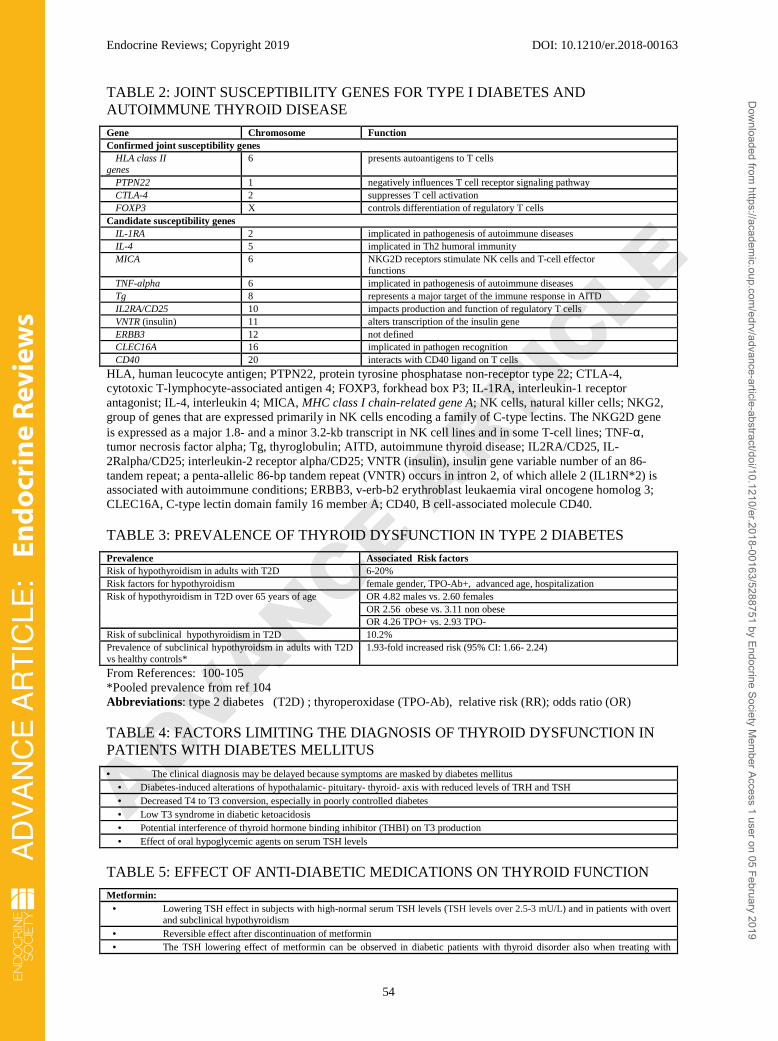
ADVANCE ARTIC
LE
Endocrine Reviews; Copyright 2019 DOI: 10.1210/er.2018-00163
54
TABLE 2: JOINT SUSCEPTIBILITY GENES FOR TYPE I DIABETES AND AUTOIMMUNE THYROID DISEASE
Gene Chromosome Function Confirmed joint susceptibility genes HLA class II genes
6 presents autoantigens to T cells
PTPN22 1 negatively influences T cell receptor signaling pathway CTLA-4 2 suppresses T cell activation FOXP3 X controls differentiation of regulatory T cells Candidate susceptibility genes IL-1RA 2 implicated in pathogenesis of autoimmune diseases IL-4 5 implicated in Th2 humoral immunity MICA 6 NKG2D receptors stimulate NK cells and T-cell effector
functions TNF-alpha 6 implicated in pathogenesis of autoimmune diseases Tg 8 represents a major target of the immune response in AITD IL2RA/CD25 10 impacts production and function of regulatory T cells VNTR (insulin) 11 alters transcription of the insulin gene ERBB3 12 not defined CLEC16A 16 implicated in pathogen recognition CD40 20 interacts with CD40 ligand on T cells
HLA, human leucocyte antigen; PTPN22, protein tyrosine phosphatase non-receptor type 22; CTLA-4, cytotoxic T-lymphocyte-associated antigen 4; FOXP3, forkhead box P3; IL-1RA, interleukin-1 receptor antagonist; IL-4, interleukin 4; MICA, MHC class I chain-related gene A; NK cells, natural killer cells; NKG2, group of genes that are expressed primarily in NK cells encoding a family of C-type lectins. The NKG2D gene is expressed as a major 1.8- and a minor 3.2-kb transcript in NK cell lines and in some T-cell lines; TNF-α, tumor necrosis factor alpha; Tg, thyroglobulin; AITD, autoimmune thyroid disease; IL2RA/CD25, IL-2Ralpha/CD25; interleukin-2 receptor alpha/CD25; VNTR (insulin), insulin gene variable number of an 86-tandem repeat; a penta-allelic 86-bp tandem repeat (VNTR) occurs in intron 2, of which allele 2 (IL1RN*2) is associated with autoimmune conditions; ERBB3, v-erb-b2 erythroblast leukaemia viral oncogene homolog 3; CLEC16A, C-type lectin domain family 16 member A; CD40, B cell-associated molecule CD40.
TABLE 3: PREVALENCE OF THYROID DYSFUNCTION IN TYPE 2 DIABETES
Prevalence Associated Risk factors Risk of hypothyroidism in adults with T2D 6-20% Risk factors for hypothyroidism female gender, TPO-Ab+, advanced age, hospitalization Risk of hypothyroidism in T2D over 65 years of age OR 4.82 males vs. 2.60 females
OR 2.56 obese vs. 3.11 non obese OR 4.26 TPO+ vs. 2.93 TPO-
Risk of subclinical hypothyroidism in T2D 10.2% Prevalence of subclinical hypothyroidsm in adults with T2D vs healthy controls*
1.93-fold increased risk (95% CI: 1.66- 2.24)
From References: 100-105 *Pooled prevalence from ref 104 Abbreviations: type 2 diabetes (T2D) ; thyroperoxidase (TPO-Ab), relative risk (RR); odds ratio (OR)
TABLE 4: FACTORS LIMITING THE DIAGNOSIS OF THYROID DYSFUNCTION IN PATIENTS WITH DIABETES MELLITUS
• The clinical diagnosis may be delayed because symptoms are masked by diabetes mellitus • Diabetes-induced alterations of hypothalamic- pituitary- thyroid- axis with reduced levels of TRH and TSH • Decreased T4 to T3 conversion, especially in poorly controlled diabetes • Low T3 syndrome in diabetic ketoacidosis • Potential interference of thyroid hormone binding inhibitor (THBI) on T3 production • Effect of oral hypoglycemic agents on serum TSH levels
TABLE 5: EFFECT OF ANTI-DIABETIC MEDICATIONS ON THYROID FUNCTION
Metformin: • Lowering TSH effect in subjects with high-normal serum TSH levels (TSH levels over 2.5-3 mU/L) and in patients with overt
and subclinical hypothyroidism • Reversible effect after discontinuation of metformin • The TSH lowering effect of metformin can be observed in diabetic patients with thyroid disorder also when treating with
AD
VA
NC
E A
RT
ICLE
:En
do
crin
e R
evie
ws
Dow
nloaded from https://academ
ic.oup.com/edrv/advance-article-abstract/doi/10.1210/er.2018-00163/5288751 by Endocrine Society M
ember Access 1 user on 05 February 2019
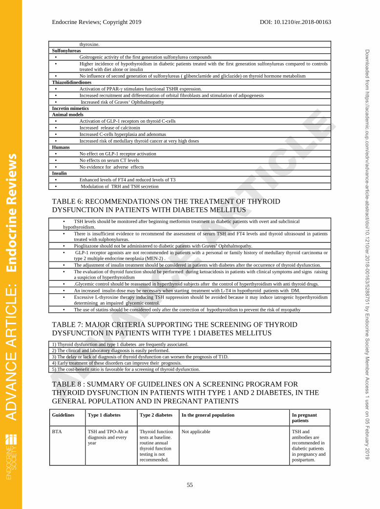
ADVANCE ARTIC
LE
Endocrine Reviews; Copyright 2019 DOI: 10.1210/er.2018-00163
55
thyroxine. Sulfonylureas • Goitrogenic activity of the first generation sulfonylurea compounds • Higher incidence of hypothyroidism in diabetic patients treated with the first generation sulfonylureas compared to controls
treated with diet alone or insulin • No influence of second generation of sulfonylureas ( glibenclamide and gliclazide) on thyroid hormone metabolism Thiazolidinediones • Activation of PPAR-γ stimulates functional TSHR expression. • Increased recruitment and differentiation of orbital fibroblasts and stimulation of adipogenesis • Increased risk of Graves‘ Ophthalmopathy Incretin mimetics Animal models • Activation of GLP-1 receptors on thyroid C-cells • Increased release of calcitonin • Increased C-cells hyperplasia and adenomas • Increased risk of medullary thyroid cancer at very high doses Humans • No effect on GLP-1 receptor activation • No effects on serum CT levels • No evidence for adverse effects Insulin • Enhanced levels of FT4 and reduced levels of T3 • Modulation of TRH and TSH secretion
TABLE 6: RECOMMENDATIONS ON THE TREATMENT OF THYROID DYSFUNCTION IN PATIENTS WITH DIABETES MELLITUS
• TSH levels should be monitored after beginning metformin treatment in diabetic patients with overt and subclinical hypothyroidism. • There is insufficient evidence to recommend the assessment of serum TSH and FT4 levels and thyroid ultrasound in patients
treated with sulphonylureas. • Pioglitazone should not be administered to diabetic patients with Graves’ Ophthalmopathy. • GLP-1 receptor agonists are not recommended in patients with a personal or family history of medullary thyroid carcinoma or
type 2 multiple endocrine neoplasia (MEN‐2) . • The adjustment of insulin treatment should be considered in patients with diabetes after the occurrence of thyroid dysfunction. • The evaluation of thyroid function should be performed during ketoacidosis in patients with clinical symptoms and signs raising
a suspicion of hyperthyroidism • .Glycemic control should be reassessed in hyperthyroid subjects after the control of hyperthyroidism with anti thyroid drugs. • An increased insulin dose may be necessary when starting treatment with L-T4 in hypothyroid patients with DM. • Excessive L‐thyroxine therapy inducing TSH suppression should be avoided because it may induce iatrogenic hyperthyroidism
determining an impaired glycemic control. • The use of statins should be considered only after the correction of hypothyroidism to prevent the risk of myopathy
TABLE 7: MAJOR CRITERIA SUPPORTING THE SCREENING OF THYROID DYSFUNCTION IN PATIENTS WITH TYPE 1 DIABETES MELLITUS
1) Thyroid dysfunction and type 1 diabetes are frequently associated. 2) The clinical and laboratory diagnosis is easily performed. 3) The delay or lack of diagnosis of thyroid dysfunction can worsen the prognosis of T1D. 4) Early treatment of these disorders can improve their prognosis. 5) The cost-benefit ratio is favorable for a screening of thyroid dysfunction.
TABLE 8 : SUMMARY OF GUIDELINES ON A SCREENING PROGRAM FOR THYROID DYSFUNCTION IN PATIENTS WITH TYPE 1 AND 2 DIABETES, IN THE GENERAL POPULATION AND IN PREGNANT PATIENTS
Guidelines Type 1 diabetes Type 2 diabetes In the general population In pregnant patients
BTA TSH and TPO-Ab at diagnosis and every year
Thyroid function tests at baseline. routine annual thyroid function testing is not recommended.
Not applicable TSH and antibodies are recommended in diabetic patients in pregnancy and postpartum.
AD
VA
NC
E A
RT
ICLE
:En
do
crin
e R
evie
ws
Dow
nloaded from https://academ
ic.oup.com/edrv/advance-article-abstract/doi/10.1210/er.2018-00163/5288751 by Endocrine Society M
ember Access 1 user on 05 February 2019
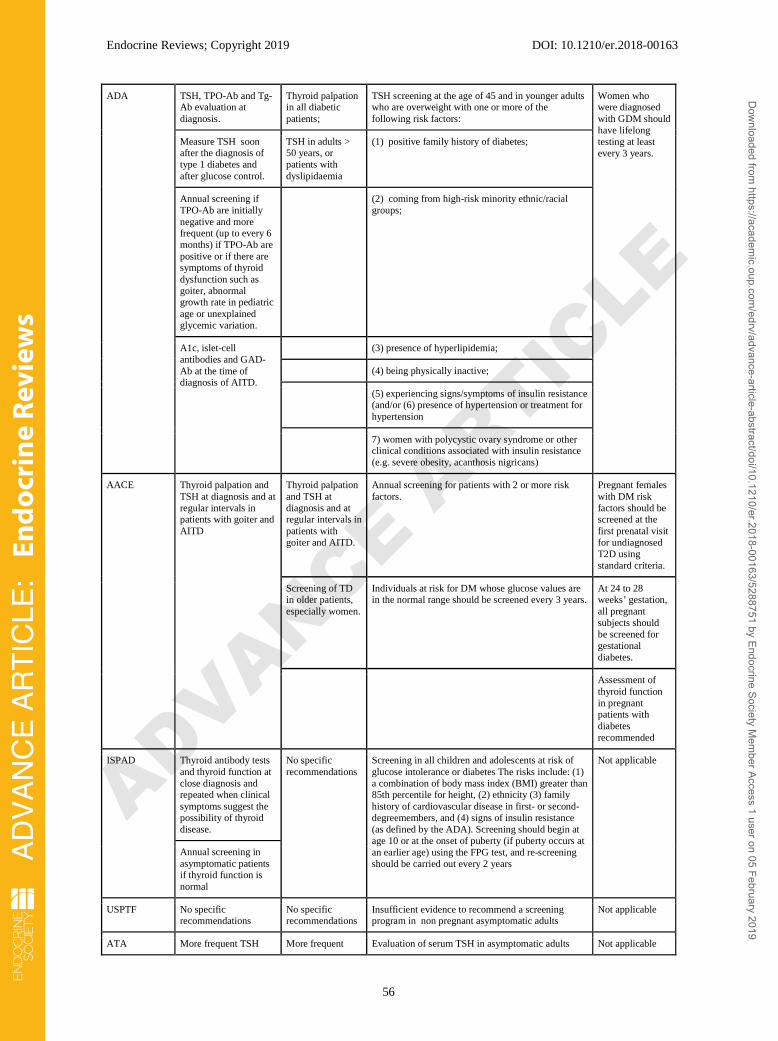
ADVANCE ARTIC
LE
Endocrine Reviews; Copyright 2019 DOI: 10.1210/er.2018-00163
56
ADA TSH, TPO-Ab and Tg-Ab evaluation at diagnosis.
Thyroid palpation in all diabetic patients;
TSH screening at the age of 45 and in younger adults who are overweight with one or more of the following risk factors:
Women who were diagnosed with GDM should have lifelong testing at least every 3 years.
Measure TSH soon after the diagnosis of type 1 diabetes and after glucose control.
TSH in adults > 50 years, or patients with dyslipidaemia
(1) positive family history of diabetes;
Annual screening if TPO-Ab are initially negative and more frequent (up to every 6 months) if TPO-Ab are positive or if there are symptoms of thyroid dysfunction such as goiter, abnormal growth rate in pediatric age or unexplained glycemic variation.
(2) coming from high-risk minority ethnic/racial groups;
A1c, islet-cell antibodies and GAD-Ab at the time of diagnosis of AITD.
(3) presence of hyperlipidemia;
(4) being physically inactive;
(5) experiencing signs/symptoms of insulin resistance (and/or (6) presence of hypertension or treatment for hypertension
7) women with polycystic ovary syndrome or other clinical conditions associated with insulin resistance (e.g. severe obesity, acanthosis nigricans)
AACE Thyroid palpation and TSH at diagnosis and at regular intervals in patients with goiter and AITD
Thyroid palpation and TSH at diagnosis and at regular intervals in patients with goiter and AITD.
Annual screening for patients with 2 or more risk factors.
Pregnant females with DM risk factors should be screened at the first prenatal visit for undiagnosed T2D using standard criteria.
Screening of TD in older patients, especially women.
Individuals at risk for DM whose glucose values are in the normal range should be screened every 3 years.
At 24 to 28 weeks’ gestation, all pregnant subjects should be screened for gestational diabetes.
Assessment of thyroid function in pregnant patients with diabetes recommended
ISPAD Thyroid antibody tests and thyroid function at close diagnosis and repeated when clinical symptoms suggest the possibility of thyroid disease.
No specific recommendations
Screening in all children and adolescents at risk of glucose intolerance or diabetes The risks include: (1) a combination of body mass index (BMI) greater than 85th percentile for height, (2) ethnicity (3) family history of cardiovascular disease in first- or second-degreemembers, and (4) signs of insulin resistance (as defined by the ADA). Screening should begin at age 10 or at the onset of puberty (if puberty occurs at an earlier age) using the FPG test, and re-screening should be carried out every 2 years
Not applicable
Annual screening in asymptomatic patients if thyroid function is normal
USPTF No specific recommendations
No specific recommendations
Insufficient evidence to recommend a screening program in non pregnant asymptomatic adults
Not applicable
ATA More frequent TSH More frequent Evaluation of serum TSH in asymptomatic adults Not applicable
AD
VA
NC
E A
RT
ICLE
:En
do
crin
e R
evie
ws
Dow
nloaded from https://academ
ic.oup.com/edrv/advance-article-abstract/doi/10.1210/er.2018-00163/5288751 by Endocrine Society M
ember Access 1 user on 05 February 2019
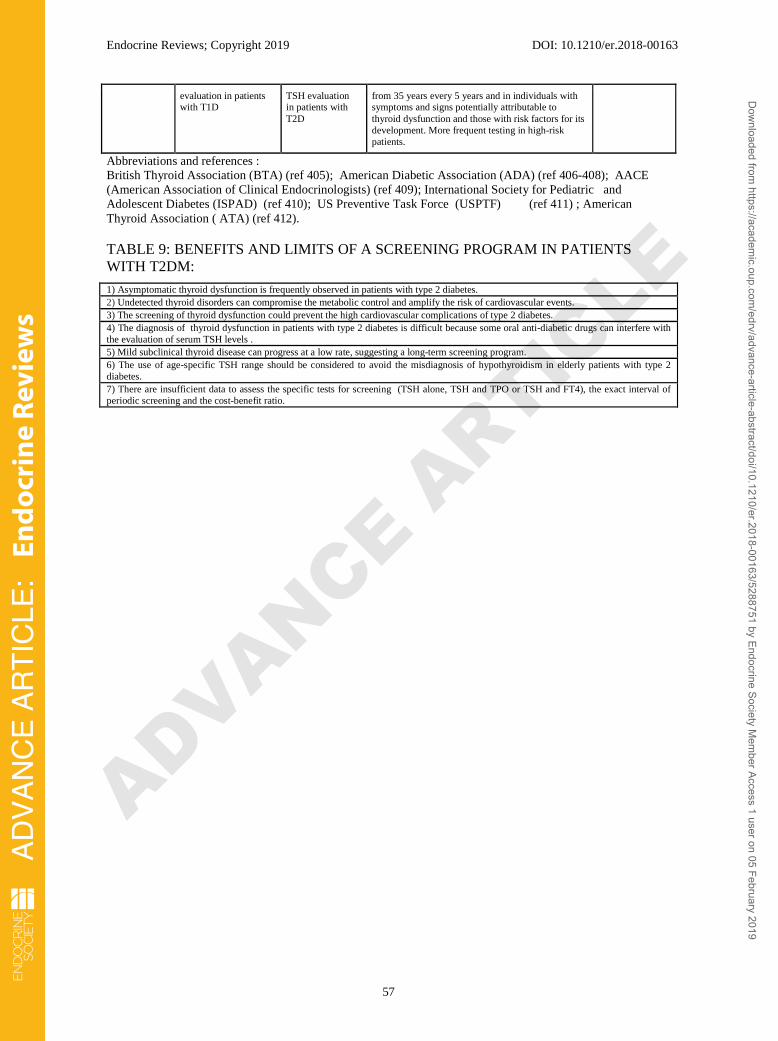
ADVANCE ARTIC
LE
Endocrine Reviews; Copyright 2019 DOI: 10.1210/er.2018-00163
57
evaluation in patients with T1D
TSH evaluation in patients with T2D
from 35 years every 5 years and in individuals with symptoms and signs potentially attributable to thyroid dysfunction and those with risk factors for its development. More frequent testing in high-risk patients.
Abbreviations and references : British Thyroid Association (BTA) (ref 405); American Diabetic Association (ADA) (ref 406-408); AACE (American Association of Clinical Endocrinologists) (ref 409); International Society for Pediatric and Adolescent Diabetes (ISPAD) (ref 410); US Preventive Task Force (USPTF) (ref 411) ; American Thyroid Association ( ATA) (ref 412).
TABLE 9: BENEFITS AND LIMITS OF A SCREENING PROGRAM IN PATIENTS WITH T2DM:
1) Asymptomatic thyroid dysfunction is frequently observed in patients with type 2 diabetes. 2) Undetected thyroid disorders can compromise the metabolic control and amplify the risk of cardiovascular events. 3) The screening of thyroid dysfunction could prevent the high cardiovascular complications of type 2 diabetes. 4) The diagnosis of thyroid dysfunction in patients with type 2 diabetes is difficult because some oral anti-diabetic drugs can interfere with the evaluation of serum TSH levels . 5) Mild subclinical thyroid disease can progress at a low rate, suggesting a long-term screening program. 6) The use of age-specific TSH range should be considered to avoid the misdiagnosis of hypothyroidism in elderly patients with type 2 diabetes. 7) There are insufficient data to assess the specific tests for screening (TSH alone, TSH and TPO or TSH and FT4), the exact interval of periodic screening and the cost-benefit ratio.
AD
VA
NC
E A
RT
ICLE
:En
do
crin
e R
evie
ws
Dow
nloaded from https://academ
ic.oup.com/edrv/advance-article-abstract/doi/10.1210/er.2018-00163/5288751 by Endocrine Society M
ember Access 1 user on 05 February 2019

UCP1ThermogenesisEnergy expenditure
Glucose uptakeGlucose transportGlucose utilizationGlycolysisEnergy expenditureUCP3
Glucose outputGluconeogenesisGlycogen synthesisInsulin sensitivityIntrahepatic lipolysis
Insulin
Insulin PP
Gut hormone
Adip
okines
Myokin
esProliferation
Paraventricular nucleus
Arcuate nucleus
MAFA
Ventromedial nucleus
Central control of appetite
SNS
TRH
POMCCART
NPY/AgRP
BDNF
Glucose absorptionInsulin production
LipolysisBodyfat
UCP1
Lipidturnover
Leptin
NE
D2
TR
Whiteadipocyte Brown
adipocyte
NE
T3
T3
T3TR
T3
β-cellmaturation
β-r
β-r
β-r bsoInsulin prod
bsoInsulin prodGlucose absoInsuli rodGlucose a
UCP1UCPUCPUCP1UCPUCPUCP1
AD
VA
NC
E A
RT
ICLE
:En
do
crin
olo
gy
AD
VA
NC
E A
RT
ICLE
:En
do
crin
olo
gy
ADVANCE ARTIC
LE
AD
VA
NC
E A
RT
ICLE
:En
do
crin
e R
evie
ws
Dow
nloaded from https://academ
ic.oup.com/edrv/advance-article-abstract/doi/10.1210/er.2018-00163/5288751 by Endocrine Society M
ember Access 1 user on 05 February 2019

Absorption of glucose
Hepatic glucose uptake
Post absorptive glycemia
Hepatic gluconeogenesis
Hepatic glycogen synthesis
Normal or plasma insulin levels with increased
glucose-stimulated insulin secretion
Glucose transport and peripheral glucose disposal
Peripheral glucose utilization
Peripheral insulin resistance
Renal insulin clearance
Exogenous insulin requirement in diabetic patients
Risk of hypoglycemic episodes in diabetic patients
Insulin dose in diabetic patients
Insulin dose after replacement therapy with L-thyroxine
Hypothyroidism
Food intake
Glucose absorption
Post absorptive plasma glycemia
Hepatic gluconeogenesis
Plasma insulin and proinsulin
Serum cholesterol and triglycerides
Hepatic insulin resistance
Glucose utilization in muscle and adipose tissue
Glucose intolerance
Lipolysis and free fatty acids
Body fat
Lactate production
Apoptosis of insulin producing cells
Glycemia
Insulin clearance
Deranged metabolic control in diabetic treated patients
Ketoacidosis
Insulin requirement in diabetic patients
Hyperthyroidism
AD
VA
NC
E A
RT
ICLE
:En
do
crin
olo
gy
AD
VA
NC
E A
RT
ICLE
:En
do
crin
olo
gy
ADVANCE ARTIC
LE
AD
VA
NC
E A
RT
ICLE
:En
do
crin
e R
evie
ws
Dow
nloaded from https://academ
ic.oup.com/edrv/advance-article-abstract/doi/10.1210/er.2018-00163/5288751 by Endocrine Society M
ember Access 1 user on 05 February 2019
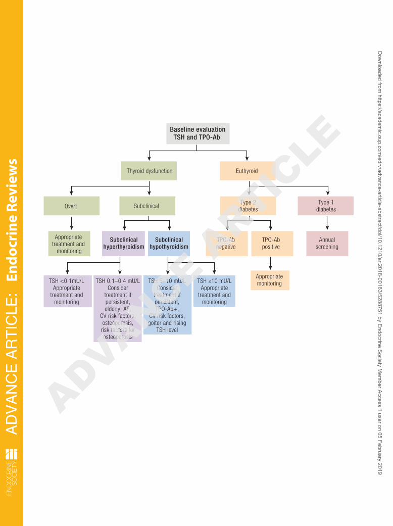
TSH 0.1–0.4 mU/LConsider
treatment ifpersistent,elderly, AF,
CV risk factors,osteoporosis,
risk factors forosteoporosis
Overt
TPO-Abnegative
TPO-Abpositive
Appropriatemonitoring
Annualscreening
Appropriate treatment and
monitoring
Subclinicalhyperthyroidism
Subclinicalhypothyroidism
Baseline evaluationTSH and TPO-Ab
EuthyroidThyroid dysfunction
SubclinicalType 2
diabetesType 1
diabetes
TSH <0.1mU/LAppropriate
treatment andmonitoring
TSH 5–10 mU/LConsider
treatment ifpersistent,TPO-Ab+,
CV risk factors,goiter and rising
TSH level
TSH ≥10 mU/LAppropriate
treatment andmonitoring
AD
VA
NC
E A
RT
ICLE
:En
do
crin
olo
gy
AD
VA
NC
E A
RT
ICLE
:En
do
crin
olo
gy
ADVANCE ARTIC
LE
AD
VA
NC
E A
RT
ICLE
:En
do
crin
e R
evie
ws
Dow
nloaded from https://academ
ic.oup.com/edrv/advance-article-abstract/doi/10.1210/er.2018-00163/5288751 by Endocrine Society M
ember Access 1 user on 05 February 2019

Cardiovascularrisk
Metabolic syndrome
Hypothyroidism
NAFLDHypertensionDyslipidemiaInsulin
resistanceObesity
Metabolic syndrome
NAFLDHypertensionDyslipidemiaInsulin
resistanceObesity
AD
VA
NC
E A
RT
ICLE
:En
do
crin
olo
gy
AD
VA
NC
E A
RT
ICLE
:En
do
crin
olo
gy
ADVANCE ARTIC
LE
AD
VA
NC
E A
RT
ICLE
:En
do
crin
e R
evie
ws
Dow
nloaded from https://academ
ic.oup.com/edrv/advance-article-abstract/doi/10.1210/er.2018-00163/5288751 by Endocrine Society M
ember Access 1 user on 05 February 2019