Embed Size (px)
Citation preview


The peritoneum is the thin serous membrane lining the walls of the abdominal and pelvic cavities and covering the abdominal and pelvic viscera.
The peritoneum can be regarded as a balloon into which organs are pressed from outside.
GENERAL ARRANGEMENT OF THE PERITONIUM

In males, this is a closed cavity; but in females, communication with the exterior occurs through the uterine tubes, the uterus, and the vagina.
GENERAL ARRANGEMENT OF THE PERITONIUM

Between the parietal peritoneum and the
fascial lining of the abdominal and pelvic
walls is a layer of connective tissue called
the extraperitoneal tissue.
It varies in amount in different regions
and, in the area of the kidneys, contains a
large amount of fat.
The visceral peritoneum is closely bound
to the underlying viscus by only a small
amount of connective tissue.
GENERAL ARRANGEMENT OF THE PERITONIUM

GENERAL ARRANGEMENT OF THE PERITONIUM
The peritoneal cavity is the largest cavity in the
body, and the surface area of the parietal and
visceral layers is enormous.
peritoneal
cavity
greater sac lesser sac

The greater sac is the
main compartment of
the peritoneal cavity
and extends from the
diaphragm down into
the pelvis.
The lesser sac is
smaller and lies
behind the stomach.
GENERAL ARRANGEMENT OF THE PERITONIUM


The greater and lesser sacs are in free communication with one another through an oval window called the opening of lesser sac, or the epiploic foramen.
The peritoneum secretes a small amount of serous fluid, the peritoneal fluid, which lubricates the surfaces of the peritoneum and allows free movement between the viscera.
GENERAL ARRANGEMENT OF THE PERITONIUM


The terms intraperitoneal and
retroperitoneal are used to describe the
relationship of various organs to their
peritoneal covering.
An organ is said to be intraperitoneal if is
almost totally covered with visceral
peritoneum, e.g., stomach, jejunum,
ileum, and spleen.
INTRAPERITONEAL AND RETROPERITONEAL
RELATIONSHIPS

Retroperitoneal organs lie behind the peritoneum and are only partially covered with visceral peritoneum.
The pancreas and the ascending and descending parts of the colon are examples of retroperitoneal organs.
No organ, however, is actually within the peritoneal cavity.
An intraperitoneal organ, such as the stomach appears to be surrounded by the peritoneal cavity but it is covered with visceral peritoneum and is attached to other organs by omenta.
INTRAPERITONEAL AND RETROPERITONEAL
RELATIONSHIPS


PERITONEAL LIGAMENTS
Peritoneal ligaments are two-layered folds of peritoneum that connect solid viscera to the abdominal walls. (They do not possess the dense fibrous tissue seen in ligaments associated with bones).
The liver, for example, is
connected to the diaphragm by the falciform ligament, the coronary ligament, and the right and left triangular ligaments.

OMENTA
Omenta are two-layered folds of peritoneum that connect the stomach to another viscus.
The greater omentum connects the greater curvature of the stomach to the transverse colon.
It hangs down like an apron in front of the coils of the small intestine and is folded back on itself to be attached to the transverse colon.


The lesser omentum suspends the lesser curvature of the stomach from the fissure of the ligamentum venosum and the porta hepatis on the undersurface of the liver.
The gastrosplenic omentum (ligament) connects the stomach to the hilum of the spleen.
OMENTA


MESENTERIES
Mesenteries are two-layered folds of peritoneum connecting parts of the intestines to the posterior abdominal wall, for example, the mesentery of the small intestine, the transverse mesocolon, and the sigmoid mesocolon.

The peritoneal ligaments, omenta, and mesenteries permit blood, lymph
vessels, and nerves to reach the viscera.


PERITONEUM AS SEEN ON TRANSVERSE SECTIONS OF
THE ABDOMEN
At the Level of the Fourth Lumbar Vertebra
The parietal peritoneum lining the anterior abdominal wall below the umbilicus is smooth, apart from the low ridges produced by the:
1. median umbilical ligament (the urachus, the remains of the fetal allantois, which passes from the apex of the bladder to the umbilicus).
2. lateral umbilical ligaments (the obliterated umbilical arteries, which pass from the internal iliac arteries to the umbilicus.

The parietal peritoneum passes onto the posterior abdominal wall and becomes continuous with the visceral peritoneum covering the sides and anterior surfaces of the ascending colon and descending colon.
PERITONEUM AS SEEN ON TRANSVERSE SECTIONS OF
THE ABDOMEN

In the region of the aorta and inferior vena cava, the parietal peritoneum becomes continuous with the mesentery of the small intestine.
Note that the peritoneum forms a continuous layer that can be traced around the abdominal cavity without interruption.
PERITONEUM AS SEEN ON TRANSVERSE SECTIONS OF
THE ABDOMEN

At the Level of the Twelfth Thoracic Vertebra
The parietal peritoneum lining the anterior abdominal wall forms a sickle-shaped fold called the falciform ligament.
This connects the anterior surface of the liver to the anterior abdominal wall above the umbilicus and to the diaphragm.
PERITONEUM AS SEEN ON TRANSVERSE SECTIONS OF
THE ABDOMEN


In the free border of the ligament, where the two layers of peritoneum are continuous with each other, lies the ligamentum teres.
This is the obliterated umbilical vein of the fetus, which passes upward to enter the groove between the quadrate lobe and the left lobe of the liver.
PERITONEUM AS SEEN ON TRANSVERSE SECTIONS OF
THE ABDOMEN


If the parietal peritoneum is
followed around the
abdominal wall on the left
side, it reaches the lateral
margin of the left kidney.
Here it becomes
continuous with the
visceral peritoneum
covering the lateral margin
and part of the anterior
surface of the left kidney.
PERITONEUM AS SEEN ON TRANSVERSE SECTIONS OF
THE ABDOMEN


The peritoneum then leaves the kidney and passes to the hilum of the spleen as the posterior layer of
the splenicorenal (lienorenal) ligament.
PERITONEUM AS SEEN ON TRANSVERSE SECTIONS OF
THE ABDOMEN

The visceral peritoneum covers the spleen and, on reaching the hilum again, is reflected onto the greater curvature of the stomach as the anterior layer of the gastrosplenic omentum (ligament).
The visceral peritoneum covers the anterior surface of the stomach and leaves the lesser curvature to form the anterior layer of the lesser omentum.
PERITONEUM AS SEEN ON TRANSVERSE SECTIONS OF
THE ABDOMEN

On the right, the lesser omentum has a free border, and here the peritoneum folds around the bile duct, hepatic artery, and portal vein.
The free border of the lesser omentum forms the anterior margin of the opening into the lesser sac.
PERITONEUM AS SEEN ON TRANSVERSE SECTIONS OF
THE ABDOMEN


The peritoneum forms the posterior layer of the lesser omentum and becomes continuous with the visceral layer of peritoneum covering the posterior wall of the stomach.
Note that here the peritoneum forms the anterior wall of the lesser sac.
PERITONEUM AS SEEN ON TRANSVERSE SECTIONS OF
THE ABDOMEN

At the greater
curvature of the
stomach, the
peritoneum leaves the
stomach, forming the
posterior layer of the
gastrosplenic
omentum (ligament),
and reaches the hilum
of the spleen.
PERITONEUM AS SEEN ON TRANSVERSE SECTIONS OF
THE ABDOMEN

Here it is reflected backward to the posterior abdominal wall, forming the anterior layer of the splenicorenal ligament.
The peritoneum now covers the anterior surface of the pancreas, the aorta, and the inferior vena cava, forming the posterior wall of the lesser sac.
PERITONEUM AS SEEN ON TRANSVERSE SECTIONS OF
THE ABDOMEN

The peritoneum passes onto the anterior surface of the right kidney and sweeps around the lateral abdominal wall to reach the anterior abdominal wall.
Once again, note that the peritoneum forms a continuous layer around the abdomen.
PERITONEUM AS SEEN ON TRANSVERSE SECTIONS OF
THE ABDOMEN
PERITONEUM AS SEEN ON SAGITTAL
SECTION OF THE ABDOMEN AND PELVIS
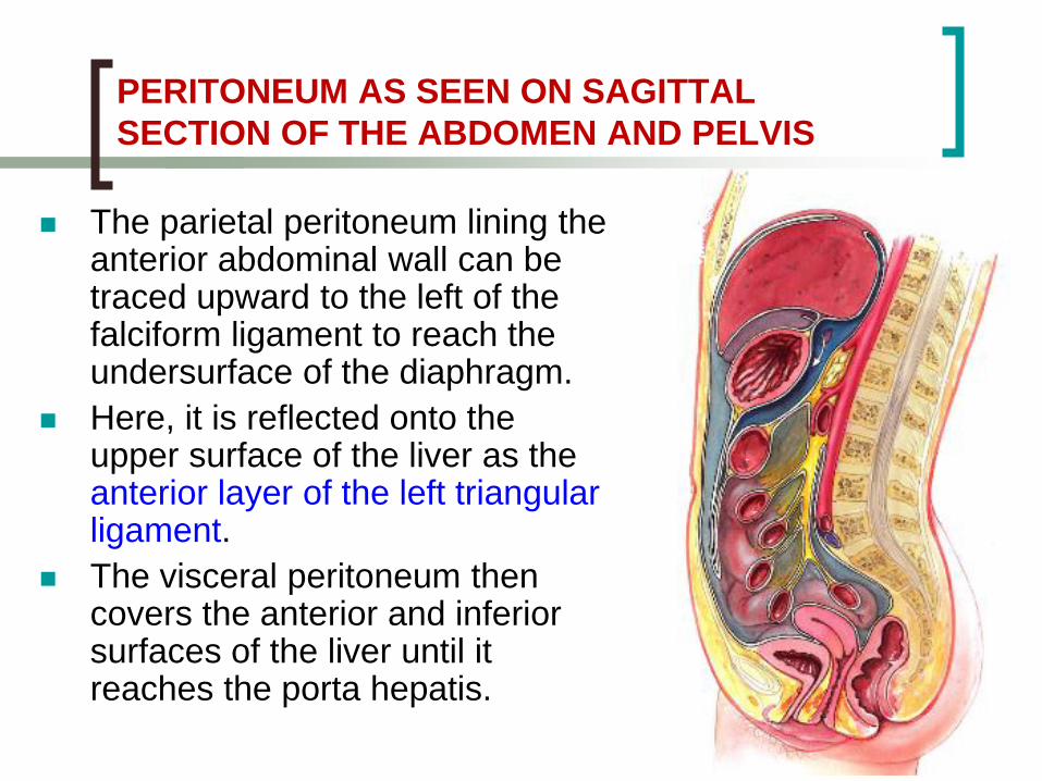
The parietal peritoneum lining the anterior abdominal wall can be traced upward to the left of the falciform ligament to reach the undersurface of the diaphragm.
Here, it is reflected onto the upper surface of the liver as the anterior layer of the left triangular ligament.
The visceral peritoneum then covers the anterior and inferior surfaces of the liver until it reaches the porta hepatis.

Here, the peritoneum passes
to the lesser curvature of the
stomach as the anterior layer
of the lesser omentum.
Having covered the anterior
surface of the stomach, the
peritoneum leaves the
greater curvature, forming the
anterior layer of the greater
omentum.
PERITONEUM AS SEEN ON SAGITTAL SECTION OF THE
ABDOMEN AND PELVIS

The greater omentum hangs down as a fold in front of the coils of intestine and contains within it the lower part of the lesser sac.
Having reached the lowest limit of the greater omentum, the peritoneum folds upward and forms the posterior layer of the greater omentum.
PERITONEUM AS SEEN ON SAGITTAL SECTION OF THE
ABDOMEN AND PELVIS

On reaching the inferior border of the transverse colon, the peritoneum covers its posterior surface and then leaves the colon to form the posterior layer of the transverse mesocolon.
The peritoneum then passes to the anterior border of the pancreas and runs downward anterior to the third part of the duodenum.
PERITONEUM AS SEEN ON SAGITTAL SECTION OF THE
ABDOMEN AND PELVIS

The peritoneum now leaves the posterior abdominal wall as the anterior layer of the mesentery of the small intestine.
The visceral peritoneum covers the jejunum and then forms the posterior layer of the mesentery.
PERITONEUM AS SEEN ON SAGITTAL SECTION OF THE
ABDOMEN AND PELVIS

On returning to the posterior abdominal wall, the peritoneum runs downward into the pelvis and covers the anterior surface of the upper part of the rectum.
From here, it is reflected onto the posterior surface of the upper part of the vagina, forming the important rectouterine pouch (pouch of Douglas).
PERITONEUM AS SEEN ON SAGITTAL SECTION OF THE
ABDOMEN AND PELVIS

In the male, the peritoneum is reflected onto the upper part of the posterior surface of the bladder and the seminal vesicles, forming the rectovesical pouch.
PERITONEUM AS SEEN ON SAGITTAL SECTION OF THE
ABDOMEN AND PELVIS
The peritoneum passes over
the upper surface of the uterus
in the female and is reflected
from its anterior surface onto
the upper surface of the
bladder.
In both sexes, the
peritoneum passes from the
bladder onto the anterior
abdominal wall.

PERITONEAL POUCHES, RECESSES, SPACES,
AND GUTTERS
Lesser Sac
The lesser sac is an extensive peritoneal pouch situated behind the lesser omentum and stomach and lying in front of structures situated on the posterior abdominal wall.
It projects upward as far as the diaphragm and downward between the layers of the greater omentum.

The lower part of
the lesser sac is
often obliterated by
the adherence of
the anterior layers
of the greater
omentum to the
posterior layers.
Lesser Sac

Its left margin
is formed by
the spleen and
the
gastrosplenic
omentum and
splenicorenal
ligaments.
Lesser Sac
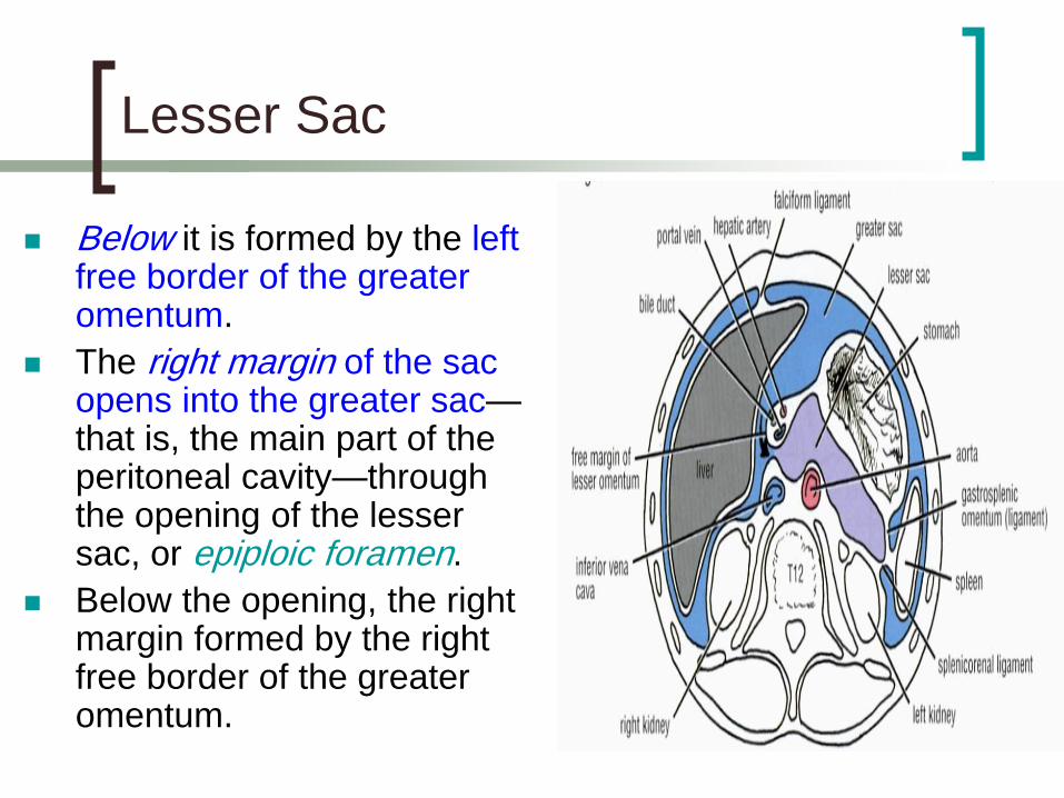
Below it is formed by the left free border of the greater omentum.
The right margin of the sac opens into the greater sac—that is, the main part of the peritoneal cavity—through the opening of the lesser sac, or epiploic foramen.
Below the opening, the right margin formed by the right free border of the greater omentum.
Lesser Sac

• Anteriorly: The free border of the lesser omentum, containing the bile duct, the hepatic artery, and the portal vein.
The bile duct lies to the right and in front, the hepatic artery lies to the left and in front, and the portal vein lies posteriorly.
Boundaries of the epiploic foramen

• Posteriorly: The
inferior vena
cava.
• Superiorly: The
caudate process
of the caudate
lobe of the liver.
• Inferiorly: The first
part of the
duodenum.
Boundaries of the epiploic foramen

Close to the duodenojejunal junction may be
four small pocket like pouches of peritoneum
called the superior, inferior, paraduodenal, and
retro duodenal recesses.
Duodenal Recesses

The presence of folds of peritoneum close to the cecum creates three peritoneal recesses: the superior , inferior, and the retrocecal recesses.
These recesses in the peritoneal lining occasionally form deep pouches.
Cecal Recesses

The intersigmoid recess is situated at the apex of the
inverted, V-shaped root of the sigmoid mesocolon.
Its mouth opens downward and lies in front of the left
ureter.
Intersigmoid Recess

The various peritoneal pouches and recesses described in the previous
paragraphs may become sites for the development of internal abdominal
herniae

The existence of the intraperitoneal subphrenic spaces is the result of the complicated arrangement of the peritoneum in the region of the liver.
The right and left anterior subphrenic spaces lie between the diaphragm and the liver, one on each side of the falciform ligament.
Subphrenic Spaces

The right posterior subphrenic space lies between the right lobe of the liver, the right kidney, and the right colic flexure.
The right extraperitoneal space lies between the layers of the coronary ligament and is therefore situated between the liver and the diaphragm.
Subphrenic Spaces

The paracolic gutters lie on the lateral and medial sides of the ascending and descending colons, respectively.
The right medial paracolic gutter is closed off from the pelvic cavity inferiorly by the mesentery of the small intestine, whereas the others are in free communication with the pelvic cavity.
Paracolic Gutters

The right lateral paracolic gutter is in communication with the right posterior subphrenic space, but the left lateral gutter is separated from the area around the spleen by the phrenicocolic ligament, a fold of peritoneum that passes from the left colic flexure to the diaphragm.
Paracolic Gutters

The subphrenic spaces and the
paracolic gutters are clinically
important because they may be sites
for the collection and movement of
infected peritoneal fluid.
Paracolic Gutters

NERVE SUPPLY OF THE PERITONEUM
The parietal peritoneum is sensitive to
pain, temperature, touch, and
pressure.
The parietal peritoneum lining the
anterior abdominal wall is supplied by
the lower six thoracic and first lumbar
nerves—that is, the same nerves that
innervate the overlying muscles and
skin.

The central part of the diaphragmatic
peritoneum is supplied by the phrenic nerves.
The peripheral part of the diaphragmatic
peritoneum is supplied by the lower six thoracic nerves.
The parietal peritoneum in the pelvis is
mainly supplied by the obturator nerve, a
branch of the lumbar plexus.
NERVE SUPPLY OF THE PERITONEUM

The visceral peritoneum is sensitive to stretch and tearing and is not sensitive to touch, pressure, or temperature.
It is supplied by autonomic afferent nerves that supply the viscera or are traveling in the mesenteries.
Overdistension of a viscus leads to the sensation of pain.
The mesenteries of the small and large intestines are sensitive to mechanical stretching.
NERVE SUPPLY OF THE PERITONEUM

FUNCTIONS OF THE PERITONEUM
The peritoneal fluid, which is pale yellow and somewhat viscid, contains leukocytes.
It is secreted by the peritoneum and ensures that the mobile viscera glide easily on one an other.
As a result of the movements of the diaphragm and the abdominal muscles, together with the peristaltic movements of the intestinal tract, the peritoneal fluid is not static.

Experimental evidence has shown that particulate matter introduced into the lower part of the peritoneal cavity reaches the subphrenic peritoneal spaces rapidly, whatever the position of the body.
It seems that intraperitoneal movement of fluid toward the diaphragm is continuous, and there it is quickly absorbed into the subperitoneal lymphatic capillaries.
FUNCTIONS OF THE PERITONEUM


This can be explained on the basis that the area of peritoneum is extensive in the region of the diaphragm and the respiratory movements of the diaphragm aid lymph flow in the lymph vessels.
The peritoneal coverings of the intestine tend to stick together in the presence of infection. The greater omentum, which is kept constantly on the move by the peristalsis of the neighboring intestinal tract, may adhere to other peritoneal surfaces around a focus of infection.
In this manner, many of the intraperitoneal infections are sealed off and remain localized.
FUNCTIONS OF THE PERITONEUM

The peritoneal folds play an important part in suspending the various organs within the peritoneal cavity and serve as a means of conveying the blood vessels, lymphatics, and nerves to these organs.
Large amounts of fat are stored in the peritoneal ligaments and mesenteries, and especially large amounts can be found in the greater omentum.
FUNCTIONS OF THE PERITONEUM

The peritoneal cavity is divided into an upper part within the abdomen and a lower part in the pelvis.
The attachment of the transverse mesocolon and the mesentery of the small intestine to the posterior abdominal wall provides natural peritoneal barriers that may hinder the movement of infected peritoneal fluid from the upper part to the lower part of the peritoneal cavity.
MOVEMENT OF PERITONEAL FLUID

It is interesting to note that when the patient is in the supine position the right subphrenic peritoneal space and the pelvic cavity are the lowest areas of the peritoneal cavity and the region of the pelvic brim is the highest area.
MOVEMENT OF PERITONEAL FLUID


PERITONEAL INFECTION
Infection may gain entrance to the peritoneal cavity through several routes:
from the interior of the gastrointestinal tract and gallbladder,
through the anterior abdominal wall,
via the uterine tubes in females ,
from the blood.
Collection of infected peritoneal fluid in one of the subphrenic spaces is often accompanied by infection of the pleural cavity.

It is common to find a localized empyerna in a patient with a subphrenic abscess.
It is believed that the infection spreads from the peritoneum to the pleura via the diaphragmatic lymph vessels.
A patient with a subphrenic abscess may complain of pain over the shoulder.
PERITONEAL INFECTION

To avoid the accumulation of infected fluid in the sub phrenic spaces and to delay the absorption of toxins from intraperitoneal infections, it is common nursing practice to sit a patient up in bed with the back at an angle of 45° In this position, the infected peritoneal
fluid tends to gravitate downward into the pelvic cavity, where the rate of toxin absorption is slow.

The greater omentum is often referred to by the surgeons as the abdominal policeman.
The lower and the right and left margins are free, and it moves about the peritoneal cavity in response to the peristaltic movements of the neighboring gut.
In the first 2 years of life it is poorly developed and thus is less protective in a young child.
THE GRAETER OMENTUM LOCALIZATION OF INFECTION

Later, however, in an acutely inflamed appendix, for example, the inflammatory exudate causes the omentum to adhere to the appendix and wrap itself around the infected organ.
By this means, the infection is often localized to a small area of the peritoneal cavity, thus saving the patient from a serious diffuse peritonitis.

GREATER OMENTUM
The greater omentum has been found to plug the neck of a hernial sac and prevent the entrance of coils of small intestine.
Surgeons sometimes use the omentum to buttress an intestinal anastomosis or in the closure of a perforated gastric or duodenal ulcer.
The greater omentum may undergo torsion, and if extensive, the blood supply to a part of it may be cut off, causing necrosis

ASCITES
Ascites is essentially an excessive accumulation of peritoneal fluid within the peritoneal cavity.
Ascites can occur secondary to hepatic cirrhosis, malignant disease, or congestive heart failure.
In a thin patient, as much as 1500 ml has to accumulate before ascites can be recognized clinically.
In obese individuals, a far greater amount has to collect before it can be detected.

PERITONEAL PAIN FROM THE PARITAL PERITONEUM
Abdominal pain originating from the parietal peritoneum is therefore of the somatic type and can be precisely localized; it is usually severe.
An inflamed parietal peritoneum is extremely sensitive to stretching.
This fact is made use of clinically in diagnosing peritonitis.

Pressure is applied to the abdominal wall with a single finger over the site of the inflammation.
The pressure is then removed by suddenly withdrawing the finger.
The abdominal wall rebounds, resulting in extreme local pain, which is known as rebound tenderness.


It should always be remembered that the parietal peritoneum in the pelvis is innervated by the obturator nerve and can be palpated by means of a rectal or vaginal examination.
An inflamed appendix may hang down into the pelvis and irritate the parietal peritoneum.
A pelvic examination can detect extreme tenderness of the parietal peritoneum on the right side.
PERITONEAL PAIN FROM THE PARITAL PERITONEUM

The visceral peritoneum, including the mesenteries, is innervated by autonomic afferent nerves.
Stretch caused by overdistension of a viscus or pulling on a mesentery gives rise to the sensation of pain.
Because the gastrointestinal tract arises embryologically as a midline structure and receives a bilateral nerve supply, pain is referred to the midline.
Pain arising from an abdominal viscus is dull and poorly localized.
PERITONEAL PAIN FROM THE VISAERAL PERITONEUM

PERITONEAL DIALYSIS
Because the peritoneum is a semipermeable membrane,allows rapid bidirectional transfer of substances across it self.
Because the surface area of the peritoneum is enormous, this transfer property has been made use of in patients with acute renal insufficiency.
The efficiency of this method is only a fraction of that achieved by hemodialysis.

A watery solution, the dialysate, is introduced through a catheter through a small midline incision through the anterior abdominal wall below the umbilicus.
The technique is the same as peritoneal lavage.
The products of metabolism, such as urea, diffuse through the peritoneal lining cells from the blood vessels into the dialysate and are removed from the patient.


INTERNAL ABDOMINAL HERNIA
Occasionally, a loop of intestine enters a peritoneal pouch or recess (e.g., the lesser sac or the duodenal recesses) and becomes strangulated at the edges of the recess.
Remember that important structures form the boundaries of the entrance into the lesser sac and that the inferior mesenteric vein often lies in the anterior wall of the paraduodenal recess.