Embed Size (px)
Citation preview

THESIS
MUSICAL NEGLECT TRAINING FOR UNILATERAL VISUAL NEGLECT IN
RIGHT HEMISPHERIC STROKE PATIENTS
Submitted by
Kyurim Kang
School of Music, Theatre and Dance
In partial fulfillment of the requirements
For the Degree of Master of Music
Colorado State University
Fort Collins, Colorado
Fall 2015
Master’s Committee: Advisor: Michael Thaut Blythe LaGasse Anna Fails
Copyright by Kyurim Kang 2015
All Rights Reserved

ii
ABSTRACT
MUSICAL NEGLECT TRAINING FOR UNILATERAL VISUAL NEGLECT IN RIGHT
HEMISPHERIC STROKE PATIENTS
The purpose of this study was to examine the immediate and longer-lasting effect of
Musical Neglect Training (MNT) on unilateral visual neglect. A single-subject design was used,
as participants served as their own control. Two individuals participated in this study.
Participants underwent two weekly 30-minute individual sessions over a time period of three
weeks, for a total of six MNT sessions for each participant. Two standardized assessments
(Albert’s and Line Bisection Test) were used. The assessments were administered immediately
before and after each of the 6 MNT sessions to assess the immediate effect of MNT. During the
training, participants played a set of horizontally arranged tone bars tuned to an ascending triads
and scales. At the endpoint of each sequence a cymbal was positioned and played to give a
strong audiovisual target in the left visual field for the participants. The experimenter provided a
chordal accompaniment on the keyboard to provide harmonic-rhythmic pacing and to cue
continuous playing to the end of the sequence. Follow-up testing was done one week after their
6th session to examine the longer-lasting effects of MNT.
Paired t-tests were used to test for statistical improvement between pre- and post-test of
interventions (immediate effects). Also, nonparametric statistics (Wilcoxon Signed Rank Test)
was also calculated in parallel with the paired t-tests due to the small sample size and possible
violations of normal distribution. For the longer-lasting effects, raw data were compared between
the average of 6 sessions’ pre-test and follow-up test since there was only one follow-up test.

iii
Both participants showed statistical improvement with Albert’s Test in the immediate
effect (Participant 1: p=.02, Participant 2: p=.01). Results for the immediate effect of MNT on
the Line Bisection Test were not significantly different, but means were lower for post-test
(Participant 1: M=24.17%, Participant 2: M=9.16%) compared to pre-test (Participant 1:
M=25.65%, Participant 2: M=10.39%), indicating positive improvement. Although not
statistically significant for the longer-lasting effect, participant 2 had a lower score (score=7)
compared to averaged pretest scores of the 6 treatment sessions (M=9.5), indicating a positive
outcome, while participant 1 was unchanged at follow-up score (score=14) compared to the
pretest average (M=14.5) in the Albert’s Test. Moreover, participant 1 showed increased
deviation percentages from the averaged pre-test (M=25.65%) to follow-up test (deviation
=27.18%), indicating no positive effect for the longer-lasting effect in the Line Bisection Test.
Participant 2 showed a decreased deviation in follow-up score (deviation=7.70%) compared to
averaged pre-test score (M=10.39%).
The study indicates MNT as a potentially positive intervention for clients with unilateral
visual neglect. Future research should employ this music-based intervention with clients in
subacute recovery stages post stroke. Furthermore, developing the intervention protocol with
increased duration and a higher number of sessions may result in stronger results. Based on the
results from this study and previous studies, research focusing on the underlying neural
mechanism and tailoring the intervention protocol appropriately to the clinical situation is
warranted.

iv
ACKNOWLEDGEMENTS
I would like to express my special appreciation and thanks to my advisor Dr. Thaut, you
have been a tremendous mentor for me. I would like to thank you for encouraging my research
and for allowing me to grow as a researcher for the future. Your advice on research has been
priceless. I would also like to thank my committee members, Dr. LaGasse and Dr. Fails for
serving as my committee members even at hardship. I also want to thank you for letting my
defense be an enjoyable moment, and for your brilliant comments and suggestions, thanks to
you. I would especially like to thank two participants for my study. All of you have been there to
support me when I collected data for my master thesis. Thanks for your smiles and
encouragements.
A special thanks to my family. Words cannot express how grateful I am to my father,
mother, and brother for all of the sacrifices that you’ve made on my behalf. Your efforts for me
were what sustained me thus far. Thank you so much and I love you all. I would also like to
thank all of my friends and Mr. Kang who supported me in writing, and helped me to strive
towards my goal.
Thank you so much.

v
TABLE OF CONTENTS
ABSTRACT .................................................................................................................................... ii
ACKNOWLEDGEMENTS ........................................................................................................... iv
CHAPTER I: INTRODUCTION .................................................................................................... 1
Purpose ........................................................................................................................................ 1
Problem/Need ............................................................................................................................. 1
Background/Rationale................................................................................................................. 3
Hypotheses .................................................................................................................................. 4
CHAPTER II: RELATED LITERATURE ..................................................................................... 5
Unilateral Visual Neglect ............................................................................................................ 5
Description .............................................................................................................................. 5
Mechanism underlying unilateral visual neglect .................................................................... 6
Therapeutic Mechanisms ............................................................................................................ 7
Music and brain plasticity ....................................................................................................... 7
Music, rhythm, and motor control. ......................................................................................... 8
Music Supports Cognitive Function ........................................................................................... 9
Neurologic Music Therapy (NMT) ........................................................................................... 10
Definition. ............................................................................................................................. 10
NMT in cognitive domain ..................................................................................................... 11
Musical Neglect Training (MNT). ........................................................................................ 12
CHAPTER III: METHODOLOGY .............................................................................................. 15
Study Design ............................................................................................................................. 15

vi
Participants ................................................................................................................................ 15
Instrumentation/Materials ......................................................................................................... 16
Procedure .................................................................................................................................. 17
Data Collection ......................................................................................................................... 18
Data Analysis ............................................................................................................................ 19
CHAPTER IV: RESULTS ............................................................................................................ 21
Participant 1 .............................................................................................................................. 21
Albert’s Test.......................................................................................................................... 21
Line bisection test ................................................................................................................. 23
Participant 2 .............................................................................................................................. 26
Albert’s Test.......................................................................................................................... 26
Line Bisection Test ............................................................................................................... 28
Combined Participant 1 and Participant 2 ................................................................................ 30
CHAPTER V: DISCUSSION ....................................................................................................... 33
REFERENCES ............................................................................................................................. 41

1
CHAPTER I: INTRODUCTION
Purpose
The presence of unilateral visual neglect may decrease functional recovery and require
more rehabilitation in patient’s daily life. There are several techniques that are used to reduce
neglect, such as visual scanning (Pizzamiglio et al., 2004), prism adaptation (Maravita et al.,
2003), limb activation (Eskes, Butler, McDonald, Harrison, & Phillips, 2003), transcutaneous
electrical nervous stimulation (TENS) technique (Beschin, Cocchini, Allen, & Sala, 2012; Rose,
Brooks, & Rizzo, 2005), and virtual reality (Rose et al., 2005). However, potential rehabilitation
interventions based on adopting auditory stimulation and musical practice for unilateral visual
neglect have not been studied in depth. With this in mind, the purpose of this study was to
examine the effectiveness of Musical Neglect Training (MNT) on unilateral visual neglect in
patients who have suffered a right hemisphere stroke. Specifically, this study was intended to
provide initial data regarding immediate and longer-lasting effectiveness of MNT on unilateral
visual neglect.
Problem/Need
Unilateral visual neglect, also called hemi-spatial neglect, spatial neglect, hemi-neglect,
is a neurological disorder characterized by a deficit in attention to stimuli on one side of the
body, mostly, contralateral to the side of the lesion (Kim et al., 1999). Unilateral visual neglect
typically occurs after right-hemisphere stroke (Cherney & Halper, 2001). According to the
website of the National Stroke Association, there are an estimated 7,000,000 stroke survivors in
the United States, and stroke is the fourth leading cause of death in America and a prominent
cause of adult disability. Only 10% of stroke survivors completely recover. Another 75% have

2
mild to severe impairments, and the remaining 15% die shortly after the stroke. These data
indicate that 75% of stroke patients still need functional rehabilitation to maximize their potential
to regain function (National Stroke Association, 2014). Motor rehabilitation after stroke is well
established (Barnes & Good, 2013). However, rehabilitation for unilateral visual neglect patients
has been limited since visual neglect can be hard to identify (Lindén, Samuelsson, Skoog, &
Blomstrand, 2005). Nevertheless, spatial neglect is a frequent outcome from right hemispheric
stroke, and therefore it is hard to estimate the actual prevalence (Ringman, Saver, Woolson,
Clarke, & Adams, 2004; Stone, Halligan, & Greenwood, 1993; Sunderland, Wade, & Hewer,
1987). Luauté, Halligan, Rode, Rossetti, and Boisson (2006) have pointed out that clinicians
recognize the presence of left visuo-spatial neglect as one of the major factors related to poor
functional outcomes. According to Barrett et al. (2006), despite the high prevalence, visual
neglect was overlooked in 61% of patients during hospitalization in the United States. Persons
who suffered from unilateral visual neglect are unable to process stimuli to one side of the body
or in the environment. The symptoms cause significant difficulties engaging in functional daily
life and crucial routines, such as eating, writing, reading, getting dressed, and combing hair.
Even though there are a number of different rehabilitation techniques/treatments that
have been developed for visual neglect, many of the existing techniques do not demonstrate
significant clinical effectiveness (Bowen, Lincoln, & Dewey, 2007; Riestra & Barrett, 2013). For
this reason, an approach, using music as a rehabilitation technique for visual neglect has been
developed by Thaut (2005) as a positive potential intervention to enable independent living in
patients with visual neglect. Specifically related to this current study, a recently published study
of neglect patients MNT study showed positive outcomes using training on a horizontally
aligned tone bar instrument (Bodak, Malhotra, Bernardi, Cocchini, & Stewart, 2014).

3
Background/Rationale
According to the Handbook of Neurologic Music Therapy edited by Thaut and
Hoemberg (2014), “Neurological Music Therapy (NMT) is a research-based system of 20
standardized clinical techniques for sensorimotor training, speech and language training, and
cognitive training” (p. 6). In NMT, therapists use music as a therapeutic tool to treat motor,
speech, and language, emotional, social, and cognitive needs of individuals. NMT is specifically
based on utilizing the physiological mechanisms inherent in music perception and production.
Some studies have introduced music as a rehabilitation stimulus in cognitive domains for
visual neglect based on the evidence that music can affect a large range of sensory and cognitive
processes. The first study investigating music with visual neglect was carried out by Hommel et
al. (1990) who examined the effectiveness of passive tactile and auditory stimuli involved with
visual neglect with 14 right hemisphere stroke patients. The researcher suggested that passive
nonverbal auditory stimuli have the potential to rehabilitate neglect patients. Specifically, they
proposed that music may preferentially engage the right hemisphere which is under aroused in
neglect syndrome. Landi et al. (1997) published a case study, with unilateral visual neglect
patients playing the piano. Not only active music playing, but also listening to music may
enhance cognitive recovery and mood after middle cerebral artery stroke (Särkämö et al., 2008).
Likewise, listening to and playing a musical instrument may enhance attention processing in the
injured brain. It is commonly agree upon that visual spatial neglect is characterized by
deficiencies in two separate neuropsychological process: the perceptual-attentional ‘where’
construct and a premotor-intentional ‘aiming’ component (Mort et al., 2003). The combination of
spatial orientation and motor execution in the spatial setup and playing of musical instruments
during MNT training may simultaneously address both critical processes effectively together.

4
Based on these clinical and mechanism research findings, the present study sought to gather
initial data on the immediate and longer-lasting effectiveness of MNT for visual neglect in right
hemisphere stroke patients.
Hypotheses
Hypothesis 1.
Ho: There will be no statistical differences in gain scores for immediate effects MNT
for pre- and post-test scores as assessed by Albert’s Test and Line Bisection Test.
Ha: There will be statistical differences in gain scores for immediate effects of MNT
for pre-and post-test scores as assessed by Albert’s Test and Line Bisection Test.
Hypothesis 2
Ho: There will be no statistical differences in gain scores for longer lasting
effects of MNT for means of 6 sessions’ pre- and follow-up test on Albert’s Test and
Line Bisection Test.
Ha: There will be statistical differences in gain scores for longer lasting effects
of MNT for mean of 6 sessions’ pre- and follow-up test scores on Albert’s Test and
Line Bisection Test.

5
CHAPTER II: RELATED LITERATURE
Unilateral Visual Neglect
Description. Unilateral (left) visual neglect is a condition that reduces a person’s ability
to attend to and process stimuli in one half of their environment. This perceptual processing
deficit can affect their daily living tasks such as eating, grooming, writing, and getting dressed,
which restricts patients who have suffered a right hemisphere stroke from being independent in
their daily life. Detecting the deficit in perception can be accomplished by tasks which require
the patient to demonstrate their perception of the world graphically. For example, when asked to
draw a picture of a flower or clock, they may fail to draw the contra-lesional side. Figure 1 is the
drawing of brain lesion that visual neglect patients usually have.
Figure 1 Common Brain Lesion in Right Hemisphere for Visual Neglect
: Parietal lobe
: Temporal lobe
: Temporo-Parietal junction (Supramarginl gyrus)
Drawing by Dr. Fails
6
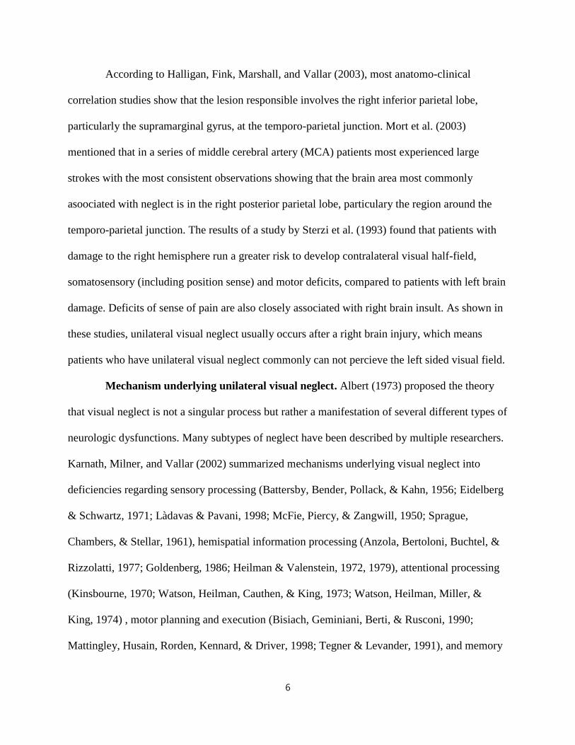
According to Halligan, Fink, Marshall, and Vallar (2003), most anatomo-clinical
correlation studies show that the lesion responsible involves the right inferior parietal lobe,
particularly the supramarginal gyrus, at the temporo-parietal junction. Mort et al. (2003)
mentioned that in a series of middle cerebral artery (MCA) patients most experienced large
strokes with the most consistent observations showing that the brain area most commonly
asoociated with neglect is in the right posterior parietal lobe, particulary the region around the
temporo-parietal junction. The results of a study by Sterzi et al. (1993) found that patients with
damage to the right hemisphere run a greater risk to develop contralateral visual half-field,
somatosensory (including position sense) and motor deficits, compared to patients with left brain
damage. Deficits of sense of pain are also closely associated with right brain insult. As shown in
these studies, unilateral visual neglect usually occurs after a right brain injury, which means
patients who have unilateral visual neglect commonly can not percieve the left sided visual field.
Mechanism underlying unilateral visual neglect. Albert (1973) proposed the theory
that visual neglect is not a singular process but rather a manifestation of several different types of
neurologic dysfunctions. Many subtypes of neglect have been described by multiple researchers.
Karnath, Milner, and Vallar (2002) summarized mechanisms underlying visual neglect into
deficiencies regarding sensory processing (Battersby, Bender, Pollack, & Kahn, 1956; Eidelberg
& Schwartz, 1971; Làdavas & Pavani, 1998; McFie, Piercy, & Zangwill, 1950; Sprague,
Chambers, & Stellar, 1961), hemispatial information processing (Anzola, Bertoloni, Buchtel, &
Rizzolatti, 1977; Goldenberg, 1986; Heilman & Valenstein, 1972, 1979), attentional processing
(Kinsbourne, 1970; Watson, Heilman, Cauthen, & King, 1973; Watson, Heilman, Miller, &
King, 1974) , motor planning and execution (Bisiach, Geminiani, Berti, & Rusconi, 1990;
Mattingley, Husain, Rorden, Kennard, & Driver, 1998; Tegner & Levander, 1991), and memory

7
(Bisiach & Luzzatti, 1978). Each of these mechanisms may be experienced in different types and
degrees of deficits according to the individual. Therefore, treating neglect may require a multi-
modal and multifaceted approach. One of approaches, which has no known harmful side effects,
is music as a sensory language stimulating cognitive, emotional, and motor functions. (Luauté,
Halligan, Rode, Rossetti & Boisson, 2006).
Therapeutic Mechanisms
Music and brain plasticity. Brain plasticity refers to the extraordinary ability of the
brain to modify its own structure and function following changes within the body or in the
external environment (Frostig, 2014). This means that the brain has the ability to change as a
result of any experiences including musical experiences, which may be used for improving visual
processing. Brain plasticity, which is the dramatic potential of the brain to reorganize itself
during activated working, has been widely studied for application in the clinical field. Recently,
brain plasticity has been implicated in the various neurologic disorders including stroke,
Parkinson’s disease, Alzheimer, Traumatic Brain Injury. Research has been published in
studying the relationship between music and brain plasticity. Using brain plasticity mechanisms,
music can be used as therapeutic tool for various functions such as motor control, cognition, and
emotion, with musical components (such as rhythm, dynamics, melody, etc.) structured to
facilitate therapeutic exercises and goals to retrain the injured brain (Jäncke, 2009; Peretz &
Zatorre, 2005; Wan & Schlaug, 2010).
Specifically related to neglect, the stimulating effect of music and rhythm on intentional
motor activity is particularly important in regard to the noted premotor intentional interruptions
in neglect states. Studies have shown the effect of auditory rhythm on motor control and motor-

8
related brain plasticity (Altenmüller, Marco‐Pallares, Münte, & Schneider, 2009; Luft et al.,
2004).
In regard to the influence of musical training on brain plasticity in cognitive attentional
functions, specifically related to pitch and melody, Moreno et al.(2009) examined whether
musical training improves non-musical brain functions such as reading and linguistic pitch
processing with 32 non-musician children over 9 months. Event-related brain potentials were
recorded while 8 year old children performed tasks. The result showed that 6 months of musical
training lead to significant improvement of behavior and influenced the development of neural
processes as reflected in specific pattern of brain waves.
Music, rhythm, and motor control. Besides studies showing the effect of music on
motor-related brain plasticity in neurorehabilitation, there are numerous kinematic and
behavioral research studies supporting applications of music and rhythm to movement
rehabilitation. Priming of movement through sound patterns, facilitating the initiation of
movement, and anticipatory timing of movement through the rhythmic structure are considered
the prime mechanisms (Thaut et al., 1999). It is well documented that even listening to rhythm
and music, without a motor task, activates motor areas in the brain, supporting a strong trigger
function for music and rhythm to initiate volitional movement. The motor system is very
sensitive to arousal by the auditory system, couples quickly into sound patterns, and this process
can take place at subliminal levels of sensory perception (Thaut et al., 1999). Furthermore,
studies have shown that music and rhythm promote motivation for movement, measuring
movement involvement with the self-reported Motor Activity Log (MAL) (Malcolm, Massie &
Thaut, 2009).

9
Based on these studies, the musical structure of MNT may have an important role in
facilitating the ‘aiming’ component and strengthening the premotor-intentional deficiencies in
neglect to aim motorial into the neglect side with playing musical patterns. This may be one
important mechanism for the role of music in successful neglect rehabilitation. However, the
neural basis of this potential mechanism has not been researched.
Music Supports Cognitive Function
Music has been used as a therapeutic tool in a variety of settings to promote not only
motor but also emotional and cognitive functions (Thaut, 2010). Of particular relevance for
neglect rehabilitation are clinical and mechanism data showing how and if music based
therapeutic exercises, especially with the NMT model, can stimulate audiovisual attention.
Also, listening to music can help to improve positive emotion and cognitive attentional
functions. An important study by Sarkamo et al. (2008) with stroke patients has shown that
listening to music can help to improve positive emotion and cognitive attentional functions. They
designed single-blind, randomized, and controlled trials to determine whether everyday music
listening can facilitate the recovery of cognitive attentional functioning in healthy subjects and in
various clinical patient groups. Participants were assigned to one of three groups: a music group
that listened to recorded music (n = 20), a language group that listened to audio books (n = 20)
and a control group that had no additional stimuli (n=20). All patients underwent a clinical
neuropsychological assessment, magnetoencephalographic (MEG) measurement, and a magnetic
resonance imaging (MRI). The results showed that recovery for participants from the music
group were better than the results for the language and control groups in areas of verbal memory,
focused attention, and less depression. The authors hypothesized that listening to music promotes
positive emotion and cognitive function, even when delivered passive-receptively.

10
Other studies have shown positive effects of music on attention in older adults (Gregory,
2002; Groene, 2001), and in traumatic brain injury rehabilitation (Barrow, Collins & Britt, 2006;
Knox, Yokota-Adachi , Kershner & Jutai, 2003). Tracking and responding to repetitive auditory
stimuli is part of the widely used and well-researched Attention Process Training by Sohlberg
and Mateer (1989). Also, Robertson et al. (1997) has shown that auditory stimuli can activate the
right hemisphere in the brain which is dominant for sustained attention and that those stimuli can
influence spatial attention including unilateral visual neglect. Finally, two recent studies, which
are very important for MNT, showed that music and rhythm can also improve visual attention
(Miller, Carlson & McCauley, 2013; Escoffier, Herrmann, & Schirmer, 2015). These findings
are very important for understanding the perceptual-cognitive mechanisms of MNT.
Neurologic Music Therapy (NMT)
MNT is part of NMT. This section will therefore (a) briefly review the treatment system
and theory of NMT, and b) the existing research evidence specifically for MNT.
Definition. NMT has been developed to facilitate sensorimotor, speech/language, and
cognitive rehabilitation and is based on scientific evidence. Specifically, NMT is based on the
Rational Scientific Mediating Model (R-SMM). The R-SMM is an epistemological model to
adopt the concept of music as a mediating stimulus for non-musical behavior, and it has four
levels to provide the transformational framework for music from nonmusical behavior. The four
stages are musical response models (logical foundations of musical behaviors), nonmusical
parallel models (processes in nonmusical brain and behavior function), mediating models
(influence of music on nonmusical brain and behavior function), and clinical research models

11
(therapeutic effects of music) (Thaut, 2008, p. 118). These levels have an important role as a
linkage to using music as a therapeutic tool from basic to applied research.
The four essential paradigms of NMT for rehabilitation are following: (1) neuroscience-
guided rehabilitation which is based on data and concepts from brain research and clinical
studies, (2) learning and training models which is using rhythm motor learning and training to
organize therapeutic interventions and stimuli to enhance cognitive, speech, and language
training, (3) cortical plasticity models that can drive neural network patterns through temporal
modulation of sensory input, music as complex, rhythmically organized, and spectrally diverse
language, and (4) neurological facilitation models to enhance motor, speech/language, and
cognitive functions via auditory rhythmicity, musical patterns (Thaut, 2008, p. 129).
NMT in cognitive domain. For cognitive deficits, NMT technique has been used as an
arousal tool for attention/perception, memory, and executive function. For attention/perception,
Musical Sensory Orientation Training (MSOT), Musical Neglect Training (MNT), Auditory
Perception Training (APT), and Musical Attention Control Training (MACT) are used. For
memory, Musical Mnemonics Training (MMT) and Associative Mood and Memory Training
(AMMT) are used. Finally, Musical Executive Function Training (MEFT) are used to practice
executive function skills, such as organization, decision making, comprehension, and problem
solving. Rhythm, a key element of music not only affects sensorimotor and speech areas, but
also, influences cognitive areas by recognizing the importance of temporal organization (Thaut,
Peterson, McIntosh, 2005). Attention and perception are crucial for functional outcomes with
any population. Numerous clinical research studies have shown that musical attention training
improves attention (Gregory, 2002; Knox, Yokota-Adachi, Kershner, & Jutai, 2003; Wolfe &

12
Noguchi, 2009) and memories (Simmons-Stern, Budson, & Ally, 2010; Thaut, Peterson,
McIntosh, & Hoemberg, 2014).
Musical Neglect Training (MNT). MNT is one of the NMT techniques in cognitive
rehabilitation. The exercise protocols for NMT are fairly standardized with little variation (Thaut
& Hoemberg, 2014). To focus attention to the neglect visual field, MNT uses musical exercises
which are structured in pitch, time and tempo, and musical equipment (tone bars, keyboards,
drums) set up in appropriate configurations. A second application type in MNT consists of
receptive music listening to stimulate hemispheric brain arousal while engaging in exercises
addressing visual neglect or inattention (Thaut, 2014). Af ter the initial study by Hommel et al.
(1990), studies have shown that an auditory stimulus promotes improvements in the patient’s
visual field, supporting the potential of MNT as a rehabilitation technique for neglect deficits.
Frassinetti, Bolognini, Bottari, Bonora, and Ladavas (2005) studied 21 patients with brain
damage to investigate the possibility that bimodal audiovisual stimulation of the affected
heimfield can improve visual perception in the blind hemifield. The results showed that the
patients with visual disorders largly benefitted from multisensory integration, and the authors
suggested a possibility of recovery from visual and spatial impairments.
Van Vleet and Robertson (2006) indicated that an auditory event can ameliorate spatial
and non-spatially lateralized attention deficits in a patient with neglect. Also, a functional
magnetic resonance imaging (fMRI) study completed by Soto et al. (2009) supported that
positive affect, generated by preferred music, can decrease visual neglect by increasing
attentional resources. In this study, Soto and colleagues induced a plesant and positive affective
response in patients with chronic visual neglect by allowing them to listen to their pleasant
preferred music. The authors found enhanced activity in the orbitofrontal cortex and the

13
cingulate gyrus associated with emotional responses when tasks were performed with preferred
music relative to unpreferred music.
Some clinical research has indicated that MNT can be effective for improving visual
field to visual neglect patients. Noto, Mouri, Amimoto, Sugimoto, and Futaki (1999) examined
the effects of limb activation using a xylophone with unilateral spatial neglect, for a 49-years-
old, right-handed woman who suffered cerebral infarction, and demonstrated a clear left
unilateral spatial neglect. In this study, the xylophone was placed to ascend the keys from right to
left. Significant improvement was observed in the intervention phase compared with the
baseline. These results suggested that unilateral spatial neglect field could be reduced by
triggering the right hemisphere concerning to the motor intention in exploring the musical scale
from right to left, and music as the stimulus of tone and rhythm.
Recent study by Bodak et al. (2014) found evidence that active music-making with
horizontally aligned instruments may help neglect patients attend more to their affected side. In
this study, two patients with left neglect had four weekly 30-minute music sessions. During these
sessions, participants played the scales and familiar melodies on tone bars from right to left. The
researchers collected the data three times during a preliminary baseline phase, before and after
the four intervention sessions during the intervention phase to define short-term effects, and 1
week after the last intervention session to investigate long-lasting effects from two cancellation
test (Mesulam shape, Behavioral Inattention Test (BIT) star), the neglect subtest from the
computerize TAP (Test of Attentional Performance) battery, and the Line Bisection Test. These
clinical studies support that MNT has a strong potential for use in rehabilitation for neglect
patients. Another recent study by Bernardi et al. (2015) assessed whether the auditory stimulus
provided by a music scale could improve spatial field with keyboard in right brain damaged

14
patients with left spatial neglect. Eleven right hemisphere stroke patients with left spatial neglect,
12 patients without neglect, and 12 age-matched healthy participants played descending scales
on a music keyboard. Specifically, researchers set the counterbalanced design to determine
whether the auditory stimulus provided by a music scale or not with congruent sound, no-sound,
or random sound feedback provided by the keyboard. The results showed congruent sound
influenced in spatial exploration by patients with left neglect compared to both silence and
random sound condition. This study supports that performing a scale with congruent sounds
would trigger at some extent preserved auditory and spatial awareness.
Experimental research to examine the effectiveness of MNT is still limited because of
small sample sizes and few studies. Active research is necessary in order to fill the theoretical
and applicative gaps found in the literature concerning music and, more specifically, musical
practice. Based on these clinical evidence-based research, this study is an applied study using
MNT to provide initial information regarding immediate and longer-lasting outcomes for
participants who are diagnosed with visual neglect after right hemisphere stroke.
15
CHAPTER III: METHODOLOGY
Study Design
A single subject design was used in this study to determine the immediate and longer-
lasting effectiveness of MNT for two individual patients with unilateral visual neglect from right
hemispheric stroke. This study followed a pre-and post- test design to observe behavior before
and after interventions with participants who were acting as their own control to see the
immediate effects, and follow-up test to determine the longer-lasting effects. The experiments
consisted of two phases: (1) pre- and posttests around 6 MNT interventions, and (2) follow-up
test measures with no intervention one week after the end of the session.
Participants
All participants gave written consent in accordance with the policies of the Institutional
Review Board in Colorado State University. Two participants with right hemisphere stroke who
were diagnosed with a neglect syndrome were recruited for this study. Both participants met the
following criteria: (1) right handed, (2) medically stable, (3) no previous music therapy
experiences, (4) no hearing impairments, and (5) no cognitive deficits. Exclusion criteria were
(1) hemianopia, and (2) previous music therapy treatment experiences.
Participant 1
Participant 1 was a 62 years old female who sustained an ischemic stroke 2 years and 2
months ago. Her medical documentation showed that she suffered from a stroke in the right
internal capsule and medial right temporal lobe resulting in left hemiparesis and loss of attention
in left side with decreased functional performance. Hypertension and diabetes were causes of
stroke. She described her vision saying that she seemed to lose half side of screen in the world.

16
Participant 2
Participant 2 was a 69 years old male who sustained a stroke that occurred in May 2005.
His ischemic stroke in right parietal lobe region from the atrial fibrillation resulted in left
hemiparesis, and sensory impairment in the left side. He described that he could button on the
right side of his shirts without seeing, but could not do this on the left side. Also, he said that he
seemed to lose quarter to half field of vision on the left side.
Instrumentation/Materials
The Albert’s Test (Line Cancellation Test) and the Line Bisection Test are used
commonly to assess neglect (Lee et al., 2004). The Albert’s Test is a screening tool used to
detect the presence of unilateral visual neglect in patients with stroke. Participants are required to
crossing out 40 black lines that are randomly orientated on a sheet of paper. The test paper was
placed on a table and presented in front of a patient, parallel to their midsection. The
experimenter asked the participant to cross over all of the lines they see in front of them. Patients
had a maximum of 5 minutes to complete the activity. Assessment tools to be used for this test
included an A4 sheet (11x8.5-inch), with 40 lines 2 centimeters in length and a pencil (Barrett et
al., 2006). Internal consistency reliability is not shown in previous studies, but excellent test-
retest reliability is reported with a score of r=0.79 (Sea & Henderson, 1994). For validity,
Albert’s Test has excellent correlation with the Line Bisection Test (r=0.85) (Agrell, Dehlin, &
Dahlgren, 1997).
The Line Bisection Test is also a screening tool used to diagnose and assess neglect.
Participants must place a mark with a pencil in the center of horizontal lines. A displacement of
the mark towards the side of the brain lesion is assessed as a symptom of neglect. The equipment
17
for this test is an A4 sheet (11x8.5-inch) with 17 vertically staggered horizontal lines and a
pencil (Strokeengine, 2014). Menon & Korner-Bitensky (2004) found the Line Bisection Test
showed strong test-retest reliability, construct validity, convergent and discriminant validity, and
criterion validity. Bailey, Riddoch and Crome (2004) reported the intraclass correlation
coefficient was excellent for neglect patients (ICC=0.97).
Procedure
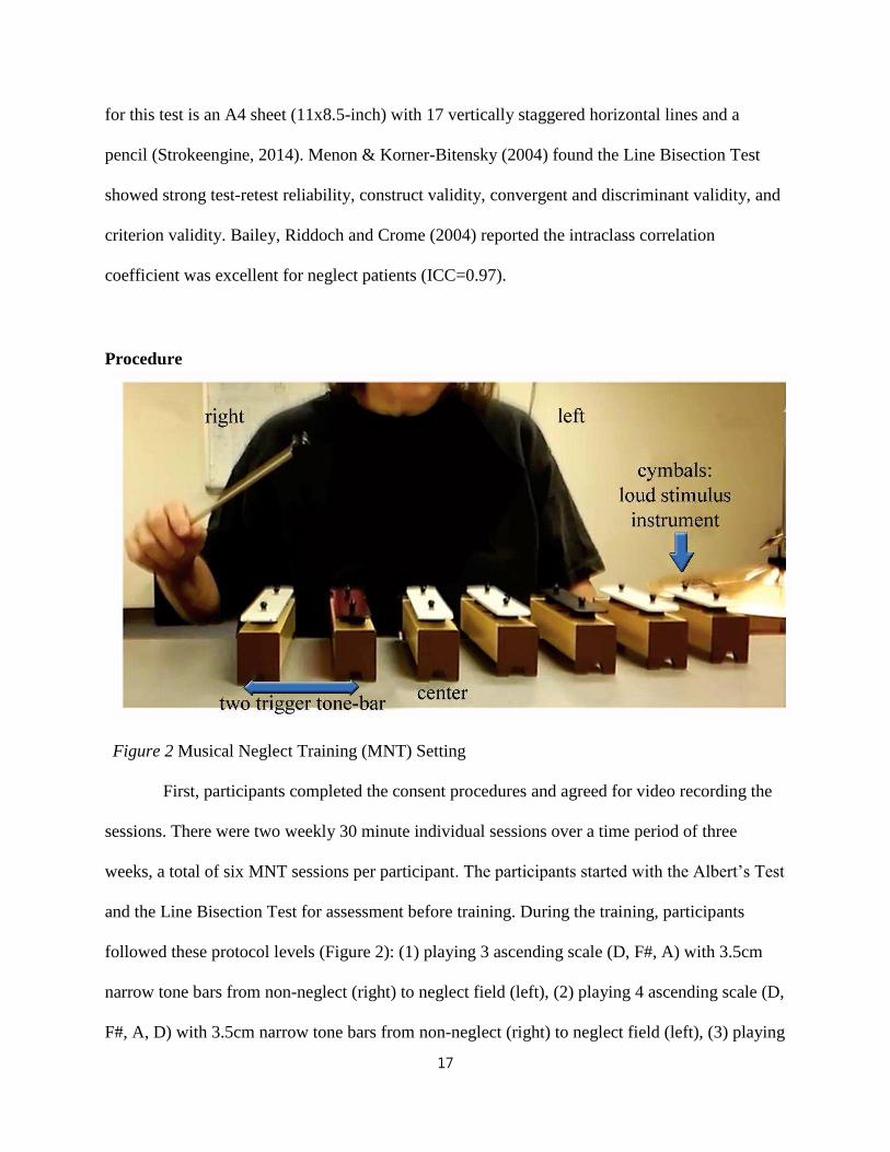
Figure 2 Musical Neglect Training (MNT) Setting
First, participants completed the consent procedures and agreed for video recording the
sessions. There were two weekly 30 minute individual sessions over a time period of three
weeks, a total of six MNT sessions per participant. The participants started with the Albert’s Test
and the Line Bisection Test for assessment before training. During the training, participants
followed these protocol levels (Figure 2): (1) playing 3 ascending scale (D, F#, A) with 3.5cm
narrow tone bars from non-neglect (right) to neglect field (left), (2) playing 4 ascending scale (D,
F#, A, D) with 3.5cm narrow tone bars from non-neglect (right) to neglect field (left), (3) playing

18
5 ascending scale (D, F#, A, C#, D) with 3.5cm narrow tone bars from non-neglect (right) to
neglect field (left), (4) playing 6 ascending scale (D, E, F#, A, C#, D) with 3.5cm narrow tone
bars from non-neglect (right) to neglect field (left), (5) playing full ascending scale (D, E, F#, G,
A, B, C#, D) with 3.5cm narrow tone bars from non-neglect (right) to neglect field (left).
Participants sat comfortably in a chair, playing the tone bars on the desk with the non-paretic
side (right). In each level, first tone bar (D) was put in the center of participants. Also, two tone
bars (B, C#) were set to the right side before starting the scale or triad to start the playing
movement from right to left in the healthy visual field. A cymbal was located in the last position
to give a strong sound target for completion of the pattern. In each pattern, the cymbals’ edge
matched the end of the very last tone bar’s edge. The experimenter was positioned on the
patient’s non-neglect side (right) to give instruction and play a chordal accompaniment for each
tone bar pitch. Verbal and rhythmic cues were given to help anticipation and completion.
Participants repeated each pattern 5 times before moving to the next step. The Albert’s test and
the Line Bisection test were given to participants after training to observe immediate effects.
Data Collection
The experimenter collected the data for all dependent variables pre-and post-
interventions and follow-up stage. The Albert’s Test and the Line Bisection Test were collected
before and after every 6 each interventions to examine the immediate effects. Also, same
assessments, Albert’s Test and Line Bisection Test were collected at the follow-up test to
determine the effectiveness of MNT and the longer-lasting effects. Also, combined data from
participant 1 and 2 were calculated with descriptive statistics only.

19
Data Analysis
The Albert’s Test was scored as the number of uncrossed lines on the left side. The test
paper consisted of 17 lines on each side, and 4 lines in the center. The Line Bisection Test was
calculated as percentage of the deviation from the true center of the line. There were 17 horizon
lines, which had different length and calculated by the following methods: the means of the
deviations of all 17 lines were divided by center point length of all 17 lines/17, and shown as
percentage. The formula was [Sum of all deviations/ Sum of all center point length of 17
lines)/17].
Descriptive statistics, means and standard deviation were calculated for both tests
(Albert’s Test and Line Bisection Test) to display in numbers and graphically pre- and post-test
results. Paired sample t-tests were calculated to examine significant differences, and compare
means by subtracting pre- intervention scores from post- test scores. Nonparametric statistics
(Wilcoxon sign-ranked test) was also calculated in parallel with the paired t-tests due to the small
sample size and possible violations of normal distribution. For the longer-lasting effects,
statistical analysis was not done because there were only one follow-up test data per subject.
Raw data were compared between all 6 averaged sessions pre-test and follow-up test. Also,
descriptive statistics only were calculated for the data of the two participants combined for
training trials, post- vs follow up, and pre- vs follow up. Figure 3 illustrates the summary of
study design, data collection, and data analysis.

20
Figure 3: Summary of study design, data collection, and data analysis
Intervention
•6 MNT sessions•Albert's test •Line Bisection Test• Immediate effect(pre-and post-)
Follow up
•One week after 6th session•Albert's Test•Line Bisection Test•Longer-lasting effect (Averaged Pre vs Follow up)

21
CHAPTER IV: RESULTS
Data were collected from two participants using the Albert’s Test and Line Bisection
Test. Pre-and post-tests of each 6 sessions for the immediate effects, and follow-up test for the
longer-lasting effects were compared with descriptive statistics, paired t-tests, and non-
parametric tests (Wilcoxon Signed Rank Test). Also, data from participant 1 and participant 2
were combined and displayed using descriptive statistics only.
Participant 1
Albert’s Test. Figure 4 shows score differences between pre and post-test of 6 sessions,
and between averaged pre and post-test of 6 sessions.
Figure 4 Descriptive Data for Participant 1’s Albert’s Test
All scores from pre to post-test of six sessions decreased, except 3rd session (remained
same). The scores were calculated as the number of uncrossed line, so a reduced score meant
improvement. The mean of the scores that the participant did not mark for pretest was 14.5 and
13 at post-test. Pre-test and post-test difference of the means of all 6 sessions’ indicated positive
0
2
4
6
8
10
12
14
16
1st 2nd 3rd 4th 5th 6th
Pre-test 14 15 13 14 15 16
Post-test 12 11 13 13 14 15
Averaged Pre 14.5
Averaged Post 13
Pre-test
Post-test
Averaged Pre
Averaged Post

22
outcomes regarding immediate effects – but with small differences only (14.5 to 13). Also, it is
interesting to note that the 6th session was worse than the pre-test in the 1st session.
A Wilcoxon signed rank test was conducted to determine whether there was a difference
in the ranking of pre-and post-test by MNT sessions. Results of that analysis indicated that there
was a significant difference in pre-and post-interventions, Z = -2.06, p < .05 (Table 1).
Table 1 Wilcoxon Signed Rank Test for Albert’s Test in Participant 1
Posttest -Pretest
Z -2.060
Asymp. Sig. (2-tailed) *.039
Note: *p < .05, two-tailed
Table 2 shows a paired t-test for participant 1 in Albert’s Test. There was a significant
difference in mean scores before and after MNT training (t=-2.67, p<.05)
Table 2 Paired t-test for Albert’s Test of Participation 1
Pre-test Post-Test
Mean 14.5 13
Standard Deviation 1.05 1.41
t -2.67
p-value (one-tailed) *0.02
p-value (two-tailed) †0.04
Note: *p < .05, one-tailed, †p < .05, two-tailed

23
Average of all 6 sessions’ pre-test and follow-up test were compared to observe the
longer-lasting effect. Figure 6 shows raw score differences between averaged pre-test of 6
sessions and follow-up test. This bar graph of raw scores slightly decreased (from 14.5 to 14).
Figure 6 Participant 1’s Averaged Pre-test and Follow-up Test Score
Line bisection test. Line Bisection Test was analyzed by following methods. Means of
all 17 lines’ deviation were divided by center point length of all 17 lines/17, and shown with
percentage. The formula is [(Sum of all 17 lines’ deviation/ Sum of all center point length of 17
lines)/17].
Figure 7 presents percentage differences between pre and post-test of 6 sessions, and
averaged pre and post-test in Line Bisection Test.
Figure 7 Descriptive Data for Participant 1’s Line Bisection Test
02468
1012141618
Averaged Pre-test Follow-up test
14.5 14
score
0.00%
10.00%
20.00%
30.00%
40.00%
1st 2nd 3rd 4th 5th 6th
Pre-test 27.87%21.31%19.81%21.91%32.04%30.93%
Post-test 32.86%17.42%16.12%24.87%35.23%18.55%
Averaged Pre 25.65%
Averaged Post 24.17%
Pre-test
Post-test
Averaged Pre
Averaged Post

24
Decreased percentage meant positive effects; because shorter deviation from the center
point indicated having better awareness of left side. (All the deviation from the center was right
side- it meant that participant who has unilateral visual neglect’s center leaned to right side
because of their left side visual field loss). These percentages of deviation in all 6 sessions were
not seen as sustainable. Three sessions (2nd, 3rd, and 6th) showed improvements, but other three
sessions (1st, 4th, and 5th) did not indicate improvements. However, overall, once averaged pre-
test and averaged post-test was compared, the deviation from the center declined from 25.65% to
24.17%, about 1.5% decreased. The interesting result was that participant 1 showed obvious
decreased percentage of deviation from 30.93% to 18.55%, about 12% decreased.
Table 3 shows indicated that there was no significant difference in pre-and post-
interventions, Z = -2.06, p =.60.
Table 3 Wilcoxon Signed Rank Test for Line Bisection Test in Participant 1
Posttest -Pretest
Z -0.52
Asymp. Sig. (2-tailed) .60
Note: *p < .05, two-tailed
Table 4 shows the paired t-test of participant’s Line Bisection Test. With the t-test (t=-
0.55) for Line Bisection Test, there was no significant improvement. Even though, mean of pre-
test to mean of post-test showed a decrease, inferential statistics did not show significant
differences.

25
For the longer-lasting effect, averaged pre-test percentage deviation of 6 sessions and
follow-up test were compared (Figure 8). Percentage of deviation in Line Bisection Test for
Participant 1 increased from 25.65% to 27.18%, which is not a positive effect.
Figure 8 Percentage of Deviation Data for Averaged Pre-test and Follow-up test
0.00%
50.00%
Averaged Pre-test Follow-up test
25.65% 27.18%
Percentage of Deviation
Table 4 Paired t-test for Line Bisection Test of Participation 1
Pre-test Post-Test
Mean 25.65% 24.17%
Standard Deviation 0.05 0.08
t -0.55
p-value (one-tailed) 0.30
p-value (two-tailed) 0.60
Note: *p < .05, two-tailed, †p < .05, one-tailed

26
Participant 2
Albert’s Test. Figure 9 displays the score differences between pre and post-test of 6
sessions, and between averaged pre and post-test in Albert’s test.
Figure 9 Descriptive Data for Participant 2’s Albert’s Test
From first session to last session, participant 2’s bar graph showed a steady decline
except from first session to second session. Also, in each session, there were all decreased scores
except last session (remained same), which indicated positive outcomes in immediate effects of
MNT. Between averaged pre-test and post-test, scores indicated a positive result.
Table 5 shows result of Wilcoxon signed rank test. Wilcoxon signed rank test indicated
post-test of intervention was statistically significantly higher than pre-test of intervention, Z=-
2.03, p < .05.
Table 5 Wilcoxon Signed Rank Test for Albert’ Test in Participant 2
Posttest -Pretest
Z -2.03
Asymp. Sig. (2-tailed) .04
Note: *p < .05, two-tailed
02468
1012
1st 2nd 3rd 4th 5th 6th
Pre-test 10 12 10 9 8 8
Post-test 7 8 8 8 7 8
Averaged Pre 9.50
Averaged Post 7.67
Pre-test
Post-test
Averaged Pre
Averaged Post

27
Table 6 shows the result of paired t-test. There was significant differences in the scores
for pre-test (M=9.5, SD=1.52), and post-test (M=7.67, SD=0.52); t=-3.05, p <.05. These data
supported the significant differences in immediate effects of MNT technique.
Figure 10 indicates compared scores between averaged all pre-test of 6 sessions and
follow-up test to determine the longer-lasting effect in Albert’s test for participant 2. Score was
decreased from averaged pre-test of 6 sessions (score=9.5) to follow-up test (score=7) that
showed positive outcome from bar graph.
Figure 10 Participant 2’s Averaged Pre-test and Follow-up Test Score
0
5
10
Averaged Pre-test Follow-up test
9.5
7
score
Table 6 Paired t-test for Albert’s Test for Participant 2
Pre-test Post-Test
Mean 9.50 7.67
Standard Deviation 1.52 0.52
t -3.05
p-value (one-tailed) *0.01
p-value (two-tailed) †0.03
Note: *p < .05, one-tailed, †p < .05, two-tailed

28
Line Bisection Test. Figure 11 showed percentage of deviation differences between pre
and post-test of 6 sessions, and between averaged pre and post-test in Line Bisection Test.
Figure 11 Descriptive Data for Participant 2’s Line Bisection Test
From this data, there was a gradually negative trend from first session to last session,
except three times (2nd, 4th, and 6th) that were very slightly increased (2nd: 1%, 4th:0.1%, and
6th: 0.05%). However, the averaged percentage of deviation from pre-test to post-test decreased
about 1.2%.
Table 7 shows the result of Wilcoxon signed rank test. Wilcoxon signed rank test
indicated post-test of intervention was no statistically significantly improvement, Z=-2.03, p
=.17.
Table 7 Wilcoxon Signed Rank Test for Line Bisection Test in Participant 2
Posttest -Pretest
Z -1.57
Asymp. Sig. (2-tailed) .17
Note: *p < .05, two-tailed
0.00%
5.00%
10.00%
15.00%
1st 2nd 3rd 4th 5th 6th
Pre-test 14.53%11.14%9.92%8.35%10.57%7.83%
Post-test 10.17%12.10%9.40%8.48%6.92%7.88%
Averaged Pre 10.39%
Averaged Post 9.16%
Pre-test
Post-test
Averaged Pre
Averaged Post

29
Table 8 shows paired t-test of participant 2’s Line Bisection Test. With the t-test, one-
paired p-value was 0.11 and two-paired p-value was 0.22, which did not show significant
improvement.
Table 8 Paired t-test for Line Bisection Test for Participant 2
Pre-test Post-Test
Mean 10.39% 9.16%
Standard Deviation 0.02 0.02
t -1.37
p-value (one-tailed) 0.11
p-value (two-tailed) 0.23
Note: *p < .05, two-tailed, †p < .05, one-tailed
To see the longer-lasting effect, same as Albert’s test, averaged pre-test of 6 sessions and
follow-up test were compared, and percentage decreased from 10.39% to 7.70% (Figure 12).
Figure 12 Percentage of Deviation Data for Averaged Pre-test and Follow-up test
0.00%
10.00%
20.00%
Averaged Pre-test Follow-up test
10.39%
7.70%
Percentage of Deviation

30
Combined Participant 1 and Participant 2
Data from participant 1 and participant 2 were combined, and compared between
averaged pre-test of 6 sessions vs averaged post-test of 6 sessions, post-test of 6 sessions vs
follow-up test, and averaged pre-test of 6 sessions vs follow-up test for both assessment, Albert’s
test and Line Bisection test. Figure 13 showed summary of mean of combined data of participant
1 and 2 in Albert’s and Line Bisection Test graphically.
Albert’s Test Line Bisection Test
Figure 13 Compare Combined Means (Averaged Pre vs Averaged Post, Averaged Post vs Follow-up, Averaged Pre vs Follow-up)
Data of participant 1 and participant 2 were combined since their clinical symptoms
were similar and also to generalize the overall effects of MNT. Descriptive statistics were
calculated for combined data (Table 9 and Table 10). Means and standard deviation of combined
averaged pre-test, averaged post-test, and follow-up test were compared. Averaged pre vs
averaged post (M=12, SD=3.54 to M=10.34, SD=3.77) and averaged pre vs follow-up (M=12,
SD=3.54 to M=10.50, SD=4.95) for Albert’s test showed improvement except comparing
9.50
10.00
10.50
11.00
11.50
12.00
12.00
10.34 10.3410.50
12.00
10.50
15.50%
16.00%
16.50%
17.00%
17.50%
18.00%
18.50%
18.02%
16.67% 16.67%
17.44%
18.02%
17.44%

31
between averaged post and follow-up (M=10.34, SD=3.77 to M=10.50, SD=4.95). Similarly,
results from Line Bisection Test showed improvements in averaged pre vs averaged post
(M=18.02, SD=10.79 to M=16.67, SD=10.61) and averaged pre vs follow-up (M=18.02,
SD=10.79 to M=17.44, SD=13.77) except comparing between averaged post and follow-up
(M=16.67, SD=10.61 to M=17.44, SD=13.77), which supported the immediate and longer-lasting
effects.
Table 9 Descriptive Statistics for Combined Data in Albert’s Test Averaged Pre vs Averaged Post
N Minimum Maximum Mean Std. Deviation Averaged Pre- test
2 9.50 14.50 12.00 3.54
Averaged Post-test
2 7.67 13.00 10.34 3.77
Averaged Post vs Follow up
N Minimum Maximum Mean Std. Deviation Averaged Post-test
2 7.67 13.00 10.34 3.77
Follow up test 2 7.00 14.00 10.50 4.95
Averaged Pre vs Follow up
N Minimum Maximum Mean Std. Deviation Averaged Pre- test
2 9.50 14.50 12.00 3.54
Follow up test 2 7.00 14.00 10.50 4.95

32
Table 10 Descriptive Statistics for Combined Data in Line Bisection Test
Averaged Pre vs Averaged Post
N Minimum Maximum Mean Std. Deviation Averaged Pre- test
2 10.39 25.65 18.02 10.79
Averaged Post-test
2 9.16 24.17 16.67 10.61
Averaged Post vs Follow up
N Minimum Maximum Mean Std. Deviation Averaged Post-test
2 9.16 24.17 16.67 10.61
Follow up test 2 7.70 27.18 17.44 13.77
Averaged Pre vs Follow up
N Minimum Maximum Mean Std. Deviation Averaged Pre- test
2 10.39 25.65 18.02 10.79
Follow up test 2 7.70 27.18 17.44 13.77

33
CHAPTER V: DISCUSSION
The purpose of this study was to examine the effectiveness of MNT on unilateral visual
neglect for the immediate and longer-lasting effect on chronic unilateral visual neglect patients.
The hypotheses of this study were that 1) participants would show improvement on scores of
Alberts and Line Bisection Tests immediately after each of six sessions, and 2) participants
would show better scores after one week at follow-up testing to examine the longer-lasting
effects of MNT training on the Alberts and Line Bisection Test. Results from paired t-tests
indicate that both participant 1(t=-2.67, p < .05) and participant 2 (t= -3.05, p < .05) showed
significant improvement in immediate effects with Albert’s Test. Results from Wilcoxon Singed
Rank Test also showed statistically significant improvement in immediate effects from
participant 1 (Z = -2.06, p < .05) and participant 2 (Z=-2.03, p < .05). Comparing between the
raw scores of averaged pre-test and follow-up test, both participants (participant 1 with 14.5 to
14, participant 2 with 9.5 to 7) had decreased scores, which means positive effects of MNT as
measured by the Albert’s Test. Reduced numbers of uncrossed lines indicated an increase in the
participant’s attention to the left visual field.
Participant 1 showed interesting result in Albert’s Test. Looking at the overall scores of
all 6 sessions, the scores gradually increased, which did not indicate a positive outcome. The
score on the pre-test of 1st session was 14, and at the 6th session was 16. Also, the score in post-
test was increased from 12 to 15. However, at each session - with the exception of the third -
participant 1’s score decreased. Overall, even though participant 1 did not show a steady decline
across 6 sessions, she showed improvement in each individual session, meaning possibly that she
benefited from the immediate training but showed no carry-over from session to session. On the
other hand, participant 2 showed improvement across all 6 sessions. Interestingly, there was a

34
drastic improvement in the first 3 sessions, and differences scores between pre and post-test were
getting smaller to the end (posttest – pretest from 1st session to 6th session= -3, -4, -2, -1-1, 0).
There were no significant differences in immediate effect with Line Bisection Test for
both participants. Participant 1 showed some positive outcomes in each session, but they were
not sustained overall. However, when comparing between averaged pre-test of 6 sessions and
averaged post-test, the percentage of deviations from the central point decreased. Same as for the
Albert’s Test, decreased percentage meant a positive result in Line Bisection Test. Reduced
percentage of deviations indicated that participant’s attention expanded to the left side.
Especially participant 1 showed a dramatic decrease of percentage of deviation from 30.93% to
18.55% in 6th session (last session).
Participant 2 also did not show significant differences, but his percentage of deviations
gradually decreased from 1st to 6th session from 14.53% to 7.88%. For the longer-lasting effect of
Line Bisection Test, participant 1 showed an increase in percentage of deviation from averaged
pre-test to follow-up test, which did not result in a positive outcome. Participant 2 had a positive
outcome in the longer-lasting effect from 10.39% to 7.70%.
Interestingly, both participants showed significant improvements in Albert’s Test, but
did not show significance on the Line Bisection Test. Especially, participant 1 reported
difficulties to do the Line Bisection Test. She exhibited frustration sometimes with this test by
stopping during testing. In contrast, when participant 2 took the Line Bisection Test, he said that
the Line Bisection Test seemed to be easier than Albert’s Test. Albert’s Test focuses on
horizontal work and the Line bisection Test focuses on vertical perception along long distance
view point. Especially, each line in the Line Bisection Test needs horizontal work to find the
center point, but participants needed vertical work to find the central line for all 17 lines during

35
assessment. This might have been the reason for the different results between Albert’s Test and
Line Bisection Test (Figure 14).
Albert’s Test Line Bisection Test
Figure 14 Compare between Pattern of Albert’s Test and Line Bisection Test
To determine the overall differences between pre-test vs post-test, post-test vs follow-up
test, and pre-test vs follow-up test in Albert’s Test and Line Bisection Test, data collected from
both participants were combined and displayed descriptively.
Interestingly, both Albert’s Test and Line Bisection Test displayed similar pattern
(Figure 13). The largest differences was shown in immediate effects (averaged pre-test vs
averaged post-test for both assessment, Albert’s Test and Line Bisection Test. The second largest
difference was in the longer-lasting effects (averaged pre-test and follow-up test) for both
assessments. Also, another interesting outcome was that comparisons between averaged post-test
and follow-up test did not show positive results (increased scores in Albert’s Test and increased
deviation percentage in Line Bisection Test) for both tests. However, the combined scores should
be treated with great caution because there were only 2 subjects involved and both subjects
showed great individual differences in their etiology and their test performances.

36
There are several factors that could have led to differences in the results of this study.
One could have been that each participant had a different onset of stroke. Participant 1 suffered
from stroke in 2013, and participant 2 suffered from stroke in 2005. Table 11 showed differences
scores in Albert’s Test and percentage of deviation in Line Bisection Test from participant 1 and
participant 2.
Table 11 Summary of Data from Participant 1 and Participant 2 in Albert’s Test and Line Bisection Test
Albert’s Test Line Bisection Test
Participant 1 Participant 2 Participant 1 Participant 2
Averaged Pre-Test 14.5 9.5 25.65% 10.39%
Averaged Post-Test 13 7.67 24.17% 9.16%
Follow-up Test 14 7 27.18% 7.70%
All scores of participant 2, (averaged pre-test, averaged post-test, and follow-up test)
were lower than participant 1’s score in Albert’s Test, which meant that participant 2 had a less
impaired visual field than participant 1 from the onset of the study. Similar results were shown in
Line Bisection Test. The overall threshold of attention on the left side in participant 2 was higher
than participant 1 in both tests, Albert’s Test and Line Bisection Test. One possible explanation
is that during 10 years following his stroke, participant 2 would have been working on
compensatory strategies to be alert regarding his spatial awareness of the left side in order to
accommodate his daily life. On the other hand, participant 1 had a stroke two years ago. She has
not had any rehabilitation training for visual neglect. She mentioned that she was learning to scan
left by herself for 2 years. Ten years training versus 2 years training may account for the

37
different outcomes in both assessments. Visual neglect patients were able to anticipate their
skewed spatial orientation and to compensate for neglect in structured and predictable
circumstances as time went on (Jehkonen et al., 2000).
A second factor could have been that each participant had a different musical experience
background. Participant 1 belonged to a musical environment. Her father is a pianist, and other
siblings are also musicians. Especially, she also has been participating in a string trio in the
community as an amateur violinist. In contrast, participant 2 has been in exposed musical
environments (attending concerts, listening to music privately) but has no active experiences like
participant 1. This factor could be one of the reasons that they showed different outcomes.
However, the non-musician participant benefited more, making the musician argument probably
less meaningful than other factors.
In addition, behavior patterns, personal attitude, etc. could have influenced the different
results between participant 1 and participant 2. However, some common positive outcomes from
the data in this study provide support that an active music intervention using a horizontally
aligned tone bar sequence (MNT) may help both participants to improve their visual field. Self-
reports also indicated that they enjoyed the exercises.
The current study has presented the positive potentials of MNT for visual neglect
patients in both, immediate and longer-lasting effects. Two other aspects were used in this study:
(1) sequential aspects: putting the cymbals as a loud stimulation instruments after playing the
simple scales to help participants to expect the final completion of each sequence, and (2)
systematic aspects by accompanying participants’ scale playing with chords cueing their motor
movements to the next tone bar.

38
Even though both participants showed a significant improvement in immediate effects
and had trends that suggested the possibility of longer-term improvement, this study had several
limitations, which would impact the strength of the findings. The first limitation was the size of
the sample in this study (n=2). Because only two participants participated in this study, the
generalizability to larger sample sizes is limited. The second limitation was the time elapsed
since suffering a stroke. Both participants were chronic visual neglect patients. Especially,
participant 2 has been almost 10 years since he suffered from stroke. Even though he could not
attend to his left visual field perfectly, he has been training by himself to scan the left visual
field. This factor could influence the results. However, the study adds new data regarding the use
of MNT with highly persistent visual neglect since most studies have investigated subacute
patient groups. Third, there are clear limitation in interpretation the statistical results that the data
were analyzed on an individual subject basis which prohibit large sample generalizability.
The recommendation for the future research could be a rearrangement of the limitations
of this study as previously mentioned. First, a larger sample size would increase the external
validity of MNT’s effectiveness on unilateral visual neglect. Second, it would be desirable to
recruit patients with a more recent onset of visual neglect so as to remove the complicating
factors of other forms of rehabilitation and self-training that participant 2 had experienced.. With
acute or subacute visual neglect patients one would be able to investigate demonstrate a more
controlled effect of MNT. Also, if further research would expand the study design by including
several base lines and follow-up tests, research would be strengthened to determine the
effectiveness of MNT. The desired goal of therapy is the longer-lasting effects and the transition
those effects to quality of daily living. The researcher would need to have several follow-up tests
rather than only one follow-up test on which to perform statistical analysis. Also, longer protocol

39
and/or more sessions would provide better statistical power to the results. Additionally, it would
be interesting to compare between auditory effects and motor effects to find an extended visual
field separately. Researcher will be able to set up both ways to determine which components
between auditory and motor lead their attention more to discover the left visual. First, auditory
only design would be set up by setting tone bar in the center point only or playing different
recording sounds only in the left side. Second, motor only design would be set up by using
muffled soundless tone bar. Using a design like this would help to separate the effect of the
auditory feedback from the motor component. Nevertheless, it is noteworthy that the study was
able to investigate the effectiveness of MNT with long-term persistent neglect. Additionally,
small sample studies are important for clinical research when large homogeneous patient groups
are not available which can be quite common in neurorehabilitation units.
In conclusion, the purpose of this study was to determine the effectiveness of MNT in
providing immediate and longer-lasting improvement of left-sided visual neglect. Both
participants showed significance immediate effects when tested by Albert’s Test. Also, trends
toward positive outcomes were shown in the longer-lasting effects. Even though neither
participants did showed significant effect in Line Bisection Test for the immediate and longer-
lasting effects, they still showed improvement of their visual field. To generalize the
effectiveness of NMT, further study would include a larger sample size, recruiting acute or
subacute visual neglect patients, and adjusting the length and level of intervention to the
individuals. Moreover, a sufficient high quality of randomize control trials with appropriate
functional measures will provide strong evidence for clinical practice (Bowen & Lincoln, 2007).
Better understanding is needed of the neurophysiological and biomechanical mechanisms that
underlie compensation-related learning of functional tasks after stroke (Kwakkel, 2006). In

40
addition, a better understanding of the neural mechanisms will help to adjust knowledge
appropriately to the clinical situation.

41
REFERENCES
Agrell, B. M., Dehlin, O. I., & Dahlgren, C. j. (1997). Neglect in elderly stroke patients: a
comparison of five tests. Psychiatry and clinical neurosciences, 51(5), 295-300. doi:
10.1111/j.1440-1819.1997.tb03201.x
Albert, M. L. (1973). A simple test of visual neglect. Neurology.
Albert, M. L., Sparks, R. W., & Helm, N. A. (1973). Melodic intonation therapy for aphasia.
Archives of Neurology, 29(2), 130-131. doi: 10.1001/archneur.1973.00490260074018
Altenmüller, E., Marco‐ Pallares, J., Münte, T., & Schneider, S. (2009). Neural Reorganization
Underlies Improvement in Stroke‐ induced Motor Dysfunction by Music‐ supported
Therapy. Annals of the New York Academy of Sciences, 1169(1), 395-405. doi:
10.1111/j.1749-6632.2009.04580.x
Anzola, G., Bertoloni, G., Buchtel, H., & Rizzolatti, G. (1977). Spatial compatibility and
anatomical factors in simple and choice reaction time. Neuropsychologia, 15(2), 295-302.
doi: 10.1016/0028-3932(77)90038-0
Bailey, M. J., Riddoch, M. J., & Crome, P. (2004). Test–retest stability of three tests for
unilateral visual neglect in patients with stroke: Star Cancellation, Line Bisection, and the
Baking Tray Task. Neuropsychological Rehabilitation, 14(4), 403-419. doi:
10.1080/09602010343000282
Barnes, M. P., & Good, D. C. (Eds.). (2013). Neurological Rehabilitation: Handbook of Clinical
Neurology (Vol. 110). Newnes.
Barrett, A. M., Buxbaum, L. J., Coslett, H. B., Edwards, E., Heilman, K. M., Hillis, A. E., . . .
Robertson, I. H. (2006). Cognitive rehabilitation interventions for neglect and related

42
disorders: moving from bench to bedside in stroke patients. Journal of cognitive
neuroscience, 18(7), 1223-1236. doi:10.1162/jocn.2006.18.7.1223
Barrow, I. M., Collins, J. N., & Britt, L. D. (2006). The influence of an auditory distraction on
rapid naming after a mild traumatic brain injury: A longitudinal study. Journal of Trauma
and Acute Care Surgery, 61(5), 1142-1149.
Basso, A., & Capitani, E. (1985). Spared musical abilities in a conductor with global aphasia and
ideomotor apraxia. Journal of Neurology, Neurosurgery & Psychiatry, 48(5), 407-412.
Battersby, W. S., Bender, M. B., Pollack, M., & Kahn, R. L. (1956). Unilateral “spatial
agnosia”(“inattention”) in patients with cerebral lesions. Brain, 79(1), 68-93. doi:
10.1093/brain/79.1.68
Bernardi, N. F., Cioffi, M. C., Ronchi, R., Maravita, A., Bricolo, E., Zigiotto, L., ... & Vallar, G.
(2015). Improving left spatial neglect through music scale playing. Journal of
neuropsychology. doi: 10.1111/jnp.12078
Beschin, N., Cocchini, G., Allen, R., & Sala, S. D. (2012). Anosognosia and neglect respond
differently to the same treatments. Neuropsychological Rehabilitation, 22(4), 550-562.
doi: 10.1080/09602011.2012.669353
Bisiach, E., Geminiani, G., Berti, A., & Rusconi, M. (1990). Perceptual and premotor factors of
unilateral neglect. Neurology, 40(8), 1278-1278. doi: 10.1212/WNL.40.8.1278
Bisiach, E., & Luzzatti, C. (1978). Unilateral neglect of representational space. Cortex, 14(1),
129-133. doi: 10.1016/S0010-9452(78)80016-1
Bodak, R., Malhotra, P., Bernardi, N. F., Cocchini, G., & Stewart, L. (2014). Reducing chronic
visuo-spatial neglect following right hemisphere stroke through instrument playing.
Frontiers in Human Neuroscience, 8, 413. doi: 10.3389/fnhum.2014.00413

43
Bowen, A., Lincoln, N., & Dewey, M. (2007). Cognitive rehabilitation for spatial neglect
following stroke. Cochrane Database Syst Rev, 2. doi:
10.1002/14651858.CD003586.pub2
Bowen, A., & Lincoln, N. (2007). Cognitive rehabilitation for spatial neglect following stroke.
The Cochrane Library. doi: 10.1002/14651858.CD003586.pub2
Cherney, L. R., & Halper, A. S. (2001). Unilateral visual neglect in right-hemisphere
stroke: a longitudinal study. Brain Injury, 15(7), 585-592. doi:
10.1080/02699050117415
Dalla Bella, S., & Penhune, V. B. (2009). The neurosciences and music III: Disorders and
plasticity (Vol. 1169). John Wiley & Sons.
Edwards, D. F., Hahn, M. G., Baum, C. M., Perlmutter, M. S., Sheedy, C., & Dromerick,
A. W. (2006). Screening patients with stroke for rehabilitation needs: validation
of the post-stroke rehabilitation guidelines. Neurorehabilitation and Neural
Repair, 20(1), 42-48. doi: 10.1177/1545968305283038
Eidelberg, E., & Schwartz, A. (1971). Experimental analysis of the extinction phenomenon in
monkeys. Brain, 94(1), 91-108. doi: 10.1093/brain/94.1.91
Escoffier, N., Herrmann, C. S., & Schirmer, A. (2015). Auditory rhythms entrain visual
processes in the human brain: Evidence from evoked oscillations and event-related
potentials. NeuroImage, 111, 267-276.
Eskes, G. A., Butler, B., McDonald, A., Harrison, E. R., & Phillips, S. J. (2003). Limb
activation effects in hemispatial neglect. Archives of physical medicine and
rehabilitation, 84(3), 323-328. doi: doi:10.1053/apmr.2003.50012
Frassinetti, F., Bolognini, N., Bottari, D., Bonora, A., & Ladavas, E. (2005). Audiovisual

44
integration in patients with visual deficit. Journal of cognitive neuroscience,
17(9), 1442-1452. doi: 10.1162/0898929054985446
Frostig, R. (2014). What is Brain Plasticity? Retrieved 08.20., 2014, from
http://www.brainfacts.org/about-neuroscience/ask-an-expert/articles/2012/what-is-brain-
plasticity
Goldenberg, G. (1986). Neglect in a patient with partial callosal disconnection.
Neuropsychologia, 24(3), 397-403. doi: 10.1016/0028-3932(86)90025-4
Gregory, D. (2002). Music listening for maintaining attention of older adults with cognitive
impairments. Journal of Music Therapy, 39(4), 244-264. doi: 10.1093/jmt/39.4.244
Groene, R. (2000). The effect of presentation and accompaniment styles on attentional and
responsive behaviors of participantswith dementia diagnoses.Journal of music
therapy, 38(1), 36-50.
Halligan, P. W., Fink, G. R., Marshall, J. C., & Vallar, G. (2003). Spatial cognition: evidence
from visual neglect. Trends in cognitive sciences, 7(3), 125-133. doi:10.1016/S1364-
6613(03)00032-9
Heilman, K. M., & Valenstein, E. (1972). Frontal lobe neglect in man. Neurology, 22(6), 660-
660. doi: 10.1212/WNL.22.6.660
Heilman, K. M., & Valenstein, E. (1979). Mechanisms underlying hemispatial neglect. Annals of
neurology, 5(2), 166-170.
Hommel, M., Peres, B., Pollak, P., Memin, B., Besson, G., Gaio, J.-M., & Perret, J. (1990).
Effects of passive tactile and auditory stimuli on left visual neglect. Archives of
Neurology, 47(5), 573-576. doi: 10.1001/archneur.1990.00530050097018
Jäncke, L. (2009). The plastic human brain.

45
Jehkonen, M., Ahonen, J. P., Dastidar, P., Koivisto, A. M., Laippala, P., Vilkki, J., & Molnar, G.
(2000). Visual neglect as a predictor of functional outcome one year after stroke. Acta
Neurologica Scandinavica, 101(3), 195-201. doi: 10.1034/j.1600-
0404.2000.101003195.x
Karnath, H.-O., Milner, A. D., & Vallar, G. (2002). The cognitive and neural bases of spatial
neglect: Oxford University Press Oxford:.
Kim, M., Na, D., Kim, G., Adair, J., Lee, K., & Heilman, K. (1999). Ipsilesional neglect:
behavioural and anatomical features. Journal of Neurology, Neurosurgery & Psychiatry,
67(1), 35-38. doi: 10.1136/jnnp.67.1.35
Kim, J., Wigram, T., & Gold, C. (2008). The effects of improvisational music therapy on joint
attention behaviors in autistic children: a randomized controlled study. Journal of autism
and developmental disorders, 38(9), 1758-1766. doi:10.1007/s10803-008-0566-6
Kinsbourne, M. (1970). A model for the mechanism of unilateral neglect of space.
Knox, R., Yokota-Adachi, H., Kershner, J., & Jutai, J. (2003). Musical attention training
program and alternating attention in brain injury: an initial report. Music Therapy
Perspectives, 21(2), 99-104. doi: 10.1093/mtp/21.2.99
Kwakkel, G. (2006). Impact of intensity of practice after stroke: issues for consideration.
Disability and rehabilitation, 28(13-14), 823-830. doi:10.1080/09638280500534861
Làdavas, E., & Pavani, F. (1998). Neuropsychological evidence of the functional integration of
visual, auditory and proprioceptive spatial maps. Neuroreport, 9(6), 1195-1200.
Landi, F., Zuccala, G., Cocchi, A., Bernabei, R., Tafani, A., & Carbonin, P. (1997). Playing
piano in visuospatial neglect: a case study. Journal of neurology, neurosurgery, and
psychiatry, 62(5), 543.

46
Luauté, J., Halligan, P., Rode, G., Rossetti, Y., & Boisson, D. (2006). Visuo-spatial
neglect: a systematic review of current interventions and their effectiveness.
Neuroscience & Biobehavioral Reviews, 30(7), 961-982.
doi:10.1016/j.neubiorev.2006.03.001
Luft, A. R., McCombe-Waller, S., Whitall, J., Forrester, L. W., Macko, R., Sorkin, J. D., ... &
Hanley, D. F. (2004). Repetitive bilateral arm training and motor cortex activation in
chronic stroke: a randomized controlled trial. Jama,292(15), 1853-1861.
Lee, B., Kang, S., Park, J., Son, Y., Lee, K., Adair, J., . . . Na, D. (2004). The Character-line
Bisection Task: a new test for hemispatial neglect. Neuropsychologia, 42(12), 1715-1724.
doi: 10.1016/j.neuropsychologia.2004.02.015
Lindén, T., Samuelsson, H., Skoog, I., & Blomstrand, C. (2005). Visual neglect and cognitive
impairment in elderly patients late after stroke. Acta Neurologica Scandinavica, 111(3),
163-168. doi: 10.1111/j.1600-0404.2005.00391.x
Malcolm, M. P., Massie, C., & Thaut, M. (2009). Rhythmic auditory-motor entrainment
improves hemiparetic arm kinematics during reaching movements: a pilot study. Topics
in stroke rehabilitation, 16(1), 69-79. DOI: 10.1310/tsr1601-69
Maravita, A., McNeil, J., Malhotra, P., Greenwood, R., Husain, M., & Driver, J. (2003). Prism
adaptation can improve contralesional tactile perception in neglect. Neurology, 60(11),
1829-1831. doi: http://dx.doi.org/10.1212/WNL.60.11.1829
Marsh, N. V., & Kersel, D. A. (1993). Screening tests for visual neglect following stroke.
Neuropsychological Rehabilitation, 3(3), 245-257. doi: 10.1080/09602019308401439

47
Mattingley, J. B., Husain, M., Rorden, C., Kennard, C., & Driver, J. (1998). Motor role of human
inferior parietal lobe revealed in unilateral neglect patients. Nature, 392(6672), 179-182.
doi: 10.1038/32413
McFie, J., Piercy, M., & Zangwill, O. (1950). Visualspatial agnosia associated with lesions of the
right cerebral hemisphere. Brain: a journal of neurology. doi: 10.1093/brain/73.2.167
McIntosh, G. C., Brown, S. H., Rice, R. R., & Thaut, M. H. (1997). Rhythmic auditory-motor
facilitation of gait patterns in patients with Parkinson's disease. Journal of Neurology,
Neurosurgery & Psychiatry, 62(1), 22-26. doi:10.1136/jnnp.62.1.22
Menon, A., & Korner-Bitensky, N. (2004). Evaluating unilateral spatial neglect post stroke:
Working your way through the maze of assessment choices. Topics in stroke
rehabilitation, 11(3), 41-66. doi: 10.1310/KQWL-3HQL-4KNM-5F4U
Miller, J. E., Carlson, L. A., & McAuley, J. D. (2012). When what you hear influences when you
see listening to an auditory rhythm influences the temporal allocation of visual
attention. Psychological science, 0956797612446707.
Moreno, S., Marques, C., Santos, A., Santos, M., Castro, S. L., & Besson, M. (2009). Musical
training influences linguistic abilities in 8-year-old children: more evidence for brain
plasticity. Cerebral Cortex, 19(3), 712-723. doi: 10.1093/cercor/bhn120
Moreno, S., Bialystok, E., Barac, R., Schellenberg, E. G., Cepeda, N. J., & Chau, T.
(2011). Short-term music training enhances verbal intelligence and executive function.
Psychological science, 22(11), 1425-1433. doi: 10.1177/0956797611416999
Mort DJ, Malhotra P, Mannan SK, Rorden C, Pambakian A, Kennard C, Husain M
(2003). The anatomy of visual neglect. Brain 126 (9), 1986-1997. doi:
http://dx.doi.org/10.1093/brain/awg200
48
National Stoke Association. (2014). Understand Stroke. Retrieved 08.20, 2014, from
http://www.stroke.org/understand-stroke
Noto, S., Fumiko, M., Amimoto, K., Sugimoto, S.,& Futaki, T. (1999). [Effect of
“xylophone therapy” for a patient of unilateral spatial neglect]. Journal of the Japanese
Occupational Therapy Association, 18, 126–133.
Peretz, I., & Zatorre, R. J. (2005). Brain organization for music processing. Annu. Rev.
Psychol., 56, 89-114. doi: 10.1146/annurev.psych.56.091103.070225
Pizzamiglio, L., Fasotti, L., Jehkonen, M., Antonucci, G., Magnotti, L., Boelen, D., & Asa, S.
(2004). The use of optokinetic stimulation in rehabilitation of the hemineglect disorder.
Cortex, 40(3), 441-450. doi:10.1016/S0010-9452(08)70138-2
Riestra, A. R., & Barrett, A. M. (2013). Rehabilitation of spatial neglect. Handbook of clinical
neurology, 110, 347.
Ringman, J., Saver, J., Woolson, R., Clarke, W., & Adams, H. (2004). Frequency, risk factors,
anatomy, and course of unilateral neglect in an acute stroke cohort. Neurology, 63(3),
468-474. doi: 10.1212/01.WNL.0000133011.10689.CE
Robertson, I. H., Manly, T., Beschin, N., Daini, R., Haeske-Dewick, H., Hömberg, V., ... &
Weber, E. (1997). Auditory sustained attention is a marker of unilateral spatial
neglect. Neuropsychologia, 35(12), 1527-1532.
Rose, F. D., Brooks, B. M., & Rizzo, A. A. (2005). Virtual reality in brain damage rehabilitation:
review. CyberPsychology & Behavior, 8(3), 241-262. doi:10.1089/cpb.2005.8.241
Särkämö, T., Tervaniemi, M., Laitinen, S., Forsblom, A., Soinila, S., Mikkonen, M., . . . Laine,
M. (2008). Music listening enhances cognitive recovery and mood after middle cerebral
artery stroke. Brain, 131(3), 866-876. doi: 10.1093/brain/awn013
49
Sea, M. J. C., & Henderson, A. (1994). The reliability and validity of visuospatial inattention
tests with stroke patients. Occupational Therapy International, 1(1), 36-48. doi:
10.1002/oti.6150010106
Simmons-Stern, N. R., Budson, A. E., & Ally, B. A. (2010). Music as a memory enhancer in
patients with Alzheimer's disease. Neuropsychologia, 48(10), 3164-3167. doi:
10.1016/j.neuropsychologia.2010.04.033
Soto, D., Funes, M. J., Guzmán-García, A., Warbrick, T., Rotshtein, P., & Humphreys, G. W.
(2009). Pleasant music overcomes the loss of awareness in patients with visual neglect.
Proceedings of the National Academy of Sciences, 106(14), 6011-6016. doi:
10.1073/pnas.0811681106
Sprague, J. M., Chambers, W. W., & Stellar, E. (1961). Attentive, Affective, and
Adaptive Behavior in the Cat Sensory deprivation of the forebrain by lesions in
the brain stem results in striking behavioral abnormalities. Science, 133(3447),
165-173. doi: 10.1126/science.133.3447.165
Sterzi, R., Bottini, G., Celani, M., Righetti, E., Lamassa, M., Ricci, S., & Vallar, G. (1993).
Hemianopia, hemianaesthesia, and hemiplegia after right and left hemisphere damage. A
hemispheric difference. Journal of Neurology, Neurosurgery & Psychiatry, 56(3), 308-
310. doi: 10.1136/jnnp.56.3.308
Stone, S., Halligan, P., & Greenwood, R. (1993). The incidence of neglect phenomena and
related disorders in patients with an acute right or left hemisphere stroke. Age and
Ageing, 22(1), 46-52. doi: 10.1093/ageing/22.1.46
Strokeengine. (2014b). Line Bisection Test. Retrieved 08.20, 2014, from
http://strokengine.ca/assess/module_lbt_indepth-en.html

50
Sunderland, A., Wade, D. T., & Hewer, R. L. (1987). The natural history of visual neglect after
stroke Indications from two methods of assessment. Disability & Rehabilitation, 9(2), 55-
59. DOI:10.3109/03790798709166235
Tegner, R., & Levander, M. (1991). Through a looking glass. A new technique to demonstrate
directional hypokinesia in unilateral neglect. Brain, 114(4), 1943-1951. doi:
10.1093/brain/114.4.1943
Thaut, M. (2010). Neurologic Music Therapy in Cognitive Rehabilitation. Music Perception
27:281-285
Thaut, M., Kenyon, G., Schauer, M., & McIntosh, G. (1999). The connection between
rhythmicity and brain function. Engineering in Medicine and Biology Magazine, IEEE,
18(2), 101-108. 10.1109/51.752991
Thaut, M., McIntosh, G., Rice, R., Miller, R., Rathbun, J., & Brault, J. (1996). Rhythmic
auditory stimulation in gait training for Parkinson's disease patients. Movement disorders,
11(2), 193-200.
Thaut, M., Peterson, D., McIntosh, G. C., & Hoemberg, V. (2014). musical mnemonics aid
verbal memory and induce learning related brain plasticity in multiple sclerosis. Frontiers
in Human Neuroscience, 8, 395. doi: 10.3389/fnhum.2014.00395
Thaut, M. H. (2008). Rhythm, Music, and the Brain. UK: Routledge.
Thaut, M. H., & Hoemberg, V. (2014). Handbook of neurologic music therapy: Oxford
University Press.
Van Vleet, T. M., & Robertson, L. C. (2006). Cross-modal interactions in time and space:
auditory influence on visual attention in hemispatial neglect. Journal of cognitive
neuroscience, 18(8), 1368-1379. doi:10.1162/jocn.2006.18.8.1368

51
Wan, C. Y., & Schlaug, G. (2010). Music making as a tool for promoting brain plasticity across
the life span. The Neuroscientist, 16(5), 566-577. doi: 10.1177/1073858410377805
Watson, R. T., Heilman, K. M., Cauthen, J. C., & King, F. A. (1973). Neglect after
cingulectomy. Neurology.
Watson, R. T., Heilman, K. M., Miller, B. D., & King, F. A. (1974). Neglect after mesencephalic
reticular formation lesions. Neurology, 24(3), 294-294. doi: 10.1212/WNL.24.3.294
Wolfe, D. E., & Noguchi, L. K. (2009). The use of music with young children to improve
sustained attention during a vigilance task in the presence of auditory distractions.
Journal of Music Therapy, 46(1), 69-82. doi: 10.1093/jmt/46.1.69