Embed Size (px)
Citation preview
| AML ACCORDING TO WHO: NEWER APPROACHES TO THERAPY |
Therapy-related myeloid neoplasms: does knowing the originhelp to guide treatment?
Michael Heuser
Hannover Medical School, Hannover, Germany
Therapy-relatedmyeloid neoplasms (t-MN) combine t-MDS and therapy related acutemyeloid leukemia (t-AML) patientsin one entity because of their similar pathogenesis, rapid progression from t-MDS to t-AML, and their equally poorprognosis. Treatment with epipodophyllotoxins like etoposide has been associated with a short interval betweentreatment and development of t-AML, with fusion oncogenes like KMT2A/MLL-MLLT3 and a better prognosis. In contrast,treatment with alkylating agents has been associated with a longer latency, an initial MDS phase, adverse cytogenetics,and a poor prognosis. The pathogenesis of t-MN can be explained by direct induction of an oncogene through chro-mosomal translocations, induction of genetic instability, or selection of a preexisting treatment-resistant hematopoieticstem cell clone. Recent evidence has highlighted the importance of the last mechanism and explains the high frequencyof TP53 mutations in patients with t-MN. After previous cytotoxic therapy, patients present with specific vulnerabilities,especially evident from the high nonrelapse mortality in patients with t-MN after allogeneic hematopoietic cell trans-plantation. Here, the prognostic impact of currently known risk factors and the therapeutic options in different patientsubgroups will be discussed.
Learning Objectives
• Therapy-related myeloid neoplasms (t-MN) are a subgroup ofthe WHO 2016 classification of AML comprising t-MDS andt-AML
• Seven percent of adult patients with AML have t-AML• Fifteen percent of patients with t-AML present with favorablerisk fusion genes (CBF, PML-RARA), 50% have adversecytogenetics, and the most frequent molecular aberration int-AML and t-MDS affects TP53 (33%)
• Patients are treated according their genetic risk profile, andminimal residual disease assessment helps to guide allogeneictransplantation for patients with favorable risk
Epidemiology of therapy-related myeloid neoplasmsClassificationTherapy-related myeloid neoplasms (t-MN) are a subgroup of acutemyeloid leukemia (AML) in the revised 2016 World Health Orga-nization classification comprising myelodysplastic syndrome (t-MDS)and acute myeloid leukemia (t-AML) patients who were exposed tocytotoxic or radiation therapy for an unrelated malignancy or auto-immune disease (eg, multiple sclerosis or rheumatologic disease).1
t-MDS and t-AML are combined in the group of t-MN because nomajor differences in outcome between these 2 categorieswere noted.2 Therevised 2016 classification recommends that the associated cytogeneticabnormality should be identified in the final diagnosis, and the familyhistory should be considered, especially regarding cancer suscepti-bility.1 t-MN should be distinguished fromAMLwithmyelodysplasia-related changes (often called secondary AML), which is diagnosed
if 50% or more of the bone marrow cells are dysplastic in at least2 lineages, if the patient had a previous diagnosis of MDS or MDS/MPN, or if myelodysplasia-associated cytogenetic aberrations arepresent.1
Population-based incidence rates and prevalenceLarge series of population-based AML registries consistently reporta t-AML frequency of ~7%.3-5 In a Swedish population-based study,the median age of patients with t-AMLwas comparable with de novoAML patients (both 70 years).5 Women predominate in t-AML,because the most frequent primary malignancy is breast cancer,followed by non-Hodgkin lymphoma.3 Based on the Surveillance,Epidemiology and End Results (SEER) database, the risk for t-AMLafter chemotherapy treatment of the first primary malignancy is4.7-fold higher than the risk for AML in the general population.6
SEER data show that 10 years after the start of chemotherapy, theexcess absolute risk of developing AML is 2.15 cases in 1000women with breast cancer and 5.8 cases in 1000 patients withHodgkin lymphoma compared with the general population.6 Thecumulative risk of developing t-AML after 6 years was 0.9% inpatients of the German Hodgkin Study Group.7 The t-AML risk hasincreased during the last 3 decades for non-Hodgkin lymphoma,declined for ovarian cancer and multiple myeloma, and remainedconstant for breast cancer and Hodgkin lymphoma,6 possiblyreflecting changes in the use of cytotoxic regimens for these diseasesover time.
A large cooperative study investigated epidemiology and outcome of1837 t-MDS patients.8 Median age was 68 years and cytogenetic riskaccording to the revised International Prognostic Scoring System(IPSS-R) was 2% very good, 36% good, 17% intermediate, 15%
Conflict-of-interest disclosure: M.H. is on the Board of Directors or an advisory committee for Sunesis and Novartis, and has received research funding from Sunesis,Novartis, Pfizer, Tetralogic, Karyopharm, BerGenBio, and Bayer.
Off-label drug use: None disclosed.
24 American Society of Hematology
poor, and 31% very poor. IPSS-R was very low in 8%, low in 20%,intermediate in 17%, high in 23%, and very high in 32%. The mostfrequent primary diseases were non-Hodgkin lymphoma (28%),breast cancer (16%), multiple myeloma (6%), and prostate cancer(6%). The median time for progression from t-MDS to overt AML is4 to 7 months.9
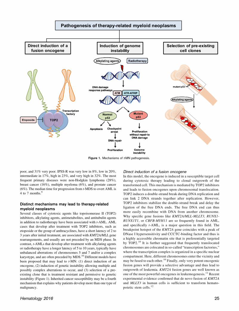
Distinct mechanisms may lead to therapy-relatedmyeloid neoplasmsSeveral classes of cytotoxic agents like topoisomerase II (TOP2)inhibitors, alkylating agents, antimetabolites, and antitubulin agentsin addition to radiotherapy have been associated with t-AML. AMLcases that develop after treatment with TOP2 inhibitors, such asetoposide or the group of anthracyclines, have a short latency of 2 to3 years after initial treatment, are associated with KMT2A/MLL generearrangements, and usually are not preceded by an MDS phase. Incontrast, t-AMLs that develop after treatment with alkylating agentsor radiotherapy have a longer latency of 5 to 10 years, typically haveunbalanced aberrations of chromosomes 5 and 7 and/or a complexkaryotype, and are often preceded by MDS.10 Different models havebeen proposed that may lead to t-MN: (1) direct induction of anoncogene, (2) induction of genetic instability allowing multiple andpossibly complex aberrations to occur, and (3) selection of a pre-existing clone that is treatment resistant and permissive to geneticinstability (Figure 1). Inherited cancer susceptibility may be a fourthmechanism that explains why patients develop more than one type ofmalignancy.
Direct induction of a fusion oncogeneIn this model, the oncogene is induced in a susceptible target cellduring cytotoxic therapy leading to clonal outgrowth of thetransformed cell. This mechanism is mediated by TOP2 inhibitorsand leads to fusion oncogenes upon chromosomal translocation.TOP2 induces a double-strand break during DNA replication andcan link 2 DNA strands together after replication. However,TOP2 inhibitors stabilize the double-strand break and delay theligation of the free DNA ends. The free DNA end can thusmore easily recombine with DNA from another chromosome.Why specific gene fusions like KMT2A/MLL-MLLT3, RUNX1-RUNX1T1, or CBFB-MYH11 are so frequently found in AML,and specifically t-AML, is a major question in this field. Thebreakpoint hotspot of the KMT2A gene coincides with a peak ofDNase I hypersensitivity and CCCTC-binding factor and thus isa highly accessible chromatin site that is preferentially targetedby TOP2.10 It is further suggested that frequently translocatedchromosomes are colocated in so-called “transcription factories,”where the transcription complex is organized in a specific nuclearcompartment. Here, different chromosomes enter the vicinity andmay be fused to each other.10 Finally, only very potent oncogenicfusion genes will provide a selective advantage and thus lead tooutgrowth of leukemia. KMT2A fusion genes are well known asone of the most powerful oncogenes in leukemogenesis.11 Recentexperimental evidence confirmed that de novo fusion of KMT2Aand MLLT3 in human cells is sufficient to transform hemato-poietic stem cells.12
Figure 1. Mechanisms of t-MN pathogenesis.
Hematology 2016 25
Induction of genetic instabilityAnother mechanism could be that cytotoxic therapy induces aber-rations, which lead to genetic instability and later to the acquisitionof leukemogenic aberrations (Figure 1). This might explain the longlatency of t-MNs and high frequency of complex cytogenetic ab-errations found in patients with previous alkylating chemotherapy orradiotherapy, but there are little experimental data to support thishypothesis. In support of increased genotoxicity of previous che-motherapy, Itzhar and colleagues evaluated copy number alterations(CNA) by array comparative genomic hybridization and found onaverage 3.46 CNAs in t-AML compared with 1.9 CNAs in de novoAML.13 However, Wong and colleagues found a similar number ofsingle nucleotide variants, indels, and transversions upon genome-wide sequencing in t-AML compared with de novo AML.14 Analternative mechanism has therefore been proposed, which suggestsselection of preexisting transformed and treatment-resistant hema-topoietic clones during chemotherapy.
Selection of preexisting hematopoietic cell clonesStrong evidence for this third mechanism of clonal selection in thepathogenesis of t-MN has been provided by Wong and colleagues.14
They found a significantly higher frequency of mutations in TP53and ABC transporters in t-AML and t-MDS compared with de novoAML, as also reported in other patient series.15,16 Most importantly,the specific TP53mutation was also found in hematopoietic cells 3 to6 years before the onset of t-MN at low frequency (0.003%-0.7%) in2 patients even before the application of chemotherapy. Moreover,in 9 of 19 older cancer-free individuals (68-89 years old), TP53mutations were found in peripheral blood with a low variantallele frequency of 0.01% to 0.37%. In a mouse model of bothwild-type and heterozygous Tp531/2 hematopoietic stem/progenitorcells, the Tp531/2 cells preferentially expanded after exposure tochemotherapy.14
Clonal hematopoiesis of indeterminate potential as a risk factorfor t-MN. This observation has been corroborated by recent datashowing an age-dependent increase of clonal hematopoiesis in healthyindividuals that is most likely driven by leukemia-associated mutationsin DNMT3A, TET2, ASXL1, JAK2, PPM1D, TP53, SF3B1, BCORL1,and others.17-19 Clonal hematopoiesis without cell dysplasia and blastincrease (ie, not fulfilling the criteria forMDS orAML) has been termedclonal hematopoiesis of indeterminate potential (CHIP).20 Individualswith CHIP have a 13-fold increased risk of developing a hematologicmalignancy, and the data by Wong et al suggest that this risk is in-creased in the context of cytotoxic therapy, at least if a TP53mutation ispresent.14 Interestingly, somatic mutations in PPM1D have been foundin CHIP and also in peripheral blood of patients with breast, ovarian,and lung cancer (in ~1% of patients).21-23 PPM1D is a serine/threoninephosphatase that negatively regulates p53.24 Truncating PPM1D mu-tations are considered gain-of-function mutations that suppress p53activity, impair the p53-dependent G1 checkpoint, and thus may lead tochemotherapy resistance and clonal outgrowth under chemotherapy.25
In patients with ovarian cancer, PPM1D mutations were not foundbefore treatment, but were present after treatment in 0.37% of casescompared with a frequency of 0.03% in controls.26 In another study ofovarian cancer, the frequency of somatic mosaic PPM1D mutations inperipheral blood mononuclear cells was significantly associated withprior chemotherapy, and the variant allele frequency increased in 85%of the patients during chemotherapy.22 In the same study, TP53 mu-tations developed in peripheral blood cells from 2 of 15 patients ofwhom sequential samples during chemotherapy were available.
In summary, recent evidence suggests that cells harboring somati-cally acquired preexisting mutations in the p53 pathway accumulateunder the selective pressure of chemotherapy and give rise to clonalhematopoiesis that may evolve into MDS or AML after additionalgenetic events (Figure 1). The incidence of CHIP under chemo-therapy and the rate of transformation should be studied in the future.
Inherited cancer susceptibilityA few years ago, the German AML Study Group (AMLSG) pub-lished data on the latency from diagnosis of the primary malignancyto t-AML.3 Seven percent of a cohort of 2835 patients with AMLdeveloped t-AML after chemotherapy and/or radiotherapy for theprimary malignancy, with a median latency of 4.04 years. However,another 3% developed AML after a diagnosis of an independentmalignancy that had never been treated with chemotherapy or ra-diotherapy. Compared with t-AML patients, these patients moreoften had prostate cancer (23% vs 9%), bladder cancer (9% vs 1%),and renal cell carcinoma (9% vs 2%), but less often had breast cancer(10% vs 52%).3 AML developed in these patients with no history ofchemotherapy or radiotherapy, with a median latency of 5 years,which is similar to that of patients with t-AML. Thus, a numberof t-AMLs may not be induced or selected by the previous treatmentbut may actually be caused by an inherited cancer susceptibility.Germline variants in the DNA damage response pathway have beenassociated with an increased risk of t-MN (genes like BRCA1,BRCA2, BARD1, TP53,27,28 RAD51, and HLX1,29 Fanconi genes,30
and the anti-apoptotic gene BCL2L10),31 supporting an effect ofcancer susceptibility to AML risk in some patients. Although thepatient numbers are small in the aforementioned studies, the avail-able data suggest that genetic cancer susceptibility contributes to therisk of myeloid neoplasms, especially under cytotoxic therapy.
Molecular genetics of t-MNIn our updated series of the German AML study group of 230patients with t-AML, 280 patients with secondary AML (sAML)after MDS and 3144 patients with de novo AMLwho were all treatedwith intensive chemotherapy, the most frequent cytogenetic ab-normalities were complex karyotype, monosomy 5 and 7, and ab-normalities of chromosome 17 (Figure 2), which are all associatedwith an unfavorable prognosis. These aberrations were most frequentin t-AML, less frequent in sAML, and least frequent in de novoAML. Comparison of the relative mutation frequencies in t-AML,sAML, and de novo AML showed that the fusion genes RUNX1-RUNXT1, CBFB-MYH11, and PML-RARA, which are all associatedwith a favorable prognosis, had a similar frequency in t-AML and denovo AML, but were less frequent in sAML. The frequency of AML-defining mutations in NPM1 and FLT3, which are associated witha favorable and unfavorable prognosis, respectively, were highestin de novo AML and similarly reduced in t-AML and sAML. TheKMT2A-MLLT3 fusion was more frequent in t-AML than in de novoor sAML (Figure 2).
Whole-genome and targeted-sequencing studies showed that TP53is the most frequently mutated gene in patients with t-MN.14-16
Approximately one third of t-AML and t-MDS patients present witha TP53mutation (Figure 3).14 Other genes with a mutation frequencyof.5% in t-AML includeNPM1, FLT3-ITD, ABC transporter genes,NRAS, KRAS and PTPN11, TET2, DNMT3A, IDH1 and IDH2,STAG2, RUNX1, and ASXL1. In t-MDS, RUNX1 and ASXL1 aremore often mutated compared with t-AML; ABC transporter genesand DNMT3A have a similar mutation frequency in t-MDS andt-AML, and the other genes are less frequently mutated in patients
26 American Society of Hematology

with t-MDS than in those with t-AML.14 Targeted sequencing withan extended gene panel did not identify any molecular aberration in9% of the patients (n 5 10). However, 8 of these 10 patients had atleast 1 cytogenetic aberration, leaving only a small proportion withouta detectable genetic abnormality.14 In summary, fusion genes withfavorable prognosis are found in ~15% of patients with t-AML, andcytogenetic aberrations with unfavorable prognosis including t(9;11)are detected in ~50% of patients with t-AML.3 Christiansen et al founda high frequency of mutations in the RAS/RAF pathway in patientswith t-AML (41%), but a rather low frequency in patients with t-MDS(8%).32 Interestingly, 3 of 5 patients with t(9;11) and the KMT2A-MLLT3 fusion had the BRAF V600E mutation.32
Prognosis and treatment of t-MNThe prognosis of therapy-related acute promyelocytic leukemia(t-APL) and core binding factor (CBF) leukemia (t(8;21) and inv(16)or t(16;16)) has been considered comparable with their cytogeneticde novo AML counterpart.33 t-AMLwith adverse cytogenetic risk hasas dismal an outcome as de novo AML with adverse cytogenetics.Therefore, it was suggested to treat patients with t-AML as those withde novo AML according to their cytogenetic risk profile.34,35 Severalfactors may complicate the treatment of patients with t-AML, such asorgan dysfunction from prior therapy, depletion of normal hema-topoietic stem cells, damage to marrow stroma, chronic immuno-suppression, and refractoriness to transfusion support.35 The prognosisand therapy of patients with t-AML will be discussed by geneticsubgroup in the following section (Figure 4).
Therapy-related acute promyelocytic leukemiaExcellent response rates, low relapse rates, and exceptional overallsurvival (OS) were recently reported for patients with APL treatedwith ATRA and arsenic trioxide.36,37 In a series of 29 t-APL patients,19 were treated with ATRA and arsenic trioxide and 10 were treatedwith ATRA and chemotherapy.38 The complete response (CR) ratein patients with t-APL treated with ATRA and arsenic was 89%compared with 70% in the ATRA and chemotherapy group (P5 not
significant). The CR rate in the ATRA and arsenic group wascomparable with a cohort of 85 patients with de novo APL (CR94%).38 Three-year OS was 65% in both groups (ATRA with arsenicand ATRA with chemotherapy groups). Therefore, ATRA and ar-senic trioxide should be considered the standard of care in low- andintermediate-risk APL patients, independent of the etiology, andhigh-risk t-APL patients should be treated according to the localstandard for high-risk de novo APL patients.
t-AML with t(8;21);RUNX1-RUNX1T1t-AML patients with t(8;21) have a high response rate after intensiveinduction therapy, but these patients have a shorter overall survival(OS) compared with de novo AML with t(8;21) in 2 of 3 studies.Comparison of the outcome of 13 t-AML patients with t(8;21) to 38de novo AML patients with t(8;21) by Gustafson et al revealed thatthe t-AML group achieved CR at similar frequency (91% vs 95%),but relapsed more often (70% vs 39%) and had decreased median OS(19 months vs.37 months).39 Krauth et al observed 2-year survivalrates of 46.8% for 16 t-AML and 76.4% for 95 de novo AML patientswith t(8;21).40 However, comparable outcomes were observed for 9t-AML and 128 de novo AML patients with t(8;21) in the AMLSGseries.3
t-AML patients with t(8;21) should be treated with standard in-duction and consolidation treatment, and response should be mon-itored by molecular minimal residual disease (MRD) assessment. Ifa positive MRD result indicates treatment failure, allogeneic he-matopoietic cell transplantation (HCT) should be discussed with the
Figure 3. Frequency of molecular aberrations in t-AML (n 5 52) andt-MDS (n5 59) patients based on data byWong et al.14 Samples from 22patients were sequenced by whole-genome sequencing and samplesfrom 89 patients were sequenced with a gene panel covering 149 genes.
Figure 2. Frequency of cytogenetic aberrations in t-AML, secondary, andde novo AML (AMLSG registry data).
Hematology 2016 27

patient. MRD studies for t-AML patients with t(8;21) have not beenpublished. However, for de novo AML patients with t(8;21) whoachieved CR, a ,3-log reduction of transcript levels after consol-idation 241 identified high-risk patients with OS after conventionalchemotherapy of only 27%, whereas allogeneic HCT increased OS to72% in these high-risk patients.41
In contrast, in low-risk patients with a .3-log reduction of tran-script levels after the second consolidation cycle had an OS of100% after conventional chemotherapy, and allogeneic HCTresulted in an OS of 76%.41 Because the risk of nonrelapse mor-tality after allogeneic HCT is high in t-AML, and a negative MRDresult indicates disease control by chemotherapy, allogeneic HCTshould be withheld in t-AML patients with negative MRD forRUNX1-RUNX1T1.
t-AML with inv16/t(16;16); CBFB/MYH11t-AML with inv(16) or t(16;16) had a high response rate but poorsurvival in 2 independent studies. Borthakur et al assessed theoutcome of 17 t-AML patients with CBF leukemia (13 with inv(16),4 with t(8;21)) and 171 de novo CBF AML patients.42 Although theCR rate was 92% in the total cohort and the relapse rate wascomparable in t-AML patients (33%) and de novo AML patients(36%), median OS was only 1.9 years in t-AML patients butlonger than 5 years in de novo AML patients. In the study byKayser et al, comparison of 15 t-AML patients with inv(16) or t(16;16) with 142 de novo AML patients with inv(16) or t(16;16)revealed t-AML as a significant adverse prognostic factor witha hazard ratio of 2.35.3
As for patients with t(8;21)-positive t-AML, t-AML patients with inv(16) or t(16;16) should be treated with standard induction andconsolidation therapy and monitored by MRD assessment. Allo-geneic HCT should be discussed with the patient if a positive MRDresult indicates treatment failure. A negative MRD result indicatesdisease control by chemotherapy and allogeneic HCT should bewithheld. In a study with 115 CBFB/MYH11 positive de novo andsecondary AML patients in complete remission, CBFB/MYH11copy numbers .10 in peripheral blood or .50 in bone marrow(per 105 ABL copies) after the end of consolidation were associated
with an estimated relapse of nearly 100% and an estimated 5-yearsurvival of 57% (if.10 copies in peripheral blood) and 25% (if.50copies in bone marrow).43 The efficacy of allogeneic HCT in theseMRD-positive patients has not been reported yet.
t-AML with t(9;11); KMT2A-MLLT3Adult patients with this translocation are specifically enriched in thegroup of t-AML patients and account for 11% of t-AML patients inthe series of the AMLSG.3 A meta-analysis based on individualpatient data of younger patients with 11q23 translocations evaluatedthe prognostic impact in 180 AML patients.44 Sixteen percent of thepatients had t-AML, and 42% of these patients had t(9;11). Seventy-one percent of the patients achieved CR and the median OS was19.6 months (4-year OS was 29%). Secondary AML (includingt-AML and AML after MDS) was an independent negative riskfactor for OS. In a donor/no donor analysis of 65 patients with t(9;11),of whom 51% had secondary or t-AML, patients with an availabledonor in first remission had improved OS (5-year OS without donor21%, with donor 51%).45 Based on these data, allogeneic HCTin first CR should be recommended for eligible t-AML patients witht(9;11).
t-AML with NPM1 mutationSpecific data for the prognostic impact of NPM1mutations in t-AMLare currently not available. Although the frequency of NPM1 mu-tations is lower in t-AML than in de novo AML, it is still one ofthe most frequent mutations in this AML cohort. NPM1-mutatedAML with normal cytogenetics and wild-type FLT3 belongs to theEuropean LeukemiaNet (ELN) favorable risk group. Within theGerman-Austrian study group, we treat these patients with intensiveinduction and consolidation therapy without a strict upper age limit.Because the treatment response can be monitored in the majority ofNPM1-mutated patients by MRD, this approach is also feasible fort-AML patients with mutant NPM1. The cutoff of.200 NPM1 copiesper 104 ABL copies after completion of consolidation therapy has beenestablished by the AMLSG as a strong risk factor for relapse.46 Iveyand colleagues suggested an alternative cutoff to discriminate patientsat high risk of relapse (ie, positive NPM1 MRD in peripheral bloodafter 2 cycles of intensive chemotherapy).47 Allogeneic HCT should beconsidered in MRD-positive patients after completion of consolidation.
Figure 4. Algorithm for the treatment of t-MN patients. *None of the mentioned treatments are currently approved for AML. alloHCT, allogeneichematopoietic stem cell transplantation; CAR, chimeric antigen receptor; CBF, core-binding factor leukemia (ie, AML with t(8;21), inv(16), or t(16;16));CR1, first complete remission; DA, daunorubicine and cytarabine; ITD, internal tandem duplication.
28 American Society of Hematology
In a retrospective analysis of the ALFA0702 trial presented at the ASHmeeting in 2015, allogeneic HCT significantly improved OS of NPM1mutated nonfavorable ELN patients with insufficient MRD reduction,but not in patients with .4-log MRD reduction.48 Patients withconcomitant FLT3-ITD and/or adverse cytogenetic aberrations shouldbe advised to undergo allogeneic HCT in first CR.
Other adult t-AML patientsAdult AML patients with ELN risk classification higher than fa-vorable have a poor outcome when treated with chemotherapy alone.It would be desirable to identify chemotherapy-responsive patientsfrom this large patient group and especially in t-AML patients. Withthis aim, Lindsley and colleagues defined a set of 8 genes (SRSF2,SF3B1, U2AF1, ZRSR2, ASXL1, EZH2, BCOR, or STAG2) thatidentified secondary AML patients with .95% specificity if at leastone of these genes was mutated.49 This algorithm was then applied toa cohort of 101 t-AML patients, of whom 33% harbored secondaryAML-type mutations, 23% had TP53 mutations, and 47% hadmutations common in de novo AML (like NPM1, FLT3, DNMT3A,etc.). t-AML patients with secondary-type mutations had similarpatient characteristics as de novo AML patients with secondary-typemutations, confirming that t-AML patients represent a heterogeneousgroup of patients, with one third being similar to AML cases thatevolved from MDS. Although CR rates were comparable betweenthe 3 genetically defined t-AML groups, patients with secondary-type and TP53 mutations significantly more often required 2 in-duction courses to achieve CR (55% of patients) compared withpatients with de novo AML mutations (7% of patients), suggestingrelative chemotherapy resistance. Whether this translates into higherrelapse rates and shorter survival remain to be shown.49 This mo-lecular classification refines the cytogenetic classification of t-AMLand should be further evaluated as a prognostic and predictive markerin t-AML patients.
Currently, all eligible t-AML patients with an ELN score higher thanfavorable should be considered as candidates for allogeneic HCT inCR1, if a suitable related or unrelated donor is available, and theirinclusion in clinical trials should be encouraged.
Is there a better induction regimen for t-AML patients than 713?This question has not been specifically addressed, but inductionoutcomes were reported for patients with unfavorable cytogeneticsthat are frequently found in t-AML patients. High-dose daunorubicinas used in the E1900 trial resulted in a median OS of 10.6 months inpatients with unfavorable cytogenetics, which was comparable withthe median OS of 10.2 months in the standard dose arm.50 Ina randomized trial of the Polish Adult Leukaemia Group, the additionof cladribine to daunorubicin and cytarabine resulted in significantlyhigher CR rates after 2 cycles of induction (67.5% vs 56%) andmedian 3-year OS of 36% vs 20% compared with daunorubicin andcytarabine in patients with unfavorable cytogenetics.51 Becausecladribine is not licensed for AML and has not been tested in a largenumber of t-AML patients, 713 induction should be consideredstandard in t-AML patients eligible for intensive chemotherapy.
Older AML patients who are unlikely to benefit fromstandard induction chemotherapyThese patients constitute the majority of AML patients and are verydifficult to treat. t-AML patients are on average older than de novoAML patients and are frequently not eligible for intensive treatment.These patients should be primarily treated in clinical trials. Outside of
trials, hypomethylating agents, low-dose cytarabine, and best sup-portive care are currently available treatment options. Several studiesreported the effect of hypomethylating agents in t-MDS and t-AMLpatients and found similar efficacy in t-MN patients compared withde novo MDS or AML patients.52-54 Overall response rates withazacitidine or decitabine were 38% to 42%, with a CR rate of 14% to21%, similar to the 45% overall response rate in de novo AMLpatients.52 In the study by Bally et al, 71% of t-MN patients hada complex karyotype compared with 43% in de novo AML. OS wasshorter in t-MN patients, with 14% at 2 years compared with 34% inde novo AML; however, multivariate analysis revealed that onlycytogenetics and age, but not etiology, were independent predictorsof survival.52
Low-dose cytarabine was compared with hydroxyurea in a ran-domized trial including 121 de novo AML, 53 sAML, and 28 MDSpatients.55 However, the number of sAML patients with t-AML wasnot specified. In the entire population, low-dose cytarabine resultedin a higher CR rate (18% vs 1%) and better OS (hazard ratio, 0.6;95% confidence interval, 0.44-0.81). A subgroup analysis revealedno benefit in patients with adverse cytogenetics, sAML, and MDS.
In summary, hypomethylating agents seem to have a similar activityin t-MN patients as in de novo AML and can be applied, if ap-propriate clinical trials are not available. Low-dose cytarabine maybe effective in some patients with t-AML, but no effect should beexpected in patients with adverse cytogenetics.
Treatment-related MDSIn the aforementioned large cooperative study of 1837 t-MDS pa-tients, the median OS was 16 months, and allogeneic HCT wasperformed in 16% of patients who had a median survival of24 months.8 The discriminatory power of the IPSS-R was inferior int-MDS compared with de novo MDS patients and a revised prog-nostic model was proposed.8 Another study compared the IPSS-Rscore in t-MN patients to de novo MDS patients and found that theIPSS-R score can distinguish the 5 prognostic subgroups, but OSwas shorter in t-MN patients than in de novo MDS patients, par-ticularly in the very-low-risk and low-risk groups.56 The mediansurvival in t-MDS patients with very-low-, low-, and intermediate-risk IPSS-R was 56.5, 21.7, and 15.8 months, respectively, whereasde novo MDS patients had median survival of 105.6, 63.6, and36 months.56 Thus, the survival of t-MDS patients with low-riskIPSS-R was shorter than the survival of de novo MDS patients withintermediate-risk IPSS-R. t-MDS patients should be treated inclinical trials whenever possible, and allogeneic HCT should beoffered to eligible patients with IPSS-R low or higher if a suitabledonor is available.
The question of whether MDS patients should be treated with cy-toreductive agents before allogeneic HCT was addressed in ret-rospective studies. MDS patients who received azacitidine beforetransplantation or who were transplanted up-front without cytor-eduction had similar outcomes after 3 years.57 Similarly, treatmentof MDS patients with azacitidine or intensive chemotherapy beforeallogeneic HCT resulted in comparable outcome after 3 years(OS 55% vs 48%, relapse 40% vs 37%, and nonrelapse mor-tality (NRM) 19% vs 20% for azacitidine vs intensive chemo-therapy, respectively).58 Therefore, allogeneic HCT should beplanned as quickly as possible. If a delay in transplantation isexpected, hypomethylating agents or intensive chemotherapymay be considered.58
Hematology 2016 29
Allogeneic HCT for t-MN patientsRisk factors for overall survivalAllogeneic HCT is often the only curative approach for t-MN pa-tients. Several studies reported the outcomes of t-MN patients afterallogeneic HCT, each including.250 patients.59-61 Overall survivalof t-MN patients at 3 to 5 years after allogeneic HCT was 22% to35%.60,61 Five adverse risk factors for OS were identified: age .35years, poor-risk cytogenetics, t-AML not in remission or advancedt-MDS, and donor other than an HLA-identical sibling or a partiallyor well-matched unrelated donor.61 Patients with none of these riskfactors had a predicted 5-year OS of 50%, whereas patients with 3risk factors had a predicted survival of 10% at 5 years.
High NRM in t-MN patients undergoing allogeneic HCTThe cumulative incidence of relapse at 3 to 5 years was 31% to 36%.These studies noted high NRM of 32% to 41% at 1 year and up to61% at 5 years. The analysis of the CIBMTR identified age .35years, a lower Karnofsky performance score, and t-MDS beforetransplantation as unfavorable risk factors for NRM.61 An obviousapproach to reduce the high NRM would be reduced-intensity con-ditioning. However, reduced-intensity conditioning resulted in similarNRM rates as myeloablative conditioning in the CIBMTR and EBMTseries.60,61 However, the study by Kroger et al compared NRM ratesbefore 1998 and since 1998 and found a significantly reduced NRM inmore recent years.60
When outcome of allogeneic HCT in t-MN patients was comparedwith de novo MDS/AML patients, no differences were noted forrelapse and NRM after adjustment for disease category, age, andcytogenetics.59 Relapse rates were similar within low- and high-riskcytogenetic groups (according to IPSS), whereas patients withintermediate-risk cytogenetics and t-MN had a higher relapse ratecompared with de novo MDS/AML patients.59 Armand and col-leagues compared the outcome after allogeneic HCT in 80 t-MDS/t-AML patients with 476 de novo MDS/AML patients who werestratified into 3 cytogenetic risk groups. Disease etiology did notaffect OS, incidence of relapse, or NRM, emphasizing that geneticrisk rather than previous treatment affects the outcome after allo-geneic HCT.62 Thus, allogeneic HCT is an important treatmentoption in t-MN patients, and age and cytogenetics are the mostimportant prognostic factors. Novel strategies are required to reduceNRM without compromising antileukemic activity.
Novel treatment approaches for t-MNTargeted therapies should be evaluated in t-MN patients as in denovo AML patients. Promising data have been reported for multi-kinase and FLT3 inhibitors (midostaurin, sorafenib, quizartinib) andfor IDH1 and IDH2 inhibitors. A large randomized phase 3 trial ofmidostaurin or placebo with intensive induction and consolidationtherapy was presented at the ASH meeting in 2015 and showeda significantly improved OS in midostaurin-treated de novo AMLpatients.63 Midostaurin use in t-AML patients should be studied inthe future. The DOT1L inhibitor EPZ-5676 inhibits the interaction ofDOT1L with KMT2A and therefore may be of specific relevance tot-AML with t(9;11). Initial results of a phase 1 clinical trial withEPZ-5676 presented at the ASH 2015 meeting indicated an ac-ceptable safety profile and some clinical activity as demonstrated bya marrow response and resolution of leukemia cutis.64
CPX-351 is a liposomal formulation of cytarabine and daunorubicinpackaged in liposomes at a 5:1 molar ratio, which improved survival
compared with the same drugs administered conventionally in an-imal models of leukemia.65
In a randomized phase 3 trial in newly diagnosed older secondaryAML patients including 20% t-AML patients, CPX-351 was com-pared with 713 induction.66 The CR/CRi rate was higher in theCPX-351 compared with the 713 cohort (47.7% vs 33.3%, P5 .016).OS was significantly improved with CPX-351, resulting in a medianOS of 9.6 vs 6 months in the 713 group (P 5 .005), and 60-daymortality was in favor of CPX-351. Thus, CPX-351 may becomea new treatment option in sAML and specifically in t-MN patients.
Because of the high incidence of mutations in the RAS pathway,inhibitors of RAS signaling are of special interest in t-MN patients.A recent phase 1/2 trial of trametinib, an inhibitor of mitogen-activated protein kinase 1 (MEK1) and MEK2, showed an overallresponse rate of 20% in relapsed/refractory AML and MDS patientswith NRAS or KRAS mutations, whereas only 3% of RAS wild-typepatients responded.67
t-MN with mutated TP53 or complex chromosome aberrations areunlikely to be cured with targeted agents or with chemotherapy,because the checkpoint for apoptosis and cell-cycle arrest is de-fective, thus allowing the aberrant cell to escape the selectivepressure of specific pathway inhibitors. Conceptually, an immuno-logic approach should be more successful because the immune attackis independent of genetic aberrations. It was suggested that cancercells with multiple aberrations are more immunogenic than cells withone or few aberrations, and thus novel immunologic therapies likechimeric antigen receptor T cells, bispecific antibodies, and checkpointinhibitors should be evaluated in t-MN patients with a high-riskgenetic profile.
Summary and outlookSignificant progress has been made in the understanding of thepathogenesis of therapy-related myeloid neoplasms. The direct in-duction of fusion oncogenes by epipodophyllotoxins is likely a resultof both stochastic and deterministic processes in the cell. In addition,strong evidence was recently obtained indicating that a significantnumber of t-MNs develop through clonal selection of preexisting,treatment-resistant clones, which exhibit a high frequency of p53pathway mutations (Figure 1).
Therapy-related acute promyelocytic leukemia has a high remissionrate with ATRA and arsenic trioxide therapy and should be treated asde novo APL. Patients with therapy-related CBF leukemias seem tohave a worse prognosis than their de novo AML counterparts.Nevertheless, we recommend initial intensive chemotherapy in thesepatients and the decision for allogeneic HCT should be based on theresults of MRD assessment (Figure 4). In most other patients, al-logeneic HCT in CR1 is the preferred treatment option for eligiblepatients, whereas treatment in clinical trials and with hypomethylatingagents are preferred options for the remaining or relapsed/refractorypatients.
Patients with t-MN often have complex cytogenetics and TP53mutations and we know that chemotherapy can hardly overcome theunderlying genetic defects. Therefore, it is hoped that novel im-munologic approaches can attack these aberrant cells and eventuallylead to improved treatment results in this underserved patientpopulation.
30 American Society of Hematology
AcknowledgmentsThe author thanks Arnold Ganser, Hartmut Dohner, Felicitas Thol,Nicolaus Kroger, and Michael Morgan for their review and helpfuldiscussions andMichelleMaria Araujo Cruz for her help with figure 1.
This review was supported by an ERC grant under the EuropeanUnion’s Horizon 2020 research and innovation program (No. 638035).M.H. is a Heisenberg chair of the DFG (HE 5240/6-1).
CorrespondenceMichael Heuser, Department of Hematology, Hemostasis, Oncol-ogy, and Stem Cell Transplantation, Hannover Medical School,Carl-Neuberg Strasse 1, 30625 Hannover, Germany; e-mail: [email protected].
References1. Arber DA, Orazi A, Hasserjian R, et al. The 2016 revision to the World
Health Organization classification of myeloid neoplasms and acuteleukemia. Blood. 2016;127(20):2391-2405; Advance online publication.
2. Arber DA, Vardiman JW, Brunning RD, et al. Acute myeloid leukaemiaand related precursor neoplasms. In: Swerdlow SH, Campo E, Harris NL,et al, eds. WHO Classification of Tumours of Haematopoietic andLymphoid Tissues, 4th ed. Geneva, Switzerland: WHO PRESS; 2008:109-148
3. Kayser S, Dohner K, Krauter J, et al; German-Austrian AMLSG. Theimpact of therapy-related acute myeloid leukemia (AML) on outcome in2853 adult patients with newly diagnosed AML. Blood. 2011;117(7):2137-2145.
4. Granfeldt Østgard LS, Medeiros BC, Sengeløv H, et al. Epidemiologyand Clinical Significance of Secondary and Therapy-Related AcuteMyeloid Leukemia: A National Population-Based Cohort Study. J ClinOncol. 2015;33(31):3641-3649.
5. Hulegardh E, Nilsson C, Lazarevic V, et al. Characterization andprognostic features of secondary acute myeloid leukemia in a population-based setting: a report from the Swedish Acute Leukemia Registry. Am JHematol. 2015;90(3):208-214.
6. Morton LM, Dores GM, Tucker MA, et al. Evolving risk of therapy-related acute myeloid leukemia following cancer chemotherapy amongadults in the United States, 1975-2008. Blood. 2013;121(15):2996-3004.
7. Eichenauer DA, Thielen I, Haverkamp H, et al. Therapy-related acutemyeloid leukemia and myelodysplastic syndromes in patients withHodgkin lymphoma: a report from the German Hodgkin Study Group.Blood. 2014;123(11):1658-1664.
8. Kuendgen A, Tuechler H, Nomdedeu M, et al. An analysis of prognostickmarkers and the performance of scoring systems in 1837 patients withtherapy-related myelodysplastic syndrome—a study of the InternationalWorking Group (IWG-PM) for Myelodysplastic Syndromes (MDS).ASH Annual Meeting. 2015;2015; Abstract 609.
9. Godley L, Larson RA. The Syndrome of Therapy-Related Myelodys-plasia and Myeloid Leukemia. In: Bennett JM, ed. The MyelodysplasticSyndromes. New York, Basel: Marcel Dekker Inc.; 2002:139-176
10. Cowell IG, Austin CA. Mechanism of generation of therapy relatedleukemia in response to anti-topoisomerase II agents. Int J Environ ResPublic Health. 2012;9(6):2075-2091.
11. Barabe F, Kennedy JA, Hope KJ, Dick JE. Modeling the initiation andprogression of human acute leukemia in mice. Science. 2007;316(5824):600-604.
12. Reimer J, Knoess S, Labuhn M, Charpentier EM, Klusmann J-H, HecklD. Crispr-Cas9 induced MLL-rearrangements cause clonal outgrowth inCD341 hematopoietic stem cells. ASH Annual Meeting. 2015;2015;Abstract 165.
13. Itzhar N, Dessen P, Toujani S, et al. Chromosomal minimal criticalregions in therapy-related leukemia appear different from those of denovo leukemia by high-resolution aCGH. PLoS One. 2011;6(2):e16623.
14. Wong TN, Ramsingh G, Young AL, et al. Role of TP53 mutations in theorigin and evolution of therapy-related acute myeloid leukaemia. Nature.2015;518(7540):552-555.
15. Shih AH, Chung SS, Dolezal EK, et al. Mutational analysis of therapy-related myelodysplastic syndromes and acute myelogenous leukemia.Haematologica. 2013;98(6):908-912.
16. Ok CY, Patel KP, Garcia-Manero G, et al. Mutational profiling oftherapy-related myelodysplastic syndromes and acute myeloid leukemiaby next generation sequencing, a comparison with de novo diseases. LeukRes. 2015;39(3):348-354.
17. Jaiswal S, Fontanillas P, Flannick J, et al. Age-related clonal hemato-poiesis associated with adverse outcomes. N Engl J Med. 2014;371(26):2488-2498.
18. Genovese G, Kahler AK, Handsaker RE, et al. Clonal hematopoiesis andblood-cancer risk inferred from blood DNA sequence. N Engl J Med.2014;371(26):2477-2487.
19. Xie M, Lu C, Wang J, et al. Age-related mutations associated with clonalhematopoietic expansion and malignancies. Nat Med. 2014;20(12):1472-1478.
20. Steensma DP, Bejar R, Jaiswal S, et al. Clonal hematopoiesis of in-determinate potential and its distinction from myelodysplastic syn-dromes. Blood. 2015;126(1):9-16.
21. Ruark E, Snape K, Humburg P, et al; Breast and Ovarian Cancer Sus-ceptibility Collaboration; Wellcome Trust Case Control Consortium.Mosaic PPM1D mutations are associated with predisposition to breastand ovarian cancer. Nature. 2013;493(7432):406-410.
22. Swisher EM, Harrell MI, Norquist BM, et al. Somatic Mosaic Mutationsin PPM1D and TP53 in the Blood of Women With Ovarian Carcinoma.JAMA Oncol. 2016;2(3):370-372.
23. Zajkowicz A, Butkiewicz D, Drosik A, Giglok M, Suwinski R, Rusin M.Truncating mutations of PPM1D are found in blood DNA samples oflung cancer patients. Br J Cancer. 2015;112(6):1114-1120.
24. Le Guezennec X, Bulavin DV. WIP1 phosphatase at the crossroads ofcancer and aging. Trends Biochem Sci. 2010;35(2):109-114.
25. Kleiblova P, Shaltiel IA, Benada J, et al. Gain-of-function mutations ofPPM1D/Wip1 impair the p53-dependent G1 checkpoint. J Cell Biol.2013;201(4):511-521.
26. Pharoah PD, Song H, Dicks E, et al; Australian Ovarian Cancer StudyGroup; Ovarian Cancer Association Consortium. PPM1D MosaicTruncating Variants in Ovarian Cancer CasesMay Be Treatment-RelatedSomatic Mutations. J Natl Cancer Inst. 2016;108(3):108.
27. Schulz E, Valentin A, Ulz P, et al. Germline mutations in the DNAdamage response genes BRCA1, BRCA2, BARD1 and TP53 in patientswith therapy related myeloid neoplasms. J Med Genet. 2012;49(7):422-428.
28. Churpek JE, Marquez R, Neistadt B, et al. Inherited mutations in cancersusceptibility genes are common among survivors of breast cancer whodevelop therapy-related leukemia. Cancer. 2016;122(2):304-311.
29. Jawad M, Seedhouse CH, Russell N, Plumb M. Polymorphisms inhuman homeobox HLX1 and DNA repair RAD51 genes increase therisk of therapy-related acute myeloid leukemia. Blood. 2006;108(12):3916-3918.
30. Voso MT, Fabiani E, Zang Z, et al. Fanconi anemia gene variants intherapy-related myeloid neoplasms. Blood Cancer J. 2015;5:e323.
31. Fabiani E, Fianchi L, Falconi G, et al. The BCL2L10 Leu21Arg variantand risk of therapy-related myeloid neoplasms and de novo myelo-dysplastic syndromes. Leuk Lymphoma. 2014;55(7):1538-1543.
32. Christiansen DH, Andersen MK, Desta F, Pedersen-Bjergaard J. Mu-tations of genes in the receptor tyrosine kinase (RTK)/RAS-BRAF signaltransduction pathway in therapy-related myelodysplasia and acute my-eloid leukemia. Leukemia. 2005;19(12):2232-2240.
33. Klimek VM, Tray NJ. Therapy-related myeloid neoplasms: what’s ina name? Curr Opin Hematol. 2016;23(2):161-166.
34. Churpek JE, Larson RA. The evolving challenge of therapy-relatedmyeloid neoplasms. Best Pract Res Clin Haematol. 2013;26(4):309-317.
35. Dohner H, Estey EH, Amadori S, et al; European LeukemiaNet. Di-agnosis and management of acute myeloid leukemia in adults:
Hematology 2016 31
recommendations from an international expert panel, on behalf of theEuropean LeukemiaNet. Blood. 2010;115(3):453-474.
36. Lo-Coco F, Avvisati G, Vignetti M, et al; Gruppo Italiano MalattieEmatologiche dell’Adulto; German-Austrian Acute Myeloid LeukemiaStudy Group; Study Alliance Leukemia. Retinoic acid and arsenic tri-oxide for acute promyelocytic leukemia. N Engl J Med. 2013;369(2):111-121.
37. Burnett AK, Russell NH, Hills RK, et al; UK National Cancer ResearchInstitute Acute Myeloid Leukaemia Working Group. Arsenic trioxideand all-trans retinoic acid treatment for acute promyelocytic leukaemiain all risk groups (AML17): results of a randomised, controlled, phase 3trial. Lancet Oncol. 2015;16(13):1295-1305.
38. Dayyani F, Kantarjian H, O’Brien S, et al. Outcome of therapy-relatedacute promyelocytic leukemia with or without arsenic trioxide asa component of frontline therapy. Cancer. 2011;117(1):110-115.
39. Gustafson SA, Lin P, Chen SS, et al. Therapy-related acute myeloidleukemia with t(8;21) (q22;q22) shares many features with de novo acutemyeloid leukemia with t(8;21)(q22;q22) but does not have a favorableoutcome. Am J Clin Pathol. 2009;131(5):647-655.
40. Krauth MT, Eder C, Alpermann T, et al. High number of additionalgenetic lesions in acute myeloid leukemia with t(8;21)/RUNX1-RUNX1T1: frequency and impact on clinical outcome. Leukemia.2014;28(7):1449-1458.
41. Zhu HH, Zhang XH, Qin YZ, et al. MRD-directed risk stratificationtreatment may improve outcomes of t(8;21) AML in the first completeremission: results from the AML05 multicenter trial. Blood. 2013;121(20):4056-4062.
42. Borthakur G, Lin E, Jain N, et al. Survival is poorer in patients withsecondary core-binding factor acute myelogenous leukemia comparedwith de novo core-binding factor leukemia. Cancer. 2009;115(14):3217-3221.
43. Yin JA, O’Brien MA, Hills RK, Daly SB, Wheatley K, Burnett AK.Minimal residual disease monitoring by quantitative RT-PCR in corebinding factor AML allows risk stratification and predicts relapse: resultsof the United Kingdom MRC AML-15 trial. Blood. 2012;120(14):2826-2835.
44. Krauter J, Wagner K, Schafer I, et al. Prognostic factors in adult patientsup to 60 years old with acute myeloid leukemia and translocations ofchromosome band 11q23: individual patient data-based meta-analysis ofthe German Acute Myeloid Leukemia Intergroup. J Clin Oncol. 2009;27(18):3000-3006.
45. Chen Y, Kantarjian H, Pierce S, et al. Prognostic significance of 11q23aberrations in adult acute myeloid leukemia and the role of allogeneicstem cell transplantation. Leukemia. 2013;27(4):836-842.
46. Kronke J, Schlenk RF, Jensen KO, et al. Monitoring of minimal residualdisease in NPM1-mutated acute myeloid leukemia: a study from theGerman-Austrian acute myeloid leukemia study group. J Clin Oncol.2011;29(19):2709-2716.
47. Ivey A, Hills RK, Simpson MA, et al; UK National Cancer ResearchInstitute AMLWorkingGroup. Assessment ofMinimal Residual Diseasein Standard-Risk AML. N Engl J Med. 2016;374(5):422-433.
48. Balsat M, Renneville A, Thomas X, et al. NPM1minimal residual diseaseas prognostic and predictive factor in young adults with acute myeloidleukemia: a study by the French ALFA Group. Blood. 2015:126:2581.
49. Lindsley RC, Mar BG, Mazzola E, et al. Acute myeloid leukemia on-togeny is defined by distinct somatic mutations. Blood. 2015;125(9):1367-1376.
50. Luskin MR, Lee JW, Fernandez HF, et al. Benefit of high-dose dau-norubicin in AML induction extends across cytogenetic and moleculargroups. Blood. 2016;127(12):1551-1558.
51. Holowiecki J, Grosicki S, Giebel S, et al. Cladribine, but not fludarabine,added to daunorubicin and cytarabine during induction prolongs survivalof patients with acute myeloid leukemia: a multicenter, randomized phaseIII study. J Clin Oncol. 2012;30(20):2441-2448.
52. Bally C, Thepot S, Quesnel B, et al. Azacitidine in the treatment oftherapy related myelodysplastic syndrome and acute myeloid leukemia
(tMDS/AML): a report on 54 patients by the Groupe Francophone DesMyelodysplasies (GFM). Leuk Res. 2013;37(6):637-640.
53. Fianchi L, Criscuolo M, Lunghi M, et al. Outcome of therapy-relatedmyeloid neoplasms treated with azacitidine. J Hematol Oncol. 2012;5:44.
54. Klimek VM, Dolezal EK, Tees MT, et al. Efficacy of hypomethylatingagents in therapy-related myelodysplastic syndromes. Leuk Res. 2012;36(9):1093-1097.
55. Burnett AK, Milligan D, Prentice AG, et al. A comparison of low-dosecytarabine and hydroxyurea with or without all-trans retinoic acid foracute myeloid leukemia and high-risk myelodysplastic syndrome inpatients not considered fit for intensive treatment. Cancer. 2007;109(6):1114-1124.
56. Ok CY, Hasserjian RP, Fox PS, et al. Application of the internationalprognostic scoring system-revised in therapy-related myelodysplasticsyndromes and oligoblastic acute myeloid leukemia. Leukemia. 2014;28(1):185-189.
57. Damaj G, Mohty M, Robin M, et al. Upfront allogeneic stem celltransplantation after reduced-intensity/nonmyeloablative conditioningfor patients with myelodysplastic syndrome: a study by the SocieteFrançaise de Greffe de Moelle et de Therapie Cellulaire. Biol BloodMarrow Transplant. 2014;20(9):1349-1355.
58. Damaj G, Duhamel A, Robin M, et al. Impact of azacitidine beforeallogeneic stem-cell transplantation for myelodysplastic syndromes:a study by the Societe Française de Greffe de Moelle et de Therapie-Cellulaire and the Groupe-Francophone des Myelodysplasies. J ClinOncol. 2012;30(36):4533-4540.
59. Chang C, Storer BE, Scott BL, et al. Hematopoietic cell transplantation inpatients with myelodysplastic syndrome or acute myeloid leukemiaarising from myelodysplastic syndrome: similar outcomes in patientswith de novo disease and disease following prior therapy or antecedenthematologic disorders. Blood. 2007;110(4):1379-1387.
60. Kroger N, Brand R, van Biezen A, et al; Myelodysplastic SyndromesSubcommittee of The Chronic Leukaemia Working Party of EuropeanGroup for Blood and Marrow Transplantation (EBMT). Risk factors fortherapy-related myelodysplastic syndrome and acute myeloid leukemiatreated with allogeneic stem cell transplantation. Haematologica. 2009;94(4):542-549.
61. Litzow MR, Tarima S, Perez WS, et al. Allogeneic transplantation fortherapy-related myelodysplastic syndrome and acute myeloid leukemia.Blood. 2010;115(9):1850-1857.
62. Armand P, Kim HT, DeAngelo DJ, et al. Impact of cytogenetics onoutcome of de novo and therapy-related AML and MDS after allogeneictransplantation. Biol Blood Marrow Transplant. 2007;13(6):655-664.
63. Stone RM, Mandrekar S, Sanford BL, et al. The multi-kinase inhibitormidostaurin prolongs survival sompared with placebo in combinationwith daunorubicin/cytarabine induction, high-dose C consolidation, and asmaintenance therapy in newly diagnosed acute myeloid leukemia (AML)patients age 18-60 with FLT3 mutations: an international prospectiverandomized placebo-controlled double-blind trial (CALGB 10603/RATIFY [Alliance]). ASH Annual Meeting. 2015;2015. Abstract 6.
64. Stein EM, Garcia-Manero G, Rizzieri DA, et al. A phase 1 study of theDOT1L inhibitor, pinometostat (EPZ-5676) in adults with relapsed orrefractory leukemia: safety. clinical activity, exposure and target in-hibition [abstract]. Blood. 2015;126(23). Abstract 2547.
65. Tardi P, Johnstone S, Harasym N, et al. In vivo maintenance of syn-ergistic cytarabine:daunorubicin ratios greatly enhances therapeutic ef-ficacy. Leuk Res. 2009;33(1):129-139.
66. Lancet JE, Uy GL, Cortes JE, et al. Final results of a phase III randomizedtrial of CPX-351 versus 713 in older patients with newly diagnosed highrisk (secondary) AML. ASCO Annual Meeting. 2016: Abstract 7000.
67. Borthakur G, Popplewell L, Boyiadzis M, et al. Activity of the oralmitogen-activated protein kinase kinase inhibitor trametinib in RAS-mutant relapsed or refractory myeloid malignancies. Cancer. 2016;122(12):1871-1879; Advance online publication.
32 American Society of Hematology


![Therapy-related Myeloid Neoplasms Following …Radiation therapy & t-MN • Have modern RT techniques [mega-voltage linear accelerators; intensity-modulated radiation therapy (IMRT)]](https://img.dokumen.tips/doc/110x75/5ed204196731c53a5734c2cd/therapy-related-myeloid-neoplasms-following-radiation-therapy-t-mn-a-have.jpg)