Embed Size (px)
Citation preview
Therapeutic Effects of 15 Hz Pulsed ElectromagneticField on Diabetic Peripheral Neuropathy inStreptozotocin-Treated RatsTao Lei1., Da Jing1., Kangning Xie1., Maogang Jiang1, Feijiang Li1, Jing Cai2, Xiaoming Wu1, Chi Tang1,
Qiaoling Xu3, Juan Liu1, Wei Guo1, Guanghao Shen1*, Erping Luo1*
1 School of Biomedical Engineering, Fourth Military Medical University, Xi’an, China, 2Department of Neurology, Xijing Hospital, Fourth Military Medical University, Xi’an,
China, 3 School of Nursing, Fourth Military Medical University, Xi’an, China
Abstract
Although numerous clinical studies have reported that pulsed electromagnetic fields (PEMF) have a neuroprotective role inpatients with diabetic peripheral neuropathy (DPN), the application of PEMF for clinic is still controversial. The present studywas designed to investigate whether PEMF has therapeutic potential in relieving peripheral neuropathic symptoms instreptozotocin (STZ)-induced diabetic rats. Adult male Sprague–Dawley rats were randomly divided into three weight-matched groups (eight in each group): the non-diabetic control group (Control), diabetes mellitus with 15 Hz PEMFexposure group (DM+PEMF) which were subjected to daily 8-h PEMF exposure for 7 weeks and diabetes mellitus with shamPEMF exposure group (DM). Signs and symptoms of DPN in STZ-treated rats were investigated by using behavioral assays.Meanwhile, ultrastructural examination and immunohistochemical study for vascular endothelial growth factor (VEGF) ofsciatic nerve were also performed. During a 7-week experimental observation, we found that PEMF stimulation did not alterhyperglycemia and weight loss in STZ-treated rats with DPN. However, PEMF stimulation attenuated the development ofthe abnormalities observed in STZ-treated rats with DPN, which were demonstrated by increased hind paw withdrawalthreshold to mechanical and thermal stimuli, slighter demyelination and axon enlargement and less VEGF immunostainingof sciatic nerve compared to those of the DM group. The current study demonstrates that treatment with PEMF mightprevent the development of abnormalities observed in animal models for DPN. It is suggested that PEMF might have directcorrective effects on injured nerves and would be a potentially promising non-invasive therapeutic tool for the treatment ofDPN.
Citation: Lei T, Jing D, Xie K, Jiang M, Li F, et al. (2013) Therapeutic Effects of 15 Hz Pulsed Electromagnetic Field on Diabetic Peripheral Neuropathy inStreptozotocin-Treated Rats. PLoS ONE 8(4): e61414. doi:10.1371/journal.pone.0061414
Editor: Maria Rosaria Scarfi, National Research Council, Italy
Received December 11, 2012; Accepted March 8, 2013; Published April 18, 2013
Copyright: � 2013 Lei et al. This is an open-access article distributed under the terms of the Creative Commons Attribution License, which permits unrestricteduse, distribution, and reproduction in any medium, provided the original author and source are credited.
Funding: This work was supported by grants from the National Natural Science Foundation of China (Grant no.31000491, 51077128, 31000381 and 31270889).The funders had no role in study design, data collection and analysis, decision to publish, or preparation of the manuscript.
Competing Interests: The authors have declared that no competing interests exist.
* E-mail: [email protected] (GS); [email protected] (EL)
. These authors contributed equally to this work.
Introduction
Diabetic peripheral neuropathy (DPN) is generally considered
to be one of the most common complications of diabetes
mellitus, affecting both types of diabetes equally [1–3]. Studies
suggest that about 30% of patients with diabetes mellitus are
affected by DPN and 16–26% of diabetic patients experience
chronic pain [4].
DPN is characterized by aberrant symptoms of stimulus-evoked
pain including allodynia and hyperalgesia [5], and it often leads to
mood and sleep disturbance, and thus can substantially impair the
quality and expectancy of life [6,7]. Therefore, it imposes a huge
burden on both individuals and society, and represents a major
public health problem. However, beyond the careful management
of the diabetes itself via glycemic control and pain relief for
neuropathy, no treatment for DPN exists [8,9]. Potential toxicity,
poor tolerability and ineffectiveness for some percent of diabetic
patients are major disadvantages of the current therapeutic
options. For this reason, there is a need to explore other non-
pharmacological novel therapeutic modalities with efficacy and
safety, particularly when diabetic patients require a combined
treatment with an oral antidiabetic drug to prevent the de-
velopment of DPN.
Numerous clinical studies have reported that pulsed electro-
magnetic fields (PEMF) are able to modify some parameters of
nerve function in diabetic patients [10,11], and a voluminous
amount of literature has suggested that PEMF can stimulate nerve
growth, regeneration, and functional recovery of nerves in cells
in vitro or in animal models with nerve disease [12–16]. However,
the application of PEMF for clinic is still controversial [17].
Therefore, more research is needed to confirm the therapeutic
effects of PEMF on DPN and then to justify the applicability of
PEMF for clinical practice. Since few studies have examined the
effects of PEMF on neuropathy induced by diabetes mellitus in
animals at present, this study aimed to test whether PEMF has
therapeutic potential in relieving diabetes-induced neuropathy in
animals.
PLOS ONE | www.plosone.org 1 April 2013 | Volume 8 | Issue 4 | e61414
Streptozotocin (STZ)-induced diabetic rat model has been used
extensively as a model of DPN to demonstrate many abnormalities
observed in patients with DPN and to assess the efficacies of
potential therapeutic interventions [18–20]. Diabetic rats develop
tactile allodynia and hyperalgesia to mechanical or thermal stimuli
in the hind paws two or three weeks after STZ injection [5,21,22].
In the current study, we examined the effects of whole-body
exposure to 15 Hz PEMF whose peak magnetic flux density
(MFD) was approximately 1.661023 T on improving signs and
symptoms of DPN in STZ-treated rats by using behavioral assays.
The PEFM was generated by a modified Helmholtz coils and the
exposure duration was 8 hours everyday, 6 days a week for 7
weeks. Meanwhile, ultrastructural examination and immunohis-
tochemical study for vascular endothelial growth factor (VEGF) of
sciatic nerve were also performed seven weeks after PEMF
stimulation. Moreover, the potential action mechanism of PEMF
on DPN was preliminarily investigated.
Methods
Experimental DiabetesThirty adult male Sprague–Dawley rats, weighting 350620 g,
were provided by Animal Center of the Fourth Military Medical
University and housed in a room (Animal Center of the Fourth
Military Medical University, Xi’an, China) with controlled
temperature (2361uC), relative humidity (50,60%), and alter-
nately light-dark cycle (12 h/12 h), with access to standard pellet
and clean water. Diabetes mellitus was induced by an in-
traperitoneal injection of STZ (Sigma Chemicals, St. Louis,
MO, USA) at 45 mg/kg in freshly prepared 0.1 mM citrate buffer
(pH 4.5) after an overnight fast [23]. Confirmation of hypergly-
cemia was made three days after STZ injection, and only STZ-
treated rats whose glucose concentration of the tail venous blood
measured by OneTouch SureStep Plus glucometer (Lifescan,
Milpitas, CA, USA) was higher than 20 mM were considered as
qualified diabetic models [24]. Six rats were excluded from the
study after confirmation of success of diabetic models because of
low blood glucose levels. The rest of rats were randomized into
three weight-matched groups (eight in each group): the non-
diabetic control group (Control), diabetes mellitus with sham
PEMF exposure group (DM), diabetes mellitus with PEMF
exposure group (DM+PEMF) which were subjected to whole-
body exposure to PEMF 8 hours (09:00–17:00) everyday, 6 days
a week for 7 weeks. Although the same PEMF apparatus was
employed in DM group, the PEMF stimulation was not activated.
PEMF stimulation was carried out the next day after confirmation
of hyperglycemia. The current study was performed in adherence
to the National Institutes of Health guidelines for the use of
experimental animals, and all animal protocols were approved by
the Committee for Ethical Use of Experimental Animals of the
Fourth Military Medical University.
PEMF ApparatusThree identical coils with coil diameters of 800 mm constituted
the PEMF stimulation apparatus (the modified Helmholtz coils).
The coils were in series connection and placed coaxially with
a distance of 304 mm apart (Fig. 1A). Each coil was made up of
enameled coated copper wire with 0.8 mm diameter. The
assembly of three identical coils significantly upgraded the
uniformity of MFD by decreasing the deviation of the MFD
between the central reference point (origin, center of the middle
coil) and other areas in the magnetic field [25].
The MFD value along the O–X direction (axial direction of the
coil, the coordinate system meets right-hand rule) is expressed as:
B xð Þ~ m0NIR2
2R2z azxð Þ2h i{3=2
(
z R2z a{xð Þ2h i{3=2
zk R2zx2� �{3=2
):
Where m0 is the permeability of vacuum, I is the current
through the coils, R is the radius of the coils, a is the distance
between the central coil and the outside coil, x is the abscissa
relative to origin, N is the number of turns of the outside coil,
and k6N is the number of turns of the middle coil. By setting the
parameters a=0.7601R and k=0.5315, the second and fourth
derivative of B(x) will become zero at the position of origin and
then the maximum uniformity of the MFD will be obtained [25].
In the present study, we set the number of turns of the two
outside coils as 500. Therefore, the number of turns of the
central coil has been determined as 266. Besides, the distance
between the central coil and the outside coil was approximately
304 mm. The modified Helmholtz coils were wired to a pulse
generator (GHY-III, FMMU, Xi’an, China; China Patent
no.ZL02224739.4) which produced a PEMF signal (Fig. 1A).
The open-circuit voltage waveform of the PEFM consisted of
a pulsed burst (burst width, 5 ms; burst wait, 60 ms; pulse width,
0.2 ms; pulse wait, 0.02 ms; pulse rise and fall time: 0.3 ms,2.0 ms ) repeated at 15 Hz (Fig. 1B). The reason for selecting this
particular waveform was that it had been proven to be effective
in diabetes-induced diseases in our previous studies which were
performed by our study group over a long period of time [26–
30].
Two cubic plastic rat cages containing rats in PEFM group
were put in the center of every two neighboring coils (the length
of the cage was along O-Y direction) and cages were supported
by stands to let the activities of rats restrict on the XY plane
which had higher intensity and better uniformity of MFD
(Fig. 1A). Moreover, whole body exposure to PEMF for rats
was applied eight hours everyday. The distribution of the peak
MFD was measured by using a Gaussmeter (Model 455 DMP
Gaussmeter, Lake Shore Cryotronics, USA), and the measure-
ment result was (1.660.1)61023 T (average 6 standard
deviation) in the exposure area (cage: 50 cm long, 20 cm wide
and 15 cm high).
A small resistor of 2 V was placed in series with the modified
Helmholtz coils. The voltage drop across the resistor was observed
with an oscilloscope (Agilent 6000 Series, Agilent Technologies,
Inc., Santa Clara, CA). Peak value of voltage drop was observed to
calculate the peak value of current in the coils so as to obtain the
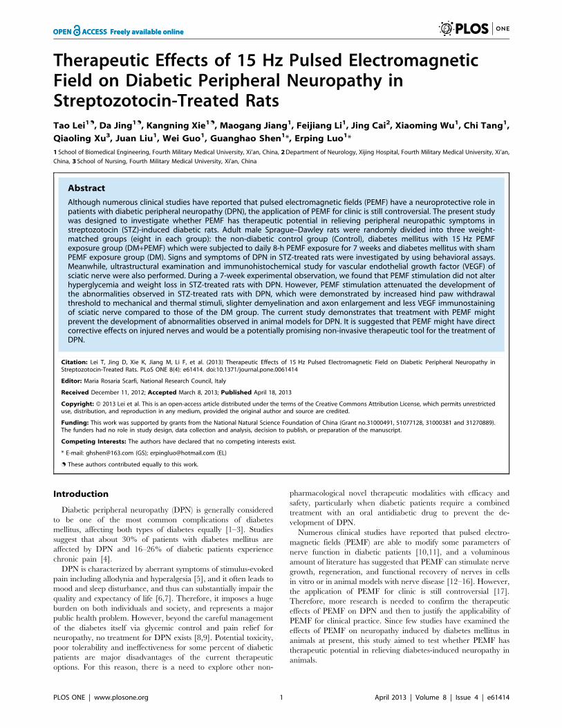
peak value of MFD. In order to make the distribution of MFD in
the modified Helmholtz coils more intuitive, the finite element
engineering software called COMSOL Multiphysics (v4.3 COM-
SOL AB, Burlington, MA, USA) was applied to simulate the three
dimensional distribution of the MFD in the modified Helmholtz
coils when the current in the coils reached peak value (approx-
imately 1.5A). A physics-controlled mesh setting whose element
size is coarse was employed to avoid memory overflow caused by
too many mesh elements. By establishing the geometric model,
setting boundary conditions, meshing models and obtaining
numerical solutions, the distributions of MFD of modified
Helmholtz coils are shown in Fig. 2. We can find that the MFD
on rats’ behavior plane (XY plane) was uniform and the peak
MFD was about 1.661023 T which approximately coincided with
the practical measurement result ((1.660.1)61023 T).
Electromagnetic Field on Diabetic Neuropathy
PLOS ONE | www.plosone.org 2 April 2013 | Volume 8 | Issue 4 | e61414

Evaluation of Mechanical AllodyniaTactile allodynia was assessed by measuring the hind paw
withdrawal threshold to the application of a calibrated series of 6
von Frey filaments (bending forces of 2, 4, 6, 8, 10 and 15 g)
(Stoelting, Wood Dale, IL, USA) using a modification of the up-
down method [31]. Rats were placed in acrylic cages with a wire
grid oor and allowed to sit in a quiet room for 30 min before
beginning the tests. Starting with the filament that has the lowest
Figure 1. Schematic drawing of PEMF exposure system and PEMF pulse protocol. (A) Modified Helmholtz coils consisted of three identicalcoils with diameters of 800 mm which were in series connection and mounted coaxially at a distance of 304 mm apart. Two cubic plastic rat cageswhose length was along O–Y direction (Origin is the center of the middle coil, O–X direction is the axial direction of the coil and the coordinatesystem meets right-hand rule) were put in the center of every two neighboring coils and cages were supported by stands to let the activities of ratsrestrict on the XY plane. The modified Helmholtz coils were wired to the GHY-III pulse generator. (B) The pulse stimulator (GHY-III) generated anopen-circuit voltage waveform of PEMF with a repetitive burst frequency at 15 Hz (burst width, 5 ms; burst wait, 60 ms; pulse width, 0.2 ms; pulsewait, 0.02 ms; pulse rise and fall time: 0.3 ms, 2.0 ms).doi:10.1371/journal.pone.0061414.g001
Electromagnetic Field on Diabetic Neuropathy
PLOS ONE | www.plosone.org 3 April 2013 | Volume 8 | Issue 4 | e61414

force (2 g), the filament was applied perpendicularly to the mid-
plantar surface of hind paw with sufficient force to cause the
filament to buckle slightly. Brisk withdrawal or hind paw flinching
was considered as the positive response. Each filament was applied
five times to each hind paw (for 6–8 s per stimulation, with
a stimulus interval of 1–2 min). Minimum recording of five
positive responses (50%) out of 10 stimulations for both paws was
considered to be the mechanical withdrawal threshold (MWT) (in
grams). Absence of a response (less than five withdrawals)
prompted use of the next graded filament. The cut-off of a 15 g
filament was selected as the upper limit for testing, since stiffer
filaments tended to raise the entire limb rather than to buckle,
substantially changing the nature of the stimulus. A significant
decrease in the threshold of hind paw withdrawal in response to
the mechanical stimulus was interpreted as indicating the presence
of mechanical allodynia as compared to the baseline threshold.
Evaluation of Thermal HyperalgesiaThe thermal stimulation system (Commat, Ankara, Turkey)
consisting of a clear plastic chamber (10620624 cm3) that sits on
a clear smooth glass oor was used to assess thermal hyperalgesia by
measuring nociceptive thermal threshold. Before beginning the
test, rats were placed individually in the chamber and allowed
approximately 30 min to acclimate to the testing environment. A
radiant heat source (8 V, 50 W halogen bulb) mounted on
a movable holder below a glass pane was positioned to deliver
a thermal stimulus to the mid-plantar region of the hind paw. The
intensity of the heat stimulus was maintained constant throughout
all experiments. When the rat feels pain and withdraws its paw,
a photocell detects interruption of a light beam reection, the
infrared generator is automatically switched off, and the timer
stops, determining the withdrawal threshold. Each rat was
unilaterally tested three times at 3-min interval. The average of
the three measurements was taken as thermal withdrawal
threshold (TWT). In order to avoid excessive suffering of rats,
the thermal source was automatically discontinued after 25 s (cut-
off latency) if the rat fails to withdraw its paw. A significant
decrease in the latency of hind paw withdrawal in response to the
noxious thermal stimulus was interpreted as indicating the
presence of thermal hyperalgesia as compared to the baseline
latency.
Time Course for Measurements of Weight, BloodGlucose, Mechanical Allodynia and Thermal HyperalgesiaThe weights, blood glucose levels and responses to mechanical
and thermal stimuli of all rats were evaluated prior to STZ
administration, and there were no statistically significant differ-
ences for these parameters among three groups. Weights and
blood glucose levels of all rats were regularly measured in the
Friday morning (9:00–11:00) in weeks 0, 1, 3, 5 and 7 after PEMF
stimulation. Mechanical allodynia and thermal hyperalgesia were
evaluated at the Friday night (19:00–23:00) in weeks 0, 1, 3, 5 and
7 after PEMF stimulation. For these two tests, measurements were
done by two experimenters who were not aware of the treatment
groups respectively and responsible for each test until the study
finished.
Ultrastructural Examination of Sciatic NerveSeven weeks after PEMF stimulation in STZ-treated rats with
DPN, all rats were anesthetized by an intraperitoneal injection of
7% chloral hydrate solution (0.45 ml/100 g) prior to collecting the
sciatic nerve [32]. The distal part of the sciatic nerve was dissected
and post fixed by immersion in the fixative solution (2%
Figure 2. The three dimensional distribution of peak MFD atXY plane (the activity plane of rats) of modified Helmholtzcoils and two dimensional distribution of peak MFD on O–Xand O–Y cut line of XY plane when the current in the coilsreached peak value (approximately 1.5 A). (A) Three dimensionaldistribution of peak MFD at XY plane of modified Helmholtz coils whosehomogeneous color (blue) indicates the peak MFD at XY plane wasuniform and approximately 1.661023 T and red arrows indicates theinstantaneous direction of MFD. (B) Two dimensional distribution ofpeak MFD on O–X cut line of XY plane whose major parts at the activityplane of rats was uniform and approximately 1.661023 T. (C) Twodimensional distribution of peak MFD on O–Y cut line of XY planewhose major parts at the activity plane of rats was uniform andapproximately 1.661023 T.doi:10.1371/journal.pone.0061414.g002
Electromagnetic Field on Diabetic Neuropathy
PLOS ONE | www.plosone.org 4 April 2013 | Volume 8 | Issue 4 | e61414

paraformaldehyde, 2% glutaraldehyde, 0.1 M cacodylate buffer at
pH 7.3) for 2 h at 4uC, and washed in 0.1 M cacodylate buffer,
and osmicated for 4 h in 1% OsO4 (Fluka). Nerves were rinsed in
0.1 M cacodylate buffer, dehydrated and embedded in epoxy 812-
Araldite (Polysciences). Ultra-thin sections (80 nm) were sub-
sequently cut, collected on cellodincoated single slot grids and
stained with uranyl acetate and lead citrate. Photographs were
obtained using a transmission electron microscope (JEM-2000EX,
Japan) operated at 80 keV.
Immunohistochemical Study for VEGF of Sciatic NerveThe distal part of the sciatic nerve was dissected and post fixed
by immersion in the fixative solution (10% paraformaldehyde),
and routine paraffin embedding was performed. Longitudinal
sections (12 mm) of sciatic nerve were thaw-mounted onto
Superfrost Plus Slides (VWR). Sections were washed with PBS,
and then incubated in blocking buffer (10% normal goat serum,
0.2% Triton-X 100 in PBS) for 1 h at room temperature. Slides
were incubated overnight at 4uC with primary antibodies (in
blocking buffer): anti–VEGF (1:100; EMD Millipore, USA). Slides
were washed and then incubated with goat anti-rabbit IgG (1:200;
Jackson ImmunoResearch, USA) for 1 h at room temperature.
Digital images were acquired using light microscope (ECLIP-
SE50i, Nikon, Japan).
Statistical AnalysisStatistical analyses were carried out using SPSS (version 14.0,
SPSS, IL, USA). All values were expressed as means 6 standard
error of the mean (SEM). P,0.05 was considered statistically
significant. Data sets (body weight, blood glucose level and
mechanical and thermal withdrawal threshold) of time course
study were analyzed by two-way repeated measures analysis of
variance (ANOVA). All results were interpreted using the Green-
hous–Geisser correction to reduce the probability of obtaining
a significant result by chance alone. Between subject factors
consisted of intervention (Control, DM and DM+PEMF) and
within subject factors consisted of time (weeks 0, 1, 3, 5 and 7 after
PEMF stimulation) resulted in a 365 ANOVA. Data was analyzed
for intervention and time main effects. Bonferroni-adjusted
pairwise comparisons were performed for multiple comparisons
of the means between the groups. PEMF effect would be indicated
by a significant main effect for intervention.
Results
Body Weight and Whole Blood Glucose LevelTwo-way repeated measures ANOVA with a Greenhouse-
Geisser correction determined that a significant main effect for
time (F (2.109, 75.936) = 9.502, P,0.001) was found for means of
body weight throughout the time course. The body weight differed
significantly between time points. Post hoc tests using the
Bonferroni correction revealed that the mean body weight of
DM group and DM+PEMF group were significantly lower than in
the Control group (P,0.01) (Fig. 3). After STZ injection, rats
consistently lost weight. Although there was slightly less loss of the
body weight in PEMF treated diabetic rats, no significant
difference between DM+PEMF group and DM group was found
(P.0.05). PEMF stimulation did not significantly affect the loss of
body weight caused by diabetes.
Similarly, a significant main effect for time (F (1.841,
38.651) = 92.331, P,0.001) was observed for average blood
glucose level throughout the time course. The average blood
glucose level differed significantly between time points. Bonfer-
roni-adjusted pairwise comparisons revealed that the average
blood glucose levels of DM group and DM+PEMF group were
significantly higher than in the Control group (P,0.01) (Fig. 4).
STZ administration caused a rapid elevation of average blood
glucose levels (.500 mg/dl) within one week, which persisted for
up to 7 weeks. Although the blood glucose levels were slightly
lower in PEMF treated diabetic rats, there was no significant
difference between DM+PEMF group and DM group (P.0.05).
PEMF stimulation did not significantly affect the hyperglycemia
caused by diabetes.
Effects of PEMF on Mechanical AllodyniaA significant main effect for time (F (2.975, 62.470) = 176.065,
P,0.001) was observed for MWT throughout the time course.
MWT differed significantly between time points. Bonferroni-
adjusted pairwise comparisons revealed that the MWT of DM
group and DM+PEMF group were significantly lower than in the
Control group (P,0.01) (Fig. 5). There was a significant difference
between DM+PEMF group and DM group (P,0.05) (Fig. 5).
PEMF stimulation significantly prevented the development of
hypersensitivity to mechanical stimulus in diabetic rats (Fig. 5).
Figure 3. Trends of body weight in Control, DM and DM+PEMFgroups in weeks 0, 1, 3, 5 and 7 after PEMF stimulation. Data arepresented as means 6 SEM for 8 rats in each group. **P,0.01,statistically significant compared to the Control group (Bonferroni-adjusted pairwise comparison regarding the main group effect aftertwo-way repeated measures ANOVA).doi:10.1371/journal.pone.0061414.g003
Figure 4. Trends of blood glucose levels in Control, DM andDM+PEMF groups in weeks 0, 1, 3, 5 and 7 after PEMFstimulation. Data are presented as means 6 SEM for 8 rats in eachgroup. **P,0.01, statistically significant compared to the Control group(Bonferroni-adjusted pairwise comparison regarding the main groupeffect after two-way repeated measures ANOVA).doi:10.1371/journal.pone.0061414.g004
Electromagnetic Field on Diabetic Neuropathy
PLOS ONE | www.plosone.org 5 April 2013 | Volume 8 | Issue 4 | e61414

Effects of PEMF on Thermal HyperalgesiaA significant main effect for time (F (2.564, 53.840) = 56.742,
P,0.001) was observed for TWT. TWT differed significantly
between time points. Bonferroni-adjusted pairwise comparisons
revealed that the TWT of DM group and DM+PEMF group were
significantly lower than in the Control group (P,0.01) (Fig. 6).
There was a significant difference between DM+PEMF group and
DM group (P,0.05) (Fig. 6). PEMF stimulation significantly
prevented the development of hypersensitivity to noxious thermal
stimulus in diabetic rats (Fig. 6).
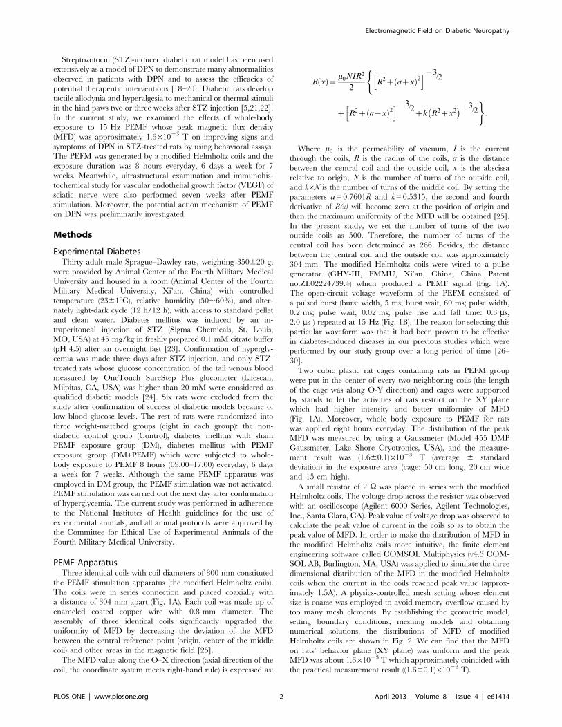
Electron Microscopy of Sciatic NerveUltrastructural examination of sciatic nerve was obtained by
using transmission electron microscopy after a 7-week experimen-
tal period in all rats. In Control group, myelinated fiber with
normal structure and morphology was observed (Fig. 7A). In DM
group, some evidences of axonal degeneration such as de-
myelination and axon enlargement were observed. Myelin sheath
showed infolding, splitting, swelling and deformation, and layers
were separated or disappeared (Fig. 7B). In DM+PEMF group,
myelin sheath of sciatic nerve was abnormal, the densities of layer
on myelin sheath were uneven and rarefaction, but the damage
was slighter than in the DM group (Fig. 7C). Seven-week exposure
to PEMF stimulation partially prevented the development of
axonal degeneration in STZ-treated rats with DPN.
Immunostaining for VEGF in Sciatic NerveAfter a 7-week experimental period, no VEGF immunostaining
in sciatic nerve was seen in Control group (Fig. 8A). In contrast,
diabetic rats with DPN showed intense VEGF immunostaining in
sciatic nerve (Fig. 8B). Diabetic animals treated with PEMF
stimulation showed less VEGF immunostaining intensity in sciatic
nerve compared to that of the DM group (Fig. 8C).
Discussion
Our data in the present study support the hypothesis that PEMF
might play a therapeutic role in the development of DPN in STZ-
treated rats. Efficacy was evaluated by the assessment of
hypersensitivity using behavioral assays of neuropathic pain that
included hind paw withdrawal threshold to non-noxious mechan-
ical stimuli (mechanical allodynia) and thermal hind paw
withdrawal latency to noxious heat stimuli (thermal hyperalgesia).
In our experiments, 3 weeks after the STZ injection, rats with
diabetes developed mechanical allodynia and thermal hyperalge-
sia, which is consistent with previous studies which demonstrated
that DPN often comes alone with altered sensitivity by producing
both allodynia and hyperalgesia both in STZ-induced diabetic
animal models and diabetic patients [33–35]. Our results also
revealed that application of PEMF attenuated the development of
painful DPN. PEMF stimulation showed protective effects to non-
noxious mechanical stimuli and noxious heat stimuli and caused
an increase in hind paw withdrawal threshold to mechanical
stimuli and response time to thermal pain compared to the
diabetic rats with sham PEMF stimulation. Our findings are
similar to a previous investigation which asserted that treatment
with PEMF may prevent the development or may reverse the
abnormalities observed in animal models for painful DPN [36].
Although different types of PEMF were employed by these two
studies, the same anti-neuropathic pain efficacy emerged.
In the current investigation, a marked decrease in body weight
of diabetic rats was observed on week 3 as compared to non-
diabetic control rats. The reduction in body weight is probably
related to the osmotic diuresis and dehydration induced by
diabetic hyperglycemia [36,37]. Meanwhile, blood glucose level
rose immediately after the STZ injection, reached quite a high
level at first week, and then remained approximately at a stable
value. The results of the current study have confirmed previous
findings that blood glucose level is elevated and body weight is
decreased in diabetic rats after STZ administration [38,39]. Our
results also revealed that PEMF stimulation did not significantly
prevent the weight loss caused by diabetes, which is consistent to
a previous investigation [36]. However, contrary to the findings
researched by Mert et al. [36], who observed that PEMF had
efficacy in anti-hyperglycemia in diabetic rats, we found that the
application of PEMF did not significantly alter hyperglycemia in
diabetic rats during the whole experimental observation (7 weeks).
This finding is consistent with the fact that the hematoxylin and
Figure 5. Trends of MWT in Control, DM and DM+PEMF groupsin weeks 0, 1, 3, 5 and 7 after PEMF stimulation. Data arepresented as means 6 SEM for 8 rats in each group. **P,0.01,statistically significant compared to the Control group, #P,0.05,statistically significant compared to the DM group (Bonferroni-adjustedpairwise comparison regarding the main group effect after two-wayrepeated measures ANOVA).doi:10.1371/journal.pone.0061414.g005
Figure 6. Trends of TWT in Control, DM and DM+PEMF groupsin weeks 0, 1, 3, 5 and 7 after PEMF stimulation. Data arepresented as means 6 SEM for 8 rats in each group. **P,0.01,statistically significant compared to the Control group, #P,0.05,statistically significant compared to the DM group (Bonferroni-adjustedpairwise comparison regarding the main group effect after two-wayrepeated measures ANOVA).doi:10.1371/journal.pone.0061414.g006
Electromagnetic Field on Diabetic Neuropathy
PLOS ONE | www.plosone.org 6 April 2013 | Volume 8 | Issue 4 | e61414

eosin staining for pancreatic islets in diabetic rats with PEMF
stimulation and sham PEMF stimulation showed similar atrophy
and reduction in cell numbers (not illustrated) in the current study.
The different effects of PEMF in hyperglycemia might be ascribed
to the different types of PEMF adopted by Mert et al. and us.
These inconsistent findings concerning PEMF effects on DPN
often come from varying stimulation parameters and exposure
durations [40].
Obviously, a prerequisite for a clear understanding of the
pathophysiological mechanisms of neuropathic appearance and
treatment is to know if there are definite structural changes in the
nerve fibers and to what extent they exist in the patients or
experimental animal models. The pathology of DPN is charac-
terized by progressive nerve fiber loss [41]. Our morphological
analysis was performed on the sciatic nerve because the common
type of DPN associated with diabetes in humans is the loss of the
distal region of long and large-diameter axons. In the present
study, some evidences of axonal degeneration such as demyelin-
ation and axon enlargement were observed in STZ-treated rats
with DPN. Similar results were also reported by other investigators
[42,43]. The sciatic nerve degeneration associated with morpho-
logic changes was confirmed by hyperalgesia and allodynia in
diabetic rats with neuropathy in our study, which is consistent with
the findings that pathological changes in diabetic rats with DPN
are characteristically associated with altered pain sensitivity [44].
In addition to this, our morphological study of sciatic nerve
revealed that long-term PEMF stimulation partially attenuated the
development of axonal degeneration observed in STZ-treated rats
with DPN, which appears to be seldom reported in animal models
for DPN by other investigators.
Our findings demonstrate that diabetic rats with DPN express
VEGF in peripheral nerves such as sciatic nerve, while adult and
healthy rats did not express the VEGF. Similar findings were also
revealed by a previous study [24]. Since it is known that
angiogenesis takes primarily place in metabolically altered or in
injured peripheral nerves and VEGF has demonstrated neuro-
trophic functions in both central and peripheral neurons [45–47],
it is not surprising to find elevated levels of the most potent
vascular growth substance in peripheral nerves of diabetic rats
with DPN. Intriguingly, on the one hand, direct neuroprotective
role for VEGF comes from both in vitro and in vivo studies [8],
but on the other hand, a potential consequence of high levels of
VEGF observed in diabetes will be enhanced vascular permeabil-
ity which often results in the extravasation of plasma protein as
well as the formation of lesions in peripheral nerves. This
abnormal angiogenesis caused by up-regulated VEGF expression
initiates chronic insidious progressive damage and loss in un-
myelinated and myelinated peripheral nerve fibers [48]. The fact
that sciatic nerve of diabetic rats with DPN over seven weeks’
PEMF stimulation showed less VEGF immunostaining might
indicate that restitution of nerve function induced by PEMF
stimulation leads to down-regulation for VEGF, what’s more, the
down-regulated VEGF might in turn cause less damage to
peripheral nerve fibers.
This present experimental study demonstrated that treatment
with PEMF may attenuate the development of abnormalities
observed in animal models for DPN. However, the underlying
mechanism of PEMF on DPN is still ambiguous. Previous study
reported that PEMF had an significant anti-hyperglycemia efficacy
in diabetic rats, and this PEMF-induced reduction in blood
glucose level could have a positive effect on nerve function that
may result in diminished pain intensity [36]. However, the
significant anti-hyperglycemia efficacy of PEMF stimulation was
not observed in STZ-treated rats with DPN during the whole
experimental observation (7 weeks) in our study. Therefore, we
hypothesized that long-term PEMF stimulation would have direct
corrective effects on injured nerves, which might lead to
diminished pain intensity observed in present studies. Moreover,
our hypothesis is supported by in vitro and in vivo studies which
Figure 7. Electron micrographs of sciatic nerves in Control, DM,and DM+PEMF groups after a 7-week experimental period inall rats (magnification:66000). (A) Control group: Myelinated fiberhad normal structure and morphology. Myelin sheath was in integrityand lined up in order. (B) DM group: Demyelination and axonenlargement were observed. Myelin sheath showed infolding, splitting,swelling and deformation, and layers were separated or disappeared.(C) DM+PEMF group: Myelin sheath of sciatic nerve was abnormal, thedensities of layer on myelin sheath were uneven and rarefaction, butthe damage was slighter than in the DM group.doi:10.1371/journal.pone.0061414.g007
Electromagnetic Field on Diabetic Neuropathy
PLOS ONE | www.plosone.org 7 April 2013 | Volume 8 | Issue 4 | e61414

have indicated that PEMF stimulation can accelerate nerve
conduction velocity and increase compound action potentials of
sciatic nerve, enhance nerve growth factor levels, and reduce both
oxidative damage and neuronal loss [13,15,16].
In summary, the results from our present study demonstrate
that treatment with PEMF might prevent the development of
abnormalities observed in animal models for DPN. Moreover, it is
suggested that PEMF might have direct corrective effects on
injured nerves and would be a potentially promising non-invasive
therapeutic tool for the treatment of DPN. However, further
research is required to elucidate the specific mechanisms of PEMF
on DPN and to confirm the applicability of PEMF for clinical
practice.
Acknowledgments
The authors would like to thank M. Ye (Department of Physiology, Xijing
hospital, Fourth Military Medical University), H. Dong (Department of
Pathology, Fourth Military Medical University) and X. Huang (De-
partment of Immunology, Fourth Military Medical University) for their
excellent technical assistance.
Author Contributions
Conceived and designed the experiments: DJ GS EL TL KX. Performed
the experiments: TL MJ FL JC. Analyzed the data: TL MJ. Contributed
reagents/materials/analysis tools: XW CT QX JL WG. Wrote the paper:
TL KX.
References
1. Imperatore G, Knowler WC, Pettitt DJ, Kobes S, Bennett PH, et al. (2000)
Segregation analysis of diabetic nephropathy in Pima Indians. Diabetes 49:
1049–1056.
2. Raine AE (1993) Epidemiology, development and treatment of end-stage renal
failure in type 2 (non-insulin-dependent) diabetic patients in Europe.
Diabetologia 36: 1099–1104.
3. Sima AA, Kamiya H (2006) Diabetic neuropathy differs in type 1 and type 2
diabetes. Ann N Y Acad Sci 1084: 235–249.
4. Jensen TS, Backonja MM, Hernandez JS, Tesfaye S, Valensi P, et al. (2006)
New perspectives on the management of diabetic peripheral neuropathic pain.
Diab Vasc Dis Res 3: 108–119.
5. Rondon LJ, Privat AM, Daulhac L, Davin N, Mazur A, et al. (2010) Magnesium
attenuates chronic hypersensitivity and spinal cord NMDA receptor phosphor-
ylation in a rat model of diabetic neuropathic pain. J Physiol 588: 4205–4215.
6. Sator-Katzenschlager SM, Schiesser AW, Kozek-Langenecker SA, Benetka G,
Langer G, et al. (2003) Does pain relief improve pain behavior and mood in
chronic pain patients? Anesth Analg 97: 791–797.
7. Schmader KE (2002) Epidemiology and impact on quality of life of postherpetic
neuralgia and painful diabetic neuropathy. Clin J Pain 18: 350–354.
8. Price SA, Dent C, Duran-Jimenez B, Liang Y, Zhang L, et al. (2006) Gene
transfer of an engineered transcription factor promoting expression of VEGF-A
protects against experimental diabetic neuropathy. Diabetes 55: 1847–1854.
9. Ziegler D (2008) Treatment of diabetic neuropathy and neuropathic pain: how
far have we come? Diabetes Care 31 Suppl 2: S255–261.
10. Szymborska-Kajanek A, Strzelczyk JK, Karasek D, Rawwash HA, Biniszkiewicz
T, et al. (2010) Impact of low-frequency pulsed magnetic fields on defensin and
CRP concentrations in patients with painful diabetic polyneuropathy and in
healthy subjects. Electromagn Biol Med 29: 19–25.
11. Wrobel MP, Szymborska-Kajanek A, Wystrychowski G, Biniszkiewicz T,
Sieron-Stoltny K, et al. (2008) Impact of low frequency pulsed magnetic fields
on pain intensity, quality of life and sleep disturbances in patients with painful
diabetic polyneuropathy. Diabetes Metab 34: 349–354.
12. Walker JL, Evans JM, Resig P, Guarnieri S, Meade P, et al. (1994) Enhancement
of functional recovery following a crush lesion to the rat sciatic nerve by
exposure to pulsed electromagnetic fields. Exp Neurol 125: 302–305.
13. Mert T, Gunay I, Gocmen C, Kaya M, Polat S (2006) Regenerative effects of
pulsed magnetic field on injured peripheral nerves. Altern Ther Health Med 12:
42–49.
14. Macias MY, Battocletti JH, Sutton CH, Pintar FA, Maiman DJ (2000) Directed
and enhanced neurite growth with pulsed magnetic field stimulation.
Bioelectromagnetics 21: 272–286.
15. Kim S, Im WS, Kang L, Lee ST, Chu K, et al. (2008) The application of
magnets directs the orientation of neurite outgrowth in cultured human neuronal
cells. J Neurosci Methods 174: 91–96.
16. Tasset I, Medina FJ, Jimena I, Aguera E, Gascon F, et al. (2012)
Neuroprotective effects of extremely low-frequency electromagnetic fields on
a Huntington’s disease rat model: effects on neurotrophic factors and neuronal
density. Neuroscience 209: 54–63.
17. Bril V, England J, Franklin GM, Backonja M, Cohen J, et al. (2011) Evidence-
based guideline: Treatment of painful diabetic neuropathy: report of the
American Academy of Neurology, the American Association of Neuromuscular
and Electrodiagnostic Medicine, and the American Academy of Physical
Medicine and Rehabilitation. Neurology 76: 1758–1765.
18. Kim H, Sasaki T, Maeda K, Koya D, Kashiwagi A, et al. (2003) Protein kinase
Cbeta selective inhibitor LY333531 attenuates diabetic hyperalgesia through
ameliorating cGMP level of dorsal root ganglion neurons. Diabetes 52: 2102–
2109.
19. Jolivalt CG, Jiang Y, Freshwater JD, Bartoszyk GD, Calcutt NA (2006)
Dynorphin A, kappa opioid receptors and the antinociceptive efficacy of
asimadoline in streptozotocin-induced diabetic rats. Diabetologia 49: 2775–
2785.
20. Cameron NE, Tuck Z, McCabe L, Cotter MA (2001) Effect of the hydroxyl
radical scavenger, dimethylthiourea, on peripheral nerve tissue perfusion,
conduction velocity and nociception in experimental diabetes. Diabetologia
44: 1161–1169.
21. Berti-Mattera LN, Larkin B, Hourmouzis Z, Kern TS, Siegel RE (2011) NF-
kappaB subunits are differentially distributed in cells of lumbar dorsal root
ganglia in naive and diabetic rats. Neurosci Lett 490: 41–45.
22. Yamamoto H, Shimoshige Y, Yamaji T, Murai N, Aoki T, et al. (2009)
Pharmacological characterization of standard analgesics on mechanical
allodynia in streptozotocin-induced diabetic rats. Neuropharmacology 57:
403–408.
Figure 8. Immunohistochemical staining for VEGF in sciatic nerves in Control, DM and DM+PEMF groups after a 7-weekexperimental period in all rats (magnification: 6200). (A) Control group: No VEGF immunostaining in sciatic nerve. (B) DM group: IntenseVEGF immunostaining in sciatic nerve. (C) DM+PEMF group: Less VEGF immunostaining intensity in sciatic nerve compared to that of the DM group.doi:10.1371/journal.pone.0061414.g008
Electromagnetic Field on Diabetic Neuropathy
PLOS ONE | www.plosone.org 8 April 2013 | Volume 8 | Issue 4 | e61414

23. Stevens MJ, Li F, Drel VR, Abatan OI, Kim H, et al. (2007) Nicotinamide
reverses neurological and neurovascular deficits in streptozotocin diabetic rats.J Pharmacol Exp Ther 320: 458–464.
24. Samii A, Unger J, Lange W (1999) Vascular endothelial growth factor expression
in peripheral nerves and dorsal root ganglia in diabetic neuropathy in rats.Neurosci Lett 262: 159–162.
25. Wang J, She S, Zhang S (2001) An improved Helmholtz coil and analysis of itsmagnetic field homogeneity. Rev Sci Instrum 20: 6–9.
26. Jing D, Cai J, Shen G, Huang J, Li F, et al. (2011) The preventive effects of
pulsed electromagnetic fields on diabetic bone loss in streptozotocin-treated rats.Osteoporos Int 22: 1885–1895.
27. Jing D, Shen G, Huang J, Xie K, Cai J, et al. (2010) Circadian rhythm affects thepreventive role of pulsed electromagnetic fields on ovariectomy-induced
osteoporosis in rats. Bone 46: 487–495.28. Li C, Liu Z, Zhang R, Luo E, Shen G, et al. (2008) Therapeutical effect of pulse
electricmagnetic field on postmenopausal osteoporosis. Chin J Osteoporos 14:
52–54.29. Luo E, Shen G, Xie K, Wu X, Xu Q, et al. (2007) Alimentary hyperlipemia of
rabbits is affected by exposure to low-intensity pulsed magnetic fields.Bioelectromagnetics 28: 608–614.
30. Yu L, Luo E, Han L (2004) Preventive effect of low intensity pulse
electromagnetic fields on osteoporosis of ovariectomized rats. Chin J ClinRehabil 8: 3590–3591.
31. Chaplan SR, Bach FW, Pogrel JW, Chung JM, Yaksh TL (1994) Quantitativeassessment of tactile allodynia in the rat paw. J Neurosci Methods 53: 55–63.
32. Chen RJ, Lin CC, Ju MS (2010) In situ transverse elasticity and blood perfusionchange of sciatic nerves in normal and diabetic rats. Clin Biomech (Bristol,
Avon) 25: 409–414.
33. Calcutt NA (2004) Experimental models of painful diabetic neuropathy. J NeurolSci 220: 137–139.
34. Fox A, Eastwood C, Gentry C, Manning D, Urban L (1999) Critical evaluationof the streptozotocin model of painful diabetic neuropathy in the rat. Pain 81:
307–316.
35. Thomas PK (1997) Classification, differential diagnosis, and staging of diabeticperipheral neuropathy. Diabetes 46 Suppl 2: S54–57.
36. Mert T, Gunay I, Ocal I (2010) Neurobiological effects of pulsed magnetic fieldon diabetes-induced neuropathy. Bioelectromagnetics 31: 39–47.
37. Malcangio M, Tomlinson DR (1998) A pharmacologic analysis of mechanical
hyperalgesia in streptozotocin/diabetic rats. Pain 76: 151–157.
38. Hoybergs YM, Biermans RL, Meert TF (2008) The impact of bodyweight and
body condition on behavioral testing for painful diabetic neuropathy in the
streptozotocin rat model. Neurosci Lett 436: 13–18.
39. Wuarin-Bierman L, Zahnd GR, Kaufmann F, Burcklen L, Adler J (1987)
Hyperalgesia in spontaneous and experimental animal models of diabetic
neuropathy. Diabetologia 30: 653–658.
40. Pieber K, Herceg M, Paternostro-Sluga T (2010) Electrotherapy for the
treatment of painful diabetic peripheral neuropathy: a review. J Rehabil Med 42:
289–295.
41. Calcutt NA, Backonja MM (2007) Pathogenesis of pain in peripheral diabetic
neuropathy. Curr Diab Rep 7: 429–434.
42. Liu GS, Shi JY, Lai CL, Hong YR, Shin SJ, et al. (2009) Peripheral gene transfer
of glial cell-derived neurotrophic factor ameliorates neuropathic deficits in
diabetic rats. Hum Gene Ther 20: 715–727.
43. Tan AM, Samad OA, Fischer TZ, Zhao P, Persson AK, et al. (2012)
Maladaptive dendritic spine remodeling contributes to diabetic neuropathic
pain. J Neurosci 32: 6795–6807.
44. Renno WM, Saleh F, Klepacek I, Al-Awadi F (2006) Talin immunogold density
increases in sciatic nerve of diabetic rats after nerve growth factor treatment.
Medicina (Kaunas) 42: 147–163.
45. Sondell M, Lundborg G, Kanje M (1999) Vascular endothelial growth factor
stimulates Schwann cell invasion and neovascularization of acellular nerve
grafts. Brain Res 846: 219–228.
46. Sondell M, Lundborg G, Kanje M (1999) Vascular endothelial growth factor has
neurotrophic activity and stimulates axonal outgrowth, enhancing cell survival
and Schwann cell proliferation in the peripheral nervous system. J Neurosci 19:
5731–5740.
47. Sondell M, Sundler F, Kanje M (2000) Vascular endothelial growth factor is
a neurotrophic factor which stimulates axonal outgrowth through the flk-1
receptor. Eur J Neurosci 12: 4243–4254.
48. Stevens MJ, Obrosova I, Cao X, Van Huysen C, Greene DA (2000) Effects of
DL-alpha-lipoic acid on peripheral nerve conduction, blood flow, energy
metabolism, and oxidative stress in experimental diabetic neuropathy. Diabetes
49: 1006–1015.
Electromagnetic Field on Diabetic Neuropathy
PLOS ONE | www.plosone.org 9 April 2013 | Volume 8 | Issue 4 | e61414