Embed Size (px)
Citation preview

[CANCER RESEARCH 62, 5657–5663, October 15, 2002]
Advances in Brief
The Use of Interleukin 12-secreting Neural Stem Cells for the Treatment ofIntracranial Glioma1
Moneeb Ehtesham, Peter Kabos, Andrea Kabosova, Toomas Neuman, Keith L. Black, and John S. Yu2
Maxine Dunitz Neurosurgical Institute, Cedars-Sinai Medical Center, Los Angeles, California 90048
Abstract
Neural stem cells (NSCs) are capable of tracking migrating glioma cells.To exploit this tropism to generate an antitumor T-cell response, partic-ularly against disseminating tumor pockets, we inoculated intracranialglioma-bearing mice with interleukin 12 (IL-12) producing NSCs. Intra-tumoral therapy with IL-12-secreting NSCs prolonged survival comparedto treatment with nonsecretory NSCs or saline. NSCs demonstrated strongtropism for disseminating glioma, and IL-12-secreting NSC therapy wasassociated with enhanced T-cell infiltration in tumor microsatellites andlong-term antitumor immunity. These results indicate that the use oftumor tracking NSCs represents a potent new therapeutic modality forglioma.
Introduction
Current therapy for intracranial glioma centers on surgical resectionwith adjuvant radio- or chemotherapy. Despite refinements in thesetechniques, tumor recurrence is common, and the prognosis for pa-tients with malignant brain tumors remains poor (1). The failure ofsurgical resection, and other clinical and experimental therapies toeffectively cure glioma is based partly on their inability to targetdisseminated tumor that has interspersed itself with normal brainparenchyma, often at great distance from the main tumor mass. Thiscan consist of thin tumor extensions growing out from the coreneoplasm deep into adjacent tissue or can comprise of independentmicrosatellites that have migrated away from the primary tumor.Surgical resection, even with the removal of a significant amount ofperitumoral normal tissue, cannot eliminate these neoplastic reser-voirs, which eventually serve as sources for recurrence. Because oftheir large number and varied locations, these tumor pockets alsoremain refractory to other conventional and experimental treatmentstrategies such as stereotactic radiosurgery or cellular and/or genetherapy strategies focused on delivering therapeutic genes or geneproducts directly into the tumor bed or postsurgical tumor cavity.
One possible method to target disseminated tumor islands is the useof NSCs, which can display intracranial migratory activity similar tothat of glioma cells (2). NSCs have been shown to be capable ofactively tracking migrating glioma cells within the brain (3) and havebeen used to deliver the therapeutic cytokine IL-43 to glioma in vivowith encouraging results (4). Therefore, the use of NSCs for thedelivery of therapeutic gene products to migratory tumor islands mayrepresent a viable modality for targeting these otherwise difficult toaccess neoplastic reservoirs. With the aim of additionally investigat-
ing this therapeutic strategy, we describe the use of IL-12-secretingNSCs for the treatment of intracranial glioma. We demonstrate that inC57Bl/6 mice bearing GL26 gliomas, intratumoral inoculation ofIL-12-secreting NSCs significantly prolonged survival and resulted inlong-term antitumor immunity. NSCs were generally found inter-spersed within the main tumor mass and were also present in smalltumor islands detached from the primary tumor body. In addition,NSCs could be seen actively tracking outgrowths from the main tumorthat extended deep into adjacent normal tissue. We also demonstratethat the prolonged survival seen in animals treated with IL-12-secret-ing NSCs was associated with significantly increased intratumoralCD4� and CD8� T-cell infiltration, particularly at the tumor/normaltissue boundary and in migrating tumor microsatellites.
Materials and Methods
Cell Culture and Virus. GL26 cells (murine glioma) and NIH-3T3 cells(murine fibroblasts) were grown in DMEM (Life Technologies, Inc., Gaith-ersburg, MD) supplemented with 10% fetal bovine serum (Gemini Bioprod-ucts, Calabasas, CA), 2 mM L-glutamine, 100 �g/ml penicillin, and 100units/ml streptomycin (Life Technologies, Inc.). The recombinant replication-deficient adenoviral vectors bearing the genes for murine interleukin 12(AdmIL-12) and �-galactosidase (AdLacZ) were constructed as described pre-viously (5, 6).
NSC Harvest, Culture, and Differentiation. NSCs were harvested fromthe frontoparietal regions of day 15 fetal C57Bl/6 mice as described previously(7). The cells were grown in suspension in DMEM/F12 medium with B-27supplement (Life Technologies, Inc.), 20 ng/ml murine epidermal growthfactor, and 20 ng/ml human basic fibroblast growth factor (Peprotech, RockyHill, NJ). NSCs were differentiated in 24-well plates in DMEM/F12 mediumsupplemented with 1% fetal bovine serum (Gemini Biotechnologies), 1 �M
retinoic acid, and 1 mM dibutyryl- cyclic AMP (Sigma, St. Louis, MO).In Vitro Infection of NSC and 3T3 with Adenoviral Vectors. NSC and
NIH-3T3 cells were infected with 100 multiplicity of infection of eitherAdmIL-12 or AdLacZ. NIH-3T3 cells were irradiated (5000 rads) to inducegrowth arrest. To ensure appropriate infection, neurospheres were disaggre-gated by vigorous pipetting before exposure to adenovirus. Forty-eight h afterinfection, supernatant was collected and tested for IL-12 content using anELISA (BD PharMingen, San Diego, CA), and cells were stained for �-galactosidase expression using an X-Gal substrate as per standard protocol.Before in vivo inoculation, all of the virally infected cells were washed threetimes with PBS (pH 7.4; �50 ml/wash) to ensure that final inoculae weredevoid of free viral particles.
Inoculation of Established Intracranial Gliomas with NSC. C57Bl/6mice (6–8 weeks old; Harlan Sprague Dawley, Indianapolis, IN) were anes-thetized with i.p. ketamine and xylazine, and stereotactically inoculated with104 GL26 glioma cells in 2.5 �l of 1.2% methylcellulose/MEM in the rightcorpus striatum. For survival experiments, the animals were divided into fivegroups on day 2 after tumor implantation and treated with intratumoral inoc-ulations of saline (5 �l; n � 9), 2 � 105 NSC-IL-12 (n � 10), NSC-LacZ(n � 9), NSC-mock (n � 9), or 3T3-IL-12 (n � 9) in 5 �l of serum andvirus-free medium, injected directly into the established tumor using the sameburr hole and stereotactic coordinates. Animals used for histological evaluationwere treated similarly on day 7 after tumor implantation. To determine whetherNSCs were capable of migrating across the brain toward a tumor in theopposite hemisphere, animals were inoculated with 2.5 � 105 NSC-LacZ in
Received 4/17/02; accepted 9/4/02.The costs of publication of this article were defrayed in part by the payment of page
charges. This article must therefore be hereby marked advertisement in accordance with18 U.S.C. Section 1734 solely to indicate this fact.
1 Supported in part by NIH Grant NS02232 (to J. S. Y.).2 To whom requests for reprints should be addressed, at Maxine Dunitz Neurosurgical
Institute, Suite 800E, 8631 West 3rd Street, Los Angeles, CA 90048. Phone: (310) 423-0845; Fax: (310) 423-0810; E-mail: [email protected].
3 The abbreviations used are: IL, interleukin; NSC, neural stem cell; X-Gal, 5-bromo-4-chloro-3-indolyl-�-D-galactopyranoside; NSC-IL-12, IL-12-secreting NSC; NSC-LacZ,�-galactosidase expressing NSC; NSC-mock, mock-infected NSC; 3T3-IL-12, IL-12-expressing NIH-3T3.
5657
the left corpus striatum contralateral to a 7-day established intracranial glioma.The stereotactic coordinates used for contralateral NSC inoculation were themirror opposite of those used for initial tumor and ipsilateral NSC implanta-tions. All of the animal use was performed in strict accordance with AnimalCare and Use Committee guidelines in force at Cedars-Sinai Medical Center.
Confirmation of in Vivo Cytokine Secretion. Intracranial tumor tissuewas harvested from GL26 glioma-bearing mice treated 1 week earlier withintratumoral inoculations of NSC-IL-12, 3T3-IL-12, or NSC-LacZ (n � 3 foreach group). Tumors for each group were pooled separately, weighed, and thenphysically disrupted in medium using blunt dissection. Cellular debris wasremoved by centrifugation and the supernatant analyzed by an IL-12 ELISA.Cytokine quantities for each group were normalized to pooled tumor weight.
Immunocyto- and Histochemistry. Primary and differentiated NSCs werefixed with acetone/methanol and stained in situ using antinestin, anti-�IIItubulin, anti-CNPase, and anti-GFAP (1:100 each; Chemicon, Temecula, CA)primary antibodies, and a Cy3-conjugated secondary antimouse IgG antibody(1:400; Chemicon). The staining was visualized under a fluorescence micro-scope (Olympus, Melville, NY).
Flow cytometry was performed on intracranial tumor tissue harvested fromGL26 glioma-bearing mice treated 1 week earlier with intratumoral inocula-tions of saline, NSC-IL-12, NSC-LacZ, NSC-mock, 3T3-IL-12, or nonsecre-tory 3T3 cells. Tumor tissue from each group was pooled separately and thenphysically disrupted in medium using blunt dissection. A single cell suspensionwas prepared by passing the disassociated tissue through syringe needles of
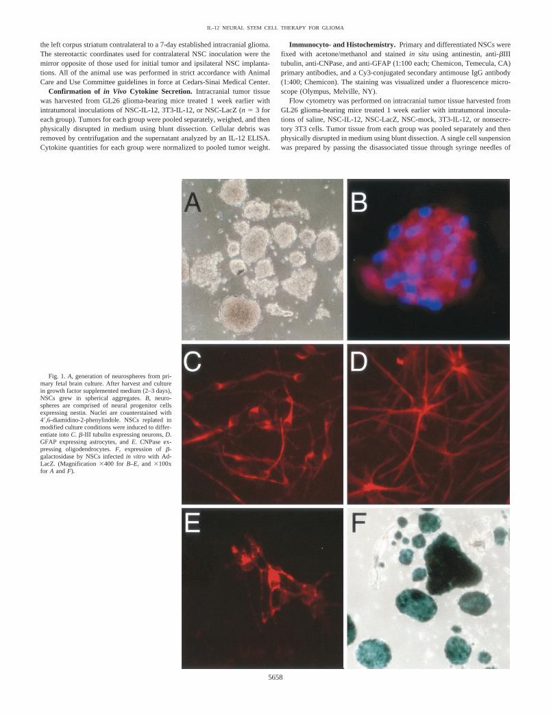
Fig. 1. A, generation of neurospheres from pri-mary fetal brain culture. After harvest and culturein growth factor supplemented medium (2–3 days),NSCs grew in spherical aggregates. B, neuro-spheres are comprised of neural progenitor cellsexpressing nestin. Nuclei are counterstained with4�,6-diamidino-2-phenylindole. NSCs replated inmodified culture conditions were induced to differ-entiate into C. �-III tubulin expressing neurons, D.GFAP expressing astrocytes, and E. CNPase ex-pressing oligodendrocytes. F, expression of �-galactosidase by NSCs infected in vitro with Ad-LacZ. (Magnification �400 for B–E, and �100xfor A and F).
5658
IL-12 NEURAL STEM CELL THERAPY FOR GLIOMA

progressively decreasing bore. Viable cells from each group (2 � 106) werestained at 4°C for 30 min with antimouse CD4-FITC and antimouse CD8-PE(1:12 each, clones H129.19 and 53–6.7, respectively; BD PharMingen). Sam-ples were then fixed with 1% paraformaldehyde before analysis on a FACStarII cell sorter. Flow cytometric gates for viable lymphocytes were establishedon the basis of forward and side scatter, and control stains from mouse spleen.
For immunohistochemistry, brains harvested from treated mice euthanizedusing CO2 asphyxiation were frozen in dry ice, sectioned using a cryostat,mounted on slides, and allowed to air dry. Sections used for X-Gal detectionwere fixed with 0.05% glutaraldehyde, stained as per standard protocol, andcounterstained with neutral red before mounting. For other stains, brain sec-tions were fixed in acetone and stained using primary antibodies for CD4 andCD8 (1:100 each; clones H129.19 and 53–6.7 respectively; BD PharMingen),a fibroblast specific marker (1:100; clone ER-TR7; Cedarlane Laboratories,Hornby, Ontario, Canada), �-galactosidase protein (1:100; MAB1802; Chemi-con), and IL-12 (1:50; H-306; Santa Cruz Biotechnology, Santa Cruz, CA).Secondary staining was then performed with the Vectastain Elite ABC kit(Vector Laboratories, Burlingame, CA). Slides were developed with diamino-benzidine (Sigma) and counterstained with hematoxylin before final mounting.
Results
Neurospheres are Comprised of Progenitor Cells That CanDifferentiate into Neurons, Astrocytes, and Oligodendrocytes.We isolated primary NSCs from fetal C57Bl/6 mice. These cells grewprimarily as spherical aggregates (Fig. 1A) and were comprisedmainly of cells that stained strongly for nestin, a marker of neuralprogenitor cells (Fig. 1B). We confirmed their pluripotency by in vitrodifferentiation in medium supplemented with serum, retinoic acid, andcyclic AMP. After 7 days of culture, cells with well-differentiatedmorphology were visible, and these contained populations expressingmarkers specific for neurons (�III-tubulin), astrocytes (GFAP), andoligodendrocytes (CNPase; Fig. 1, C–E, respectively).
In Vitro Expression of �-Galactosidase and Secretion of IL-12by Infected NSC and NIH-3T3. To confirm biologically relevantsecretion of cytokine, supernatant from NSC and NIH-3T3 cellsinfected with AdmIL-12 was analyzed using ELISA. NSC and NIH-3T3 cells infected with AdmIL-12 were found to secrete 70 and 64.8ng/106 cells per 48 h, respectively. In addition, NSCs infected withAdLacZ were stained in vitro with an X-Gal substrate and found toexpress high levels of �-galactosidase 48 h after viral infection (Fig.1F). To determine the effect of our viral vectors on NSC viability, wefollowed adenoviral-infected NSCs in vitro and found them to main-tain a viability in excess of 95% (as determined by trypan blueexclusion staining) up to 10 days after viral infection (data notshown).
Glioma-bearing Mice Treated with IL-12-secreting NSCs Dem-onstrate Prolonged Survival and Long-Term Immunity. To deter-mine whether inoculation of NSCs into established gliomas effected atherapeutic benefit, we delivered NSC-IL-12, NSC-LacZ, NSC-mock,3T3-IL-12, or normal saline into established intracranial gliomas inC57Bl/6 mice. NSC-IL-12-treated mice demonstrated significantlyprolonged survival compared with saline and nonsecretory NSC-treated controls (P � 0.03, NSC-IL-12 versus NSC-mock; P � 0.01,NSC-IL-12 versus NSC-LacZ; P � 0.01, NSC-IL-12 versus saline;Log rank), with 30% of treated animals surviving beyond day 60 aftertumor implantation (Fig. 2). However, 3T3-IL-12 therapy producedno long-term survivors and did not result in statistically enhancedsurvival compared with treatment with NSC-mock, NSC-LacZ, orsaline. The observable difference in survival between NSC-IL-12-treated animals and 3T3-IL-12 therapy could, however, not be verifiedstatistically (P � 0.08; Log rank). There were no detectable differ-ences in survival between animals treated with NSC-mock, NSC-LacZ, or saline.
Three months after initial tumor implantation, NSC-IL-12-treated
mice that survived long term (n � 3) were rechallenged intracraniallywith 2.5 � 104 GL-26 cells. At the same time, naı̈ve animals (n � 5)received similar GL26 implantations. All 5 of the control animals diedwithin 30 days after implantation, whereas the NSC-IL-12-treatedsurvivors survived beyond day 120 after the tumor rechallenge.
In Vivo Transplanted NSC and 3T3 Remain Viable and SecreteSignificant Levels of Cytokine. To ascertain whether our trans-planted 3T3 and NSCs survived in vivo for a sufficient length of timeto allow a therapeutic effect to ensue, we harvested and stainedsections from tumor-bearing brains treated 18 days earlier with intra-tumoral inoculations of 3T3-IL-12, NSC-IL-12, or NSC-LacZ. Immu-nohistochemistry was performed for a fibroblast specific marker,�-galactosidase protein, and IL-12 cytokine. Tumors treated 18 daysearlier with 3T3-IL-12 demonstrated positive staining for a fibroblast-specific marker, indicating the presence of fibroblasts within thetumor (Fig. 3A). NSC-LacZ- or NSC-IL-12-treated brains were de-void of any fibroblast staining (not shown and Fig. 3C, respectively).Tumors inoculated with NSC-LacZ demonstrated numerous �-galac-tosidase-positive cellular clusters within the tumor mass and in tumoroutgrowths (Fig. 3B) confirming the presence of NSC-LacZ in thesesections. We were also able to detect IL-12-positive cells within3T3-IL-12- and NSC-IL-12-treated tumors (not shown and Fig. 3D,respectively) 18 days after inoculation. These results indicate that bothtransplanted 3T3 and NSCs survived in significant quantity withintreated tumors and continued to produce the specific gene productsthey had been engineered to express.
To quantify the amount of cytokine secreted in vivo, tumors fromglioma-bearing mice treated with NSC-IL-12, 3T3-IL-12, and NSC-LacZ were harvested, processed, and analyzed for IL-12 content usingELISA. Tissue from both NSC-IL-12- and 3T3-IL-12-treated tumorswere found to have significant and comparable levels of IL-12 [40.3(�3.4) pg/mg and 43.2 (�3.2) pg/mg, respectively]. In contrast tu-mors from NSC-LacZ-treated controls contained negligible levels ofcytokine in the tumor bed [1.1 (�0.1) pg/mg].
NSC Migrate Extensively through the Tumor Mass and AreStrongly Tropic for Disseminating Tumor Cells. To determinewhether intratumorally inoculated NSCs demonstrated tropism fordisseminating glioma cells, brains from NSC-LacZ-treated mice wereharvested and stained with an X-Gal substrate to detect the presenceof �-galactosidase-expressing NSCs. NSC-LacZ cells were readilyidentifiable, dispersed within treated tumors, and were clearly visibletracking glioma cells as they migrated away from the main tumor
Fig. 2. Kaplan-Meier survival curve. Two days after intracranial implantation of 104
GL26 tumor cells, mice were inoculated intratumorally with NSC-IL-12, 3T3-IL-12,NSC-LacZ, NSC-mock, or saline, and then followed for survival.
5659
IL-12 NEURAL STEM CELL THERAPY FOR GLIOMA

mass. We were able to detect several distinct patterns of tumor spreadand found NSCs tracking migrating glioma cells in each case. Theseincluded thin outgrowths of tumor deep into adjacent normal brain(Fig. 4, A and B), direct extension of the tumor mass into adjacenttissue (Fig. 4C), migration of glioma cells along established whitematter tracts (Fig. 4D), and dissemination of solitary tumor pockets atconsiderable distance from the primary tumor bed (Fig. 4, E and F).
NSCs inoculated into the corpus striatum contralateral to the site oftumor implantation did not randomly disperse into adjacent normaltissue (Fig. 4G, left panel) nor could they be seen migrating to anydistant nontumorous region of the brain. However, some NSCs werevisible tracking directly across the brain into the immediate vicinity ofthe tumor (Fig. 4G, center panel) and into the tumor itself (Fig. 4G,right panel). We also found that NSCs inoculated into nontumor-bearing brains did not randomly dissipate into adjacent tissue or to the
contralateral hemisphere (Fig. 4H). These results indicate that NSCsexhibit strong, specific tropism for intracranial tumor.
Tumors Treated with IL-12-secreting NSCs Demonstrate En-hanced CD4� and CD8� T-Cell Infiltration. On the basis of theknown ability of IL-12 to promote antitumor cell-mediated immunity,we wished to assess whether the survival benefit observed withNSC-IL-12 therapy was associated with enhanced intratumoral T-cellinfiltration. Tumors from NSC-IL-12-treated animals demonstratedrobust infiltration with CD4� and CD8� T cells (Fig. 5A, left panel;Fig. 5D). 3T3-IL-12 therapy also resulted in increased intratumoralT-cell infiltration (Fig. 5A, center panel; Fig. 5B), which was signif-icantly higher than the immune cell infiltration seen in GL-26 tumorsinoculated with nonsecretory 3T3 (data not shown).
In addition to enhancing T-cell infiltration within the tumor paren-chyma, NSC-IL-12 therapy also resulted in the accumulation of nu-
Fig. 3. Immunohistochemistry for in vivo trans-planted 3T3 and NSCs. A, 3T3-IL-12-treated tu-mors were stained for a fibroblast specific marker18 days after inoculation. Positively staining fibro-blasts could be detected throughout the tumor. B,NSC-LacZ inoculated glioma-bearing brains werestained for �-galactosidase. Numerous positive cel-lular clusters were visible within the tumor. C,NSC-IL-12-treated tumors when stained for thesame fibroblast specific marker used in A demon-strate no staining. D, tumor outgrowths from NSC-IL-12-treated gliomas demonstrated cells thatstained positive (brown) for IL-12, indicating thatthey were infiltrated with NSC-IL-12. Right panelillustrates IL-12-positive cells (brown) interspersedwith tumor cells (blue) in tumor outgrowth. T,tumor and N, normal tissue. Right panel imagesrepresent high power magnification of boxed areasin corresponding left panels.
5660
IL-12 NEURAL STEM CELL THERAPY FOR GLIOMA

merous CD4� and CD8� T-cell aggregates along the tumor/normaltissue boundary, and in tumor outgrowths and microsatellites (Fig.5D; Fig. 5E, left panel), which was not seen in 3T3-IL-12-treatedtumors (Fig. 5B; and Fig. 5E, right panel).
T-cell infiltration, as determined by flow cytometry and immuno-histochemistry, in NSC-LacZ (Fig. 5A, right panel; Fig. 5C), NSC-mock (data not shown), and saline-treated tumors (data not shown)was low and comparably negligible.
Discussion
The inevitability of tumor recurrence after surgical resection andstandard adjuvant therapy remains an unfortunate reality for the vastmajority of patients with malignant glioma. The refractoriness of theseneoplasms to treatment possibly stems from several independentmechanisms. This may include the documented ability of gliomas toexert immunosuppressive effects at both the systemic (8) and localintratumoral (9) levels. In addition, malignant gliomas are highlyinvasive in nature (10), which provides them with the ability toinfiltrate deep into normal tissue, thereby establishing microscopicreservoirs from which regrowth can occur after surgical resection. The
utilization of IL-12-secreting NSCs represents a novel method aimedat targeting both these phenomena. IL-12 is a potent tumoricidalcytokine (11) with demonstrated efficacy against intracranial glioma(5, 12). NSCs, with their extensive migratory and tumor trackingcharacteristics (3), are capable of delivering this therapeutic protein toneoplastic pockets separate from the main tumor mass.
The inoculation of NSCs engineered to secrete IL-12 resulted insignificant prolongation of survival in intracranial glioma-bearingmice. To determine the potential role of NSC migration in this result,we compared the survival benefit offered by NSC IL-12 secretion tothat conferred by IL-12 secretion by nonmigratory NIH-3T3 cells,which produce similar levels of cytokine to NSC-IL-12 in vitro and invivo. 3T3-IL-12 treatment did not result in statistically significantsurvival compared with saline and nonsecretory NSC inoculated con-trols. In contrast, NSC-IL-12 therapy was able to demonstrate astatistically significant enhancement in survival compared with NSC-LacZ, NSC-mock, and saline treated controls, with 30% of NSC-IL-12 treated animals surviving beyond 60 days after tumor implan-tation. NSC-IL-12-treated long-term survivors were also immune tointracranial tumor rechallenge. This may be a direct consequence of
Fig. 4. NSCs display strong tropism for dissem-inating glioma in vivo. Tumors from glioma-bear-ing mice inoculated with NSC-LacZ were stainedwith X-Gal and counterstained with neutral red.NSCs appear blue as they express �-galactosidasewhereas tumor appears as hypercellular areas stain-ing intensely with neutral red. T, tumor mass,outgrowths, and microsatellites. Arrows indicatedisseminating NSC closely following migratingpockets of tumor. Four distinct patterns of tumorspread were detected, and NSCs were found track-ing migrating glioma in each case. A, thin out-growth of tumor cells deep into adjacent normalbrain. B, high power magnification of area boxed inA. C, direct extension of tumor mass into adjacenttissue. D, migration of glioma cells away from theprimary tumor bed along a white matter tract. E,tumor microsatellite independent of main tumormass. F, high power photomicrograph of microsat-ellite in E demonstrating �-galactosidase-positiveNSCs interspersed with tumor cells exhibiting vis-ible mitoses. G, inoculation of NSCs into cerebralhemisphere contralateral to existing tumor. Leftpanel represents portion of left cerebral hemispherewhere NSCs were inoculated and illustrates thatNSCs do not randomly dissipate into adjacent non-tumorous tissue. Center panel shows tumor-bearingportion of right cerebral hemisphere demonstratingspecific, nonrandom, migration of NSCs across thebrain into the vicinity of the tumor in the oppositehemisphere (inset box). Right panel demonstrates asecond tumor-bearing portion of a similarly treatedbrain. NSC-LacZ are visible, interspersed withinthe tumor mass (inset box), contralateral to theirsite of inoculation. H, NSC-LacZ when adminis-tered in a nontumor-bearing brain remain confinedto the needle tract (left panel) and do not randomlydisseminate into adjacent tissue or to the contralat-eral hemisphere (right panel).
5661
IL-12 NEURAL STEM CELL THERAPY FOR GLIOMA

the ability of NSCs to target migrating tumor cells, as confirmed byour observation that NSCs exhibit strong tropism for disseminatingtumor in vivo. Of significance was our inability to detect any survivalbenefit with the use of NSC therapy alone, as described previously(4). Both NSC-mock and NSC-LacZ-treated animals demonstratedsurvival similar to saline inoculated controls. This represents a sig-nificant divergence from the results forwarded by Benedetti et al. (4),in which intratumoral delivery of noncytokine-secreting NSCs alsoresulted in significantly prolonged survival in intracranial glioma-bearing rodents. This may possibly be explained by their use of adifferent cell line (GL261) in their murine glioma model, which isdistinct and has different characteristics from the GL26 gliomas weused in our study (13, 14).
These results demonstrate, for the first time, the ability of primaryNSCs to intersperse themselves in neoplastic microsatellites locateddeep within normal tissue, at significant distance from the primarytumor mass. Aboody et al. (3) described previously the tropism of animmortalized NSC line for intracranial glioma. Our results confirmthat primary, autologous, nontransformed NSCs are also capable ofdemonstrating potent migratory activity in vivo, a finding that was notreported by Benedetti et al. (4) in their primary NSC treatment model.Of significance was our finding that NSC inoculated into normalcorpus striatum contralateral to an existing tumor did not randomlydisperse throughout the brain. Rather, these cells exhibited specificmigration to the tumor in the opposite hemisphere, indicating that ourobserved dissemination of NSC was not a random phenomenon, but
Fig. 5. Intratumoral CD4� and CD8� T-cellinfiltration. A, flow cytometry analysis demonstrat-ing robust intratumoral T-cell infiltration in NSC-IL-12 (left panel) and 3T3-IL-12 (center panel)inoculated gliomas. T-cell content of NSC-LacZ-treated tumors (right panel) was much lower andcomparable with infiltration seen in NSC-mock andsaline-inoculated gliomas (data not shown). B,3T3-IL-12-treated tumors demonstrate CD4� (leftpanel) and CD8� (center and right panels) T-cellinfiltration, with immunohistochemically positivecells interspersed in tumor tissue. C, NSC-LacZ-treated tumors display negligible infiltration of tu-mors by CD4� (left panel) or CD8� (center andright panels) T cells. D, NSC-IL-12 treated glioma-bearing brains demonstrate robust infiltration oftumors with CD4� (left panel) and CD8� (centerand right panels) T cells, with numerous aggregatesalong the tumor/normal tissue boundary (arrows)E, comparison of T-cell infiltration in comparableoutgrowths from primary tumor bed. Tumor mic-rosatellites in NSC-IL-12-treated tumors demon-strated robust CD8� T-cell infiltration (left panel),whereas those in 3T3-IL-12 inoculated brains didnot (right panel). T, tumor and N, normal braintissue. Right panels in B–D, high power images ofboxed areas in respective center panels. Magnifi-cation �100 for left and center panels in B–D,�400 for right panels in B–D, and �200 for bothpanels in E.
5662
IL-12 NEURAL STEM CELL THERAPY FOR GLIOMA

was in fact a process resulting from a strong, specific tropism of NSCsfor intracranial glioma. The underlying basis of this tropism is unclearand may be the result of a chemotactic gradient resulting from theproduction of chemokines from tumor cells that are actively infiltrat-ing normal tissue.
We have shown previously that in situ IL-12 gene transfer inintracranial glioma results in potent Th-1-mediated antitumor cyto-toxic immunity (5). On the basis of this experience, we wished todetermine whether the survival benefit associated with NSC-IL-12therapy was accompanied by increased intratumoral CD4� andCD8� T-cell infiltration. Both NSC-IL-12- and 3T3-IL-12-treatedtumors demonstrated robust T-cell infiltration. However, in contrast to3T3-IL-12-treated tumors, NSC-IL-12 inoculated brains also demon-strated large, numerous CD4� and CD8� T-cell aggregates alongtheir tumor margins and in migrating tumor microsatellites. BecauseNSCs also concentrate along the tumor/normal tissue boundary whilealigning themselves with tumor cells that have begun to migrate, theselymphocytic aggregates may represent a chemotactic colocalization ofT cells with marginating and migrating IL-12 producing NSCs. Thesurvival benefit observed with NSC-IL-12 therapy may, therefore, bea consequence of IL-12-induced taxis of tumoricidal T cells to dis-seminating tumor pockets being tracked by migrating NSC-IL-12cells.
The current focus of experimental NSC therapeutics is primarilybased around their use in the treatment of neurodegenerative disordersand stroke. We have demonstrated that combining the tumoricidalpotency of IL-12 with the extensive tumor tracking capability ofNSCs results in a synergistic therapeutic benefit. This additionallyextends the scope of NSC therapy to include their use as vehicles forprotein delivery to in vivo glioma, and, therefore, represents a prom-ising new treatment modality for malignant brain tumors.
Acknowledgments
We thank Mervin Gutierrez for invaluable technical support and for assist-ance in digitizing and drafting the color figures, and Nancy Chung for hertechnical assistance with this project.
References
1. Surawicz, T. S., Davis, F., Freels, S., Laws, E. R., Jr., and Menck, H. R. Brain tumorsurvival: results from the National Cancer Data Base. J. Neurooncol., 40: 151–160,1998.
2. Dirks, P. B. Glioma migration: clues from the biology of neural progenitor cells andembryonic CNS cell migration. J. Neurooncol., 53: 203–212, 2001.
3. Aboody, K. S., Brown, A., Rainov, N. G., Bower, K. A., Liu, S., Yang, W., Small,J. E., Herrlinger, U., Ourednik, V., Black, P. M., Breakefield, X. O., and Snyder, E. Y.From the cover: neural stem cells display extensive tropism for pathology in adultbrain: evidence from intracranial gliomas. Proc. Natl. Acad. Sci. USA, 97: 12846–12851, 2000.
4. Benedetti, S., Pirola, B., Pollo, B., Magrassi, L., Bruzzone, M. G., Rigamonti, D.,Galli, R., Selleri, S., Di Meco, F., De Fraja, C., Vescovi, A., Cattaneo, E., andFinocchiaro, G. Gene therapy of experimental brain tumors using neural progenitorcells. Nat. Med., 6: 447–450, 2000.
5. Liu, Y., Ehtesham, M., Samoto, K., Wheeler, C. J., Thompson, R. C., Villareal, L. P.,Black, K. L., and Yu, J. S. In situ adenoviral interleukin 12 gene transfer conferspotent and long-lasting cytotoxic immunity in glioma. Cancer Gene Ther., 9: 9–15,2002.
6. Schaack, J., Langer, S., and Guo, X. Efficient selection of recombinant adenovirusesby vectors that express �-galactosidase. J. Virol., 69: 3920–3923, 1995.
7. Martens, D. J., Tropepe, V., and van Der Kooy, D. Separate proliferation kinetics offibroblast growth factor-responsive and epidermal growth factor-responsive neuralcells within the embryonic forebrain germinal zone. J. Neurosci., 20: 1085–1095,2000.
8. Morford, L. A., Elliott, L. H., Carlson, S. L., Brooks, W. H., and Roszman, T. L. Tcell receptor-mediated signaling is defective in T cells obtained from patients withprimary intracranial tumors. J. Immunol., 159: 4415–4425, 1997.
9. Black, K. L., Chen, K., Becker, D. P., and Merrill, J. E. Inflammatory leukocytesassociated with increased immunosuppression by glioblastoma. J. Neurosurg., 77:120–126, 1992.
10. Lopes, M. B. S, VandenBerg, S. R., and Scheithauer, B. W. Histopathology, immu-nochemistry, and ultrastructure of brain tumors. In: A. H. Kaye and E. R. Laws, Jr.(eds.), Brain Tumors, pp. 125–162. Edinburgh: Churchill Livingstone, 1995.
11. Nastala, C. L., Edington, H. D., McKinney, T. G., Tahara, H., Nalesnik, M. A.,Brunda, M. J., Gately, M. K., Wolf, S. F., Schreiber, R. D., and Storkus, W. J.Recombinant IL-12 administration induces tumor regression in association withIFN-� production. J. Immunol., 153: 1697–1706, 1994.
12. Jean, W. C., Spellman, S. R., Wallenfriedman, M. A., Hall, W. A., and Low, W. C.Interleukin-12-based immunotherapy against rat 9L glioma. Neurosurgery, 42: 850–856, 1998.
13. Ausman, J. I., Shapiro, W. R., and Rall, D. P. Studies on the chemotherapy ofexperimental brain tumors: development of an experimental model. Cancer Res., 30:2394–2400, 1970.
14. Akbasak, A., Oldfield, E. H., and Saris, S. C. Expression and modulation of majorhistocompatibility antigens on murine primary brain tumor in vitro. J. Neurosurg., 75:922–929, 1991.
5663
IL-12 NEURAL STEM CELL THERAPY FOR GLIOMA