Embed Size (px)
Citation preview
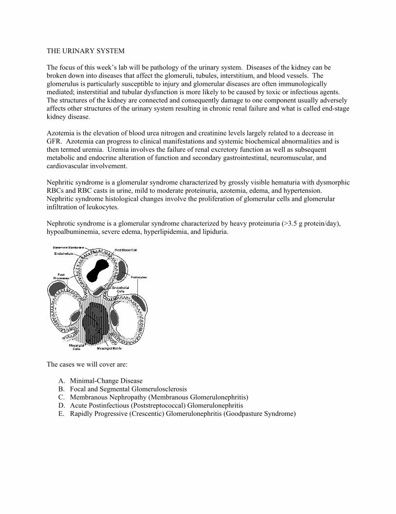
THE URINARY SYSTEM The focus of this week’s lab will be pathology of the urinary system. Diseases of the kidney can be broken down into diseases that affect the glomeruli, tubules, interstitium, and blood vessels. The glomerulus is particularly susceptible to injury and glomerular diseases are often immunologically mediated; insterstitial and tubular dysfunction is more likely to be caused by toxic or infectious agents. The structures of the kidney are connected and consequently damage to one component usually adversely affects other structures of the urinary system resulting in chronic renal failure and what is called end-stage kidney disease. Azotemia is the elevation of blood urea nitrogen and creatinine levels largely related to a decrease in GFR. Azotemia can progress to clinical manifestations and systemic biochemical abnormalities and is then termed uremia. Uremia involves the failure of renal excretory function as well as subsequent metabolic and endocrine alteration of function and secondary gastrointestinal, neuromuscular, and cardiovascular involvement. Nephritic syndrome is a glomerular syndrome characterized by grossly visible hematuria with dysmorphic RBCs and RBC casts in urine, mild to moderate proteinuria, azotemia, edema, and hypertension. Nephritic syndrome histological changes involve the proliferation of glomerular cells and glomerular infiltration of leukocytes. Nephrotic syndrome is a glomerular syndrome characterized by heavy proteinuria (>3.5 g protein/day), hypoalbuminemia, severe edema, hyperlipidemia, and lipiduria.
The cases we will cover are:
A. Minimal-Change Disease B. Focal and Segmental Glomerulosclerosis C. Membranous Nephropathy (Membranous Glomerulonephritis) D. Acute Postinfectious (Poststreptococcal) Glomerulonephritis E. Rapidly Progressive (Crescentic) Glomerulonephritis (Goodpasture Syndrome)
A. MINIMAL-CHANGE DISEASE
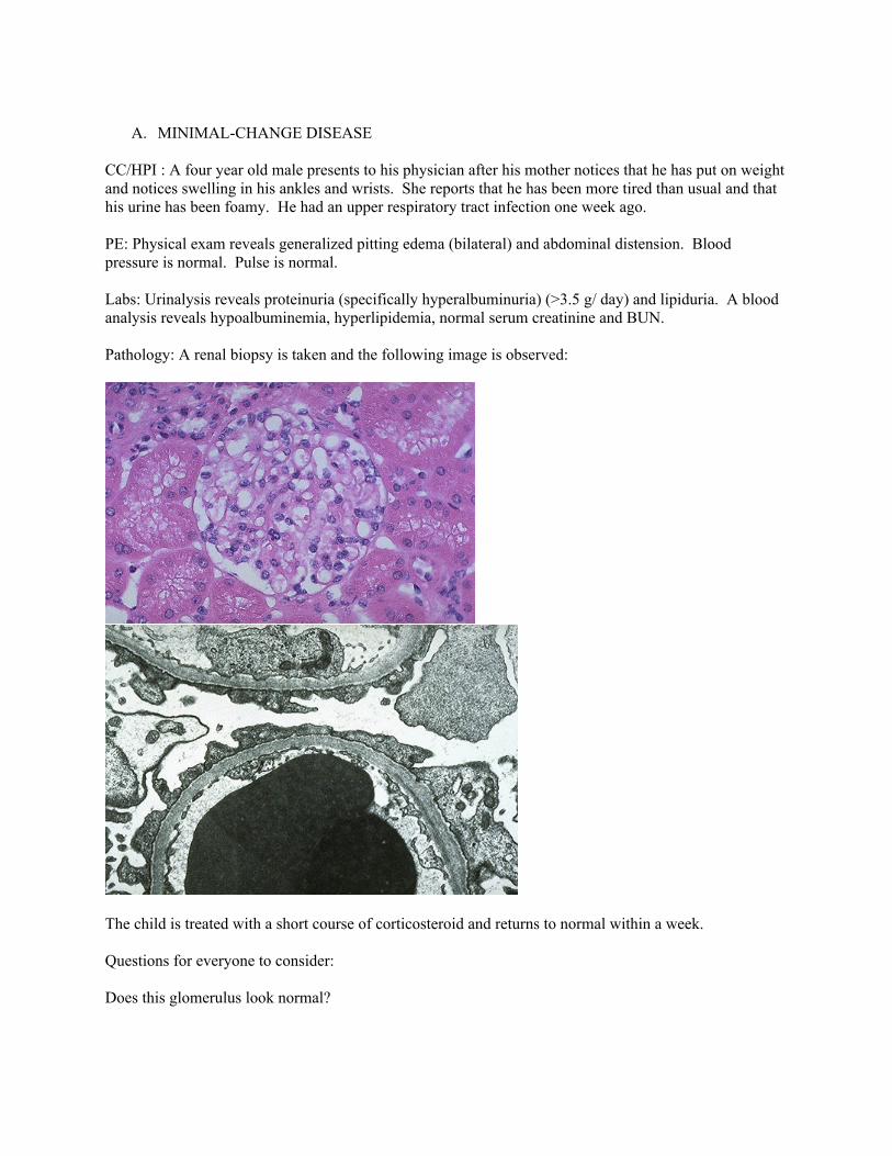
CC/HPI : A four year old male presents to his physician after his mother notices that he has put on weight and notices swelling in his ankles and wrists. She reports that he has been more tired than usual and that his urine has been foamy. He had an upper respiratory tract infection one week ago. PE: Physical exam reveals generalized pitting edema (bilateral) and abdominal distension. Blood pressure is normal. Pulse is normal. Labs: Urinalysis reveals proteinuria (specifically hyperalbuminuria) (>3.5 g/ day) and lipiduria. A blood analysis reveals hypoalbuminemia, hyperlipidemia, normal serum creatinine and BUN. Pathology: A renal biopsy is taken and the following image is observed:
The child is treated with a short course of corticosteroid and returns to normal within a week. Questions for everyone to consider: Does this glomerulus look normal?
Hypoalbuminemia and proteinuria suggest that what structures shown in the above picture are likely to be damaged?
Questions if you have been assigned this case: What is the structural defect in this disease (seen in the EM image)? Does this patient exhibit nephritic or nephrotic syndrome? Why is edema present in this patient?
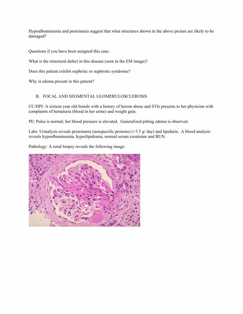
B. FOCAL AND SEGMENTAL GLOMERULOSCLEROSIS CC/HPI: A sixteen year old female with a history of heroin abuse and STIs presents to her physician with complaints of hematuria (blood in her urine) and weight gain. PE: Pulse is normal, her blood pressure is elevated. Generalized pitting edema is observed. Labs: Urinalysis reveals proteinuria (nonspecific proteins) (>3.5 g/ day) and lipiduria. A blood analysis reveals hypoalbuminemia, hyperlipidemia, normal serum creatinine and BUN. Pathology: A renal biopsy reveals the following image:
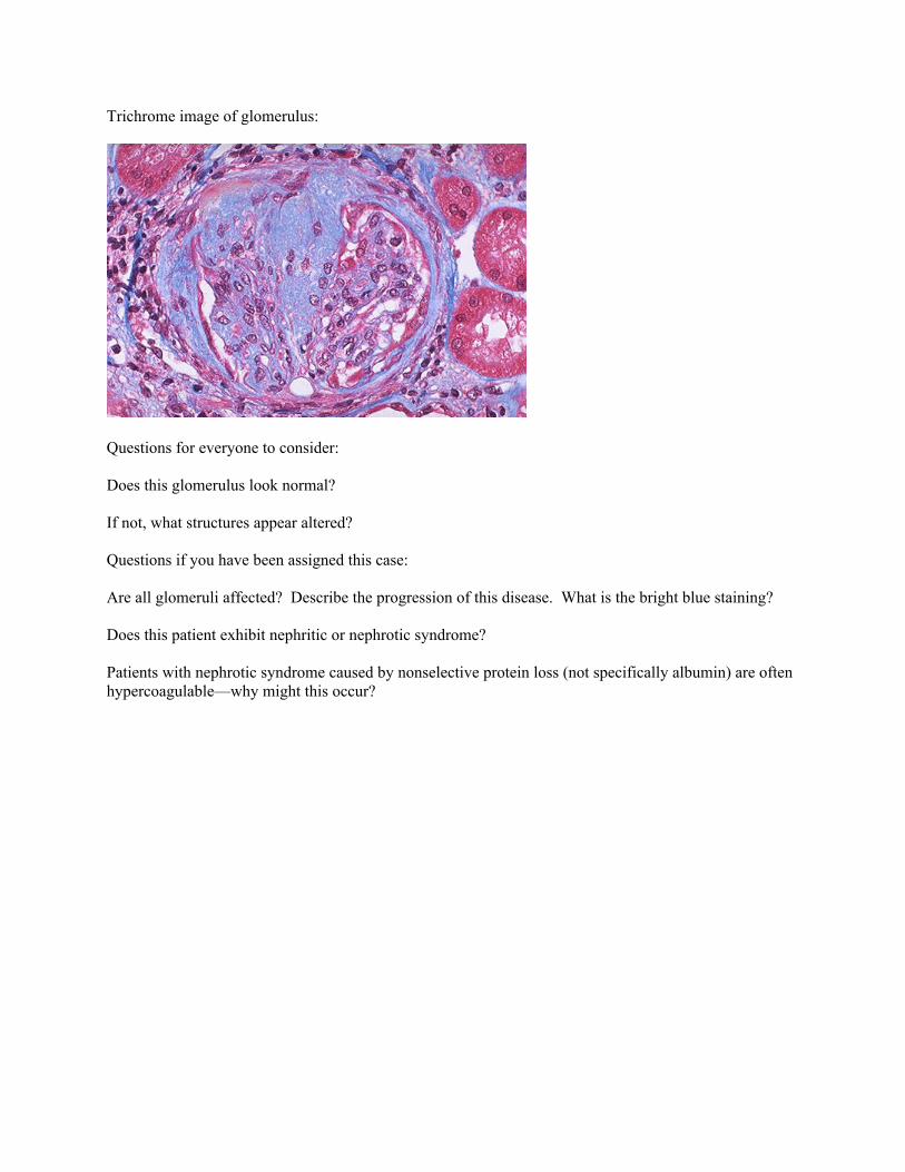
Trichrome image of glomerulus:
Questions for everyone to consider: Does this glomerulus look normal?
If not, what structures appear altered? Questions if you have been assigned this case: Are all glomeruli affected? Describe the progression of this disease. What is the bright blue staining? Does this patient exhibit nephritic or nephrotic syndrome?
Patients with nephrotic syndrome caused by nonselective protein loss (not specifically albumin) are often hypercoagulable—why might this occur?
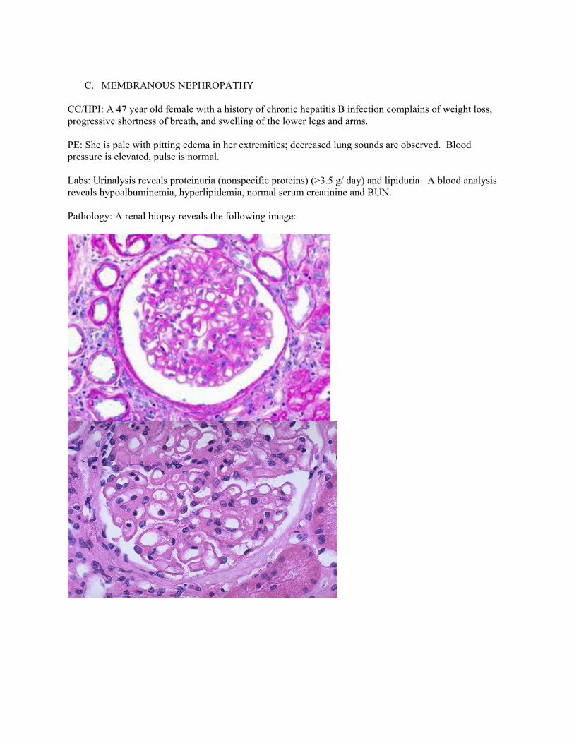
C. MEMBRANOUS NEPHROPATHY
CC/HPI: A 47 year old female with a history of chronic hepatitis B infection complains of weight loss, progressive shortness of breath, and swelling of the lower legs and arms. PE: She is pale with pitting edema in her extremities; decreased lung sounds are observed. Blood pressure is elevated, pulse is normal. Labs: Urinalysis reveals proteinuria (nonspecific proteins) (>3.5 g/ day) and lipiduria. A blood analysis reveals hypoalbuminemia, hyperlipidemia, normal serum creatinine and BUN. Pathology: A renal biopsy reveals the following image:
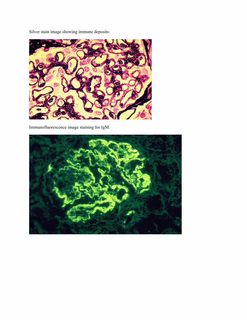
Silver stain image showing immune deposits:
Immunofluorescence image staining for IgM:
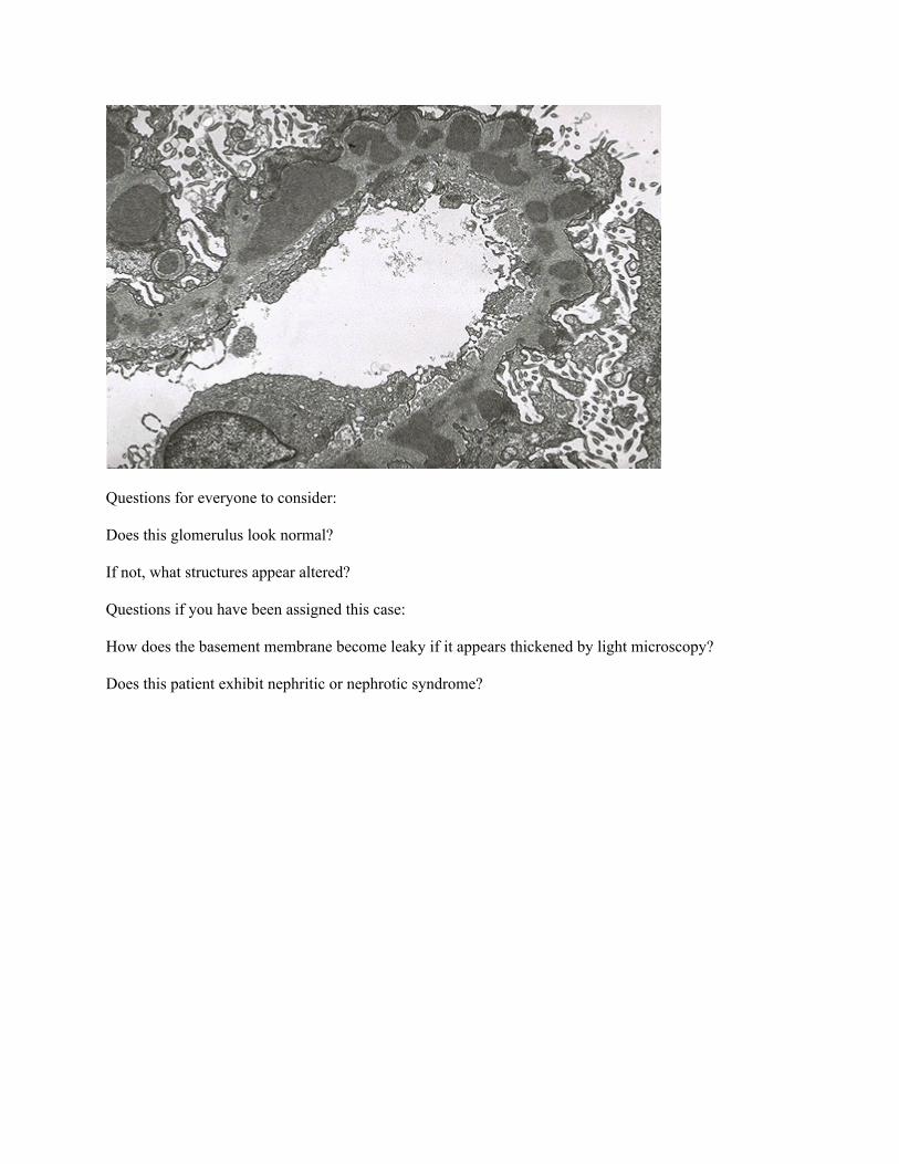
Questions for everyone to consider: Does this glomerulus look normal?
If not, what structures appear altered? Questions if you have been assigned this case: How does the basement membrane become leaky if it appears thickened by light microscopy? Does this patient exhibit nephritic or nephrotic syndrome?
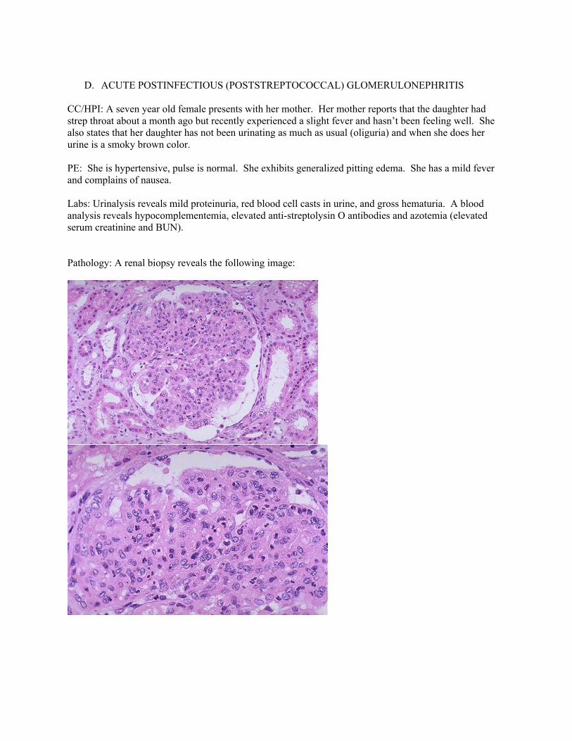
D. ACUTE POSTINFECTIOUS (POSTSTREPTOCOCCAL) GLOMERULONEPHRITIS
CC/HPI: A seven year old female presents with her mother. Her mother reports that the daughter had strep throat about a month ago but recently experienced a slight fever and hasn’t been feeling well. She also states that her daughter has not been urinating as much as usual (oliguria) and when she does her urine is a smoky brown color. PE: She is hypertensive, pulse is normal. She exhibits generalized pitting edema. She has a mild fever and complains of nausea. Labs: Urinalysis reveals mild proteinuria, red blood cell casts in urine, and gross hematuria. A blood analysis reveals hypocomplementemia, elevated anti-streptolysin O antibodies and azotemia (elevated serum creatinine and BUN). Pathology: A renal biopsy reveals the following image:
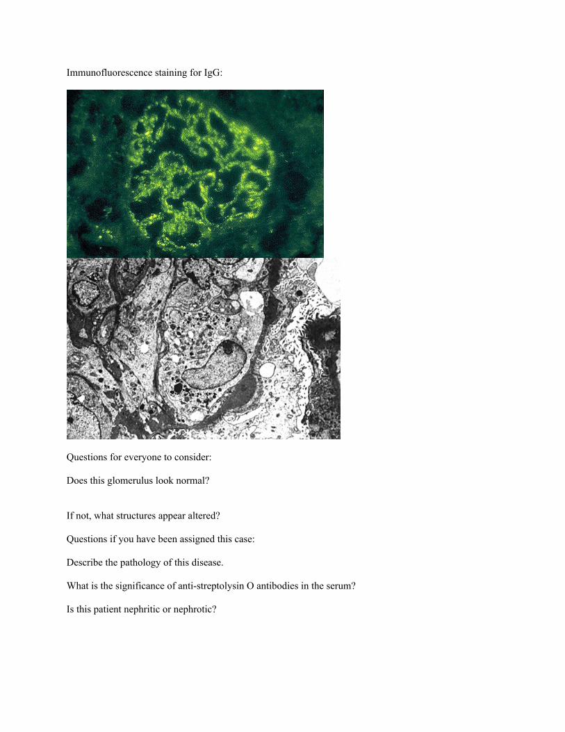
Immunofluorescence staining for IgG:
Questions for everyone to consider: Does this glomerulus look normal?
If not, what structures appear altered? Questions if you have been assigned this case: Describe the pathology of this disease. What is the significance of anti-streptolysin O antibodies in the serum? Is this patient nephritic or nephrotic?

E. RAPDILY PROGRESSIVE (CRESCENTERIC) GLOMERULONEPHRITIS (GOODPASTURE SYNDROME)
CC/HPI: A 25 year old male complains of a chronic cough for several months accompanied by lightheadedness, fatigue, and malaise. Yesterday he coughed up blood. He admits to heavy smoking. He also reports intermittent fever and headaches. He complains that he hasn’t been urinating as frequently and when he does it is dark orange in color. PE: Pulmonary crackles are present bilaterally. Labs: A blood analysis demonstrates azotemia (elevated BUN and creatinine) and the presence of antiglomerular abasement membrane antibodies. A urinalysis demonstrates oliguria, hematuria, and proteinuria. Pathology: A renal biopsy reveals the following image: PAS stain of glomeruli:
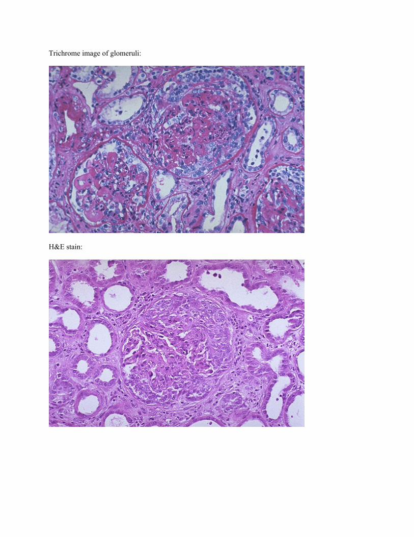
Trichrome image of glomeruli:
H&E stain:
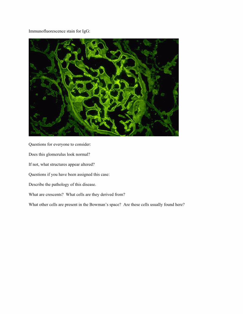
Immunofluorescence stain for IgG:
Questions for everyone to consider: Does this glomerulus look normal?
If not, what structures appear altered? Questions if you have been assigned this case: Describe the pathology of this disease. What are crescents? What cells are they derived from? What other cells are present in the Bowman’s space? Are these cells usually found here?