Embed Size (px)
Citation preview
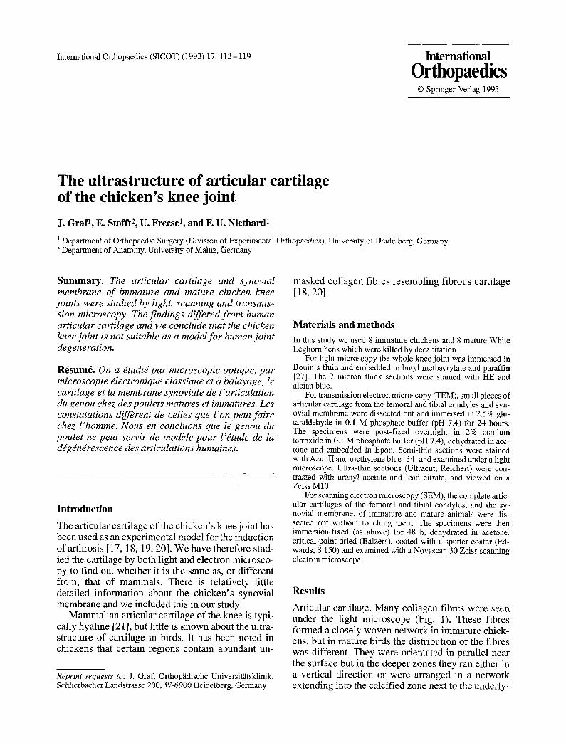
Intemational Orthopaedics (SICOT) (1993) 17:113 - 119 International Orthopaedics
© Springer-Verlag 1993
The ultrastructure of articular cartilage of the chicken's knee joint
J. Graf 1, E. Stofft 2, U. Freese 1, and F. U. Niethard 1
~ Department of Orthopaedic Surgery (Division of Experimental Orthopaedics), University of Heidelberg, Germany ~ Department of Anatomy, University of Mainz, Germany
S u m m a r y . The articular cartilage and synovial membrane of immature and mature chicken knee joints were studied by light, scanning and transmis- sion microscopy. The findings differed from human articular cartilage and we conclude that the chicken knee joint is not suitable as a model for human joint degeneration.
R~sum~. On a dtudid par microscopie optique, par microscopie ~lectronique classique et & balayage, le cartilage et la membrane synoviale de l'articulation du genou chez des poulets matures et immatures. Les constatations diffbrent de celles que l' on peut faire chez l'homme. Nous en concluons que le genou du poulet ne peut servir de modble pour l'ttude de la d~g~ndrescence des articulations humaines.
Introduct ion
The articular cartilage of the chicken ' s knee joint has been used as an exper imental model for the induction of arthrosis [17, 18, 19, 20]. We have therefore stud- ied the cartilage by both light and electron microsco- py to find out whether it is the same as, or different from, that of mammals . There is relatively little detailed information about the chicken ' s synovial membrane and we included this in our study.
M a m m a l i a n articular cartilage of the knee is typi- cally hyaline [21], but little is known about the ultra- structure of cartilage in birds. It has been noted in chickens that certain regions contain abundant un-
Reprint requests to: J. Graf, Orthop~idische Universit~tsklinik, Schlierbacher Landstrasse 200, W-6900 Heidelberg, Germany
masked collagen fibres resembling fibrous cartilage [18,20].
Materials and m e t h o d s
In this study we used 8 immature chickens and 8 mature White Leghorn hens which were killed by decapitation.
For light microscopy the whole knee joint was immersed in Bouin's fluid and embedded in butyl methacrylate and paraffin [27]. The 7 micron thick sections were stained with HE and alcian blue.
For transmission electron microscopy (TEM), small pieces of articular cartilage from the femoral and tibial condyles and syn- ovial membrane were dissected out and immersed in 2.5% glu- taraldehyde in 0.1 M phosphate buffer (pH 7.4) for 24 hours. The specimens were post-fixed overnight in 2% osmium tetroxide in 0.1 M phosphate buffer (pH 7.4), dehydrated in ace L tone and embedded in Epon. Semi-thin sections were stained with Azur II and methylene blue [34] and examined under a light microscope. Ultra-thin sections (Ultracut, Reichert) were con- trasted with uranyl acetate and lead citrate, and viewed on a Zeiss M10.
For scanning electron microscopy (SEM), the complete artic- ular cartilages of the femoral and tibial condyles, and the sy- novial membrane, of immature and mature animals were dis- sected out without touching them. The specimens were then immersion-fixed (as above) for 48 h, dehydrated in acetone, critical-point dried (Balzers), coated with a sputter coater (Ed- wards, S 150) and examined with a Novascan 30 Zeiss scanning electron microscope.
Results
Articular cartilage. Many collagen fibres were seen under the light microscope (Fig. 1). These fibres formed a closely woven network in immature chick- ens, but in mature birds the distribution of the fibres was different. They were orientated in parallel near the surface but in the deeper zones they ran either in a vertical direction or were arranged in a ne twork extending into the calcified zone next to the underly-

114 J. Graf et al.: Articular cartilage of the chicken's knee joint
Fig. 1. Light microscopy of chicken femoral articular cartilage. Numerous unmasked collagen fibres are present. × 200
Fig. 2. Blood vessels are present within the cartilage. × 400
Fig. 3. SEM of collagen fibrils running at a tangent to the surface of the joint cavity, x 10 000
Fig. 4. SEM of a chondrocyte lying in a lacuna and surrounded by a collagenous network, x 10000
ing bone. Most chondrocytes were arranged in pairs and were surrounded by a distinct layer of capsular matrix, even near the bone, and only rarely formed groups of 4 cells in the middle and superficial layers. Near the convex surface, single chondrocytes which lacked a distinct territorial or capsular matrix were seen. A characteristic feature was the presence of blood vessels within the cartilage (Fig. 2).
SEM showed that the articular surface was basi- cally the same in immature and mature specimens
and was absolutely smooth without undulations, humps or pits [14].
At higher magnification the smooth surface was seen to consist of a layer of round matrix granules of varying size, collectively termed the surface coat [ 1, 14]. No fibrils were found lying between the matrix granules. In immature specimens the surface was interrupted, whereas in mature specimens relatively large polymorphic areas lacking a surface coat were seen. At the bot tom of these shallow pits, bundles of

J. Graf et al.: Articular cartilage of the chicken' s knee joint 115
Fig. 5. TEM of a chon&-ocyte showing a wealth of fine filamentous fibres surrounding the nucleus as a result of which the cells organelles have acquired a peripheral location, x 12 500
Fig. 6. A chondrocyte containing well developed Golgi complexes and filaments, x 25 000
collagen were seen running parallel to the surface. The surface coat was lacking in the oval zones which clearly differed from the polymorphic zones. These oval zones represented the entrance into deeper cavi- ties surrounded by collagen fibrils arranged in a basket-like fashion. In some instances, the "basket" exhibited gaps and individual chondrocytes sur- rounded by collagen fibrils together with finely fila- mentous material could be identified (Figs. 3 and 4). There were abundant intracellular filaments measur- ing 8 - 1 2 nm.
In the immature chickens, there were more fila- ments in the superficial chondrocytes than in the deeper cells. In mature birds, the cytoplasm of the chondrocytes was often almost entirely filled with these filaments so that only a narrow zone of cytosol, without cytoplasmic organelles, was seen around the nucleus (Fig. 5). Invariably, the usual cell organelles were confined to a thin subplasmalemmal zone. The straight filaments were not randomly orientated, but formed groups of longitudinally or transversely sec- tioned elements, giving the impression of a densely
knit network. Similar findings were obtained with regard to cytoplasmic organelles and inclusions in chondrocytes of other species.
In immature chickens, the chondrocytes contained a large amount of rough endoplasmic reticulum, free polysomes and a well developed Golgi complex (Fig. 6). Mitochondria were moderately abundant and lipid droplets were scarce.
In mature birds, rough endoplasmic reticulum and polysomes were less plentiful than in immature chickens, and lipid droplets were increased.
The synovial membrane, together with a subsy- novial layer, could be clearly distinguished in imma- ture and mature birds. The synovium consisted of from one to three layers of relatively small oval or elongated lining cells, with round or oval eccentri- cally located nuclei and one or two large nucleoli. The free surface of the lining cells was relatively smooth and did not bulge towards the articular cav- ity; their cytoplasm appeared homogeneous. The subsynovial layer consisted of loosely arranged con- nective tissue with a few adipose cells in immature

116 J. Graf et al.: Articular cartilage of the chicken's knee joint
Fig. 7. Semi-thin section showing synovial membrane composed of loosely arranged lining cells and a subsynovial layer, x 200
Fig. 8. SEM of the intimal layer showing only elongated B cells with some granules on their surface, x 10000
chickens, but there were large numbers of these cells in mature birds. Blood vessels and nerve fibres were restricted to this zone (Fig. 7).
SEM showed that the synovial membrane in im- mature and mature beds differed in appearance. In immature specimens, all the cells at the surface of the lining layer appeared elongated and relatively smooth, and were usually arranged parallel to one another. The gaps between the lining cells were filled with delicate fibrils which formed what looked like intercellular bridges (Fig. 8).
In mature birds, two types of lining cells were differentiated. One had relatively smooth contours and a few cytoplasmic processes while the other was characterised by numerous irregular protrusions, giving the cell a cauliflower-like appearance [8] which was seen to bear microvilli under higher mag- nification. The round granules were distinctly larger in the mature birds, and there was a clear increase in the amount of intercellular material compared to im- mature chickens.
TEM showed that the well known A and B cells in synovial membrane could be clearly distinguished in both immature and mature birds. The more abundant B cells, which correspond to the smooth-surface elongated cells seen under SEM, were spindle- shaped and contained the usual organelles, much rough endoplasmic reticulum, prominent Golgi ap- paratus and a moderate number of mitochondria.
Small lipid droplets and filaments of indetermi- nate length with a diameter of approximately 10 nm were more abundant in mature than immature birds. Secretory vesicles measuring 150 to 240 nm were few in number and were seen in mature specimens only.
The basically round A cells were characterised by a large number of long filopodia extending in all directions, an abundance of large vacuoles 0.275 to 0.350 nm in diameter, small vesicles, moderate num- bers of mitochondria, what looked like primary lyso- somes 0.14 to 0.175 nm in diameter, a prominent Golgi apparatus and sparse rough reticulum (Fig. 9).

J. Graf et al.: Articular cartilage of the chicken's knee joint 117
Fig. 9. TEM of lining cells. A cell with smooth-walled vacuoles, mitochondria, filopodia and scanty rough endoplasmic reticulum. x 4070
Fig. 10. B cells showing well developed rough endoplasmic reticulum and small vesicles. The matrix with collagen fibres covers the surface of the lining cells, x 5000
The cells did not differ in appearance in immature or mature birds, but they were more plentiful in mature than immature specimens. The location of A cells underwent pronounced changes with age. In imma- ture chickens these cells were relatively sparse and restricted to the deepest layers of the synovial mem- brane, so that its surface was made up of B cells only (Fig. 10). This is in contrast to mature birds in whom the A cells were found in all layers, some being superficial and in contact with the articular cavity.
A basal lamina was not detected in the region of the synovial membrane. In addition to the A and B cells, some mast ceils were seen in all layers, sometimes bordering on the articular cavity. Fibro- blasts were restricted to the subsynovial layer.
Discussion
Our study shows that that structure of the cartilage of the chicken knee joint differs in a number of ways from the corresponding cartilage in mammals.
Light microscopy demonstrates that in addition to the presence of blood vessels the most conspicuous difference was the regular occurrence of unmasked collagen fibres in chicken cartilage. In human and mammalian cartilage the unmasking of collagen fibres is commonly regarded as a sign of ageing [4, 21, 28] or degeneration [2, 6, 11]. The presence of clearly visible collagen fibres in immature and ma- ture specimens is against this interpretation in chick- ens.
There were also definite differences in the ar- rangement of the collagen fibres. In the hyaline car- tilage of humans and other mammals, superficial fibres run parallel to the articular surface; a second deeper layer runs parallel to the surface for a short distance only, and then bends towards the deeper zones to form arcades which superficially merge with adjacent arcades, so that the fibres cross each other forming acute angles [6, 16, 21, 40]. In chick- ens, only some fibres are arranged in this way, and most form bundles which run in different direc- tions. Therefore, the four zones described by

118 J. Graf et al.: Articular cartilage of the chicken's knee joint
Beninghoff [6] were not seen in our specimens. Oth- er authors are of the opinion that chicken and rat articular cartilage are basically similar in structure [12, 18, 20], but they distinguish between zones of typical hyaline cartilage and fibrous cartilage.
As a result of the disposition of the collagen fibres in chickens, the chondrocytes are diffusely arranged and lack the typical columnar pattern found in mam- malian cartilage [6, 11, 21]. Another dissimilarity is that chondrocytes in chicken cartilage usually form nests of two, and not four or more as is the case in mammals [2, 35, 36].
SEM studies show that in immature chickens the surface of the articular cartilage is absolutely smooth, whereas in mammals ridges and undulations are seen [29, 32, 33]. In mature birds the surface does not show humps and pits [10, 13, 14, 29], or undula- tions [7, 11, 32] which are likely to represent prepa- ration artefacts [16, 37]. Although we were able to avoid the occurrence of oval depressions at the bot- tom of which collagen fibres are seen in mammals [37], they occurred in mature and not in immature chickens. Whether or not these were artefacts in chickens remains to be elucidated.
In contrast to the oval zones, the polymorphous areas lacking a surface coat and revealing collagen fibres are interpreted as a sign of degeneration. Iden- tical changes were demonstrated in human articular cartilage at various stages of osteoarthrosis [2, 7, 11, 26, 33] and in guinea pigs which were subjected to enforced long-term running [37].
TEM reveals that chicken chondrocytes may con- tain large amounts of intracytoplasmic filaments, especially in mature birds. This is clearly in contrast to mammals [5, 31] in whom the fibrils have been interpreted as signs of ageing and/or degeneration [5, 21, 35]. Although in our specimens the fibrils increase with age, their presence in immature animals contradicts the idea that they represent re- gressive changes.
In view of all these features it is evident that chicken cartilage is neither a typical hyaline or a typical fibrous cartilage. It is structurally more closely related to fibrous cartilage, but functionally closer to hyaline cartilage; we are inclined to call it fibrous hyaline cartilage for the present.
The ultrastructure of chicken synovial membrane conforms to that in mammals. Both A and B cells have been described previously [3, 9, 22, 24, 25, 30, 38, 39]. The so-called intermediate cells, which are intermediate in appearance between A and B cells [3, 23, 24] were not seen in chickens. The lack of intermediate cells has also been noted in rat synovial membrane [15].
A basal lamina demarcating intima from the sub- synovial layer has been reported [22], but we did not find this in our specimens. The absence of a basal lamina in human synovium has been recognised [3].
We think therefore that it is not correct to use chickens for research into experimental osteoarthritis because of these anatomical differences.
References
1. Altwein MTh (1979) Rasterelektronenmikroskopische Un- tersuchungen zur Feinstruktur der Oberfl~iche des Gelenk- knorpels im Verlauf der tierexperimentellen Arthrose. In- auguraldissertation, Universitiit Bonn
2. Annefeld M (1982) Der Chondrocyt, das lebende Element des Gelenkknorpels. Gelenkknorpel und Arthrose. Docu- menta Geigy, pp 29-40
3. Barland P, Novikoff AB, Hamermann D (1962) Electron microscopy of the human synovial membrane. J Cell Biol 14:207-220
4. Barnett CH, Davies DV, Mac Conaill MA (1961) Synovial joints, their structure and mechanics. Longmans, London
5. Barnett CH, Cochrane W, Palfrey AJ (1963) Age changes in articular cartilage of rabbits. Ann Rheum Dis 22:389-400
6. Benninghoff A (1925) Der funktionelle Ban des Hyalinknor- pels. Ergeb Anat Ewgsch 26:1-54
7. Clarke JC (1971) Human articular surface contours and re- lated surface depression. Ann Rheum Dis 30a: 15
8. Date K (1980) Scanning electron microscope studies on the synovial membrane. Arch Histol Jpn 42:517- 531
9. Fujita T, Inoue H, Kodama T (1968) Scanning electron mi- croscopy of the normal and rheumatoid synovial mem- branes. Arch Histol Jpn 29:511-522 Gardner DL, Woodward DH (1969) Scanning electron mi- croscopy and replica studies of articular surfaces of guinea- pig synovial joints. Ann Rheum Dis 28:379-391 Ghadially FN (1981) Structure and function of articular car- tilage. Clinics in rheumatic diseases 7. Saunders, London Ghadially FN, Roy S (1969) Ultrastructure of synovial joints in health and disease. Butterworth, London Ghadially FN, Moshurak EM, Thomas I (1977) Humps on young human and rabbit articular cartilage. J Anat 124: 425-435 Ghadially FN, Yong NK, Lalonde JMA (1982) A transmis- sion electron microscopic comparison of the articular sur- face of cartilage processed attached to bone and detached from bone. J Anat 135:685-706 Graabaeck PM (1982) Ultrastructural evidence for two distinct types of synoviocytes in rat synovial membrane. J Ultrastruct Res 78:321-339 Graf J (1983) Der hyaline Knorpel im Experiment - Eine rasterelektronenmikroskopische Analyse der Oberfl~ichen- struktur des Gelenkknorpels. Inauguraldissertation, Univer- sit,it, Mainz Kalbhen DA (1981) Experimentelle-pharmakologische Prii- fung der antiarthrotischen Wirkung des Knochenknorpel- markextraktes Rumalon. Therapiewoche 31: 4983 - 5001 Kalbhen DA (1982) Aithrosis deformans. Experimentell- pharmakologische Studien und ihre klinische Bedeutung. Euler, Basel Kalbhen DA, Blum U (1977) Therapeutisches Konzept und experimentelle Best/itigung ftir ein neues Arthrose-Modell am Versuchstier. Artzneim Forsch 27: 527- 531
10.
11.
12.
13.
14.
15.
16.
17.
18.
19.

J. Graf et al.: Articular cartilage of the chicken' s knee joint 119
20. Kalbhen DA, Scherbach E, Felten K (1981) Histologische Untersuchungen zur antiarthrotischen Wirkung yon Tribe- nosid bei der tierexperimentellen Gonarthrose. Z Rheumatol 40: 72- 86
21. Knese KH (1979) Sttitzgewebe und Skeletsystem. Springer, Berlin Heidelberg New York
22. Langer E, Huth F (1960) Untersuchungen tiber den submi- kroskopischen Bau der Synovialmembran. Z Zellforsch 51: 547 - 599
23. Linck G, Porte A (1978 a) B-cells of the synovial membrane. A comparative ultrastructural study in some mammals. Cell Tissue Res 187:251-261
24. Linck G, Porte A (1978 b) B-cells of the synovial membrane. II Differentiation during development of the synovial cavity in the mouse. Cell Tissue Res 195:251-265
25. Linck G, Porte A (1981) Cytophysiology o# the synovial membrane: distinction of two cell types of the intima re- vealed by their reaction with horseradish peroxidase and iron saccarate in mouse. Biol Cell 42: 147-152
26. Longmore RB, Gardner DL (1975) Development with age of human articular cartilage surface structure. A survey by in- terference microscopy of the lateral femoral condyle. Ann Rheum Dis 34:26 - 37
27. McMillan PJ, Engen PL, Dalgleish A, McMillan J (1983) Improvement of the methacrylate - Paraffin embedment. Stain Technology 58: 125-130
28. Meachim G, Roy S (1969) Surface ultrastructure of mature adult human articular cartilage. J Bone Jt Surg 51:529
29. Mow C, Lai M, Eisenfeld J, Redler I (1974) Some surface characteristics of articular cartilage. II On the stability of articular surface and a possible biomechanical factor in eti- ology of chondrodegeneration. J Biomechanics 7:457-468
30. Okada Y, Nakanishi I, Kajikawa K (1981) Ultrastructure of the mouse synovial membrane. Arthritis Rheum 24: 835-843
31. Palfrey AJ, Davies DV (1966) The fine structure of chondro- cytes. J Anat 100:213-226
32. Puhl W (1974) Die Mikromorphologie gesunder Gelenk- knorpeloberfl~ichen. Z Orthop 112:262-272
33, Redler I, Zirnny M (1970) Scanning electron microscopy of normal and abnormal articular cartilage and synovium. J Bone Jt Surg [Am] 52:1395 - 1404
34. Richardson KG, Jarett L, Finke EH (1960) Embedding in epoxy resins for ultrathin sectioning in electron microscopy. Stain Techno135:313-323
35. Roy S, Meachim G (1963) Chondrocyte ultrastructure in adult human articular cartilage. Ann Rheum Dis 27: 544-558
36. Silberberg R (1968) Ultrastructure of articular cartilage in health and disease. Clin arthop 57:233
37. Stofft E, Graf J (1983) Rasterelektronenmikroskopische Un- tersuchungen des hyalinen Gelenkknorpels. Acta Anat 116: 114-125
38. Wyllie J, More R, Haust D (1964) The fine structure of normal guinea-pig synovium. Lab Invest 13: 1254-1263
39. Wyllie J, Haust D, More R (1966) The fine structure of synovial lining cells in rheumatoid arthritis. Lab Invest 15:519
40. Zambrano NZ, Montes GS, Shigihara KM, Sanchez EM, Junqueira LCV (1982) Collagen arrangement in cartilages Acta Anat 113:26-38


![Cartilage - facultymembers.sbu.ac.irfacultymembers.sbu.ac.ir/rajabi/ppt toPDF/Cartilage [Compatibility Mode].pdfFibrocartilage • Fibrous Cartilage • is a form of connective tissue](https://img.dokumen.tips/doc/110x75/6012989a4318862a0e5813ae/cartilage-topdfcartilage-compatibility-modepdf-fibrocartilage-a-fibrous.jpg)