Embed Size (px)
Citation preview

Greenson Sprue 19/3/11
1
The surgical pathology of malabsorption
By Joel K. Greenson, M.D.
How not to sprue-up a small bowel biopsy Talk Overview
�Biopsy issues�Classic Histology�Response to Treatment�Marsh 1 lesion�Histologic mimics
– Peptic duodenitis, Tropical sprue, Bacterial overgrowth, Autoimmune enteropathy
*

Greenson Sprue 19/3/11
2
Where to Biopsy?How many Biopsies?
�Most studies suggest that 4 biopsies are optimum– One study suggested 5 with one biopsy being from the bulb
�Recent pediatric studies have found 10% of kids have involvement in the bulb only and that 10% have non-diagnostic findings in the bulb with Marsh 3 lesions more distally.�Probably best to biopsy both bulb and distal duodenum
and put in separate jarsRashid M. BMC Gastroenterol 9;78:2009.Weir DC Am J Gastroenterol 105;207-12:2010.

Greenson Sprue 19/3/11
3

Greenson Sprue 19/3/11
4
Disorders of MalabsorptionClassification
�Normal mucosal histology�Non-specific inflammatory and architectural changes�Demonstrable infectious agents�Immunodeficiency present�Misc. entities with characteristic findings
Cause of Celiac Disease
Alcohol InsolubleGlutenin
Alcohol SolubleGliadin
Water InsolubleFractionGluten
Water SolubleFraction
StarchFat
FiberProtein
Wheat Flour

Greenson Sprue 19/3/11
5
Celiac DiseaseHistopathology - prior to Tx
�Flat biopsy with surface damage�Increased Intraepithelial lymphocytes�Increased lamina propria inflammation
– Plasma cells�Increased crypt mitoses

Greenson Sprue 19/3/11
6
Classification of Celiac Lesions
Marsh 3A Marsh 3B Marsh 3C

Greenson Sprue 19/3/11
7
Celiac DiseaseHistopathology - Shortly after Tx�Marked clinical improvement�Surface epithelium restored�Slight return of villi�Other findings unchanged
Gluten Free Diet - 2 Weeks
Celiac DiseaseHistopathology - Long term Tx�Continued clinical improvement�Further return of villi�Mitotic rate subsides�Chronic inflammation subsides

Greenson Sprue 19/3/11
8
Celiac DiseaseGluten Challenge
�Epithelial lymphocytes increase�Epithelial damage to upper villi�Full-blown lesion develops later
Celiac DiseasePathogenic Factors
�Genetic Aspects– Familial Occurrence (11-22% first degree relative)– Identical Twin Concordance (70%)– HLA Associations ( DQ2, B8)�Environmental Factors
– Dietary Gluten– Twin non-concordance rate of 30%; separate onsets– ?Viral exposure (Adenovirus type 12)

Greenson Sprue 19/3/11
9
Protein Sequence Homology Serologic MarkersIn Celiac Disease
Marker Sensitivity SpecificityAnti-gliadin 31-100% 85-100%Anti-reticulin 42-100% 95-100%Anti-endomysium 60-100% 95-100%Tissue Transglut 85-100% 92-97%
Schuppan D. Gastroenterol 2000:119;234-242
Schuppan et al. Gastro 2009;137:1912-33
Pinier et al, Am J Gastroenterol 105:2551-2561;2010
Greenson Sprue 19/3/11
10
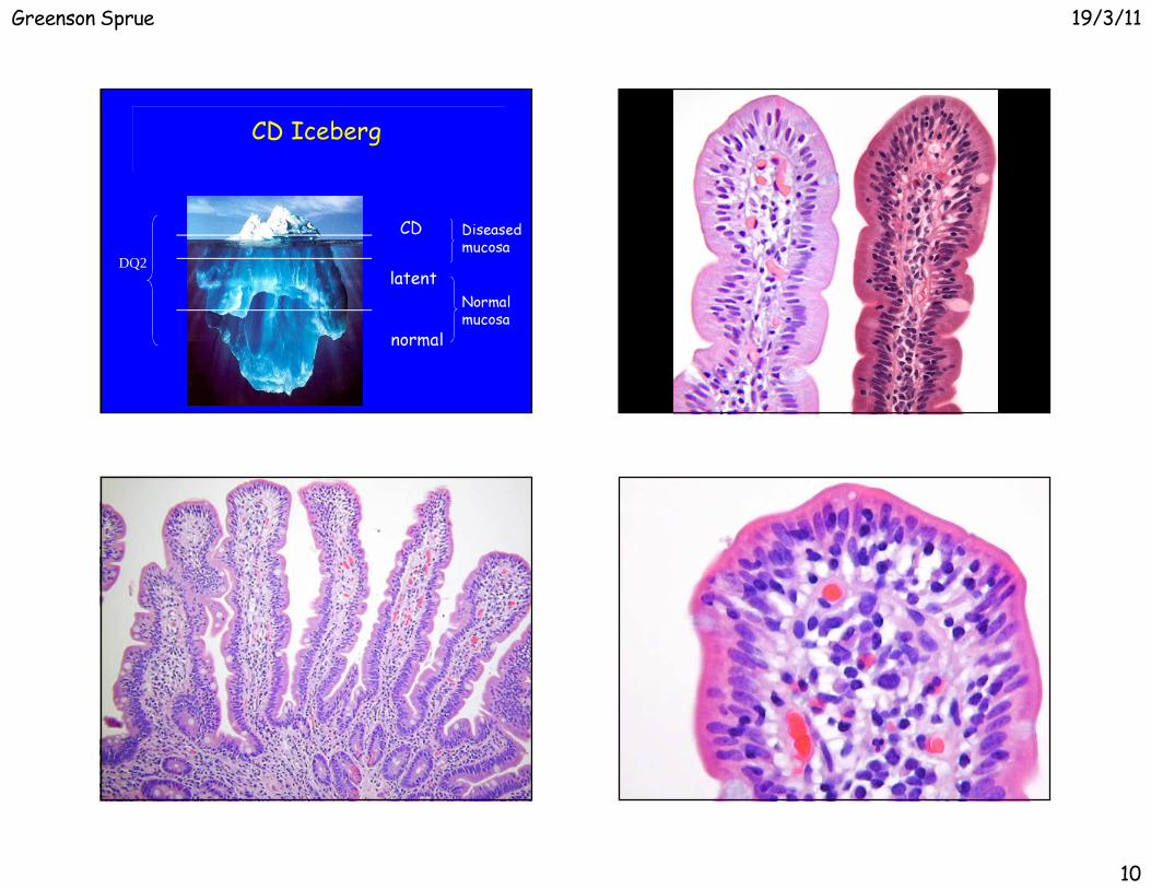
CD Iceberg
latent
normal
Diseased mucosa
Normal mucosa
DQ2___________________________
___________________________
___________________________ CD

Greenson Sprue 19/3/11
11
How many IELs are abnormal?
�>25/100 epithelial cells�>40/100 epithelial cells�>12/20 epithelial cells on the tips of villi
– Decrescendo pattern is normal– Diffuse pattern is abnormal – Goldstein Am J Clin Pathol 116;63-71,2001�>8/20 epithelial cells in the tips of villi
– CD3 stains– Biagi et al J Clin Pathol 57;835-839, 2004
But what does it all mean?�2-3 % of small bowel biopsies have normal architecture
with increased IELs�Depending on the type of study and the country the
study was carried out in, anywhere from 9 to 40% of such cases represent (pre) celiac disease.– Whether such patients need any therapy is controversial
Brown I,et al. Arch Pathol Lab Med 130;1020-25, 2006
Normal Architecture Increased IELs
� Gluten Sensitive Enteropathy– Early type 1 lesion or treated sprue�Other food hypersensitivity� H. Pylori (usually only in bulb)� Autoimmune conditions (RA, SLE, MS, Graves, Hashimoto’s,
Diabetes)� Post-infection� Drugs (NSAIDs, PPIs??)� Bacterial Overgrowth�Obesity� Crohn’s disease and Ulcerative colitis

Greenson Sprue 19/3/11
12
Results
Other Diagnoses: Graft versus Host Disease, Combined Variable Immunodeficiency, Diabetes mellitus 1, Juvenile Rheumatoid Arthritis, Systemic Lupus Erythematosis, Tropical Sprue, Ulcerative Colitis
Diseases Associated with Marsh 1 Lesions
CD, 19
Idiopathic, 31
NSAID, 17
Crohn's, 7
Bacterial Overgrowth, 7
H. pylori, 7
IBS, 9 Other, 7
Celiac DiseaseComplications
�Refractory Celiac Disease�Ulcers of Small Bowel�Collagenous Sprue�Malignancy
– T cell Lymphoma of gut and regional nodes– Adenocarcinoma of small bowel– Squamous cell carcinoma of esophagus and oropharynx
Refractory Celiac Disease�Develops in about 5% of celiac patients
– Malabsorption, diarrhea, pain, wt loss�Divided into types I and II�Type I RCD: IELs are normal / not clonal
– better prognosis– Can progress to Type II�Type II RCD: IELs are aberrant / clonal
– 50% mortality rate
Refractory Celiac Disease�IELs in Celiac disease and type I RCD are CD3 + and CD8 +�IELs in type II RCD are CD3 + and CD8 -
– Will have T-cell gene rearrangements– Will also loose staining for T-cell receptor αβ

Greenson Sprue 19/3/11
13
CD3 CD8
Small Intestinal Ulcers In Celiac Disease

Greenson Sprue 19/3/11
14
LYMPHOMA INCELIAC DISEASE

Greenson Sprue 19/3/11
15
Malabsorption
Remain Well
Benign Ulcer Refractory Celiac Disease Lymphoma
Deterioration
Response No Response(Refractory Sprue)
Gluten Free Diet
Sprue-like Changes
Celiac Disease Histologic Mimics
�Celiac-related– Lymphoma (EATCL)– Collagenous Sprue�Other luminal antigens other than gluten/gliadin
– Soy protein�General
– Peptic duodenitis– Tropical Sprue, Bacterial overgrowth– Autoimmune enteropathy– Infections/immunodeficiencies– Crohn’s disease
Greenson Sprue 19/3/11
16
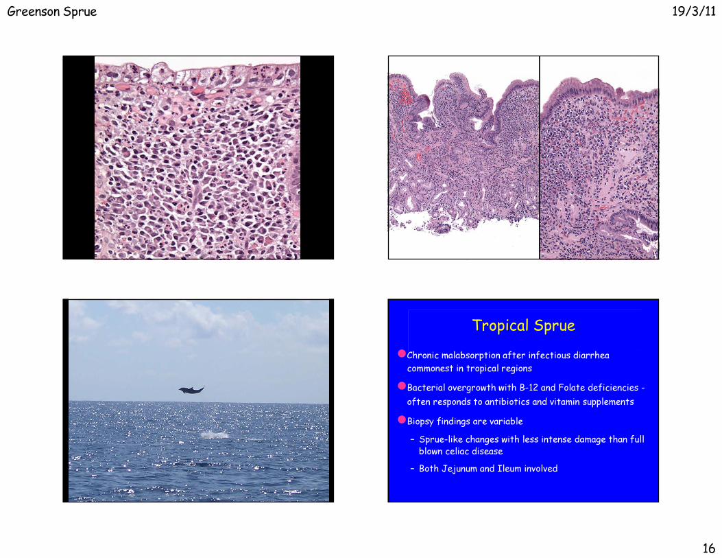
Tropical Sprue�Chronic malabsorption after infectious diarrhea
commonest in tropical regions�Bacterial overgrowth with B-12 and Folate deficiencies -
often responds to antibiotics and vitamin supplements�Biopsy findings are variable
– Sprue-like changes with less intense damage than full blown celiac disease
– Both Jejunum and Ileum involved

Greenson Sprue 19/3/11
17
Stasis Syndrome(Bacterial Overgrowth)
�Crohn’s Disease�Diverticular Disease�Scleroderma�Pseudo-obstruction�Post-Surgical
– Blind loop or Pouch– Entero-enterostomy– Afferent loop– Fistulae- Adhesions/partial obstruction

Greenson Sprue 19/3/11
18
Bacterial OvergrowthBiopsy Findings
�Irregular Villi�Surface cell damage�Plasmacytosis�Neutrophils�Crypt Hyperplasia�“Doesn’t fit”

Greenson Sprue 19/3/11
19
Autoimmune Enteropathy�Childhood onset - usually prior to age 1
– Intractable diarrhea not relieved by TPN– Anti-enterocyte antibodies (requires indirect immunofluorescence)– Other autoantibodies (islet cell, parietal cell)– FOXP3 mutation– X-linked with polyendocrinopathy�Also Adult onset (not well known)
– Anti-enterocyte antibodies or anti-goblet cell antibodies

Greenson Sprue 19/3/11
20
Pouchitis and Diversion ColitisIt’s the Surgeon’s Fault!
Greenson Sprue 19/3/11
21
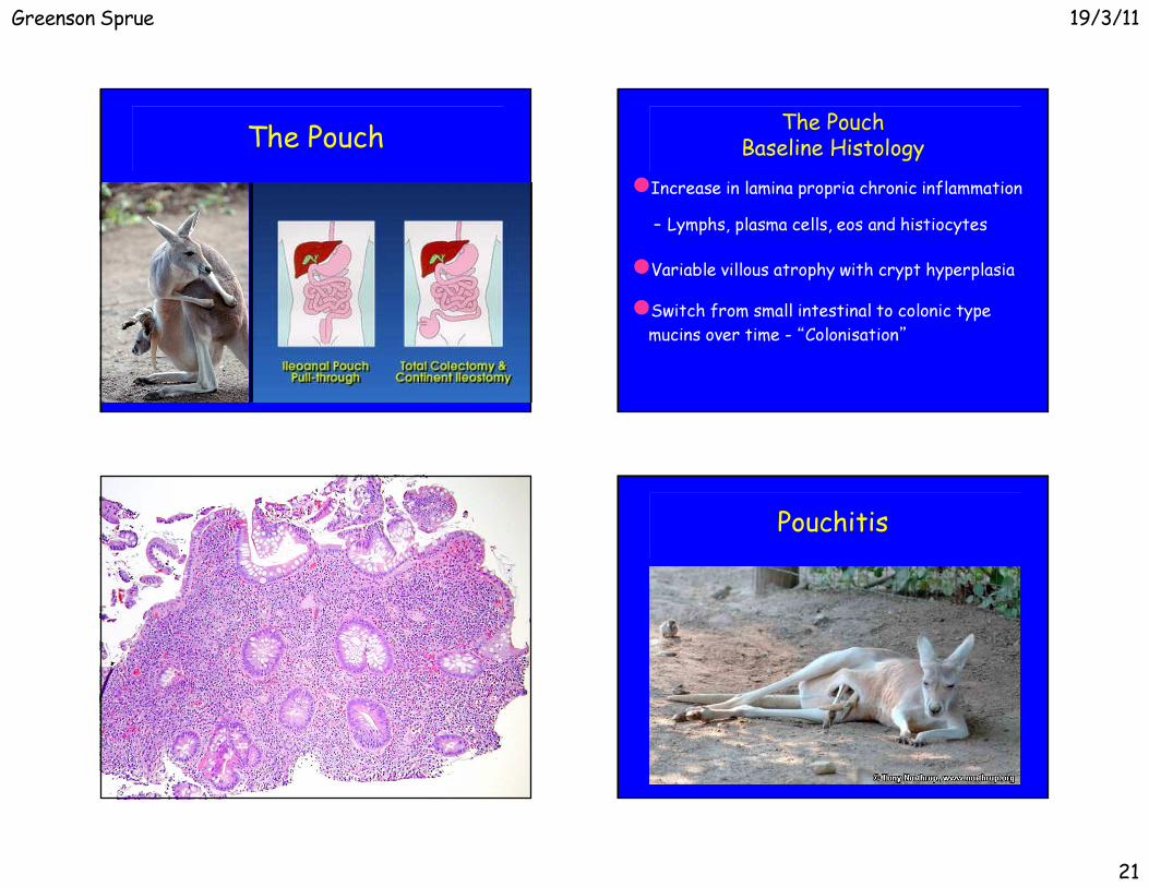
The Pouch The PouchBaseline Histology
�Increase in lamina propria chronic inflammation– Lymphs, plasma cells, eos and histiocytes�Variable villous atrophy with crypt hyperplasia�Switch from small intestinal to colonic type mucins over time - “Colonisation”
Pouchitis

Greenson Sprue 19/3/11
22
PouchitisClinical Symptoms
�Occurs in 7-50% of pouch patients (avg. 32%)�Abdominal pain and fever �Bloody stools, increased frequency, incontinence�Often responds to antibiotics�10-20% refractory to therapy -?chronic IBD
– Much more common in UC than FAP patients– Highest incidence in UC patients with PSC
PouchitisClinical Syndromes
�Responsive to Antibiotics (Flagyl)– Bacterial overgrowth�Refractory Pouchitis
– Irritable pouch syndrome - no path changes– Short strip pouchitis - UC in retained rectal mucosa
– Chronic primary refractory pouchitis - active inflammation in bxs.
PouchitisHistopathology
�Villous blunting and chronic inflammtion are part of the “baseline pouch” and these changes do not correlate with clinical symptoms�Active inflammation does correlate with clinical symptoms– Erosions– Ulcers– Cryptitis
Greenson Sprue 19/3/11
23
The End