Embed Size (px)
Citation preview

i
The sciatic nerve division in the gluteal region in a South African population:
An anatomical study
by
Bryan Jason Bergsteedt (BScHons)
Thesis presented in fulfilment of the requirements for the degree of
Master of Science in the Faculty of Medicine and Health Sciences at
Stellenbosch University
Supervisor: Ms LM Greyling
April 2019

ii
DECLARATION
By submitting this thesis, I declare that the entirety of the work contained therein is my own,
original work, that I am the sole author thereof (save to the extent explicitly otherwise stated),
that reproduction and publication thereof by Stellenbosch University will not infringe any third
party rights and that I have not previously in its entirety or in part submitted it for obtaining any
qualification.
April 2019
Bryan Jason Bergsteedt
Copyright © 2019 Stellenbosch University
All rights reserved
Stellenbosch University https://scholar.sun.ac.za
iii
ABSTRACT
The sciatic nerve is repeatedly involved in the daily medical practices of anaesthesia, neurology,
orthopaedics and rehabilitative medicine. The sciatic nerve, and its branches, are some of the
most frequently injured nerves within the human body. A possible reason for injury could be
related to an inadequate knowledge of the anatomical variations of this nerve. Adequate
understanding of the anatomical variability within the gluteal region is vital for appropriate
diagnosis, potential treatment of gluteal pathology and pain and population-specific anomalies.
To the author’s best knowledge, no previous study has described the anatomical variations in
relation to the piriformis and sciatic nerve bifurcation within the South African population.
Therefore, the aim of the study is to report the prevalence of anatomical variations within the
course of the sciatic nerve in relation to the piriformis muscle. Additionally, to report the
prevalence of the variations in the level of the sciatic nerve bifurcation. Lastly, to analyse the
typical sciatic nerve and piriformis morphomety. The results obtained will be a comparison
between sides, sexes, and population groups.
For the purpose of this study, lower limbs (𝑁 = 340) from 170 South African cadavers were
selected for dissection and morphological analysis. These specimens consisted of 191 males
and 149 females, and comprised of three South African subpopulation groups, namely,
White/Caucasian (𝑛 = 232), Mixed race (𝑛 = 78) and South African Black (𝑛 = 30). The
variations were recorded, classified and described. Piriformis and sciatic nerve parameters were
measured morphometrically using a digital sliding calliper, and statistically analysed.
Analysis of the relationship between piriformis and the sciatic nerve resulted in 43 (12.65%)
specimens that presented variations in the morphology, while 297 (87.35%) specimens
presented normal anatomical features. Variations of these structures occurred predominantly in
the South African White/Caucasian population. The bifurcation of the sciatic nerve occurred
mainly in the popliteal fossa proper (79.6%). The width of the sciatic nerve was significantly
larger in the White/Caucasian group (𝑝 < 0.05), in comparison to the other two groups. The
mean length of the sciatic nerve was significantly larger in the male specimens (𝑝 < 0.05) in
comparison to the female specimens.
It was found that the sciatic nerve commonly entered the gluteal region as a single trunk,
through the infra-piriform space, inferior to the piriformis muscle. However, variations in the
anatomy of the sciatic nerve are common, and are vital in assessing clinical risk, and avoiding
debilitating injury or incorrect pain diagnoses. To maintain best possible clinical practices
requires regularly updated clinical skills in relation to accurate and relevant new anatomical
Stellenbosch University https://scholar.sun.ac.za

iv
knowledge. It is for this reason that studies, such as this one, ensure that vital research
contributions are available for best clinical practice. Clear uniform landmarks for morphometric
analysis of the sciatic nerve and piriformis needs to be established in order to create uniformity
and understanding of results. Additionally, there is a need for the increase in published literature
for the South African subpopulation groups in order to strengthen comparisons and conclusions
of reported research. Researchers also need to research variations in larger groups within the
South African population.
Stellenbosch University https://scholar.sun.ac.za
v
OPSOMMING
N. ischiadicus is dikwels betrokke in mediese praktyke soos narkose, neurologie, ortopedie en
rehabilitasie geneeskunde. N. ischiadicus, sy vertakkings, is ook die senuwee in die menslike
liggaam wat die meeste beseer word. Onvoldoende kennis oor anatomiese variasies van die
senuwee is ’n moontlike rede tot beserings. Dit is van kardinale belang om voldoende kennis
van die anatomiese variasies in die gluteale gebied op te doen, vir korrekte diagnose en
potensiële behandeling van gluteale patologie en -pyn, asook van bevolkingspesifieke
anomalieë.
Geen vorige studies wat die verskille in die verhouding tussen m. piriformis en bifurkasie van
n. isciadicus beskryf, bestaan, sover die navorser kon bepaal, vir die bevolkingsgroepe in Suid-
Afrika nie. Die doel van hierdie studie was dus om verslag te doen oor die voorkoms van
anatomiese variasies in die verloop van n. ischiadicus in verhouding tot m. piriformis. Die
voorkoms van variasies op die vlak van bifurkasie van n. ischiadicus is ook bestudeer. Laastens
is ‘n tipiese n.ischiadicus en m. piriformis morfometries geanaliseer. Die resultate is tussen
linker en regter kante van die liggaam, geslagte en bevolkingsgroepe vergelyk.
Vir die doel van hierdie studie is onderste ledemate (𝑁 = 340) van 170 Suid-Afrikaanse
kadawers vir disseksie en morfologiese analise, geselekteer. Die liggame wat bestudeer is, het
bestaan uit 191 mans en 149 vroue, en is oor drie Suid-Afrikaanse bevolkingsgroepe versprei,
naamlik Wit (𝑛 = 232), Kleurling (𝑛 = 78) en Swart (𝑛 = 30). Verskille is bestudeer,
geklassifiseer en beskryf. Morfometriese afmetings van m. piriformis- en n. isciadicus is met
behulp van ’n digitale gly-meetpasser gedoen en statisties geanaliseer.
Na analise van die verhouding tussen m. piriformis en n. ischiadicus, is bevind dat in 43
(12.65%) van die liggame daar morfologie verskille is, terwyl in 297 (87.35%) van die liggame
‘n normale anatomies patroon vertoon. Daar is bevind dat verskille in morfologie van die
strukture hoofsaaklik in die Wit Suid-Afrikaanse bevolkingsgroep voorgekom. Bifurkasie van
n. ischiadicus het hoofsaaklik in die popliteale fossa self (79.6%) voorgekom. Die breedte van
n. ischiadicus was noemenswaardig groter in die Wit bevolkings groep wat bestudeer is (p <
0.05) in vergelyking met die ander twee bevolkings groep is. Die gemiddelde lengte van n.
ischiadicus was noemenswaardig langer in die manlike groep (p < 0.05), teenoor die van die
vroulik groep.
N. ischiadicus gaan gewoonlik as ’n enkele stam die gluteale gebied binne. Die senuwee beweeg
inferior tot m. piriformis. Variasies in die anatomie van n. ischiadicus kom algemeen voor.
Kennis van hierdie wariasies is klinies belangrik om risiko’s te beperk en om debiliterende
Stellenbosch University https://scholar.sun.ac.za

vi
beserings of foutiewe diagnose van pyn te voorkom. Ten einde die handhawing van die beste
moontlike kliniese praktyk te verseker, vereis dit dat kliniese vaardighede, met betrekking tot
akkurate en relevante anatomiese kennis, op ’n konstante basis verfyn word, en gereeld
opgedateer word. Dit is om hierdie rede dat studies soos hierdie belangrike bydraes lewer vir
die beste kliniese uitkomste. Duidelike, uniforme landmerke vir die morfometriese analise van
n. ischiadicus en m. piriformis moet bepaal word om sodoende uniformiteit te verseker.
Daarbenewens is daar ook ’n behoefte vir meer gepubliseerde literatuur oor Suid-Afrikaanse
bevolkingsgroepe, sodat vergelykings en gevolgtrekkings wat gerapporteer word, ondersteun
kan word. Daar is ook ‘n behoefte vir groter studies om beter verteen woordigig van
verskillende bevolkings groepe in Suid Afrika te beskryf.
Stellenbosch University https://scholar.sun.ac.za
vii
ACKNOWLEDGEMENTS
The completion of this masters thesis would not have been possible without the support,
guidance, assistance and contributions from the following individuals and institutions:
- To my supervisor, Ms Linda Greyling, for her guidance, expertise and kindness. She
took me on as a student after adversity, and still managed to make it memorable.
- To my fellow colleagues and postgraduate students at the division of Clinical Anatomy,
at Stellenbosch University, for their constant support and assistance throughout the
duration of this thesis. Thank you to Prof Ben Page for his unwavering support; Dr
Betha Bastiaanse for her patience and wisdom; and Mrs Mandi Alblas for all her
assistance, and for listening to my ideas and complaints. To the new head of the division,
Dr Karin Baatjes. I look forward to my future at the division, and growth under her
leadership.
- To Prof Hans Strijdom, my mentor, for his support, understanding, and patience this
year. I look forward to our time together and I am thankful for the guidance and wisdom.
- To the School of Anatomical Sciences, at the University of the Witwatersrand, and
especially Dr Nanette Briers, for going the extra mile to make me feel welcome and part
of the team during the short time during which data collection was completed.
- To the division of Clinical Anatomy, at the University of Cape Town, and especially
Mr Michael Cassar, for the assistance and freedom to complete data collection.
- To Prof Martin Kidd for assistance with statistical analysis.
- Finally, thank you to my loved ones. To my friends, and especially Kim, Lyndon, Sarah,
Ziningi, Mohapi, Waseemah, Ndivhuwo, Heike, Ed and Elri. You have helped more
than you know. I cannot wait to grow old with you all. To my extended family, your
love and support are all I need at the end of a long day. To my partner and best friend
Nicola Jo Bruns, I could not have completed this thesis without you there, right next to
me at every step along this journey. Thank you for your constant encouragement and
compassion; I love you. Thank you to George and Joanne Bruns, I appreciate you more
than you know. Thank you to my siblings, Matthew and Darren, for being a phone call
away and always cheering me up when I need it most; I love you. Thank you to Allison
Evette Bergsteedt, who is the reason why I constantly strive for better. Your strength,
love and laughter nourishes my soul, and are constant reminders to never give up.
This thesis is dedicated to my parents:
Allison Evette Bergsteedt & Craig Kelvin Bergsteedt
While this will never be sufficient, thank you for all the sacrifices that were made, and the
unrelenting support in providing me with the opportunities that were never available to you.
“True love is selfless. It is prepared to sacrifice”
Stellenbosch University https://scholar.sun.ac.za

viii
CONTENTS
DECLARATION ....................................................................................................................... ii
ABSTRACT .............................................................................................................................. iii
OPSOMMING ........................................................................................................................... v
ACKNOWLEDGEMENTS ..................................................................................................... vii
CONTENTS ............................................................................................................................ viii
TABLES .................................................................................................................................... xi
FIGURES ................................................................................................................................. xii
ABBREVIATIONS .................................................................................................................. xv
MATHEMATICAL SYMBOLS AND UNITS ..................................................................... xvii
CHAPTER ONE: INTRODUCTION ....................................................................................... 1
CHAPTER TWO: LITERATURE REVIEW ........................................................................... 4
2.1 Regional Anatomy ....................................................................................................... 5
2.1.1 Muscles of the Gluteal Region ............................................................................. 6
2.1.2 Sub-gluteal Space ................................................................................................. 6
2.1.3 Piriformis .............................................................................................................. 8
2.1.4 The Sciatic Nerve ............................................................................................... 10
2.2 Anatomical variations within this region................................................................... 12
2.2.1 The Relationship between the Sciatic Nerve and Piriformis .............................. 12
2.2.2 The Level of Sciatic Nerve Division .................................................................. 23
2.2.3 Literature in Africa ............................................................................................. 24
2.3 Clinical Consideration ............................................................................................... 26
2.3.1 Pain ..................................................................................................................... 26
2.4.2 Sciatic Nerve Block ............................................................................................ 28
2.4.3 Intramuscular Injections ..................................................................................... 29
2.4.4 Piriformis Syndrome .......................................................................................... 30
CHAPTER THREE: AIMS AND OBJECTIVES .................................................................... 34
Stellenbosch University https://scholar.sun.ac.za

ix
3.1 Problem Statement ..................................................................................................... 35
3.2 Aims........................................................................................................................... 35
3.3 Objectives .................................................................................................................. 36
CHAPTER FOUR: MATERIALS AND METHODS ............................................................. 37
4.1 Ethical approval ......................................................................................................... 38
4.2 Acquisition of Cadavers ............................................................................................ 38
4.3 Study Material ........................................................................................................... 39
4.4 Measurement of the structures ................................................................................... 42
4.4.1 Piriformis muscle ............................................................................................... 42
4.4.2 Sciatic nerve ....................................................................................................... 45
4.4.3 Sciatic nerve bifurcation level index .................................................................. 47
4.5 Documentation of the variations ................................................................................ 49
4.6 Statistical Analysis .................................................................................................... 49
CHAPTER FIVE: RESULTS ................................................................................................. 50
5.1 Overview ................................................................................................................... 51
5.2 Anatomical variations of piriformis .......................................................................... 51
5.2.1 Overview ............................................................................................................ 51
5.2.2 Type A ................................................................................................................ 53
5.2.3 Type B ................................................................................................................ 55
5.2.4 Type C ................................................................................................................ 58
5.2.5 Additional Variation ........................................................................................... 61
5.2.6 Fused piriformis muscle and gluteus medius muscle ......................................... 62
5.3 Variations in the sciatic nerve bifurcation ................................................................. 65
5.3.1 Overview ............................................................................................................ 65
5.3.2 Type A ................................................................................................................ 67
5.3.3 Type D ................................................................................................................ 69
5.3.4 Type E ................................................................................................................ 69
5.3.5 Type F ................................................................................................................ 69
Stellenbosch University https://scholar.sun.ac.za

x
5.4 Morphometric Measurements .................................................................................... 70
5.4.1 Measurements of piriformis ............................................................................... 72
5.4.1.1 Medial width of piriformis .......................................................................... 72
5.4.1.2 Lateral width of piriformis .......................................................................... 73
5.4.1.3 Length of the superior border of piriformis ................................................ 76
5.4.1.4 Length of the inferior border of piriformis ................................................. 77
5.4.2 Measurements related to the sciatic nerve .......................................................... 78
5.4.2.1 Width of the sciatic nerve ........................................................................... 78
5.4.2.2 Length of the sciatic nerve .......................................................................... 80
5.4.2.3 Length of the thigh ...................................................................................... 82
CHAPTER SIX: DISCUSSION ............................................................................................. 85
6.1 Anatomical variations of piriformis .......................................................................... 86
6.2 Variations in the sciatic nerve bifurcation level ........................................................ 88
6.3 Morphometric Analysis ............................................................................................. 91
CHAPTER SEVEN: CONCLUSION ...................................................................................... 94
LIMITATIONS AND FUTURE STUDIES ............................................................................ 96
REFERENCES ......................................................................................................................... 98
Stellenbosch University https://scholar.sun.ac.za
xi
TABLES
Table 2.1: Contents of the sub-gluteal space. ............................................................................. 8
Table 2.2: Summary of the possible variations in the relationship between the sciatic nerve and
piriformis. ................................................................................................................................. 21
Table 2.3: Shows basic details of previous studies conducted on the sciatic nerve variations in
relation to piriformis. ................................................................................................................ 22
Table 2.4: The level of sciatic nerve division in the lower limb. ............................................. 23
Table 4.1: Basic information regarding the specimens used in the present study. .................. 39
Table 4.2: Parameters of the morphometric measurements. .................................................... 42
Table 4.3: Theoretical summary of the sciatic nerve bifurcation level index. ......................... 48
Table 5.1: A summary of piriformis and sciatic nerve variations found in the study, along with
additional sample characteristics. ............................................................................................. 52
Table 5.2: Summary of the sciatic nerve bifurcation level classified in the study, along with
additional sample characteristics. ............................................................................................. 66
Table 5.3: Summary of the morphometric measurements conducted in present study (measured
in mm). ..................................................................................................................................... 71
Stellenbosch University https://scholar.sun.ac.za
xii
FIGURES
Figure 2.1: The superficial (A) and deep (B) muscles of the gluteal region. ............................. 5
Figure 2.2: The normal anatomy of the subgluteal space.. ........................................................ 7
Figure 2.3: The Piriformis muscle, along with important structures in the gluteal region ........ 9
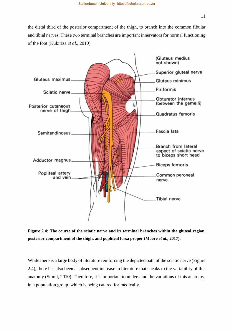
Figure 2.4: The course of the sciatic nerve and its terminal branches within the gluteal region,
posterior compartment of the thigh, and popliteal fossa proper. .............................................. 11
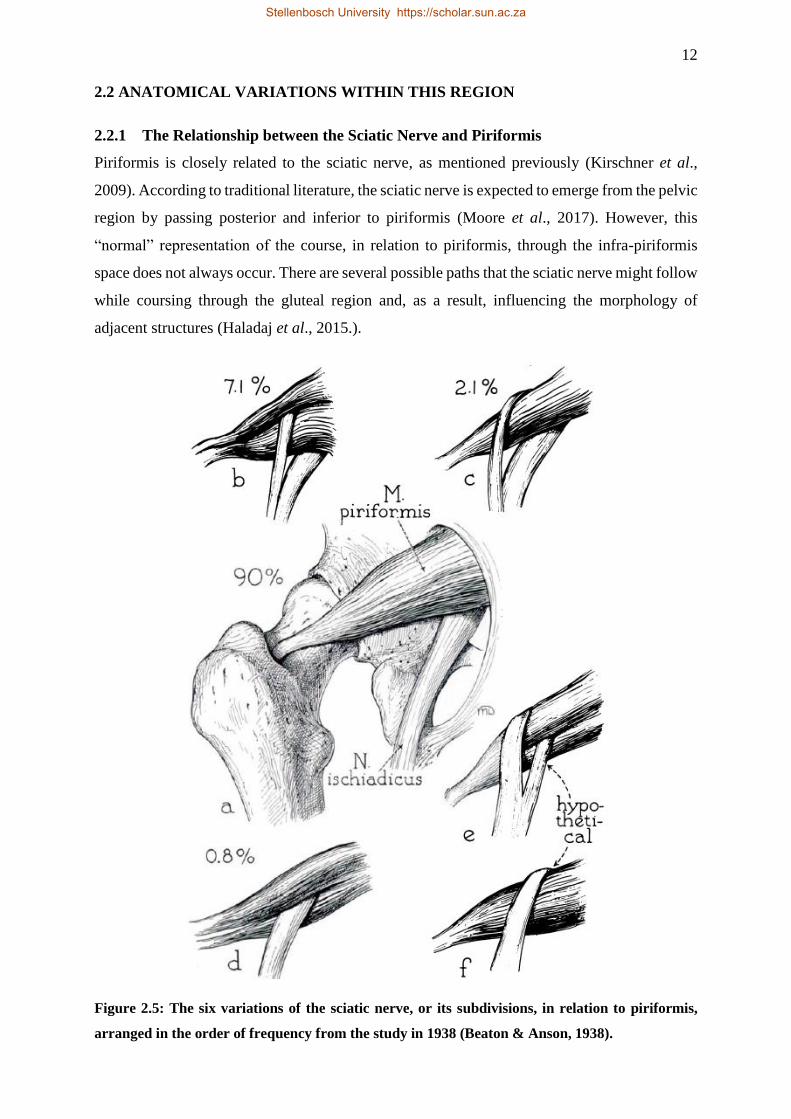
Figure 2.5: The six variations of the sciatic nerve, or its subdivisions, in relation to piriformis,
arranged in the order of frequency from the study in 1938 (Beaton & Anson, 1938). ............ 12
Figure 2.6: The variations in the relationship between piriformis and the sciatic nerve, within
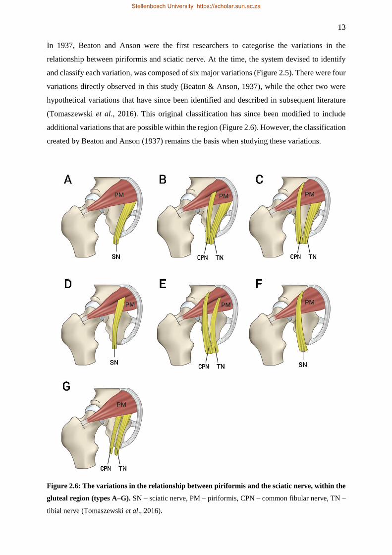
the gluteal region (types A–G). ................................................................................................ 13
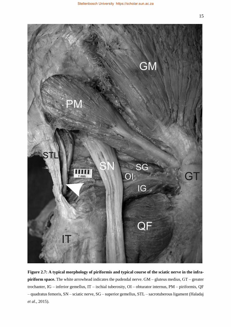
Figure 2.7: A typical morphology of piriformis and typical course of the sciatic nerve in the
infra-piriform space. ................................................................................................................. 15
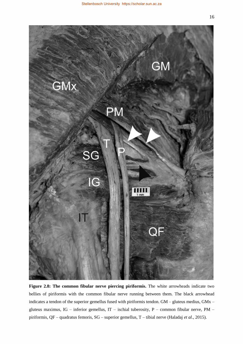
Figure 2.8: The common fibular nerve piercing piriformis. .................................................... 16
Figure 2.9: Common fibular nerve running through the supra-piriform space. The tibial nerve
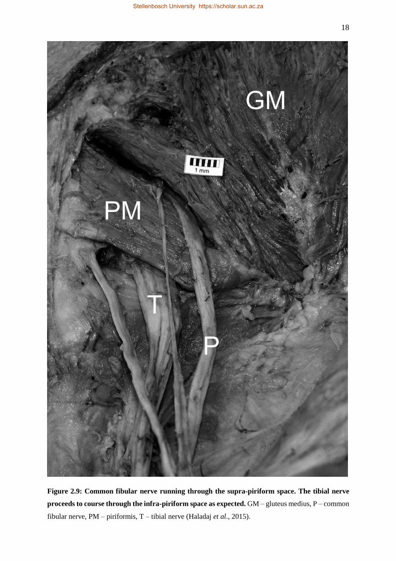
proceeds to course through the infra-piriform space as expected. ........................................... 18
Figure 2.10: Fusion of piriformis and gluteus medius. ............................................................ 19
Figure 2.11: Map depicting countries in Africa where previous sciatic nerve and piriformis
variation related research have been published.. ...................................................................... 24
Figure 4.1: Summary of methodology. Specimens were collected and grouped according to the
two overarching variations observed: the variation in the relationship between piriformis and
the sciatic nerve, and the variation in the level of bifurcation of the sciatic nerve. ................. 41
Figure 4.2: Measurement of the medial width of piriformis, using the sliding digital calliper.
.................................................................................................................................................. 43
Figure 4.3: Measurement of the lateral width of piriformis, at the middle of the muscle belly.
This measurement was taken using a sliding digital calliper. .................................................. 43
Figure 4.4: Measurement of the superior border of piriformis, using the sliding digital calliper.
.................................................................................................................................................. 44
Figure 4.5: Measurement of the inferior border of piriformis, using a sliding digital calliper. 44
Figure 4.6: Measurement of the width of the sciatic nerve, as the nerve becomes visible at the
inferior border of piriformis. .................................................................................................... 45
Figure 4.7: Measurement of the length of the sciatic nerve, from the level of the inferior border
of piriformis, to its terminal bifurcation in the posterior compartment of the thigh. ............... 46
Stellenbosch University https://scholar.sun.ac.za
xiii
Figure 4.8: Measurement of the thigh length, from the most proximal point on the greater
trochanter of the femur, to the middle of the popliteal fossa proper. ....................................... 46
Figure 5.1: The typical morphology of piriformis and course of the sciatic nerve, in a right
gluteal region. ........................................................................................................................... 53
Figure 5.2: The typical morphology of piriformis and typical course of the sciatic nerve in the
infra-piriform space. ................................................................................................................. 54
Figure 5.3: The common fibular nerve piercing piriformis.. ................................................... 55
Figure 5.4: The common fibular nerve piercing piriformis. .................................................... 56
Figure 5.5: The common fibular nerve piercing piriformis. .................................................... 57
Figure 5.6: The common fibular nerve runs through the supra-piriform space. ...................... 58
Figure 5.7: The common fibular nerve runs through the supra-piriform space. ...................... 59
Figure 5.8: The common fibular nerve runs through the supra-piriform space. ...................... 60
Figure 5.9: The common fibular nerve runs through the supra-piriform space. ..................... 61
Figure 5.10: Fusion of piriformis and gluteus medius in the left gluteal region. ..................... 62
Figure 5.11: Fusion of piriformis and gluteus medius in the right gluteal region. ................... 63
Figure 5.12: Fusion of piriformis and gluteus medius in the right gluteal region. ................... 64
Figure 5.13: Fusion of piriformis and gluteus medius in the left gluteal region.. .................... 64
Figure 5.14: Sciatic nerve bifurcating superiorly, within the pelvic region. ............................ 67
Figure 5.15: The common fibular and tibial nerve converge, within the gluteal region, forming
the sciatic nerve.. ...................................................................................................................... 68
Figure 5.16: A histogram summarising the measurements for the medial width of piriformis.
.................................................................................................................................................. 72
Figure 5.17: A histogram summarising the measurements for the lateral width of piriformis. 73
Figure 5.18: The effect that sex and side has on the lateral width of piriformis. ..................... 74
Figure 5.19: The effect of population group and side on the lateral width of piriformis. ........ 75
Figure 5.20: A histogram summarising the measurements for the superior border length of the
piriformis. ................................................................................................................................. 76
Figure 5.21: A histogram summarising the inferior border length of piriformis measurements.
.................................................................................................................................................. 77
Figure 5.22: A histogram summarising the measurements for the width of the sciatic nerve. 78
Figure 5.23: The effect of population group and side to evaluate the width of the sciatic nerve.
.................................................................................................................................................. 79
Figure 5.24: A histogram summarising the measurements for the length of the sciatic nerve 80
Figure 5.25: The effect that sex and side has on the length of the sciatic nerve. ..................... 81
Figure 5.26: A histogram summarising the measurements for the length of the thigh. ........... 82
Stellenbosch University https://scholar.sun.ac.za

xiv
Figure 5.27: The effect of sex and side on the length of the thigh. .......................................... 83
Figure 5.28: The effect of population group and side on the length of the thigh. .................... 84
Stellenbosch University https://scholar.sun.ac.za

xv
ABBREVIATIONS
CPN Common fibular nerve
GM Gluteus Medius
GM&PM Fused gluteus medius and piriformis
GMin Gluteus minimus
GMx Gluteus maximus
GT Greater trochanter
IG Inferior gemellus
IGV Inferior gluteal vessels
IT Ischial tuberosity
LWP Lateral width of piriformis
MWP Medial width of piriformis
OI Obturator internus
P Common fibular nerve
PIBL Piriformis inferior border length
PM Piriformis muscle
PM1 Superior piriformis belly
PM2 Inferior piriformis belly
PSBL Piriformis superior border length
QF Quadratus femoris
SG Superior gemellus
SGV Superior gluteal vessels
SN Sciatic nerve
SNBLI Sciatic nerve bifurcation level index
SNL Sciatic nerve length
SNW Sciatic nerve width
Stellenbosch University https://scholar.sun.ac.za

xvi
SP Sacral plexus
SU Stellenbosch University
STL Sacrotuberous ligament
T Tibial nerve
TCox Triceps coxae muscle group
TL Thigh length
TN Tibial nerve
TNB1 Tibial nerve branch one
TNB2 Tibial nerve branch two
UCT University of Cape Town
UP University of Pretoria
Wits University of the Witwatersrand
Stellenbosch University https://scholar.sun.ac.za

xvii
MATHEMATICAL SYMBOLS AND UNITS
≅ Approximately equal to
= Equal to
𝓍
𝓎 Fraction (division)
> Greater than
< Lesser than
≤ Lesser than or equal to
× Multiply
% Percentage
± Plus / minus
cm centimetre
mm Millimetre
Stellenbosch University https://scholar.sun.ac.za

1
CHAPTER ONE:
INTRODUCTION
Stellenbosch University https://scholar.sun.ac.za
2
The sciatic nerve is constantly involved in the daily medical practices of anaesthesia, neurology,
orthopaedics, and rehabilitative medicine (Brooks et al., 2011). The sciatic nerve, and its
branches, are also the most frequently injured nerves within the human body (Kumar et al.,
2011; Budhiraja et al., 2016). This nerve is unique because of its extensive course throughout
the gluteal region, and lower limbs (Kotian et al., 2015). This intricately long course makes the
nerve vulnerable to injury from various medical causes (Prakash et al., 2010; Kotian et al.,
2015; Budhiraja et al., 2016). Another reason for injury could be related to an inadequate
knowledge regarding anatomical variations of the nerve (Budhiraja et al., 2016; Kiros &
Woldeyes, 2015). Therefore, knowledge of the nerve, and its variations, are of vital concern in
clinical science (Prakash et al., 2010; Ogeng'o et al., 2011).
The anatomy of the sciatic nerve has been described extensively in literature (Kanawati, 2014).
However, this is not the case for the variations of the sciatic nerve, as well as the subsequent
variations of structures closely related to, and innervated by this nerve, within the gluteal region
(Budhiraja et al., 2016). One such association, which is of increasing concern, is the close
relationship between piriformis and the sciatic nerve (Brooks et al., 2011). The interest in this
relationship is largely due to its possible involvement in producing sciatic-related pain and
discomfort. The existence of variations in the anatomy of the sciatic nerve only increases the
risk of damage to this nerve during clinical and surgical procedures (Smoll, 2010). Several
authors have identified variations of the levels of the sciatic nerve bifurcations into its tibial and
common fibular branches. The branching variation could occur proximally, close to its origin
from the sacral plexus, or more distally, within the popliteal fossa proper. Should the branching
occur superiorly, proximal to the piriformis, there is a possibility of variations in the
morphology of piriformis itself. These anatomical variations could give rise to a number of
possible debilitating conditions within the region innervated by the sciatic nerve. Sufficient
knowledge about the variations of the sciatic nerve, and its bifurcation, are fundamental in the
prevention of injury to the nerve (Ogeng'o et al., 2011). A number of techniques performed in,
and out, of the operating theatre often require access to this region. If complications within the
gluteal region occur, resulting from surgical complications or the presence of unknown
variations within the region, there could be considerable and permanent impairment to the
structures of the lower limb. These complications range from general irritation, to loss of
sensation in the posterior compartment of the thigh, foot, or the entire lower limb (Kiros &
Woldeyes, 2015).
Stellenbosch University https://scholar.sun.ac.za
3
Variations of the sciatic nerve, and its bifurcation, occurs with varying prevalence
(Tomaszewski et al., 2016) amongst different population groups across the world. While
extensive research on the sciatic nerve is done in Northern and Western regions of the world,
this is not the case for the African continent. Upon reviewing literature, only six academic
papers were found from medical universities in Africa, namely: Kenya, Ethiopia, Uganda, and
Nigeria (Ogeng'o et al., 2011; Kiros & Woldeyes, 2015). Considering the lack of research on
variations of the sciatic nerve for the African population (Ogeng'o et al., 2011), this research
hopes to address some of the problems by looking at the prevalence of sciatic nerve variations,
and related variations of the piriformis muscle, in a South Africa population. Dissected cadaver
lower limbs will be used for the study. According to Prakash et al. (2010), the cadaver is the
best means to study anatomy at this level, even with advances in medical technology (Smoll,
2010; Brooks et al., 2011).
Stellenbosch University https://scholar.sun.ac.za

4
CHAPTER TWO:
LITERATURE REVIEW
Stellenbosch University https://scholar.sun.ac.za
5
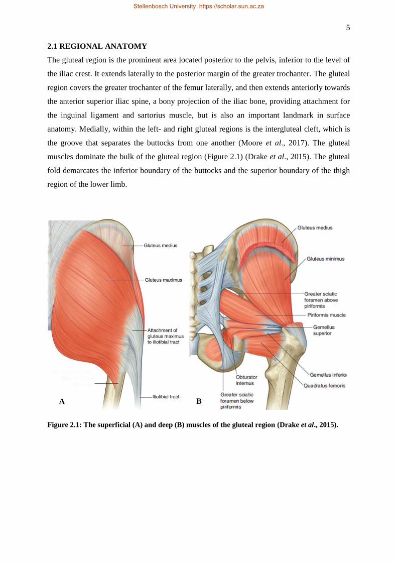
2.1 REGIONAL ANATOMY
The gluteal region is the prominent area located posterior to the pelvis, inferior to the level of
the iliac crest. It extends laterally to the posterior margin of the greater trochanter. The gluteal
region covers the greater trochanter of the femur laterally, and then extends anteriorly towards
the anterior superior iliac spine, a bony projection of the iliac bone, providing attachment for
the inguinal ligament and sartorius muscle, but is also an important landmark in surface
anatomy. Medially, within the left- and right gluteal regions is the intergluteal cleft, which is
the groove that separates the buttocks from one another (Moore et al., 2017). The gluteal
muscles dominate the bulk of the gluteal region (Figure 2.1) (Drake et al., 2015). The gluteal
fold demarcates the inferior boundary of the buttocks and the superior boundary of the thigh
region of the lower limb.
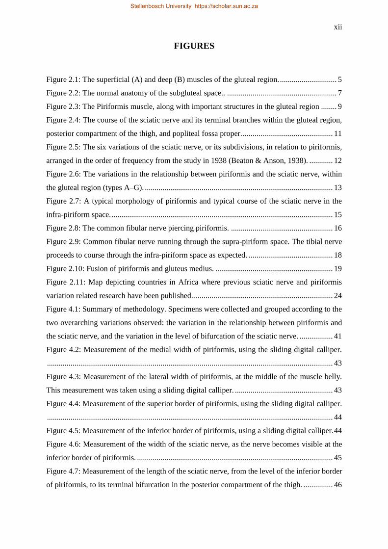
Figure 2.1: The superficial (A) and deep (B) muscles of the gluteal region (Drake et al., 2015).
B A
Stellenbosch University https://scholar.sun.ac.za

6
2.1.1 Muscles of the Gluteal Region
The muscles of the gluteal region (Figure 2.1) share a common compartment, but are organised
into two layers, namely the superficial- and deep layers (Moore et al., 2017). The superficial
layer of muscles of the gluteal region consists of the three large overlapping glutei – the gluteus
maximus, medius, and minimus. The last muscle in this layer is the tensor fasciae latae (Moore
et al., 2017). The latter is closely associated with gluteus maximus as both muscles’ fibres join
and attach to a common insertion structure, known as the iliotibial tract. The superficial gluteal
muscles are all proximally attached to the posterolateral surface and margins of the ala of the
ilium, and have common insertion points on various sites of the proximal femur, or iliotibial
tract, laterally (Hansen, 2014). The glutei muscles are mainly responsible for abduction and
extension of the hip, and extension, abduction, and medial rotation of the thigh (Drake et al.,
2015).
The deep layer of muscles in the gluteal region consists of smaller muscles, such as piriformis,
obturator internus, superior and inferior gemelli, and quadratus femoris. The inferior half of
gluteus maximus covers them all (Hansen, 2014). These muscles all have distal attachments on
or adjacent to the intertrochanteric crest of the femur. These muscles are lateral rotators of the
thigh, but they also stabilize the hip joint, working with the strong ligaments of the hip joint to
steady the femoral head in the acetabulum (Moore et al., 2017).
2.1.2 Sub-gluteal Space
The sub-gluteal space, also known as the deep gluteal space, represents the cellular and fatty
tissue located between the middle and deep gluteal aponeurosis layers (Hernando et al., 2015).
The sub-gluteal space largely represents the space deep to the belly of gluteus maximus (Byrd,
2015). The inferior margin of the sub-gluteal space is continuous with the superior contents of
the posterior compartment of the thigh, with the proximal attachment of the hamstring muscles
at the ischial tuberosity structurally demarcating this border (Park et al., 2016). The lateral lip
of the linea aspera and gluteal tuberosity of the proximal femur mark the most lateral boundary
of this space, along with the lateral fusion of both the middle and deep gluteal aponeurosis
layers, which extend up to tensor fascia latae via the iliotibial tract. The posterior surface of the
femoral neck, associated posterior acetabular column, hip joint capsule, and the greater- and
lesser trochanters of the femur all contribute in forming the anterior limit of this space (Martin
et al., 2015). The inferior margin of the superior aspect of the sciatic notch forms the superior
boundary. Lastly, the sacrotuberous ligament and the falciform fascia jointly create the medial
limit of the sub-gluteal space (Figure 2.2) (Hernando et al., 2015).
Stellenbosch University https://scholar.sun.ac.za
7
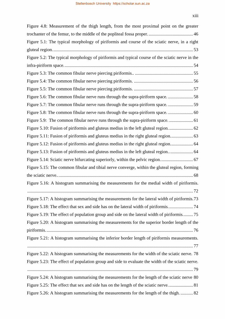
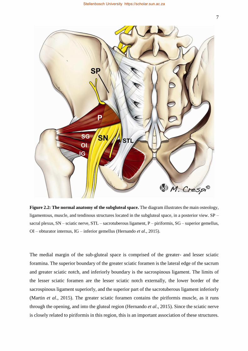
Figure 2.2: The normal anatomy of the subgluteal space. The diagram illustrates the main osteology,
ligamentous, muscle, and tendinous structures located in the subgluteal space, in a posterior view. SP –
sacral plexus, SN – sciatic nerve, STL – sacrotuberous ligament, P – piriformis, SG – superior gemellus,
OI – obturator internus, IG – inferior gemellus (Hernando et al., 2015).
The medial margin of the sub-gluteal space is comprised of the greater- and lesser sciatic
foramina. The superior boundary of the greater sciatic foramen is the lateral edge of the sacrum
and greater sciatic notch, and inferiorly boundary is the sacrospinous ligament. The limits of
the lesser sciatic foramen are the lesser sciatic notch externally, the lower border of the
sacrospinous ligament superiorly, and the superior part of the sacrotuberous ligament inferiorly
(Martin et al., 2015). The greater sciatic foramen contains the piriformis muscle, as it runs
through the opening, and into the gluteal region (Hernando et al., 2015). Since the sciatic nerve
is closely related to piriformis in this region, this is an important association of these structures.
Stellenbosch University https://scholar.sun.ac.za
8
Table 2.1: Contents of the sub-gluteal space.
Contents
• Superior and inferior gluteal nerves
• Superior and inferior gluteal blood vessels
• Ischium
• Sacrotuberous- and sacrospinous ligaments
• Sciatic nerve
• Piriformis
• Obturator Internus
• Obturator Externus
• Gemelli
• Quadratus femoris
• Hamstring muscle group
The sub-gluteal space contain an array of structures, ranging from vascular and neural to
muscular tissue (Park et al., 2016). The contents of the sub-gluteal space are listed above, in
table 2.1 (Byrd, 2015). In the sub-gluteal space, the sciatic nerve enters the pelvic region
through the greater sciatic foramen, to pass inferior to piriformis (Knudsen, Mei-Dan & Brick,
2016). The sciatic nerve has a significant mobility associated with overall hip movement
(Coppieters et al., 2006). According to Coppieters et al. (2006), the sciatic nerve has a range of
28 mm of displacement during hip flexion (Martin et al., 2015). Under normal circumstances,
the sciatic nerve is able to stretch and glide to accommodate moderate strain or compression
associated with joint movement (Martin et al., 2010). However, this motion is affected by
anatomical variations in both the sciatic nerve and piriformis in 16.2% of the population, as
reported by Smoll (2010). Any of the contents of the sub-gluteal space, and the wide range of
variability in displacement of the sciatic nerve in this space, can cause sciatic nerve entrapment-
related syndromes (Park et al., 2016).
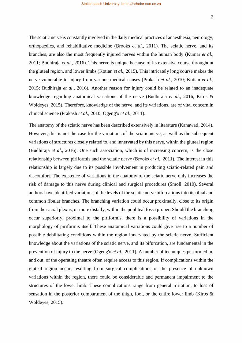
2.1.3 Piriformis
Piriformis is a flat, triangular muscle. It belongs to the deep muscle group within the gluteal
region, where piriformis is the most superiorly placed muscle of the group (Drake et al., 2015).
In the gluteal region, piriformis is located deep to the large gluteus maximus and proximal to
the obturator internus, where the obturator internus is part of the triceps coxae group, along
with the superior and inferior gemelli (Pecina et al., 2001). Piriformis arises from the
Stellenbosch University https://scholar.sun.ac.za
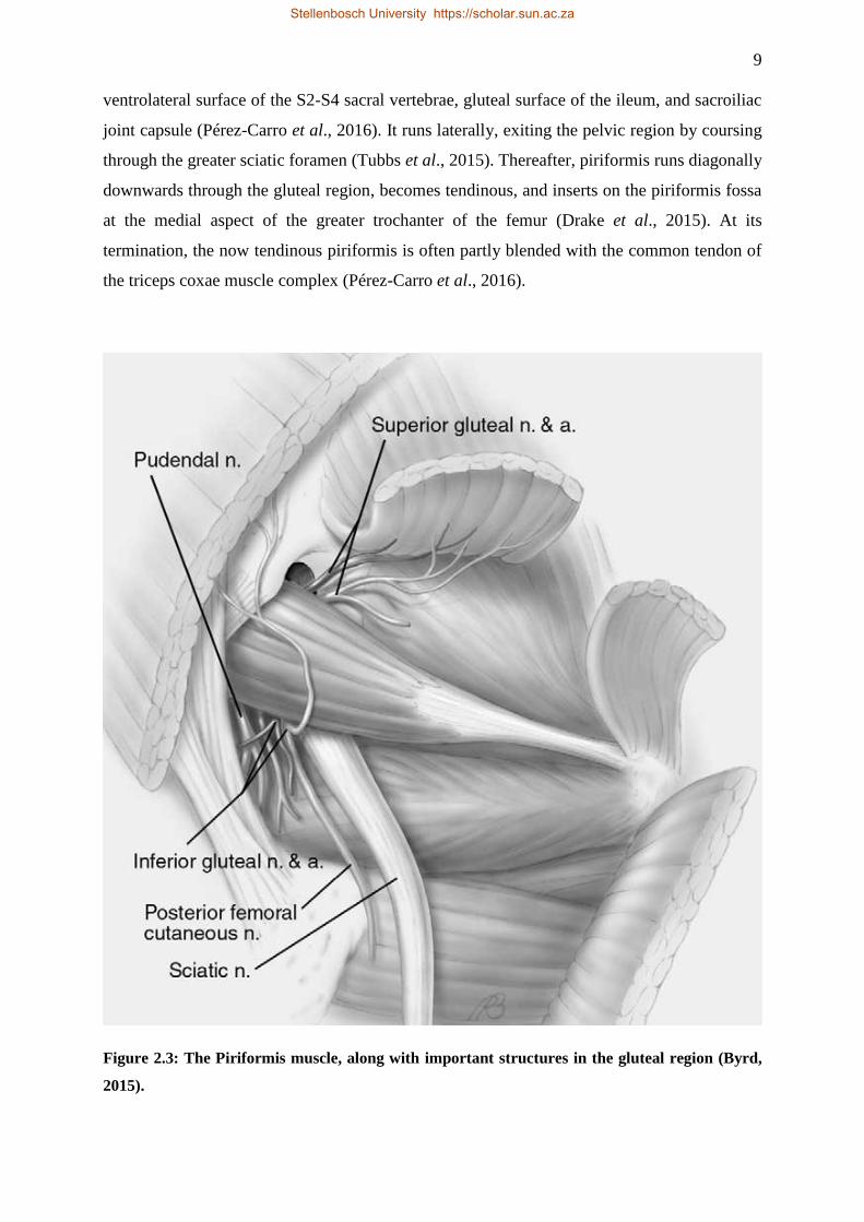
9
ventrolateral surface of the S2-S4 sacral vertebrae, gluteal surface of the ileum, and sacroiliac
joint capsule (Pérez-Carro et al., 2016). It runs laterally, exiting the pelvic region by coursing
through the greater sciatic foramen (Tubbs et al., 2015). Thereafter, piriformis runs diagonally
downwards through the gluteal region, becomes tendinous, and inserts on the piriformis fossa
at the medial aspect of the greater trochanter of the femur (Drake et al., 2015). At its
termination, the now tendinous piriformis is often partly blended with the common tendon of
the triceps coxae muscle complex (Pérez-Carro et al., 2016).
Figure 2.3: The Piriformis muscle, along with important structures in the gluteal region (Byrd,
2015).
Stellenbosch University https://scholar.sun.ac.za

10
The main functions of the piriformis involve abduction of the flexed thigh, and lateral rotation
of the extended thigh (Moore et al., 2017). During walking, abduction of the flexed thigh is
important, because it shifts the body weight to the opposite side of the body and ultimately
prevents falling (Siddiq et al., 2017). Additionally, piriformis stabilizes the femoral head in the
acetabulum. This stabilization is essential for postural stability during ambulation, as well as
standing (Tubbs et al., 2015).
Due to the muscle’s central position in the area, piriformis is the landmark structure of the
gluteal region (Moore et al., 2017). Consequently, piriformis provides the key to understanding
relationships in the pelvic region, and gluteal region. Additionally, piriformis demarcates the
two zones of structural passage within the gluteal region. The passage superior to piriformis,
through which the superior gluteal nerve and vessels course, is known as the supra-piriform
space. Conversely, the passage inferior to piriformis, in the gluteal region, is infrequently
referred to as the infra-piriform space (Knudsen et al., 2016). Piriformis thus determines the
nomenclature of the vessels and neurovascular structures in the region, depending on the course
of these vessels in relation to piriformis (Pérez-Carro et al., 2016). While the superior gluteal
nerves and vessels traverse the supra-piriformis space, the inferior gluteal and pudendal nerves,
and the sciatic nerve passes through the infra-piriformis space (Michel et al., 2013). For these
various reasons, the relationship shared by the sciatic nerve and piriformis becomes a focal
point in the gluteal region.
2.1.4 The Sciatic Nerve
The sciatic nerve is the largest nerve in the human body, as well as in many other animals
(Saritha et al., 2012). There are two reasons for this; one being that there are numerous neural
fibres that gather to produce this nerve, and secondly, due to the extensive size of the gluteal
region and lower limb, in other words, the regions innervated by this nerve (Figure 2.4) (Saleh
et al., 2009). The sciatic nerve develops from the lumbo-sacral plexus, from the fourth lumbar
to the third sacral (L4-L5 and S1-S3) spinal nerves. The nerve often has a maximum width of
two centimetres (cm) or more, and can reach a diameter of over 0.5 cm, as it passes posterior
and inferior to piriformis (Saleh et al., 2009). The sciatic nerve is described as the nerve with
the largest diameter in the body. The nerve carries both motor and sensory fibres. The sciatic
nerve enters the gluteal region, through the lower part of the greater sciatic foramen, below
piriformis, as a single nerve encompassed by a single epineural sheath (Kotian et al., 2015),
and thereafter continues into the lower limb. The sciatic nerve is the chief innervator of the
posterior compartment of the thigh, the anterior, lateral and posterior compartments of the lower
leg, and dorsal and plantar regions of the foot. Generally, this nerve terminates in approximately
Stellenbosch University https://scholar.sun.ac.za
11
the distal third of the posterior compartment of the thigh, to branch into the common fibular
and tibial nerves. These two terminal branches are important innervators for normal functioning
of the foot (Kukiriza et al., 2010).
Figure 2.4: The course of the sciatic nerve and its terminal branches within the gluteal region,
posterior compartment of the thigh, and popliteal fossa proper (Moore et al., 2017).
While there is a large body of literature reinforcing the depicted path of the sciatic nerve (Figure
2.4), there has also been a subsequent increase in literature that speaks to the variability of this
anatomy (Smoll, 2010). Therefore, it is important to understand the variations of this anatomy,
in a population group, which is being catered for medically.
Stellenbosch University https://scholar.sun.ac.za
12
2.2 ANATOMICAL VARIATIONS WITHIN THIS REGION
2.2.1 The Relationship between the Sciatic Nerve and Piriformis
Piriformis is closely related to the sciatic nerve, as mentioned previously (Kirschner et al.,
2009). According to traditional literature, the sciatic nerve is expected to emerge from the pelvic
region by passing posterior and inferior to piriformis (Moore et al., 2017). However, this
“normal” representation of the course, in relation to piriformis, through the infra-piriformis
space does not always occur. There are several possible paths that the sciatic nerve might follow
while coursing through the gluteal region and, as a result, influencing the morphology of
adjacent structures (Haladaj et al., 2015.).
Figure 2.5: The six variations of the sciatic nerve, or its subdivisions, in relation to piriformis,
arranged in the order of frequency from the study in 1938 (Beaton & Anson, 1938).
Stellenbosch University https://scholar.sun.ac.za
13
In 1937, Beaton and Anson were the first researchers to categorise the variations in the
relationship between piriformis and sciatic nerve. At the time, the system devised to identify
and classify each variation, was composed of six major variations (Figure 2.5). There were four
variations directly observed in this study (Beaton & Anson, 1937), while the other two were
hypothetical variations that have since been identified and described in subsequent literature
(Tomaszewski et al., 2016). This original classification has since been modified to include
additional variations that are possible within the region (Figure 2.6). However, the classification
created by Beaton and Anson (1937) remains the basis when studying these variations.
Figure 2.6: The variations in the relationship between piriformis and the sciatic nerve, within the
gluteal region (types A–G). SN – sciatic nerve, PM – piriformis, CPN – common fibular nerve, TN –
tibial nerve (Tomaszewski et al., 2016).
Stellenbosch University https://scholar.sun.ac.za
14
The variations categorized in the literature (Figure 2.6), can broadly be subdivided into two
groups. The first group comprises of variations where the sciatic nerve enters the gluteal region,
posterior and inferior to piriformis, as a single common trunk, comprised of the common fibular
and tibial nerves. The second group is categorised by a sciatic nerve that has bifurcated
proximally, within the pelvic region, into its terminal branches, the common fibular and tibial
nerves (Tomaszewski et al., 2016). The most important aspect about the two subgroups is how
the bifurcated sciatic nerve exits in relation to piriformis (Guvencer et al., 2008). It is for this
reason that the aforementioned classifications (Figure 2.6) rely heavily on how structural
morphology impacts on neighbouring structures (Natsis et al., 2014).
The classification, type A (Figure 2.6), is generally the most common morphological
representation of these structures, where the sciatic nerve enters the gluteal region as a single
entity, posterior and inferior piriformis, which is also a single, undivided muscle. According to
a meta-analysis conducted by Tomaszewski et al. (2016), this occurrence had a pooled
prevalence of 85.2% in 7068 lower limbs investigated. This is accurately visualised in figure
2.7, where a typical course of the sciatic nerve passes posteriorly and inferior to a typical
piriformis muscle.
Type B, is a variation that has a pooled prevalence of 9.8%, far less than that of type A
(Tomaszewski et al., 2016). In this version of the variation, the sciatic nerve is already
bifurcated, where both the common fibular and tibial nerves exit the pelvis as separate entities
within the gluteal region. Furthermore, the common fibular nerve is piercing piriformis, which
is now split into two different bellies of the same muscle, with the same origin and insertion
points. Moreover, the tibial nerve courses below piriformis, which is the expected course for
the undivided sciatic nerve (Figure 2.6). This is also depicted in figure 2.8, by Haladaj et al.
(2015), where the common fibular nerve is clearly seen piercing piriformis, thereby separating
piriformis into two bellies. In this image, the larger tibial division of the sciatic nerve is passing
posterior and inferior to the smaller, inferiorly placed belly of piriformis (Figure 2.8).
Furthermore, additional variations that have occurred in the right gluteal region of this lower
limb can be observed, as a result of the variation of the sciatic nerve and piriformis (Haladaj et
al., 2015). In this image (Figure 2.8), a black arrowhead is used to indicate the tendon of
superior gemellus, which has fused with the piriformis tendon. While this tendon would
traditionally attach to the trochanteric fossa of the femur, distally (Moore et al., 2017), as can
be seen in figure 2.7, this is not the case in figure 2.8, where piriformis and sciatic nerve
variations have subsequently effected other structures in this region.
Stellenbosch University https://scholar.sun.ac.za
15
Figure 2.7: A typical morphology of piriformis and typical course of the sciatic nerve in the infra-
piriform space. The white arrowhead indicates the pudendal nerve. GM – gluteus medius, GT – greater
trochanter, IG – inferior gemellus, IT – ischial tuberosity, OI – obturator internus, PM – piriformis, QF
– quadratus femoris, SN – sciatic nerve, SG – superior gemellus, STL – sacrotuberous ligament (Haladaj
et al., 2015).
Stellenbosch University https://scholar.sun.ac.za
16
Figure 2.8: The common fibular nerve piercing piriformis. The white arrowheads indicate two
bellies of piriformis with the common fibular nerve running between them. The black arrowhead
indicates a tendon of the superior gemellus fused with piriformis tendon. GM – gluteus medius, GMx –
gluteus maximus, IG – inferior gemellus, IT – ischial tuberosity, P – common fibular nerve, PM –
piriformis, QF – quadratus femoris, SG – superior gemellus, T – tibial nerve (Haladaj et al., 2015).
Stellenbosch University https://scholar.sun.ac.za

17
The third variation in the series, type C, is where the sciatic nerve has bifurcated prior to exiting
the pelvis (Figure 2.9). In this instance, the common fibular nerve exits along with the superior
gluteal vessels, in the supra-piriform space, before proceeding to travel inferiorly, into the lower
limb. The tibial nerve, on the other hand, exits within the infra-piriform space, as is traditionally
expected from a common sciatic nerve, as seen in the most frequently occurring type A.
Therefore, the two terminal branches of the sciatic nerve wrap around a complete piriformis
without piercing the muscle.
In type D, the sciatic nerve pierces piriformis. Here, there are two bellies of piriformis, with the
same origin and insertion points. The sciatic nerve remains unbranched, but does not travel
posterior and inferior to piriformis, though the infra-piriform space, as expected. Instead, the
defining feature of type C is that the intact sciatic nerve pierces piriformis. Therefore, the nerve
will travel through piriformis, before traveling inferior, to the posterior compartment of the
thigh, where it will proceed to bifurcate into its terminal branches, the common fibular and
tibial nerves (Figure 2.5).
Type E has many similarities to the second variation (type B). Once again, this type of variation
presents the terminal branches of the sciatic nerve entering the gluteal region separately. The
common fibular nerve enters through the supra-piriform space with the superior gluteal vessels,
and crosses piriformis before traveling inferiorly, towards the posterior compartment of the
thigh. The tibial nerve exits the pelvic region and pierces piriformis, thereby creating two
muscle bellies (Figure 2.5). In this variation, no sciatic nerve branch runs posterior and inferior
to piriformis, through the infra-piriform space.
Type F is another variation where the sciatic nerve does not branch in the pelvic region. Rather,
the sciatic nerve enters the gluteal region unbranched, but travels through the supra-piriform
space, accompanied by the superior gluteal vessels (Figure 2.5). The sciatic nerve enters the
gluteal region superior to piriformis, crosses piriformis, and then proceeds to follow its expected
route inferiorly towards the posterior compartment of the thigh.
The last variation expected in the gluteal region is type G (Figure 2.5). This variation is not too
different from type A. However, in this instance the sciatic nerve has already bifurcated into
terminal branches when it exits the pelvic region. Thus, the structures entering the gluteal region
include the common fibular and tibial nerves. These bifurcated terminal branches of the sciatic
nerve do not pierce piriformis. None of the two branches enters the gluteal region through the
supra-piriformis region, and instead course posterior and then inferior to piriformis, as two
separate nerves, through the infra-piriform space.
Stellenbosch University https://scholar.sun.ac.za
18
Figure 2.9: Common fibular nerve running through the supra-piriform space. The tibial nerve
proceeds to course through the infra-piriform space as expected. GM – gluteus medius, P – common
fibular nerve, PM – piriformis, T – tibial nerve (Haladaj et al., 2015).
Stellenbosch University https://scholar.sun.ac.za

19
Figure 2.10: Fusion of piriformis and gluteus medius. The white arrowhead indicates the superior
gluteal vessels, running between fibres of the fused muscle belly. GM & PM – fused gluteus medius and
piriformis, GT – greater trochanter, SN – sciatic nerve (Haladaj et al., 2015).
Stellenbosch University https://scholar.sun.ac.za
20
An additional variation, not described in previous literature, can occur in the gluteal region.
This variation involves gluteus medius and piriformis, and is not part of the standard
classification of sciatic nerve variations in the gluteal region, as seen in figure 2.5. This variation
occurs when gluteus medius fuses with piriformis, resulting in one continuous muscle layer that
occupies a large area within the deeper layer of the gluteal region and deep to gluteus maximus.
This variation is important because it has a significant effect on the gluteal vessels that enter
the gluteal region. Once the muscles are fused, the supra-piriform space is eliminated. This
means that there is no room for the superior gluteal vessels to course though the opening that
would normally be present between gluteus medius and piriformis. Therefore, the superior
gluteal vessels runs along the middle of this fused muscle. Recently, the variation was
mentioned by Haladaj et al., in 2016, and can be seen in figure 2.10. Furthermore, should any
one of the types A-G occur in conjunction with the fused gluteus medius and piriformis, the
sciatic nerve or its terminal branches, will be affected, along with the superior gluteal vessels.
The types A, B and C have been reported frequently in literature. According to Tomaszewski
et al. (2016), types D, E, F, and G are rarer, and reported in less than 1% of the populations
studied. In the study conducted by Beaton and Anson, in 1938, the types E and F were only
hypothesized, and not seen in their study sample. Moreover, type G was not found and described
by Beton and Anson (1937), but was added as a modification of the different types of variations
at a much later stage (Guvencer et al., 2008). The possible variations that can occur in the
gluteal region, namely types A-G, and shown in figure 2.5, have been summarised in table 2.2,
below.
Stellenbosch University https://scholar.sun.ac.za
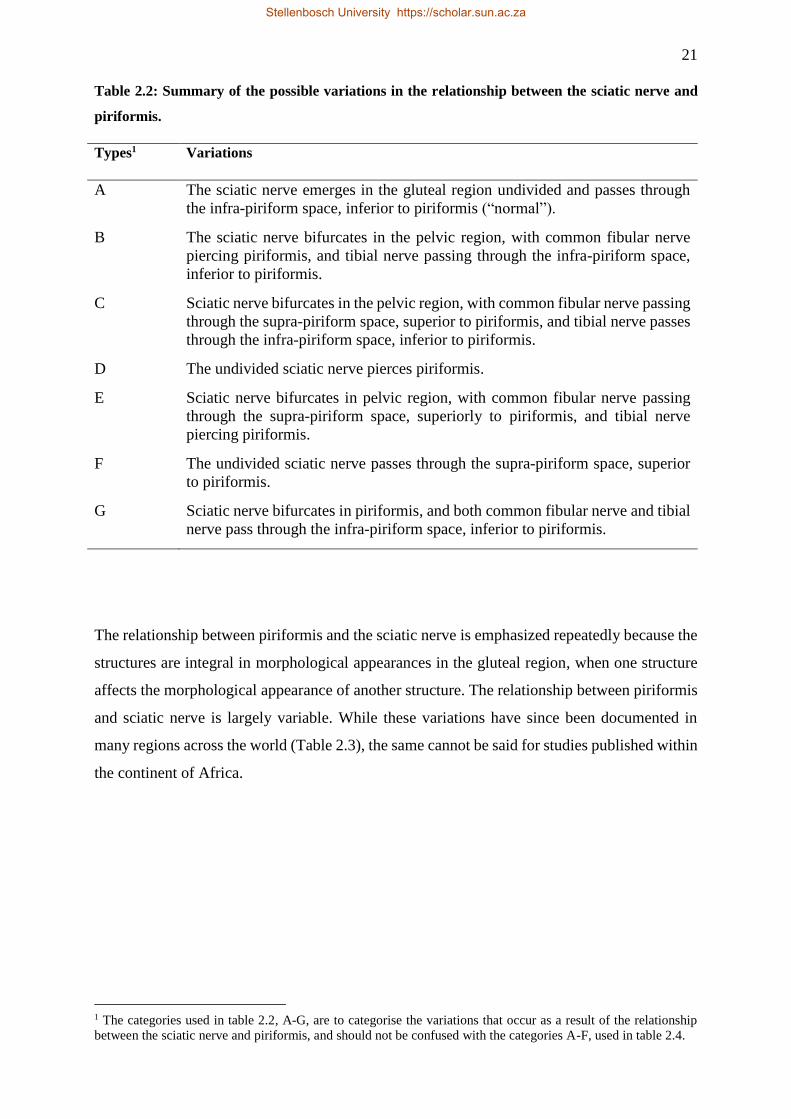
21
Table 2.2: Summary of the possible variations in the relationship between the sciatic nerve and
piriformis.
Types1 Variations
A The sciatic nerve emerges in the gluteal region undivided and passes through
the infra-piriform space, inferior to piriformis (“normal”).
B The sciatic nerve bifurcates in the pelvic region, with common fibular nerve
piercing piriformis, and tibial nerve passing through the infra-piriform space,
inferior to piriformis.
C Sciatic nerve bifurcates in the pelvic region, with common fibular nerve passing
through the supra-piriform space, superior to piriformis, and tibial nerve passes
through the infra-piriform space, inferior to piriformis.
D The undivided sciatic nerve pierces piriformis.
E Sciatic nerve bifurcates in pelvic region, with common fibular nerve passing
through the supra-piriform space, superiorly to piriformis, and tibial nerve
piercing piriformis.
F The undivided sciatic nerve passes through the supra-piriform space, superior
to piriformis.
G Sciatic nerve bifurcates in piriformis, and both common fibular nerve and tibial
nerve pass through the infra-piriform space, inferior to piriformis.
The relationship between piriformis and the sciatic nerve is emphasized repeatedly because the
structures are integral in morphological appearances in the gluteal region, when one structure
affects the morphological appearance of another structure. The relationship between piriformis
and sciatic nerve is largely variable. While these variations have since been documented in
many regions across the world (Table 2.3), the same cannot be said for studies published within
the continent of Africa.
1 The categories used in table 2.2, A-G, are to categorise the variations that occur as a result of the relationship
between the sciatic nerve and piriformis, and should not be confused with the categories A-F, used in table 2.4.
Stellenbosch University https://scholar.sun.ac.za
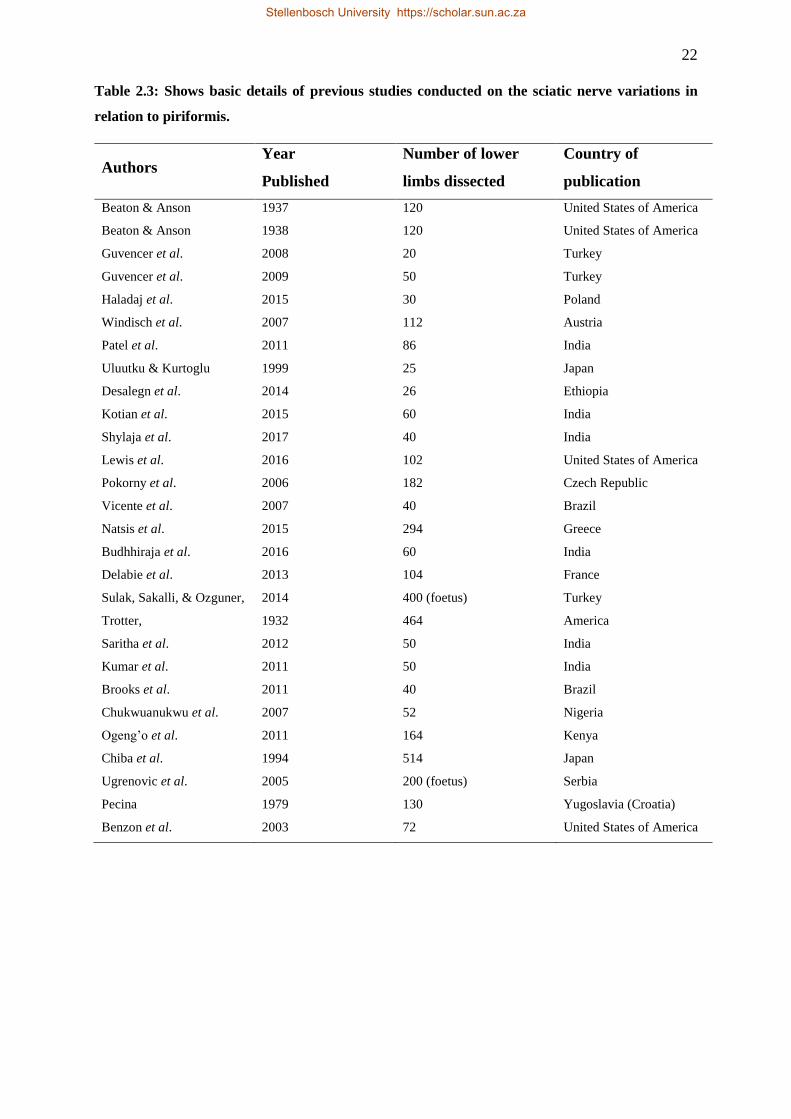
22
Table 2.3: Shows basic details of previous studies conducted on the sciatic nerve variations in
relation to piriformis.
Authors Year
Published
Number of lower
limbs dissected
Country of
publication
Beaton & Anson 1937 120 United States of America
Beaton & Anson 1938 120 United States of America
Guvencer et al. 2008 20 Turkey
Guvencer et al. 2009 50 Turkey
Haladaj et al. 2015 30 Poland
Windisch et al. 2007 112 Austria
Patel et al. 2011 86 India
Uluutku & Kurtoglu 1999 25 Japan
Desalegn et al. 2014 26 Ethiopia
Kotian et al. 2015 60 India
Shylaja et al. 2017 40 India
Lewis et al. 2016 102 United States of America
Pokorny et al. 2006 182 Czech Republic
Vicente et al. 2007 40 Brazil
Natsis et al. 2015 294 Greece
Budhhiraja et al. 2016 60 India
Delabie et al. 2013 104 France
Sulak, Sakalli, & Ozguner, 2014 400 (foetus) Turkey
Trotter, 1932 464 America
Saritha et al. 2012 50 India
Kumar et al. 2011 50 India
Brooks et al. 2011 40 Brazil
Chukwuanukwu et al. 2007 52 Nigeria
Ogeng’o et al. 2011 164 Kenya
Chiba et al. 1994 514 Japan
Ugrenovic et al. 2005 200 (foetus) Serbia
Pecina 1979 130 Yugoslavia (Croatia)
Benzon et al. 2003 72 United States of America
Stellenbosch University https://scholar.sun.ac.za
23
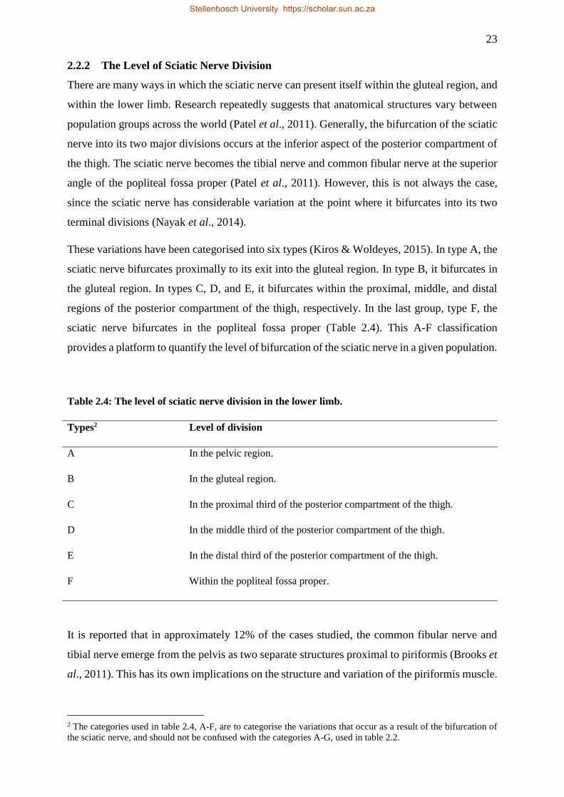
2.2.2 The Level of Sciatic Nerve Division
There are many ways in which the sciatic nerve can present itself within the gluteal region, and
within the lower limb. Research repeatedly suggests that anatomical structures vary between
population groups across the world (Patel et al., 2011). Generally, the bifurcation of the sciatic
nerve into its two major divisions occurs at the inferior aspect of the posterior compartment of
the thigh. The sciatic nerve becomes the tibial nerve and common fibular nerve at the superior
angle of the popliteal fossa proper (Patel et al., 2011). However, this is not always the case,
since the sciatic nerve has considerable variation at the point where it bifurcates into its two
terminal divisions (Nayak et al., 2014).
These variations have been categorised into six types (Kiros & Woldeyes, 2015). In type A, the
sciatic nerve bifurcates proximally to its exit into the gluteal region. In type B, it bifurcates in
the gluteal region. In types C, D, and E, it bifurcates within the proximal, middle, and distal
regions of the posterior compartment of the thigh, respectively. In the last group, type F, the
sciatic nerve bifurcates in the popliteal fossa proper (Table 2.4). This A-F classification
provides a platform to quantify the level of bifurcation of the sciatic nerve in a given population.
Table 2.4: The level of sciatic nerve division in the lower limb.
Types2 Level of division
A In the pelvic region.
B In the gluteal region.
C In the proximal third of the posterior compartment of the thigh.
D In the middle third of the posterior compartment of the thigh.
E In the distal third of the posterior compartment of the thigh.
F Within the popliteal fossa proper.
It is reported that in approximately 12% of the cases studied, the common fibular nerve and
tibial nerve emerge from the pelvis as two separate structures proximal to piriformis (Brooks et
al., 2011). This has its own implications on the structure and variation of the piriformis muscle.
2 The categories used in table 2.4, A-F, are to categorise the variations that occur as a result of the bifurcation of
the sciatic nerve, and should not be confused with the categories A-G, used in table 2.2.
Stellenbosch University https://scholar.sun.ac.za
24
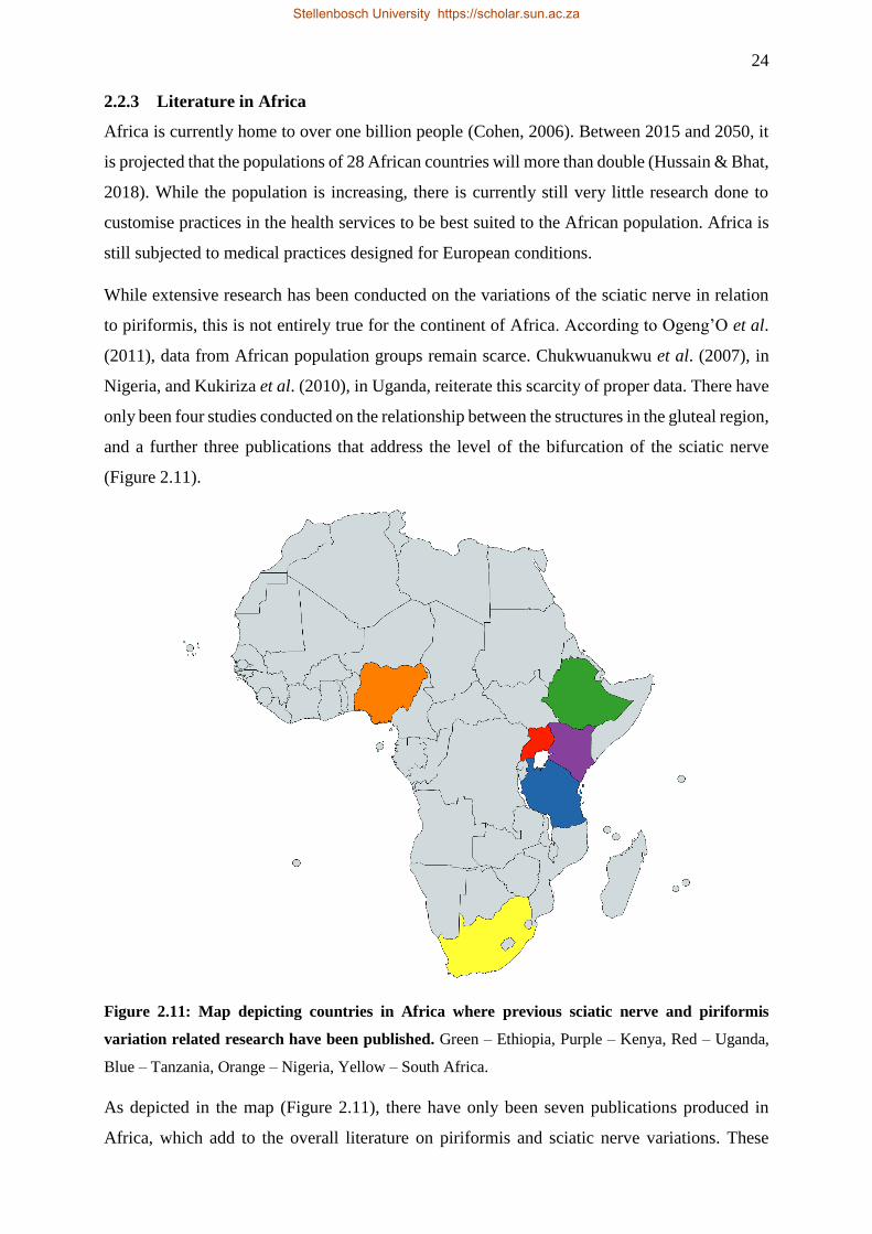
2.2.3 Literature in Africa
Africa is currently home to over one billion people (Cohen, 2006). Between 2015 and 2050, it
is projected that the populations of 28 African countries will more than double (Hussain & Bhat,
2018). While the population is increasing, there is currently still very little research done to
customise practices in the health services to be best suited to the African population. Africa is
still subjected to medical practices designed for European conditions.
While extensive research has been conducted on the variations of the sciatic nerve in relation
to piriformis, this is not entirely true for the continent of Africa. According to Ogeng’O et al.
(2011), data from African population groups remain scarce. Chukwuanukwu et al. (2007), in
Nigeria, and Kukiriza et al. (2010), in Uganda, reiterate this scarcity of proper data. There have
only been four studies conducted on the relationship between the structures in the gluteal region,
and a further three publications that address the level of the bifurcation of the sciatic nerve
(Figure 2.11).
Figure 2.11: Map depicting countries in Africa where previous sciatic nerve and piriformis
variation related research have been published. Green – Ethiopia, Purple – Kenya, Red – Uganda,
Blue – Tanzania, Orange – Nigeria, Yellow – South Africa.
As depicted in the map (Figure 2.11), there have only been seven publications produced in
Africa, which add to the overall literature on piriformis and sciatic nerve variations. These
Stellenbosch University https://scholar.sun.ac.za
25
countries include Ethiopia, where three known studies have been conducted, and Kenya,
Uganda, Tanzania, and Nigeria with one publication each, at the time the present study was
conducted (Figure 2.11). It should be noted that South Africa has been highlighted on the map
to indicate where the current study is being conducted.
In studies published on Ethiopian, Kenyan, and Nigerian population groups, where sciatic nerve
and resultant piriformis variations were observed, only 156 cadavers were assessed across the
three studies. This sample size is far smaller in relation to research conducted elsewhere in the
world (Ogeng’o et al., 2011). While a prevalence for variations between the structures in the
pelvic- or gluteal region is reported to be between 10-20% for the European population
(Pokorny et al., 2006; Natsis et al., 2014; Delabie et al., 2013), the prevalence of this varied
association was far less, at only 3.8% in the Nigerian population (Chukwuanukwu et al., 2007).
Comparatively, there is still a lack of data that speaks to the variations in an African population
compared with that elsewhere. It is for this reason that the current study aims to add meaningful
data to existing literature for the South African population.
Piriformis is closely related to the sciatic nerve, which makes it possible that trauma and
inflammation in piriformis might be clinically represented by sciatic-related pain (Hopayian et
al., 2010). The clinical setting requires that there is an accurate knowledge of the sciatic nerve,
which is important for the purpose of multiple procedures performed in this area. It is paramount
that variations are documented extensively. It is for this reason that the variations are prone to
develop into case specific pathologies in patients. No patient wants to live through pain and
surgical intervention on a continued basis. Due to the aforementioned, literature regarding
clinical conditions related to the sciatic nerve and piriformis will be reviewed; namely how this
nerve is fundamental in producing debilitating and even chronic sources of pain in and around
the gluteal region if not handled effectively. Literature will also focus on sciatic nerve blocks,
intramuscular injections and the role that the sciatic nerve and muscles of the gluteal region
play in piriformis syndrome.
Stellenbosch University https://scholar.sun.ac.za
26
2.3 CLINICAL CONSIDERATION
2.3.1 Pain
Pain is a major global health problem (Goldberg & McGee, 2011), and it has been estimated
that 1 in 5 adults suffer from pain. Another 1 in 10 adults are diagnosed with chronic pain each
year. Chronic pain has recently been highlighted as the leading cause for disability across the
globe (Cooper et al., 2016). The high prevalence of chronic pain and the negative effect on
society provides justification for regarding chronic pain as a public health priority (Häuser et
al., 2015). Subsequently, this is one of the most debated issues in public health worldwide (Hoy
et al., 2014). This information demands continued research with the focus on pain.
Pain can progressively become worse, resulting in chronic pain. Chronic pain can be described
as pain that perseveres past what is considered ‘normal’ healing time (Treede et al., 2015). The
result of this is a condition that is debilitating, and places the patient on medication and/or rest
for extended periods at a time. Similar to many other chronic non-communicable diseases,
chronic pain is typically accompanied by somatic and mental comorbidities (Goldberg &
McGee, 2011). Therefore, the burden of chronic pain in terms of health care or unemployment
could be confounded with one of these associated diseases (Dominick et al., 2012). Thus, there
is a need for further research in this area (Häuser et al., 2015). If anything is to change, pain
needs to be clearly defined as being of disease status on itself, rather than as a symptom of a
disease (McGee et al., 2011).
One of the largest contributors to disability (Dunn et al., 2013), particularly in the Western
world, is chronic musculoskeletal conditions (Stubbs et al., 2014). The results of the 2010
Global Burden of Disease study confirms that the prevalence and burden from musculoskeletal
conditions are exceptionally high throughout the world (Hoy et al., 2014). Gore et al. (2012),
maintain that lower back pain is the most prevalent musculoskeletal condition. According to
Gouveia et al. (2015), lower back pain is defined as pain in the back area, from the lower margin
of the twelfth ribs to the lower gluteal folds, with or without pain referred to the lower limbs.
At its most extreme, lower back pain can present itself as an extremely severe and persistent
pain in some patients, leading to chronic low back (Overaas et al., 2017).
Pain affects all population groups, regardless of age, sex, income, ethnicity, or geography
(Goldberg & McGee, 2011). The prevalence rate for pain is expected to increase as people
continue to live longer (Reid et al., 2015). According to Stubbs et al. (2014), and Leopoldino
et al. (2016), older populations are increasing worldwide. By 2035, an estimated one quarter of
the population in the European Union will be aged 65 or older (Reid et al., 2015). Williams et
Stellenbosch University https://scholar.sun.ac.za
27
al. (2015) notes that with the rapid growth in numbers of older adults in low and middle-income
countries, the back- and musculoskeletal pain burden will grow significantly in the coming
decades. Therefore, the public health impact of pain is ever rising. This data only emphasises
the urgency to identify the cause of musculoskeletal-related pain, and ensure accurate diagnoses
when treating patients.
Pain in the gluteal region is becoming far more recognised by clinicians, and is an ever
increasing complaint encountered in orthopaedic practices (Martin & Sekiya, 2008; Martin et
al., 2014), with an estimated prevalence of between 10-25% amongst the population of
industrialised nations (Meknas et al., 2011). According to Battaglia, D’Angelo and Kettne
(2016), hip- and gluteal pain is common and effects approximately 14% of the population over
the age of 60 years. Since people over 65 years-of-age account for 65% of admissions to
hospitals, and 40% of primary care expenditure, it is important for clinicians to adjust to this
reality (Abdulla et al., 2013). The possible causes of pain in the gluteal region are extensive
(Hartvigsen et al., 2013; Martin et al., 2014), and might include referred symptoms from
lumbosacral spine, sacroiliac joint (Buijs, Visser & Groen, 2007), the hip joint, or extra-articular
structures of the hip region (Frank et al., 2010). The deep rotators of the hip joint (Cox &
Bakkum, 2005; Meknas et al., 2003), the hamstring group (Puranen & Orava, 1988; Frank et
al., 2010), tendinopathy of gluteus medius and gluteus minimus (Kingzett-Taylor et al., 1999),
or an entrapment of the pudendal or sciatic nerves (Martin et al., 2010), could be extra-articular
sources of gluteal pain. Entrapment of the sciatic nerve can present itself in many different
forms within the gluteal region and lower limb, namely; an abnormal anatomical relationship
between the sciatic nerve and vascular tissue found in the deep gluteal region (Martin et al.,
2010), the muscle and tendon complexes of piriformis (Freiberg, 1934; Robinson, 1947; Lee et
al., 2016) obturator internus and gemelli (Cox & Bakkum, 2005; Meknas et al., 2003), or
proximal hamstrings (Puranen & Orava, 1988; Martin et al., 2010). Due to the many sources of
gluteal pain from various intra- and extra-articular structures found in relation to the sciatic
nerve, it can be difficult to determine the specific source through a clinical examination (Buijs
et al., 2007). Therefore, it is paramount that there is adequate knowledge of the anatomical
variations within the region for appropriate diagnosis, and potential treatment of the pain
(Grassi et al., 2003; Martin et al., 2014).
Stellenbosch University https://scholar.sun.ac.za
28
2.4.2 Sciatic Nerve Block
Significant advances have been made in regional techniques for localised nerve blocks. This
has primarily been driven by the need to produce effective analgesia during the postoperative
period (Murray et al., 2010). Additionally, modern medical care practices demand a shorter
hospital stay, early mobilisation of the individual, and overall improved patient experiences
(Murray et al., 2010).
Regional anaesthesia has become increasingly popular for procedures performed on the lower
limb because this improves the quality of pain relief postoperatively (di Benedetto et al., 2001).
The sciatic nerve block is frequently used for anaesthesia or analgesia when performing
procedures on the lower limb (Karmakar et al., 2007). This procedure is well established, and
is performed using a variety of different techniques and approaches. The nerve can be blocked
at any point along its course within the lower limb, provided there is sufficient ultrasound
images to guide this procedure (Marhofer et al., 2010).
Although the sciatic nerve has the largest cross-sectional diameter in the human body, its
ultrasonography imaging can, however, not be easy to interpret (Moayeri et al., 2010). Imaging
of the sciatic nerve can be challenging for a number of reasons. There are many other
surrounding structures within the gluteal region, which means that the large amount of muscle
and adipose tissue around the nerve can impair its visualisation (Marhofer et al., 2010).
Moreover, inadequate probe manipulation has the potential to cause serious complications to
the patient. The risk of injury caused by the intraneural injection is increased, as multiple
redirections of the needle would often be required in order to deposit the anaesthetic solution
correctly near the deeply situated nerve (Yamamoto et al., 2014). It is common for most of the
techniques currently in use to rely on anatomical landmarks identified on the surface, which is
largely unreliable and has the potential for many errors. While these anatomical landmarks are
correct in providing clues to where the sciatic nerve is situated, it is by no means accurate, and
these landmarks are only a guide. These landmarks do not work for all patients, as they do not
make provision for anatomical variations, or varying body sizes (Karmakar et al., 2007). Most
importantly, the effective identification of this nerve within the gluteal region requires extensive
knowledge of the topography of the gluteal region, and internal architecture of the nerves and
vessels within adjacent structures (Moayeri et al., 2010).
As discussed previously, the sciatic nerve anatomy has been described in many medical
textbooks used throughout the world. However, as with many other structures in the body, there
are varieties of different ways that the sciatic nerve is distributed within the gluteal region. The
bifurcation of this nerve proximal to the gluteal region may lead to subsequent failure of
Stellenbosch University https://scholar.sun.ac.za
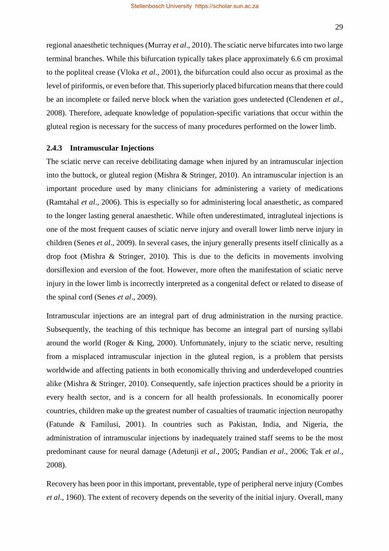
29
regional anaesthetic techniques (Murray et al., 2010). The sciatic nerve bifurcates into two large
terminal branches. While this bifurcation typically takes place approximately 6.6 cm proximal
to the popliteal crease (Vloka et al., 2001), the bifurcation could also occur as proximal as the
level of piriformis, or even before that. This superiorly placed bifurcation means that there could
be an incomplete or failed nerve block when the variation goes undetected (Clendenen et al.,
2008). Therefore, adequate knowledge of population-specific variations that occur within the
gluteal region is necessary for the success of many procedures performed on the lower limb.
2.4.3 Intramuscular Injections
The sciatic nerve can receive debilitating damage when injured by an intramuscular injection
into the buttock, or gluteal region (Mishra & Stringer, 2010). An intramuscular injection is an
important procedure used by many clinicians for administering a variety of medications
(Ramtahal et al., 2006). This is especially so for administering local anaesthetic, as compared
to the longer lasting general anaesthetic. While often underestimated, intragluteal injections is
one of the most frequent causes of sciatic nerve injury and overall lower limb nerve injury in
children (Senes et al., 2009). In several cases, the injury generally presents itself clinically as a
drop foot (Mishra & Stringer, 2010). This is due to the deficits in movements involving
dorsiflexion and eversion of the foot. However, more often the manifestation of sciatic nerve
injury in the lower limb is incorrectly interpreted as a congenital defect or related to disease of
the spinal cord (Senes et al., 2009).
Intramuscular injections are an integral part of drug administration in the nursing practice.
Subsequently, the teaching of this technique has become an integral part of nursing syllabi
around the world (Roger & King, 2000). Unfortunately, injury to the sciatic nerve, resulting
from a misplaced intramuscular injection in the gluteal region, is a problem that persists
worldwide and affecting patients in both economically thriving and underdeveloped countries
alike (Mishra & Stringer, 2010). Consequently, safe injection practices should be a priority in
every health sector, and is a concern for all health professionals. In economically poorer
countries, children make up the greatest number of casualties of traumatic injection neuropathy
(Fatunde & Familusi, 2001). In countries such as Pakistan, India, and Nigeria, the
administration of intramuscular injections by inadequately trained staff seems to be the most
predominant cause for neural damage (Adetunji et al., 2005; Pandian et al., 2006; Tak et al.,
2008).
Recovery has been poor in this important, preventable, type of peripheral nerve injury (Combes
et al., 1960). The extent of recovery depends on the severity of the initial injury. Overall, many
Stellenbosch University https://scholar.sun.ac.za
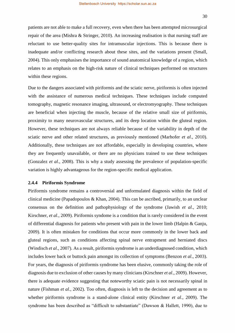
30
patients are not able to make a full recovery, even when there has been attempted microsurgical
repair of the area (Mishra & Stringer, 2010). An increasing realisation is that nursing staff are
reluctant to use better-quality sites for intramuscular injections. This is because there is
inadequate and/or conflicting research about these sites, and the variations present (Small,
2004). This only emphasises the importance of sound anatomical knowledge of a region, which
relates to an emphasis on the high-risk nature of clinical techniques performed on structures
within these regions.
Due to the dangers associated with piriformis and the sciatic nerve, piriformis is often injected
with the assistance of numerous medical techniques. These techniques include computed
tomography, magnetic resonance imaging, ultrasound, or electromyography. These techniques
are beneficial when injecting the muscle, because of the relative small size of piriformis,
proximity to many neurovascular structures, and its deep location within the gluteal region.
However, these techniques are not always reliable because of the variability in depth of the
sciatic nerve and other related structures, as previously mentioned (Marhofer et al., 2010).
Additionally, these techniques are not affordable, especially in developing countries, where
they are frequently unavailable, or there are no physicians trained to use these techniques
(Gonzalez et al., 2008). This is why a study assessing the prevalence of population-specific
variation is highly advantageous for the region-specific medical application.
2.4.4 Piriformis Syndrome
Piriformis syndrome remains a controversial and unformulated diagnosis within the field of
clinical medicine (Papadopoulos & Khan, 2004). This can be ascribed, primarily, to an unclear
consensus on the definition and pathophysiology of the syndrome (Jawish et al., 2010;
Kirschner, et al., 2009). Piriformis syndome is a condition that is rarely considered in the event
of differential diagnosis for patients who present with pain in the lower limb (Halpin & Ganju,
2009). It is often mistaken for conditions that occur more commonly in the lower back and
gluteal regions, such as conditions affecting spinal nerve entrapment and herniated discs
(Windisch et al., 2007). As a result, piriformis syndrome is an underdiagnosed condition, which
includes lower back or buttock pain amongst its collection of symptoms (Benzon et al., 2003).
For years, the diagnosis of piriformis syndrome has been elusive, commonly taking the role of
diagnosis due to exclusion of other causes by many clinicians (Kirschner et al., 2009). However,
there is adequate evidence suggesting that noteworthy sciatic pain is not necessarily spinal in
nature (Fishman et al., 2002). Too often, diagnosis is left to the decision and agreement as to
whether piriformis syndrome is a stand-alone clinical entity (Kirschner et al., 2009). The
syndrome has been described as “difficult to substantiate” (Dawson & Hallett, 1990), due to
Stellenbosch University https://scholar.sun.ac.za
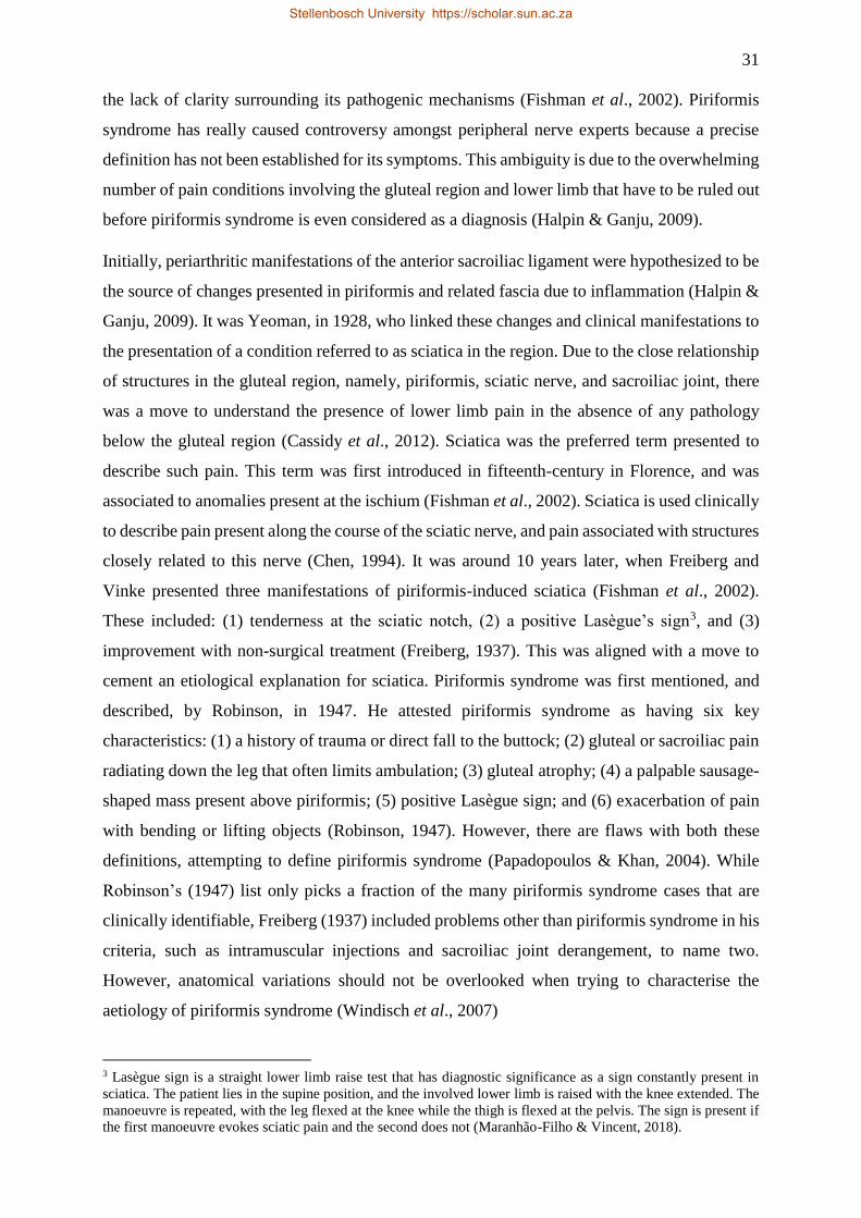
31
the lack of clarity surrounding its pathogenic mechanisms (Fishman et al., 2002). Piriformis
syndrome has really caused controversy amongst peripheral nerve experts because a precise
definition has not been established for its symptoms. This ambiguity is due to the overwhelming
number of pain conditions involving the gluteal region and lower limb that have to be ruled out
before piriformis syndrome is even considered as a diagnosis (Halpin & Ganju, 2009).
Initially, periarthritic manifestations of the anterior sacroiliac ligament were hypothesized to be
the source of changes presented in piriformis and related fascia due to inflammation (Halpin &
Ganju, 2009). It was Yeoman, in 1928, who linked these changes and clinical manifestations to
the presentation of a condition referred to as sciatica in the region. Due to the close relationship
of structures in the gluteal region, namely, piriformis, sciatic nerve, and sacroiliac joint, there
was a move to understand the presence of lower limb pain in the absence of any pathology
below the gluteal region (Cassidy et al., 2012). Sciatica was the preferred term presented to
describe such pain. This term was first introduced in fifteenth-century in Florence, and was
associated to anomalies present at the ischium (Fishman et al., 2002). Sciatica is used clinically
to describe pain present along the course of the sciatic nerve, and pain associated with structures
closely related to this nerve (Chen, 1994). It was around 10 years later, when Freiberg and
Vinke presented three manifestations of piriformis-induced sciatica (Fishman et al., 2002).
These included: (1) tenderness at the sciatic notch, (2) a positive Lasègue’s sign3, and (3)
improvement with non-surgical treatment (Freiberg, 1937). This was aligned with a move to
cement an etiological explanation for sciatica. Piriformis syndrome was first mentioned, and
described, by Robinson, in 1947. He attested piriformis syndrome as having six key
characteristics: (1) a history of trauma or direct fall to the buttock; (2) gluteal or sacroiliac pain
radiating down the leg that often limits ambulation; (3) gluteal atrophy; (4) a palpable sausage-
shaped mass present above piriformis; (5) positive Lasègue sign; and (6) exacerbation of pain
with bending or lifting objects (Robinson, 1947). However, there are flaws with both these
definitions, attempting to define piriformis syndrome (Papadopoulos & Khan, 2004). While
Robinson’s (1947) list only picks a fraction of the many piriformis syndrome cases that are
clinically identifiable, Freiberg (1937) included problems other than piriformis syndrome in his
criteria, such as intramuscular injections and sacroiliac joint derangement, to name two.
However, anatomical variations should not be overlooked when trying to characterise the
aetiology of piriformis syndrome (Windisch et al., 2007)
3 Lasègue sign is a straight lower limb raise test that has diagnostic significance as a sign constantly present in
sciatica. The patient lies in the supine position, and the involved lower limb is raised with the knee extended. The
manoeuvre is repeated, with the leg flexed at the knee while the thigh is flexed at the pelvis. The sign is present if
the first manoeuvre evokes sciatic pain and the second does not (Maranhão-Filho & Vincent, 2018).
Stellenbosch University https://scholar.sun.ac.za
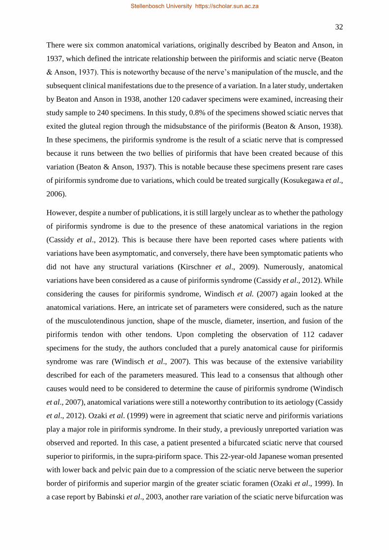
32
There were six common anatomical variations, originally described by Beaton and Anson, in
1937, which defined the intricate relationship between the piriformis and sciatic nerve (Beaton
& Anson, 1937). This is noteworthy because of the nerve’s manipulation of the muscle, and the
subsequent clinical manifestations due to the presence of a variation. In a later study, undertaken
by Beaton and Anson in 1938, another 120 cadaver specimens were examined, increasing their
study sample to 240 specimens. In this study, 0.8% of the specimens showed sciatic nerves that
exited the gluteal region through the midsubstance of the piriformis (Beaton & Anson, 1938).
In these specimens, the piriformis syndrome is the result of a sciatic nerve that is compressed
because it runs between the two bellies of piriformis that have been created because of this
variation (Beaton & Anson, 1937). This is notable because these specimens present rare cases
of piriformis syndrome due to variations, which could be treated surgically (Kosukegawa et al.,
2006).
However, despite a number of publications, it is still largely unclear as to whether the pathology
of piriformis syndrome is due to the presence of these anatomical variations in the region
(Cassidy et al., 2012). This is because there have been reported cases where patients with
variations have been asymptomatic, and conversely, there have been symptomatic patients who
did not have any structural variations (Kirschner et al., 2009). Numerously, anatomical
variations have been considered as a cause of piriformis syndrome (Cassidy et al., 2012). While
considering the causes for piriformis syndrome, Windisch et al. (2007) again looked at the
anatomical variations. Here, an intricate set of parameters were considered, such as the nature
of the musculotendinous junction, shape of the muscle, diameter, insertion, and fusion of the
piriformis tendon with other tendons. Upon completing the observation of 112 cadaver
specimens for the study, the authors concluded that a purely anatomical cause for piriformis
syndrome was rare (Windisch et al., 2007). This was because of the extensive variability
described for each of the parameters measured. This lead to a consensus that although other
causes would need to be considered to determine the cause of piriformis syndrome (Windisch
et al., 2007), anatomical variations were still a noteworthy contribution to its aetiology (Cassidy
et al., 2012). Ozaki et al. (1999) were in agreement that sciatic nerve and piriformis variations
play a major role in piriformis syndrome. In their study, a previously unreported variation was
observed and reported. In this case, a patient presented a bifurcated sciatic nerve that coursed
superior to piriformis, in the supra-piriform space. This 22-year-old Japanese woman presented
with lower back and pelvic pain due to a compression of the sciatic nerve between the superior
border of piriformis and superior margin of the greater sciatic foramen (Ozaki et al., 1999). In
a case report by Babinski et al., 2003, another rare variation of the sciatic nerve bifurcation was
Stellenbosch University https://scholar.sun.ac.za
33
described. In this case, the tibial nerve passed inferior to the superior gemellus, while the
common fibular nerve passed superior to this muscle. The conclusion here was that such
variations might be conducive to the prevalence of piriformis syndrome (Babinski et al., 2003).
In yet another rare case report, a similar type of variation was observed, namely, piriformis,
which had been divided into two bellies, compressed the entire sciatic nerve. Upon intervention,
the piriformis muscle was resected, resulting in the disappearance of the pain (Kosukegawa et
al., 2006). Once again, Kosukegawa et al. (2006) highlighted the definitive contribution that
anatomical variations have on the clinical manifestation of piriformis syndrome.
Scientific specialists in the field have studied the detection of piriformis and sciatic nerve
related variations. These variations can be identified by the use of radiological techniques, and
in almost all operative procedures within the gluteal region. However, it is important to
remember that, even with increased technological advances, the cadaver is the best means to
study the anatomy of the sciatic nerve and piriformis, along with the prevalence of variations
in the morphology of these structures (Smoll, 2010; Prakash et al., 2010).
Stellenbosch University https://scholar.sun.ac.za

34
CHAPTER THREE:
AIMS AND OBJECTIVES
Stellenbosch University https://scholar.sun.ac.za
35
3.1 PROBLEM STATEMENT
The intricacy of the sciatic nerve and piriformis variations have been discussed in detail, and
the clinical implications resulting from these variations have been looked at as well. Following
the review of literature, it is clear that it is important to understand the sciatic nerve, along with
the variations that are most prevalent. While the variations of piriformis and the sciatic nerve
have been documented in the past, there is a need for population specific data. This is the case
for the Americas, Europe, and Asia. As far as it is understood, there have been very few studies
of this calibre undertaken or published on the African continent. Moreover, to the author’s
knowledge, there have been no formal publications of this kind within South Africa to date.
The primary aim of the present study is to describe and document the anatomical and
morphological variations of piriformis in relation to the sciatic nerve bifurcation within the
South African population. Additionally, to morphometrically analyse normal parameters of the
sciatic nerve and piriformis muscle. Furthermore, the secondary aim of the present study is to
assess the level of bifurcation of the sciatic nerve within the lower limbs of the South African
population. Thereafter, comparisons of the variations described will be made between right and
left lower limbs, male and female specimens, and between the three population groups
available, namely; White/Caucasian, Mixed Race and South African Black groups, within
South Africa. Although there is extensive diversity amongst the smaller subpopulation groups
within South Africa, this research will speak to the South African population as a whole.
Furthermore, the researcher will attempt to provide detailed results for the South African
population groups and their variation classifications where statistically possible.
3.2 AIMS
The aims of the present study are to:
(1) Report on the prevalence of anatomical variations in the course of the sciatic nerve in
relation to piriformis;
(2) Report on the prevalence of variations in the level of sciatic nerve bifurcation into the
tibial and common fibular nerves either within the pelvic region, gluteal region,
posterior compartment of the thigh, or popliteal fossa proper, within the lower limb;
(3) Analyse morphometric measurements for the typical sciatic nerve and piriformis
morphology;
Stellenbosch University https://scholar.sun.ac.za

36
(4) Compare results found between right and left lower limbs, male and female specimens,
and in the three South African subpopulation groups.
3.3 OBJECTIVES
In order to achieve the aims of this study, the following objectives were set:
i. To observe the variations of the sciatic nerve in relation to piriformis variation
a. Sciatic nerve exits the pelvic region and continues undivided, below piriformis.
b. Sciatic nerve bifurcates in the pelvic region, with the common fibular nerve
piercing piriformis, and the tibial nerve emerging below piriformis.
c. Sciatic nerve bifurcates in the pelvic region, with the common fibular nerve
coursing inferiorly across piriformis, and tibial nerve emerging below
piriformis.
d. Sciatic nerve exits the pelvic region undivided, piercing piriformis.
e. Sciatic nerve bifurcates in the pelvic region, with the common fibular nerve
coursing inferiorly across piriformis, and the tibial nerve piercing piriformis.
f. Sciatic nerve exits the pelvic region undivided, coursing through the supra-
piriform space and then inferiorly across piriformis.
g. Sciatic nerve bifurcates in the pelvic region, with both the common fibular nerve
and tibial nerve coursing separately below piriformis.
ii. To observe the level of sciatic nerve bifurcation within the lower limb:
a. Within the pelvic region, proximal to piriformis;
b. Within the gluteal region;
c. In the proximal first third of the posterior compartment of the thigh;
d. In the middle third of the posterior compartment of the thigh;
e. In the distal third of the posterior compartment of the thigh;
f. Within the popliteal fossa proper.
iii. Take morphometric measurements of selected piriformis and sciatic nerve
parameters;
iv. Analyse datasets of the sciatic nerve and piriformis between:
a. right and left lower limbs,
b. male and female specimens
c. cadavers from the three different South African population groups.
Stellenbosch University https://scholar.sun.ac.za

37
CHAPTER FOUR:
MATERIALS AND
METHODS
Stellenbosch University https://scholar.sun.ac.za

38
4.1 ETHICAL APPROVAL
Ethical approval was obtained from the Stellenbosch University’s Health Research Ethics
Committee; reference number S18/02/032. The Health Research Ethics Committee complies
with the South African National Health Act No. 61 2003, sections 61 to 64 as it allows the use
of cadavers for research purposes and medical training (National Health Act, 2003).
Additionally, this act pertains to health research and adheres to the United States Code of
Federal Regulations Title 45 Part 46. This committee abides by the ethical norms and principles
for research, established by the Declaration of Helsinki, the South African Medical Research
Council Guidelines as well as the Guidelines for Ethical Research: Principles Structures and
Processes 2004 (Department of Health).
4.2 ACQUISITION OF CADAVERS
For this research project, cadavers were sourced from three universities within South Africa:
Division of Clinical Anatomy, at Stellenbosch University (SU), School of Anatomical Sciences,
at the University of the Witwatersrand (Wits), and the Division of Clinical Anatomy and
Biological Anthropology, at the University of Cape Town (UCT).
The departments and divisions obtained cadavers primarily by making use of two methods.
Firstly, cadavers are sourced through informed consent. Prior to death, a person provides
consent that their body is donated for the purpose of medical research and education. Secondly,
cadavers that are not claimed from the state by relatives within a 30-day period are
automatically donated to a university within the region for the purpose of medical research and
education. Concerning the latter arrangement, consent is not formally given, instead consent is
granted by the inspector of anatomy for the Western Cape, on behalf of the person, their family,
and government. Furthermore, a detailed account of the cadaver’s location is kept. In both
instances, no personal information regarding cadavers is made available.
Cadavers will each receive a university-specific number for storage and identification purposes,
with the inclusion of basic cause and date of death, sex and age. No personal or medical
information regarding any of the cadavers were used when conducting this present study. The
cadavers will remain anonymous throughout the entirety of the study.
Stellenbosch University https://scholar.sun.ac.za
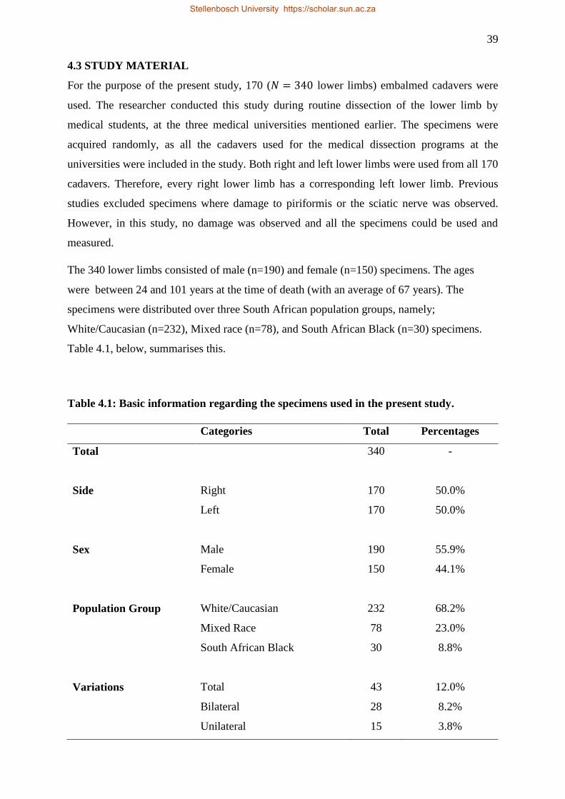
39
4.3 STUDY MATERIAL
For the purpose of the present study, 170 (𝑁 = 340 lower limbs) embalmed cadavers were
used. The researcher conducted this study during routine dissection of the lower limb by
medical students, at the three medical universities mentioned earlier. The specimens were
acquired randomly, as all the cadavers used for the medical dissection programs at the
universities were included in the study. Both right and left lower limbs were used from all 170
cadavers. Therefore, every right lower limb has a corresponding left lower limb. Previous
studies excluded specimens where damage to piriformis or the sciatic nerve was observed.
However, in this study, no damage was observed and all the specimens could be used and
measured.
The 340 lower limbs consisted of male (n=190) and female (n=150) specimens. The ages
were between 24 and 101 years at the time of death (with an average of 67 years). The
specimens were distributed over three South African population groups, namely;
White/Caucasian (n=232), Mixed race (n=78), and South African Black (n=30) specimens.
Table 4.1, below, summarises this.
Table 4.1: Basic information regarding the specimens used in the present study.
Categories Total Percentages
Total 340 -
Side Right 170 50.0%
Left 170 50.0%
Sex Male 190 55.9%
Female 150 44.1%
Population Group White/Caucasian 232 68.2%
Mixed Race 78 23.0%
South African Black 30 8.8%
Variations Total 43 12.0%
Bilateral 28 8.2%
Unilateral 15 3.8%
Stellenbosch University https://scholar.sun.ac.za

40
The present study is descriptive and observational in nature. The emergence of the sciatic nerve,
in relation to piriformis, within the gluteal region, was observed and documented.
Subsequently, the anatomical relationship between the sciatic nerve and piriformis was
classified according to the adjusted Beaton and Anson (1937) classification, into the seven
known types (Table 2.2). Additionally, the lower limbs were observed in order to study the
sciatic nerve, and its level of bifurcation into the tibial and common peroneal nerves. This was
categorised into six separate classifications (A-F) (Table 2.4), depending on the level of
bifurcation of the sciatic nerve into its branches. Figure 4.1 depicts a summary of this
methodology for the study. Specimens were collected and grouped according to the two
overarching variations observed: the variation in the relationship between piriformis and the
sciatic nerve, and the variation in the level of bifurcation of the sciatic nerve.
Dissection and classification of variations were done as follows: if students at specific
institutions were not dissecting this region, the researcher performed the dissection on the
gluteal regions and lower limbs. Where these regions were dissected as part of the curriculum,
the students dissected these regions under constant supervision of the primary researcher. In
this way, although various individuals were responsible for dissection, the methodology stayed
the same. The researcher was solely responsible for the classification of any variation
discovered within the gluteal region or at the level of bifurcation.
Stellenbosch University https://scholar.sun.ac.za
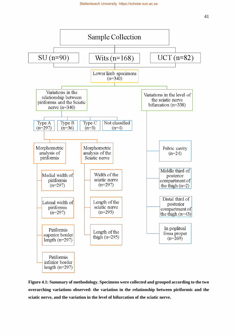
41
Figure 4.1: Summary of methodology. Specimens were collected and grouped according to the two
overarching variations observed: the variation in the relationship between piriformis and the
sciatic nerve, and the variation in the level of bifurcation of the sciatic nerve.
Stellenbosch University https://scholar.sun.ac.za
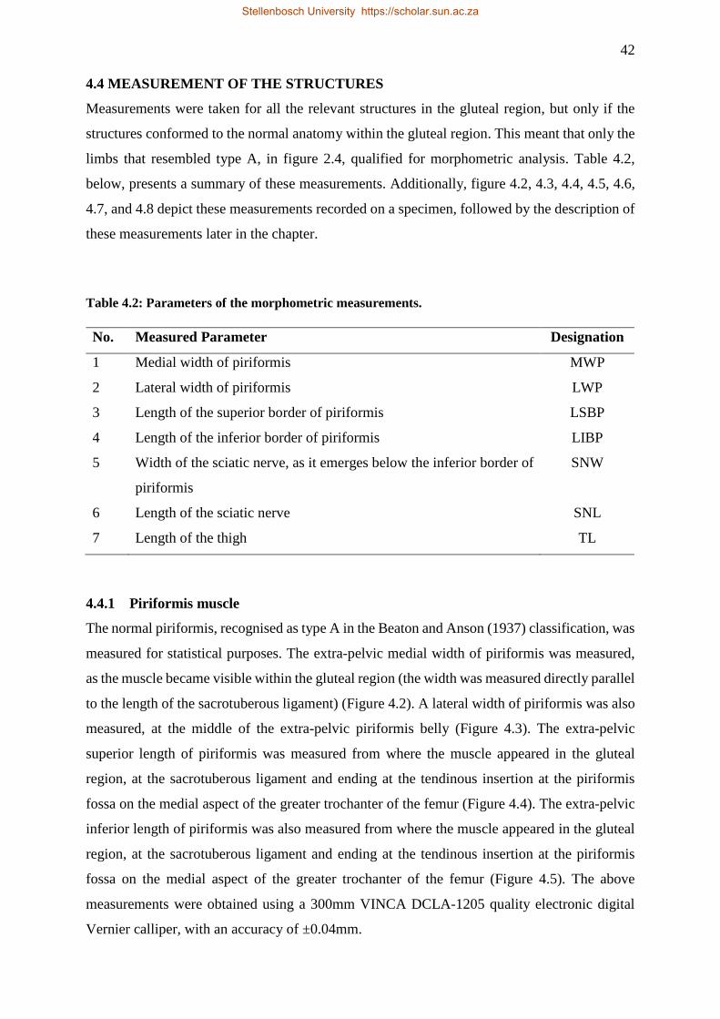
42
4.4 MEASUREMENT OF THE STRUCTURES
Measurements were taken for all the relevant structures in the gluteal region, but only if the
structures conformed to the normal anatomy within the gluteal region. This meant that only the
limbs that resembled type A, in figure 2.4, qualified for morphometric analysis. Table 4.2,
below, presents a summary of these measurements. Additionally, figure 4.2, 4.3, 4.4, 4.5, 4.6,
4.7, and 4.8 depict these measurements recorded on a specimen, followed by the description of
these measurements later in the chapter.
Table 4.2: Parameters of the morphometric measurements.
No. Measured Parameter Designation
1 Medial width of piriformis MWP
2 Lateral width of piriformis LWP
3 Length of the superior border of piriformis LSBP
4 Length of the inferior border of piriformis LIBP
5 Width of the sciatic nerve, as it emerges below the inferior border of
piriformis
SNW
6 Length of the sciatic nerve SNL
7 Length of the thigh TL
4.4.1 Piriformis muscle
The normal piriformis, recognised as type A in the Beaton and Anson (1937) classification, was
measured for statistical purposes. The extra-pelvic medial width of piriformis was measured,
as the muscle became visible within the gluteal region (the width was measured directly parallel
to the length of the sacrotuberous ligament) (Figure 4.2). A lateral width of piriformis was also
measured, at the middle of the extra-pelvic piriformis belly (Figure 4.3). The extra-pelvic
superior length of piriformis was measured from where the muscle appeared in the gluteal
region, at the sacrotuberous ligament and ending at the tendinous insertion at the piriformis
fossa on the medial aspect of the greater trochanter of the femur (Figure 4.4). The extra-pelvic
inferior length of piriformis was also measured from where the muscle appeared in the gluteal
region, at the sacrotuberous ligament and ending at the tendinous insertion at the piriformis
fossa on the medial aspect of the greater trochanter of the femur (Figure 4.5). The above
measurements were obtained using a 300mm VINCA DCLA-1205 quality electronic digital
Vernier calliper, with an accuracy of ±0.04mm.
Stellenbosch University https://scholar.sun.ac.za
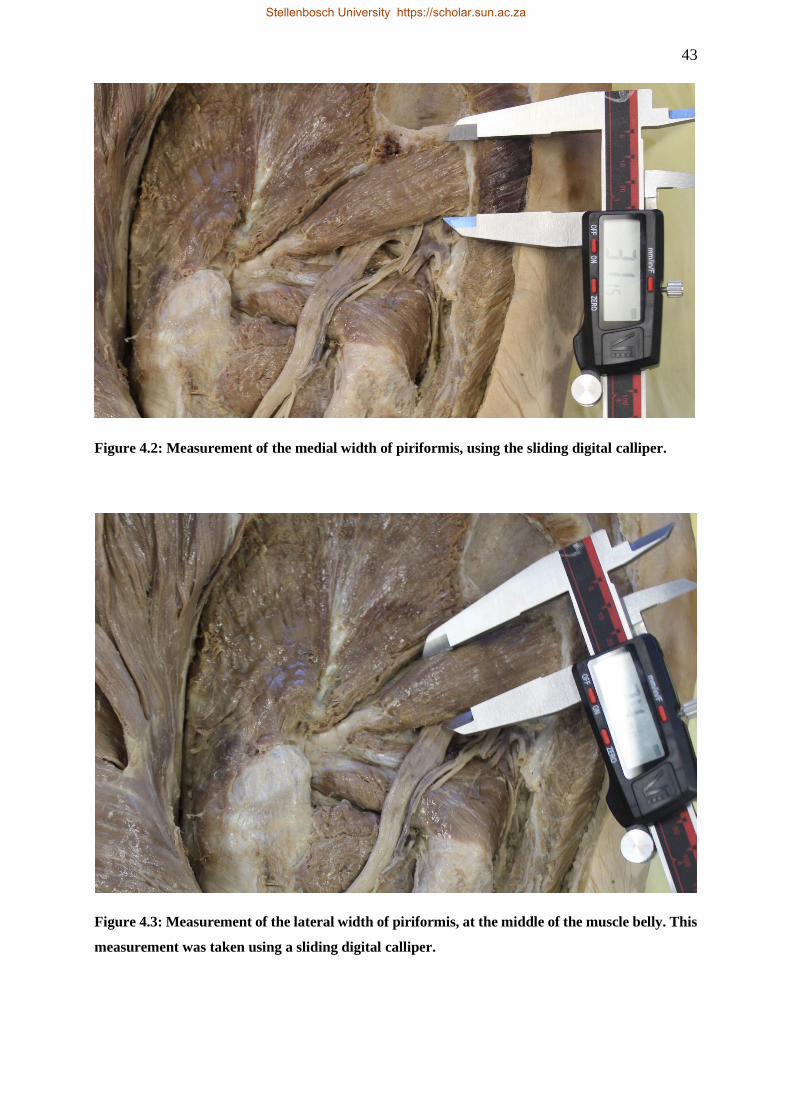
43
Figure 4.2: Measurement of the medial width of piriformis, using the sliding digital calliper.
Figure 4.3: Measurement of the lateral width of piriformis, at the middle of the muscle belly. This
measurement was taken using a sliding digital calliper.
Stellenbosch University https://scholar.sun.ac.za
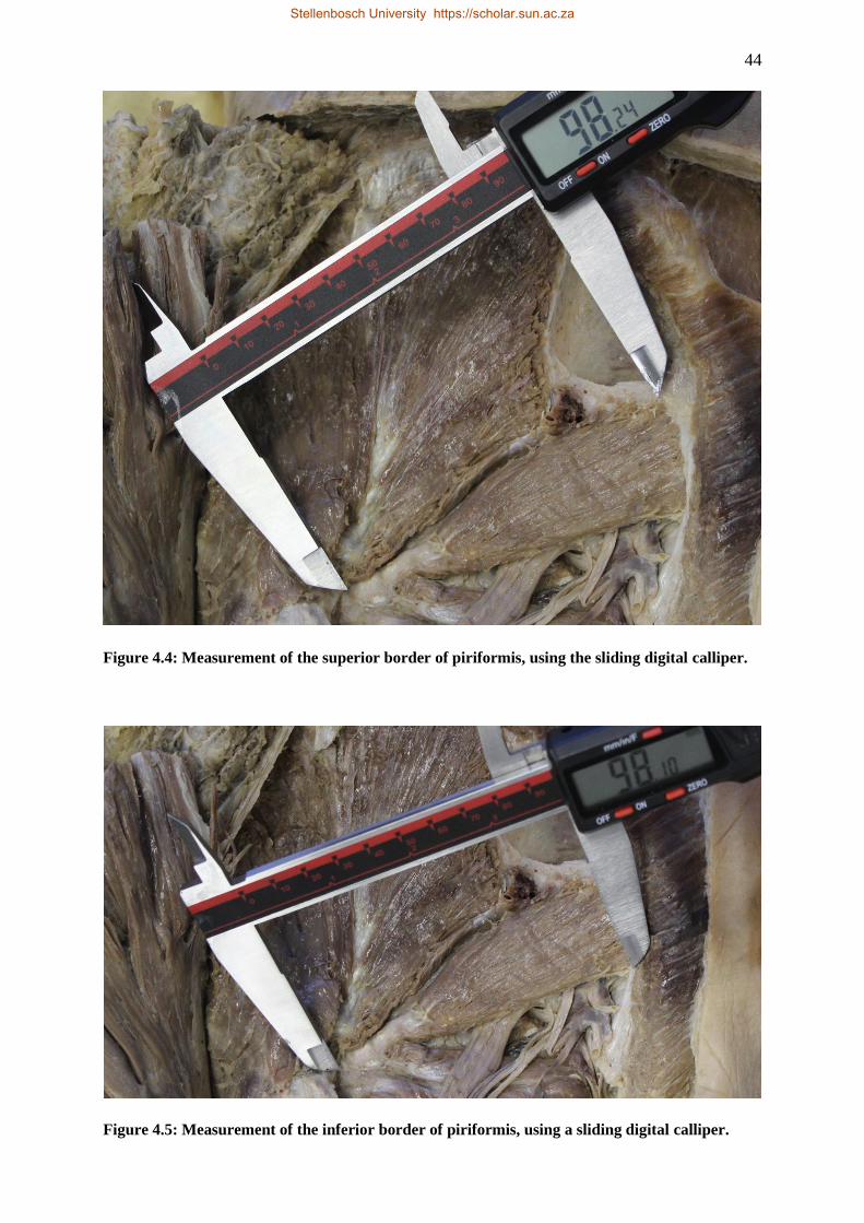
44
Figure 4.4: Measurement of the superior border of piriformis, using the sliding digital calliper.
Figure 4.5: Measurement of the inferior border of piriformis, using a sliding digital calliper.
Stellenbosch University https://scholar.sun.ac.za
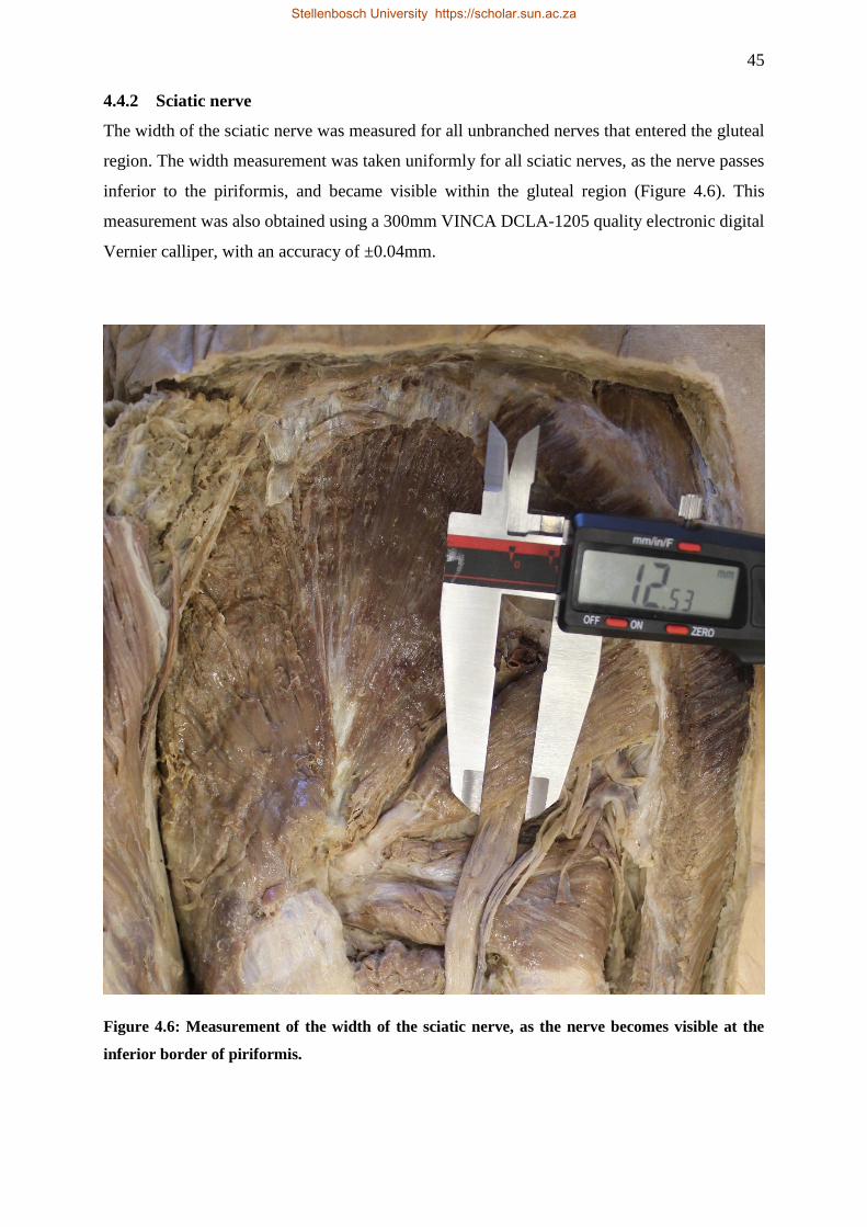
45
4.4.2 Sciatic nerve
The width of the sciatic nerve was measured for all unbranched nerves that entered the gluteal
region. The width measurement was taken uniformly for all sciatic nerves, as the nerve passes
inferior to the piriformis, and became visible within the gluteal region (Figure 4.6). This
measurement was also obtained using a 300mm VINCA DCLA-1205 quality electronic digital
Vernier calliper, with an accuracy of ±0.04mm.
Figure 4.6: Measurement of the width of the sciatic nerve, as the nerve becomes visible at the
inferior border of piriformis.
Stellenbosch University https://scholar.sun.ac.za
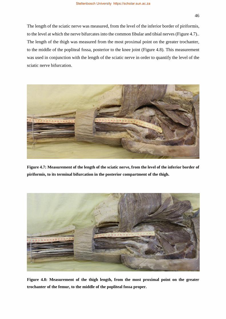
46
The length of the sciatic nerve was measured, from the level of the inferior border of piriformis,
to the level at which the nerve bifurcates into the common fibular and tibial nerves (Figure 4.7)..
The length of the thigh was measured from the most proximal point on the greater trochanter,
to the middle of the popliteal fossa, posterior to the knee joint (Figure 4.8). This measurement
was used in conjunction with the length of the sciatic nerve in order to quantify the level of the
sciatic nerve bifurcation.
Figure 4.7: Measurement of the length of the sciatic nerve, from the level of the inferior border of
piriformis, to its terminal bifurcation in the posterior compartment of the thigh.
Figure 4.8: Measurement of the thigh length, from the most proximal point on the greater
trochanter of the femur, to the middle of the popliteal fossa proper.
Stellenbosch University https://scholar.sun.ac.za
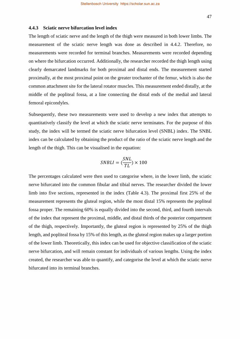
47
4.4.3 Sciatic nerve bifurcation level index
The length of sciatic nerve and the length of the thigh were measured in both lower limbs. The
measurement of the sciatic nerve length was done as described in 4.4.2. Therefore, no
measurements were recorded for terminal branches. Measurements were recorded depending
on where the bifurcation occurred. Additionally, the researcher recorded the thigh length using
clearly demarcated landmarks for both proximal and distal ends. The measurement started
proximally, at the most proximal point on the greater trochanter of the femur, which is also the
common attachment site for the lateral rotator muscles. This measurement ended distally, at the
middle of the popliteal fossa, at a line connecting the distal ends of the medial and lateral
femoral epicondyles.
Subsequently, these two measurements were used to develop a new index that attempts to
quantitatively classify the level at which the sciatic nerve terminates. For the purpose of this
study, the index will be termed the sciatic nerve bifurcation level (SNBL) index. The SNBL
index can be calculated by obtaining the product of the ratio of the sciatic nerve length and the
length of the thigh. This can be visualised in the equation:
𝑆𝑁𝐵𝐿𝐼 = (𝑆𝑁𝐿
𝑇𝐿) × 100
The percentages calculated were then used to categorise where, in the lower limb, the sciatic
nerve bifurcated into the common fibular and tibial nerves. The researcher divided the lower
limb into five sections, represented in the index (Table 4.3). The proximal first 25% of the
measurement represents the gluteal region, while the most distal 15% represents the popliteal
fossa proper. The remaining 60% is equally divided into the second, third, and fourth intervals
of the index that represent the proximal, middle, and distal thirds of the posterior compartment
of the thigh, respectively. Importantly, the gluteal region is represented by 25% of the thigh
length, and popliteal fossa by 15% of this length, as the gluteal region makes up a larger portion
of the lower limb. Theoretically, this index can be used for objective classification of the sciatic
nerve bifurcation, and will remain constant for individuals of various lengths. Using the index
created, the researcher was able to quantify, and categorise the level at which the sciatic nerve
bifurcated into its terminal branches.
Stellenbosch University https://scholar.sun.ac.za
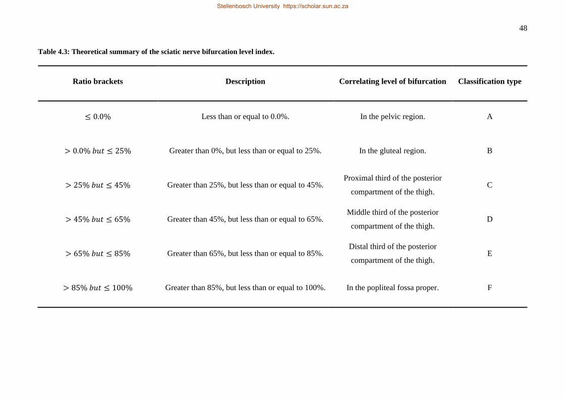
48
Table 4.3: Theoretical summary of the sciatic nerve bifurcation level index.
Ratio brackets Description Correlating level of bifurcation Classification type
≤ 0.0% Less than or equal to 0.0%. In the pelvic region. A
> 0.0% 𝑏𝑢𝑡 ≤ 25% Greater than 0%, but less than or equal to 25%. In the gluteal region. B
> 25% 𝑏𝑢𝑡 ≤ 45% Greater than 25%, but less than or equal to 45%. Proximal third of the posterior
compartment of the thigh. C
> 45% 𝑏𝑢𝑡 ≤ 65% Greater than 45%, but less than or equal to 65%. Middle third of the posterior
compartment of the thigh. D
> 65% 𝑏𝑢𝑡 ≤ 85% Greater than 65%, but less than or equal to 85%. Distal third of the posterior
compartment of the thigh. E
> 85% 𝑏𝑢𝑡 ≤ 100% Greater than 85%, but less than or equal to 100%. In the popliteal fossa proper. F
Stellenbosch University https://scholar.sun.ac.za
Stellenbosch University https://scholar.sun.ac.za

49
4.5 DOCUMENTATION OF THE VARIATIONS
The parameters of the sciatic nerve and piriformis were measured for every cadaver in the
sample group, regardless of whether a variation was present or not. Images were only taken of
the lower limb if the specimens presenting anatomical variations. A Canon 600D 18.0
megapixel camera was used for capturing all the images. The lens has four-stop image stabilizer
technology, which improves performance during low-light conditions, as can be expected in an
artificially lighted dissection hall.
4.6 STATISTICAL ANALYSIS
For statistical analysis, a biostatistician, Professor Martin Kidd, at the Centre for Statistical
Consultation, Department of Statistics and Actuarial Sciences, Stellenbosch University, was
consulted for quantitative and descriptive statistics of the study data. Statistical analysis was
conducted with Statistica® version 13.2 (Dell, 2014, USA) software. Comparisons were made
between the left and right sides of the lower limbs, sex, and population groups.
Stellenbosch University https://scholar.sun.ac.za

50
CHAPTER FIVE:
RESULTS
Stellenbosch University https://scholar.sun.ac.za
51
5.1 OVERVIEW
The following chapter will provide descriptive, statistical, and graphical representation of the
morphology of piriformis and the sciatic nerve, as well as the prevalence of anatomical
variations of these two structures, within a South African sample population. In the present
study, the researcher studied 340 lower limbs in order to assess the anatomy of piriformis and
its relation to the sciatic nerve. This included an equal number of right and left lower limbs
from 170 cadavers. The researcher interpreted the results for the variations of the piriformis,
variation in the level of sciatic nerve bifurcation, and the morphometric measurements of the
“normal” anatomy.
5.2 ANATOMICAL VARIATIONS OF PIRIFORMIS
5.2.1 Overview
The researcher classified specimens into two overarching subgroups. The first subgroup made
up the total number of specimens that presented with no variation in the sciatic nerve and
piriformis within the gluteal region. This group consisted out of 297 (87.35%) lower limbs.
Additionally, the investigator classified them under type A, according to the modified
classification, created by Beaton and Anson, in 1937. The researcher recorded morphometric
measurements of the sciatic nerve, the piriformis, and the length of the thigh for these
specimens.
The second subgroup consisted of the remaining 43 (12.65%) lower limbs that showed variation
in the sciatic nerve and piriformis anatomy within the gluteal region. As these specimens
presented anatomical variations, they did not conform to the standard criteria needed for the
morphometric measurements. The researcher will proceed to discuss the different variation
classifications within this subgroup, namely types B and C, as well as variations that do not
conform to the standard classification system, along with respective prevalence. Table 5.1,
below, provides a summary of the seven different classification categories, descriptions, and
findings of the present study.
Stellenbosch University https://scholar.sun.ac.za
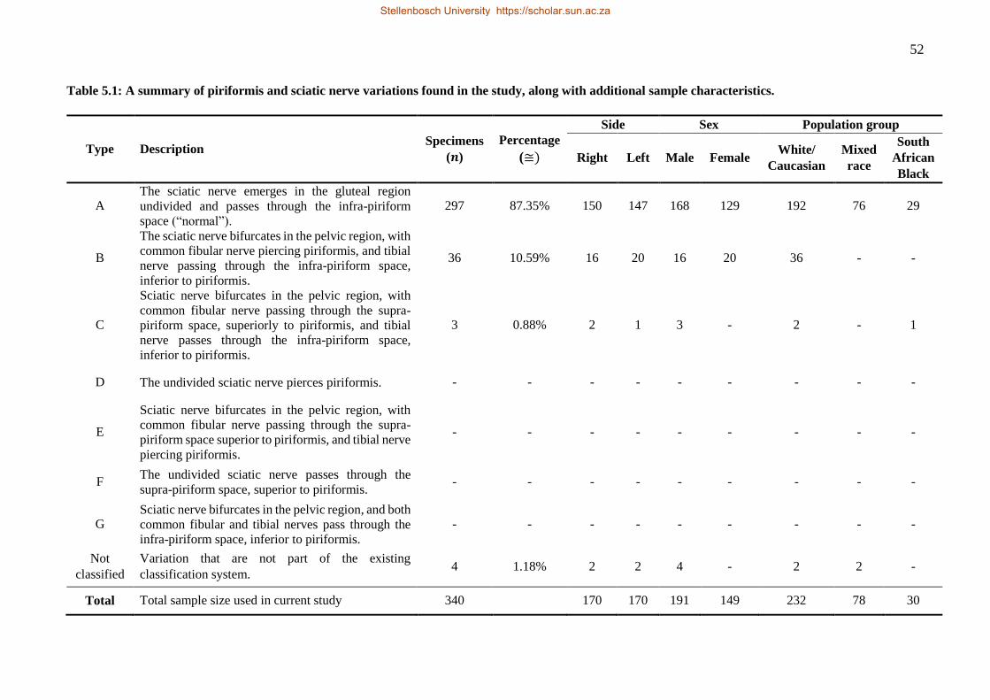
52
Table 5.1: A summary of piriformis and sciatic nerve variations found in the study, along with additional sample characteristics.
Type Description Specimens
(𝒏)
Percentage
(≅)
Side Sex Population group
Right Left Male Female White/
Caucasian
Mixed
race
South
African
Black
A The sciatic nerve emerges in the gluteal region
undivided and passes through the infra-piriform
space (“normal”).
297 87.35% 150 147 168 129 192 76 29
B
The sciatic nerve bifurcates in the pelvic region, with
common fibular nerve piercing piriformis, and tibial
nerve passing through the infra-piriform space,
inferior to piriformis.
36 10.59% 16 20 16 20 36 - -
C
Sciatic nerve bifurcates in the pelvic region, with
common fibular nerve passing through the supra-
piriform space, superiorly to piriformis, and tibial
nerve passes through the infra-piriform space,
inferior to piriformis.
3 0.88% 2 1 3 - 2 - 1
D The undivided sciatic nerve pierces piriformis. - - - - - - - - -
E
Sciatic nerve bifurcates in the pelvic region, with
common fibular nerve passing through the supra-
piriform space superior to piriformis, and tibial nerve
piercing piriformis.
- - - - - - - - -
F The undivided sciatic nerve passes through the
supra-piriform space, superior to piriformis. - - - - - - - - -
G Sciatic nerve bifurcates in the pelvic region, and both
common fibular and tibial nerves pass through the
infra-piriform space, inferior to piriformis.
- - - - - - - - -
Not
classified
Variation that are not part of the existing
classification system. 4 1.18% 2 2 4 - 2 2 -
Total Total sample size used in current study 340 170 170 191 149 232 78 30
Stellenbosch University https://scholar.sun.ac.za
Stellenbosch University https://scholar.sun.ac.za
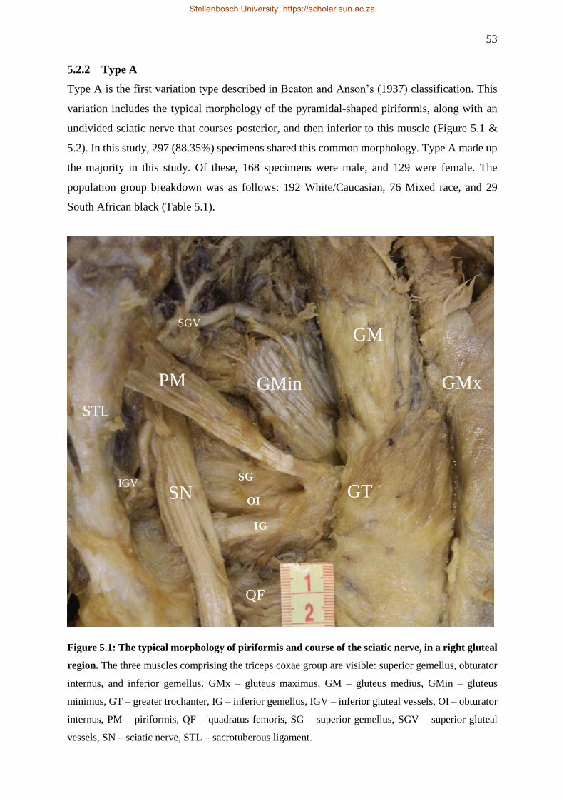
53
5.2.2 Type A
Type A is the first variation type described in Beaton and Anson’s (1937) classification. This
variation includes the typical morphology of the pyramidal-shaped piriformis, along with an
undivided sciatic nerve that courses posterior, and then inferior to this muscle (Figure 5.1 &
5.2). In this study, 297 (88.35%) specimens shared this common morphology. Type A made up
the majority in this study. Of these, 168 specimens were male, and 129 were female. The
population group breakdown was as follows: 192 White/Caucasian, 76 Mixed race, and 29
South African black (Table 5.1).
Figure 5.1: The typical morphology of piriformis and course of the sciatic nerve, in a right gluteal
region. The three muscles comprising the triceps coxae group are visible: superior gemellus, obturator
internus, and inferior gemellus. GMx – gluteus maximus, GM – gluteus medius, GMin – gluteus
minimus, GT – greater trochanter, IG – inferior gemellus, IGV – inferior gluteal vessels, OI – obturator
internus, PM – piriformis, QF – quadratus femoris, SG – superior gemellus, SGV – superior gluteal
vessels, SN – sciatic nerve, STL – sacrotuberous ligament.
PM
SN
GMin
GM
GT SG
OI
IG
SGV
IGV
GMx
QF
STL
Stellenbosch University https://scholar.sun.ac.za
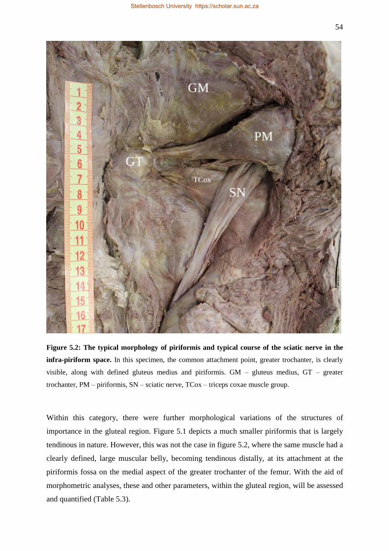
54
Figure 5.2: The typical morphology of piriformis and typical course of the sciatic nerve in the
infra-piriform space. In this specimen, the common attachment point, greater trochanter, is clearly
visible, along with defined gluteus medius and piriformis. GM – gluteus medius, GT – greater
trochanter, PM – piriformis, SN – sciatic nerve, TCox – triceps coxae muscle group.
Within this category, there were further morphological variations of the structures of
importance in the gluteal region. Figure 5.1 depicts a much smaller piriformis that is largely
tendinous in nature. However, this was not the case in figure 5.2, where the same muscle had a
clearly defined, large muscular belly, becoming tendinous distally, at its attachment at the
piriformis fossa on the medial aspect of the greater trochanter of the femur. With the aid of
morphometric analyses, these and other parameters, within the gluteal region, will be assessed
and quantified (Table 5.3).
PM
GT
GM
SN
TCox
Stellenbosch University https://scholar.sun.ac.za
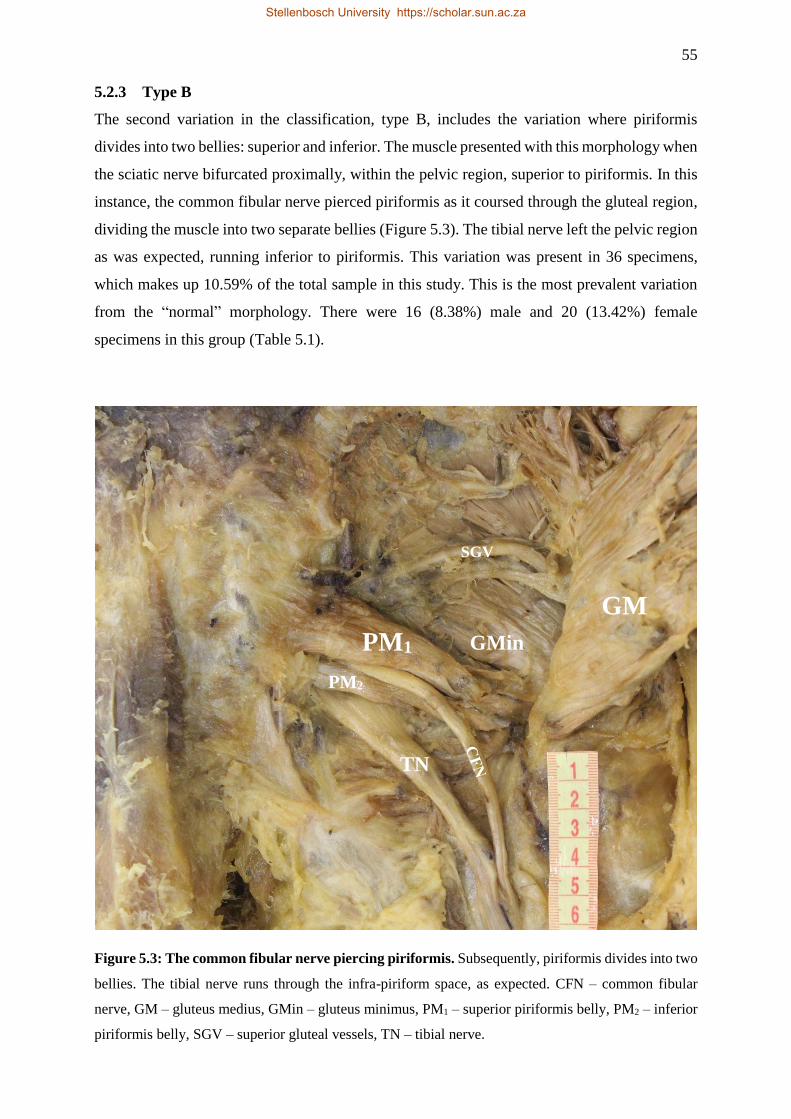
55
5.2.3 Type B
The second variation in the classification, type B, includes the variation where piriformis
divides into two bellies: superior and inferior. The muscle presented with this morphology when
the sciatic nerve bifurcated proximally, within the pelvic region, superior to piriformis. In this
instance, the common fibular nerve pierced piriformis as it coursed through the gluteal region,
dividing the muscle into two separate bellies (Figure 5.3). The tibial nerve left the pelvic region
as was expected, running inferior to piriformis. This variation was present in 36 specimens,
which makes up 10.59% of the total sample in this study. This is the most prevalent variation
from the “normal” morphology. There were 16 (8.38%) male and 20 (13.42%) female
specimens in this group (Table 5.1).
Figure 5.3: The common fibular nerve piercing piriformis. Subsequently, piriformis divides into two
bellies. The tibial nerve runs through the infra-piriform space, as expected. CFN – common fibular
nerve, GM – gluteus medius, GMin – gluteus minimus, PM1 – superior piriformis belly, PM2 – inferior
piriformis belly, SGV – superior gluteal vessels, TN – tibial nerve.
PM1
TN
GMin
GM
PM2
SGV
Stellenbosch University https://scholar.sun.ac.za
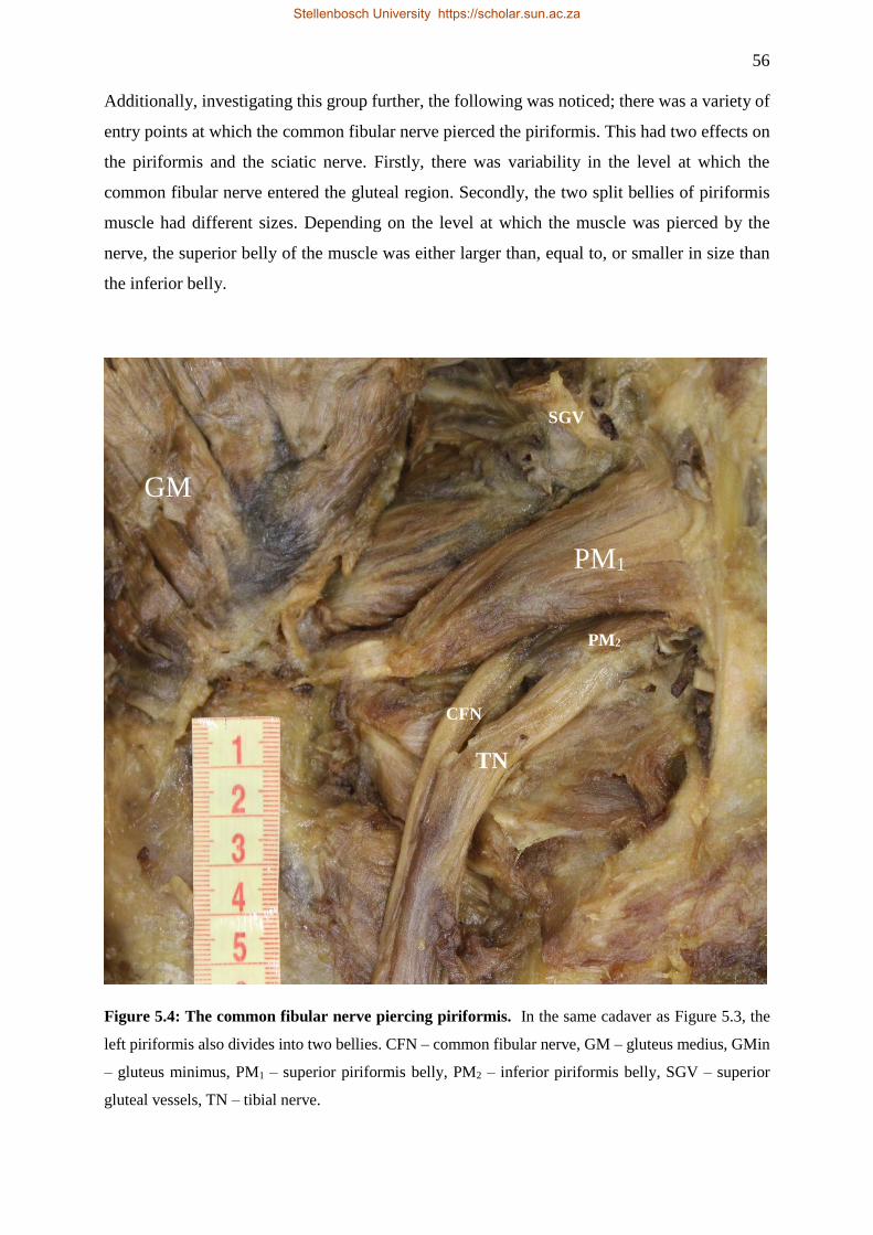
56
Additionally, investigating this group further, the following was noticed; there was a variety of
entry points at which the common fibular nerve pierced the piriformis. This had two effects on
the piriformis and the sciatic nerve. Firstly, there was variability in the level at which the
common fibular nerve entered the gluteal region. Secondly, the two split bellies of piriformis
muscle had different sizes. Depending on the level at which the muscle was pierced by the
nerve, the superior belly of the muscle was either larger than, equal to, or smaller in size than
the inferior belly.
Figure 5.4: The common fibular nerve piercing piriformis. In the same cadaver as Figure 5.3, the
left piriformis also divides into two bellies. CFN – common fibular nerve, GM – gluteus medius, GMin
– gluteus minimus, PM1 – superior piriformis belly, PM2 – inferior piriformis belly, SGV – superior
gluteal vessels, TN – tibial nerve.
PM1
TN
GM
CFN
PM2
SGV
Stellenbosch University https://scholar.sun.ac.za
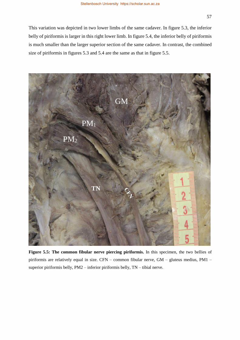
57
This variation was depicted in two lower limbs of the same cadaver. In figure 5.3, the inferior
belly of piriformis is larger in this right lower limb. In figure 5.4, the inferior belly of piriformis
is much smaller than the larger superior section of the same cadaver. In contrast, the combined
size of piriformis in figures 5.3 and 5.4 are the same as that in figure 5.5.
Figure 5.5: The common fibular nerve piercing piriformis. In this specimen, the two bellies of
piriformis are relatively equal in size. CFN – common fibular nerve, GM – gluteus medius, PM1 –
superior piriformis belly, PM2 – inferior piriformis belly, TN – tibial nerve.
PM1
TN
GM
PM2
Stellenbosch University https://scholar.sun.ac.za
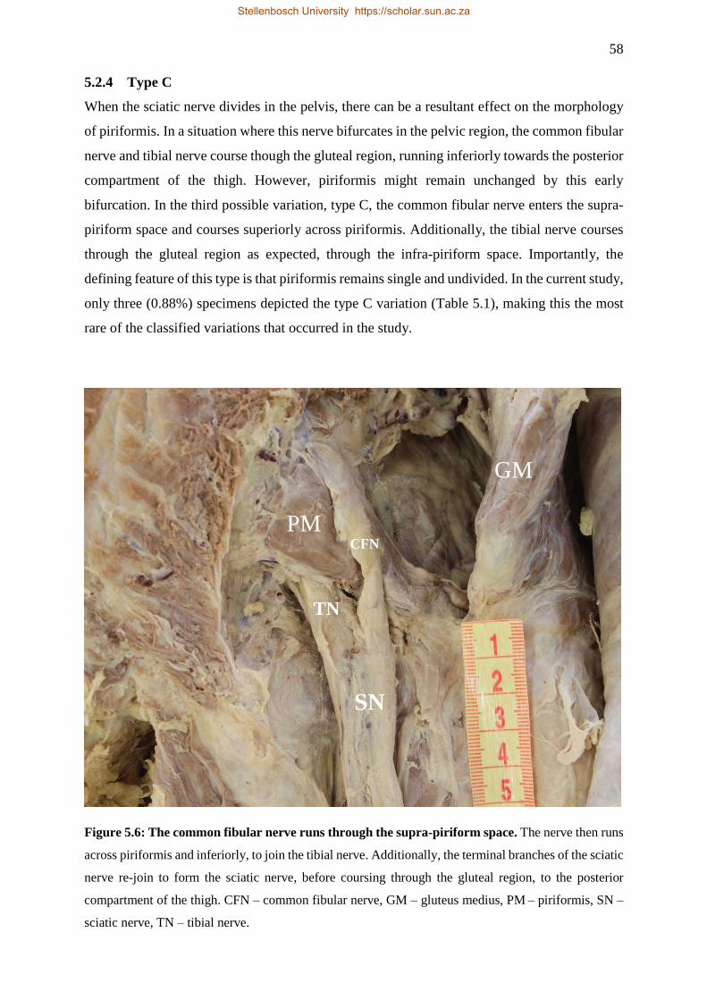
58
5.2.4 Type C
When the sciatic nerve divides in the pelvis, there can be a resultant effect on the morphology
of piriformis. In a situation where this nerve bifurcates in the pelvic region, the common fibular
nerve and tibial nerve course though the gluteal region, running inferiorly towards the posterior
compartment of the thigh. However, piriformis might remain unchanged by this early
bifurcation. In the third possible variation, type C, the common fibular nerve enters the supra-
piriform space and courses superiorly across piriformis. Additionally, the tibial nerve courses
through the gluteal region as expected, through the infra-piriform space. Importantly, the
defining feature of this type is that piriformis remains single and undivided. In the current study,
only three (0.88%) specimens depicted the type C variation (Table 5.1), making this the most
rare of the classified variations that occurred in the study.
Figure 5.6: The common fibular nerve runs through the supra-piriform space. The nerve then runs
across piriformis and inferiorly, to join the tibial nerve. Additionally, the terminal branches of the sciatic
nerve re-join to form the sciatic nerve, before coursing through the gluteal region, to the posterior
compartment of the thigh. CFN – common fibular nerve, GM – gluteus medius, PM – piriformis, SN –
sciatic nerve, TN – tibial nerve.
PM
TN
GM
CFN
SN
Stellenbosch University https://scholar.sun.ac.za

59
Figure 5.7: The common fibular nerve runs through the supra-piriform space. The nerve crosses
piriformis, and passes inferiorly, to join tibial nerve inferiorly. CFN – common fibular nerve, GMin –
gluteus minimus, GT – greater trochanter, PM – piriformis, SN – sciatic nerve, TN – tibial nerve.
This variation occurred in two limbs of the same individual, a 71-year-old White/Caucasian
female, with bilateral symmetry in the variation between piriformis and the sciatic nerve (Figure
5.6 & 5.7). The remaining one specimen is that of a right lower limb that belonged to a South
African black male, aged 37 years at the time of death (Figure 5.8). This cadaver did not present
with bilateral symmetry, and the left limb is categorised under type A, as its anatomy depicts a
normal piriformis and sciatic nerve morphology.
As shown in figures 5.6, 5.7, and 5.8, piriformis remains unseparated, despite the fact that the
sciatic nerve bifurcated in the pelvis. In addition, the common fibular nerve and tibial nerve re-
join in both left and right gluteal regions (Figure 5.6 & 5.7), to course through the respective
lower limbs as unbranched sciatic nerves. In the remaining specimen (Figure 5.8), the sciatic
nerve terminates in the pelvic region, and its terminal branches, the common fibular and tibial
nerves, course superficial and deep to piriformis, and into the posterior compartment of the
thigh, as two distinct entities.
PM
TN
GMin
CFN
SN
GT
Stellenbosch University https://scholar.sun.ac.za
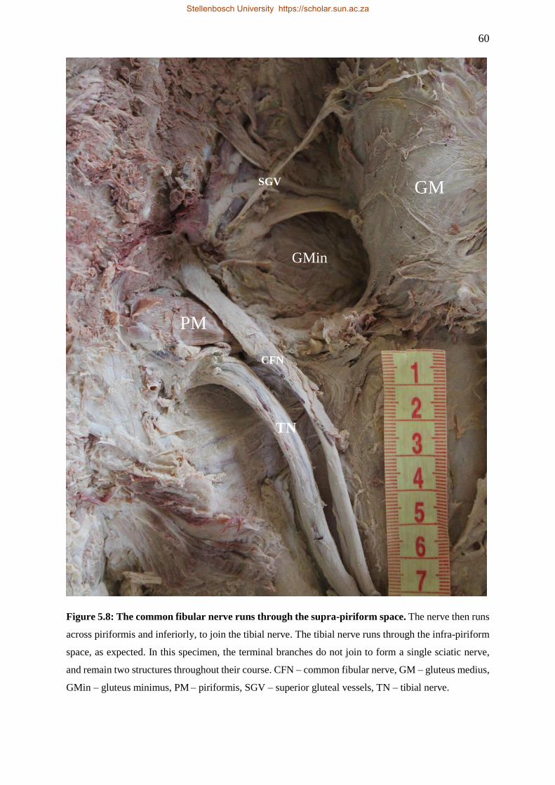
60
Figure 5.8: The common fibular nerve runs through the supra-piriform space. The nerve then runs
across piriformis and inferiorly, to join the tibial nerve. The tibial nerve runs through the infra-piriform
space, as expected. In this specimen, the terminal branches do not join to form a single sciatic nerve,
and remain two structures throughout their course. CFN – common fibular nerve, GM – gluteus medius,
GMin – gluteus minimus, PM – piriformis, SGV – superior gluteal vessels, TN – tibial nerve.
PM
TN
GMin
CFN
SGV GM
Stellenbosch University https://scholar.sun.ac.za
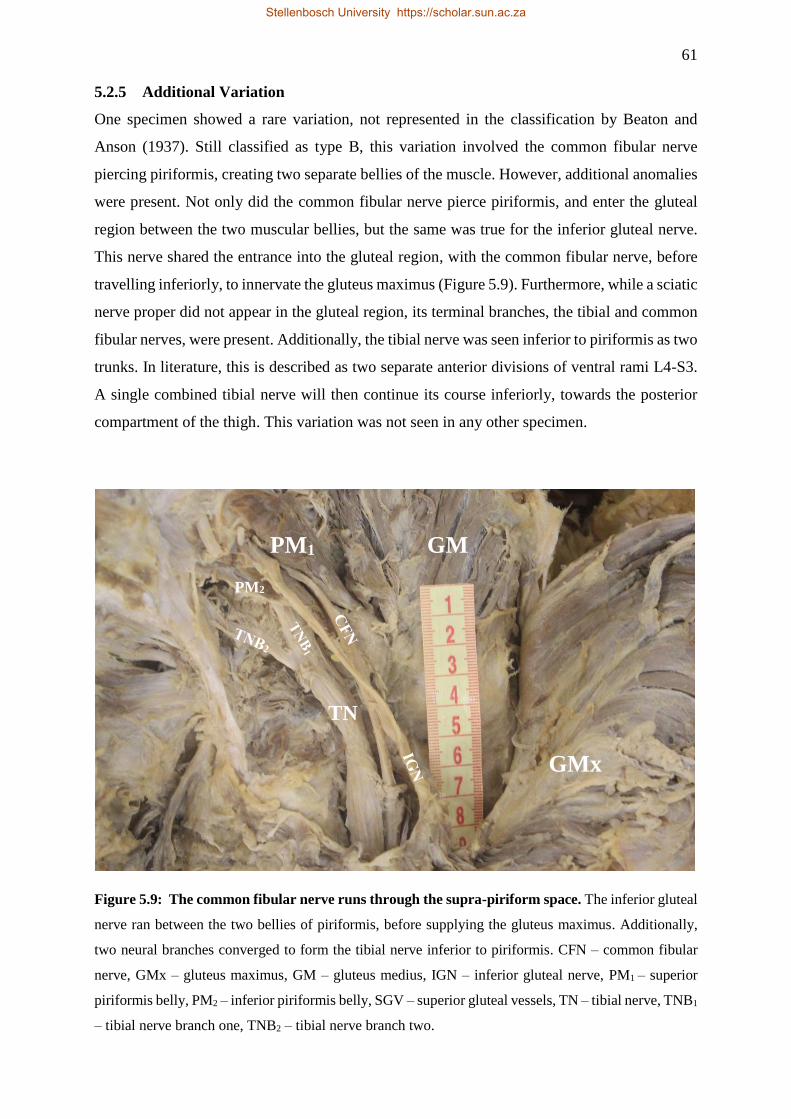
61
5.2.5 Additional Variation
One specimen showed a rare variation, not represented in the classification by Beaton and
Anson (1937). Still classified as type B, this variation involved the common fibular nerve
piercing piriformis, creating two separate bellies of the muscle. However, additional anomalies
were present. Not only did the common fibular nerve pierce piriformis, and enter the gluteal
region between the two muscular bellies, but the same was true for the inferior gluteal nerve.
This nerve shared the entrance into the gluteal region, with the common fibular nerve, before
travelling inferiorly, to innervate the gluteus maximus (Figure 5.9). Furthermore, while a sciatic
nerve proper did not appear in the gluteal region, its terminal branches, the tibial and common
fibular nerves, were present. Additionally, the tibial nerve was seen inferior to piriformis as two
trunks. In literature, this is described as two separate anterior divisions of ventral rami L4-S3.
A single combined tibial nerve will then continue its course inferiorly, towards the posterior
compartment of the thigh. This variation was not seen in any other specimen.
Figure 5.9: The common fibular nerve runs through the supra-piriform space. The inferior gluteal
nerve ran between the two bellies of piriformis, before supplying the gluteus maximus. Additionally,
two neural branches converged to form the tibial nerve inferior to piriformis. CFN – common fibular
nerve, GMx – gluteus maximus, GM – gluteus medius, IGN – inferior gluteal nerve, PM1 – superior
piriformis belly, PM2 – inferior piriformis belly, SGV – superior gluteal vessels, TN – tibial nerve, TNB1
– tibial nerve branch one, TNB2 – tibial nerve branch two.
PM1
TN
GM
PM2
SGV
GMx
Stellenbosch University https://scholar.sun.ac.za
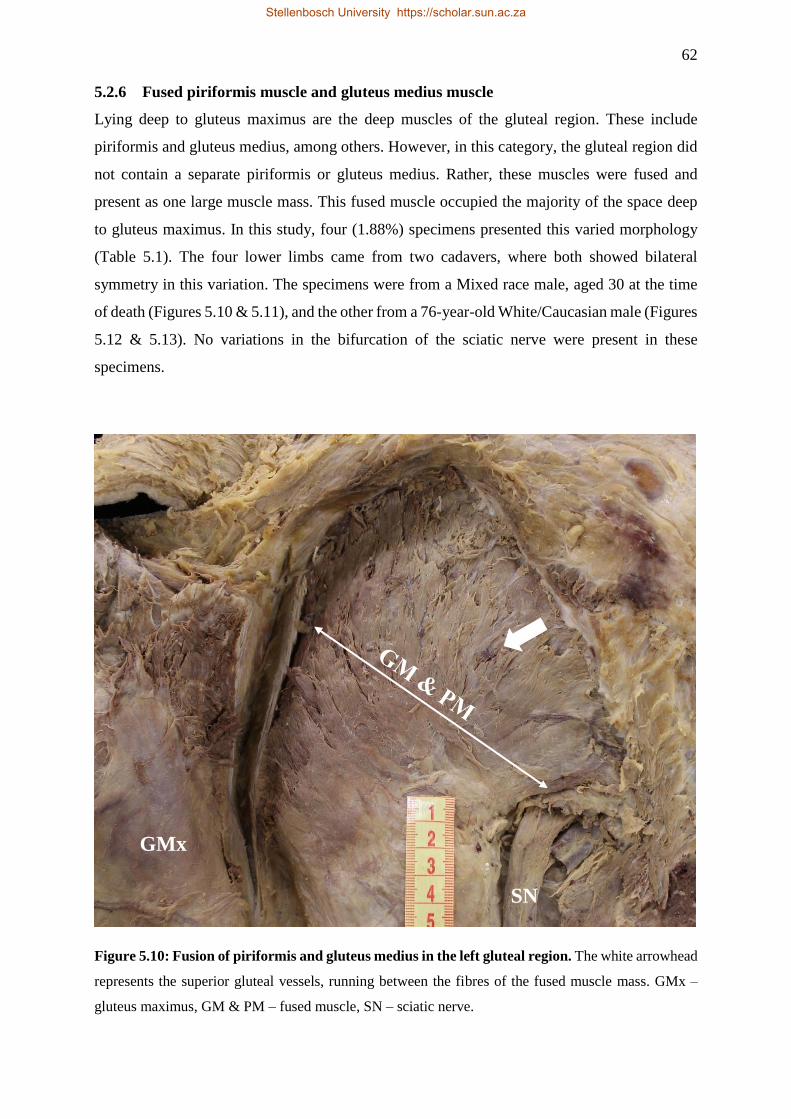
62
5.2.6 Fused piriformis muscle and gluteus medius muscle
Lying deep to gluteus maximus are the deep muscles of the gluteal region. These include
piriformis and gluteus medius, among others. However, in this category, the gluteal region did
not contain a separate piriformis or gluteus medius. Rather, these muscles were fused and
present as one large muscle mass. This fused muscle occupied the majority of the space deep
to gluteus maximus. In this study, four (1.88%) specimens presented this varied morphology
(Table 5.1). The four lower limbs came from two cadavers, where both showed bilateral
symmetry in this variation. The specimens were from a Mixed race male, aged 30 at the time
of death (Figures 5.10 & 5.11), and the other from a 76-year-old White/Caucasian male (Figures
5.12 & 5.13). No variations in the bifurcation of the sciatic nerve were present in these
specimens.
Figure 5.10: Fusion of piriformis and gluteus medius in the left gluteal region. The white arrowhead
represents the superior gluteal vessels, running between the fibres of the fused muscle mass. GMx –
gluteus maximus, GM & PM – fused muscle, SN – sciatic nerve.
SN
GMx
Stellenbosch University https://scholar.sun.ac.za
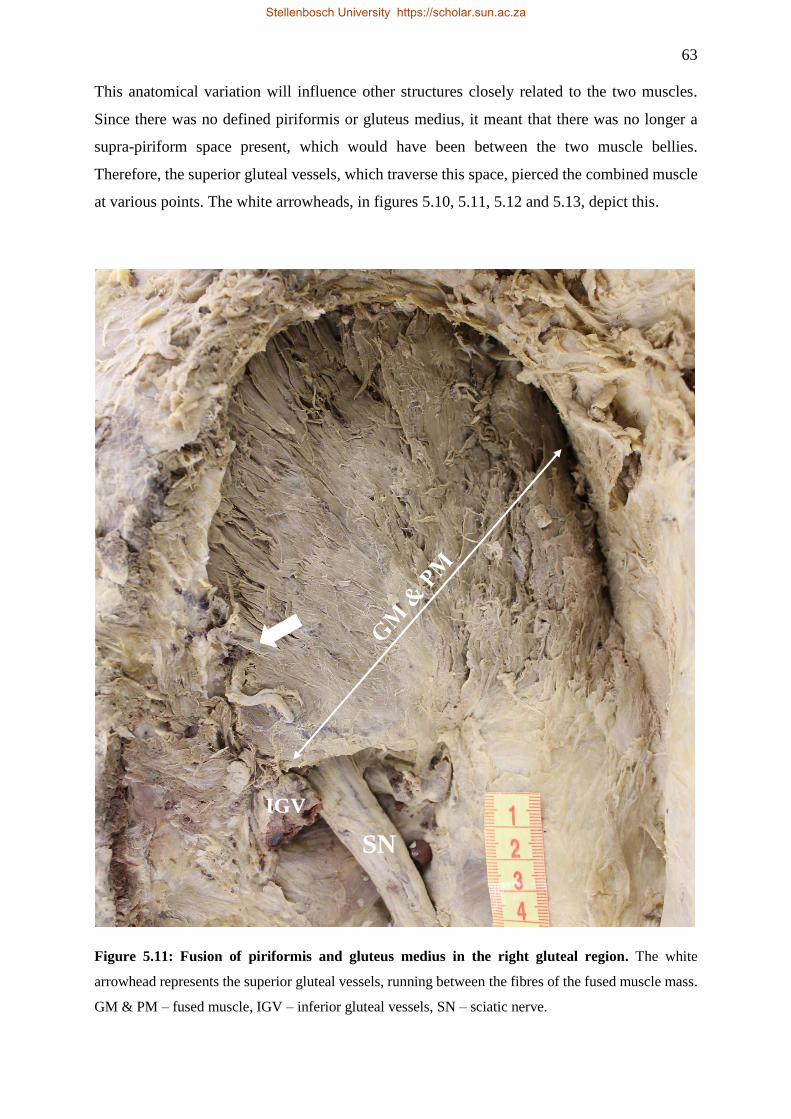
63
This anatomical variation will influence other structures closely related to the two muscles.
Since there was no defined piriformis or gluteus medius, it meant that there was no longer a
supra-piriform space present, which would have been between the two muscle bellies.
Therefore, the superior gluteal vessels, which traverse this space, pierced the combined muscle
at various points. The white arrowheads, in figures 5.10, 5.11, 5.12 and 5.13, depict this.
Figure 5.11: Fusion of piriformis and gluteus medius in the right gluteal region. The white
arrowhead represents the superior gluteal vessels, running between the fibres of the fused muscle mass.
GM & PM – fused muscle, IGV – inferior gluteal vessels, SN – sciatic nerve.
SN
IGV
Stellenbosch University https://scholar.sun.ac.za
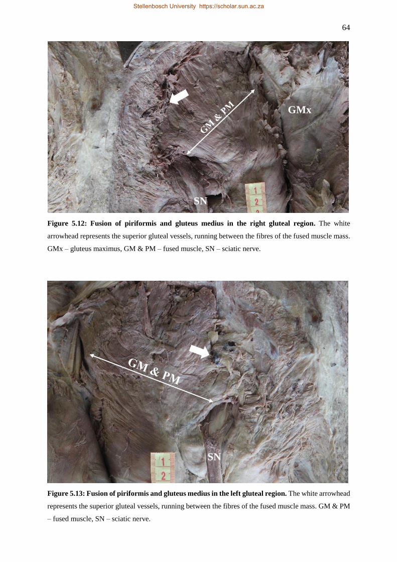
64
Figure 5.12: Fusion of piriformis and gluteus medius in the right gluteal region. The white
arrowhead represents the superior gluteal vessels, running between the fibres of the fused muscle mass.
GMx – gluteus maximus, GM & PM – fused muscle, SN – sciatic nerve.
Figure 5.13: Fusion of piriformis and gluteus medius in the left gluteal region. The white arrowhead
represents the superior gluteal vessels, running between the fibres of the fused muscle mass. GM & PM
– fused muscle, SN – sciatic nerve.
GMx
SN
SN
Stellenbosch University https://scholar.sun.ac.za

65
5.3 VARIATIONS IN THE SCIATIC NERVE BIFURCATION
5.3.1 Overview
In the present study, 338 lower limbs were examined to classify the level at which the sciatic
nerve bifurcated into the common fibular and tibial nerves. This group consisted of 189 male
specimens, and 149 female specimens. Additional specimen characteristics are summarised in
table 5.2, below. There were two specimens, from the same cadaver, that were excluded from
the classification process because this cadaver had undergone a bilateral amputation above the
knee joint. This meant that there was insufficient evidence of where the bifurcation of the nerve
was. Table 5.2 consists of accurate classification, descriptions, and a summary concerning the
variations in the bifurcation of the sciatic nerve for the 338 specimens available, because of the
new SNBL index.
Stellenbosch University https://scholar.sun.ac.za
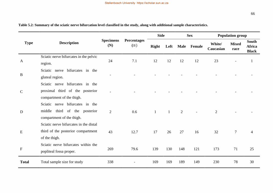
66
Table 5.2: Summary of the sciatic nerve bifurcation level classified in the study, along with additional sample characteristics.
Type Description Specimens
(N)
Percentages
(≅)
Side Sex Population group
Right Left Male Female White/
Caucasian
Mixed
race
South
Africa
Black
A Sciatic nerve bifurcates in the pelvic
region. 24 7.1 12 12 12 12 23 - 1
B Sciatic nerve bifurcates in the
gluteal region. - - - - - - - - -
C
Sciatic nerve bifurcates in the
proximal third of the posterior
compartment of the thigh.
- - - - - - - - -
D
Sciatic nerve bifurcates in the
middle third of the posterior
compartment of the thigh.
2 0.6 1 1 2 - 2 - -
E
Sciatic nerve bifurcates in the distal
third of the posterior compartment
of the thigh.
43 12.7 17 26 27 16 32 7 4
F Sciatic nerve bifurcates within the
popliteal fossa proper. 269 79.6 139 130 148 121 173 71 25
Total Total sample size for study 338 - 169 169 189 149 230 78 30
Stellenbosch University https://scholar.sun.ac.za
Stellenbosch University https://scholar.sun.ac.za
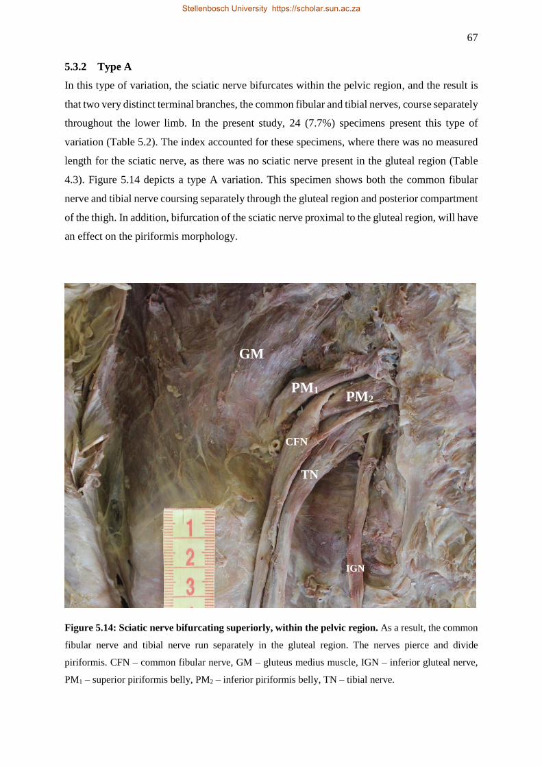
67
5.3.2 Type A
In this type of variation, the sciatic nerve bifurcates within the pelvic region, and the result is
that two very distinct terminal branches, the common fibular and tibial nerves, course separately
throughout the lower limb. In the present study, 24 (7.7%) specimens present this type of
variation (Table 5.2). The index accounted for these specimens, where there was no measured
length for the sciatic nerve, as there was no sciatic nerve present in the gluteal region (Table
4.3). Figure 5.14 depicts a type A variation. This specimen shows both the common fibular
nerve and tibial nerve coursing separately through the gluteal region and posterior compartment
of the thigh. In addition, bifurcation of the sciatic nerve proximal to the gluteal region, will have
an effect on the piriformis morphology.
Figure 5.14: Sciatic nerve bifurcating superiorly, within the pelvic region. As a result, the common
fibular nerve and tibial nerve run separately in the gluteal region. The nerves pierce and divide
piriformis. CFN – common fibular nerve, GM – gluteus medius muscle, IGN – inferior gluteal nerve,
PM1 – superior piriformis belly, PM2 – inferior piriformis belly, TN – tibial nerve.
PM2
TN
GM
CFN
PM1
IGN
Stellenbosch University https://scholar.sun.ac.za
68
In 15 specimens, the sciatic nerve bifurcated prior to entering the gluteal region, and resulted
in similar variations seen in figure 5.14, with subsequent variations to piriformis. However,
these specimens were not added to the total in this bifurcation category, because the branches
converged into a single nerve inferior to piriformis (Figure 5.15). These nerves went on to
follow the more traditional patterns of bifurcation, where terminal bifurcation took place in the
distal third of the posterior compartment of the thigh, for one of the specimens, and 14
specimens where the sciatic nerve branched further down, bifurcating into the common fibular
and tibial nerves within the popliteal fossa proper. The discussion, below, concerns the last two
types of bifurcation variations.
Figure 5.15: The common fibular and tibial nerve converge, within the gluteal region, forming the
sciatic nerve. Piriformis divided into two bellies, pierced by the common fibular nerve. CFN – common
fibular nerve, GMx – gluteus maximus, GM – gluteus maximus, PM1 – superior piriformis belly, PM1
– inferior piriformis belly, SN – sciatic nerve, TN – tibial nerve. IGV – inferior gluteal vessels.
PM2
TN
GM
CFN
PM1
SN
GMx
IGV
Stellenbosch University https://scholar.sun.ac.za
69
5.3.3 Type D
Type D represents the fourth classification in the possible bifurcation level of the sciatic nerve.
This type is characterised by a sciatic nerve that bifurcates in the middle third of the posterior
compartment of the thigh. In this study, this type of variation was rare, with only two (0.6%)
specimens presenting with a sciatic nerve that bifurcated at this level (Table 5.2). According to
table 4.3, this category was quantified by using the ratio of the sciatic nerve length to the length
of the thigh, and accepting all the values that fall within a range greater than but not equal to
45%, and lesser than and equal to 65%.
5.3.4 Type E
In this study, 43 (12.7%) specimens conform to the requirements for this category (Table 5.2).
For this category, type E, specimens were only selected where the calculated index ratio was
greater than but not equal to 65%, and lesser than or equal to 85% (Table 4.3). The researcher
added one specimen, similar to the one depicted in figure 5.15, to the total, where the sciatic
nerve bifurcated prior to the gluteal region. In this specimen, the branches rejoin to form a single
sciatic nerve inferior to piriformis. The sciatic nerve then bifurcated lower down into its
terminal branches within the distal third of the posterior compartment of the thigh. Generally,
the sciatic nerve bifurcation occurs within this region, namely proximal to the superior border
of the popliteal fossa. The common fibular and tibial nerves will then course to their respective
compartments in the leg.
5.3.5 Type F
In the final classification, the sciatic nerve bifurcates most distally, within the popliteal fossa
proper. This type of variation made up the majority of the variations seen in the present study.
In the present study, the researcher classified 269 (79.6%) specimens as type F (Table 5.2). The
researcher quantified this classification by including all the specimens with an index value that
was greater than but not equal to 85% (Table 4.3). The 14 specimens, which had sciatic nerves
that bifurcated within the gluteal region, only to fuse and reform the sciatic nerve inferior to
piriformis (Figure 5.15), were added to the total specimens in this category. This was done
because the sciatic nerve in these 14 specimens bifurcated within the popliteal fossa proper,
after unification higher up.
Stellenbosch University https://scholar.sun.ac.za
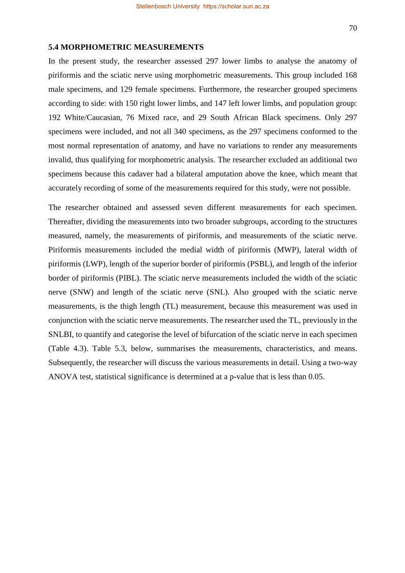
70
5.4 MORPHOMETRIC MEASUREMENTS
In the present study, the researcher assessed 297 lower limbs to analyse the anatomy of
piriformis and the sciatic nerve using morphometric measurements. This group included 168
male specimens, and 129 female specimens. Furthermore, the researcher grouped specimens
according to side: with 150 right lower limbs, and 147 left lower limbs, and population group:
192 White/Caucasian, 76 Mixed race, and 29 South African Black specimens. Only 297
specimens were included, and not all 340 specimens, as the 297 specimens conformed to the
most normal representation of anatomy, and have no variations to render any measurements
invalid, thus qualifying for morphometric analysis. The researcher excluded an additional two
specimens because this cadaver had a bilateral amputation above the knee, which meant that
accurately recording of some of the measurements required for this study, were not possible.
The researcher obtained and assessed seven different measurements for each specimen.
Thereafter, dividing the measurements into two broader subgroups, according to the structures
measured, namely, the measurements of piriformis, and measurements of the sciatic nerve.
Piriformis measurements included the medial width of piriformis (MWP), lateral width of
piriformis (LWP), length of the superior border of piriformis (PSBL), and length of the inferior
border of piriformis (PIBL). The sciatic nerve measurements included the width of the sciatic
nerve (SNW) and length of the sciatic nerve (SNL). Also grouped with the sciatic nerve
measurements, is the thigh length (TL) measurement, because this measurement was used in
conjunction with the sciatic nerve measurements. The researcher used the TL, previously in the
SNLBI, to quantify and categorise the level of bifurcation of the sciatic nerve in each specimen
(Table 4.3). Table 5.3, below, summarises the measurements, characteristics, and means.
Subsequently, the researcher will discuss the various measurements in detail. Using a two-way
ANOVA test, statistical significance is determined at a p-value that is less than 0.05.
Stellenbosch University https://scholar.sun.ac.za
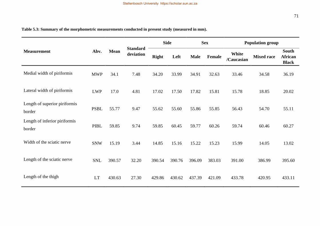
71
Table 5.3: Summary of the morphometric measurements conducted in present study (measured in mm).
Measurement Abv. Mean Standard
deviation
Side Sex Population group
Right Left Male Female White
/Caucasian Mixed race
South
African
Black
Medial width of piriformis MWP 34.1 7.48 34.20 33.99 34.91 32.63 33.46 34.58 36.19
Lateral width of piriformis LWP 17.0 4.81 17.02 17.50 17.82 15.81 15.78 18.85 20.02
Length of superior piriformis
border PSBL 55.77 9.47 55.62 55.60 55.86 55.85 56.43 54.70 55.11
Length of inferior piriformis
border PIBL 59.85 9.74 59.85 60.45 59.77 60.26 59.74 60.46 60.27
Width of the sciatic nerve SNW 15.19 3.44 14.85 15.16 15.22 15.23 15.99 14.05 13.02
Length of the sciatic nerve SNL 390.57 32.20 390.54 390.76 396.09 383.03 391.00 386.99 395.60
Length of the thigh LT 430.63 27.30 429.86 430.62 437.39 421.09 433.78 420.95 433.11
Stellenbosch University https://scholar.sun.ac.za
Stellenbosch University https://scholar.sun.ac.za
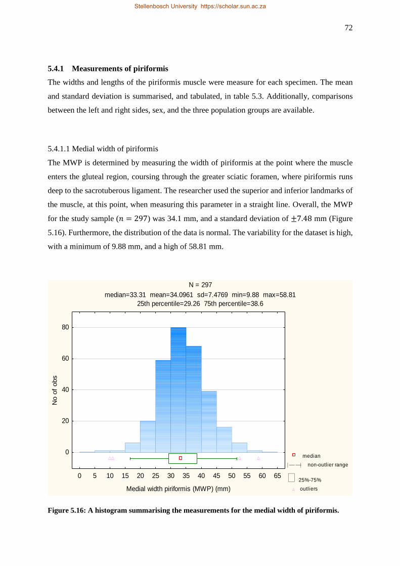
72
5.4.1 Measurements of piriformis
The widths and lengths of the piriformis muscle were measure for each specimen. The mean
and standard deviation is summarised, and tabulated, in table 5.3. Additionally, comparisons
between the left and right sides, sex, and the three population groups are available.
5.4.1.1 Medial width of piriformis
The MWP is determined by measuring the width of piriformis at the point where the muscle
enters the gluteal region, coursing through the greater sciatic foramen, where piriformis runs
deep to the sacrotuberous ligament. The researcher used the superior and inferior landmarks of
the muscle, at this point, when measuring this parameter in a straight line. Overall, the MWP
for the study sample (𝑛 = 297) was 34.1 mm, and a standard deviation of ±7.48 mm (Figure
5.16). Furthermore, the distribution of the data is normal. The variability for the dataset is high,
with a minimum of 9.88 mm, and a high of 58.81 mm.
N = 297
median=33.31 mean=34.0961 sd=7.4769 min=9.88 max=58.81
25th percentile=29.26 75th percentile=38.6
median
non-outlier range
25%-75%
outliers
0 5 10 15 20 25 30 35 40 45 50 55 60 65
Medial width piriformis (MWP) (mm)
0
20
40
60
80
No o
f obs
Figure 5.16: A histogram summarising the measurements for the medial width of piriformis.
Stellenbosch University https://scholar.sun.ac.za
73
5.4.1.2 Lateral width of piriformis
The researcher obtained the LWP by measuring the vertical width of piriformis mid-section of
the muscle belly. Again, the researcher measured this vertical width in a straight line. The LWP
was narrow compared to the MWP, with a mean of 17.01 mm for all specimens (𝑛 = 297)
(Table 5.3). In accordance with a smaller measurement, the calculated standard deviation for
this mean is ±4.81 mm (Figure 5.17). The LWP mean is consistent with the expected size of
the muscle at this point, becoming tendinous and increasingly narrow, ahead of its distal
attachment to the piriformis fossa at the medial aspect of the greater trochanter of the femur.
N = 297
median=16.74 mean=17.0051 sd=4.8114 min=4.19 max=36.35
25th percentile=13.95 75th percentile=19.53
median
non-outlier range
25%-75%
outliers
0 5 10 15 20 25 30 35 40
Lateral width piriformis (LWP) (mm)
0
30
60
90
120
No o
f obs
Figure 5.17: A histogram summarising the measurements for the lateral width of piriformis.
As illustrated in figure 5.18, the two different box plots represent the different comparisons
made regarding the LWP. In this test, the effect of both side and sex are used to understand the
LWP. As depicted, a high significant difference is observed between the right lower limbs of
male and female specimens (𝑝 < 0.01), and similarly, a high significant difference is observed
when comparing left lower limbs that belonged to male and female specimens (𝑝 = 0.01).
Therefore, a highly significant difference exists between male (blue) and female (red) LWP,
Stellenbosch University https://scholar.sun.ac.za
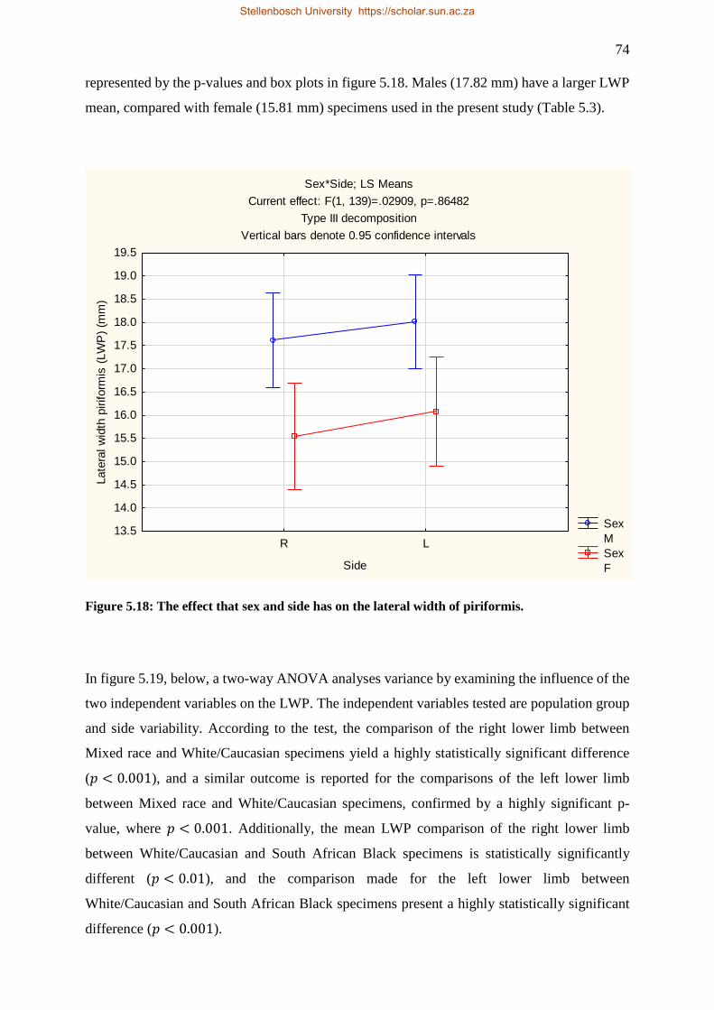
74
represented by the p-values and box plots in figure 5.18. Males (17.82 mm) have a larger LWP
mean, compared with female (15.81 mm) specimens used in the present study (Table 5.3).
Sex*Side; LS Means
Current effect: F(1, 139)=.02909, p=.86482
Type III decomposition
Vertical bars denote 0.95 confidence intervals
Sex
M
Sex
F
R L
Side
13.5
14.0
14.5
15.0
15.5
16.0
16.5
17.0
17.5
18.0
18.5
19.0
19.5
Late
ral w
idth
piriform
is (
LW
P)
(mm
)
Figure 5.18: The effect that sex and side has on the lateral width of piriformis.
In figure 5.19, below, a two-way ANOVA analyses variance by examining the influence of the
two independent variables on the LWP. The independent variables tested are population group
and side variability. According to the test, the comparison of the right lower limb between
Mixed race and White/Caucasian specimens yield a highly statistically significant difference
(𝑝 < 0.001), and a similar outcome is reported for the comparisons of the left lower limb
between Mixed race and White/Caucasian specimens, confirmed by a highly significant p-
value, where 𝑝 < 0.001. Additionally, the mean LWP comparison of the right lower limb
between White/Caucasian and South African Black specimens is statistically significantly
different (𝑝 < 0.01), and the comparison made for the left lower limb between
White/Caucasian and South African Black specimens present a highly statistically significant
difference (𝑝 < 0.001).
Stellenbosch University https://scholar.sun.ac.za
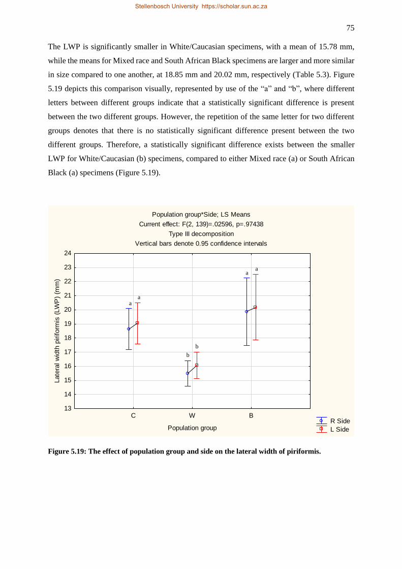
75
The LWP is significantly smaller in White/Caucasian specimens, with a mean of 15.78 mm,
while the means for Mixed race and South African Black specimens are larger and more similar
in size compared to one another, at 18.85 mm and 20.02 mm, respectively (Table 5.3). Figure
5.19 depicts this comparison visually, represented by use of the “a” and “b”, where different
letters between different groups indicate that a statistically significant difference is present
between the two different groups. However, the repetition of the same letter for two different
groups denotes that there is no statistically significant difference present between the two
different groups. Therefore, a statistically significant difference exists between the smaller
LWP for White/Caucasian (b) specimens, compared to either Mixed race (a) or South African
Black (a) specimens (Figure 5.19).
Population group*Side; LS Means
Current effect: F(2, 139)=.02596, p=.97438
Type III decomposition
Vertical bars denote 0.95 confidence intervals
R Side
L Side
C W B
Population group
13
14
15
16
17
18
19
20
21
22
23
24
Late
ral w
idth
piriform
is (
LW
P)
(mm
)
Figure 5.19: The effect of population group and side on the lateral width of piriformis.
a a
a a
b
b
Stellenbosch University https://scholar.sun.ac.za
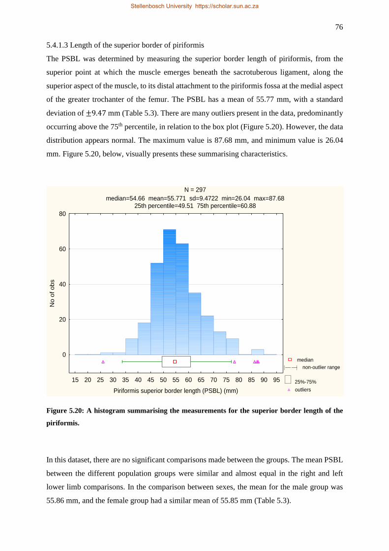
76
5.4.1.3 Length of the superior border of piriformis
The PSBL was determined by measuring the superior border length of piriformis, from the
superior point at which the muscle emerges beneath the sacrotuberous ligament, along the
superior aspect of the muscle, to its distal attachment to the piriformis fossa at the medial aspect
of the greater trochanter of the femur. The PSBL has a mean of 55.77 mm, with a standard
deviation of ±9.47 mm (Table 5.3). There are many outliers present in the data, predominantly
occurring above the 75th percentile, in relation to the box plot (Figure 5.20). However, the data
distribution appears normal. The maximum value is 87.68 mm, and minimum value is 26.04
mm. Figure 5.20, below, visually presents these summarising characteristics.
N = 297
median=54.66 mean=55.771 sd=9.4722 min=26.04 max=87.6825th percentile=49.51 75th percentile=60.88
median
non-outlier range
25%-75%
outliers
15 20 25 30 35 40 45 50 55 60 65 70 75 80 85 90 95
Piriformis superior border length (PSBL) (mm)
0
20
40
60
80
No o
f obs
Figure 5.20: A histogram summarising the measurements for the superior border length of the
piriformis.
In this dataset, there are no significant comparisons made between the groups. The mean PSBL
between the different population groups were similar and almost equal in the right and left
lower limb comparisons. In the comparison between sexes, the mean for the male group was
55.86 mm, and the female group had a similar mean of 55.85 mm (Table 5.3).
Stellenbosch University https://scholar.sun.ac.za

77
5.4.1.4 Length of the inferior border of piriformis
The researcher obtained the LIBP measurement by measuring the length of the inferior border
of piriformis, from the inferior point at which the muscle emerged beneath the sacrotuberous
ligament, along the inferior aspect of the muscle, to its distal attachment to the piriformis fossa
at the medial aspect of the greater trochanter of the femur.
Figure 5.21 visually summarises the LIBP measurement results for the total study sample (𝑁 =
297). This length has a mean value of 59.85 mm, and an even distribution amongst the entire
sample group. Furthermore, the LIBP comparisons present no statistical values of interest when
represented graphically. Therefore, the researcher observed no statistically significant
differences for comparisons between sexes and side.
N = 297
median=59.54 mean=59.8497 sd=9.7359 min=30.16 max=92.0325th percentile=53.81 75th percentile=64.79
median
non-outlier range
25%-75%
outliers
20 25 30 35 40 45 50 55 60 65 70 75 80 85 90 95 100
Piriformis inferior border length (PIBL) (mm)
0
20
40
60
80
No o
f obs
Figure 5.21: A histogram summarising the inferior border length of piriformis measurements.
Very similar means seen in the comparison between the White/Caucasian, Mixed race, and
South African Black population groups. Table 5.3 depicts the similarities between the three
mean values, where the mean values for the White/Caucasian, Mixed race, and South African
Black specimens are 59.74 mm, 60.46 mm, and 60.27 mm, respectively.
Stellenbosch University https://scholar.sun.ac.za
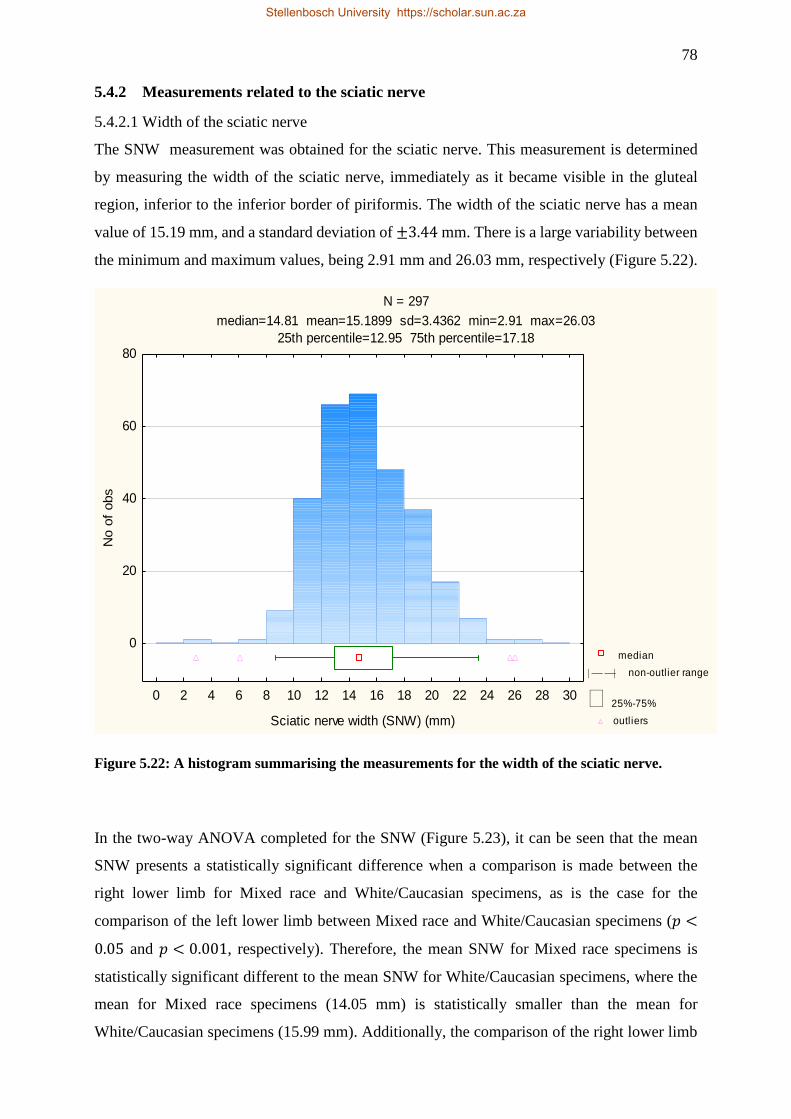
78
5.4.2 Measurements related to the sciatic nerve
5.4.2.1 Width of the sciatic nerve
The SNW measurement was obtained for the sciatic nerve. This measurement is determined
by measuring the width of the sciatic nerve, immediately as it became visible in the gluteal
region, inferior to the inferior border of piriformis. The width of the sciatic nerve has a mean
value of 15.19 mm, and a standard deviation of ±3.44 mm. There is a large variability between
the minimum and maximum values, being 2.91 mm and 26.03 mm, respectively (Figure 5.22).
N = 297
median=14.81 mean=15.1899 sd=3.4362 min=2.91 max=26.03
25th percentile=12.95 75th percentile=17.18
median
non-outlier range
25%-75%
outliers
0 2 4 6 8 10 12 14 16 18 20 22 24 26 28 30
Sciatic nerve width (SNW) (mm)
0
20
40
60
80
No o
f obs
Figure 5.22: A histogram summarising the measurements for the width of the sciatic nerve.
In the two-way ANOVA completed for the SNW (Figure 5.23), it can be seen that the mean
SNW presents a statistically significant difference when a comparison is made between the
right lower limb for Mixed race and White/Caucasian specimens, as is the case for the
comparison of the left lower limb between Mixed race and White/Caucasian specimens (𝑝 <
0.05 and 𝑝 < 0.001, respectively). Therefore, the mean SNW for Mixed race specimens is
statistically significant different to the mean SNW for White/Caucasian specimens, where the
mean for Mixed race specimens (14.05 mm) is statistically smaller than the mean for
White/Caucasian specimens (15.99 mm). Additionally, the comparison of the right lower limb
Stellenbosch University https://scholar.sun.ac.za
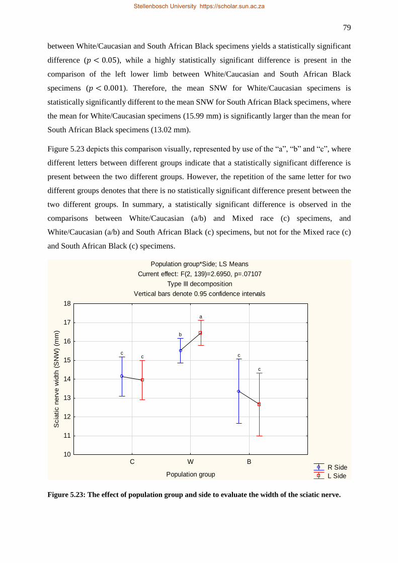
79
between White/Caucasian and South African Black specimens yields a statistically significant
difference (𝑝 < 0.05), while a highly statistically significant difference is present in the
comparison of the left lower limb between White/Caucasian and South African Black
specimens (𝑝 < 0.001). Therefore, the mean SNW for White/Caucasian specimens is
statistically significantly different to the mean SNW for South African Black specimens, where
the mean for White/Caucasian specimens (15.99 mm) is significantly larger than the mean for
South African Black specimens (13.02 mm).
Figure 5.23 depicts this comparison visually, represented by use of the “a”, “b” and “c”, where
different letters between different groups indicate that a statistically significant difference is
present between the two different groups. However, the repetition of the same letter for two
different groups denotes that there is no statistically significant difference present between the
two different groups. In summary, a statistically significant difference is observed in the
comparisons between White/Caucasian (a/b) and Mixed race (c) specimens, and
White/Caucasian (a/b) and South African Black (c) specimens, but not for the Mixed race (c)
and South African Black (c) specimens.
Population group*Side; LS Means
Current effect: F(2, 139)=2.6950, p=.07107
Type III decomposition
Vertical bars denote 0.95 confidence intervals
R Side
L Side
C W B
Population group
10
11
12
13
14
15
16
17
18
Scia
tic n
erv
e w
idth
(S
NW
) (m
m)
cc
b
a
c
c
Figure 5.23: The effect of population group and side to evaluate the width of the sciatic nerve.
Stellenbosch University https://scholar.sun.ac.za
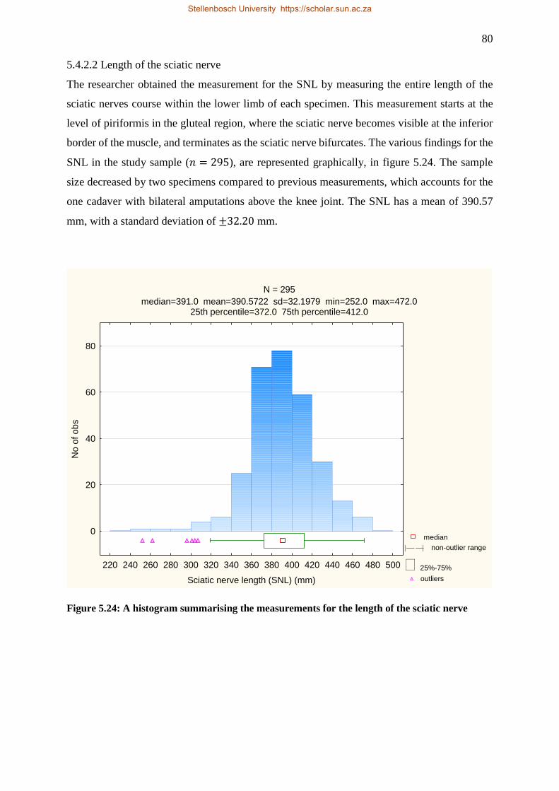
80
5.4.2.2 Length of the sciatic nerve
The researcher obtained the measurement for the SNL by measuring the entire length of the
sciatic nerves course within the lower limb of each specimen. This measurement starts at the
level of piriformis in the gluteal region, where the sciatic nerve becomes visible at the inferior
border of the muscle, and terminates as the sciatic nerve bifurcates. The various findings for the
SNL in the study sample (𝑛 = 295), are represented graphically, in figure 5.24. The sample
size decreased by two specimens compared to previous measurements, which accounts for the
one cadaver with bilateral amputations above the knee joint. The SNL has a mean of 390.57
mm, with a standard deviation of ±32.20 mm.
N = 295
median=391.0 mean=390.5722 sd=32.1979 min=252.0 max=472.025th percentile=372.0 75th percentile=412.0
median
non-outlier range
25%-75%
outliers
220 240 260 280 300 320 340 360 380 400 420 440 460 480 500
Sciatic nerve length (SNL) (mm)
0
20
40
60
80
No
of o
bs
Figure 5.24: A histogram summarising the measurements for the length of the sciatic nerve
Stellenbosch University https://scholar.sun.ac.za
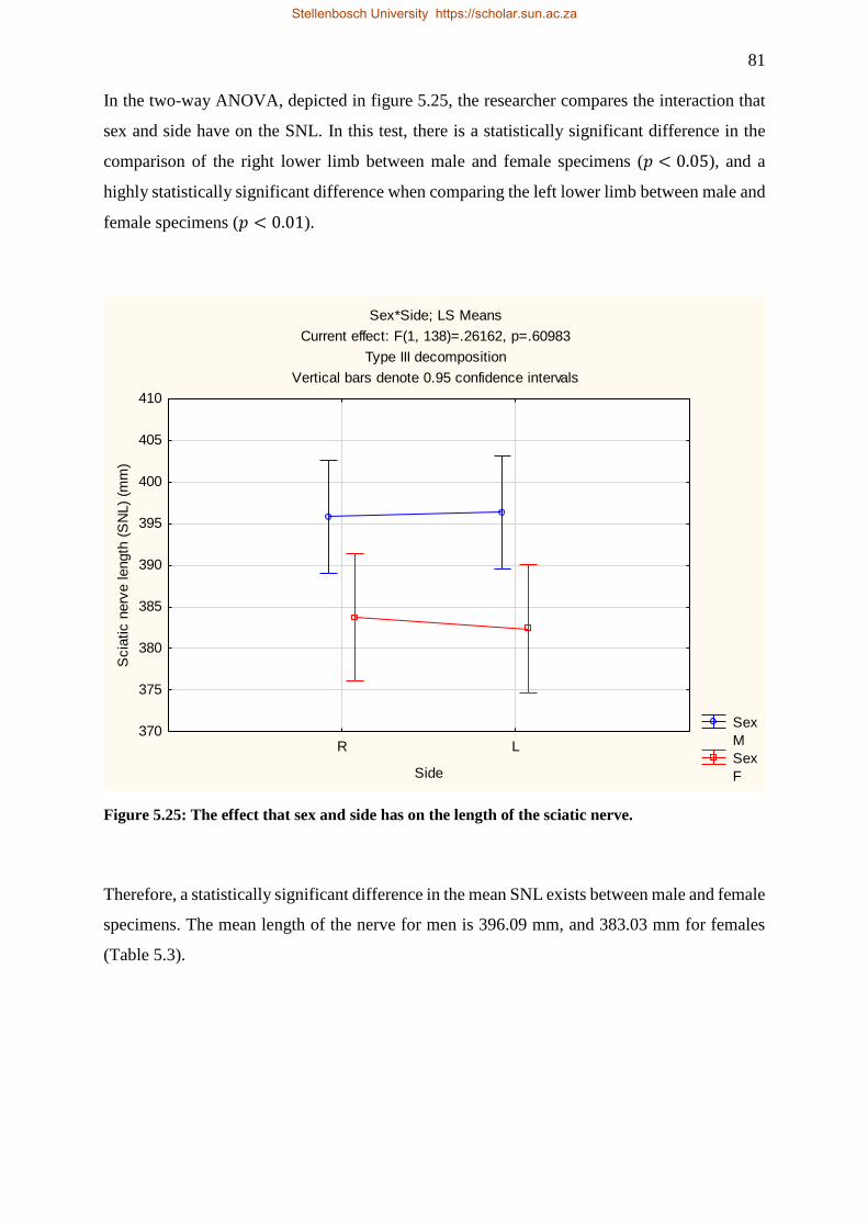
81
In the two-way ANOVA, depicted in figure 5.25, the researcher compares the interaction that
sex and side have on the SNL. In this test, there is a statistically significant difference in the
comparison of the right lower limb between male and female specimens (𝑝 < 0.05), and a
highly statistically significant difference when comparing the left lower limb between male and
female specimens (𝑝 < 0.01).
Sex*Side; LS Means
Current effect: F(1, 138)=.26162, p=.60983
Type III decomposition
Vertical bars denote 0.95 confidence intervals
Sex
M
Sex
F
R L
Side
370
375
380
385
390
395
400
405
410
Scia
tic n
erv
e length
(S
NL)
(mm
)
Figure 5.25: The effect that sex and side has on the length of the sciatic nerve.
Therefore, a statistically significant difference in the mean SNL exists between male and female
specimens. The mean length of the nerve for men is 396.09 mm, and 383.03 mm for females
(Table 5.3).
Stellenbosch University https://scholar.sun.ac.za
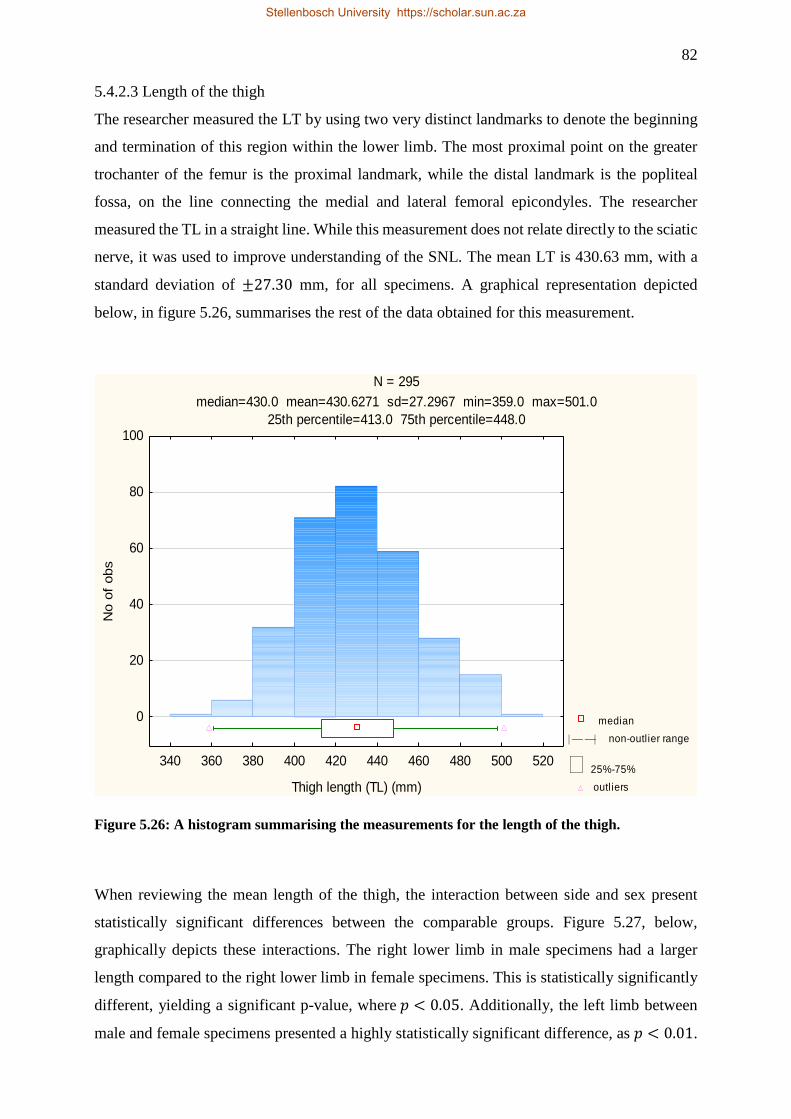
82
5.4.2.3 Length of the thigh
The researcher measured the LT by using two very distinct landmarks to denote the beginning
and termination of this region within the lower limb. The most proximal point on the greater
trochanter of the femur is the proximal landmark, while the distal landmark is the popliteal
fossa, on the line connecting the medial and lateral femoral epicondyles. The researcher
measured the TL in a straight line. While this measurement does not relate directly to the sciatic
nerve, it was used to improve understanding of the SNL. The mean LT is 430.63 mm, with a
standard deviation of ±27.30 mm, for all specimens. A graphical representation depicted
below, in figure 5.26, summarises the rest of the data obtained for this measurement.
N = 295
median=430.0 mean=430.6271 sd=27.2967 min=359.0 max=501.0
25th percentile=413.0 75th percentile=448.0
median
non-outlier range
25%-75%
outliers
340 360 380 400 420 440 460 480 500 520
Thigh length (TL) (mm)
0
20
40
60
80
100
No o
f obs
Figure 5.26: A histogram summarising the measurements for the length of the thigh.
When reviewing the mean length of the thigh, the interaction between side and sex present
statistically significant differences between the comparable groups. Figure 5.27, below,
graphically depicts these interactions. The right lower limb in male specimens had a larger
length compared to the right lower limb in female specimens. This is statistically significantly
different, yielding a significant p-value, where 𝑝 < 0.05. Additionally, the left limb between
male and female specimens presented a highly statistically significant difference, as 𝑝 < 0.01.
Stellenbosch University https://scholar.sun.ac.za
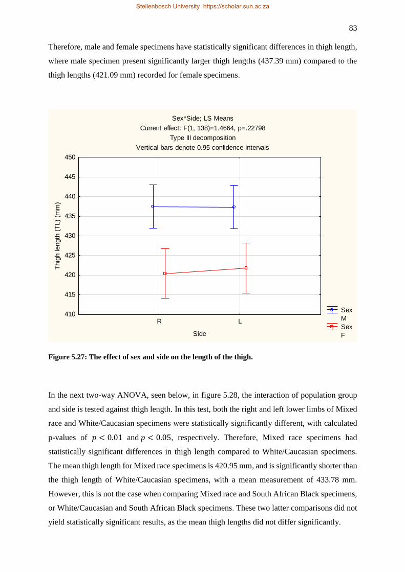
83
Therefore, male and female specimens have statistically significant differences in thigh length,
where male specimen present significantly larger thigh lengths (437.39 mm) compared to the
thigh lengths (421.09 mm) recorded for female specimens.
Sex*Side; LS Means
Current effect: F(1, 138)=1.4664, p=.22798
Type III decomposition
Vertical bars denote 0.95 confidence intervals
Sex
M
Sex
F
R L
Side
410
415
420
425
430
435
440
445
450
Thig
h length
(T
L)
(mm
)
Figure 5.27: The effect of sex and side on the length of the thigh.
In the next two-way ANOVA, seen below, in figure 5.28, the interaction of population group
and side is tested against thigh length. In this test, both the right and left lower limbs of Mixed
race and White/Caucasian specimens were statistically significantly different, with calculated
p-values of 𝑝 < 0.01 and 𝑝 < 0.05, respectively. Therefore, Mixed race specimens had
statistically significant differences in thigh length compared to White/Caucasian specimens.
The mean thigh length for Mixed race specimens is 420.95 mm, and is significantly shorter than
the thigh length of White/Caucasian specimens, with a mean measurement of 433.78 mm.
However, this is not the case when comparing Mixed race and South African Black specimens,
or White/Caucasian and South African Black specimens. These two latter comparisons did not
yield statistically significant results, as the mean thigh lengths did not differ significantly.
Stellenbosch University https://scholar.sun.ac.za
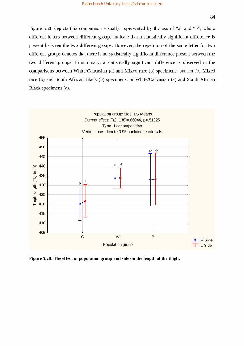
84
Figure 5.28 depicts this comparison visually, represented by the use of “a” and “b”, where
different letters between different groups indicate that a statistically significant difference is
present between the two different groups. However, the repetition of the same letter for two
different groups denotes that there is no statistically significant difference present between the
two different groups. In summary, a statistically significant difference is observed in the
comparisons between White/Caucasian (a) and Mixed race (b) specimens, but not for Mixed
race (b) and South African Black (b) specimens, or White/Caucasian (a) and South African
Black specimens (a).
Population group*Side; LS Means
Current effect: F(2, 138)=.66044, p=.51825
Type III decomposition
Vertical bars denote 0.95 confidence intervals
R Side
L Side
C W B
Population group
405
410
415
420
425
430
435
440
445
450
455
Thig
h length
(T
L)
(mm
)
Figure 5.28: The effect of population group and side on the length of the thigh.
a a
ab ab
b b
Stellenbosch University https://scholar.sun.ac.za

85
CHAPTER SIX:
DISCUSSION
Stellenbosch University https://scholar.sun.ac.za
86
Damage resulting from injections into the sciatic nerve or structures closely related to the nerve
is the most frequent cause of nerve injuries in children (Senes et al., 2009). Although the
intramuscular injection is considered a basic skill, it must be treated with due diligence. To
avoid complications, medical staff need to understand regional anatomy properly. Additionally,
clinicians need to understand the different injection sites, along with advantages and
disadvantages for each site (Small, 2004). Too often, local anatomical variations predispose
injury to nerves. These variations include abnormal subdivision of the piriformis due to
proximal bifurcation of the sciatic nerve into its terminal branches. Regardless of what
procedure the clinician intends to follow, good clinical judgement using the best evidence
available on case-specific assessment will result in the best outcome (Small, 2004). Therefore,
maintaining the best possible clinical practices requires up to date relevant anatomical
knowledge. It is for this reason that studies, such as the present one, ensures that vital “new”
information is available.
6.1 ANATOMICAL VARIATIONS OF PIRIFORMIS
In the present study, the researcher examined 340 specimens to study and describe the variation
in the relationship between the sciatic nerve and piriformis. By comparison, this study had a
much larger sample size than many other studies published previously.
Similar to previous studies published, type A in the classification, was the most prevalent type
of morphology. In a similar study conducted by Natsis et al. (2014), type A was present in
93.6% of the specimens studied. However, 76% of the specimens presented type A morphology
in the study conducted by Guvencer et al. (2009), which helps to understand the prevalence
range of this type. In a meta-analysis conducted by Tomaszewski et al. (2016), the population
prevalence for the normal pattern was 85.2%. According to Pokorny et al. (2006), 79.1% of the
cases had an undivided sciatic nerve that passed distally below a complete piriformis,
resembling type A. Furthermore, in two studies conducted in Brazil, where 40 specimens were
used in both studies, type A was seen in 85% (Vicente et al., 2007), and 90% of the specimens
(Brooks et al., 2011), respectively. In the present study, the researcher observed this
morphology in 297 (87.35%) specimens. The prevalence in the present study is therefore similar
to the prevalence documented elsewhere in the world.
Stellenbosch University https://scholar.sun.ac.za
87
The current study had 43 (12.65%) specimens, in 28 (16.47%) cadavers, which presented
variations in the course of the sciatic nerve in relation to piriformis. Again, these numbers are
consistent with literature published previously. A more detailed description of each type of
variation, observed in the present study, will follow.
Numerous studies (Chiba et al., 1992; Pecina, 1979; Pokorny et al., 2006; Haladaj et al., 2015)
have published the prevalence of each category described by Beaton & Anson et al. (1937) in
detail. Overwhelmingly, previous literature points to type B as the most prevalent deviation
from the normal morphology. The results in the present study confirm this assumption, as type
B made up the majority of all variations seen. Piriformis was pierced by the common fibular
nerve in 36 (10.59%) of specimens in the present study. In a study conducted on 514 lower
limbs, Chiba et al. (1992) found that this variation occurred in as many as 38% of the cases,
which is the highest prevalence reported in literature. Subsequent literature has reported a
prevalence of between 10-20%. Guvencer et al. (2009), Haladaj et al. (2015), Pokorny et al.
(2006) and Moore et al. (2017) have confirmed this, with a reported prevalence of 16%, 20%,
14.3%, and 12%, respectively. However, the prevalence was lower in literature published in
Africa. In the study conducted by Ogeng’o et al. (2011), the prevalence for the common fibular
nerve division of the sciatic nerve piercing piriformis was 7.9%, and only occurred in 13 of the
164 specimens examined. Berihu and Debeb (2015) saw similar numbers, where 9% of the
specimens observed belonged to the type B variation. Furthermore, Chukwuanukwu et al.
(2007) reported no variations that resembled type B. The present study confirms the lower
prevalence of this variation in the African population.
The third variation in the classification, type C, is rarer than the previous type, and occurs when
the common fibular nerve runs through the supra-piriform space, superficially across piriformis,
while the tibial nerve courses through the infra-piriform space, as normal. In the meta-analysis
conducted by Tomaszewski et al. (2016), this variation had a prevalence of 1.9%, with no major
sex or geographical variations amongst populations (Tomaszewski et al., 2016). Pokorny et al.
(2006) largely reinforces this, where 4.4% of the specimens presented this variation. Other
prevalence amongst different authors include: 6% of the cases published by Okraszewska et al.
(2002), 1.5% of the specimens studied by Ugrenovic et al. (2005), and 2.4% in the Black
Kenyan population (Ogeng’o et al., 2011). Additionally, only one specimen presented this
variation type in a study conducted by Chukwuanukwu et al. (2007), resulting in a 3.8%
prevalence of this variation in this Nigerian population group. Only three (0.88%) specimens
presented this variation, in the present study (Table 5.1).
Stellenbosch University https://scholar.sun.ac.za
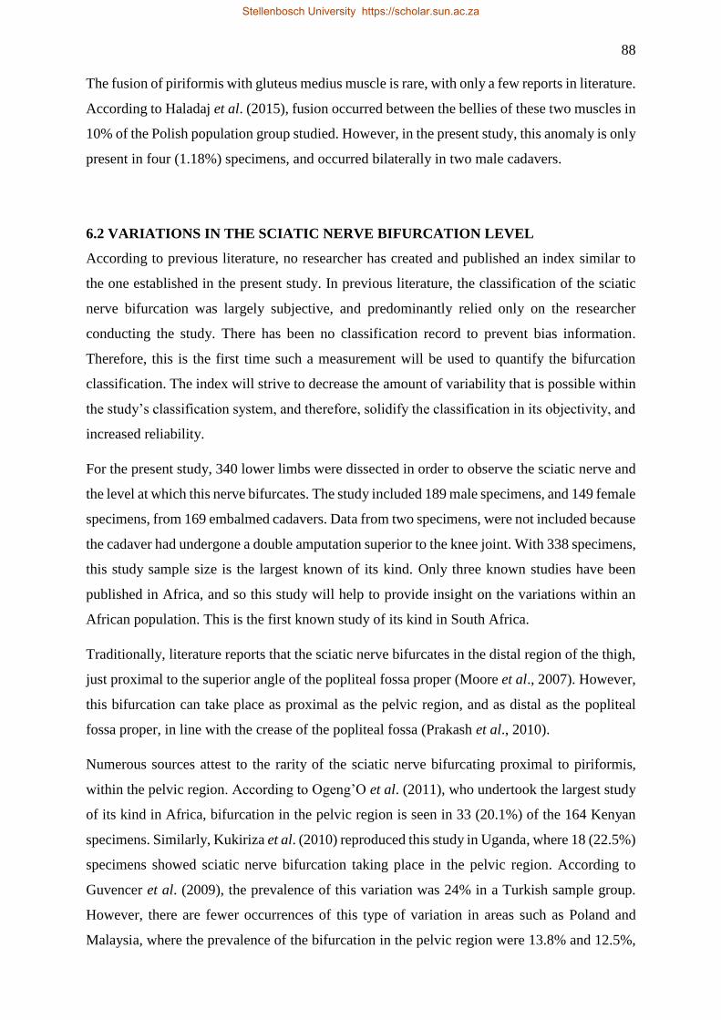
88
The fusion of piriformis with gluteus medius muscle is rare, with only a few reports in literature.
According to Haladaj et al. (2015), fusion occurred between the bellies of these two muscles in
10% of the Polish population group studied. However, in the present study, this anomaly is only
present in four (1.18%) specimens, and occurred bilaterally in two male cadavers.
6.2 VARIATIONS IN THE SCIATIC NERVE BIFURCATION LEVEL
According to previous literature, no researcher has created and published an index similar to
the one established in the present study. In previous literature, the classification of the sciatic
nerve bifurcation was largely subjective, and predominantly relied only on the researcher
conducting the study. There has been no classification record to prevent bias information.
Therefore, this is the first time such a measurement will be used to quantify the bifurcation
classification. The index will strive to decrease the amount of variability that is possible within
the study’s classification system, and therefore, solidify the classification in its objectivity, and
increased reliability.
For the present study, 340 lower limbs were dissected in order to observe the sciatic nerve and
the level at which this nerve bifurcates. The study included 189 male specimens, and 149 female
specimens, from 169 embalmed cadavers. Data from two specimens, were not included because
the cadaver had undergone a double amputation superior to the knee joint. With 338 specimens,
this study sample size is the largest known of its kind. Only three known studies have been
published in Africa, and so this study will help to provide insight on the variations within an
African population. This is the first known study of its kind in South Africa.
Traditionally, literature reports that the sciatic nerve bifurcates in the distal region of the thigh,
just proximal to the superior angle of the popliteal fossa proper (Moore et al., 2007). However,
this bifurcation can take place as proximal as the pelvic region, and as distal as the popliteal
fossa proper, in line with the crease of the popliteal fossa (Prakash et al., 2010).
Numerous sources attest to the rarity of the sciatic nerve bifurcating proximal to piriformis,
within the pelvic region. According to Ogeng’O et al. (2011), who undertook the largest study
of its kind in Africa, bifurcation in the pelvic region is seen in 33 (20.1%) of the 164 Kenyan
specimens. Similarly, Kukiriza et al. (2010) reproduced this study in Uganda, where 18 (22.5%)
specimens showed sciatic nerve bifurcation taking place in the pelvic region. According to
Guvencer et al. (2009), the prevalence of this variation was 24% in a Turkish sample group.
However, there are fewer occurrences of this type of variation in areas such as Poland and
Malaysia, where the prevalence of the bifurcation in the pelvic region were 13.8% and 12.5%,
Stellenbosch University https://scholar.sun.ac.za
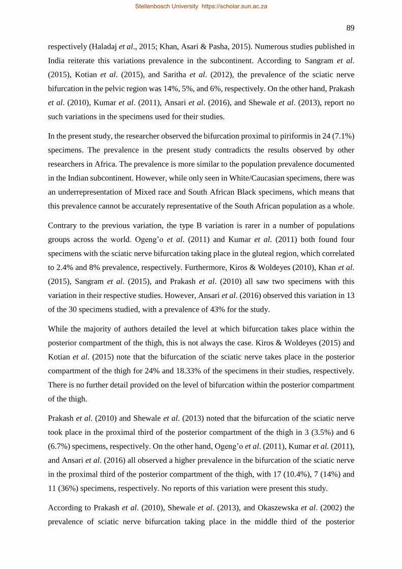
89
respectively (Haladaj et al., 2015; Khan, Asari & Pasha, 2015). Numerous studies published in
India reiterate this variations prevalence in the subcontinent. According to Sangram et al.
(2015), Kotian et al. (2015), and Saritha et al. (2012), the prevalence of the sciatic nerve
bifurcation in the pelvic region was 14%, 5%, and 6%, respectively. On the other hand, Prakash
et al. (2010), Kumar et al. (2011), Ansari et al. (2016), and Shewale et al. (2013), report no
such variations in the specimens used for their studies.
In the present study, the researcher observed the bifurcation proximal to piriformis in 24 (7.1%)
specimens. The prevalence in the present study contradicts the results observed by other
researchers in Africa. The prevalence is more similar to the population prevalence documented
in the Indian subcontinent. However, while only seen in White/Caucasian specimens, there was
an underrepresentation of Mixed race and South African Black specimens, which means that
this prevalence cannot be accurately representative of the South African population as a whole.
Contrary to the previous variation, the type B variation is rarer in a number of populations
groups across the world. Ogeng’o et al. (2011) and Kumar et al. (2011) both found four
specimens with the sciatic nerve bifurcation taking place in the gluteal region, which correlated
to 2.4% and 8% prevalence, respectively. Furthermore, Kiros & Woldeyes (2010), Khan et al.
(2015), Sangram et al. (2015), and Prakash et al. (2010) all saw two specimens with this
variation in their respective studies. However, Ansari et al. (2016) observed this variation in 13
of the 30 specimens studied, with a prevalence of 43% for the study.
While the majority of authors detailed the level at which bifurcation takes place within the
posterior compartment of the thigh, this is not always the case. Kiros & Woldeyes (2015) and
Kotian et al. (2015) note that the bifurcation of the sciatic nerve takes place in the posterior
compartment of the thigh for 24% and 18.33% of the specimens in their studies, respectively.
There is no further detail provided on the level of bifurcation within the posterior compartment
of the thigh.
Prakash et al. (2010) and Shewale et al. (2013) noted that the bifurcation of the sciatic nerve
took place in the proximal third of the posterior compartment of the thigh in 3 (3.5%) and 6
(6.7%) specimens, respectively. On the other hand, Ogeng’o et al. (2011), Kumar et al. (2011),
and Ansari et al. (2016) all observed a higher prevalence in the bifurcation of the sciatic nerve
in the proximal third of the posterior compartment of the thigh, with 17 (10.4%), 7 (14%) and
11 (36%) specimens, respectively. No reports of this variation were present this study.
According to Prakash et al. (2010), Shewale et al. (2013), and Okaszewska et al. (2002) the
prevalence of sciatic nerve bifurcation taking place in the middle third of the posterior
Stellenbosch University https://scholar.sun.ac.za
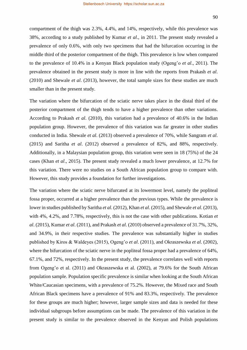
90
compartment of the thigh was 2.3%, 4.4%, and 14%, respectively, while this prevalence was
38%, according to a study published by Kumar et al., in 2011. The present study revealed a
prevalence of only 0.6%, with only two specimens that had the bifurcation occurring in the
middle third of the posterior compartment of the thigh. This prevalence is low when compared
to the prevalence of 10.4% in a Kenyan Black population study (Ogeng’o et al., 2011). The
prevalence obtained in the present study is more in line with the reports from Prakash et al.
(2010) and Shewale et al. (2013), however, the total sample sizes for these studies are much
smaller than in the present study.
The variation where the bifurcation of the sciatic nerve takes place in the distal third of the
posterior compartment of the thigh tends to have a higher prevalence than other variations.
According to Prakash et al. (2010), this variation had a prevalence of 40.6% in the Indian
population group. However, the prevalence of this variation was far greater in other studies
conducted in India. Shewale et al. (2013) observed a prevalence of 70%, while Sangram et al.
(2015) and Saritha et al. (2012) observed a prevalence of 82%, and 88%, respectively.
Additionally, in a Malaysian population group, this variation were seen in 18 (75%) of the 24
cases (Khan et al., 2015). The present study revealed a much lower prevalence, at 12.7% for
this variation. There were no studies on a South African population group to compare with.
However, this study provides a foundation for further investigations.
The variation where the sciatic nerve bifurcated at its lowermost level, namely the popliteal
fossa proper, occurred at a higher prevalence than the previous types. While the prevalence is
lower in studies published by Saritha et al. (2012), Khan et al. (2015), and Shewale et al. (2013),
with 4%, 4.2%, and 7.78%, respectively, this is not the case with other publications. Kotian et
al. (2015), Kumar et al. (2011), and Prakash et al. (2010) observed a prevalence of 31.7%, 32%,
and 34.9%, in their respective studies. The prevalence was substantially higher in studies
published by Kiros & Waldeyes (2015), Ogeng’o et al. (2011), and Okraszewska et al. (2002),
where the bifurcation of the sciatic nerve in the popliteal fossa proper had a prevalence of 64%,
67.1%, and 72%, respectively. In the present study, the prevalence correlates well with reports
from Ogeng’o et al. (2011) and Okraszewska et al. (2002), at 79.6% for the South African
population sample. Population specific prevalence is similar when looking at the South African
White/Caucasian specimens, with a prevalence of 75.2%. However, the Mixed race and South
African Black specimens have a prevalence of 91% and 83.3%, respectively. The prevalence
for these groups are much higher; however, larger sample sizes and data is needed for these
individual subgroups before assumptions can be made. The prevalence of this variation in the
present study is similar to the prevalence observed in the Kenyan and Polish populations
Stellenbosch University https://scholar.sun.ac.za
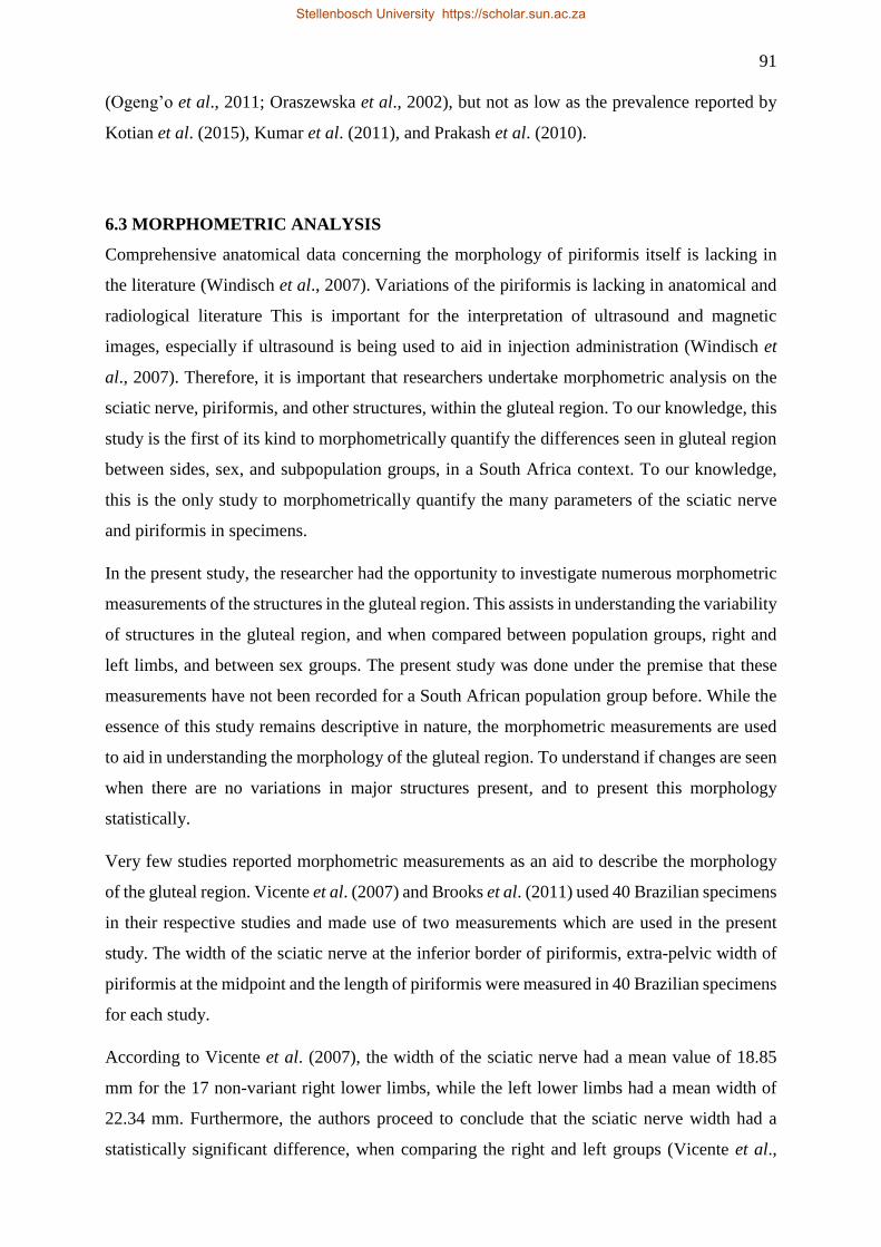
91
(Ogeng’o et al., 2011; Oraszewska et al., 2002), but not as low as the prevalence reported by
Kotian et al. (2015), Kumar et al. (2011), and Prakash et al. (2010).
6.3 MORPHOMETRIC ANALYSIS
Comprehensive anatomical data concerning the morphology of piriformis itself is lacking in
the literature (Windisch et al., 2007). Variations of the piriformis is lacking in anatomical and
radiological literature This is important for the interpretation of ultrasound and magnetic
images, especially if ultrasound is being used to aid in injection administration (Windisch et
al., 2007). Therefore, it is important that researchers undertake morphometric analysis on the
sciatic nerve, piriformis, and other structures, within the gluteal region. To our knowledge, this
study is the first of its kind to morphometrically quantify the differences seen in gluteal region
between sides, sex, and subpopulation groups, in a South Africa context. To our knowledge,
this is the only study to morphometrically quantify the many parameters of the sciatic nerve
and piriformis in specimens.
In the present study, the researcher had the opportunity to investigate numerous morphometric
measurements of the structures in the gluteal region. This assists in understanding the variability
of structures in the gluteal region, and when compared between population groups, right and
left limbs, and between sex groups. The present study was done under the premise that these
measurements have not been recorded for a South African population group before. While the
essence of this study remains descriptive in nature, the morphometric measurements are used
to aid in understanding the morphology of the gluteal region. To understand if changes are seen
when there are no variations in major structures present, and to present this morphology
statistically.
Very few studies reported morphometric measurements as an aid to describe the morphology
of the gluteal region. Vicente et al. (2007) and Brooks et al. (2011) used 40 Brazilian specimens
in their respective studies and made use of two measurements which are used in the present
study. The width of the sciatic nerve at the inferior border of piriformis, extra-pelvic width of
piriformis at the midpoint and the length of piriformis were measured in 40 Brazilian specimens
for each study.
According to Vicente et al. (2007), the width of the sciatic nerve had a mean value of 18.85
mm for the 17 non-variant right lower limbs, while the left lower limbs had a mean width of
22.34 mm. Furthermore, the authors proceed to conclude that the sciatic nerve width had a
statistically significant difference, when comparing the right and left groups (Vicente et al.,
Stellenbosch University https://scholar.sun.ac.za
92
2007). In addition, Brooks et al. (2011) observed similar values in their specimens, where the
mean sciatic nerve width in the 18 non-variant right lower limbs was 19.45 mm, while the
sciatic nerve width in the 18 left lower limbs was 19.46 mm. However, since these numbers
were almost equal in value, no statistical significance was calculated when comparing the two
groups. Additionally, Brooks et al. (2011) goes on to state that the study sample did not allow
for statistical comparisons and conclusions because the sample sizes were so small. This begs
the question as to whether Vicente et al. (2007) were able to accurately draw significant
conclusions from their sample, as the sample sizes in both Brazilian studies were identical at
40 specimens. In a meta-analysis completed by Tomaszewski et al. (2016), morphometric data
from three studies (𝑛 = 252), which included the study by Brooks et al. (2011), resulted in a
pooled mean width of 15.55 mm for the sciatic nerve. Haladaj et al. (2015) included the width
of the sciatic nerve within their study. In the 20 Turkish specimens included in the study by
Guvencer et al. (2008), he calculated the mean width of the sciatic nerve at 17.00 ± 3.70 mm in
the infra-piriform space.
In the present study, the sciatic nerve has a mean width of 15.19 mm, with a standard deviation
of ±3.44 mm, for all 297 specimens included in the calculation. This width is similar to that
calculated by Tomaszewski et al. (2016), and it is possible that these values are similar due to
comparable sample sizes in both their study and the present study. Vicente et al. (2007) and
Brooks et al. (2011) have widths that are slightly larger, but this could be due to the much
smaller sample sizes used in their studies.
In addition, there were significant statistical differences when comparing means of the sciatic
nerve width between the population groups. There were significant statistical differences seen
between White/Caucasian and South African Black sample specimens, and White/Caucasian
and Mixed Race specimens, with the width in the White/Caucasian specimens being the largest
(15.99 mm). However, there are less Mixed race and South African Black specimens, compared
to White/Caucasian specimens, in the current study. Therefore, it is uncertain whether the
significant statistical differences observed are due to differences in population group
morphology, or due to the lack of Mixed race and South African Black specimens, which
increases sample bias, and variability in the data.
Very few studies make use of the extra-pelvic length of piriformis (Vicente et al., 2007; Brooks
et al., 2011; Haladaj et al., 2015) as a component of comparison. According to Vicente et al.
(2007), the mean extra-pelvic length of piriformis in right lower limbs was 76.38 mm, while
the left mean was 79.51 mm. Similar measurements were observed in the 40 specimens included
in the study by Brooks et al. (2011), with 74.52 mm, and 78.45 mm observed for the same
Stellenbosch University https://scholar.sun.ac.za
93
groups, respectively. In addition, a mean measurement of 81 mm was calculated for the superior
border length of piriformis, and 82 mm for the inferior border length of piriformis (Haladaj et
al., 2015). In the present study, this was very different, at a mean of 55.77 mm for the superior
border length, and 59.85 mm for the inferior border length. This is significantly shorter
compared to measurements published elsewhere, even though comparable reference points
were used.
The last comparable measurement between the present study and previously studies is the extra-
pelvic width of piriformis at the midline of the muscle belly. Haladaj et al. (2015) found a mean
width of 32 mm, while Brooks et al. (2011) recorded right and left lower limb widths of 23.19
mm and 22.37 mm, respectively. Additionally, another publication recorded widths of 22.98
mm and 21.84 mm for the right and left limbs, respectively (Vicente et al., 2007). The widths
recorded in the present study have a mean value of 17.0 mm, where the mean width
measurement for the different population groups, namely; White/Caucasian, Mixed Race and
South African Black, were 15.78 mm, 18.85 mm, and 20.02 mm, respectively. When comparing
the widths of piriformis in the present study to literature published previously, the extra-pelvic
width of piriformis is smaller in the present study reports. There are notable difference between
subpopulation groups in the South African sample, with the mean width of piriformis for
White/Caucasian specimens being statistically smaller than Mixed race or South African Black
specimens. This comparison between population groups was statistically significant in the
present study; however, a larger Mixed race and South African Black sample size is needed to
further validate these findings.
Overall, there are no studies published in South Africa, or Africa that make use of these
morphometric measurements, and thus, the researcher has no existing data to compare the
results with. However, this study attempts to provide a foundation of literature for a South
African population sample.
Stellenbosch University https://scholar.sun.ac.za

94
CHAPTER SEVEN:
CONCLUSION
Stellenbosch University https://scholar.sun.ac.za
95
In conclusion, the sciatic nerve plays an important role in the clinical intervention that takes
place within the gluteal region. While researchers have described this nerve and its branches
extensively in literature, the same is not true for the variations of the sciatic nerve, and
especially the variation in the relations between the sciatic nerve and piriformis muscle. This is
especially true for the continent of Africa, and its different population groups.
The present study attempts to change this, and provides a foundation of literature for the
description of the morphology of the gluteal region in a South African population. Therefore,
this study has set the stage for research into the variations to described morphology, and the
prevalence of these variations in the South African population.
The present study has described the possible types of variations in the relationship between the
sciatic nerve and piriformis. While this is the first known study based on the South African
population, preliminary conclusions can be drawn. In the present study, a large study sample
was used. This study sample has proven that the prevalence of piriformis variations, and related
sciatic nerve variations, are comparable with the prevalence obtained in other regions of the
world. The prevalence of variations is 12.5%, and is important for clinicians, as this translates
to about one in every 10 South African patients that might present a variation in the gluteal
region. Furthermore, when looking at specimens that belong to the South African
White/Caucasian population group, the prevalence of variations is 17%. This translates to
almost one in every five patients that might present with a variation in the gluteal region.
However, further studies are needed in order to confidently reveal trends about the variation
prevalence in the Mixed race and South African Black population groups, which makes up the
majority of the population in South Africa.
The present study provides a foundation of literature for the South African population.
However, further studies are needed for comparison and to solidify the results found in the
present study. While there are significantly more variations present in a South African
White/Caucasian population, the underrepresentation of South African Mixed race and South
African Black specimens in this study, means that this assumption remains largely inconclusive.
Thus, speaking to the aim of the research, the prevalence has been reported for the variations
in the relationship between the piriformis and sciatic nerve. This is also true for the prevalence
of the variation in the sciatic nerve bifurcation levels. The morphometric analysis of piriformis
and the sciatic nerve has not been documented intensively, thus detailed descriptions were given
on these structures in the gluteal region. Additionally, comparisons were made between the
sides, sexes, and different subpopulation groups.
Stellenbosch University https://scholar.sun.ac.za
96
LIMITATIONS AND FUTURE STUDIES
The study sample comprised of all the cadavers that were dissected by the undergraduate
medical students, during the dissection for the respective undergraduate anatomy programmes.
Therefore, the study sample is not an accurate and true reflection of the South African
population. A limitation of the study is that results obtained by the researcher does not directly
reflect the anatomy of the South African population, but only the sample that was incorporated
in the present study.
The study could have benefited from a longer experimental period, as this would have allowed
for the use of a larger sample size. Thus, the study was limited by the number of cadavers used
at the three South African universities. A larger sample size could ensure that additional rare
variations and anomalies are identified and analysed. The 340 lower limbs used is a
considerably larger sample than previously reported studies.
The medical students dissected the cadavers that constituted the majority of the sample for this
study. This meant that further dissection by the researcher was not always possible, in order to
reveal additional anatomical landmarks in the gluteal region. These landmarks would have
allowed for the measurement of additional morphometric points of importance, such as the
placement of the sciatic nerve in relation to the medial and lateral osteological structures of the
gluteal region, or the variability in the motility of the sciatic nerve during extension and flexion
of the lower limb. Future studies should include the use of additional landmarks for detailed
morphometric and anthropometric analysis, where possible.
In the present study, there was an unequal distribution of specimens in the different population
and sex groups, which could have resulted in bias results reported. This is especially true for
the population groups where the researcher obtained a larger number of White/Caucasian
specimens, compared to fewer Mixed race and South African Black specimens. While the
results might tend to the conclusion that there is a higher prevalence of variations in the
relationship between piriformis and the sciatic nerve in the South African White/Caucasian
population group, the lack of Mixed race and South African Black specimens could have meant
that fewer specimens with variations were incorporated into the present study. Therefore, future
studies should include larger numbers of Mixed race and South African Black specimens, as
these subpopulation groups make up a significant majority in South Africa.
Lastly, to the researcher’s knowledge, this is the first study of its kind in South Africa.
Therefore, no literature is available that can be compared to the results obtained in the present
study. Additionally, very few studies make use of the many morphometric measurements used
Stellenbosch University https://scholar.sun.ac.za

97
in the present study. Future studies on the South African population are needed for comparison,
in order to reiterate the results found in the present study.
Stellenbosch University https://scholar.sun.ac.za
98
REFERENCES
Abdulla, A., Adams, N., Bone, M., Elliott, A.M., Gaffin, J., Jones, D., Knaggs, R., Martin, D.,
Sampson, L. & Schofield, P. 2013. Guidance on the management of pain in older people. Age
and Ageing, 42:i1-i57.
Adetunji, O., Olusola, E., Joseph, A., Dare, O., Ademola, O. & Segun, O. 2005. Injection-
Induced Sciatic Nerve Injuries among Children Seen At a Nigerian Physiotherapy Unit. The
Internal Journal of Third World Medicine, 3(2):1–4.
Ansari, M.S., Singla, M., Ravi, K.S. & Singh, R. 2016. Anatomical Variations in the Division
and Innervation of the Sciatic Nerve with its Clinical Consequences. International Journal of
Anatomy, Radiology and Surgery, 5(3):AO22–AO25.
Babinski, M.A., Machado, F.A. & Costa, W.S. 2003. A Rare Variation in the High Division of
the Sciatic Nerve Surrounding the Superior Gemellus Muscle. European Journal of
Morphology, 41(1):41-42.
Battaglia, P.J., D'Angelo, K. & Kettner, N.W. 2016. Posterior, Lateral, and Anterior Hip Pain
Due to Musculoskeletal Origin: A Narrative Literature Review of History, Physical
Examination, and Diagnostic Imaging. Journal of Chiropractic Medicine, 15(4):281-293.
Beaton, L.E. & Anson, B.J. 1937. The Relation of the Sciatic Nerve and of its Subdivisions to
the Piriformis Muscle. The Anatomical Record, 70(1):1-5.
Beaton, L.E. & Anson, B.J. 1938. The Sciatic Nerve and the Piriformis Muscle: Their
Interrelation A Possible Cause of Coccygodynia. The Journal of Bone and Joint Surgery,
20(3):686-688.
Benzon, H.T., Katz, J.A., Benzon, H.A. & Iqbal, M.S. 2003. Piriformis Syndrome: Anatomic
Considerations, a New Injection Technique, and a Review of the Literature. Anesthesiology,
98(6):1442-1448.
Brooks, J.B.B., Silva, C.A.C., Soares, S.A., Kai, M.R., Cabral, R.H. & Fragoso, Y.D. 2011.
Anatomical variations of the sciatic nerve in a group of Brazilian Cadavers. Revista Dor. São
Paulo, 12(4):332-336.
Budhiraja, V., Rastogi, R., Jain, S.K., Sharma, N., Garg, R. & Nafees, H. 2016. Variation
Relation of the Sciatic Nerve to the Piriformis Muscle: A Cadaveric Study from North India.
Argentine Journal of Clinical Anatomy, 8(1):38-42.
Stellenbosch University https://scholar.sun.ac.za
99
Buijs, E., Visser, L. & Groen, G. 2007. Sciatica and the sacroiliac joint: a forgotten concept.
British Journal of Anaesthesia, 99(5):713–716.
Byrd, J.W.T. 2015. Disorders of the Peritrochanteric and Deep Gluteal Space: New Frontiers
for Arthroscopy. Sports Medicine and Arthroscopy Review, 23(4):221-231.
Cassidy, L., Walters, A., Bubb, K., Shoja M.M., Tubbs, R.S. & Loukas, M. 2012. Piriformis
Syndrome: Implications of Anatomical Variations, Diagnostic Techniques, and Treatment
Options. Surgical and Radiologic Anatomy, 34:479-486.
Chen, W.S. 1994. Bipartite piriformis muscle: an unusual cause of sciatic nerve entrapment.
Pain, 58:269-272.
Chiba, S., Ishibashi, Y. & Kasai, T. 1994. Perforation of dorsal branches of the sacral nerve
plexus through the piriformis muscle and its relation to changes of segmental arrangements of
the vertebral column and others. Kaibogaku Zasshi, 69(3):281–305.
Chukwuanukwu, T.O.G., Ukoha, U.U., Chykwujekwu, I.E., Asomugha, A.L., Oranusi, C.K.,
Anyabolu, A.E., Nzeako, H.C., Nwajagu, G.I. & Ashaolu, J.O. 2007. Bilateral High Division
of the Sciatic Nerve: Incidence and Clinical Implications in Nigeria. Tropical Journal of
Medical Research, 11(2):12-13.
Clendenen, S.R., York, J.E., Wang, R.D. & Greengrass, R.A. 2008. Three-dimensional
ultrasound-assisted popliteal catheter placement revealing aberrant anatomy: implications for
block failure. Acta Anaesthesiologica Scandinavica, 52(10):1429–1431.
Cohen, B. 2006. Urbanization in developing countries: Current trends, future projections, and
key challenges for sustainability. Technology in Society, 28:63–80.
Combes, M.A., Clark, W.K. & Gregory, F.C. 1960. Sciatic nerve injury in infants. Recognition
and prevention of impairment resulting from intragluteal injections. The Journal of the
American Medical Association, 173(12):1336–1339.
Cooper, N.A., Scavo, K.M., Strickland, K.J., Tipayamongkol, N., Nicholson, J.D., Bewyer,
D.C. & Sluka, K.A. 2016. Prevalence of gluteus medius weakness in people with chronic low
back pain compared to healthy controls. European Spine Journal, 25:1258-1265.
Coppieters, M.W., Alshami, A.M., Babri, A.S., Souvlis, T., Kippers, V. & Hodges, P.W. 2006.
Strain and Excursion of the Sciatic, Tibial, and Plantar Nerves during a Modified Straight Leg
Raising Test. Journal of orthopaedic Research, 24(9):1883-1889.
Stellenbosch University https://scholar.sun.ac.za
100
Cox, J.M. & Bakkum, B.W. 2005. Possible Generators of Retrotrochanteric Gluteal and Thigh
Pain: The Gemelli–Obturator Internus Complex. Journal of Manipulative and Physiological
Therapeutics, 28(7):534–538.
Dawson, D.M. & Hallett, M. 1990. Entrapment Neuropathies. 2nd ed. Boston: Little Brown &
Company.
Delabie, A., Peltier, J., Havet, E., Page, C., Foulon, P. & Le Gars, D. 2013. Relationships
between piriformis muscle and sciatic nerve: Radioanatomical study with 104 buttocks. Journal
of Morphology, 97(316):12–18.
Desalegn, M. & Tesfay, A. 2014. Variations of Sciatic Nerve Its Exit in Relation to Piriformis
Muscle in the Northern Ethiopia. International Journal of Pharma Sciences and Research,
5(12):953–956.
di Benedetto, P., Bertini, L., Casati, A., Borghi, B., Albertin, A. & Fanelli, G. 2001. A New
Posterior Approach to the Sciatic Nerve Block: A Prospective, Randomized Comparison with
the Classic Posterior Approach. Anesthesia & Analgesia, 93:1040-1044.
Dominick, C.H., Blyth, F.M. & Nicholas, M.K. 2012. Unpacking the burden: Understanding
the relationships between chronic pain and comorbidity in the general population. Pain,
153:293-304.
Drake, R.L., Vogl, A.W. & Mitchell, A.W.M. 2015. Grey's Anatomy for Students. 3rd Edition
ed. Philadelphia: Churchill Livingstone Elsevier.
Dunn, K.M., Campbell, P. & Jordan, K.P. 2013. Long-term trajectories of back pain: cohort
study with 7-year follow-up. BMJ Open, 3(e003838).
Fatunde, O.J. & Familusi, J.B. 2001. Injection-induced Sicatic Nerve Injury in Nigerian
Children. Central African Journal of Medicine, 47(2):35-38.
Fishman, L.M., Dombi, G.W., Michaelsen, C., Ringel, S., Rozbruch, J., Rosner, B. & Weber,
C. 2002. Piriformis Syndrome: Diagnosis, Treatment, and Outcome - a 10-year Study. Archives
of Physical Medicine and Rehabilitation, 83:295-301.
Frank, R.M., Slabaugh, M.A., Grumet, R.C., Virkus, W.W., Bush-Joseph, C.A. & Nho, S.J.
2010. Posterior Hip Pain in an Athletic Population: Differential Diagnosis and Treatment
Options. Sports Health, 2(3):237–246.
Stellenbosch University https://scholar.sun.ac.za

101
Freiberg, A.H. 1937. Sciatic Pain and its Relief by Operations on Muscle and Fascia. Archives
of Surgery, 34(2):337-350.
Goldberg, D.S. & McGee, S.J. 2011. Pain as a global public health priority. BMC Public Health,
11:770.
Gonzalez, P., Pepper, M., Sullivan, W. & Akuthota, V. 2008. Confirmation of Needle
Placement within the Piriformis Muscle of a Cadaveric Specimen using Anatomical Landmarks
and Fluoroscopic Guidance. Pain Physician Journal, 11(3):327-331.
Gore, M., Sadosky, A., Stacey, B.R., Tai, K. & Leslie, D. 2012. The Burden of Chronic Low
Back Pain: Clinical Comorbidities, Treatment Patterns, and Health Care Costs in Usual Care
Settings. Spine, 37(11):E668-E677.
Gouveia, N., Rodrigues, A., Eusébio, M., Ramiro, S., Machado, P., Canhão, H. & Branco, J.C.
2015. Prevalence and social burden of active chronic low back pain in the adult Portuguese
population: results from a national survey. Rheumatology International, 36(2):183-197.
Grassi, W., Filippucci, E., Carotti, M. & Salaffi, F. 2003. Imaging modalities for identifying
the origin of regional musculoskeletal pain. Best Practice & Research Clinical Rheumatology,
17(1):17-32.
Guvencer, M., Akyer, P., Iyem, C., Tetik, S. & Naderi, S. 2008. Anatomic considerations and
the relationship between the piriformis muscle and the sciatic nerve. Surgical and Radiologic
Anatomy, 30:467–474.
Guvencer, M., Iyem, C., Akyer, P., Tetik, S. & Naderi, S. 2009. Variations in the High Division
of the Sciatic Nerve and Relationship between the Sciatic Nerve and the Piriformis. Turkish
Neurosurgery, 19:139-144.
Haladaj, R., Pingot, M., Polguj, M., Wysiadecki, G. & Topol, M. 2015. Anthropometric Study
of the Piriformis Muscle and Sciatic Nerve: A Morphological Analysis in the Polish Population.
Medical Science Monitor, 21:3760-3768.
Halpin, R.J. & Ganju, A. 2009. Piriformis Syndrome: A Real Pain in the Buttock?
Neurosurgery, 65:A197-A202.
Hansen, J.T. 2014. Netter's Clinical Anatomy. 3rd Edition ed. Philadelphia: Elsevier Saunders.
Stellenbosch University https://scholar.sun.ac.za

102
Hartvigsen, J., Davidsen, M., Hestbaek, L., Sogaard, K. & Roos, E.M. 2013. Patterns of
musculoskeletal pain in the population: A latent class analysis using a nationally representative
interviewer-based survey of 4817 Danes. European Journal of Pain, 17:453-460.
Häuser, W., Wolfe, F., Henningsen, P., Schmutzer, G., Brähler, E. & Hinz, A. 2014. Untying
chronic pain: prevalence and societal burden of chronic pain stages in the general population -
a cross-sectional survey. BMC Public Health, 14(352).
Hernando, M.F., Cerezal, L. & Pérez-Carro, L. 2015. Deep gluteal syndrome: anatomy,
imaging, and management of sciatic nerve entrapments in the subgluteal space. Skeletal
Radiology, 44:919-934.
Hopayian, K., Song, F., Riera, R. & Sambandan, S. 2010. The clinical features of the piriformis
syndrome: a systematic review. European Spine Journal, 12(4):2095-2109.
Hoy, D.G., Smith, E., Cross, M., Sanchez-Riera, L., Blyth, F. M., Buchbinder, R., Woolf, A.D.,
Driscoll, T., Brooks, P. & March, L.M. 2014. Reflecting on the global burden of
musculoskeletal conditions: lessons learnt from the Global Burden of Disease 2010 Study and
the next steps forward. Annals of the Rheumatic Diseases, 74:4-7.
Hoy, D.G., March, L.M., Brooks, P., Blyth, F.M., Woolf, A.D., Bain, C., Williams, G., Smith,
E., Vos, T., Barendregt, J., Murray, C., Burstein, R. & Buchbinder, R. 2014. The global burden
of low back pain: estimates from the Global Burden of Disease 2010 study. Annals of the
Rheumatic Diseases, 73:968-974.
Hussain, M. & Bhat, N. 2018. World population statistics: some key findings. The research
journal of social sciences, 9(4):16-25.
Jawish, R.M., Assoum, H.A. & Khamis, C.F. 2010. Anatomical, Clinical and Electrical
Observations in Piriformis Syndrome. Journal of Orthopaedic Surgery and Research, 5(3):1-
7.
Kanawati, A.J. 2014. Variations of the sciatic nerve anatomy and blood supply in the gluteal
region: a review of the literature. ANZ Journal of Surgery, 84:816-819.
Karmakar, M.K., Kwok, W.H., Ho, A.M., Tsang, K., Chui, P.T. & Gin, T. 2007. Ultrasound-
guided sciatic nerve block: description of a new approach at the subgluteal place. British
Journal of Anaesthesia, 98(3):390-395.
Khan, A.A., Asari, M.A. & Pasha, M.A. 2015. The sciatic nerve in human cadavers-high
division or low formation. Folia Morphologica, 75(3):306–310.
Stellenbosch University https://scholar.sun.ac.za
103
Kingzett-Taylor, A., Tirman, P.F.J., Feller, J., McGann, W., Prieto, V., Wischner, T., Cameron,
J.A., Cvitanic, O. & Genant, H.K. 1999. Tendinosis and Tears of Gluteus Medius and Minimus
Muscles as a Cause of Hip Pain: MR Imaging Findings. American Journal of Roentgenology,
173(4):1123–1126.
Kiros, M.D. & Woldeyes, D.H. 2015. Anatomical variations in the level of bifurcation of the
sciatic nerve in Ethiopia. Journal of Experimental and Clinical Anatomy, 14(1):1-4.
Kirschner, J.S., Foye, P.M. & Cole, J.L. 2009. Piriformis Syndrome, Diagnosis and Treatment.
Muscle and Nerve, 40:10-18.
Knudsen, J.S., Mei-Dan, O. & Brick, M.J. 2016. Piriformis Syndrome and Endoscopic Sciatic
Neurolysis. Sports Medicine and Arthroscopy Review, 24(1).
Kosukegawa, I., Yoshimoto, M., Isogai, S., Nonaka, S. & Yamashita, T. 2006. Piriformis
Syndrome Resulting from a rare Anatomic Variation. The Spine Journal, 31(18):E664-E666.
Kotian, S.R., Sinha, A., Souza, A.S.D. & Sumalatha, S. 2015. Variations of the sciatic nerve
and its relation with the piriformis muscle in South Indian Population. Journal of Experimental
and Integrative Medicine, 5(3):144-158.
Kubota, K., Noguchi, I. & Nakano, T. 1960. Rare Types of Relation between the Sciatic Nerve
and the Piriformis Muscle. Okajimas Folia Anatomica Japonica, 36(5):329-343.
Kukiriza, J., Kiryowa, H., Turyabahika, J., Ochieng, J. & Ibingira, C.B.R. 2010. Levels of
Bifurcation of the Sciatic Nerve among Ugandans at School of Biomedical Sciences Makerere
and Mulago Hospital Uganda. East and Central African Journal of Surgery, 15(2):69-75.
Kumar, M.T., Srimathi, R.A. & Latha, S. 2011. A Cadaveric Study of Sciatic Nerve and its
Level of Bifurcation. Journal of Clinical and Diagnostic Research, 5(8):1502-1504.
Lee, D.W., Lim, C.H., Han, H.Y. & Kim, W.M. 2016. Chronic pelvic pain arising from
dysfunctional stabilizing muscles of the hip joint and pelvis. Korean Journal of Pain,
29(4):274-276.
Leopoldino, A.A.O., Diz, J.B.M., Martins, V.T., Henschke, N., Pereira, L.S.M., Dias, R.C. &
Oliveira, V.C. 2016. Prevalence of low back pain in older Brazilians: a systematic review with
meta-analysis. Revista Brasileira de Reumatologia, 56(3):258-269.
Lewis, S., Jurak, J., Lee, C., Lewis, R. & Gest, T. 2016. Anatomical Variations of the Sciatic
Nerve, in Relation to the Piriformis Muscle. Translational Research in Anatomy, 5:15–19.
Stellenbosch University https://scholar.sun.ac.za
104
Maranhão-Filho, P. & Vincent, M. 2018. Lazarević-Lasègue sign. Arquivos de Neuro-
Psiquiatria 76(6):421-423.
Marhofer, P., Harrop-Griffiths, W., Willschke, H. & Kirchmair, L. 2010. Fifteen years of
ultrasound guidance in regional anaesthesia: Part 2—Recent developments in block techniques.
British Journal of Anaesthesia, 106(4):673-683.
Martin, H.D., Kivlan, B.R., Palmer, I.J. & Martin, R.L. 2014. Diagnostic accuracy of
clinicaltests for sciatic nerve entrapment in the gluteal region. Knee Surgery, Sports
Traumatology, Arthroscopy, 22:882-888.
Martin, H.D., Shears, S.A., Johnson, J.C., Smathers, A.M. & Palmer, I.J. 2010. The Endoscopic
Treatment of Sciatic Nerve Entrapment/Deep Gluteal Syndrome. Arthroscopy: The Journal of
Arthroscopic and Related Surgery, 27(2):172-181.
Martin, H.D., Reddy, M. & Gomez-Hoyos, J. 2015. Deep Gluteal Syndrome. Journal of Hip
Preservation Surgery, 2(2):99-107.
Martin, R.L. & Sekiya, J.K. 2008. The Interrater Reliability of four Clinical Tests Used to
Assess Individuals with Musculoskeletal Hip Pain. Journal of Orthopaedic & Sports Physical
Therapy, 38(2):71-77.
McGee, S.J., Kaylor, B.D., Emmott, H. & Christopher, M.J. 2011. Defining Chronic Pain
Ethics. Pain Medicine, 12:1376-1384.
Meknas, K., Christensen, A. & Johansen, O. 2003. The internal obturator muscle may cause
sciatic pain. Pain, 104:375 – 380.
Meknas, K., Johansen, O. & Kartus, J. 2011. Retro-trochanteric sciatica-like pain: current
concept. Knee Surgery, Sports Traumatology, Arthroscopy, 19:1971–1985.
Michel, F., Decavel, P., Toussirot, E., Tatu, L., Aleton, E., Monnier, G., Garbuio, P. & Parratte,
B. 2013. The piriformis muscle syndrome: An exploration of anatomical context,
pathophysiological hypotheses and diagnostic criteria. Annals of Physical and Rehabilitation
Medicine, 56:300-311.
Mishra, P. & Stringer, M.D. 2010. Sciatic nerve injury from intramuscular injection: a persistent
and global problem. The International Journal of Clinical Practice, 64(11):1573-1579.
Stellenbosch University https://scholar.sun.ac.za
105
Moayeri, N., van Geffen, G.J., Bruhn, J., Chan, V.W. & Groen, G.J. 2010. Correlation Among
Ultrasound, Cross-Sectional Anatomy, and Histology of the Sciatic Nerve: A Review. Regional
Anesthesia and Pain Medicine, 35(5):442-449.
Moore, K.L., Dalley, A.F. & Agur, A.M.R. 2017. Clinically Oriented Anatomy. 8th Edition ed.
Baltimore: Lippincott Williams & Wilkins.
Murray, J.M., Derbyshire, S. & Shields, M.O. 2010. Lower Limb Blocks. Anaesthesia: Journal
of the Association of Anaesthetists of Great Britain and Ireland, 65(1):57-66.
Natsis, K., Totlis, T., Konstantinidis, G.A., Paraskevas, G., Piagkou, M. & Koebke, J. 2014.
Anatomical variations between the sciatic nerve and the piriformis muscle: a contribution to
surgical anatomy in piriformis syndrome. Surgical and Radiologic Anatomy, 36:273-280.
Nayak, S.B., George, B.M. & Mishra, S. 2014. A Sciatic Nerve with Three Roots and its
Perforation by the Enlarged Ischiadic Artery. Anatomical Science International, 89:118-121.
Ogeng'o, J.A., El-Busaidy, H., Mwika, P.M., Khanbhai, M.M. & Munguti, J. 2011. Variant
anatomy of sciatic nerve in a Black Kenyan population. Folia Morphologica, 70(3):175-179.
Overaas, C.K., Johansson, M.S., de Campos, T.F., Ferreira, M.L., Natvig, B., Mork, P.J. &
Hartvigsen, J. 2017. Prevalence and pattern of co-occurring musculoskeletal pain and its
association with back-related disability among people with persistent low back pain: protocol
for a systematic review and meta-analysis. Systematic Reviews, 6(258).
Ozaki, S., Hamabe, T. & Muro, T. 1999. Piriformis Syndrome Resulting from an Anomalous
Relationship between the Sciatic Nerve and Piriformis Muscle. Orthopedics, 22(8):771-772.
Pais, D., Casal, D., Bettencourt Pires, M. A., Furtado, A., Bilhim, T., Angelica-Almeida, M. &
Goyri-O'Neill, J. 2013. Sciatic Nerve High Division: Two Different Anatomical Variants. Acta
Med Port, 26(3):208-211.
Pandian, J.D., Bose, S., Daniel, V., Signh, Y. & Abraham, A.P. 2006. Nerve injuries following
intramuscular injections: a clinical and neurophysiological study from Northwest India. Journal
of the Peripheral Nervous System, 11:165-171.
Papadopoulos, E.C. & Khan, S.N. 2004. Piriformis syndrome and low back pain: a new
classification and review of the literature. The Orthopaedic Clinics of North America, 35:65-
71.
Stellenbosch University https://scholar.sun.ac.za

106
Park, M., Yoon, S., Jung, S. & Kim, S. 2016. Clinical results of endoscopic sciatic nerve
decompression for deep gluteal syndrome: mean 2-year follow-up. BMC Musculoskeletal
Disorders, 17(218).
Patel, S., Shah, M., Vora, R., Zalawadia, A. & Rathod, S.P. 2011. A Variation in the High
Division of the Sciatic Nerve and Its Relation with Piriformis Muscle. National Journal of
Medical Research, 1:27-30.
Pecina, M.M. 1979. Contribution to the Etiological Explanation of the Piriformis Syndrome.
Acta Anatomica, 105:181-187.
Pecina, M.M., Markiewitz, A.D. & Krmpotic-Nemanic, J. 2001. Tunnel Syndromes: Peripheral
Nerve Compression Syndromes. 3rd Edition ed. Florida: CRC Press.
Pérez-Carro, L. Hernando, M.F., Cerezal, L., Navarro, I.S., Fernandez, A.A. & Castillo A.O.
2016. Deep gluteal space problems: piriformis syndrome, ischiofemoral impingement and
sciatic nerve release. Muscles, Ligaments and Tendons Journal, 6(3):384-396
Pokorny, D., Jahoda, D., Veigl, D., Pinskerova, V. & Sosna, A. 2006. Topographic variations
of the relationship of the sciatic nerve and the piriformis muscle and its relevance to palsy after
total hip arthroplasty. Journal of surgical and radiological anatomy, 28:88–91.
Prakash, B.A.K., Devi, M.N., Sridevi, N.S., Rao, P.K. & Singh, G., 2010. Sciatic nerve division:
a cadaver study in the Indian population and review of the literature. Singapore Medical
Journal, 51(9):721–723.
Puranen, J. & Orava, S. 1988. The hamstring syndrome: A new diagnosis of gluteal sciatic
pain. The American Journal of Sports Medicine, 16(5):517–521.
Ramtahal, J., Ramlakhan, S. & Singh, K. 2006. Sciatic nerve injury following intramuscular
injection: A case report and review of the literature. Journal of Neuroscience Nursing,
38(4):238-240.
Reid, M.C., Eccleston, C. & Pillemer, K. 2015. Management of chronic pain in older adults.
BMJ, 350(532):27-30.
Robinson, D.R. 1947. Piriformis Syndrome in Relation to Sciatic Pain. American Journal of
Surgery, 73(3):355-358.
Roger, M.A. & King, L. 2000. Drawing up and administering intramuscular injections: a review
of the literature. Journal of Advanced Nursing, 31(3):574-582.
Stellenbosch University https://scholar.sun.ac.za
107
Saleh, H.A.M., El-fark, M.M.O. & Abdel-Hamid, G.A. 2009. Anatomical variation of sciatic
nerve division in the popliteal fossa and its implication in popliteal nerve blockade. Folia
Morphologica, 68(4):256-259.
Sangram, S., Samanta, C., Paul, M., Biswas, S., Ghosh, R., Sarkar, A., Pradhan, S. & Majumdar,
S. 2015. A Study on the Bifurcation of Sciatic Nerve with its Clinical Significance. Indian
Journal of Basic and Applied Medical Research, 4(3):34-44.
Saritha, S., Kumar, P.M. & Supriya, G. 2012. Anatomical Variations in the Bifurcation of the
Sciatic Nerve, a Cadaveric Study and its Clinical Implications. Journal of Anatomy and
Physiology, 2(5):1-4.
Senes, F.M., Campus, R., Becchetti, F. & Catena, N. 2009. Sciatic Nerve Injection Palsy in the
Child: Early Microsurgical Treatment and Long-Term Results. Journal of Microsurgery, 443-
448.
Shylaja, D.K. & Ramesh, B.R. 2017. Study of anatomical variations of structures in relation to
piriformis muscle. International Journal of Anatomy and Research, 5(3.3):4373–4377.
Siddiq, A.B., Hossain, S., Uddin, M.M., Jahan, I., Khasru, M.R., Haider, N.M. & Rasker, J.J.
2017. Piriformis syndrome: a case series of 31 Bangladeshi people with literature review.
European Journal of Orthopaedic Surgery & Traumatology, 27:193-203.
Small, S.P. 2004. Preventing sciatic nerve injury from intramuscular injections: literature
review. Journal of Advanced Nursing, 47(3):287–296.
Smoll, N.R. 2010. Variations of the Piriformis and Sciatic Nerve with Clinical Consequence:
A Review. Journal of Clinical Anatomy, 23:8-17.
Stubbs, B., Schofield, P. & Patchay, S. 2014. Mobility Limitations and Fall-Related Factors
Contribute to the Reduced Health-Related Quality of Life in Older Adults With Chronic
Musculoskeletal Pain. Pain Practice, 16(10):80-89
Sulak, O., Sakalli, B. & Ozguner, G. 2014. Anatomical Relation between Sciatic Nerve and
Piriformis Musce and its Bifurcation Level during Foetal Period in Human. Surgical and
Radiologic Anatomy, 36:265-272.
Tak, S.R., Dar, G.N., Halwai, M.A. & Mir, M.R. 2008. Post-injection nerve injuries in Kashmir:
A menace Overlooked. Journal of Research in Medical Sciences, 13(5):244-247.
Stellenbosch University https://scholar.sun.ac.za

108
Tomaszewski, K.A., Graves, M.J., Henry, B.M., Popieluszko, P., Roy, J., Pekala, P.A., Hsieh,
W.C., Vikse, J. & Walocha, J.A. 2016. Surgical Anatomy of the Sciatic Nerve: A Meta-
Analysis. Journal of Orthopaedic Research, 34(10):1820-1827.
Treede, R., Rief, W., Barke, A., Aziz, Q., Bennett, M.L., Benoliel, R. & Wang, S. 2015. A
classification of chronic pain for ICD-11. Pain Journal, 156(6):1003-1007.
Trotter, M. 1938. The Relation of the Sciatic Nerve to the Piriformis Muscle in American
Whites and Negroes. Anatomical Records, 52:321-323.
Tubbs, R.I., Barton, J.C., Watson, C.C., Kollias, T., Ward, R.J., Loukas, M., Barbaro, N.M. &
Cohen-Gadol, A.A. 2015. A novel method for sciatic nerve decompression: Cadaveric
feasibility study with potential application to patients with piriformis syndrome. Translational
Research in Anatomy 1:40-43.
Ugrenović, S., Jovanovic, I., Krstic, V., Stojanovic, V., Vasovic, L., Antic, S. & Pavlovic, S.
2005. The level of the sciatic nerve division and its relations to the piriform muscle.
Vojnosanitetski Pregled, 62(1):45–49.
Uluutku, M.H. & Kurtoglu, Z. 1999. Variations of Nerves Located in Deep Gluteal Region.
Okajimas Folia Aanatomica Japonica, 76(5): 273–276.
Vicente, E.J., Viotto, M.J., Barbosa, C.A. & Vicente, P.C. 2007. Study on Anatomical
Relationships and Variations between the Sciatic Nerve and Piriformis Muscle. Revista
Brasileira de Fisioterapia, 11:227-232.
Vloka, J.D., Hadzic, A., April, E. & Thys, D.M. 2001. The division of the sciatic nerve in the
popliteal fossa: anatomical implications for popliteal nerve blockade. Anesthesia & Analgesia
92:215–217.
Williams, J.S., Ng, N., Peltzer, K., Yawson, A., Biritwum, R., Maximova, T., Wu, F.,
Arokiasamy, P., Kowal, P. & Chatterji, S. 2015. Risk Factors and Disability Associated with
Low Back Pain in Older Adults in Low- and Middle-Income Countries. Results from the WHO
Study on Global AGEing and Adult Health (SAGE). PLoS ONE, 10(6).
Windisch, G., Braun, E.M. & Anderhuber, F. 2007. Piriformis Muscle: Clinical Anatomy and
Consideration of the Piriformis Syndrome. Surgical and Radiologic Anatomy, 29:37-45.
Yamamoto, H., Sakura, S., Wada, M. & Shido, A. 2014. A Prospective, Randomized
Comparison Between Single- and Multiple-Injection Techniques for Ultrasound-Guided
Subgluteal Sciatic Nerve Block. Anesthesia & Analgesia, 119(6):1442-1448.
Stellenbosch University https://scholar.sun.ac.za

109
Yeoman, W. 1928. The Relation of Arthritis of the Sacroiliac Joint to Sciatica. Lancet, 2:1119-
1122.
Stellenbosch University https://scholar.sun.ac.za