Embed Size (px)
Citation preview
Effects of sciatic nerve transection onultrastructure, NADPH-diaphorase reaction
and serotonin-, tyrosine hydroxylase-,c-Fos-, glucose transporter 1- and 3-like
immunoreactivities in frog dorsal root ganglion
F. Rigon1, D. Rossato1, V.B. Auler1, L. Dal Bosco1, M.C. Faccioni-Heuser2,3 and W.A. Partata1
1Departamento de Fisiologia, Instituto de Ciencias Basicas da Saude, Universidade Federal do Rio Grande do Sul,
Porto Alegre, RS, Brasil2Departamento de Ciencias Morfologicas, Universidade Federal do Rio Grande do Sul, Porto Alegre, RS, Brasil
3Centro de Microscopia Eletronica, Universidade Federal do Rio Grande do Sul, Porto Alegre, RS, Brasil
Abstract
Frogs have been used as an alternative model to study pain mechanisms. Since we did not find any reports on the effects of
sciatic nerve transection (SNT) on the ultrastructure and pattern of metabolic substances in frog dorsal root ganglion (DRG) cells,
in the present study, 18 adult male frogs (Rana catesbeiana) were divided into three experimental groups: naive (frogs not
subjected to surgical manipulation), sham (frogs in which all surgical procedures to expose the sciatic nerve were used except
transection of the nerve), and SNT (frogs in which the sciatic nerve was exposed and transected). After 3 days, the bilateral DRG
of the sciatic nerve was collected and used for transmission electron microscopy. Immunohistochemistry was used to detect
reactivity for glucose transporter (Glut) types 1 and 3, tyrosine hydroxylase, serotonin and c-Fos, as well as nicotinamide adenine
dinucleotide phosphate diaphorase (NADPH-diaphorase). SNT induced more mitochondria with vacuolation in neurons, satellite
glial cells (SGCs) with more cytoplasmic extensions emerging from cell bodies, as well as more ribosomes, rough endoplasmic
reticulum, intermediate filaments and mitochondria. c-Fos immunoreactivity was found in neuronal nuclei. More neurons and
SGCs surrounded by tyrosine hydroxylase-like immunoreactivity were found. No change occurred in serotonin- and Glut1- and
Glut3-like immunoreactivity. NADPH-diaphorase occurred in more neurons and SGCs. No sign of SGC proliferation was
observed. Since the changes of frog DRG in response to nerve injury are similar to those of mammals, frogs should be a valid
experimental model for the study of the effects of SNT, a condition that still has many unanswered questions.
Key words: Axotomy; Immunoreactivity; Histochemistry
Introduction
In amphibians, as in mammals, the dorsal root
ganglion (DRG) is composed of three classes of neurons
classified according to morphology and function. In
addition to neurons, myelinated and unmyelinated fibers,
macrophages, fibroblasts, and satellite glial cells (SGCs)
are also resident components (1,2). In this ganglion some
neurons show reactivity to nicotinamide adenine dinucleo-
tide phosphate diaphorase (NADPH-diaphorase) (3,4),
which is considered to be equivalent to nitric oxide (NO)
synthase, the enzyme responsible for NO synthesis (5).
DRG cells also respond to application of serotonin (6,7)
and catecholamine (8).
Amphibians have been used as a model for the study
of pain mechanisms (9-15). The reasons for using
amphibians in pain research are varied. According to
Stevens (9), the use of these animals provides a
phylogenetic perspective on the mechanisms of pain
research. Other issues include the simplicity of the
amphibian central nervous system, the economic advan-
tage of using these animals, and ethical considerations
about conducting pain research in non-mammalian
vertebrate species. In this context, frog spinal cord and
DRG have been used to demonstrate the changes in
different neurotransmitters and neuropeptides after sciatic
Correspondence: W.A. Partata, Departamento de Fisiologia, Instituto de Ciencias Basicas da Saude, UFRGS, 90050-170 Porto
Alegre, RS, Brasil. Fax: ++55-51-3308-3166. E-mail: [email protected]
Received November 22, 2012. Accepted March 18, 2013. First published online May 24, 2013.
Brazilian Journal of Medical and Biological Research (2013) 46: 513-520, http://dx.doi.org/10.1590/1414-431X20132853
ISSN 1414-431X
www.bjournal.com.br Braz J Med Biol Res 46(6) 2013

nerve transection (SNT), one of the models that mimic the
clinical conditions of neuropathic pain (16-18). These
studies reported alterations that shared similarities with
those observed in mammals, while others were unique to
this animal species. Moreover, further studies are
necessary to better understand the effects of SNT on
frog nervous tissue to support the use of this model in this
experimental condition.
In mammals, serotonin transporter deficiency attenu-
ated the mechanical allodynia and heat hyperalgesia,
symptoms frequently observed in neuropathic pain (19).
Tyrosine hydroxylase, a rate-limiting enzyme responsible
for catalyzing the conversion of L-tyrosine to the precursor
of dopamine and then norepinephrine and epinephrine,
shows changes in its immunoreactivity pattern in DRG
cells after SNT (20). Glucose transport (Glut) also
appears to be modulated by noxious stimuli and denerva-
tion (21). These experimental conditions upregulated
c-Fos, a protein that is regarded as a marker of neural
activation by noxious stimulation (22). SNT also increases
NADPH-diaphorase staining in mammalian DRG cells
(23,24). SGCs, cells that support DRG neurons both
physically and metabolically (25), also change their
ultrastructure after peripheral nerve lesion (26).
In order to elucidate the effects of SNT on the
ultrastructure and pattern of metabolic substances in
bullfrog DRG cells, we used transmission electron
microscopy to reveal the effects on the ultrastructure of
these cells, and light microscopy to demonstrate the
distribution of NADPH-diaphorase reaction and the
pattern of Glut1- and Glut3-, serotonin-, tyrosine hydro-
xylase-, and c-Fos-like immunoreactivity. The experiment
was performed 3 days after SNT because previous
studies demonstrated that the functional changes in frog
nervous tissue are already present 3 days after axotomy
(16-18). We think that these findings will determine if
these responses are similar across amphibians and
mammals, potentially increasing our knowledge of the
effects of SNT on frog nervous tissue.
Material and Methods
AnimalsEighteen adult male frogs, Rana catesbeiana, weigh-
ing 100-200 g were obtained from Ranasul (Brazil). Upon
arrival at the laboratory they were housed in cages with
water and kept under natural conditions of temperature
and photoperiod. The animals were fed specific food
ad libitum and acclimated to laboratory conditions for at
least 2 weeks before being used. They were divided into 3
experimental groups of 6 animals each: naive (animals did
not undergo surgical manipulation), sham (animals in
which all surgical procedures to expose the sciatic nerve
were used except transection of this nerve), and SNT
(animals in which the sciatic nerve was exposed and
transected). For the surgical procedures, frogs were
anesthetized intramuscularly with 3% prilocaine
(Prilonest1, DFL Industria e Comercio S.A., Brazil;
0.1 mL/100 g body weight). In the SNT group, the right
sciatic nerve was exposed and transected approximately
5 mm distal to the sciatic notch. Flexion and ocular
reflexes were used to monitor the anesthesic effect. After
surgery, the muscle and skin layer were immediately
sutured with thread and a topical antibiotic was applied.
The animals were killed 3 days after the procedure. The
experimental protocol followed the NIH Guide for the Care
and Use of Laboratory Animals (NIH publication 85-23,
revised 1985) and was approved by the Neuroscience
Graduate Committee of Instituto de Ciencias Basicas da
Saude, Universidade Federal do Rio Grande do Sul.
Transmission electron microscopyThe bilateral DRG of the sciatic nerve were dissected
out within 3 min after frog decapitation. They were fixed
immediately by immersion in 2% paraformaldehyde, 1.5%
glutaraldehyde (Sigma, USA) and 0.1 M phosphate buffer
(PB), pH 7.3, for 1 h. The material was washed in the
same buffer and postfixed in 1% osmium tetroxide
(Sigma) diluted in PB for 1 h at room temperature. Next,
the sections were washed in PB and subsequently
dehydrated with an ascending series of acetone, and
then embedded in Araldite (Durcupan, Fluka,
Switzerland). Semithin sections (1 mM) were obtained
using an ultramicrotome (MT6000-XL, RMC, USA) with a
diamond knife (Diatome, Switzerland) and stained with
1% toluidine blue for examination under a light micro-
scope. Ultrathin sections (70 nm) were cut with the same
ultramicrotome using a diamond knife (Drukker, The
Netherlands). These sections were stained with 2% uranyl
acetate (Merck, Germany) followed by 1% lead citrate and
examined with a JEM 120 EX II electron microscope
(Joel, Japan).
Histochemistry and immunohistochemistryFor the NADPH-diaphorase and immunohistochem-
ical procedure, the frogs were decerebrated and after a
brief saline flush they were perfused intracardially with 4%
paraformaldehyde in 0.1 M PB, pH 7.4. The DRG were
quickly dissected out, immersed in the same fixative
solution for 4 h and then cryoprotected in 15 and 30%
sucrose solutions in PB at 46C. Serial coronal sections
(50 mM) were obtained with a cryostat and collected in
cold phosphate-buffered saline (PBS).
For the NADPH-diaphorase procedure, free-floating
sections were pre-incubated in 10 mL PB containing
12 mL Triton X-100 for 10 min. The sections were then
transferred to fresh NADPH-diaphorase medium contain-
ing 0.5 mg/mL b-NADPH, 0.2 mg/mL nitroblue tetrazo-
lium, and 0.2 M PB containing 12 mL Triton X-100. After
pre-incubation at room temperature for 5 min under
continuous shaking, they were incubated at 376C for
4 h. The reaction was stopped by the addition of excess
514 F. Rigon et al.
Braz J Med Biol Res 46(6) 2013 www.bjournal.com.br

0.1 M PB. Control sections were incubated in a reaction
medium without substrate.
For immunohistochemistry, the sections were treated
with 3% hydrogen peroxide in 10% methanol for 30 min,
washed with PBS for a further 30 min and incubated for
30 min in 3% normal goat serum in PBS containing 0.4%
Triton X-100 (PBS-T). The sections were incubated over-
night with gentle shaking at 46C with a primary antibody
[c-Fos, a polyclonal antibody against 4-17 amino acids,
diluted 1:700 (Calbiochem, Germany); Glut1, a polyclonal
antibody that recognizes ,42- to 45-kDa protein, diluted
1:1000 (Sigma); Glut3, a polyclonal antibody that recog-
nizes the C-terminal sequence of the protein, diluted
1:1000 (Sigma); serotonin, a polyclonal antibody diluted
1:1200 (Sigma); tyrosine hydroxylase, a polyclonal anti-
body diluted 1:1000 (Calbiochem)]. The primary antibody
was then removed and the sections washed in PBS-T for
30 min. The sections were then immersed in a secondary
antibody (anti-IgG, Sigma), diluted 1:50 in PBS-T for 2 h at
room temperature with gentle shaking. After washing with
PBS-T for 30 min, a peroxidase anti-peroxidase soluble
complex andibody (Sigma) diluted 1:500 was applied for
2 h at room temperature. The samples were then washed
in PBS and incubated in a solution of 3,39-diaminobenzi-
dine tetrahydrochloride (60 mg/100 mL, Sigma) and
0.005% (v/v) hydrogen peroxide in PBS. Specific immu-
nostaining was abolished when the primary antibody was
omitted from the staining sequence.
After the histochemical and immunohistochemical
procedures, the sections were mounted onto gelatinized
slides, dehydrated, cleared, and covered with Entellan
(Merck). The sections were examined and photographed
with a Nikon Optiphot-2 microscope equipped with a
Nikon FX-35DX camera (Japan).
Results
In naive animals, DRG neurons had a typical aspect.
Ultrastructurally, some neurons exhibited electron-dense
cytoplasm while others showed intermediate electron
densities. Neuronal cell bodies (Figure 1A) were
ensheathed by SGCs. Sometimes only a thin ring of
SGC cytoplasm was observed around the soma of the
sensory neuron (Figure 1B). In the SGC cytoplasm, there
were well-developed rough endoplasmic reticulum (RER),
ribosomes and mitochondria (Figures 1 and 2A-C).
Polyribosomes with characteristic rosettes were also
found. SGCs had a fusiform shape and were separated
from adjacent neurons by a space of about 20 nm.
Lamellar cytoplasmic expansions emerging from SGCs
and projections from neurons were commonly observed in
this space (Figure 1A,B).
Three days after SNT, several sensory neurons showed
increased size, mitochondrial accumulation, and vacuola-
tion in their cytoplasm (Figure 1C). Some mitochondria
were so dilated and vacuolated that there were empty
spaces (Figure 1D). The nuclei of these neurons began to
take an irregular shape. SGCs did not show these changes.
The nucleus of SGCs showed a characteristic chromatin
condensation, with heterochromatin attached to the nuclear
membrane. The nuclear envelope showed regular outlines
and no changes were observed in its pores (Figure 2D). A
10-12-nm thick plasma membrane was observed in SGCs
and no interruptions along this membrane were seen.
Although no statistical analysis was performed, more
ribosomes, RER, intermediate filaments, and mitochondria
were observed in SGC cytoplasm (Figure 2D-F). Many free
ribosomes, polysomes, RER and mitochondria were evenly
distributed throughout the SGC cytoplasm, while intermedi-
ate filaments (8-10 nm in diameter) were more common in
the perinuclear region, which seemed to form a very dense
network. Mitochondria and RER generally appeared nor-
mal. Mitochondria showed intracristal spaces of normal
aspect. While in most cells the RER showed the usual
features, sometimes it appeared to be a little dilated. Many
lamellar cytoplasmic expansions emerged from SGCs.
These expansions had different diameters and lengths
and assumed an irregular shape (Figure 3). Although no
statistical analysis was performed, the number of SGCs did
not seem to be altered after SNT. In the DRG of sham
animals, no ultrastructural change was seen in ganglion
cells (data not shown).
In naive animals, a strong NADPH-diaphorase reac-
tion occurred in medium and small neurons and in SGCs.
In neurons, the positive reaction was more common in
medium-sized neurons. The positive SGCs surrounded
either positive or non-positive large, medium or small
neurons (Figure 4A). Immunoreactivity for c-Fos was
found predominantly in the cytoplasm of large and
medium neurons (Figure 4C), but no immunoreaction
was found in SGCs. Tyrosine hydroxylase-like immuno-
reactivity was found surrounding neurons (Figure 4E) and
SGCs. This glial cell did not show immunoreactivity to
serotonin. Some serotonin-like immunoreactivity was
found in a few neurons (Figure 5A). While Glut3-like
immunoreactivity was found in neurons (Figure 5B),
Glut1-like immunoreactivity was located in the capsule
of the DRG, blood vessels and SGCs.
SNT also caused changes in histochemical and
immunohistochemical patterns. The NADPH-diaphorase
reaction was more common in neurons from ipsilateral
ganglia. This reaction was strong and prevailed in medium
and small neurons (Figure 4B). The number of positive
SGCs was also higher after SNT, regardless of whether
these cells surrounded positive large, medium or small
neurons. Immunoreactivity for c-Fos was found in a larger
number of nuclei of sensory neurons (Figure 4D), but it was
observed in the cytoplasm of only few neurons. SGCs did
not exhibit this immunoreactivity. The number of DRG
neurons and SGCs surrounded by tyrosine hydroxylase-like
immunoreactivity was also increased after SNT (Figure 4F).
However, no change was found in the serotonin immuno-
Histological analysis of frog spinal ganglia after axotomy 515
www.bjournal.com.br Braz J Med Biol Res 46(6) 2013

reactivity pattern. Similarly, Glut1 and Glut3 immunoreac-
tivity did not change after SNT (data not shown).
Discussion
In naive frogs, our results confirmed previous reports
(1-4). SNT, in turn, induced prominent morphological
changes in the ipsilateral ganglion from axotomized frogs.
Most of these changes were similar to those described in
mammals. Differently from mammals, however, SNT did
not induce proliferation of SGCs 3 days after the surgical
procedure in frogs. SGC proliferation appears to be part of
the glial responses to nerve injury in mammalian sensory
ganglia (26-29). According to Humbertson Jr. et al. (27),
the SGC/neuron ratio begins to change on the first day
after axotomy, with its value doubling at 6 days and
returning to baseline values around day 18. A possible
explanation for this difference may be the nervous
system’s slower metabolic rate in frogs than in mammals
(30). If this is true, we can think that the proliferation of
SGCs may be occurring in a slower fashion in frog DRG.
Clarification of this issue depends on the demonstration of
morphological changes in SGCs from axotomized frogs at
later times. Research into this matter is underway in our
Figure 1. Cross-section of a naive (A, B) and transected (C, D) Rana catesbeiana’s dorsal root ganglion. A, Nuclear satellite glial cells
(NSGC) close to a sensory neuron (NEU). B, Lamellar cytoplasmic expansions (Le) from a satellite glial cell near the neuronal
projection (NPL). C, Sensory NEU in the chromatolysis process. Note the mitochondrial (M) accumulation in the neuronal cytoplasm
and the characteristic chromatin condensation in the NSGC. D, Sensory NEU in the chromatolysis process and vacuolated M. Scale
bars: A (0.2 mm); B (5 mm), C and D (2 mm).
516 F. Rigon et al.
Braz J Med Biol Res 46(6) 2013 www.bjournal.com.br
laboratory. Supportive of the continuation of this research
line is the report of increased numbers of SGCs 30 and 90
days after nerve transection in mammals (26).
Some differences between frog and rat responses
appear to be common. While moderate neuropeptide Y
immunoreactivity was found in normal frog DRG, no
immunoreaction to this neuropeptide was observed in rat
spinal ganglia. SNT increased neuropeptide Y immuno-
reaction in ipsilateral and contralateral ganglia of the
frogs, while this increase was only seen ipsilaterally in rats
(18). It is probable that the differences between frogs and
rats represent peculiar responses of frogs. Nevertheless,
they do not preclude the use of frogs to study the effects
of SNT on nervous tissue. Similar to mammals (31,32),
the axotomized frogs’ DRG exhibited sensory neurons
with enlarged perikarya, swelling in mitochondria and a
nuclear membrane with enfolding and indentation. In
SGCs there were more intermediate filaments, ribo-
somes, endoplasmic reticulum, and mitochondria.
Histochemical and immunohistochemical changes were
also similar to those described in mammals (2,22-24,33-37),
with an increase in NADPH-diaphorase reaction and in
immunoreactivity to c-Fos and tyrosine hydroxylase, but
with no change in serotonin, Glut1 or Glut3 immunoreac-
tivity. Thus, these changes may be playing the same
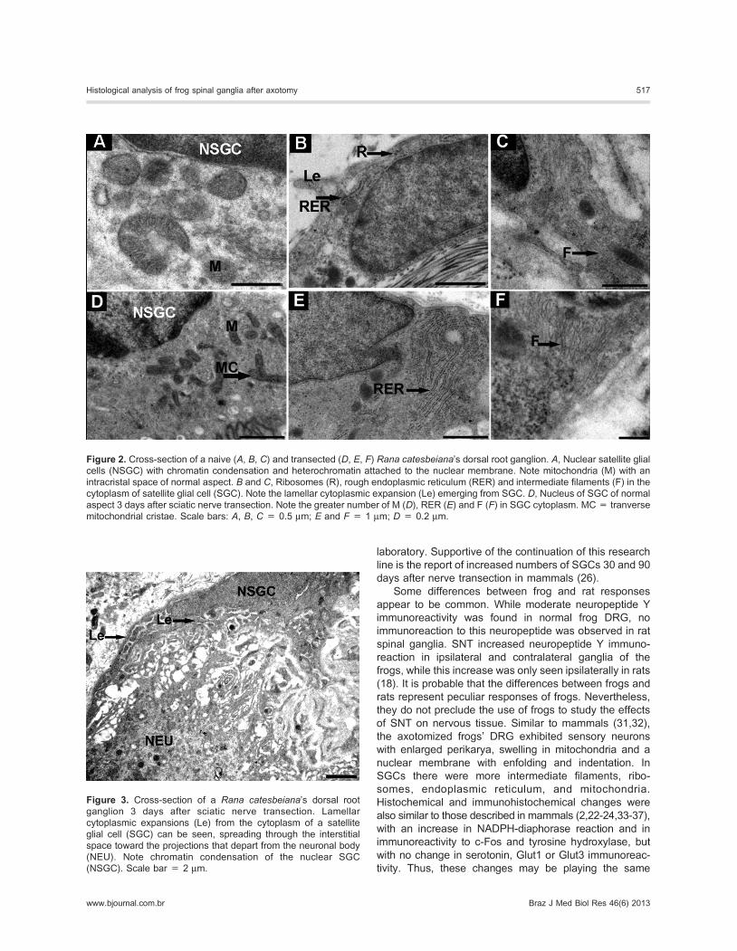
Figure 2. Cross-section of a naive (A, B, C) and transected (D, E, F) Rana catesbeiana’s dorsal root ganglion. A, Nuclear satellite glial
cells (NSGC) with chromatin condensation and heterochromatin attached to the nuclear membrane. Note mitochondria (M) with an
intracristal space of normal aspect. B and C, Ribosomes (R), rough endoplasmic reticulum (RER) and intermediate filaments (F) in the
cytoplasm of satellite glial cell (SGC). Note the lamellar cytoplasmic expansion (Le) emerging from SGC. D, Nucleus of SGC of normal
aspect 3 days after sciatic nerve transection. Note the greater number of M (D), RER (E) and F (F) in SGC cytoplasm. MC = tranverse
mitochondrial cristae. Scale bars: A, B, C = 0.5 mm; E and F = 1 mm; D = 0.2 mm.
Figure 3. Cross-section of a Rana catesbeiana’s dorsal root
ganglion 3 days after sciatic nerve transection. Lamellar
cytoplasmic expansions (Le) from the cytoplasm of a satellite
glial cell (SGC) can be seen, spreading through the interstitial
space toward the projections that depart from the neuronal body
(NEU). Note chromatin condensation of the nuclear SGC
(NSGC). Scale bar = 2 mm.
Histological analysis of frog spinal ganglia after axotomy 517
www.bjournal.com.br Braz J Med Biol Res 46(6) 2013

functional role in frogs. Because total mitochondrial mass
and maximum rate of oxygen consumption appear to be
directly linked in mammals (38), the increased number of
mitochondria found in frog DRG may indicate a higher
ability of SGCs to produce energy. This hypothesis was
suggested to explain the enlargement of mitochondria in
SGCs from mammalian DRG observed after peripheral
nerve injury (29). The greater number of ribosomes and
RER reinforces the higher activation of SGCs from frog
DRG as early as 3 days after SNT. This higher activation of
these cells also occurs in mammals (39). Another change
that reinforces the activation of SGCs in frog DRG in
response to SNT is the increased NADPH-diaphorase
reaction in these cells and sensory neurons. In mammals, it
was suggested that neuronal NO signals satellite glia in
axotomized DRG to neutralize the cytotoxic effect of
inducible NO synthase by inducing neurotrophic factors in
the glial cell (40). A similar hypothesis may be raised for
frogs to explain the increased NADPH-diaphorase reaction
in parallel to the ultrastructural changes in SGCs.
Overall, the present study provides evidence that SNT
induces ultrastructural, histochemical and immunohisto-
chemical changes in frog DRG that are very similar to
those described in mammals. The difference appears to
be the beginning of SGC proliferation. Thus, our results
support the use of frogs to study the effects of SNT, a
model of neuropathic pain, on nervous tissue. The use of
frogs in these studies provides knowledge not only about
this issue, which still has many unanswered questions,
but also about the evolution of these responses in
vertebrates.
Acknowledgments
Research supported by FAPERGS and CNPq.
Figure 4. Cross-section of a naive (A, C, E) and transected (B, D,F)Rana catesbeiana’s dorsal root ganglion. Note the larger number
of cells positive for the NADPH-diaphorase reaction (B), the c-Fos-
like immunoreactivity in the nucleus of the neuron (D), and themore
intense tyrosine hydroxylase-like immunoreactivity surrounding
neurons (F) 3 days after sciatic nerve transection. Arrows point to
neurons positive for NADPH-diaphorase (A) and c-FOS (C). Scalebars: A and B = 20 mm, C = 10 mm, D and F = 40 mm.
Figure 5. Serotonin- (A) and Glut3-like (B) immunoreactivity in a
medium neuron of the dorsal root ganglion from a naive frog.
Scale bars: A = 20 mm; B = 40 mm.
518 F. Rigon et al.
Braz J Med Biol Res 46(6) 2013 www.bjournal.com.br

References
1. Ten Donkelaar HJ. Anurans. In: Nieuwenhuys R, Ten
Donkellar HJ, Nicholson C (Editors), The central nervous
system of vertebrates. Berlin: Springer-Verlag; 1998. p
1151-1314.
2. Matsuda S, Kobayashi N, Terashita T, Shimokawa T,
Shigemoto K, Mominoki K, et al. Phylogenetic investigation
of Dogiel’s pericellular nests and Cajal’s initial glomeruli in
the dorsal root ganglion. J Comp Neurol 2005; 491: 234-
245, doi: 10.1002/cne.20713.
3. Crowe MJ, Brown TJ, Bresnahan JC, Beattie MS.
Distribution of NADPH-diaphorase reactivity in the spinal
cord of metamorphosing and adult Xenopus laevis. Brain
Res Dev Brain Res 1995; 86: 155-166, doi: 10.1016/0165-
3806(95)00021-5.
4. Cristino L, Florenzano F, Bentivoglio M, Guglielmotti V.
Nitric oxide synthase expression and cell changes in dorsal
root ganglia and spinal dorsal horn of developing and adult
Rana esculenta indicate a role of nitric oxide in limb
metamorphosis. J Comp Neurol 2004; 472: 423-436, doi:
10.1002/cne.20057.
5. Hope BT, Michael GJ, Knigge KM, Vincent SR. Neuronal
NADPH diaphorase is a nitric oxide synthase. Proc Natl Acad
Sci U S A 1991; 88: 2811-2814, doi: 10.1073/pnas.88.7.2811.
6. HolzGG,AndersonEG.Theactionsofserotoninon frogprimary
afferent terminals and cell bodies. Comp Biochem Physiol C
1984; 77: 13-21, doi: 10.1016/0742-8413(84)90124-5.
7. Philippi M, Vyklicky L, Kuffler DP, Orkand RK. Serotonin-
and proton-induced and modified ionic currents in frog
sensory neurons. J Neurosci Res 1995; 40: 387-395, doi:
10.1002/jnr.490400313.
8. Chen Y, Wang AJ, Ma YL. [Effects of morphine on
sensitivities of alpha-adrenoceptors in toad spinal ganglion
neurons]. Zhongguo Yao Li Xue Bao 1993; 14: 417-420.
9. Stevens CW. Opioid research in amphibians: an alternative
pain model yielding insights on the evolution of opioid
receptors. Brain Res Brain Res Rev 2004; 46: 204-215, doi:
10.1016/j.brainresrev.2004.07.003.
10. Stevens CW, Brasel CM, Mohan S. Cloning and bioinfor-
matics of amphibian mu, delta, kappa, and nociceptin opioid
receptors expressed in brain tissue: evidence for opioid
receptor divergence in mammals. Neurosci Lett 2007; 419:
189-194, doi: 10.1016/j.neulet.2007.04.014.
11. Stevens CW, Martin KK, Stahlheber BW. Nociceptin
produces antinociception after spinal administration in
amphibians. Pharmacol Biochem Behav 2009; 91: 436-
440, doi: 10.1016/j.pbb.2008.08.022.
12. Chen NC, Srinivasan RC, Shauver MJ, Chung KC. A
systematic review of outcomes of fasciotomy, aponeurot-
omy, and collagenase treatments for Dupuytren’s contrac-
ture. Hand 2011; 6: 250-255, doi: 10.1007/s11552-011-
9326-8.
13. Coble DJ, Taylor DK, Mook DM. Analgesic effects of
meloxicam, morphine sulfate, flunixin meglumine, and
xylazine hydrochloride in African-clawed frogs (Xenopus
laevis). J Am Assoc Lab Anim Sci 2011; 50: 355-360.
14. Ohkita M, Saito S, Imagawa T, Takahashi K, Tominaga M,
Ohta T. Molecular cloning and functional characterization of
Xenopus tropicalis frog transient receptor potential vanilloid
1 reveal its functional evolution for heat, acid, and capsaicin
sensitivities in terrestrial vertebrates. J Biol Chem 2012;
287: 2388-2397, doi: 10.1074/jbc.M111.305698.
15. Saito S, Nakatsuka K, Takahashi K, Fukuta N, Imagawa T,
Ohta T, et al. Analysis of transient receptor potential ankyrin
1 (TRPA1) in frogs and lizards illuminates both nociceptive
heat and chemical sensitivities and coexpression with TRP
vanilloid 1 (TRPV1) in ancestral vertebrates. J Biol Chem
2012; 287: 30743-30754, doi: 10.1074/jbc.M112.362194.
16. Partata WA, Cerveira JF, Xavier LL, Viola GG, Achaval M.
Sciatic nerve transection decrease substance P immunor-
eactivity in the lumbosacral spinal cord of the frog (Rana
catesbeiana). Comp Biochem Physiol B Biochem Mol Biol
2002; 131: 807-814, doi: 10.1016/S1096-4959(02)00041-6.
17. Guedes RP, Marchi MI, Viola GG, Xavier LL, Achaval M,
Partata WA. Somatostatin-, calcitonin gene-related peptide,
and gamma-aminobutyric acid-like immunoreactivitity in the
frog lumbosacral spinal cord: distribution and effects of sciatic
nerve transection. Comp Biochem Physiol B Biochem Mol
Biol 2004; 138: 19-28, doi: 10.1016/j.cbpc.2004.01.004.
18. Guedes RP, Marchi MI, Achaval M, Partata WA. Complete
sciatic nerve transection induces increase of neuropeptide Y-
like immunoreactivity in primary sensory neurons and spinal
cord of frogs. Comp Biochem Physiol A Mol Integr Physiol
2004; 139: 461-467, doi: 10.1016/j.cbpb.2004.10.006.
19. Hansen N, Uceyler N, Palm F, Zelenka M, Biko L, Lesch KP,
et al. Serotonin transporter deficiency protects mice from
mechanical allodynia and heat hyperalgesia in vincristine
neuropathy. Neurosci Lett 2011; 495: 93-97, doi: 10.1016/
j.neulet.2011.03.035.
20. Joseph L, Butera RJ. High-frequency stimulation selectively
blocks different types of fibers in frog sciatic nerve. IEEE
Trans Neural Syst Rehabil Eng 2011; 19: 550-557, doi:
10.1109/TNSRE.2011.2163082.
21. Stark B, Carlstedt T, Cullheim S, Risling M. Developmental
and lesion-induced changes in the distribution of the
glucose transporter Glut-1 in the central and peripheral
nervous system. Exp Brain Res 2000; 131: 74-84, doi:
10.1007/s002219900300.
22. Soares HD, Chen SC, Morgan JI. Differential and prolonged
expression of Fos-lacZ and Jun-lacZ in neurons, glia, and
muscle following sciatic nerve damage. Exp Neurol 2001;
167: 1-14, doi: 10.1006/exnr.2000.7558.
23. Keilhoff G, Fansa H, Wolf G. Nitric oxide synthase, an
essential factor in peripheral nerve regeneration. Cell Mol
Biol 2003; 49: 885-897.
24. Lukacova N, Cizkova D, Krizanova O, Pavel J, Marsala M,
Marsala J. Peripheral axotomy affects nicotinamide adenine
dinucleotide phosphate diaphorase and nitric oxide
synthases in the spinal cord of the rabbit. J Neurosci Res
2003; 71: 300-313, doi: 10.1002/jnr.10470.
25. Hanani M. Satellite glial cells in sensory ganglia: from form
to function. Brain Res Rev 2005; 48: 457-476, doi: 10.1016/
j.brainresrev.2004.09.001.
26. Arkhipova SS, Raginov IS, Mukhitov AR, Chelyshev YA.
Satellite cells of sensory neurons after various types of
sciatic nerve trauma in the rat. Neurosci Behav Physiol
2010; 40: 609-614, doi: 10.1007/s11055-010-9303-7.
27. Humbertson A Jr, Zimmermann E, Leedy M. A chronological
study of mitotic activity in satellite cell hyperplasia associated
Histological analysis of frog spinal ganglia after axotomy 519
www.bjournal.com.br Braz J Med Biol Res 46(6) 2013

with chromatolytic neurons. Z Zellforsch Mikrosk Anat 1969;
100: 507-515, doi: 10.1007/BF00344371.
28. Pannese E. The structure of the perineuronal sheath of
satellite glial cells (SGCs) in sensory ganglia. Neuron Glia
Biol 2010; 6: 3-10, doi: 10.1017/S1740925X10000037.
29. Hanani M. Satellite glial cells in sympathetic and para-
sympathetic ganglia: in search of function. Brain Res Rev
2010; 64: 304-327, doi: 10.1016/j.brainresrev.2010.04.009.
30. McDougal DB Jr, Holowach J, Howe MC, Jones EM,
Thomas CA. The effects of anoxia upon energy sources and
selected metabolic intermediates in the brains of fish, frog
and turtle. J Neurochem 1968; 15: 577-588, doi: 10.1111/
j.1471-4159.1968.tb08956.x.
31. Oliveira AL. Apoptosis of sensory neurons and satellite cells
after sciatic nerve transection in C57BL/6J mice. Braz J Med
Biol Res 2001; 34: 375-380, doi: 10.1590/S0100-
879X2001000300012.
32. Atlasi MA, Mehdizadeh M, Bahadori MH, Joghataei MT.
Morphological identification of cell death in dorsal root
ganglion neurons following peripheral nerve injury and
repair in adult rat. Iran Biomed J 2009; 13: 65-72.
33. Lukacova N, Davidova A, Kolesar D, Kolesarova M,
Schreiberova A, Lackova M, et al. The effect of N-nitro-L-
arginine and aminoguanidine treatment on changes in
constitutive and inducible nitric oxide synthases in the
spinal cord after sciatic nerve transection. Int J Mol Med
2008; 21: 413-421.
34. Lin CT, Tsai YJ, Chen SH, Wang HY, Lin LH, Lue JH. Early
expression of injury-induced neuropeptide Y in primary
sensory neurons and the cuneate nucleus in diabetic rats
with median nerve transection. J Chem Neuroanat 2010; 40:
102-111, doi: 10.1016/j.jchemneu.2010.05.006.
35. Wang D, Gao Y, Ji H, Hong Y. Topical and systemic
administrations of ketanserin attenuate hypersensitivity and
expression of CGRP in rats with spinal nerve ligation. Eur J
Pharmacol 2010; 627: 124-130, doi : 10.1016/
j.ejphar.2009.11.011.
36. Brumovsky PR, Seroogy KB, Lundgren KH, Watanabe M,
Hokfelt T, Gebhart GF. Some lumbar sympathetic neurons
develop a glutamatergic phenotype after peripheral axotomy
with a note on VGLUT(2)-positive perineuronal baskets. Exp
Neurol 2011; 230: 258-272, doi: 10.1016/j.expneurol.
2011.05.004.
37. Xia CM, Colomb DG Jr, Akbarali HI, Qiao LY. Prolonged
sympathetic innervation of sensory neurons in rat thoraco-
lumbar dorsal root ganglia during chronic colitis.
Neurogastroenterol Motil 2011; 23: 801-e339, doi:
10.1111/j.1365-2982.2011.01728.x.
38. Hoppeler H, Hudlicka O, Uhlmann E. Relationship between
mitochondria and oxygen consumption in isolated cat
muscles. J Physiol 1987; 385: 661-675.
39. Liu FY, Sun YN, Wang FT, Li Q, Su L, Zhao ZF, et al.
Activation of satellite glial cells in lumbar dorsal root ganglia
contributes to neuropathic pain after spinal nerve ligation.Brain
Res 2012; 1427: 65-77, doi: 10.1016/j.brainres.2011.10.016.
40. Bradman MJ, Arora DK, Morris R, Thippeswamy T. How do
the satellite glia cells of the dorsal root ganglia respond to
stressed neurons? Nitric oxide saga from embryonic
development to axonal injury in adulthood. Neuron Glia
Biol 2010; 6: 11-17, doi: 10.1017/S1740925X09990494.
520 F. Rigon et al.
Braz J Med Biol Res 46(6) 2013 www.bjournal.com.br