Embed Size (px)
Citation preview

Anuar RIM, et al
71
ABSTRACTThe supracondylar humerus fracture (SCHF) in children iscommon and can be complicated with nerve injury eitherprimarily immediate post-trauma or secondarily post-treatment. The concept of neurapraxic nerve injury makesmost surgeons choose to ‘watch and see’ the nerve recoverybefore deciding second surgery if the nerve does not recover.We report three cases of nerve injury in SCHF, all of whichunderwent nerve exploration for different reasons. Earlyreduction in the Casualty is important to release the nervetension before transferring the patient to the operation room.If close reduction fails, we proceed to explore the nervetogether with open reduction of the fracture. In iatrogenicnerve injury, we recommend nerve exploration to determinethe surgical procedure that is causing the injury. Primarynerve exploration will allow early assessment of the injurednerve and minimize subsequent surgery.
Key Words: Nerve exploration, supracondylar humerus fracture, nerveinjury
INTRODUCTIONThe supracondylar humerus fracture (SCHF) is one of themost common fractures in children, predominantly theextension-type. As the Median and Radial Nerves lieanterior to the supracondylar humerus region they are at riskfor injury, primarily post-trauma either by stretching,piercing or impinging at the fracture ends or being entrappedbetween two fracture fragments (traumatic or primary nerveinjury), whereas the Ulnar Nerve injury is usually secondaryto treatment (iatrogenic or secondary nerve injury) 1.
The documented incidence of primary nerve injury in SCHFis 7 – 10% and up to 6% for secondary nerve injury 1. In theextension-type of SCHF, postero-medial displacement ofdistal fragment usually causes injury to the Radial Nerve
and postero-lateral placement is more likely to cause MedianNerve injury. In the flexion-type of SCHF, the Ulnar Nerveinjury predominates 2. Closed manipulative reduction (CMR)and percutaneous pinning are the first line treatments forSCHF 3. Various methods of pin placement have beendescribed and crossed pinning is still popular amongsurgeons. For the medial placement of the Kirschner-wire (Kwire), the mini-open technique has been proposed in order tominimize injury to the Ulnar Nerve. In lateral pinning, theMedian Nerve is at risk of injury 2.
The concept of traumatic neurapraxia and iatrogenicneurapraxia are well understood and accepted by most ofsurgeons, but some orthopaedic surgeons still believe inacute nerve decompression or nerve exploration as thesehave their own benefits. We discuss three cases of nerveinjury in SCHF which underwent nerve exploration fordifferent reasons.
CASE REPORTCase 1An 8-year old girl had fallen down from the monkey bar andsustained right elbow swelling and pain with limitation ofelbow motion. On examination, the right elbow was swollenand tender with signs of Radial Nerve palsy: unable toextend the wrist joint and reduced sensation over theanatomical snuffbox. The radiograph showed fracture of theright supracondylar humerus (Gartland III) with the distalfragment displaced postero-medially (Fig.1A). On thediagnosis of closed supracondylar fracture of the righthumerus with Radial Nerve injury, we explored the nerve inthe operation theatre before attempting closed reduction. Wefound the Radial Nerve severely stretched by the proximalfracture end and nearly lacerated (Fig.1B), and was thenreleased after fracture reduction. The fracture was held withcrossed K-wires.
The Role of Nerve Exploration in Supracondylar HumerusFracture in Children with Nerve Injury
Anuar RIM, MMed Orth, Gooi SG, MS Orth, Zulkiflee O, MS Orth
Department of Orthopaedics, Penang Hospital, Georgetown, Malaysia
Date of submission: July 2015Date of acceptance: October 2015
Corresponding Author: Mohd Anuar Ramdhan Ibrahim, Department of Orthopaedics, Penang Hospital, Jalan Residensi, 10990Georgetown, Penang Email: [email protected]
Malaysian Orthopaedic Journal 2015 Vol 9 No 3http://dx.doi.org/10.5704/MOJ.1511.019
15-B118_OA1 12/3/15 12:15 AM Page 71

Malaysian Orthopaedic Journal 2015 Vol 9 No 3 Anuar RIM, et al
72
Fig. 1a: Radiograph showing fracture of thesupracondyle of right humerus.
Fig. 1b:Radiograph showing fracture of the supracondyle of righthumerus. Yellow arrow showing radial nerve.
Fig. 2a: Radiographs showing fracture of the supracondyle of lefthumerus.
Fig. 2b:White arrow: Proximal fracture end. Yellow arrowindicates- intact Median Nerve.
15-B118_OA1 12/3/15 12:15 AM Page 72
Role of Nerve Exploration
73
Case 2A 5-year old girl had fallen on her out-stretch left hand whileplaying and sustained injury to her left elbow. Onexamination, the left elbow was tender and swollen withantecubital ecchymosis, with signs of Median Nerve injury:she was unable to flex the inter-phalangeal joint (IPJ) of theleft thumb, distal inter-phalangeal joint (DIPJ) of the leftindex finger and reduced sensation over the Median Nervedistribution. Radiography showed left supracondylarhumerus fracture (Gartland III) (Fig. 2A). As she had closedleft humerus supracondylar fracture with Median Nerveinjury, we attempted reduction under sedation in the casualty(emergency department) aiming to release the tension on thenerve. Immediately post-reduction the nerve had partiallyrecovered with residual inability to flex the inter-phalangealjoint (IPJ) of the left thumb and reduced sensation over theMedian Nerve distribution. On transferring her to theoperation theatre, we attempted closed manipulative
reduction (CMR) but failed, and proceeded with openreduction. We used this opportunity to explore the nerve andfound the Median Nerve as well as the Anterior InterosseousNerve (AIN) intact (Fig. 2B). The fracture was held withcrossed K-wires.
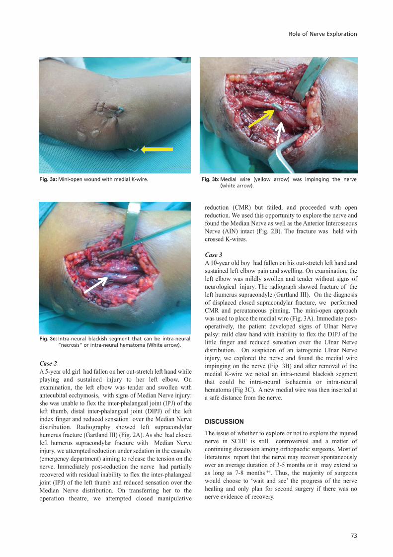
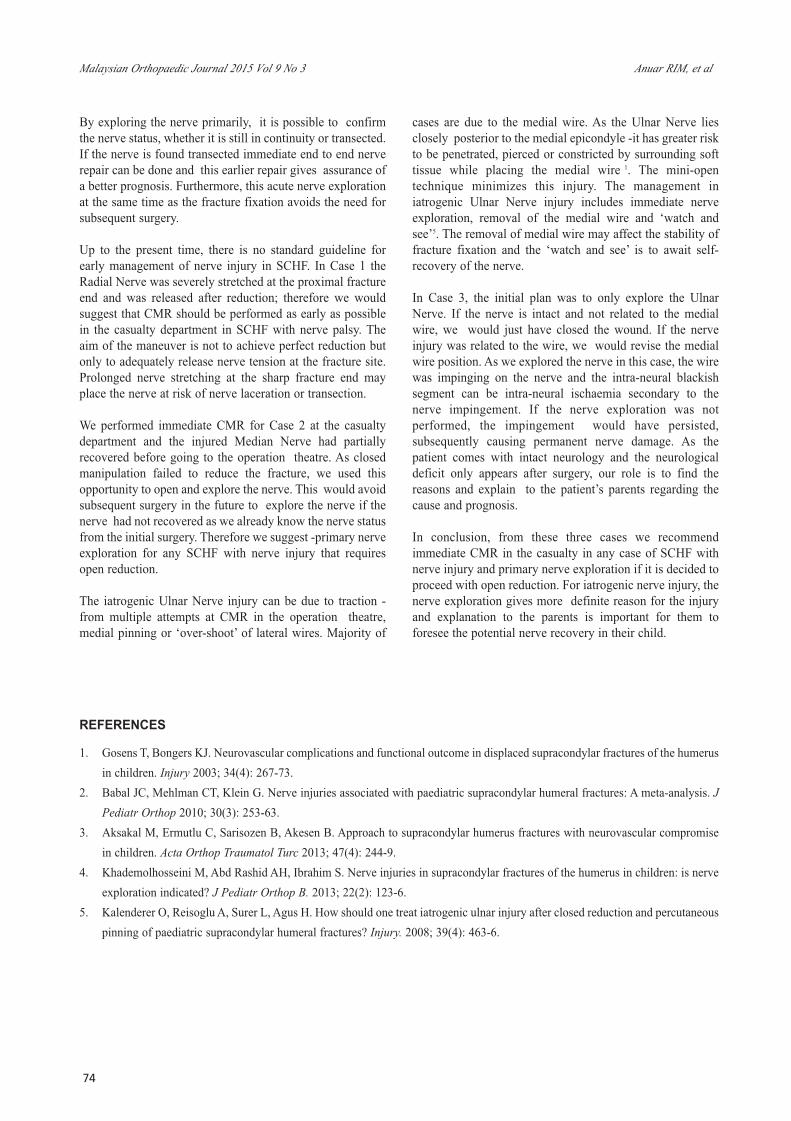
Case 3A 10-year old boy had fallen on his out-stretch left hand andsustained left elbow pain and swelling. On examination, theleft elbow was mildly swollen and tender without signs ofneurological injury. The radiograph showed fracture of theleft humerus supracondyle (Gartland III). On the diagnosisof displaced closed supracondylar fracture, we performedCMR and percutaneous pinning. The mini-open approachwas used to place the medial wire (Fig. 3A). Immediate post-operatively, the patient developed signs of Ulnar Nervepalsy: mild claw hand with inability to flex the DIPJ of thelittle finger and reduced sensation over the Ulnar Nervedistribution. On suspicion of an iatrogenic Ulnar Nerveinjury, we explored the nerve and found the medial wireimpinging on the nerve (Fig. 3B) and after removal of themedial K-wire we noted an intra-neural blackish segmentthat could be intra-neural ischaemia or intra-neuralhematoma (Fig 3C). A new medial wire was then inserted ata safe distance from the nerve.
DISCUSSIONThe issue of whether to explore or not to explore the injurednerve in SCHF is still controversial and a matter ofcontinuing discussion among orthopaedic surgeons. Most ofliteratures report that the nerve may recover spontaneouslyover an average duration of 3-5 months or it may extend toas long as 7-8 months 4-5. Thus, the majority of surgeonswould choose to ‘wait and see’ the progress of the nervehealing and only plan for second surgery if there was nonerve evidence of recovery.
Fig. 3a:Mini-open wound with medial K-wire. Fig. 3b:Medial wire (yellow arrow) was impinging the nerve(white arrow).
Fig. 3c: Intra-neural blackish segment that can be intra-neural“necrosis” or intra-neural hematoma (White arrow).
15-B118_OA1 12/3/15 12:15 AM Page 73
Malaysian Orthopaedic Journal 2015 Vol 9 No 3 Anuar RIM, et al
74
REFERENCES
1. Gosens T, Bongers KJ. Neurovascular complications and functional outcome in displaced supracondylar fractures of the humerusin children. Injury 2003; 34(4): 267-73.
2. Babal JC, Mehlman CT, Klein G. Nerve injuries associated with paediatric supracondylar humeral fractures: A meta-analysis. JPediatr Orthop 2010; 30(3): 253-63.
3. Aksakal M, Ermutlu C, Sarisozen B, Akesen B. Approach to supracondylar humerus fractures with neurovascular compromisein children. Acta Orthop Traumatol Turc 2013; 47(4): 244-9.
4. Khademolhosseini M, Abd Rashid AH, Ibrahim S. Nerve injuries in supracondylar fractures of the humerus in children: is nerveexploration indicated? J Pediatr Orthop B. 2013; 22(2): 123-6.
5. Kalenderer O, Reisoglu A, Surer L, Agus H. How should one treat iatrogenic ulnar injury after closed reduction and percutaneouspinning of paediatric supracondylar humeral fractures? Injury. 2008; 39(4): 463-6.
By exploring the nerve primarily, it is possible to confirmthe nerve status, whether it is still in continuity or transected.If the nerve is found transected immediate end to end nerverepair can be done and this earlier repair gives assurance ofa better prognosis. Furthermore, this acute nerve explorationat the same time as the fracture fixation avoids the need forsubsequent surgery.
Up to the present time, there is no standard guideline forearly management of nerve injury in SCHF. In Case 1 theRadial Nerve was severely stretched at the proximal fractureend and was released after reduction; therefore we wouldsuggest that CMR should be performed as early as possiblein the casualty department in SCHF with nerve palsy. Theaim of the maneuver is not to achieve perfect reduction butonly to adequately release nerve tension at the fracture site.Prolonged nerve stretching at the sharp fracture end mayplace the nerve at risk of nerve laceration or transection.
We performed immediate CMR for Case 2 at the casualtydepartment and the injured Median Nerve had partiallyrecovered before going to the operation theatre. As closedmanipulation failed to reduce the fracture, we used thisopportunity to open and explore the nerve. This would avoidsubsequent surgery in the future to explore the nerve if thenerve had not recovered as we already know the nerve statusfrom the initial surgery. Therefore we suggest -primary nerveexploration for any SCHF with nerve injury that requiresopen reduction.
The iatrogenic Ulnar Nerve injury can be due to traction -from multiple attempts at CMR in the operation theatre,medial pinning or ‘over-shoot’ of lateral wires. Majority of
cases are due to the medial wire. As the Ulnar Nerve liesclosely posterior to the medial epicondyle -it has greater riskto be penetrated, pierced or constricted by surrounding softtissue while placing the medial wire 1. The mini-opentechnique minimizes this injury. The management iniatrogenic Ulnar Nerve injury includes immediate nerveexploration, removal of the medial wire and ‘watch andsee’5. The removal of medial wire may affect the stability offracture fixation and the ‘watch and see’ is to await self-recovery of the nerve.
In Case 3, the initial plan was to only explore the UlnarNerve. If the nerve is intact and not related to the medialwire, we would just have closed the wound. If the nerveinjury was related to the wire, we would revise the medialwire position. As we explored the nerve in this case, the wirewas impinging on the nerve and the intra-neural blackishsegment can be intra-neural ischaemia secondary to thenerve impingement. If the nerve exploration was notperformed, the impingement would have persisted,subsequently causing permanent nerve damage. As thepatient comes with intact neurology and the neurologicaldeficit only appears after surgery, our role is to find thereasons and explain to the patient’s parents regarding thecause and prognosis.
In conclusion, from these three cases we recommendimmediate CMR in the casualty in any case of SCHF withnerve injury and primary nerve exploration if it is decided toproceed with open reduction. For iatrogenic nerve injury, thenerve exploration gives more definite reason for the injuryand explanation to the parents is important for them toforesee the potential nerve recovery in their child.
15-B118_OA1 12/3/15 12:15 AM Page 74