Embed Size (px)
Citation preview
The Role of Iron in Restless Legs Syndrome
Richard P. Allen, MD, PhD,* and Christopher J. Earley, PhD
Department of Neurology, Johns Hopkins University, Baltimore, Maryland, USA
Abstract: The impressive relief from restless legs syndrome(RLS) symptoms provided by levodopa treatment indicatesRLS is caused by a dopaminergic abnormality. But similar andmore lasting relief also occurs for iron treatment in somepatients. Thus there are two major putative causes for RLS:CNS dopaminergic abnormality and CNS iron insufficiency.This article presents the data documenting that both peripheraland CNS iron insufficiency occur with RLS symptoms. Brainiron insufficiency is supported by independently replicatedcerebrospinal fluid and brain imaging studies for patients with-out iron deficiency (ID) anemia. Autopsy studies and intrave-nous iron treatment further link brain iron insufficiency to RLS.The brain iron insufficiency in patients with RLS is now wellestablished. In this article the data are reviewed that support thefollowing postulates combining dopaminergic and iron causesof RLS: (1) All conditions that compromise iron availability
will increase the risk of RLS leading to a higher than expectedprevalence of RLS in these conditions. (2) Some patients withRLS have marginal CNS iron status that can become insuffi-cient when deprived of normal access to adequate peripheraliron or may be insufficient even with normal access to adequateperipheral iron. (3) The change or reduced CNS iron statusproduces RLS symptoms largely through its effects on thedopaminergic system and the corollary to 3. (4) Dopaminergicsystem abnormalities producing RLS symptoms will be in-cluded in those produced by brain ID. Study of the iron modelof RLS offers hope for developing new treatment approachesand perhaps methods to prevent or cure the disorder. © 2007Movement Disorder Society
Key words: iron deificieny; restless legs; dopaminel intra-venous iron.
When Ekbom provided the first modern medical de-scriptions of restless legs syndrome (RLS), he noted ahigh prevalence of iron deficiency (ID) among patientswith RLS.1 This striking relation led a contemporary ofEkbom, another Swedish neurologist Nordlander, to notonly propose that ID in some body tissue caused RLS butalso to successfully treat 21 0f 22 patients with RLS withrelatively large doses of intravenous (IV) iron.2,3 Themodern discovery of the remarkably effective dopami-nergic treatment for RLS has led to an emphasis uponfinding a dopamine pathology in RLS using techniquessimilar to those for the study of Parkinson’s Disease.These studies have generally failed to find convincingevidence in replicated studies for dopamine pathology in
RLS. That direction in research largely ignored consid-erations of the relation of iron to RLS despite the clinicalevidence showing that ID produces RLS; all conditionsthat compromise iron status increase the risk of RLS andiron treatments reduce or even cure RLS. Moreover,autopsy, cerebrospinal fluid (CSF), and brain imagingstudies document low CNS iron status for patients withRLS. In this chapter, we review the iron and RLS con-nections as found in clinical, peripheral, and CNS studiesand then advance the iron model of RLS and discuss itsimplications for unraveling the neurobiology of RLS.
CLINICAL INDICATIONS OF THE RLS–IRONCONNECTION
There are three major secondary causes of RLS: ID,end-stage renal disease, and pregnancy. In each of thesethere is a higher than expected prevalence of RLS, buteven more important is that RLS starts after these con-ditions start and commonly resolves when the conditionis corrected. These very disparate conditions share atleast one common problem. They all compromise ironsufficiency and in each case when the condition is cor-
*Correspondence to: Dr. Richard P Allen, Neurology and SleepMedicine, Johns Hopkins University, Asthma and Allergy Bldg 1B76b,5501 Hopkins Bayview Circle, Baltimore, MD 21224.E-mail: [email protected]
Received 19 January 2007; Revised 18 April 2007; Accepted 3 May2007
Published online 12 June 2007 in Wiley InterScience (www.interscience.wiley.com). DOI: 10.1002/mds.21607
Movement DisordersVol. 22, Suppl. 18, 2007, pp. S440–S448© 2007 Movement Disorder Society
S440

rected the iron problem is also corrected. The iron insuf-ficiency and not other problems in these disorders appearto produce the RLS. Thus, end stage renal disease pro-duces neuropathy but that neuropathy changes little withsuccessful kidney transplant, yet the RLS usually com-pletely disappears.4,5 Moreover, high doses of IV ironreduce the RLS symptoms in patients with end stagerenal disease.6 Similarly, the decreased blood volumethat occurs with delivery provides a rapid improvementin access to iron stores and there is a corresponding rapidremission of any RLS symptoms.7 Increasing the bodyiron stores in patients with RLS and ID can providecomplete relief from all RLS symptoms in some pa-tients.8 These considerations led to our hypothesis: Allconditions that compromise iron availability will in-crease the risk of RLS leading to a higher than expectedprevalence of RLS in these conditions.
This hypothesis has been proven correct for all suchconditions evaluated to date, eg: Gastric surgery9 andlow-density lipoprotein apheresis.10,11
It deserves note that the serology measures of ironstatus, thought the most frequently used, have limita-tions. They neither provide a direct measure of cellular orbone marrow iron status nor an accurate measure ofneuronal iron status. Despite these problems, one com-munity based study found higher serum transferrin re-ceptor indicating iron compromise for RLS compared tocontrol subjects12 but this was not found in anothersimilar study.13
IRON STATUS AND RLS
Peripheral Iron Status and RLS
Serum ferritin provides the generally accepted bestsingle measure of iron stores.14,15 The usually publishednormal values for ferritin, however, tend to confuse ironstatus evaluations since they represent population sam-ples not biological norms. When serum ferritin valueswere compared to iron status from the bone marrow inpatients thought to be iron deficient the response–ob-server curve indicated an optimum cut-off of about 45�g/L with ferritin below that indicating low peripheraliron stores (see Fig. 1).15 The phase-reactive property offerritin, however, sometimes produces falsely elevatedvalues even in the face of ID. Percent transferrin satura-tion below 20% or TIBC above 400 �g/dL both alsoindicate ID and should be used clinically given the riskof falsely elevated ferritin values. Serum transferrin re-ceptor provides a useful alternative to ferritin foryounger subjects. It does not have the phase reactiveaspect of ferritin and thus is a more sensitive measure,but unfortunately a less specific indicator of low iron
than ferritin.16 TfR also fails to discriminate between IDand anemia of chronic disease, and therefore is not auseful test for the elderly.17 Given the ferritin measure-ment problems it has been recommended that patientswith RLS with ferritin of 50 �g/L or less be consideredfor oral iron treatment.18
Serum ferritin somewhat indirectly reflects body ironstores and in two studies correlated with RLS severi-ty.19,20 Moreover, patients with RLS who develop IDshow marked exacerbation of their symptoms. Thus, anysudden worsening of RLS symptoms unrelated to med-ication changes has to be considered as likely indicatingblood loss or other cause of ID.
An un-blinded study19 and a recent double-blindcontrolled study21 documented oral iron treatment re-duces RLS symptoms, particularly for those withlower ferritin values. Another study failed to includeadequate numbers of patients with low serum ferritinand accordingly did not find any benefit from oral irontreatment.22 Thus when peripheral iron stores are cor-rectly measured and found to be low, treatment withoral iron reduces RLS symptoms. It is important tonote that there is no substance other than iron that hasbeen conclusively shown in some patients to bothcause RLS when abnormal and cure RLS when theabnormality is corrected. Some limited evidence indi-cates that for some patients the dopaminergic system fol-lows a similar although somewhat less dramatic pattern.Levodopa23,24 and dopamine agonists25,26significantly re-lieve and dopamine antagonists27 exacerbate RLS symp-
FIG. 1. ROC curve for serum ferritin criteria values (indicated inparentheses) for those below that value considered to be anemic givingfor each value the percentage with anemia correctly identified and thepercentage of those identified with anemia who do not have it. Anemiawas determined by bone marrow aspiration from 259 consecutiveeligible and consenting patients with suspected anemia. (Reproducedwith permission from Guyatt GH, et al., Am J Med, 1990, 88, 205–209,© Excerpta Medica).
ROLE OF IRON IN RESTLESS LEGS SYNDROME S441
Movement Disorders, Vol. 22, Suppl. 18, 2007
toms. The data for both the adverse and positive effects ofdopamine on RLS, are less clear than that shown foriron, nonetheless this convergence of similar strong re-sponse to change suggests a possible iron–dopamineconnection.
The recognition both of RLS as a CNS disorder andthe strong link between peripheral iron status and RLSsymptoms supports the following postulate: Patientswith RLS have marginal CNS iron status that: canbecome insufficient when deprived of normal access toadequate peripheral iron or may be insufficient evenwith normal access to adequate peripheral iron.
The recognition of the strong effects on RLS symp-toms of the changes in the dopaminergic system similarto the effects of changes in iron led us to further postu-late: the change or reduced CNS iron status producesRLS symptoms largely through its effects on the dopa-minergic system.
This postulate yields an important corollary: Dopami-nergic system abnormalities producing RLS symptomswill be included in those produced by brain ID.
This enables research into the nature of the dopamineabnormalities of RLS using in vitro and in vivo tech-niques evaluating effects of ID on the dopaminergicsystem. The implications of this corollary are reviewedlater after first evaluating the primary postulate of brainiron insufficiency in RLS.
Brain Iron Status and RLS
Brain iron concentrations differ dramatically both byareas of the brain and with normal aging.28 Magneticresonance imaging using special sequences providesmeasures of the iron concentrations in specific brain
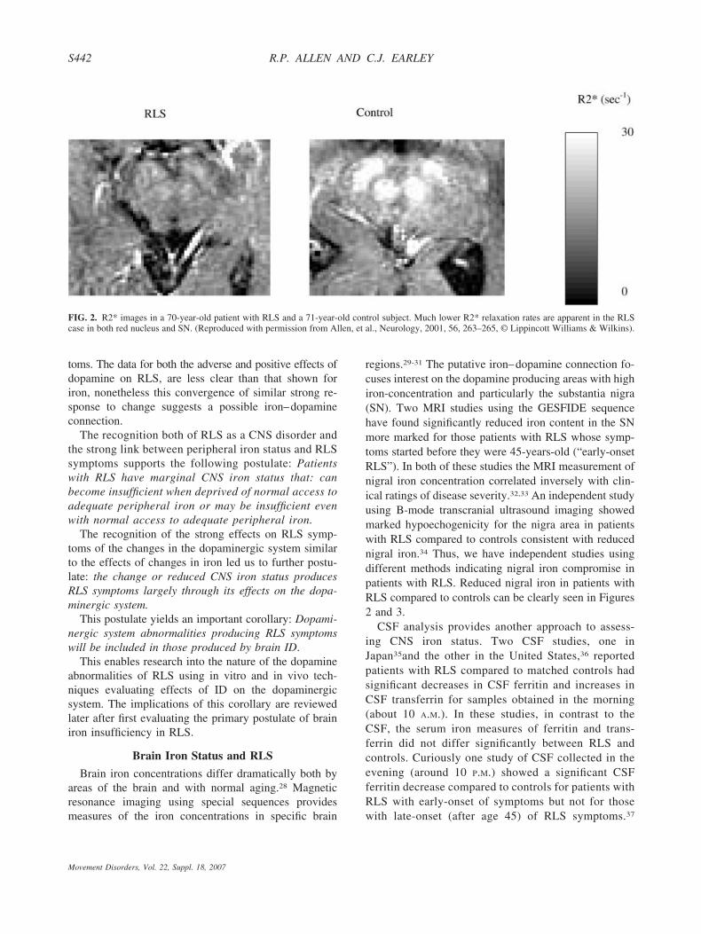
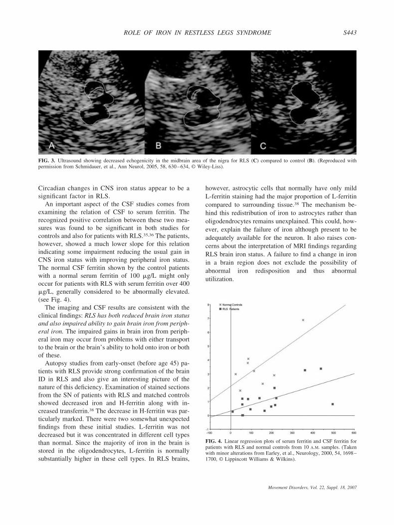
regions.29-31 The putative iron–dopamine connection fo-cuses interest on the dopamine producing areas with highiron-concentration and particularly the substantia nigra(SN). Two MRI studies using the GESFIDE sequencehave found significantly reduced iron content in the SNmore marked for those patients with RLS whose symp-toms started before they were 45-years-old (“early-onsetRLS”). In both of these studies the MRI measurement ofnigral iron concentration correlated inversely with clin-ical ratings of disease severity.32,33 An independent studyusing B-mode transcranial ultrasound imaging showedmarked hypoechogenicity for the nigra area in patientswith RLS compared to controls consistent with reducednigral iron.34 Thus, we have independent studies usingdifferent methods indicating nigral iron compromise inpatients with RLS. Reduced nigral iron in patients withRLS compared to controls can be clearly seen in Figures2 and 3.
CSF analysis provides another approach to assess-ing CNS iron status. Two CSF studies, one inJapan35and the other in the United States,36 reportedpatients with RLS compared to matched controls hadsignificant decreases in CSF ferritin and increases inCSF transferrin for samples obtained in the morning(about 10 A.M.). In these studies, in contrast to theCSF, the serum iron measures of ferritin and trans-ferrin did not differ significantly between RLS andcontrols. Curiously one study of CSF collected in theevening (around 10 P.M.) showed a significant CSFferritin decrease compared to controls for patients withRLS with early-onset of symptoms but not for thosewith late-onset (after age 45) of RLS symptoms.37
FIG. 2. R2* images in a 70-year-old patient with RLS and a 71-year-old control subject. Much lower R2* relaxation rates are apparent in the RLScase in both red nucleus and SN. (Reproduced with permission from Allen, et al., Neurology, 2001, 56, 263–265, © Lippincott Williams & Wilkins).
S442 R.P. ALLEN AND C.J. EARLEY
Movement Disorders, Vol. 22, Suppl. 18, 2007
Circadian changes in CNS iron status appear to be asignificant factor in RLS.
An important aspect of the CSF studies comes fromexamining the relation of CSF to serum ferritin. Therecognized positive correlation between these two mea-sures was found to be significant in both studies forcontrols and also for patients with RLS.35,36 The patients,however, showed a much lower slope for this relationindicating some impairment reducing the usual gain inCNS iron status with improving peripheral iron status.The normal CSF ferritin shown by the control patientswith a normal serum ferritin of 100 �g/L might onlyoccur for patients with RLS with serum ferritin over 400�g/L, generally considered to be abnormally elevated.(see Fig. 4).
The imaging and CSF results are consistent with theclinical findings: RLS has both reduced brain iron statusand also impaired ability to gain brain iron from periph-eral iron. The impaired gains in brain iron from periph-eral iron may occur from problems with either transportto the brain or the brain’s ability to hold onto iron or bothof these.
Autopsy studies from early-onset (before age 45) pa-tients with RLS provide strong confirmation of the brainID in RLS and also give an interesting picture of thenature of this deficiency. Examination of stained sectionsfrom the SN of patients with RLS and matched controlsshowed decreased iron and H-ferritin along with in-creased transferrin.38 The decrease in H-ferritin was par-ticularly marked. There were two somewhat unexpectedfindings from these initial studies. L-ferritin was notdecreased but it was concentrated in different cell typesthan normal. Since the majority of iron in the brain isstored in the oligodendrocytes, L-ferritin is normallysubstantially higher in these cell types. In RLS brains,
however, astrocytic cells that normally have only mildL-ferritin staining had the major proportion of L-ferritincompared to surrounding tissue.38 The mechanism be-hind this redistribution of iron to astrocytes rather thanoligodendrocytes remains unexplained. This could, how-ever, explain the failure of iron although present to beadequately available for the neuron. It also raises con-cerns about the interpretation of MRI findings regardingRLS brain iron status. A failure to find a change in ironin a brain region does not exclude the possibility ofabnormal iron redisposition and thus abnormalutilization.
FIG. 3. Ultrasound showing decreased echogenicity in the midbrain area of the nigra for RLS (C) compared to control (B). (Reproduced withpermission from Schmidauer, et al., Ann Neurol, 2005, 58, 630–634, © Wiley-Liss).
FIG. 4. Linear regression plots of serum ferritin and CSF ferritin forpatients with RLS and normal controls from 10 A.M. samples. (Takenwith minor alterations from Earley, et al., Neurology, 2000, 54, 1698–1700, © Lippincott Williams & Wilkins).
ROLE OF IRON IN RESTLESS LEGS SYNDROME S443
Movement Disorders, Vol. 22, Suppl. 18, 2007

The second unexpected finding was a decrease intransferrin receptor.38 An increase should occur with ID.Transferrin receptor status is post-transcriptionally regu-lated by iron regulating proteins IRP1 and IRP2. Thesewere evaluated in a study used microlaser capture tech-niques to isolate neuromelanin cells of the SN. Exami-nation of the homogenates of only the isolated neu-romelanin cells showed that RLS compared to controlshad increased IRP2 consistent with cellular ID but alsoan unexpected decreased IRP1 both in its active andaconitase form.39 The marked decrease in IRP1 mayexplain at least in part the decreased transferrin receptorpossibly limiting cellular iron access and contributing tothe brain ID or even to reduced transport across the bloodbrain barrier. The decreased IRP1 may be secondary tothe lack of availability of iron or reflect a primary pa-thology of RLS.
The homogenates of the laser-captured neuromelanincells also showed the expected decrease in H-ferritinwith no change in L-ferritin. In addition two iron trans-port proteins DMT1 and ferroportin were significantlydecreased.39
Overall, the early-onset form of RLS occurs withabnormal iron regulation in the brain that appears likelyto cause the RLS symptoms rather than the reverse.Whether or not this also occurs in some late-onset pa-tients with RLS perhaps to in a different form or degreeremains to be determined.
IRON–DOPAMINE CONNECTION
If, as seems incontrovertible, RLS links closely to ironstatus then how does reduced iron status produce theRLS symptoms? If, as was noted above, changes indopamine status also change RLS symptoms, then, aspostulated above, iron abnormality may produce thesedopaminergic changes that produce the RLS symptoms.But, how does reduced iron change the dopaminergicsystem? It should be noted that answering this questionmay reveal details of the dopamine pathology in RLSthat would otherwise be hard to directly measure forpatients with RLS. The evaluation of the effects of ironon the dopaminergic system opens a wide range of stud-ies. The iron–dopamine relation can be studied using invitro cell models with differing degrees of iron chelationand also with in vivo animal models using either dietaryinduced ID or natural strain variation in brain iron.
In vivo studies using dietary induced ID in animalsprovides an experimental model to study effects of ironchanges on the dopamine system. Dietary ID forSprague-Dawley rats starting post weaning (e.g. day 21)and continuing for 2 to 4 weeks produced, compared tocontrols, 30 to 50% lower brain iron with increased
transferrin and decreased ferritin.40 The iron concentra-tion decreases varied between brain regions. The irondecreased by 60% in the ventral midbrain which containsthe SN, 30% in he caudate-putamen, and 20% in thenucleus accumbens.41 ID lead to a decrease in striatal D1and D2 receptors. The D2 receptor density correlatedpositively with striatal iron (r � 0.91) while the D1receptor density showed no correlation with iron.41 Thestriatal dopamine transporter (DAT) density decreasedby 30% in the caudate putamen and 20% in the nucleusaccumbens for the same dietary ID protocol.42 Microdi-alysis assessment of striatal dopamine again using asimilar ID protocol showed a 53% increase in extracel-lular dopamine compared to controls that occurred forthe time period from the end of the inactive (light)through the first part of their active (dark) periods. Othertime periods were not analyzed. More recent studiesfound an increased in the nigral tyrosine hydroxylase andincrease in its phosphorylated form with dietary ID.43
Tyrosine hydroxylase, particularly in its phosphorylatedform, is also increased with iron chelation of PC12cells43,44. Thus, in vitro and in vivo data provide furtherevidence for increased dopamine production contributingto the increased extracellular dopamine.
Finally, one interesting study looking at brain homog-enates of dietary ID adult rats compared to controlsfound decreased Thy1, a protein commonly expressed onthe surface of neurons that may have an important rolefor synaptic function.45
In summary, these in vivo and in vitro studies indicatethat ID reduces striatal D2R and DAT and increasesstriatal extracellular dopamine enhancing the amplitudeof the normal circadian pattern. The increased extracel-lular dopamine could result from either increased DAproduction, decreased uptake of DA or both, but theincreased Th and pTh suggests this is at least in part aresult of increased DA production in RLS.
IRON–RLS SYMPTOM CONNECTION
ID provides an in vivo animal model expressing RLSbehavioral phenotype.
Although the sensory discomfort experienced by anpatient with RLS cannot be directly evaluated in a non-human animal model, other aspects of the syndrome canbe. In particular, the circadian pattern of activity shouldbe altered reflecting a response to the circadian variationin the RLS symptoms of urge to move the legs. TheDietary ID animal model of RLS like RLS producesbrain iron insufficiency but unlike RLS also producessystemic ID anemia. This limits the value of this modelfor evaluating most behavioral tests since the significantanemia in these animals dominates the animal’s behavior
S444 R.P. ALLEN AND C.J. EARLEY
Movement Disorders, Vol. 22, Suppl. 18, 2007
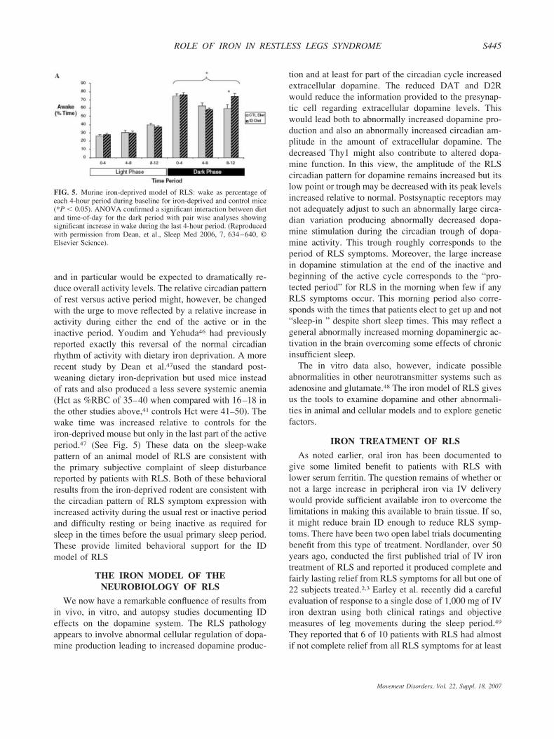
and in particular would be expected to dramatically re-duce overall activity levels. The relative circadian patternof rest versus active period might, however, be changedwith the urge to move reflected by a relative increase inactivity during either the end of the active or in theinactive period. Youdim and Yehuda46 had previouslyreported exactly this reversal of the normal circadianrhythm of activity with dietary iron deprivation. A morerecent study by Dean et al.47used the standard post-weaning dietary iron-deprivation but used mice insteadof rats and also produced a less severe systemic anemia(Hct as %RBC of 35–40 when compared with 16–18 inthe other studies above,41 controls Hct were 41–50). Thewake time was increased relative to controls for theiron-deprived mouse but only in the last part of the activeperiod.47 (See Fig. 5) These data on the sleep-wakepattern of an animal model of RLS are consistent withthe primary subjective complaint of sleep disturbancereported by patients with RLS. Both of these behavioralresults from the iron-deprived rodent are consistent withthe circadian pattern of RLS symptom expression withincreased activity during the usual rest or inactive periodand difficulty resting or being inactive as required forsleep in the times before the usual primary sleep period.These provide limited behavioral support for the IDmodel of RLS
THE IRON MODEL OF THENEUROBIOLOGY OF RLS
We now have a remarkable confluence of results fromin vivo, in vitro, and autopsy studies documenting IDeffects on the dopamine system. The RLS pathologyappears to involve abnormal cellular regulation of dopa-mine production leading to increased dopamine produc-
tion and at least for part of the circadian cycle increasedextracellular dopamine. The reduced DAT and D2Rwould reduce the information provided to the presynap-tic cell regarding extracellular dopamine levels. Thiswould lead both to abnormally increased dopamine pro-duction and also an abnormally increased circadian am-plitude in the amount of extracellular dopamine. Thedecreased Thy1 might also contribute to altered dopa-mine function. In this view, the amplitude of the RLScircadian pattern for dopamine remains increased but itslow point or trough may be decreased with its peak levelsincreased relative to normal. Postsynaptic receptors maynot adequately adjust to such an abnormally large circa-dian variation producing abnormally decreased dopa-mine stimulation during the circadian trough of dopa-mine activity. This trough roughly corresponds to theperiod of RLS symptoms. Moreover, the large increasein dopamine stimulation at the end of the inactive andbeginning of the active cycle corresponds to the “pro-tected period” for RLS in the morning when few if anyRLS symptoms occur. This morning period also corre-sponds with the times that patients elect to get up and not“sleep-in ” despite short sleep times. This may reflect ageneral abnormally increased morning dopaminergic ac-tivation in the brain overcoming some effects of chronicinsufficient sleep.
The in vitro data also, however, indicate possibleabnormalities in other neurotransmitter systems such asadenosine and glutamate.48 The iron model of RLS givesus the tools to examine dopamine and other abnormali-ties in animal and cellular models and to explore geneticfactors.
IRON TREATMENT OF RLS
As noted earlier, oral iron has been documented togive some limited benefit to patients with RLS withlower serum ferritin. The question remains of whether ornot a large increase in peripheral iron via IV deliverywould provide sufficient available iron to overcome thelimitations in making this available to brain tissue. If so,it might reduce brain ID enough to reduce RLS symp-toms. There have been two open label trials documentingbenefit from this type of treatment. Nordlander, over 50years ago, conducted the first published trial of IV irontreatment of RLS and reported it produced complete andfairly lasting relief from RLS symptoms for all but one of22 subjects treated.2,3 Earley et al. recently did a carefulevaluation of response to a single dose of 1,000 mg of IViron dextran using both clinical ratings and objectivemeasures of leg movements during the sleep period.49
They reported that 6 of 10 patients with RLS had almostif not complete relief from all RLS symptoms for at least
FIG. 5. Murine iron-deprived model of RLS: wake as percentage ofeach 4-hour period during baseline for iron-deprived and control mice(*P � 0.05). ANOVA confirmed a significant interaction between dietand time-of-day for the dark period with pair wise analyses showingsignificant increase in wake during the last 4-hour period. (Reproducedwith permission from Dean, et al., Sleep Med 2006, 7, 634–640, ©Elsevier Science).
ROLE OF IRON IN RESTLESS LEGS SYNDROME S445
Movement Disorders, Vol. 22, Suppl. 18, 2007

2 weeks and for most patients the relief lasted longer than2 months. This relief was documented both on subjectivescales and also on the objective measure of leg move-ments. Double-blind placebo-controlled studies are cur-rently being conducted. It is important to recognize thatfailure of IV iron treatment may occur, despite ID if theRLS impairment of the normal modes of brain ironacquisition and retention exceeds the capacity for cor-rection through even a very large increase in peripheralavailability of iron. The severity of the iron managementimpairment may also differ considerably between pa-tients and correction by a large influx of peripheral ironmay be possible only for those with less severe impair-ment who presumably have somewhat less severe symp-toms. It may also be that the iron treatment works betterif the higher levels of iron availability are maintained forseveral hours or even days to permit the brains impairediron management continuous access to a rich supply ofiron. Longer half-life iron formulations may therefore beneeded to produce treatment benefits.
IRON AND OTHER CAUSES OF RLS
A common and complex syndrome like RLS is un-likely to have one cause. Any number of factors otherthan iron insufficiency might produce dopaminergic orother abnormalities that could produce RLS symptoms.Theories abound, but there are only meager data showingactual clinical relations of factors other than iron anddopamine to the occurrence of RLS symptoms. Somedata indicate that RLS occurs more with the use ofSSRIs,50 with some forms of neuropathy51-53 and possi-bly with rheumatoid more than osteoarthritis arthritis54.However, even rheumatoid arthritis involves problemswith iron management and the relation to RLS may besecondary to this iron problem.55 Whether or not theother conditions associated with RLS involve iron prob-lems or interact with iron status of the patient has notbeen evaluated. It, however, seems likely that RLS willhave multiple determinants some of which may not in-volve iron at all. That iron insufficiency probably doesnot cause all of RLS does not reduce its importance asone major cause of RLS that provides us with basicinformation about RLS pathology. It may also interactwith these other determinants of RLS.
The iron abnormalities consistently found in RLSstudies appear likely to be pervasive involving not onlythe SN but probably in various degrees other dopami-nergic and other neurotransmitter systems. (see Fig. 6)The neural areas affected by RLS are similarly likely toinvolve more than one part of the nervous system. De-termining anatomical locations for RLS may tell us lessthan looking at what disrupts the neural function; thus a
strength of the iron model of RLS is that it woulddifferentially affect diverse parts of the brain consistentwith evidence for RLS pathology in several brain areasincluding the tuberoinfundibular,56 A11-spinal,57 thalam-ic,58,59 and nigrostriatal systems.38
SUMMARY: THE ROLE OF IRON IN RLS
We can no longer say the pathophysiology of RLS isunknown. Brain iron and particularly iron in the SN isreduced in RLS, particularly for the early-onset (beforeage 35–45) phenotype that tends to have greater familialoccurrence. Moreover, reducing iron availability to thebrain by any change in peripheral iron increases both thechance of developing RLS and also the severity of RLS.Replacing iron reduces RLS symptoms and in somecases completely corrects the problem. Thus, an abnor-mality in iron management compromising brain ironstatus causes RLS. There are undoubtedly other indepen-dent causes of RLS, but this is a major cause of thedisorder. Other factors may interact with the iron pathol-ogy to either enable or protect from RLS expression, butthe iron pathology remains one that is central to thedisease process in RLS.
The iron model of RLS provides powerful researchtools for assessing the neurobiology of RLS. Since ironinsufficiency causes RLS, its neurobiological effects arelikely to include the ones that cause RLS symptoms. Inparticular, it may cause RLS symptoms through its ef-
FIG. 6. The iron model of RLS. Brain ID creates an abnormality in thedopaminergic system that produces the RLS symptoms. The Brain IDmay also create abnormalities in other neurotransmitter/neuromodula-tor systems such as the opioid, glutamate, histamine, and adenosinewhich may also produce some of the RLS symptoms. This model doesnot limit the effects to any one specific brain region, although theeffects may be more easily detected in some larger and more studiedsystems (e.g. nigrostriatal system for dopamine).
S446 R.P. ALLEN AND C.J. EARLEY
Movement Disorders, Vol. 22, Suppl. 18, 2007
fects on the dopaminergic and other neurotransmittersystems (see Fig. 6). It maybe, however, that only asubset of the biological effects of ID causes RLS, par-ticularly since iron is probably only one of the causes ofRLS. Nonetheless the iron-model should reveal essentialabnormalities causing RLS symptoms. The in vivo ani-mal models of ID also produce some of the behavioralphenotypes of RLS and certainly provide a good biolog-ical model of RLS. Both the in vivo and in vitro modelsof ID reveal dopamine abnormalities that have beenlargely confirmed in autopsy studies of brains from pa-tients with RLS. These critical studies serve to confirmthe iron model of RLS and also provide a biological basisfor the dopaminergic treatment of RLS. Moreover, theID as a biological model of RLS permits evaluatinginvolvement of other neurological systems including butcertainly not limited to adenosine and glutamate. Evalu-ating these in the iron models and also autopsy or clinicalstudies would further confirm the iron model of RLS andmight indicate targets for developing or understandingmechanisms of nondopaminergic treatments for RLS.These studies will likely inform about the neurobiologynot only of RLS but also of RLS related states such as theinteresting brain state of quiet resting, a state normallyrequired before sleep but one that provokes RLS symp-toms. The iron model also provides some alternativeways forward for genetic studies both using in-bredstrains of mice and also possible endophenotypes.
Recognizing the central role of iron pathology in RLS,however, requires some refocusing of our attention. Weneed to study the abnormalities in neurological circuitsand neurotransmitter systems produced by the ID as anindication of the neurobiology of RLS. These importantstudies serve to confirm the role of iron and indicatebiological bases and targets for treatment. But, we alsomust not forget to evaluate the fundamental iron problemof RLS. We need to better determine the iron manage-ment pathology in RLS. Perhaps the iron pathology itselfcan be treated.
REFERENCES
1. Ekbom KA. Restless legs. Stockholm: Ivar Haeggstroms; 1945.2. Nordlander NB. Restless Legs. Br J Phys Med 1954;17:160–162.3. Nordlander NB. Therapy in restless legs. Acta Med Scand 1953;
145:453–457.4. Yasuda T, Nishimura A, Katsuki Y, Tsuji Y. Restless legs syn-
drome treated successfully by kidney transplantation—a case re-port. Clin Transpl 1986;138:138.
5. Winkelmann J, Stautner A, Samtleben W, Trenkwalder C. Long-term course of restless legs syndrome in dialysis patients afterkidney transplantation. M1ov Disord 2002;17:1072–1076.
6. Sloand JA, Shelly MA, Feigin A, Bernstein P, Monk RD. Adouble-blind, placebo-controlled trial of intravenous iron dextrantherapy in patients with ESRD and restless legs syndrome. Am JKidney Dis 2004;43:663–670.
7. Manconi M, Govoni V, De Vito A, et al. Pregnancy as a risk factorfor restless legs syndrome. Sleep Med 2004;5:305–308.
8. O’Keeffe ST, Noel J, Lavan JN. Restless legs syndrome in theelderly. Postgrad Med J 1993;69:701–703.
9. Ekbom KA. Restless legs syndrome after partial gastrectomy.Acute Neurol Scand 1966;42:79–89.
10. Happe S, Tings T, Schettler V, Canelo M, Paulus W, TrenkwalderC. Low-density lipoprotein apheresis and restless legs syndrome.Sleep 2003;26:A335–A336.
11. Tings T, Schettler V, Canelo M, Paulus W, Trenkwalder C. Impactof regular LDL apheresis on the development of restless legssyndrome. Mov Disord 2004;19:1072–1075.
12. Hogl B, Kiechl S, Willeit J, et al. Restless legs syndrome: acommunity-based study of prevalence, severity, and risk factors.Neurology 2005;64:1920–1924.
13. Berger K, von Eckardstein A, Trenkwalder C, Rothdach A, JunkerR, Weiland SK. Iron metabolism and the risk of restless legssyndrome in an elderly general population—the MEMO-Study.J Neurol 2002;249:1195–1199.
14. Alvarez-Ossorio L, Kirchner H, Kluter H, Schlenke P. Low ferritinlevels indicate the need for iron supplementation: strategy to min-imize iron-depletion in regular blood donors. Transfus Med 2000;10:107–112.
15. Guyatt GH, Patterson C, Ali M, et al. Diagnosis of iron-deficiencyanemia in the elderly. Am J Med 1990;88:205–209.
16. Means RT, Jr, Allen J, Sears DA, Schuster SJ. Serum solubletransferrin receptor and the prediction of marrow aspirate ironresults in a heterogeneous group of patients. Clin Lab Haematol1999;21:161–167.
17. Joosten E, Van Loon R, Billen J, Blanckaert N, Fabri R, PelemansW. Serum transferrin receptor in the evaluation of the iron status inelderly hospitalized patients with anemia. Am J Hematol 2002;69:1–6.
18. Silber MH, Ehrenberg BL, Allen RP, et al. An algorithm for themanagement of restless legs syndrome. Mayo Clin Proc 2004;79:916–922.
19. O’Keeffe ST, Gavin K, Lavan JN. Iron status and restless legssyndrome in the elderly. Age Ageing 1994;23:200–203.
20. Sun ER, Chen CA, Ho G, Earley CJ, Allen RP. Iron and therestless legs syndrome. Sleep 1998;21:371–377.
21. Wang Y, Mysliviec V, Fischer C, Dehaan P, Owshalipur D,Mysliviec A. Efficacy of iron in patients with restless legs syn-drome and a low-normal ferritin: a randomized, double-blind,placebo controlled study. In: Association of professional sleepsocieties, Sleep 2006, Salt lake City, Utah, USA, 2006. p A273.
22. Davis BJ, Rajput A, Rajput ML, Aul EA, Eichhorn GR. A ran-domized, double-blind placebo-controlled trial of iron in restlesslegs syndrome. Eur Neurol 2000;43:70–75.
23. Montplaisir J, Godbout R, Poirier G, Bedard MA. Restless legssyndrome and periodic movements in sleep: physiopathology andtreatment with L-dopa. Clin Neuropharmacol 1986;9:456–463.
24. Akpinar S. Treatment of restless legs syndrome with levodopa plusbenserazide [letter]. Arch Neurol 1982;39:739.
25. Walters AS, Ondo WG, Dreykluft T, Grunstein R, Lee D, Sethi K.Ropinirole is effective in the treatment of restless legs syndrome.TREAT RLS 2: a 12-week, double-blind, randomized, parallel-group, placebo-controlled study. Mov Disord 2004;19:1414–1423.
26. Earley CJ, Yaffee JB, Allen RP. Randomized, double-blind, pla-cebo-controlled trial of pergolide in restless legs syndrome. Neu-rology 1998;51:1599–1602.
27. Winkelmann J, Schadrack J, Wetter TC, Zieglgansberger W, Tren-kwalder C. Opioid and dopamine antagonist drug challenges inuntreated restless legs syndrome patients. Sleep Med 2001;2:57–61.
28. Bartzokis G, Beckson M, Hance DB, Marx P, Foster JA, MarderSR. MR evaluation of age-related increase of brain iron in youngadult and older normal males. Magn Reson Imaging 1997;15:29–35.
ROLE OF IRON IN RESTLESS LEGS SYNDROME S447
Movement Disorders, Vol. 22, Suppl. 18, 2007

29. Ma J, Wehrli FE. Method for image-based measurement of thereversible and irreversible contribution to the transverse relaxationrate. J Magn Reson B 1996;111:61–69.
30. Ordidge RJ, Gorell JM, Deniau JC, Knight RA, Helpern JA.Assesment of relative brain iron concentrations using T2-weightedand T2
*- weighted MRI at 3 Tesla. Magna Reson Med 1994;32:335–341.
31. Hikita T, Abe K, Sakoda S, Tanaka H, Murase K, Fujita N.Determination of transverse relaxation rate for estimating irondeposits in central nervous system. Neurosci Res 2005;51:67–71.
32. Earley CJ, Barker PB, Horska A, Allen RP. MRI-determinedregional brain iron concentrations in early- and late-onset restlesslegs syndrome. Sleep Med 2006;7:459–461.
33. Allen RP, Barker PB, Wehrl F, Song HK, Earley CJ. MRI mea-surement of brain iron in patients with restless legs syndrome.Neurology 2001;56:263–265.
34. Schmidauer C, Sojer M, Seppi K, et al. Transcranial ultrasoundshows nigral hypoechogenicity in restless legs syndrome. AnnNeurol 2005;58:630–634.
35. Mizuno S, Mihara T, Miyaoka T, Inagaki T, Horiguchi J. CSF iron,ferritin and transferrin levels in restless legs syndrome. J Sleep Res2005;14:43–47.
36. Earley CJ, Connor JR, Beard JL, Malecki EA, Epstein DK, AllenRP. Abnormalities in CSF concentrations of ferritin and transferrinin restless legs syndrome. Neurology 2000;54:1698–1700.
37. Earley CJ, Connor JR, Beard JL, Clardy SL, Allen RP. Ferritinlevels in the cerebrospinal fluid and restless legs syndrome: effectsof different clinical phenotypes. Sleep 2005;28:1069–1075.
38. Connor JR, Boyer PJ, Menzies SL, Dellinger B, Allen RP, EarleyCJ. Neuropathological examination suggests impaired brain ironacquisition in restless legs syndrome. Neurology 2003;61:304–309.
39. Connor JR, Wang XS, Patton SM, et al. Decreased transferrinreceptor expression by neuromelanin cells in restless legs syn-drome. Neurology 2004;62:1563–1567.
40. Chen Q, Connor JR, Beard JL. Brain iron, transferrin and ferritinconcentrations are altered in developing iron-deficient rats. J Nutr1995;125:1529–1535.
41. Erikson KM, Jones BC, Hess EJ, Zhang Q, Beard JL. Iron defi-ciency decreases dopamine D1 and D2 receptors in rat brain.Pharmacol Biochem Behav 2001;69:409–418.
42. Erikson KM, Jones BC, Beard JL. Iron deficiency alters dopaminetransporter functioning in rat striatum. J Nutr 2000;130:2831–2837.
43. Connor J, Wang X, Allen R, et al. The dopaminergic profile in theputamen and substantia nigra in restless leg syndrome. In: WorldAssociation of Sleep Medicine 2007, Bangkok, Thailand, 2007.
44. Wang X, Allen RP, Earley CJ, Beard JL, Connor JR. Altereddopaminergic expression in restless leg syndrome. In: Society forNeuroscience Annual Meeting, Washington, DC, 2005.
45. Wang X, Wiesinger J, Beard J, et al. Thy1 expressionin the brainis affected by iron and is decreased in restless legs syndrome.J Neurol Sci 2004;220:59–66.
46. Youdim M, Yehuda S. Iron deficiency induces reversal of dopa-mine dependent circadian cycles: differential response to d-am-phetamine and TRH. Peptides 1985;6:851–855.
47. Dean T, Jr, Allen RP, O’Donnell C P, Earley CJ. The effects ofdietary iron deprivation on murine circadian sleep architecture.Sleep Med 2006;7:634–640.
48. Gulyani S, Gleichmann M, Martin B, Mattson M, Allen R, EarleyC. The effects of low iron on adenosine and dopamine receptors.In: World Association of Sleep Medicine 2007, Bangkok, Thai-land, 2007.
49. Earley CJ, Heckler D, Allen RP. IV Iron treatment for the restlesslegs syndrome (RLS). Sleep 2001;24:A359.
50. Ohayon MM, Roth T. Prevalence of restless legs syndrome andperiodic limb movement disorder in the general population. J Psy-chosom Res 2002;53:547–554.
51. Gemignani F, Marbini A, Di Giovanni G, et al. Cryoglobulinaemicneuropathy manifesting with restless legs syndrome. J Neurol Sci1997;152:218–223.
52. Iannaccone S, Zucconi M, Marchettini P, et al. Evidence of pe-ripheral axonal neuropathy in primary restless legs syndrome. MovDisord 1995;10:2–9.
53. Schols L, Haan J, Riess O, Amoiridis G, Przuntek H. Sleepdisturbance in spinocerebellar ataxias: is the SCA3 mutation acause of restless legs syndrome? Neurology 1998;51:1603–1607.
54. Salih AM, Gray RE, Mills KR, Webley M. A clinical, serologicaland neurophysiological study of restless legs syndrome in rheu-matoid arthritis. Br J Rheumatol 1994;33:60–63.
55. Gyorfi M, Szakacs Z, Koumlves P. Restless legs syndrome andserum transferrin receptor and ferritin levels in patients with rheu-matoid arthritis. Sleep 2003;26:A334.
56. Garcia-Borreguero D, Larrosa O, Granizo JJ, de la Llave Y,Hening WA. Circadian variation in neuroendocrine response toL-dopa in patients with restless legs syndrome. Sleep 2004;27:669–673.
57. Clemens S, Rye D, Hochman S. Restless legs syndrome: revisitingthe dopamine hypothesis from the spinal cord perspective. Neu-rology 2006;67:125–130.
58. Bucher S, Seelos K, Oertel W, Reiser M, Trenkwalder C. Cerebralgenerators involved in the pathogenesis of the restless legs syn-drome. Ann Neurol 1997;41:639–645.
59. Etgen T, Draganski B, Ilg C, et al. Bilateral thalamic gray matterchanges in patients with restless legs syndrome. Neuroimage 2005;24:1242–1247.
S448 R.P. ALLEN AND C.J. EARLEY
Movement Disorders, Vol. 22, Suppl. 18, 2007


![C{DROME, - The Podiatry Institute · CHAPTER IO RESTLESS LEGS S\C{DROME, Robert M. Goecker, DPM DEFINITION AND CLINICAL FEATT]RES The restless legs syndrome (RLS) is a neuroiogic](https://img.dokumen.tips/doc/110x75/5e8041ac41545c5d275ae185/cdrome-the-podiatry-chapter-io-restless-legs-scdrome-robert-m-goecker-dpm.jpg)