Embed Size (px)
Citation preview

1
The Role of Alpha-Synuclein in Parkinson’s Disease
Manpreet Kaur Bassi
Copyright May 2014 by Manpreet Kaur Bassi and Koni Stone
Parkinson's disease (PD) is a slowly progressing neurodegenerative disorder that effects
the daily movements and coordination of a person. Currently, there are about 6.3 million people
affected worldwide and 1 million people in the United States [1]. PD belongs to the motor
system disorders that result from loss of dopamine-producing brain cells. Dopamine is a
neurotransmitter which plays an important role in movement, coordination, sleep, behavior,
mood, and pleasure. The most common feature of PD is the extreme slowness in the movements
and reflexes known as the bradykinesia. In addition to motor symptoms such as resting tremor,
inflexibility, and postural instability, there are other nonmotor symptoms experienced by a
patient include depression, rapid eye movement sleep behavior disorder, and constipation [2].
Parkinson's disease patients are found to have high level of lewy bodies in the substantia nigra (a
part of brain involved in movement, reward, and addiction). Lewy bodies are formed by
abnormal aggregation of proteins in the nerve cells of substantia nigra. Alpha-synuclein has been
found to be a major component of Lewy bodies in the PD patients. Thus, the focus of this paper
is to understand the structure and role of the alpha-synuclein in the neuronal cells involved in
Parkinson’s disease patient.
Parkinson's disease has mostly been found in people older than 60 years of age. As the
person ages, the chance of having PD increases. However, age is not the only factor involved in
having PD symptoms. About 5-10% cases of PD are inherited from parents through gene
alternations. PD can also be sporadic with no known history of PD in the family. It has been

2
really hard to pinpoint for specific causes for symptoms of PD in a person. PD is not limited to
genetics and age, it is also influenced by the environment a person lives in. Parkinson's disease is
a multifactorial disease which means it is the result of genetic and environmental factors
interacting with each other. The primary cause for the motor symptoms has been found to be the
decreasing number or absence of dopaminergic (DA) neurons within the substantia nigra (SN).
One of the reasons for this loss of SN neurons has been linked to the overexpression of alpha-
synuclein which further leads to oligmerization, fibrillation, and aggregation of this protein in the
SN neurons. The aggregated alpha-synuclein interferes with the normal function of SN neurons
and causes PD-linked symptoms [3].
Alpha-synuclein, a presynaptic neural protein, is a product of 140 amino acid sequence
lacking both tryptophan and cysteine amino acids. Human alpha-synuclein of 140 amino acid
sequence consists of three different regions. The amino acid sequence region from the first
amino acid to 60th amino acid is called N-terminal amphipathic region. This region can form
alpha chains and interact with the surface membranes of other cells, neurotransmitter vesicles,
and micelles. The region from 61th amino acid sequence to 95th is known as central hydrophobic
non-amyloid region. The last region from 96th amino acid to 140th amino acid is called C-
terminal acidic part which is highly enriched in prolines and acidic residues such as serine and
tryrosine and can be phosphorylated [3].
It had been understood in the past that alpha-synuclein only exists in monomer form;
however, this is not an accepted fact anymore. Wang and Chittulure et.al determined that in the
absence of other lipids, and micelles, the alpha-synuclein forms a tetramer structure. This
tetramer structure made up of a four-helix bundle did not aggregate under normal conditions and
was not toxic to the membranes of other cells. Only when alpha-synuclein was isolated by using
3
denaturing conditions, it lacked a stable structure and formed aggregation. In order to carry out
the experiment, Wang et al constructed an expression vector in E.coli where N-terminal GST-
tagged alpha-synuclein protein was expressed. To prevent any inclusion body formation, the
expression of GST-tagged protein was carried out at 20oC. After the extraction and purification
of protein by GST affinity chromatography, the N-terminal tag was removed by protease. The
further purification was done by size-exclusion chromatography. The protein was used to find
the normal structure of quaternary structure of alpha synuclein. Later the normal structure was
compared to structure denatured by detergent, concentration, heat, and disease-associated
mutations. To find the normal structure, first SDS/PAGE gel was run loaded with a chemical
cross-linked sample of purified protein. There were four bands seen on the gel corresponding to
the presence of tetramer structure of protein. MALDI-TOF mass spectrometry was used to
identify the bands of tetramer of alpha-synuclein. Further analysis on the electron microscopy
revealed the protein was homotetramer oligomer. By analyzing two negative bands at 222nm and
208nm and one positive band at 193nm on CD spectra charactized the presence of 65% alpha-
helix, 17% turns, and 8% unfolded. To detect if there was any hydrophobic core present in this
structure, a ThermoFluor assay was performed. It was concluded from both CD spectra and a
ThermoFluor assay that hydrophobic interactions were used to hold the subunits together [4].
The protein sample was then boiled at 95oC and a precipitation formed. The precipitated
sample did dissolved when mixed; however, the structure of the protein was disordered after
boiling. This disordered structure began to aggregate on day 4. Then, the sample was tested
against concentration and it was found that alpha-synuclein protein was more in the monomer
form than oligomer when low concentration was present. However, below 0.5mg/ml
concentration, the protein was most disordered on the CD spectra. The interesting point that was

4
seen in the concentration test was that in normal and high concentrations, the tetrameric form of
protein was not toxic to neuronal tissue and did not cause any pores in the membranes. The
results from both boiling sample test and concentration test concluded that in order to form
aggregation, the protein has to go through shape changes that makes it more prone to fibrillation
and aggregation. In disordered form, the protein was toxic at high levels and formed pores in the
membranes. Mutated proteins were also analyzed and it was found out that mutated proteins
were more disordered than tetrameric ordered form. Overall, the study showed that alpha-
synuclein existed in ordered tetrameric form in the absence of lipids bilayer or micelles. It was
concluded in vitro, and probably in vivo, there was equilibrium between monomer, tetramer
oligomer, and fibrilled proteins. This equilibrium can be shifted to either side by changing
conditions such as genetic mutations, age, environmental conditions, and intracellular processes.
When a high concentration of alpha-synuclein is present, it appeared in the experiment that less
helical structure was favored, which over time led to aggregated and amyloid fibrils product.
Being able to extract ordered tetrameric form that was resistant to aggregation has provided hope
for the possibility of using this protein structure in future to treat PD [4].
The normal function of alpha-synuclein is still under investigation. However, it has been
found that alpha synuclein helps in recycling synaptic vesicles and releasing neurotransmitters
such as dopamine. The N-terminal portion of the alpha-synuclein interacts with SNAREs
complex and helps control the exocytosis of the dopamine. The exocytosis of dopamine is
regulated by alpha-synuclein which helps in regulating SNARE's complex, vesicle pool and
dopamine reserve. In a study done by Murphy et al, the researchers showed how alpha-synuclein
regulated the size of the presynaptic vesicular pool of hippocampal neurons. First, hippocampal
neuronal cell culture was prepared from rat brain. The culture was used to prepare coverslip for
5
electron microscopy and to make tissue wells for Western blot analysis. Then cells were
incubated with different antibodies for immunocytochemistry analysis. The cells were incubated
with Syn102 antibody which recognized alpha and beta synuclein. The second antibody that was
used was SNL-1 which recognized alpha-synuclein only. The third antibody syn207 was specific
for beta-synuclein protein. To image cells, the cells were first washed and incubated with
secondary antibodies. A Laser-scanning microscope was used to image the cells. It was found
that both alpha synuclein and beta synuclein were found to be expressed exclusively at the
presynapse once the presynaptic neuronal terminals were mature. Thus, the alpha and beta
synuclein are proteins associated with presynapse. The Western Blot analysis was also done on
the cell culture. The western blot procedures revealed that alpha-synuclein level decreased over
the 3 weeks where as the level of beta-synuclein was unchanged when the neuronal cells were
maturing. The alpha synuclein was found to be 100% at first week, then 75% and about 60% on
the week 3 when neurons presynaptic terminal was matured. However, alpha-synuclein was fully
expressed only when the presynaptic terminals were fully matured. Thus, this late start
expression in neurons even though the level of alpha-synuclein was found to higher at the start of
the week 1 suggested that the alpha-synuclein protein might be important for later maintance of
neurons not the initial formation [5].
The hippocampal cell culture was also treated with antisense oligonucleotides that were
specific for alpha-synuclein region. The goal was to turn off the expression of synuclein by
antisense oligonucleotides and to see what happened to the cell expression. Also, a control cell
culture was set up which did not have antisense oligonucleotides rather had a sense nucleotide.
Both cell cultures were then analyzed by electron microscopy. The researchers found that the
expression of beta-synuclein stayed unchanged in cell culture incubated with antisense
6
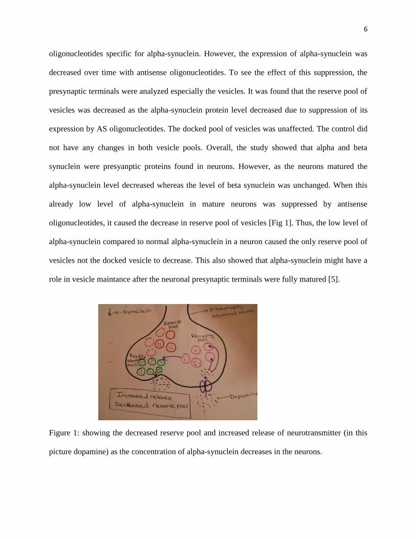
oligonucleotides specific for alpha-synuclein. However, the expression of alpha-synuclein was
decreased over time with antisense oligonucleotides. To see the effect of this suppression, the
presynaptic terminals were analyzed especially the vesicles. It was found that the reserve pool of
vesicles was decreased as the alpha-synuclein protein level decreased due to suppression of its
expression by AS oligonucleotides. The docked pool of vesicles was unaffected. The control did
not have any changes in both vesicle pools. Overall, the study showed that alpha and beta
synuclein were presyanptic proteins found in neurons. However, as the neurons matured the
alpha-synuclein level decreased whereas the level of beta synuclein was unchanged. When this
already low level of alpha-synuclein in mature neurons was suppressed by antisense
oligonucleotides, it caused the decrease in reserve pool of vesicles [Fig 1]. Thus, the low level of
alpha-synuclein compared to normal alpha-synuclein in a neuron caused the only reserve pool of
vesicles not the docked vesicle to decrease. This also showed that alpha-synuclein might have a
role in vesicle maintance after the neuronal presynaptic terminals were fully matured [5].
Figure 1: showing the decreased reserve pool and increased release of neurotransmitter (in this
picture dopamine) as the concentration of alpha-synuclein decreases in the neurons.

7
It was now known that low level of alpha-synuclein causes decrease in the reserve pool;
however, more researched was needed to see if the high level of alpha-synuclein had any effects.
A study done by Nermani et al. showed that elevated level of alpha-synuclein decreased the size
of the recycling pool of vesicles in neurons while interfering with the process of exocytosis and
enodcytosis. This overexpression of alpha-synuclein has been seen to cause considerable toxicity
in the cells. This study used neuronal cells with vesicles transfected with vesicular glutamate
transporter 1-pHluroin to directly observe the effects of overexpressed (more protein being
formed) alpha-synuclein on the cells.VGLUT1-pHluroin fluoresces when exposed to alkaline
solution. However, it fluoresced less when exposed to acidic solution. This change in pH was
really important in observing exocytosis of neural vesicles. When the vesicles were released,
VGLUT1-pHluorin came in contact with the extracellular alkaline environment and it fluoresced.
However, when the neurons were in resting state with no exocytosis of vesicles, VGLUT1-
pHluorin did not fluoresce. In order to relate this to human cells, the researchers used dopamine
human cells, and transfected them with VGLUT1-pHluroin. The change in fluorescence was
observed in normal concentration alpha-synuclein versus elevated concentration of alpha-
synuclein in dopamine neurons. When alpha-synuclein was in high amount, the VGLUT1-
pHluroin did not or little fluoresced. However, under normal concentration, the VGLUT1-
pHluroin did fluoresce. This showed that over expression of alpha-synuclein interfered with the
vesicle exocytosis and inhibited the dopamine releasing vesicles. Thus, the over-expression
caused the reduced release of dopamine neurons by interfering with exocytosis. Another
experiment was done to see what effect the over-expression had on the process of endocytosis.
Following endocytosis, the reclustering of the vesicles was also slowed in the presence of over
expressed alpha-synuclein protein. Since the vesicles could not be reclustered at a normal rate,

8
there was a decrease in the recycle pool of the vesicle. To observe this in lab, the researchers
measure the uptake of styryl dye by the neurons expressing normal and elevated level of alpha
synuclein. Styryl dyes are organic molecules with fluorescent properties and are used in the field
of neurobiology as a means of labeling recycling vesicles. The cells were given stimulant and
allowed to uptake the dye. The cells were allowed to remain in the dye solution to have a full
endocytosis process. The neurons were given another stimulant to unload the dye. The cells were
allowed to uptake the dye again. The second stimulant for unloading was used to measure the
size of the recycling pool of the vesicles. The cells expressing elevated level of alpha-synuclein
showed 50% decrease in the uptake of the dye compare to a normal cells. The rate which
endocytosis happened was not changed in both cells containing elevated and normal level of
alpha-synuclein. It was the vesicle size that was reduced and lower amount of dye was uptakened
by the cells due to high level of alpha-synuclein [Fig 2]. Thus, the high level of alpha-synuclein
not only interfered with the exocytosis process where the vesicles were not able to fuse at a
normal rate and release the contents outside but also interfered with the endocytosis process. In
endocytosis, the size of the recycle vesicle was reduces leading to lower amount of substance
uptake [Fig 2]. Overall, the study showed that alpha synuclein plays a major role in the uptake
and release of neurotransmitter [6].
9
Figure 2: showing the decreased recycling pool size and decreased release of neurotransmitter (in
this picture dopamine) as the concentration of alpha-synuclein increases in the neurons.
The experiments had shown that elevated levels of alpha-synuclein interfered with the
process of exocytosis by reducing the release of dopamine or endocytosis in which the recycling
vesicle pool size was reduced. However, there was not any study done until 2012 at the
molecular level to show how alpha-synuclein interacts with the membrane of the neurons and
interferes with their normal functions. A study done by Bong-Choi et al showed that large alpha-
synuclein oligmers interfered with SNARE complex during exocytosis and release of dopamine.
In order to obtain large oligomers, alpha-synuclein was incubated with 10-fold molar excess of
dopamine. After the incubation of alpha-synuclein, the solution was run on the SDS/PAGE to get
different bands. One of the bands that was produced belonged to the high molecular weight of
alpha synuclein oligomer. Oligomers were only produced in the presence of dopamine. In the
absence of dopamine, there were no oligomers present in the solution. Thus, high amount of
dopamine causes high molecular weight oligomers formation. The size exclusion
chromatography was used to separate the high molecular weight oligomer from the other species.
The weight of the oligomer was around 250kDa. In vitro lipid-mixing assay, the researchers
tested whether the alpha-synuclein has any effect on the SNARE-mediated lipid mixing by using
proteoliposomes. In order to carry out the lipid-mixing assay, two populations of
proteoliposomes were created. The t-SAREs constituted the t-vesicle while the v-SNARE
synaptobrevin-2 constituted the v-vesicle. Under normal conditions, when these two populations
10
of proteoliposomes were mixed, the vesicles fused together to form one vesicle. The fusion of
vesicle was monitored by FRET signal. FRET also known as the fluorescence resonance energy
transfer is used in the protein-protein interactions and in this study for vesicle-vesicle
interactions. When there was fusion of the vesicles, the signal was increased. However, when
these two proteoliposomes v-vesicle and t-vesicles were allowed to fuse in the presence of
30mM concentration of large alpha-synuclein oligomers, the fusion rate decreased to 75% with
25% of inhibition. The concentration of oligomers was given in monomer units. The same
concentration 30nM but now in the monomer form of alpha-synuclein had no effect on the
vesicle fusion. Thus, it was concluded that high molecular weight alpha-synuclein oligomers
interfered with the SNARE-mediated lipid mixing and the fusion of vesicles was inhibited [7].
To understand the mechanism behind the inhibition of SNARE-mediated lipid mixing,
more experiments were carried out by the researchers. The researchers tried to test three known
modes of interactions between alpha-synuclein and SNARE proteoliposomes to see which
interaction was common in large alpha-synuclein oligomers and SNARE proteoliposomes. The
first mode of interaction could be large alpha-synuclein oligomers interacting with a negatively
charged membrane of the vesicles. To test this hypothesis, the researchers incubated a large
amount of alpha-synuclein oligomers with protein free liposome. They stated if the oligomers did
interact with the membrane of vesicles, then there would be a decrease in the amount of free-
floating large alpha-synuclein oligomers and there was a chance of inhibition of vesicle mixing.
However, this inhibition could be overcome by addition of large amount of protein free
liposome. Addition of protein free liposome had no effect on the 50% inhibition. Even when
large amount of alpha-synuclein oligomers were added, the inhibition still stayed at 50%. The
researchers concluded that alpha-synuclein oligomers did not bind to the protein free liposome or

11
phospholipids of vesicle membrane. The second mode of interaction where oligomers caused
leakage of membranes was tested. In order to test this hypothesis, the researchers used the
sulforhodamine B (SRB) dequenching method. The vesicles were prepared from addition of
20nM of SRB to lipid film. These vesicles with concentration 10uM were incubated with 170nM
of large alpha-synuclein oligomers. To observe the dequenching of SRB ( result of membrane
disruption), SRB fluorescent emissions were monitored. There was not any noticeable increase in
the fluorescent emissions leading to the conclusion that the large oligomers of alpha-synuclein
did not cause leakage of membranes of vesicles. The last mode of interaction was that large
oligomers of alpha-synuclein actually bind to the v-SNARE synaptobrevin-2 one of the SNARE
proteins. To test this hypothesis, the researchers used coflotation assay. This assay helped to
observe the interactions between large alpha-synuclein oligomers and v-vesicle synaptobrevin-2.
The researchers obtained large alpha-oligomers bound to t-vesicles and v-vesicle. The western
blot was used to quantify the amount of alpha-oligmers bound to each vesicle. The v-vesicle had
higher amount of bounded oligomers than t-vesicles and protein free liposomes. The researchers
concluded the large oligomers of alpha-synuclein had preferential binding for v-vesicle over
other vesicles. Thus, in the exocytosis process SNAREs complex, the oligomers interacted with
the v-vesicles and caused the inhibition of formation of SNAREs complex and release of
dopamine vesicles [7].
The mode of interaction was determined; however, it was still unclear how the large
oligomer of alpha-synuclein interacted with the v-SNARE synaptobrevin-2. To determine the
mechanism behind this interaction, the researchers used mutant synpatobrevin-2 without any N-
terminal region. Their hypothesis was that if the large oligomer used N-terminal region of
synaptobrevin-2 to bind to it, then the large oligomer will no longer to bind to mutant
12
synaptobrevin-2 lacking N-terminal region. Their hypothesis was supported by the coflotation
assay which showed that oligomers had lower binding affinity for mutant synaptobrevin-2
lacking N-terminal than v-SNARE synaptobrevin-2 with N-terminal region. It was now known
that oligomers did not bind to mutant synaptobrevin-2 lacking N-terminal region, next step was
to see if the low affinity for binding also lowered the inhibitory effect of oligomers on the lipid
mixing. The paper does not give any specific method for this procedure and only stated that there
was no inhibitory effect of oligomers seen in the presence of synpatobrevin-2 lacking N-terminal
region. Thus, the researchers concluded that large oligomers of alpha-synuclein specifically bind
to N-terminal region of v-SNAREs synaptobrevin-2 to inhibit the lipid mixing in a cell [7].
The next question put up by the researchers was exactly how oligomers' binding to N-
terminal region of synaptobrevin-2 led to inhibition of lipid mixing. Their hypothesis was that
oligomers inhibit the SNAREs complex which led to vesicle reduced docking. Docking in the
molecular biology means the orientation of one substance to another which then leads to the
product. In order to relate to this concept to this situation, the orientation of v-SNAREs with t-
SNAREs was called docking and the lipid mixing was called the product. A single-vesicle assay
known as ALEX used to observe the docking and lipid-mixing. ALEX used different color for
docking, lipid-mixing steps, and unreacted t-and v-vesicles. The vesicles were incubated with the
large oligomers of alpha-synuclein for 30 minutes at 37oC. To use a control, the vesicles were
also incubated with alpha-synuclein monomer at the same conditions. It was seen that in the
presence of oligomers, the lipid-mixed vesicles were reduced and the exocytosis was decreased.
The vesicles were not able to mix with the lipid bilayer. If the lipid-mixed vesicles were low and
it was due to lipid-mixing step at the end, then there should be increase in the vesicles of the
docking step. However, the subpopulation of docking step vesicles did not change. This means

13
the oligomers inhibit the initial SNAREs complex formation of t- and v-SNAREs synaptobrevin-
2 at the docking step where v-and t-vesicle come together which will later allow the lipid layers
to mix. Overall, this study showed that the role of large oligomers of alpha-synuclein in
exocytosis process which can be formed when there was high concentration of dopamine or
mutated alpha-synuclein or other environment cause was present. The large oligomers interact
with the N-terminal region of v-vesicle snyaptobrevin2 and inhibit the initial SNAREs complex
formation with the t-vesicle. Since the SNAREs complex cannot be formed, the membranes
cannot fuse to release the substance outside [Fig 3]. Similar results were seen in vivo, where
large alpha-synuclein oligomers inhibited the exocytosis in PC12 cells. This study provided very
important insights in understanding the role of alpha-synuclein in the cells [7].
Figure 3: showing the general process of monomer alpha-synuclein forming into large alpha-
synuclein oligomers in the presence of high amount of dopamine. These large alpha-synuclein
oligomers then go bind to v-SNAREs synaptobrevin-2. The v-SNAREs can no longer bind to t-
SNARES. Since the v-SNAREs and t-SNAREs cannot interact, the fusion of two membranes
cannot be accomplished leading to inhibition of exocytosis.
14
The aggregation of alpha-synuclein in the neurons of substantia nigra in human leads to
reduce exocytosis of dopamine release. When there is high level of dopamine as seen in the past
experiments, the more alpha-synuclein oligomers will be formed leading to overstressed cell
with no exocytosis process. The overstressed cells will eventually die leading to neuronal death.
The reduced released amount of dopamine leads to movement disorders, depression, and other
motor disorders that require the dopamine to function properly. There is still more research being
done around the world to see other ways alpha-synuclein can cause the disease. It is a very
elusive process because there is more than one factor contributing to neuronal cell death.
Although alpha-synuclein has been found to be a major component of lewy bodies, there are still
other proteins that might interact and cause the cell death. Thus, more research needs to be done
to fully understand the cause behind the neuronal cell seen in PD. Due to these difficulties, there
is still no cure for Parkinson's disease. However, there are some treatments available to reduce
the aggregation of proteins or death of the neurons. These treatments are not effective to inhibit
the aggregation of proteins; thus, patient still has Parkinson's disease and symptoms associated it.
The current treatment involves medications such as substituting dopamine and therapy such as
deep brain stimulation to suppress pathological neuronal oscillations.
There are numerous studies being done to find a cure for the Parkinson's disease.
One focus of these studies is to slow or inhibit the fibrillation or aggregation of alpha-synuclein.
Aggregated alpha-synuclein causes inhibition of exocytosis and leading to high level of
dopamine in the presyanptic neuronal cells. A study done by Hyun Koo et al., the researchers
tested whether the self-fibrillation-defective α-synuclein mutants were able to prevent the
polymerization of wild-type and/or PD-linked α-synuclein variants. The cDNA that coded for
mutant self-fibrillation defective alpha-synuclein proteins was cloned into E.coli plasmid. The

15
plasmid was introduced into the E.coli and was allowed to replicate the cDNA and produce
mutant alpha-synuclein produce. The mutant protein was extracted from the cells by lysis of the
cell and then purification of the supernatant that contained the protein. The purification of the
mutant alpha-synuclein was done by anion exchange column chromatography and later on the
SDS-polyacrylamide gel. After getting purified mutant alpha-synuclein, it was used to test the
inhibition of fibrillation of the wild-type and PD-linked alpha-synuclein protein (A30P, A53T,
and E46K). These all four alpha-synuclein proteins were incubated with or without the mutant
self-fibrillation defective protein in PBS buffer. The absence of mutant alpha-synuclein was used
as a control. At certain points, the 20ul of each of the samples was taken and mixed with
thioflavin T solution to find the degree of fibril formation. Thioflavin T is a benzothiazole salt
and used as a dye to visualize and quantify the presence of misfolded protein aggregates called
amyloid When it binds to amyloid aggregates, the dye displays enhanced fluorescence.
Fluorescence emission was measured for each of the sample. As the solubility of the protein
decreased, more fluorescence color would be seen [8]. Thus, more amyloid aggregates were
present.
When the concentrations of both wild-type alpha-synuclein and PD-linked alpha-
synuclein were decreased, the time for fibrillation of proteins was increased. Thus, in the absence
of mutant self-fibrillation defective alpha-synuclein protein, the time for fibrillation is
concentration dependent. The wild-type alpha-synuclein in the presence of mutant self-
fibrillation defective alpha-synuclein protein, the fibrillation of the wild-type protein was
inhibited and almost no fibrillation was recorded by fluorescence emission. Also, all PD-linked
alpha-synuclein in the presence of mutant self-fibrillation defective alpha-synuclein protein had
slow or no fibrillation seen. [8]. Thus, the method used in this study to slow or inhibit the

16
fibrillation can serve as a therapeutic method in the future to treat Parkinson’s disease or other
disease caused by aggregation of alpha-synuclein.
In conclusion, Parkinson’s disease is the second most common neurodegenerative disease
and affects many people worldwide. It is very important in today’s world to know the causes of
Parkinson’s disease and find cure for this disease. The most studied cause for PD is the
aggregation of alpha-synuclein which is found to be a major component of lewy bodies. This
protein is found most at the presynaptic terminals of brain cells. By understanding the
mechanism involved behind the aggregated alpha-synuclein in neurodegeneration can also
greatly help with other diseases such as neurodegenerative synucleinopathies. As of this moment,
alpha-synuclein protein is found to be involved in the maintance of neurons. However, when
expressed in high concentrations, it can greatly reduce the neurotransmitter release by inhibiting
the SNAREs complex involved in the exocytosis. It not only inhibits the SNAREs complex but
decrease the recycling vesicle pool size. This leads to lower uptake of neurotransmitter at the
synapse. There are many studies being done to find cure such as the inhibition of aggregation or
fibrillation of alpha-synuclein. With new growing technology and research methods, it might be
possible to find cure in the future and help all the people suffering from this disease.

17
Work Cited
[1] "Who Is Affected by Parkinson's Disease." WebMD. WebMD, 02, Jan. 001. Web. 02 Feb.
2014. <http://www.webmd.com/parkinsons-disease/who-is-affected-by-parkinson-disease>.
[2] Anthony, Paul MA, Nico J Diederich, Rejko Kruger, and Rudi Balling. " The Hallmarks of
Parkinson's Disease. "The FEBS Journal (2013), 280:23, 5981-5993.
[3] Leonid Breydo, Jessica W. Wu, Vladimir N. Uversky, α-Synuclein misfolding and
Parkinson's disease, Biochimica et Biophysica Acta (BBA) - Molecular Basis of Disease, 1822:2,
February 2012, Pages 261-285
(http://www.sciencedirect.com/science/article/pii/S0925443911002250)
[4]. Wang, Wei, et al. "A Soluble α-synuclein Construct Forms a Dynamic Tetramer."
Proceedings of the National Academy of Sciences of the United States of America 108.43
(2011): 17797-7802. JSTOR. Web. 18 Apr. 2014.
<http://www.jstor.org.ezproxy.lib.csustan.edu:2048/stable/10.2307/41352597?ref=search-
gateway:cc95753bb5b390f6d19a58df7f3b6102>.
[5]. Murphy, Diane D., Susan M. Rueter, John Q. Trojanowski, and Virginia M. Lee.
"Synucleins Are Developmentally Expressed, and Alpha-synuclein Regulates the Size of the
Presynaptic Vesicular Pool in Primary Hippocampal Neurons." The Journal of Neuroscience
20.9 (200): 3214-220. Highwire Press E-Journals. Web. 27 Mar. 2014.
<http://www.jneurosci.org/content/20/9/3214.long>.
[6]. Nemani, Venu M., Wei Lu, Victoria Berge, Ken Nakamura, and Et Al. "Increased
Expression of Alpha-synuclein Reduces Neurotransmitter Release by Inhibiting Synaptic Vesicle
Reclustering after Endocytosis." Neuron 65.1 (2010): 66-79. Science Direct. Web. 16 Apr. 2014.
<http://p9003-

18
sfx.calstate.edu.ezproxy.lib.csustan.edu:2048/stanislaus/img/ajaxtabs/transparentpixel.png>.
[7]. Choi, Bong-Kyu, Mal-Gi Choi, Jae-Yeol Kim, Yoosoo Yang, and Et Al. "Large α-synuclein
Oligomers Inhibit Neuronal SNARE-mediated Vesicle Docking." Proceedings of the National
Academy of Sciences of the United States of America 110.10 (2013): 4087-092. Highwire Press
E-Journals. Web. 18 Apr. 2014.
<http://www.pnas.org.ezproxy.lib.csustan.edu:2048/content/110/10/4087>.
[8] Koo, Hyun-Jung, Min Yeong Choi, and Hana Im. "Aggregation-defective α-synuclein
Mutants Inhibit the Fibrillation of Parkinson’s Disease-linked α-synuclein Variants."
Biochemical and Biophysical Research Communications (2009) 386:1, 165-69.


![Preclinical development of a vaccine against oligomeric alpha-synuclein … · 2017. 11. 15. · gated alpha-synuclein [6–9]. Alpha-synuclein (a-syn) is an abundant protein in the](https://img.dokumen.tips/doc/110x75/5fc07f533588d914ed7a20f9/preclinical-development-of-a-vaccine-against-oligomeric-alpha-synuclein-2017-11.jpg)