Embed Size (px)
Citation preview

α-synuclein−lipoprotein interactions and elevatedApoE level in cerebrospinal fluid from Parkinson’sdisease patientsWojciech Paslawskia,1, Justyna Zareba-Paslawskaa,1, Xiaoqun Zhanga, Katharina Hölzla, Henrik Wadenstenb,Mohammadreza Shariatgorjib, Shorena Janelidzec, Oskar Hanssonc,d, Lars Forsgrene, Per E. Andrénb,and Per Svenningssona,2
aDepartment of Clinical Neuroscience, Neuro Svenningsson, Karolinska Institute, 171 76 Stockholm, Sweden; bBiomolecular Mass Spectrometry Imaging,National Resource for Mass Spectrometry Imaging, Science for Life Laboratory, Department of Pharmaceutical Biosciences, Uppsala University, SE-75124Uppsala, Sweden; cClinical Memory Research Unit, Faculty of Medicine, Lund University, 221 00 Lund, Sweden; dMemory Clinic, Skåne University Hospital,205 02 Malmö, Sweden; and eDepartment of Pharmacology and Clinical Neuroscience, Umeå University, 901 87 Umeå, Sweden
Edited by Anders Björklund, Lund University, Lund, Sweden, and approved June 10, 2019 (received for review December 18, 2018)
The progressive accumulation, aggregation, and spread ofα-synuclein (αSN) are common hallmarks of Parkinson’s disease (PD)pathology. Moreover, numerous proteins interact with αSN spe-cies, influencing its toxicity in the brain. In the present study, weextended analyses of αSN-interacting proteins to cerebrospinalfluid (CSF). Using coimmunoprecipitation, followed by mass spec-trometry, we found that αSN colocalize with apolipoproteins onlipoprotein vesicles. We confirmed these interactions using severalmethods, including the enrichment of lipoproteins with a recombi-nant αSN, and the subsequent uptake of prepared vesicles by humandopaminergic neuronal-like cells. Further, we report an increasedlevel of ApoE in CSF from early PD patients compared with matchedcontrols in 3 independent cohorts. Moreover, in contrast to controls,we observed the presence of ApoE-positive neuromelanin-containingdopaminergic neurons in substantia nigra of PD patients. In conclu-sion, the cooccurrence of αSN on lipoprotein vesicles, and their up-take by dopaminergic neurons along with an increase of ApoE inearly PD, proposes a mechanism(s) for αSN spreading in the extracel-lular milieu of PD.
apolipoproteins | α-synuclein | Parkinson’s disease | cerebrospinal fluid
Parkinson’s disease (PD) is the second most common neuro-degenerative disorder. Its current diagnosis is based on a
clinical examination and presence of bradykinesia, rigidity, andresting tremor (1). PD patients also suffer from nonmotorsymptoms like depression, hyposmia, sleep disorders, cognitiveimpairment, and hallucinations (2). PD is confirmed postmortemby a loss of neuromelanin-containing dopaminergic neurons, inthe substantia nigra pars compacta (SNc) (3). Several therapies,most of which enhance dopamine neurotransmission, symptom-atically relieve motor symptoms of PD, but they do not slowdown the progressive neurodegeneration (4). A second patho-logical hallmark of PD is the presence of abnormal α-synuclein(αSN)-enriched inclusions called Lewy bodies (5).Physiological αSN has been proposed to engage in synaptic
vesicle trafficking, exocytosis, regulation of neurite outgrowth, andnerve cell adhesion (6–8). αSN monomers are unstructured underphysiological conditions (9), but can associate into oligomeric (10,11) and amyloid species (5). It has been shown that αSN aggregatesare able to disrupt cell membranes (12) and spread from cell to cell(13). αSN not only self-assemble into aggregates but also interactwith a number of proteins within nerve cells, including parkin,β-amyloid, dopamine transporter, tau, 14-3-3, caveolin-1, and tu-bulin (14–17). The corresponding knowledge about αSN interac-tions with extracellular components remains sparse.Cerebrospinal fluid (CSF) reflects important aspects of the ex-
tracellular milieu of the brain. Moreover, CSF studies have pro-vided several clinically meaningful biomarkers for the diagnosis andprognosis of neurological disorders, including Alzheimer’s disease,
multiple system atrophy (MSA), and progressive supranuclearpalsy (PSP) (18, 19). Alterations in the levels of αSN species inCSF from PD patients have been reported, but the magnitudeand/or reproducibility of reported changes varies considerably(20, 21). In the present study, our aim was to identify αSN-interacting proteins in CSF and quantify their levels in CSFfrom PD patients along with age- and gender-matched controls.Moreover, we examined content validity of identified αSN inter-actors by studying a dopaminergic cell line and human brain tissuefrom SNc.
Materials and MethodsSubjects and Sample Collections. Subjects’ consent was obtained according tothe Declaration of Helsinki. All experiments involving human subjects wereapproved by the regional ethical committees at Karolinska University Hos-pital, Umeå University, and Lund University (2016/19-31/1, 2014-163-31M,2012/2224-32/4, 2011-334-31M, 09-160M, and 2008-290). Samples were collectedas described before (22–24). Briefly, the standardized lumbar puncture pro-cedure was performed sitting up, in accordance with the Alzheimer’s DiseaseNeuroimaging Initiative recommended protocol. The first 2 mL of sample
Significance
Two of the most important issues in Parkinson’s disease (PD) re-search are the identification of mechanisms underlying α-synucleincell-to-cell transfer in the nervous system and the discovery ofearly diagnostic biomarkers. Both of these issues are addressed inour current manuscript. Using multiple approaches, we presentthat α-synuclein interacts with lipoproteins within human cere-brospinal fluid and can be taken up by cells in such a state.Moreover, using cerebrospinal fluid samples from 3 large and in-dependent cohorts of patients, we demonstrate that apolipopro-tein E is elevated in early, not yet medicated, patients with PD.Finally, using postmortem brain tissue, we provide preliminaryhistological evidence that apolipoprotein E is enriched in a sub-population of dopaminergic neurons of human substantia nigra.
Author contributions: W.P., J.Z.-P., O.H., L.F., P.E.A., and P.S. designed research; W.P.,J.Z.-P., X.Z., K.H., H.W., M.S., S.J., O.H., L.F., P.E.A., and P.S. performed research; O.H.,L.F., P.E.A., and P.S. contributed new reagents/analytic tools; W.P., J.Z.-P., and P.S. ana-lyzed data; and W.P., J.Z.-P., X.Z., K.H., H.W., M.S., S.J., O.H., L.F., P.E.A., and P.S. wrotethe paper.
The authors declare no conflict of interest.
This article is a PNAS Direct Submission.
This open access article is distributed under Creative Commons Attribution-NonCommercial-NoDerivatives License 4.0 (CC BY-NC-ND).1W.P. and J.Z.-P. contributed equally to this work.2To whom correspondence may be addressed. Email: [email protected].
This article contains supporting information online at www.pnas.org/lookup/suppl/doi:10.1073/pnas.1821409116/-/DCSupplemental.
Published online July 3, 2019.
15226–15235 | PNAS | July 23, 2019 | vol. 116 | no. 30 www.pnas.org/cgi/doi/10.1073/pnas.1821409116
Dow
nloa
ded
by g
uest
on
Mar
ch 1
4, 2
020

were discarded, and between 10 and 12 mL of CSF was collected. Cell countswere measured, samples were centrifuged, aliquoted, frozen on dry ice, andstored. Time between sample collection and freezing was maximum 30 min.Blood-contaminated samples were excluded. Formalin-fixed, paraffin-embedded5-μm human brain sections, from subjects without neurological disorders (n = 2;male, age 77 y and male, age 73 y) or PD (n = 2; male, age 78 y, disease duration12 y and male, age 75 y, disease duration 12 y), were obtained from the BrainBank at Karolinska Institutet.
Chemicals and Antibodies. Unless stated otherwise, all chemicals were pur-chased from Sigma-Aldrich (Merck KGaA) andwere of an analytical grade. Allsolutions were prepared using Milli-Q deionized water (Millipore). Therecombinant ApoE was purchased from Sigma-Aldrich (SRP4696, MerckKGaA). The list of antibodies, and methods in which they were used, can befound in SI Appendix, Table S1.
Coimmunoprecipitation. Protein A/G agarose beads (Abcam) were pretreatedwith 1% BSA, dissolved in phosphate-buffered saline (PBS) buffer (20 mMphosphate, 150 mM NaCl, pH-7.4), to block any unspecific interactions. A largevolume of CSF (50mL) was pulled down from healthy controls, and endogenousimmunoglobulins were depleted from the sample during 2-h incubation withprotein A/G agarose beads, followed by elimination of beads via centrifugation.The antibody recognizing αSN, used for similar approaches previously (25–27),was added to the obtained solution and incubated overnight in 4 °C. There-after, the sample was incubated for 6 h with protein A/G agarose beads atroom temperature (RT) and spun. The beads were washed, and protein elutionwas conducted by 0.2 M glycine pH 2.6. The sample was dialyzed, lyophilized,and stored at −20 °C for further analysis. In parallel, separate positive andnegative control samples were prepared. Recombinant αSN served as a positivecontrol. The negative control was a CSF sample without addition of the primaryanti-αSN antibody.
ApoE Depletion. Depletion of ApoE-positive vesicles from human CSF wasperformed using the aforementioned coimmunoprecipitation (co-IP) ap-proach; 200 μL of CSF was used as a starting material. The procedure wasterminated after collecting the supernatant obtained after removal ofprotein A/G agarose beads bound to the anti-ApoE antibody.
ELISA. A microtiter plate was coated with the capture antibody at 10 μg/mLconcentration in carbonate/bicarbonate buffer (pH 9.6) and incubatedovernight at 4 °C. Next, the plate was washed with PBS containing 0.1% (vol/vol)Tween-20 (PBS-T), and remaining protein-binding sites were blocked for 2 hat RT with 5% nonfat dry milk in PBS. The plate was washed with PBS-T, and10 μL of each sample diluted in PBS, with or without addition of the de-tergent, was incubated for 2 h at 37 °C and washed with PBS-T. Sub-sequently, a detection antibody was added to each well, incubated for 2 h atRT, and washed with PBS-T. A secondary antibody was added to each well,incubated for 1 h at RT and washed with PBS-T. Then 3,3′,5,5′-Tetrame-thylbenzidine solution (10 μg/mL in 0.05 M phosphate-citrate buffer, pH 5.0)was added to each well and incubated for 30 min. Equal volume of a
stopping solution (2 M H2SO4) was added to each well and the opticaldensity was read at 450 nm. For detection of αSN before and after depletionof ApoE-positive vesicles chemiluminescent substrate (Thermo Fisher Scien-tific SuperSignal ELISA Pico Substrate, Thermo Fisher Scientific) was usedinstead of 3,3′,5,5′-Tetramethylbenzidine solution.
Mass Spectrometry. IP sampleswere digestedusingmodified trypsin (Promega).Obtained peptides were purified using C18 resin ZipTips (Millipore). Allsamples were dried down to approximately 10 μL using a vacuum centrifuge.Thereafter, samples were analyzed on a nanoLC system (Easy-nLC II; Thermo FisherScientific) coupled to an electrospray linear ion trap (LTQ; Thermo FisherScientific) mass spectrometer. The acquired mass spectrometry (MS)/MS datawere converted into a combined mgf-file. The mgf-files were searched againstHomo sapiens Database using a X!Tandem search engine.
Immunogold Labeling and Transmission Electron Microscopy. To remove excessproteins and analyze mostly vesicles, the CSF sample was initially filtered, andthe retained solution was collected for transmission electron microscopy(TEM) analysis. An aliquot from each sample was added to a grid, and the excesssolution was soaked off by a filter paper. The samples were incubated withprimary antibodies for 1.5 h, grids were rinsed, and the secondary antibodieswere added: goat polyclonal anti-mouse (5 nm, anti-ApoE antibody detection)or goat polyclonal anti-rabbit (10 nm, anti-αSN antibody detection) antibodiesat a final dilution of 1:100 each. The samples were examined at 80 kV.
SDS/PAGE. SDS/PAGE was performed as described before (28). Briefly, thesample was mixed with a denaturing loading buffer and boiled at 95 °C for5 min. For analysis of samples containing αSN oligomers or fibrils, the de-naturing loading buffer was additionally supplemented with urea to 10 Mfinal concentration, and incubation in 95 °C for 5 min was changed to 65 °Cfor 60 min to allow dissociation of aggregates. Next, samples were separatedusing a bis-Tris acrylamide gel using 2-(N-morpholino)ethanesulfonic acidrunning buffer. If necessary, the gel was stained using Coomassie BrilliantBlue (CBB) R-250 solution.
Western Blotting. After SDS/PAGE, gels were assembled with 0.45-μm poresize polyvinylidene difluoride membranes and overnight transfer was per-formed at 4 °C. Membranes were blocked during 1-h incubation in 5% skimmilk at RT. Next, membranes were incubated overnight at 4 °C with a pri-mary antibody diluted 1:2,000 (vol/vol) in 1% skim milk. Membranes werethereafter incubated for 2 h with appropriate horseradish peroxidase (HRP)-conjugated secondary antibody (Dako) diluted 1:10,000 (vol/vol) in 1% skimmilk at RT. The signal was developed by a Clarity Western ECL Substrate (Bio-Rad), specificity of antibodies was checked (SI Appendix, Fig. S1), and levelsof visualized proteins were estimated by measuring band intensities withImageJ (29). A colorimetric scan was used to visualize the protein ladder.
Immunohistochemistry. Human formalin-fixed, paraffin-embedded 5-μm sec-tions were deparaffinized and rehydrated, and epitope retrieval was performedon each section. The endogenous peroxidase activity was quenched, and
Table 1. Proteins identified using MS after IP against αSN from the human CSF
Sample log(e) log(I) PCM PCC UP MW Description
CSF IP −188.3 7.05 52 67 14 36.1 APOE:p, apolipoprotein E−141 5.62 71 82 11 11.4 SNCA:p, synuclein alpha−132.1 5.28 18 21 10 69.2 Serum albumin, bovine; BSA−104.2 6.84 35 44 9 30.8 APOA1:p, apolipoprotein A1−43.9 7 12 29 4 52.5 ApoJ:p, apolipoprotein J−20.2 4.8 12 15 2 24.4 Trypsin; EC 3.4.21.4−14.8 4 24 33 2 12.6 IGHV@:p, Ig heavy variable 3-23
Negative control IP −52 4.47 8.8 16 5 41.3 Ig heavy constant gamma 3−4.7 4.11 4.7 7 1 24.5 Bovine Alpha-S1-casein−4.6 3.28 2.6 4 1 36.6 IGHA2:p, Ig heavy constant α 2
Positive controlRecombinant αSN IP
−145 5.15 71 82 11 11.4 SNCA:p, synuclein alpha
−15.1 3.76 24 33 2 12.6 IGHV@:p, Ig heavy variable 3-23
Proteins identified in CSF samples, a specificity control, i.e., the sample without the αSN antibody and a positive control, i.e.,samples spiked with recombinant αSN; log(e), base-10 log of the expectation that this assignment is stochastic; log(I), base-10 log ofthe sum of the intensities of the fragment ion spectra; PCM, protein coverage measured; PCC, protein coverage corrected; UP,number of unique peptides found; MW, molecular weight (kilodaltons).
Paslawski et al. PNAS | July 23, 2019 | vol. 116 | no. 30 | 15227
NEU
ROSC
IENCE
Dow
nloa
ded
by g
uest
on
Mar
ch 1
4, 2
020
unspecific interactions were blocked using 5% goat normal serum followed byovernight incubation with the anti-ApoE antibody. Thereafter, sections wereincubated with the HRP-conjugated secondary antibody, and the peroxidaselabeling was visualized with a mix of 3,3-diaminobenzidine and nickel(SK-4100; Vector Laboratories), which yielded a blue/gray reaction product.
Densitometry. The optical density of ApoE immunoreactivity was performed onrepresentative light microscopy pictures. The digital images of SN were obtainedby a high-power objective lens (40×, CFI Plan Apo Lambda; Nikon) and analyzedby ImageJ in grayscale. From each picture, 6 random neuromelanin-positive cellswere selected. The intracellular signal was measured within the cell body in theregion where no neuromelanin was present. The extracellular signal was mea-sured in the parenchyma just next to the cell of interest. The optical density foreach cell was corrected for nonspecific background density. The data are pre-sented as arbitrary units or intracellular/extracellular signal ratio.
Preparation of the Recombinant αSN Monomers, Oligomers, and Fibrils. Therecombinant human αSN was expressed in Escherichia coli and purified asdescribed before (11). Briefly, cells were harvested and lysed. The majority of
unwanted proteins were precipitated by acidification. The solution was frac-tionated on a Q-Sepharose column. Fractions containing αSN were identifiedby SDS/PAGE and pulled together, and high molecular weight aggregates wereremoved by filtration. αSN oligomers were prepared by dissolving αSN mono-mers at 12 mg/mL followed by incubation at 37 °C with shaking. Insolublematerial was removed, and supernatant was fractioned using Superpose6 column (GE Healthcare). Oligomer fractions were collected, concentrated,and stored at 4 °C. The recombinant αSN was fibrillated by dissolving αSNmonomers at 1 mg/mL and incubated at 37 °C with shaking for 5 d. Obtainedsamples were centrifuged, obtained pellet was suspended in PBS buffer, andpreformed fibrils (PFF) were prepared by sonicating the sample to obtainunified length of fibrils. For aggregation analysis, samples were incubated withor without addition of ApoE, at a final concentration of 1 mg/mL for αSN and0.25 mg/mL for ApoE with 40 μM ThT in a Tecan Spark 10 M (Tecan Nordic AB)plate reader at 37 °C with shaking. The ThT signal was monitored at 448-nmexcitation and 485-nm emission.
Preparation of Enriched Lipoprotein Vesicles. Human plasma high-densitylipoprotein (HDL) (437647) and very low-density lipoprotein (VLDL) (437641)
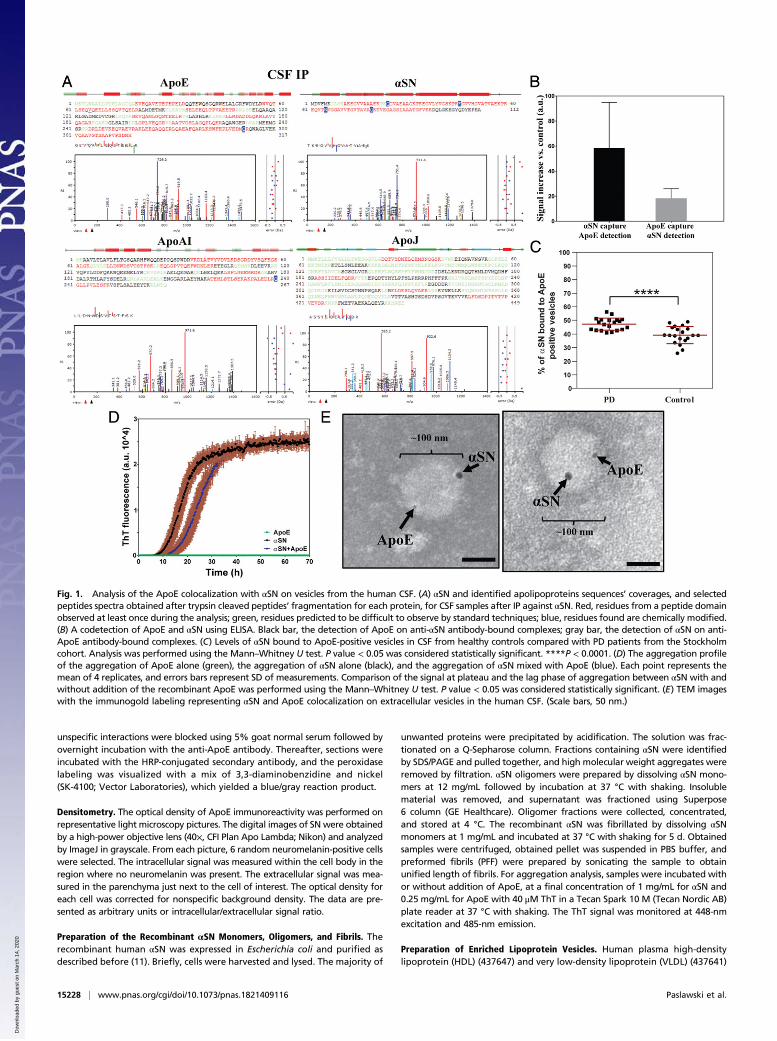
Fig. 1. Analysis of the ApoE colocalization with αSN on vesicles from the human CSF. (A) αSN and identified apolipoproteins sequences’ coverages, and selectedpeptides spectra obtained after trypsin cleaved peptides’ fragmentation for each protein, for CSF samples after IP against αSN. Red, residues from a peptide domainobserved at least once during the analysis; green, residues predicted to be difficult to observe by standard techniques; blue, residues found are chemically modified.(B) A codetection of ApoE and αSN using ELISA. Black bar, the detection of ApoE on anti-αSN antibody-bound complexes; gray bar, the detection of αSN on anti-ApoE antibody-bound complexes. (C) Levels of αSN bound to ApoE-positive vesicles in CSF from healthy controls compared with PD patients from the Stockholmcohort. Analysis was performed using the Mann–Whitney U test. P value < 0.05 was considered statistically significant. ****P < 0.0001. (D) The aggregation profileof the aggregation of ApoE alone (green), the aggregation of αSN alone (black), and the aggregation of αSN mixed with ApoE (blue). Each point represents themean of 4 replicates, and errors bars represent SD of measurements. Comparison of the signal at plateau and the lag phase of aggregation between αSN with andwithout addition of the recombinant ApoE was performed using the Mann–Whitney U test. P value < 0.05 was considered statistically significant. (E) TEM imageswith the immunogold labeling representing αSN and ApoE colocalization on extracellular vesicles in the human CSF. (Scale bars, 50 nm.)
15228 | www.pnas.org/cgi/doi/10.1073/pnas.1821409116 Paslawski et al.
Dow
nloa
ded
by g
uest
on
Mar
ch 1
4, 2
020

vesicles were purchased fromMerckMillipore. For the enrichment, 550 μg/mL(cholesterol content) lipoproteins were mixed with αSN or ApoE (11 μM finalconcentration each) and incubated for 1 h at 37 °C. For the enrichment withboth αSN and ApoE, αSN was added first and incubated for 1 h at 37 °C,followed by 1-h incubation with ApoE. Unbound proteins were removed bypassing the solution through 100-kDa or 50-kDa Amicon Ultra-0.5 Centrif-ugal Filter Units (Millipore). Finally, the sample was washed 3 times byadding PBS to the retained fraction and passing the solution through 100-kDa or 50-kDa Centrifugal Filter Units.
Lipoprotein Uptake by Dopaminergic Cells. SH-SY5Y human neuroblastomacells were routinely maintained in a Dulbecco’s Modified Eagle Medium(DMEM) modified medium supplemented with fetal bovine serum (FBS)(10%), L-alanyl-L-glutamine (2 mM), penicillin (100 μg/mL), and streptomycin(100 μg/mL). Cultures were maintained at 37 °C in 5% CO2/humidified air. Forthe uptake screening, cells were cultured in 24-well plates on laminin-coatedcover glasses at a seeding density of 1 × 10⁵ cells per well in a differentiatingmedium (DMEM modified medium supplemented with FBS [1%], L-alanyl-L-glutamine [2 mM], penicillin [100 μg/mL], streptomycin [100 μg/mL], and 10 μMretinoic acid) for 4 d. Human plasma HDL (437647; Merck Millipore), VLDL(437641; Merck Millipore), and the recombinant αSN were marked withAlexa Fluor 568 NHS-ester (A20003; Thermo Fisher Scientific) or Alexa Fluor488 NHS-ester (A20000; Thermo Fisher Scientific) according to the manu-facturer protocol. For uptake study, cell medium was changed to DMEMmodified medium supplemented with FBS (0.1%), L-alanyl-L-glutamine(2 mM), penicillin (100 μg/mL), streptomycin (100 μg/mL), and 10 μM retinoicacid. Labeled human plasma HDL and VLDL, with or without enrichmentwith the labeled monomeric αSN (enrichment protocol described above), thelabeled monomeric αSN, and vehicle controls were added to cells (finalconcentrations: 20 μM cholesterol and 1 μM αSN) and incubated for 4 h.Next, cells were washed with PBS and fixed with 4% paraformaldehyde.Subsequently, cells were stained with the Alexa Fluor 647 phalloidin dye(A22287; Thermo Fisher Scientific) according to the manufacturer protocolfollowed by a DAPI counterstaining. The single-plane images of cells wereobtained using a confocal microscopy (LSM880; Zeiss).
Separation of Lipoproteins by Density-Gradient Ultracentrifugation. Lipopro-teins were isolated from 0.1 mL of αSN−lipoprotein mix by KBr-densitygradient ultracentrifugation according to Redgrave et al. (30). Briefly, af-ter incubation, density and volume of each sample was adjusted, re-spectively, to 1.21 g/mL and 1 mL with KBr and transferred into centrifugetubes. A discontinuous density gradient was formed by layering 3 mL of1.063 g/mL salt solution above the sample, followed by 3 mL of 1.019 g/mLsalt solution and 3 mL of 1.006 g/mL salt solution. Salt solutions were pre-pared from KBr and NaCl and contained 0.1 mg/mL 2,2′,2′′,2′′′-(ethane-1,2-diyldinitrilo)tetraacetic acid. The samples were centrifuged at 386,000 × gfor 24 h at 20 °C. The 1.25-mL fractions were collected and analyzed usingthe Western blotting (WB) method.
Size-Exclusion Chromatography. The αSN−lipoprotein samples were analyzedand fractionated using Superose 6 Increase 10/300 GL column connected toan ÄKTA Explorer system (GE Healthcare) using PBS as a mobile phase. Theappearance of proteins was monitored by reading the absorbance at 280 nm.Obtained fractions were collected and analyzed using the WB method.
Sample Randomization. For each patient cohort, all available samples werechosen taking age and gender matching into consideration. Obtained CSFand plasma samples were coded and analyzed blindly. No sample was ex-cluded from the final analysis. Sampleswere decoded after experimental datawere obtained.
Statistics. Statistical analyses were performed using a GraphPad Prism soft-ware (GraphPadInc). Comparison between 2 groups was performed using aMann–Whitney U test. Multiple comparison was performed using a Kruskal−Wallis H test with a Dunn’s correction. A Spearman’s rank correlation coeffi-cient was used to analyze dependence between 2 sets of data. All statisticaltests were 2-tailed, and P value < 0.05 was considered statistically significant.
Data Availability. The data are available upon reasonable request. Due tosensitive nature of the patients’ clinical information, the ethics protocol doesnot allow open data sharing.
For more detailed description of the methods, see SI Appendix.
ResultsαSN Colocalizes with Apolipoproteins in the Human CSF. Data onαSN interactors in the extracellular milieu including CSF aresparse. Therefore, we performed co-IP (SI Appendix, Fig. S2)against αSN on CSF samples, and analyzed bounded proteinsusing MS. Unexpectedly, our data showed that αSN-boundproteins in CSF were mainly apolipoproteins, among whichApoE was the most apparent. Moreover, we identified ApoAI-and ApoJ-derived peptides (Table 1, Fig. 1A, and SI Appendix,Fig. S3). Neither αSN- nor apolipoproteins-related peptides wereidentified in the negative control (Table 1).To confirm the co-IP of αSN and ApoE, we performed ELISA
on crude CSF samples using 2 reciprocal settings: First, a capturewas made with an anti-αSN antibody and detection was performedwith an anti-ApoE antibody; second, proteins were captured usingthe antibody against ApoE, and detection was performed using theantibody against αSN. With both settings, we observed a clearELISA signal (Fig. 1B), indicating that ApoE and αSN closely in-teract in CSF. Moreover, we did not observe any unspecific inter-actions when using antibody controls (rabbit and goat IgGs), in theELISA approach, neither for recombinant proteins nor for CSFsamples (SI Appendix, Fig. S4). Furthermore, analysis of our datasuggests that around 40 to 50% of CSF αSN is associated withApoE-positive lipoproteins (SI Appendix, Fig. S4D).To confirm this finding, we performed analysis of αSN levels
before and after depletion of ApoE-positive vesicles in CSF from20 healthy controls (14/6 males/females, average age 64.10 ±8.42 y) and 20 PD patients (14/6 males/females, average age
Fig. 2. Enrichment of lipoprotein vesicles with αSN. (A) The WB analysisagainst αSN and ApoE of flow-through and retained fractions obtained afterlipoprotein enrichment with αSN and subsequent filtration through 100-kDaspin filter. (B–E) The ELISA analysis of fractions retained on the filter afterenrichment of HDL and VLDL vesicles with αSN and/or ApoE. Correspondinggraphs represent ELISA analyses using (B) the anti-ApoE capture and theanti-ApoE detection antibody, (C) the anti-ApoE capture and the anti-αSNdetection antibody, (D) the anti-αSN capture and the anti-ApoE detectionantibody, and (E) the anti-αSN capture and the anti-αSN detection antibody.
Paslawski et al. PNAS | July 23, 2019 | vol. 116 | no. 30 | 15229
NEU
ROSC
IENCE
Dow
nloa
ded
by g
uest
on
Mar
ch 1
4, 2
020

62.65 ± 12.72 y). We observed a decrease of αSN in samplesfrom both PD and control subjects (Fig. 1C and SI Appendix, Fig.S5). Moreover, the amount of αSN bound to ApoE-positive ves-icles was significantly higher in PD cases compared with healthycontrols (Fig. 1C).To investigate whether the interaction between αSN and
ApoE is direct or indirect, we analyzed kinetics of αSN fibrilformation after addition of recombinant ApoE. We did not ob-serve any significant changes in the aggregation profile of therecombinant αSN, suggesting indirect contact between theseproteins (Fig. 1D).To further examine the character of interactions observed
between αSN and ApoE, we performed TEM analysis on CSFsamples. Indeed, αSN and ApoE double immune-gold labelingrevealed a colocalization of αSN and ApoE on larger (>30 nm)vesicular structures in CSF (Fig. 1E and SI Appendix, Fig. S6A).However, it is possible that the αSN and ApoE colocalizationalso occurred on smaller vesicles, but, due to a steric hindrance,caused by a relatively large size of immunogold particles andantibodies, only a single protein could be detected on vesiclessmaller than 30 nm in size (SI Appendix, Fig. S6B).No clear lipid bilayer was observed on αSN- and ApoE-posi-
tive vesicular structures in TEM analysis (Fig. 1E), suggestingthat they are lipoprotein vesicles. This is in accordance with thedata from the MS analysis identifying only lipoproteins’ markers(Table 1, Fig. 1A, and SI Appendix, Fig. S3). We therefore in-vestigated the ability of αSN to interact with lipoprotein vesiclesin vitro. Considering that CSF lipoproteins are predominantly ofthe size and density of plasma HDL, human plasma HDLs wereincluded in the study. Moreover, due to fact that CSF HDLs aremainly ApoE-positive, contrary to plasma HDL which are mainlyApoAI-positive (31), human plasma VLDL were also used toaddress the role of ApoE-positive lipoproteins. Accordingly,we incubated human plasma HDL or VLDL vesicles withrecombinant monomeric αSN and passed the obtained samplesthrough a size-exclusion filter. Subsequent WB analysis of elu-ates showed that the recombinant monomeric αSN withoutvesicles passed entirely through the filter, while a large amountof αSN incubated with either HDL or VLDL vesicles wasretained on the filter. This confirmed αSN binding ability to li-poprotein vesicles (Fig. 2A).To verify that αSN and ApoE might colocalize on the lipo-
proteins resembling CSF lipoproteins, we performed doubleenrichment of plasma HDL and examined whether αSN- and/orApoE-enriched vesicle can be detected by ELISA settings ap-plied in the CSF study. When using both capture and detec-tion antibodies against ApoE, we obtained signal for VLDL,VLDL+αSN, and HDL+αSN+ApoE vesicles (Fig. 2B). For theanti-ApoE capture antibody and the anti-αSN detection antibody,
the signal was observed for VLDL+αSN and HDL+αSN+ApoEvesicles (Fig. 2C). For the anti-αSN capture antibody and the anti-ApoE detection antibody, the ApoE signal was observed forVLDL+αSN and HDL+αSN+ApoE vesicles (Fig. 2D). Whenusing both capture and detection antibodies against αSN, we obtainedsignal for VLDL+αSN, HDL+αSN, and HDL+αSN+ApoE vesicles(Fig. 2E). We extended our examination of αSN and lipoproteinsinteractions to include not only monomeric αSN but also oligo-meric and PFF αSN, using 2 additional methods. First, instead offiltration technique, we performed density gradient ultracentri-fugation separation followed by WB analysis (SI Appendix, Fig.S7). In this experiment, we were able to detect the 3 forms ofαSN both in protein and lipoprotein fractions. We conclude thatnot only monomeric but also oligomeric and PFF forms of αSNcan interact with lipoproteins. The fact that ultracentrifugationdetached apolipoproteins from vesicles is in agreement withprevious reports (32) (SI Appendix, Fig. S7). Therefore, to omitdetachment of proteins from vesicles, we decided to use a size-exclusion chromatography (SEC) approach, which does notintroduce any excessive forces to the sample separation. Asdescribed above, human plasma HDL or VLDL vesicles wereincubated with each of the 3 forms of αSN separately and in-jected to the column for SEC analysis. We observed a shift inretention volume after αSN and lipoprotein coincubation, in-dicating formation of larger-size complexes (SI Appendix, Fig.S8). We further confirmed that both αSN and lipoproteins arepresent in fractions collected from shifted peaks, using the WBmethod, verifying interactions of monomeric, oligomeric, andPFFs forms of αSN with lipoproteins.Moreover, using a filtration method with spin filters, presented in
Fig. 2, and SEC, we confirm that such isolation of lipoproteinsenriched with monomeric αSN does not affect the interaction be-tween monomeric αSN and lipoproteins (SI Appendix, Fig. S9).Based on the obtained results, we decided to investigate whether
ApoE, and other identified apolipoproteins, are changed in theCSF and plasma from PD patients and examine their biomarkerpotential.
Increased Level of ApoE in CSF, but Not in Plasma, from PD Patients.ApoE is the main apolipoprotein in central nervous system(CNS), and its allelic variants are strongly implicated in thepathogenesis of Alzheimer’s disease (33, 34). There is also someevidence that allelic variants of ApoE predispose to dementia inPD (35). However, no study has reported changes in ApoEprotein level in CSF or plasma from PD patients. Therefore, wemeasured ApoE in biofluids from PD patients and from matchedcontrols (SI Appendix, Tables S2 and S3). Throughout the ELISAoptimization process, we realized that the level of ApoE detectedby ELISA in the human CSF rises with increasing concentration
Table 2. Results from the statistical analysis of differences in apolipoproteins levels in CSF and plasma samples, between PD patientsand matched controls from the Stockholm cohort
Kruskal−Wallis
Analyzedapolipoprotein
Mann−Whitney CSF Plasma
CSF Controlvs. PD
Plasma Controlvs. PD
Control vs.PD treated
PD treated vs.PD untreated
Control vs.PD untreated
Control vs.PD treated
PD treated vs.PD untreated
Control vs.PD untreated
ApoAI 0.7995 0.0048 >0.9999 >0.9999 >0.9999 0.0598 >0.9999 0.0697ApoCI 0.4245 0.6176 0.0807 0.0439 >0.9999 >0.9999 0.1918 0.5398ApoE <0.0001 0.8194 0.0023 0.2830 <0.0001 0.7492 0.4278 >0.9999ApoJ 0.0116 0.639 0.0413 >0.9999 0.2601 >0.9999 0.4312 0.7826
The left part of the table represents P values obtained after analyzing apolipoproteins’ levels in PD (PD) patient set vs. healthy controls (the Mann−WhitneyU test). The right part of the table represents P values obtained after splitting the PD group into treated and untreated patients’ subsets and analyzingapolipoproteins’ levels between them and vs. healthy controls (the Kruskal−Wallis H test with a Dunn’s correction). P values < 0.05 are considered significantand are marked with a bold font.
15230 | www.pnas.org/cgi/doi/10.1073/pnas.1821409116 Paslawski et al.
Dow
nloa
ded
by g
uest
on
Mar
ch 1
4, 2
020

of detergents in a buffer, up to the point where the detergentconcentration is too high for the method to work at all. At the sametime, the level of recombinant ApoE, detected with ELISA method,was independent of a buffer’s composition (SI Appendix, Fig. S10).This effect was probably caused by the strong affinity of ApoE tolipoproteins and the steric hindrance caused by the large size of anantibody compared with the small size of lipoproteins (SI Appendix,Fig. S6B), consequently making ApoE hard to analyze without usageof strong detergents, and therefore not suitable for high-throughputmethods like ELISA. Subsequently, we decided to analyze ApoEprotein levels with SDS/PAGE followed by WB (SI Appendix, Fig.S11). We found a significant increase in the ApoE level in CSFfrom PD patients compared with matched controls in 3 independentcohorts (Table 2 and Fig. 3 A–F). Using the recombinant ApoEprotein, we estimated that the average concentration of ApoE inStockholm cohort controls was 8.0 μg/mL, while, in PD patients,the average level was 12.6 μg/mL (SI Appendix, Fig. S12), nearly
57% more. In contrast, we found no difference in the plasma levelof ApoE between controls and PD patients from Stockholm cohort(Fig. 3 G and H).The CSF level of ApoE was negatively correlated only with
Levodopa Equivalent Daily Doses score (SI Appendix, Table S4).No significant correlations were observed between the ApoE leveland disease duration, or rating scores from the unified PD ratingscale part 3, the Hoehn &Yahr scale, the nonmotor symptomsscale, Beck’s depression inventory (BDI), and Montreal cognitiveassessment (SI Appendix, Table S4). Similarly, there were no cor-relations between the plasma ApoE level and patient age or any ofthe PD rating scores (SI Appendix, Tables S4 and S5). There was nocorrelation between plasma and CSF ApoE levels (SI Appendix,Table S6).To investigate the ApoE biomarker potential for PD di-
agnosis, we performed a receiver operating characteristic (ROC)curve analysis (36, 37) for data obtained from Stockholm cohort.Our analysis showed that, at the level of 0.8 sensitivity, ∼0.6specificity was achievable (SI Appendix, Fig. S13A, solid blackline). Interestingly, with the same sensitivity of 0.8, we couldobtain around 0.7 specificity when only untreated patients wereanalyzed (SI Appendix, Fig. S13A, dashed black line).
ApoE as a Potential Biomarker for Atypical Parkinsonism. To checkwhether the ApoE increase is specific for PD, we analyzed thelevel of ApoE in CSF from patients with MSA or PSP fromUmeå and Lund cohorts. The ApoE level in MSA patients, an-other synucleinopathy, was significantly higher compared withcontrols, whereas its level in PSP patients, a tauopathy, was notsignificantly changed (SI Appendix, Fig. S14). These results furtherstrengthen the relationship between ApoE and αSN.
Uptake of αSN-Enriched Lipoprotein Vesicles in Dopaminergic Neuronal-like Cells. To investigate whether αSN-enriched lipoprotein vesiclescan be a possible mechanism of αSN uptake and spreading, weexamined the uptake of HDL and VLDL along with αSN-enriched lipoprotein vesicles in neuronal-like cells. We thusdifferentiated SH-SY5Y human dopaminergic neuroblastomacells to a neuronal-like state and incubated them with a fluo-rescently labeled monomeric αSN, HDL, and VLDL, and withdouble-labeled complexes of αSN-enriched lipoprotein vesicles.We also checked that the labeled αSN does not undergo ag-gregation or degradation/fragmentation, using both WB andSEC approaches (SI Appendix, Fig. S15). We decided not touse unlabeled recombinant αSN, due to the fact that SH-SY5Ycells, which are of human origin, express endogenous αSN.Performing αSN staining in such conditions will not allowdiscrimination between endogenous and recombinant αSN.Furthermore, we decided to use cells, recombinant proteins,and lipoproteins of human origin to omit any concerns comingfrom receptor differences across species. After 4-h incubation,we observed that the monomeric αSN alone, as well as HDLand VLDL, easily entered neuronal cells. Interestingly, weobserved the same effect for αSN-enriched HDL and VLDLvesicles (Fig. 4), confirming αSN couptake during lipoproteininternalization as a possible mechanism of αSN uptake andspreading.
Immunohistochemical Detection of ApoE in the Human SNc. To ex-amine the distribution of ApoE in the human SN, we performedimmunohistochemistry analysis on postmortem human brain tissuefrom SNc of PD patients and healthy controls. Similarly to previousreports (38, 39), we detected ApoE in paravascular spaces (SI Ap-pendix, Fig. S16 A and B). Moreover, we observed a strong region-specific ApoE signal mostly in periaqueductal gray, intercollicularnucleus, and, most importantly, SNc (SI Appendix, Fig. S17 A–H).Interestingly, in controls, most of neuromelanin-positive cells(dopaminergic neurons) in SNc were negative (or exceedingly
Fig. 3. Levels of ApoE in biofluids from Stockholm, Umeå, and Lund co-horts. (A) Stockholm cohort levels of ApoE in CSF of healthy controls com-pared with PD patients. (B) Stockholm cohort levels of ApoE in CSF ofhealthy controls compared with treated and untreated PD patients. (C)Umeå cohort levels of ApoE in CSF of healthy controls compared with PDpatients. (D) Umeå cohort levels of ApoE in CSF of healthy controls com-pared with treated and untreated PD patients. (E) Lund cohort levels ofApoE in CSF of healthy controls compared with PD patients. (F) Lund cohortlevels of ApoE in CSF of healthy controls compared with treated and un-treated PD patients. (G) Stockholm cohort levels of ApoE in plasma ofhealthy controls compared with PD patients. (H) Stockholm cohort levels ofApoE in plasma of healthy controls compared with treated and untreated PDpatients. PD, PD. Tests used were the Mann−Whitney U test for 2 groupscomparison and the Kruskal−Wallis test with the Dunn’s correction for morethan 2 groups. *P < 0.05; **P < 0.01; ****P < 0.0001.
Paslawski et al. PNAS | July 23, 2019 | vol. 116 | no. 30 | 15231
NEU
ROSC
IENCE
Dow
nloa
ded
by g
uest
on
Mar
ch 1
4, 2
020

low) for the ApoE staining (Fig. 5A). In contrast to controls, insections from PD patients, most of the dopaminergic neuronswere clearly positive for ApoE (Fig. 5A). Additionally, to con-firm observed changes and rule out optical illusion, we per-formed densitometric analysis of ApoE signal distributionbetween the intracellular compartment of dopaminergic cellsand extracellular space (Fig. 5 B–D). We detected a signifi-cantly higher signal for extracellular than intracellular ApoE incontrols (Fig. 5B), while, in PD patients, ApoE signal wasdistributed equally (Fig. 5C). Finally, we compared the ratio ofintracellular and extracellular ApoE signal in controls and PDsamples and observed a significantly lower ratio for controls,indicating changes in the ApoE gene expression and/or an in-creased uptake of ApoE in dopaminergic neurons of PD pa-tients. Importantly, we did not observe any ApoE-negativedopaminergic neurons in PD patients, while close cooccur-rences of both negative and positive ApoE dopaminergic cellswere detected in controls (SI Appendix, Fig. S17 I–K). Finally,we did not observe any signal on negative control sectionsin which the primary antibody was omitted (SI Appendix,Fig. S16C).
Decreased Level of ApoAI in Plasma, but Not in CSF, from PD Patients.We have identified ApoAI as one of the αSN-interacting pro-teins in CSF (Table 1). The level of ApoAI in CSF was notchanged between controls and PD patients (SI Appendix, Fig.S18 A and B and Table 2). However, in agreement with a pre-vious study (40), we observed a decreased plasma level of ApoAIin PD patients compared with controls (SI Appendix, Fig. S18 Cand D and Table 2). We did not observe any correlation betweenthe level of ApoAI and PD severity scores, disease duration, orage, either in CSF nor in plasma (SI Appendix, Tables S4 and S5).However, the CSF level of ApoAI significantly correlated with itsIgG and albumin content (SI Appendix, Table S5). At the sametime, we did not observe any significant changes in albumin andIgG at a group level, either in CSF or in plasma of PD patientsand controls (SI Appendix, Fig. S19 A–D). However, the numberof mononuclear cells in CSF was significantly higher in PD pa-
tients compared with controls (SI Appendix, Fig. S19 E and F),suggesting a mild inflammatory response in PD.
Elevated Levels of ApoJ and ApoCI in CSF, but Not in Plasma, from PDPatients. We also analyzed CSF and plasma levels of 2 otherapolipoproteins: ApoJ and ApoCI. The level of ApoJ was sig-nificantly increased in CSF (SI Appendix, Fig. S20 A and B andTable 2), but not in plasma (SI Appendix, Fig. S20 C and D andTable 2), of PD patients compared with controls. We did not findcorrelations between the ApoJ level and any PD severity score (SIAppendix, Table S4). However, the plasma ApoJ level negativelycorrelated with age and the albumin content and positively corre-lated with IgG levels in PD patients (SI Appendix, Table S5).Additionally, we decided to investigate the ApoCI level, due
to its high abundance in CSF. Although the level of CSF ApoCIin PD patients did not differ from controls (SI Appendix, Fig.S21A and Table 2), it was significantly higher in treated com-pared with untreated PD patients (SI Appendix, Fig. S21B andTable 2). Furthermore, the ApoCI level in CSF positively cor-related with the disease duration, and the plasma level positivelycorrelated with the BDI score (SI Appendix, Table S4). We didnot observe any difference in the level of ApoCI in plasma be-tween controls and PD patients (SI Appendix, Fig. S21 C and D).The ApoCI level from controls correlated with albumin and IgGlevels in CSF and plasma. For PD patients, ApoC1 levels correlatedwith albumin and IgG levels in CSF and the albumin level in plasma(SI Appendix, Table S5). These data suggest a possible involvementof ApoCI in neuroinflammation rather than a specific contributionto PD.
Correlations between Apolipoproteins Levels in Plasma and CSF andTheir Combined Accuracy for PD Detection. As mentioned above,our data strongly indicate differences in levels of various types ofapolipoproteins in CSF and plasma of PD patients. Therefore,we investigated whether there is any correlation between levelsof different types of apolipoproteins within and between plasmaand CSF. We observed that the increased level of ApoE in CSFcorresponded to the decreased level of ApoAI in plasma (SIAppendix, Table S6). Moreover, we noticed positive correlations
Fig. 4. Uptake of lipoprotein vesicles enriched withαSN by SH-SY5Y cells differentiated to dopaminergicneurons. Single-plain confocal images represent theuptake of lipoprotein vesicles enriched with αSN bySH-SY5Y cells differentiated to dopaminergic neu-rons. (Left to Right) A vehicle control, uptake offluorescently labeled αSN-AF488, HDL-AF568, VLDL-AF568, and double-labeled enriched lipoproteinsαSN-AF488+HDL-AF568 and αSN-AF488+VLDL-AF568.After 4 d of differentiation, cells were incubated for4 h with mixtures, then fixed, and proceeded to theF-actin staining with Phalloidin-AF647 and the nu-clear labeling with DAPI. Insets in merged panelsrepresent magnified areas showing fluorescently la-beled αSN and lipoproteins, and their colocalizationfor enriched vesicles. Images were acquired using 63×magnification objective. Insets represent further 5×digital enlargement of 63× magnification images.
15232 | www.pnas.org/cgi/doi/10.1073/pnas.1821409116 Paslawski et al.
Dow
nloa
ded
by g
uest
on
Mar
ch 1
4, 2
020

between plasma levels of ApoCI and ApoE as well as betweenCSF levels of ApoCI and ApoAI. Furthermore, we detected acorrelation between plasma levels of ApoJ and ApoAI (SIAppendix, Table S6).In addition, we examined whether a combined ROC analysis
of several apolipoproteins would increase the diagnostic accu-racy compared with the analysis of ApoE alone. Thus, we com-bined the level of ApoE in CSF with the level of ApoAI inplasma and recalculated the ROC curve (SI Appendix, Fig.S13B). We also performed the ROC analysis combining the levelof ApoAI in plasma with levels of ApoJ and ApoE in CSF (SIAppendix, Fig. S13C). Our analysis showed that, with the sensi-tivity of 0.8, combined levels did not increase the specificity (SIAppendix, Fig. S13D). However, when the sensitivity was set to0.71, we could increase the specificity from 0.70 for ApoE aloneto 0.83 when combining ApoE, ApoAI, and ApoJ levels (SIAppendix, Fig. S13D).
DiscussionDuring the last decade, a number of studies reported that αSNpossesses a membrane-binding capacity and can interact notonly with artificial lipid vesicles but also with cell membranesand extracellular vesicles, including exosomes (41). Here, usinga co-IP approach followed by MS analysis, we discovered arobust interaction between αSN and apolipoproteins in thehuman CSF. We observed the strongest signal for ApoE, andwe further confirmed this interaction using an ELISA methodrelying on different sets of antibodies. Our data suggest thatαSN−apolipoprotein interactions are indirect, since the ApoEprotein was not able to interfere with the αSN aggregation, and
we also observed an interaction of αSN with ApoE-negative li-poproteins in vitro. This interaction rather depends on the αSNpropensity to bind to the negatively charged lipid particles (42,43). Importantly, while it is vital to note that both ApoE (44) andαSN (45–47) were found on exosomes, our TEM analysisrevealed structures without a clear lipid bilayer. More impor-tantly, we did not identify any exosome markers during the MSanalysis, while we observed several apolipoproteins. Our datasuggest that αSN associates with lipoprotein particles both invitro and in human CSF. Approximately 45% of CSF αSN isbound to ApoE-positive lipoproteins. The percentage of αSNbound to ApoE-positive vesicles in CSF is significantly higher inPD patients compared with healthy controls. This is in accor-dance with the higher level of CSF ApoE in PD patients. It istherefore a rather common event and could be a major mecha-nism of αSN uptake. At the same time, combining the facts thatαSN concentration in CSF is estimated to be around 1 to 2 ng/mLand ApoE concentration is in the range of 5 to 15 μg/mL, wecan conclude that this is a rare event from the perspective oflipoproteins. Furthermore, it is likely the amount of αSN−lipo-proteins particles taken up by cells will be determined by cellularrequirements for lipoprotein cargo. Therefore, αSN−lipopro-teins particle uptake will differ between the nonmitotic cell lineand fully mature dopaminergic neurons. Moreover, we de-termined that not only αSN monomers but also oligomers andfibrils can interact with human plasma-derived HDL and VLDLvesicles in vitro. Likewise, during the preparation of this manu-script, it was reported that αSN can interact with lipoproteinsalso in human plasma (48), further confirming our observations.Finally, we are showing that not only HDL and VLDL alone, but
Fig. 5. Analysis of ApoE localization in dopaminer-gic neurons of the human SN. (A) Representativephotomicrographs of immunostaining against ApoEin human SN (light blue staining). ApoE immunore-activity in control individuals (Control 1 and Control2) present a preferential localization of ApoE to theextracellular space while dopaminergic neurons(brown, neuromelanin-pigmented cells) remain neg-ative (or at very low signal level). Immunostainingagainst ApoE in brains from PD patients (PD 1 and PD2) show an equal localization of ApoE in extracellularspace and intracellularly in dopaminergic neurons.Scale bars on PD 2 photomicrographs refer to allpictures in the same column. (B–D) The densitometricanalysis of ApoE signal distribution between dopa-minergic cells intracellular compartment and extra-cellular space in (B) controls and (C) PD patients. (D)Ratio of densitometric analysis of intracellular andextracellular ApoE signal between controls and PDpatients. Comparison was performed using theMann–Whitney U test. P value < 0.05 was consideredstatistically significant. **P < 0.01; ****P < 0.0001.
Paslawski et al. PNAS | July 23, 2019 | vol. 116 | no. 30 | 15233
NEU
ROSC
IENCE
Dow
nloa
ded
by g
uest
on
Mar
ch 1
4, 2
020

also αSN-enriched lipoprotein vesicles, undergo a cellular uptakeby dopaminergic neuronal like cells in vitro. Additionally, we ob-served compartmentalization of αSN after cellular uptake. This islikely due to the fact that the uptake of lipoproteins, similar toother endocytosis mechanisms, results in trafficking of lipopro-teins to endosomes and then to lysosomes (49, 50). Consequently,enrichment of staining in specific cellular compartments isexpected.Taken together, we believe that our present data on αSN−
lipoprotein interactions, and their cellular uptake, raise a plau-sible explanation for the αSN uptake and spreading in the brain.It is possible that the monomeric αSN, as well as hydrophobicaggregates, binds to extracellular lipoprotein particles, which arethen taken up by other cells through a lipoproteins endocytosispathway (Fig. 6). This lipoprotein-dependent mechanism maycooperate with others, including exosome-based and nanotubemodels, to propagate αSN pathology.Apolipoproteins themselves are key components of lipopro-
tein particles and are responsible for the lipid homeostasis asthey transport lipids and lipid-soluble compounds from one cellto another (51). The major apolipoprotein in CSF is ApoE,followed by ApoAI, ApoAII, ApoCs, ApoJ, and ApoD (52).The ApoE concentration in the brain is the second highest,following that of liver. In the CNS, ApoE is synthesized mainlyby astrocytes and microglia, and, to a certain extent, by immatureneurons (53). The ApoE content is high in the paravascular(glymphatic) space, a CSF flow pathway through the brain(39, 54, 55). Moreover, its presence and colocalization withamyloidß in perivascular drainage channels was linked to theApoE-dependent amyloidß clearance through the blood–brainbarrier (56–58).ApoE levels are increased in injured or stressed neurons and
therefore postulated to play a role in the growth and repair ofcells in the CNS (59). Furthermore, ApoE allelic variants arelinked to an increased risk and earlier age of onset for Alz-heimer’s disease (34) and cognitive decline in PD (35). While theinfluence of ApoE genotypes was widely studied in PD, littlework has been performed at the protein level (60, 61). We re-alized that the use of strong detergents is necessary to isolate
ApoE molecules for a proper quantification. For this reason, weanalyzed apolipoproteins levels in CSF and plasma with SDS/PAGE followed by WB. Our analysis demonstrated a significantincrease of CSF ApoE in PD patients in 3 separate cohorts.Further, by performing the ROC curve analysis, we conclude thatmeasuring apolipoproteins in CSF and plasma can be clinicallymeaningful, especially when used in combination with otherknown biomarkers.ApoE was found to be enriched in the human SN, mostly
localized in extracellular or paravascular spaces rather than inthe cytosol of dopaminergic neurons. Furthermore, in pre-liminary studies, we distinguished 2 different populations ofneuromelanin-containing dopaminergic neurons: ApoE-positiveand ApoE-negative. In controls, the number of ApoE-negativedopaminergic neurons appears higher than the number of ApoE-positive ones. Conversely, in PD patients, the vast majority ofremaining neuromelanin-containing neurons appear to beApoE-positive. However, further analyses are needed to eluci-date whether this effect is due to a higher uptake/expression oflipoproteins and/or ApoE-positive neurons being more resistantto neurodegeneration. Furthermore, the results need to be val-idated in additional PD populations. Nevertheless, our initialobservations suggest a functional involvement of ApoE in PDpathology and/or defense against it.We also studied other apolipoproteins in CSF as well as in
plasma. We did not observe any changes in the CSF ApoAI levelfrom PD patients, but, consistent with previous studies, theplasma ApoAI level was decreased (40). Contrary to ApoE,ApoAI in the brain is believed to be plasma-derived (52), while asmall amount is synthesized by endothelial cells in the cerebralvasculature (62). This, together with the identified ApoAI−αSNinteraction, suggests that toxic αSN species might be transportedwith ApoAI lipoprotein particles from the periphery and thendistributed throughout the brain.Similar to ApoE, we observed a significant increase in ApoJ
levels in CSF from PD patients. Similar to other apolipoproteins,ApoJ, also known as clusterin, is involved in a lipid transportation,but it also serves a chaperone role during the cellular stress re-sponse (63). Besides, it stabilizes a range of misfolded proteins(64). Interestingly, ApoJ is involved in the amyloidß clearancethrough blood–brain barrier (55, 65), and the level of brain ApoJincreases in numerous neurological disorders such as Alzheimer’sdisease, MSA, ischemia, and epilepsy (66).Finally, we observed a change in the CSF ApoCI level, but
only in treated PD patients. Moreover, CSF and plasma ApoCIlevels positively correlated with albumin and IgGs levels, in-dicating an association of ApoCI with blood−brain barrier in-tegrity and a mild ongoing inflammation.In summary, the presence of αSN on lipoprotein vesicles in the
human CSF, along with the presence of ApoE in some dopa-minergic neurons and the cellular uptake of αSN-enriched li-poproteins, represents a possible mechanism for αSN uptake andspreading in the brain. Finally, changes in apolipoproteins’ levelsin human CSF represent an aspect of PD pathology and can bebecome clinically meaningful, especially when used in combina-tion with other biomarkers.
ACKNOWLEDGMENTS. We thank all the subjects who participated in thisstudy. The project was financially supported by grants from the SwedishFoundation for Strategic Research; European and Swedish Research Councils;The Parkinson foundation of Sweden; Agreement on medical education andresearch (ALF) programs in Västerbotten, Skåne, and Stockholm; The Swed-ish Brain Foundation; Wenner-Gren Foundation; and Knut and AliceWallenberg Foundation. We would like to also thank Prof. Daniel Otzen fromInterdisciplinary Nanoscience Center, Aarhus University for sharing the bacte-rial plasmid for expression of recombinant human αSN.
Fig. 6. Schematic representing the ApoE and αSN interaction in the hu-man CNS and a possible spreading of a pathological αSN between cells. Thepathological and the normal αSN forms might be released form healthyand/or dying neurons to the extracellular space, where they startinteracting with lipoprotein particles released by microglia and astrocytes.Further, probably after recognition by lipoprotein receptors, they are takenup by healthy neurons. Possibly, pathological αSN aggregates, resistant toproteolytic cleavage, remain in the cell undigested and become theseed elongated by the normal αSN, which further might lead toneurodegeneration and disease spreading. Image prepared using SmartServier Medical Art (https://smart.servier.com/), which is licensed under CCBY 3.0.
15234 | www.pnas.org/cgi/doi/10.1073/pnas.1821409116 Paslawski et al.
Dow
nloa
ded
by g
uest
on
Mar
ch 1
4, 2
020

1. R. B. Postuma et al., MDS clinical diagnostic criteria for Parkinson’s disease. Mov.Disord. 30, 1591–1601 (2015).
2. K. R. Chaudhuri, A. H. V. Schapira, Non-motor symptoms of Parkinson’s disease: Do-paminergic pathophysiology and treatment. Lancet Neurol. 8, 464–474 (2009).
3. P. L. McGeer, S. Itagaki, H. Akiyama, E. G. McGeer, Rate of cell death in parkinsonismindicates active neuropathological process. Ann. Neurol. 24, 574–576 (1988).
4. W. G. Meissner et al., Priorities in Parkinson’s disease research. Nat. Rev. Drug Discov.10, 377–393 (2011).
5. M. G. Spillantini et al., Alpha-synuclein in Lewy bodies. Nature 388, 839–840 (1997).6. S. Chandra, G. Gallardo, R. Fernández-Chacón, O. M. Schlüter, T. C. Südhof, Alpha-
synuclein cooperates with CSPalpha in preventing neurodegeneration. Cell 123, 383–396 (2005).
7. J. Lautenschläger, C. F. Kaminski, G. S. Kaminski Schierle, α-Synuclein–regulator ofexocytosis, endocytosis, or both? Trends Cell Biol. 27, 468–479 (2017).
8. T. Takenouchi et al., Reduced neuritic outgrowth and cell adhesion in neuronal cellstransfected with human alpha-synuclein. Mol. Cell. Neurosci. 17, 141–150 (2001).
9. P. H. Weinreb, W. Zhen, A. W. Poon, K. A. Conway, P. T. Lansbury, NACP, a proteinimplicated in Alzheimer’s disease and learning, is natively unfolded. Biochemistry 35,13709–13715 (1996).
10. L. Giehm, D. I. Svergun, D. E. Otzen, B. Vestergaard, Low-resolution structure of avesicle disrupting α-synuclein oligomer that accumulates during fibrillation. Proc.Natl. Acad. Sci. U.S.A. 108, 3246–3251 (2011).
11. W. Paslawski, S. Mysling, K. Thomsen, T. J. Jørgensen, D. E. Otzen, Co-existence of twodifferent α-synuclein oligomers with different core structures determined by hydro-gen/deuterium exchange mass spectrometry. Angew. Chem. Int. Ed. Engl. 53, 7560–7563 (2014).
12. M. J. Volles et al., Vesicle permeabilization by protofibrillar alpha-synuclein: Impli-cations for the pathogenesis and treatment of Parkinson’s disease. Biochemistry 40,7812–7819 (2001).
13. J.-Y. Li et al., Lewy bodies in grafted neurons in subjects with Parkinson’s diseasesuggest host-to-graft disease propagation. Nat. Med. 14, 501–503 (2008).
14. F. N. Emamzadeh, Alpha-synuclein structure, functions, and interactions. J. Res. Med.Sci. 21, 29 (2016).
15. C. Betzer et al., Identification of synaptosomal proteins binding to monomeric andoligomeric α-synuclein. PLoS One 10, e0116473 (2015).
16. Y. Zhou et al., Analysis of alpha-synuclein-associated proteins by quantitative pro-teomics. J. Biol. Chem. 279, 39155–39164 (2004).
17. J. E. Payton, R. J. Perrin, D. F. Clayton, J. M. George, Protein-protein interactions ofalpha-synuclein in brain homogenates and transfected cells. Brain Res. Mol. Brain Res.95, 138–145 (2001).
18. A. Lleó et al., Cerebrospinal fluid biomarkers in trials for Alzheimer and Parkinsondiseases. Nat. Rev. Neurol. 11, 41–55 (2015).
19. N. K. Magdalinou et al., A panel of nine cerebrospinal fluid biomarkers may identifypatients with atypical parkinsonian syndromes. J. Neurol. Neurosurg. Psychiatry 86,1240–1247 (2015).
20. B. Mollenhauer et al., Investigating Synuclein Consortium of the Michael J. FoxFoundation for Parkinson’s Research, A user’s guide for α-synuclein biomarker studiesin biological fluids: Perianalytical considerations. Mov. Disord. 32, 1117–1130 (2017).
21. L. Gao et al., Cerebrospinal fluid alpha-synuclein as a biomarker for Parkinson’s dis-ease diagnosis: A systematic review and meta-analysis. Int. J. Neurosci. 125, 645–654(2015).
22. I. Björkhem et al., Oxysterols and Parkinson’s disease: Evidence that levels of 24S-hydroxycholesterol in cerebrospinal fluid correlates with the duration of the dis-ease. Neurosci. Lett. 555, 102–105 (2013).
23. S. Hall et al., Cerebrospinal fluid concentrations of inflammatory markers in Parkin-son’s disease and atypical parkinsonian disorders. Sci. Rep. 8, 13276 (2018).
24. M. Trupp et al., Metabolite and peptide levels in plasma and CSF differentiatinghealthy controls from patients with newly diagnosed Parkinson’s disease. J. Parkin-sons Dis. 4, 549–560 (2014).
25. X. Geng et al., α-Synuclein binds the K(ATP) channel at insulin-secretory granules andinhibits insulin secretion. Am. J. Physiol. Endocrinol. Metab. 300, E276–E286 (2011).
26. P. S. Guerreiro et al., LRRK2 interactions with α-synuclein in Parkinson’s disease brainsand in cell models. J. Mol. Med. (Berl.) 91, 513–522 (2013).
27. K. L. Norris et al., Convergence of parkin, PINK1, and α-synuclein on stress-inducedmitochondrial morphological remodeling. J. Biol. Chem. 290, 13862–13874 (2015).
28. W. Paslawski et al., High stability and cooperative unfolding of α-synuclein oligomers.Biochemistry 53, 6252–6263 (2014).
29. M. D. Abramoff, P. J. Magelhaes, S. J. Ram, Image processing with ImageJ. Biophoton.Int. 11, 36–42 (2004).
30. T. G. Redgrave, D. C. K. Roberts, C. E. West, Separation of plasma lipoproteins bydensity-gradient ultracentrifugation. Anal. Biochem. 65, 42–49 (1975).
31. R. W. Mahley, Central nervous system lipoproteins: ApoE and regulation of choles-terol metabolism. Arterioscler. Thromb. Vasc. Biol. 36, 1305–1315 (2016).
32. W. H. Munroe, M. L. Phillips, V. N. Schumaker, Excessive centrifugal fields damagehigh density lipoprotein. J. Lipid Res. 56, 1172–1181 (2015).
33. M. C. Chartier-Harlin et al., Apolipoprotein E, epsilon 4 allele as a major risk factor forsporadic early and late-onset forms of Alzheimer’s disease: Analysis of the19q13.2 chromosomal region. Hum. Mol. Genet. 3, 569–574 (1994).
34. C.-C. Liu, C. C. Liu, T. Kanekiyo, H. Xu, G. Bu, Apolipoprotein E and Alzheimer disease:Risk, mechanisms and therapy. Nat. Rev. Neurol. 9, 106–118 (2013). Correction in: Nat.Rev. Neurol., 9, 184 (2013).
35. K. C. Paul et al., APOE, MAPT, and COMT and Parkinson’s disease susceptibility andcognitive symptom progression. J. Parkinsons Dis. 6, 349–359 (2016).
36. C. M. Florkowski, Sensitivity, specificity, receiver-operating characteristic (ROC) curvesand likelihood ratios: Communicating the performance of diagnostic tests. Clin. Bi-ochem. Rev. 29 (suppl. 1), S83–S87 (2008).
37. K. Hajian-Tilaki, Receiver operating characteristic (ROC) curve analysis for medicaldiagnostic test evaluation. Caspian J. Intern. Med. 4, 627–635 (2013).
38. P. T. Xu et al., Specific regional transcription of apolipoprotein E in human brainneurons. Am. J. Pathol. 154, 601–611 (1999).
39. T. M. Achariyar et al., Glymphatic distribution of CSF-derived apoE into brain is iso-form specific and suppressed during sleep deprivation. Mol. Neurodegener. 11, 74(2016).
40. C. R. Swanson et al., Lower plasma apolipoprotein A1 levels are found in Parkinson’sdisease and associate with apolipoprotein A1 genotype. Mov. Disord. 30, 805–812(2015).
41. C. M. Pfefferkorn, Z. Jiang, J. C. Lee, Biophysics of α-synuclein membrane interactions.Biochim. Biophys. Acta 1818, 162–171 (2012).
42. D. Eliezer, E. Kutluay, R. Bussell , Jr, G. Browne, Conformational properties of alpha-synucleinin its free and lipid-associated states. J. Mol. Biol. 307, 1061–1073 (2001).
43. W. S. Davidson, A. Jonas, D. F. Clayton, J. M. George, Stabilization of alpha-synucleinsecondary structure upon binding to synthetic membranes. J. Biol. Chem. 273, 9443–9449 (1998).
44. E. Nikitidou et al., Increased release of apolipoprotein E in extracellular vesicles fol-lowing amyloid-β protofibril exposure of neuroglial co-cultures. J. Alzheimers Dis. 60,305–321 (2017).
45. L. Alvarez-Erviti et al., Lysosomal dysfunction increases exosome-mediated alpha-synuclein release and transmission. Neurobiol. Dis. 42, 360–367 (2011).
46. J. Ngolab et al., Brain-derived exosomes from dementia with Lewy bodies propagateα-synuclein pathology. Acta Neuropathol. Commun. 5, 46 (2017).
47. K. M. Danzer et al., Exosomal cell-to-cell transmission of alpha synuclein oligomers.Mol. Neurodegener. 7, 42 (2012).
48. F. N. Emamzadeh, D. Allsop, Alpha-synuclein interacts with lipoproteins in plasma. J.Mol. Neurosci. 63, 165–172 (2017).
49. P. Zanoni, S. Velagapudi, M. Yalcinkaya, L. Rohrer, A. von Eckardstein, Endocytosis oflipoproteins. Atherosclerosis 275, 273–295 (2018).
50. M. M. Al Gadban et al., Differential trafficking of oxidized LDL and oxidized LDLimmune complexes in macrophages: Impact on oxidative stress. PLoS One 5, e12534(2010).
51. R. W. Mahley, S. C. Rall , Jr, Apolipoprotein E: Far more than a lipid transport protein.Annu. Rev. Genomics Hum. Genet. 1, 507–537 (2000).
52. P. S. Roheim, M. Carey, T. Forte, G. L. Vega, Apolipoproteins in human cerebrospinalfluid. Proc. Natl. Acad. Sci. U.S.A. 76, 4646–4649 (1979).
53. Y. Huang, R. W. Mahley, Apolipoprotein E: Structure and function in lipid metabo-lism, neurobiology, and Alzheimer’s diseases. Neurobiol. Dis. 72, 3–12 (2014).
54. Q. Xu et al., Profile and regulation of apolipoprotein E (ApoE) expression in the CNSin mice with targeting of green fluorescent protein gene to the ApoE locus. J. Neu-rosci. 26, 4985–4994 (2006).
55. J. M. Tarasoff-Conway et al., Clearance systems in the brain—Implications for Alzheimerdisease. Nat. Rev. Neurol. 11, 457–470 (2015).
56. D. R. Thal et al., Occurrence and co-localization of amyloid beta-protein and apoli-poprotein E in perivascular drainage channels of wild-type and APP-transgenic mice.Neurobiol. Aging 28, 1221–1230 (2007).
57. H. Rolyan et al., Amyloid-beta protein modulates the perivascular clearance of neu-ronal apolipoprotein E in mouse models of Alzheimer’s disease. J. Neural Transm.(Vienna) 118, 699–712 (2011).
58. A. Elali, S. Rivest, The role of ABCB1 and ABCA1 in beta-amyloid clearance at theneurovascular unit in Alzheimer’s disease. Front. Physiol. 4, 45 (2013).
59. K. Horsburgh, D. I. Graham, J. Stewart, J. A. Nicoll, Influence of apolipoprotein Egenotype on neuronal damage and apoE immunoreactivity in human hippocampusfollowing global ischemia. J. Neuropathol. Exp. Neurol. 58, 227–234 (1999).
60. T. V. Huynh, A. A. Davis, J. D. Ulrich, D. M. Holtzman, Apolipoprotein E and Alzheimer’sdisease: The influence of apolipoprotein E on amyloid-beta and other amyloidogenicproteins. J. Lipid Res. 58, 824–836 (2017).
61. V. V. Giau, E. Bagyinszky, S. S. An, S. Y. Kim, Role of apolipoprotein E in neurode-generative diseases. Neuropsychiatr. Dis. Treat. 11, 1723–1737 (2015).
62. H. Weiler-Güttler et al., Synthesis of apolipoprotein A-1 in pig brain microvascularendothelial cells. J. Neurochem. 54, 444–450 (1990).
63. D. T. Humphreys, J. A. Carver, S. B. Easterbrook-Smith, M. R. Wilson, Clusterin haschaperone-like activity similar to that of small heat shock proteins. J. Biol. Chem. 274,6875–6881 (1999).
64. M. Freixes et al., Clusterin solubility and aggregation in Creutzfeldt-Jakob disease.Acta Neuropathol. 108, 295–301 (2004).
65. S. M. Hammad, S. Ranganathan, E. Loukinova, W. O. Twal, W. S. Argraves, Interactionof apolipoprotein J-amyloid beta-peptide complex with low density lipoproteinreceptor-related protein-2/megalin. A mechanism to prevent pathological accumu-lation of amyloid beta-peptide. J. Biol. Chem. 272, 18644–18649 (1997).
66. G. M. Pasinetti, S. A. Johnson, T. Oda, I. Rozovsky, C. E. Finch, Clusterin (SGP-2): Amultifunctional glycoprotein with regional expression in astrocytes and neurons ofthe adult rat brain. J. Comp. Neurol. 339, 387–400 (1994).
Paslawski et al. PNAS | July 23, 2019 | vol. 116 | no. 30 | 15235
NEU
ROSC
IENCE
Dow
nloa
ded
by g
uest
on
Mar
ch 1
4, 2
020


![The fibrate gemfibrozil is a NO- and haem independent ...lms.ndmctsgh.edu.tw/sysdata/70/5370/doc/0c48b6b292198045/...lipoprotein(a) [Lp(a)], LDL Low HDL • elevated VLDL, Lp(a); elevated](https://img.dokumen.tips/doc/110x75/6099bc5840e5043e3e6d9f91/the-fibrate-gemfibrozil-is-a-no-and-haem-independent-lms-lipoproteina.jpg)