Embed Size (px)
Citation preview

The Rodger C. Haggitt
Memorial Lecture
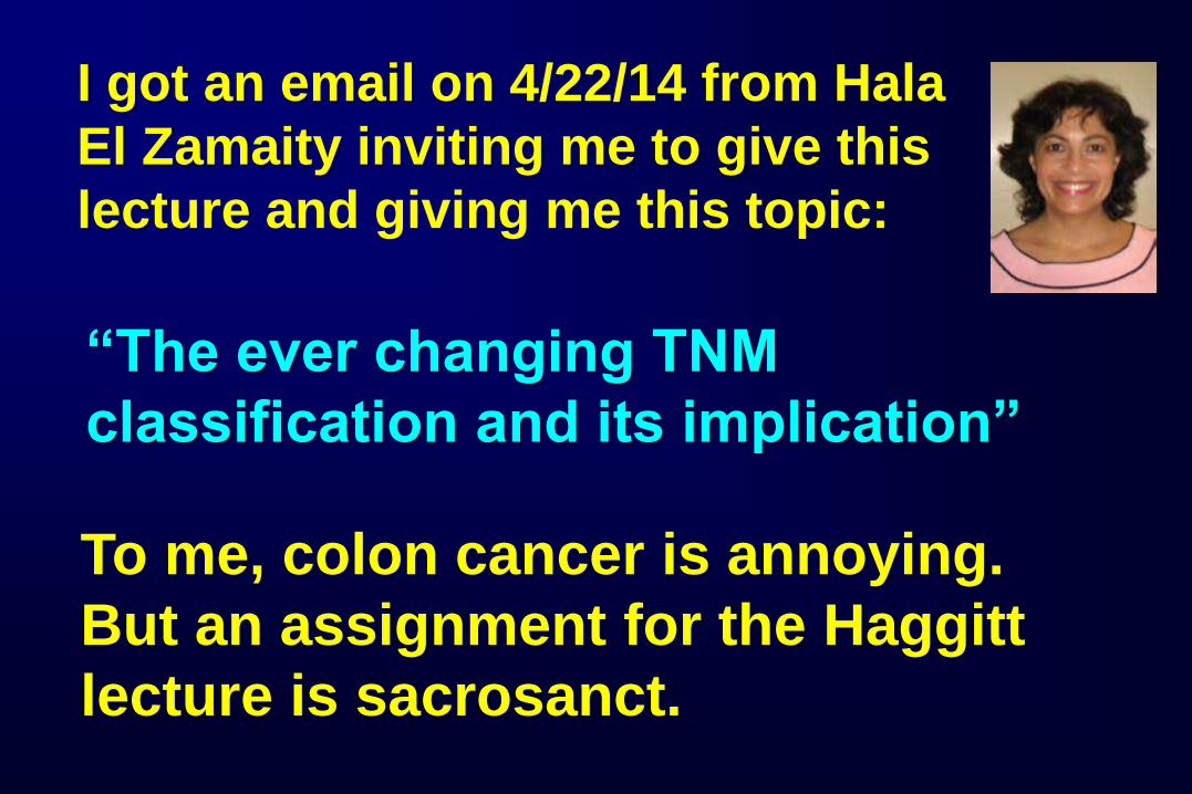
“The ever changing TNM
classification and its implication”
I got an email on 4/22/14 from Hala
El Zamaity inviting me to give this
lecture and giving me this topic:
To me, colon cancer is annoying.
But an assignment for the Haggitt
lecture is sacrosanct.

My primary
sources of
information for
this lecture
include:


My only conflict
of interest is that
I was on the site
task force for the
esophageal chapter

With a bunch of
surgeons and
oncologists who
ignored me
completely….
just like my children

I also used the 2 premier
reference sites for practicing
surgical pathologists when all
other options fail

Disclaimer:
I really wish that I could be
supported by 10 equipment
companies and another 10
reagent companies, but they
have ignored all my requests.

There is a massive amount
of published data covering
everything I will discuss.
I cannot deal with most of it,
since my assignment was to
concentrate on the AJCC
classification.

An erudite, informative paper, far
more insightful and sophisticated
than this lecture.
Mod Pathol,
Supplement 1,
2015;28:S95-S108

Histopathol,
2015;66:102-111
Another erudite, informative
paper, far more insightful and
sophisticated than this lecture.

Staging colorectal
carcinomas:
Historical perspective

4 Nodal
metastases
Transmurally
invasive
carcinoma
The model
for this
discussion

Journal of Pathology and
Bacteriology 1932;35:323
The first staging system
for this carcinoma??
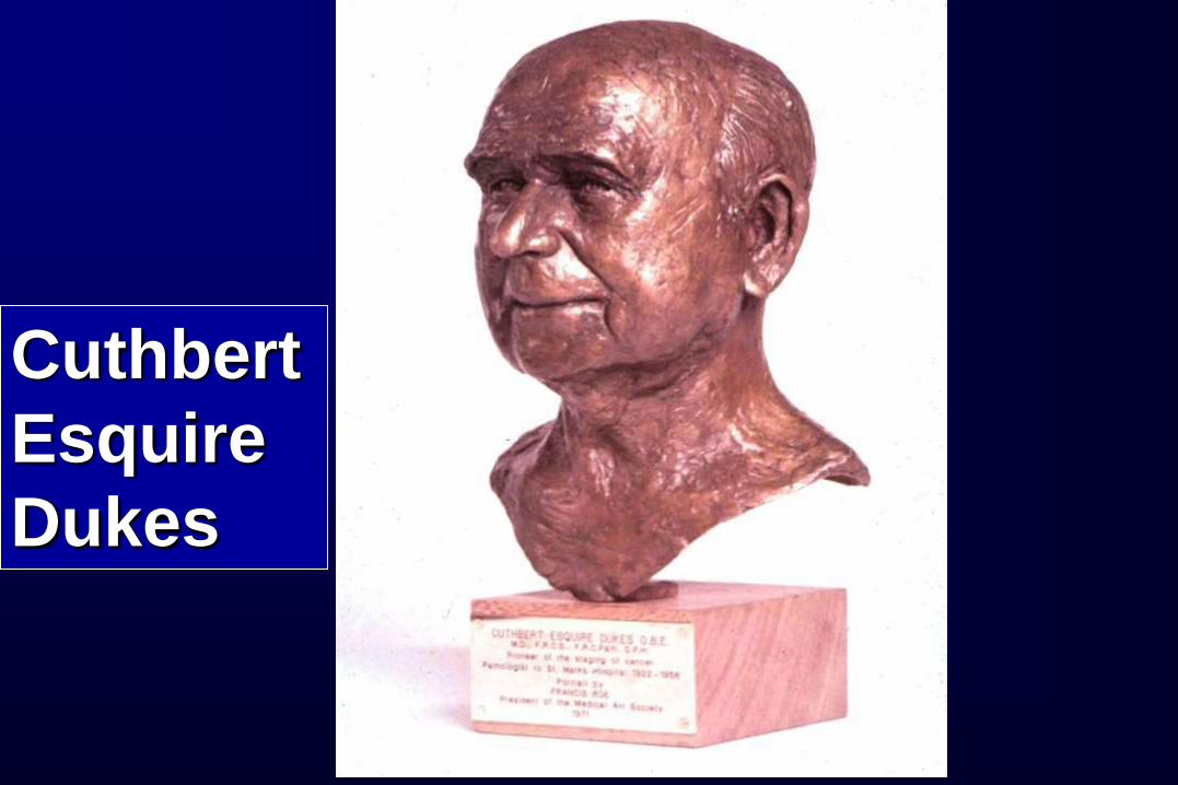
Cuthbert
Esquire
Dukes


Dukes’ staging system
was only for the rectum.
Had 2 components:
1. The extent of the
primary tumor when there
were no nodal metastases
2. Nodal metastases.

The A stage had no
subsets based on
depth
The C stage was
nodal metastases
regardless of depth
of invasion.
Distant metastases
were not included,
nor would they be
for 45 years.

+ nodes
Depth:
extension
into extra
rectal tissues
Dukes, 1932

Dukes stages and
survival were related

Mayo Clinic modification, 1949
A Limited to mucosa
B1 extending into muscularis propria
B2 penetrating muscularis propria
C B1 or B2 with nodal metastasis
Kirklin JW, Dockerty MB, Waugh JW: The role of the peritoneal
reflection in the prognosis of carcinoma of the rectum and
sigmoid colon. Surg Gynec & Obst, 88:326, 1949
The A stage, carcinoma limited to
the mucosa, is really an adenoma
The site now included the sigmoid colon

Comparison of Dukes and Kirklin et al
Dukes Kirklin et al
rectum sig-rectum
A A
B-1
B B-2
C C

+ Nodes
Depth: B2
penetrating
muscularis
Kirklin et al Staging of
a colorectal cancer
C
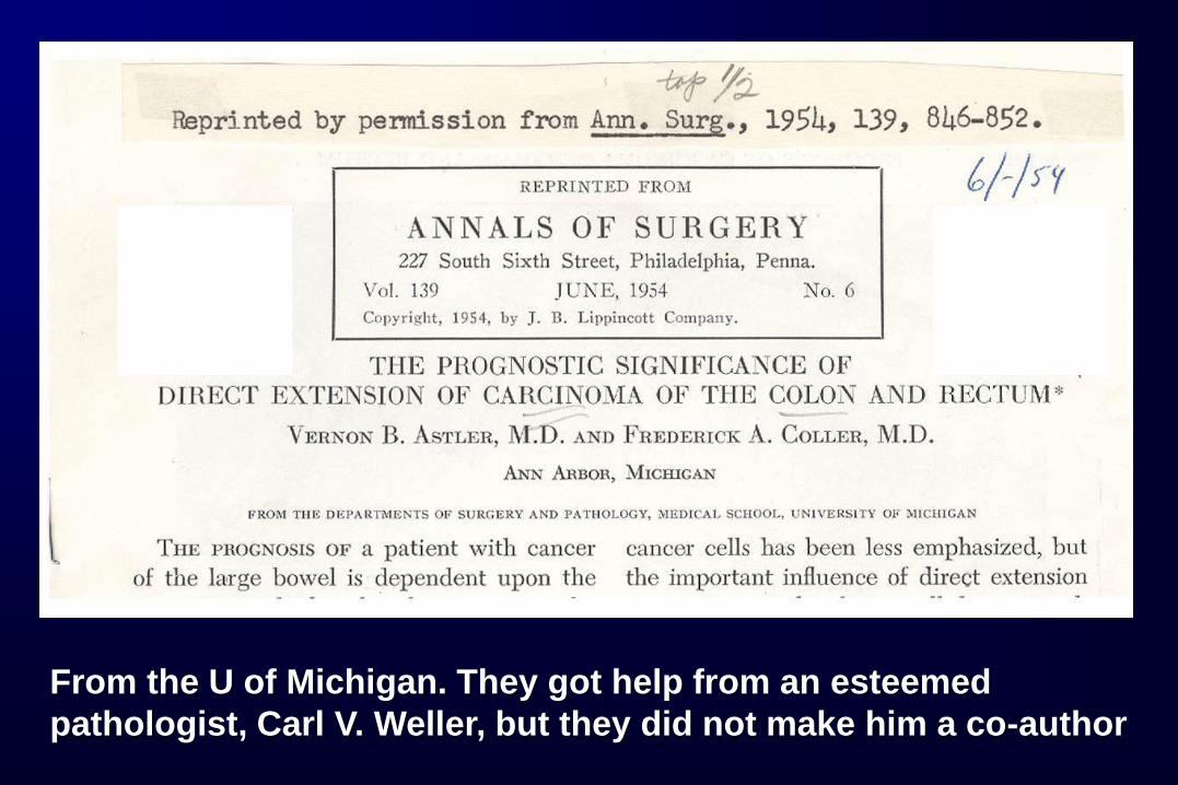
From the U of Michigan. They got help from an esteemed
pathologist, Carl V. Weller, but they did not make him a co-author
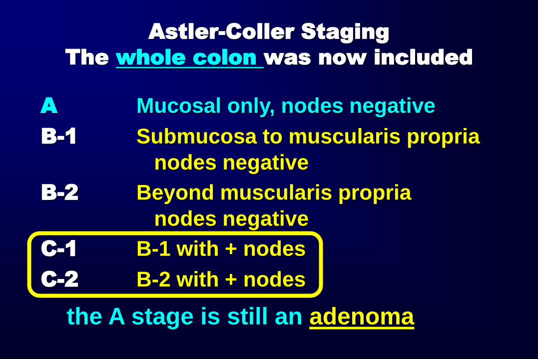
Astler-Coller Staging
The whole colon was now included
A Mucosal only, nodes negative
B-1 Submucosa to muscularis propria
nodes negative
B-2 Beyond muscularis propria
nodes negative
C-1 B-1 with + nodes
C-2 B-2 with + nodes
the A stage is still an adenoma

Astler-Coller modified Kirklin et al which modified Dukes
Dukes Kirklin et al Astler-Coller
rectum sig-rectum colorectum
A A A
B-1 B-1
B B-2 B-2
C C C-1
C-2

+ Nodes
Depth: B2
Beyond
Muscularis
Astler-Coller Staging
of a colorectal cancer
C2

5 year survival using the Astler-Coller
staging based on 352 resected cases at the
University of Michigan, 1940-1944
Stage #cases 5-yr surv
A 1 100%
B-1 48 67%
B-2 164 47%
C-1 14 43%
C-2 125 22%

Finally, the TNM staging
system was introduced
to the general public,
including the
pathologist public

A TNM system was
originally proposed in the
1950s for the clinical
staging of malignant
tumors in general by the
International Union
Against Cancer (UICC).Wood, et al. Staging of cancer of the colon and
cancer of the rectum. Cancer. 1979;43:961-968
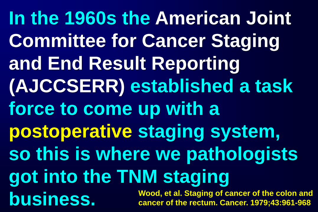
In the 1960s the American Joint
Committee for Cancer Staging
and End Result Reporting
(AJCCSERR) established a task
force to come up with a
postoperative staging system,
so this is where we pathologists
got into the TNM staging
business. Wood, et al. Staging of cancer of the colon and
cancer of the rectum. Cancer. 1979;43:961-968

This postoperative
staging system was
developed, and the
AJCCSERR
published it in the
first edition of the
Staging Manual in
1977
Wood, et al. Staging of cancer of the colon and
cancer of the rectum. Cancer. 1979;43:961-968

The A and B
of previous
systems is T
C is N
M finallyWood, et al. Staging of cancer of the colon and
cancer of the rectum. Cancer. 1979;43:961-968

Primary Tumor (T)
Tis Carcinoma in situ (no
penetration of lamina propria)
T1 Clinically benign lesion or
lesion confined to the mucosa
or submucosa
T2 Involvement of muscular wall
or serosa, no extension beyond
The AJCC 1st Ed Colorectal TNM
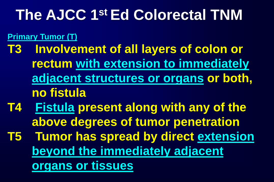
Primary Tumor (T)
T3 Involvement of all layers of colon or
rectum with extension to immediately
adjacent structures or organs or both,
no fistula
T4 Fistula present along with any of the
above degrees of tumor penetration
T5 Tumor has spread by direct extension
beyond the immediately adjacent
organs or tissues
The AJCC 1st Ed Colorectal TNM
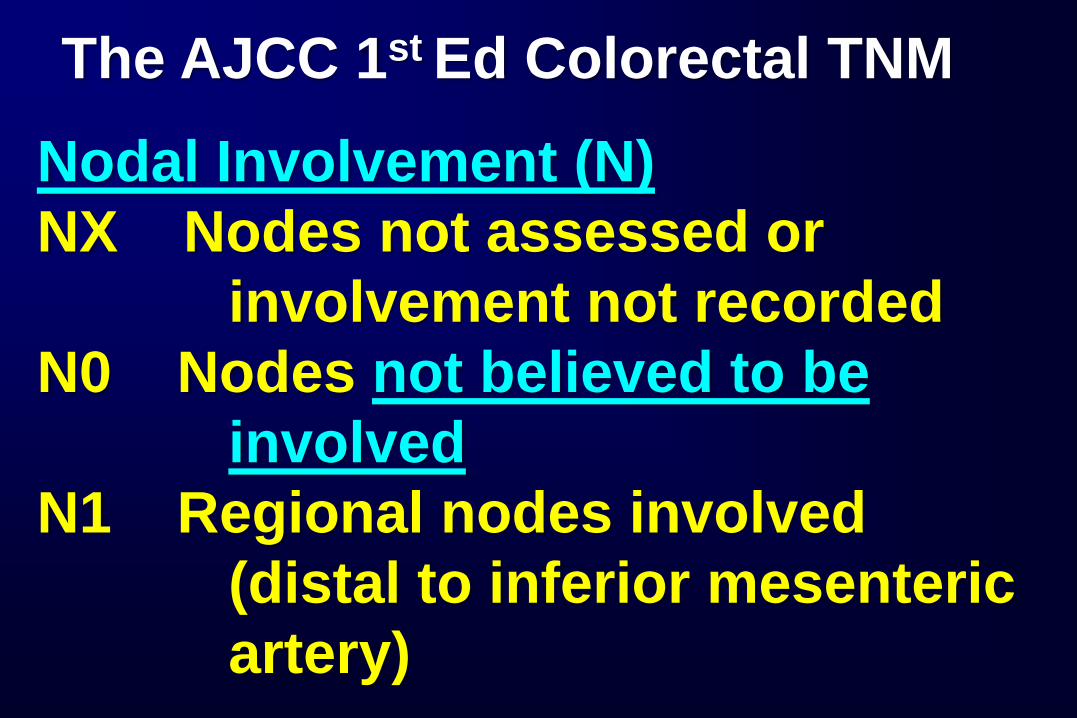
Nodal Involvement (N)
NX Nodes not assessed or
involvement not recorded
N0 Nodes not believed to be
involved
N1 Regional nodes involved
(distal to inferior mesenteric
artery)
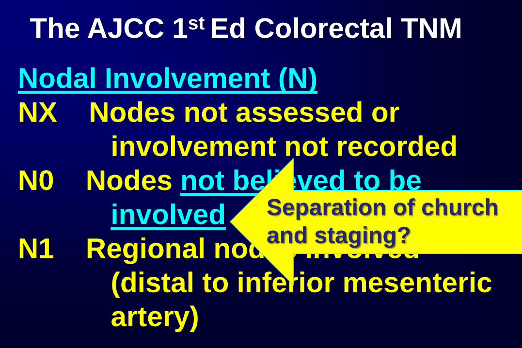
The AJCC 1st Ed Colorectal TNM

Nodal Involvement (N)
NX Nodes not assessed or
involvement not recorded
N0 Nodes not believed to be
involved
N1 Regional nodes involved
(distal to inferior mesenteric
artery)
The AJCC 1st Ed Colorectal TNM
Faith? Religion?
Nodal Involvement (N)
NX Nodes not assessed or
involvement not recorded
N0 Nodes not believed to be
involved
N1 Regional nodes involved
(distal to inferior mesenteric
artery)
The AJCC 1st Ed Colorectal TNM
Separation of church
and staging?

Distant Metastasis (M)
MX Not assessed
M0 No (known) distant
metastasis
M1 Distant metastases
present
The AJCC 1st Ed Colorectal TNM

The 2nd
Edition was
published
in 1983

1987
Why?

Fistulas were not mentioned for T
T3 was limited to pericolic adipose
T4 was extension beyond pericolic
adipose
T5 disappeared
N1 was separated into N1 and N2
based on number of positive nodes
N3 was any + node along a major vessel
3rd
The T and N changed in the 2nd (1983) and
3rd (1988) editions with no change in M.
By the 3rd edition,
1988

4th
5th
Tis Carcinoma in situ:
intraepithelial or
invasion of LP
T1 invasion of submucosa
T4 Direct invasion of other
organs or structures
and/or perforates
visceral peritoneum
DEFINITIONS as of 4th and 5th editions
N3 disappeared
1992
1997

2002
6th

2002
6th Lack of
trust?

Starting with the 6th edition,
big changes were summarized
at the beginning of the chapter

The 7th edition
from 2010 is
what we all use
now. Cancer
registries
require it.

The rest of my
commentsl, both
snide and nit-picking
deal mainly with the
7th edition

Tis
T1
PRIMARY TUMOR (T) 7th EditionTis Carcinoma in situ: intraepithelial
or invasion of lamina propria
T1 invasion of submucosa
T2 invasion of muscularis propria
T3 invasion through the muscularis
propria into pericolorectal
tissues
T4a penetrates to the surface of the
visceral peritoneum
T4b direct invaion or adherence to
other organs or structures

4th Edition
Tis = carcinoma in situ: intraepithelial or
invasion of LPT1 = tumor invades submucosa
1st and 2nd Editions:
Tis = carcinoma in situ
T1 = tumor confined to mucosa or
submucosa3rd Edition
Tis = carcinoma in situ
T1 = tumor invades submucosa
No mention of LP invasion
The Evolution of Tis and T1

The Tis saga, courtesy of the 7th
edition, AJCC Staging Manual
Carcinoma in situ, by definition,
is a non-invasive epithelium
However, in the manual,
carcinoma in situ includes
invasion as well as non-invasion
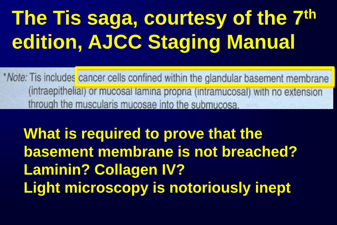
The Tis saga, courtesy of the 7th
edition, AJCC Staging Manual
What is required to prove that the
basement membrane is not breached?
Laminin? Collagen IV?
Light microscopy is notoriously inept

The Tis saga, courtesy of the 7th
edition, AJCC Staging Manual
Is there another lamina propria that
differs from the one in the mucosa?
Nit-Picking stuff:
The colon has crypts, not glands!

malignant
looking
cribriform
stuff

Looks even
worse at
high power

Should we
call this a Tis?

Has it breached
the glandular
basement
membrane?

Complex proliferation
limited to the mucosa

The stroma is pure
lamina propria

Should we
call this a Tis?

I bet this has
breached the
glandular
basement
membrane.

If we do call this
Tis, then we
have to fill out a
template, right?

Hardly anyone
calls this Tis;
HGD instead
and no template

In the esophagus
Tis = HGD
Regardless of the definition
Tis doesn’t metastasize
In the stomach
Tis = intraepithelial
without invasion of LPIn the colon
Tis = intraepithelial or
invasion of LP

In the esophagus
Tis = HGDIn the stomach
Tis = intraepithelial
without invasion of LPIn the colon
Tis = intraepithelial or
invasion of LP
Maybe, Tis should die!

Kill
this!
PRIMARY TUMOR (T) 7th EditionTis Carcinoma in situ: intraepithelial
or invasion of lamina propria
T1 Tumor invades submucosa
T2 Tumor invades muscularis propria
T3 Tumor invades through the
muscularis propria into
pericolorectal tissues
T4a Tumor penetrates to the surface
of the visceral peritoneum
T4b Tumor directly invades or is
adherent to other organs or
structures
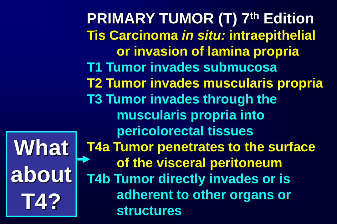
What
about
T4?
PRIMARY TUMOR (T) 7th EditionTis Carcinoma in situ: intraepithelial
or invasion of lamina propria
T1 Tumor invades submucosa
T2 Tumor invades muscularis propria
T3 Tumor invades through the
muscularis propria into
pericolorectal tissues
T4a Tumor penetrates to the surface
of the visceral peritoneum
T4b Tumor directly invades or is
adherent to other organs or
structures

T4 history: fistula timeT3 involves all layers with extention to
immediately adjacent structures or
organs, no fistula
T4 Fistula with any depth of invasion
T5 further extensionT3 invades all layers including serosa
with or without extention to
adjacent or contiguous tissues
+/- fistula
T4 direct extension beyond
contiguous tissue or immediately
adjacent organs
1st
2nd
1983

T4 history: forgotten fistulae
A time of great stability
2nd
3rd
3rd 4th5th
6th
T4: 2 components combined
Invasion of other organs or structures
Perforation of visceral peritoneum

In the 7th edition
T4 is split into 2 parts

T4a
Tumor penetrates
to the surface of the
visceral peritoneum
The T4a mess

On the surface and
growing along it
T4a

On the
surface T4a

Not on the
surface and
covered by
exudate
T4a?

Not on the
surface and
covered by
exudate
T4a?

Various definitions of a T4a tumor:
Tumor actually on the peritoneal surface
Tumor close to the surface with overlying
mesothelial hyperplasia
Tumor close to the surface with overlying
inflammation and exudate.
The AJCC Staging Manual does not give
us guidelines, thus leaving it for the
pathologist community to fight it out

On the
surface!!!
Close to the surface
with reaction!!!

Tumor directly
invades or is
adherent to other
organs or structures
The T4b mess

Urinary bladder muscularis
Sigmoid
carcinoma
pT4b

T4b :Tumor directly invades or is adherent
to other organs or structures
Tumor that is adherent to other organs or
structures, grossly, is classified as cT4b.
(Presumably, grossly means clinically,
since the c prefix is used.)
It seems logical that if tumor is found
microscopically to be in the adhesion,
then it should be classified as pT4b, but
that is not clarified.

T4b :Tumor directly invades or is adherent
to other organs or structures
It does say: However, if no tumor is
present in the adhesion, microscopically,
the classification should be pT1-4a
depending on the anatomic depth of wall
invasion.”
How can a T1 or T2 tumor
(confined to the wall) adhere
to another organ or structure?
Only with perseverance and speed

Enough of the
T stuff!
Let’s explore
N….

But then we would
lose the chance to
fight the battle of
Are you exhausted yet?
I can stop here, and you can
rest….go drinking
tumor deposits!!

Things nodal were
pretty stable from
the 1st through the
6th editions

Nodal Involvement (N) 1st Edition
NX Nodes not assessed or involvement
not recorded
N0 Nodes not believed to be involved
N1 Regional nodes involved (distal to
inferior mesenteric artery)
2nd through 6th Edition
1st
ed
6th
ed
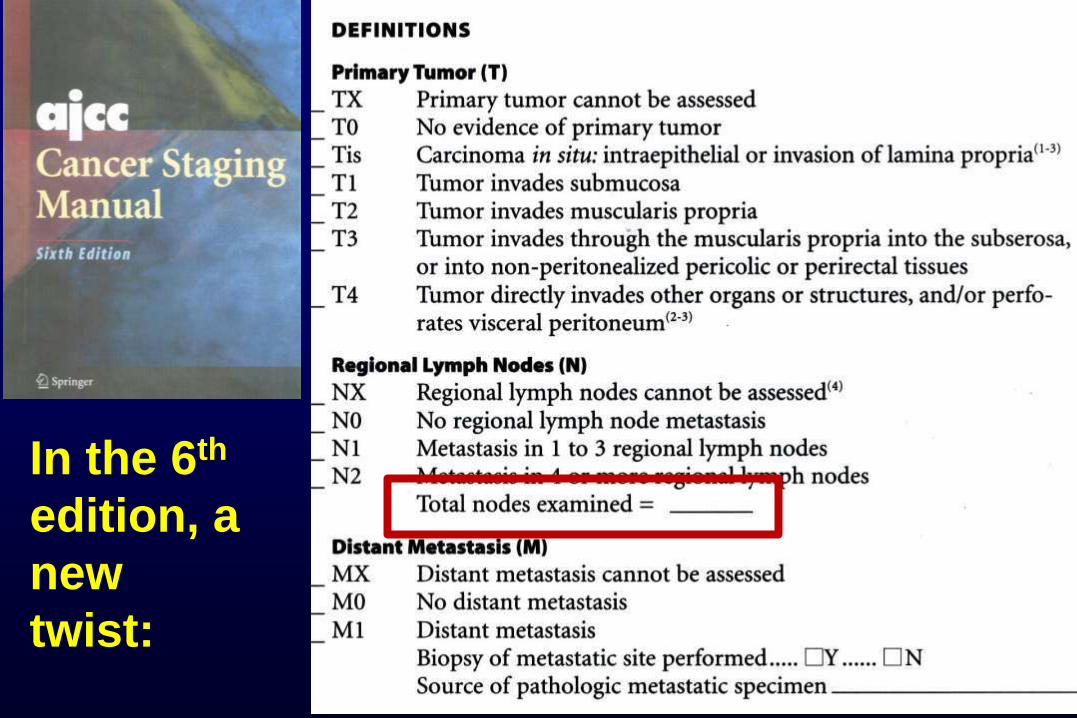
In the 6th
edition, a
new
twist:

This appears in the staging check list
at the end of the chapter, but there is no
explanation in the text for why it is included.
The number of total nodes must be
important!

However, it was not important for
long, because this was dropped
from the 7th edition, only 8 years
later.

The 7th edition
went ballistic!
N1 was split into 3 parts
N2 was split into 2 parts
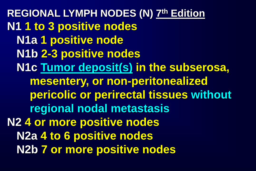
REGIONAL LYMPH NODES (N) 7th Edition
N1 1 to 3 positive nodes
N1a 1 positive node
N1b 2-3 positive nodes
N1c Tumor deposit(s) in the subserosa,
mesentery, or non-peritonealized
pericolic or perirectal tissues without
regional nodal metastasis
N2 4 or more positive nodes
N2a 4 to 6 positive nodes
N2b 7 or more positive nodes

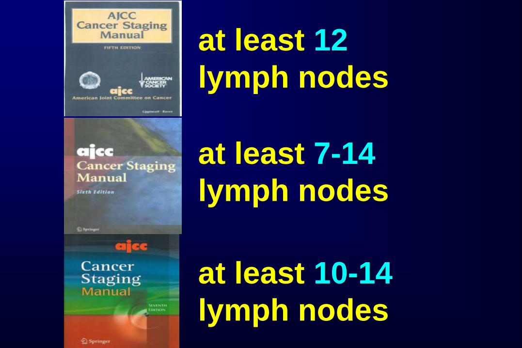
How many nodes are enough?
The first to deal
with this was
the 5th edition:
“it is desirable to
obtain at least 12
lymph nodes …..”

The 6th edition
“it is important
to obtain at
least 7-14
lymph nodes…”
How many nodes are enough?

How many nodes are enough?
The 7th edition:
“it is important to
obtain at least
10-14 lymph
nodes ….”
at least 12
lymph nodes
at least 7-14
lymph nodes
at least 10-14
lymph nodes

at least 7-14 nodes!
Is it 7? Or is it 14?
at least 10-14 nodes!
Is it 10? Or is it 14?
Are they serious?

How many is enough lymph nodes?
The 12 rule
There are studies that say that tumors
for which fewer than 12 nodes are found
do worse than tumors for which
12 or more nodes are found
3rd party payers are legislating 12 nodes
The Dutch only need 10, a 16.7% difference
The lymph node saga

Theoretically, finding more nodes
should find more nodal metastases.
So, there should be significant
upstaging of tumors with fewer
nodes found initially that have more
nodes found on re-evaluation.
If more nodes are found over time,
there should be a trend toward
upstaging.

Using SEER data, 1995-2005:
The number of lymph nodes hospitals
examine ….is not associated with staging,
use of adjuvant chemotherapy, or patient
survival.
Efforts by payers and professional
organizations to increase node examination
rates may have limited value as a public
health intervention.
Wong SL, et al. JAMA. 2007;298:2149-54
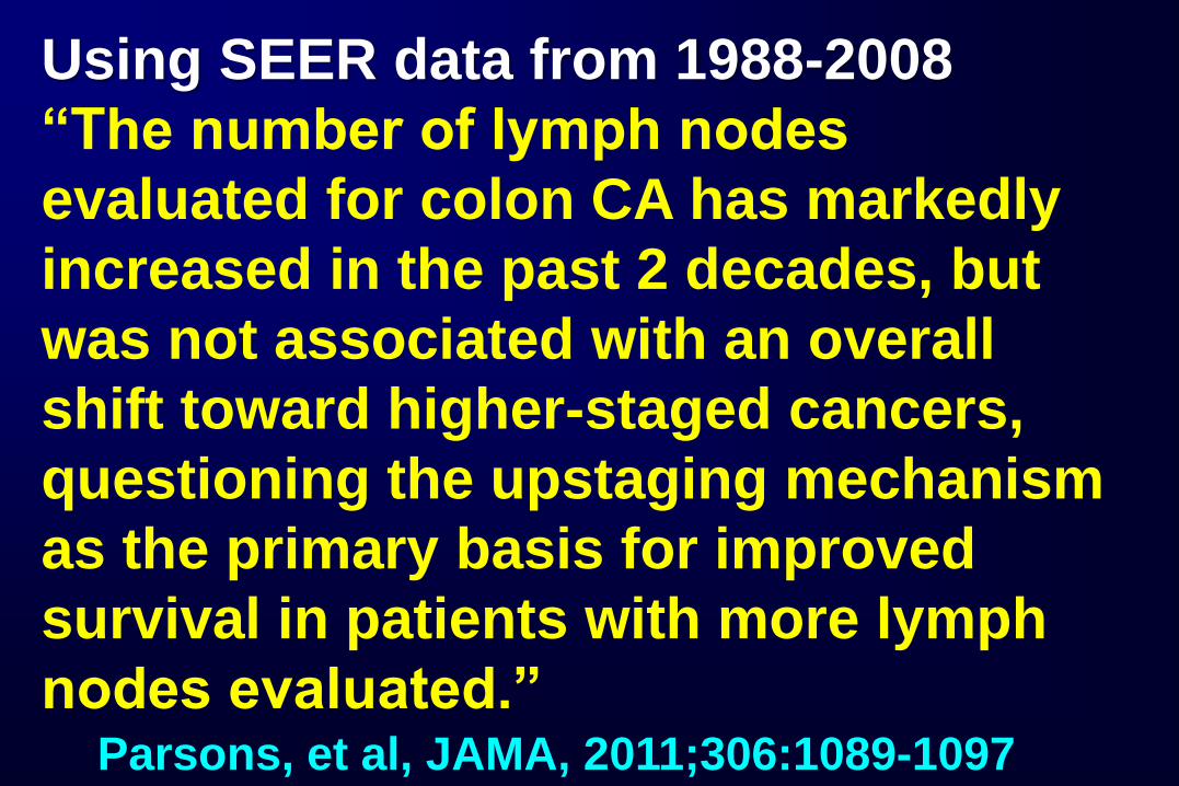
Using SEER data from 1988-2008
“The number of lymph nodes
evaluated for colon CA has markedly
increased in the past 2 decades, but
was not associated with an overall
shift toward higher-staged cancers,
questioning the upstaging mechanism
as the primary basis for improved
survival in patients with more lymph
nodes evaluated.”Parsons, et al, JAMA, 2011;306:1089-1097

These two important
epidemiologic studies
appeared in the JAMA.
Do cancer people ever read
the JAMA?
Maybe that is why they pay
no attention to the results.

Other studies
dispute this and
hold on to 12 or
some other
number

In the 7th edition, the AJCC says
10-14 nodes, and
“when fewer than the number
of nodes recommended by the
CAP have been found, it is
important that the pathologist
report the degree of diligence
of their efforts to find lymph
nodes….”

In the 7th edition, the AJCC says
10-14 nodes, and
“when fewer than the number
of nodes recommended by the
CAP have been found, it is
important that the pathologist
report the degree of diligence
of their efforts to find lymph
nodes….”
CAP: Where in the
hell did those
guys come from?
They are
everywhere!

In the 7th edition, the AJCC says
10-14 nodes, and
“when fewer than the number
of nodes recommended by the
CAP have been found, it is
important that the pathologist
report the degree of diligence
of their efforts to find lymph
nodes….”

In the 7th edition, the AJCC says
10-14 nodes, and
“when fewer than the number of
nodes recommended by the
CAP have been found, it is
important that the pathologist
report the degree of diligence
of their efforts to find lymph
nodes….”
This is an insulting demand!

Comment: I looked through the sigmoid
mesocolon 3 times, trying to find 5 more nodes,
but there were none.
I even sent through 7 blocks of the mesocolon
hoping for some tiny nodes, but there were none.
I truly apologize for my inadequacies,
and I promise to do better in the future.
Diagnosis: Sigmoid colon:
Transmurally invasive adenocarinoma.
Metastases in one of 7 nodes.
Example

I have a better suggestion:
“when fewer than the number
of nodes recommended by the
CAP have been removed, it is
important that the surgeon
report the degree of diligence
of her/his efforts to remove
enough lymph nodes….”

4+ Nodes
N2a
Depth: T3
invades
through
muscularis
TNM Staging a colorectal cancer: have to count + nodes Jan 2010
T3
N2a
M not
used
200 nodes
recovered

Stage T N M 5-Yr Surv
I T1-2 N0 M0 74-79%
IIA T3 N0 M0 67%
IIB T4a N0 M0 61%
IIC T4b N0 M0 46%
IIIA T1-2 N1/N1c M0 67-74%
T1 N2a M0 65%
IIIB T3-4a N1/N1c M0 52-58%
T3 N2a M0 43%
T1-2 N2b M0 52%
IIIC T4a N2a M0 33%
T4a N2b M0 18%
T4b N1 M0 30%
IVA Any T Any N M1a ~6%
IVB Any T Any N M1b ~5%
As of
Jan
2010
SEER
Wrong
stage?

What about M: Distant Metastasis?
1st through 6th editions:
MX cannot be assessed
M0 No distant metastasis
M1 Distant metastasis
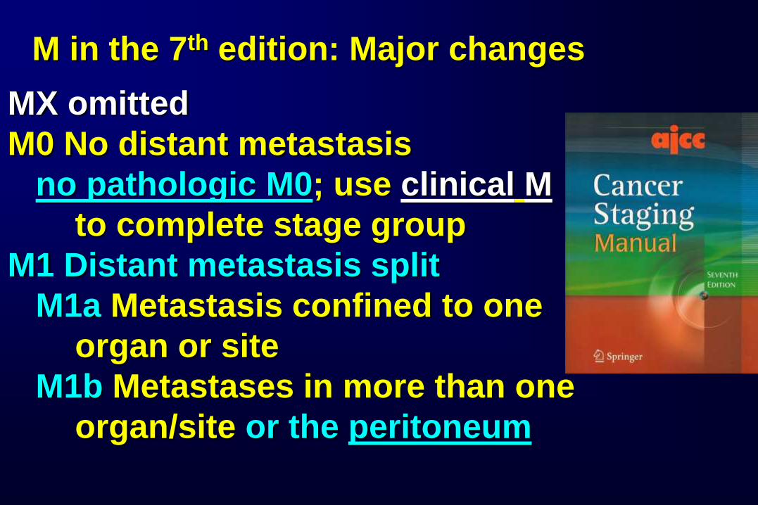
MX omitted
M0 No distant metastasis
no pathologic M0; use clinical M
to complete stage group
M1 Distant metastasis split
M1a Metastasis confined to one
organ or site
M1b Metastases in more than one
organ/site or the peritoneum
M in the 7th edition: Major changes