Embed Size (px)
Citation preview
ARTICLE
Received 26 Feb 2016 | Accepted 1 Aug 2016 | Published 26 Sep 2016
The RNA-binding protein vigilin regulates VLDLsecretion through modulation of Apob mRNAtranslationMehrpouya B. Mobin1, Stefanie Gerstberger2, Daniel Teupser3, Benedetta Campana4, Klaus Charisse5,
Markus H. Heim4, Muthiah Manoharan5, Thomas Tuschl2 & Markus Stoffel1
The liver is essential for the synthesis of plasma proteins and integration of lipid
metabolism. While the role of transcriptional networks in these processes is increasingly
understood, less is known about post-transcriptional control of gene expression by
RNA-binding proteins (RBPs). Here, we show that the RBP vigilin is upregulated in livers of
obese mice and in patients with fatty liver disease. By using in vivo, biochemical and genomic
approaches, we demonstrate that vigilin controls very-low-density lipoprotein (VLDL)
secretion through the modulation of apolipoproteinB/Apob mRNA translation. Crosslinking
studies reveal that vigilin binds to CU-rich regions in the mRNA coding sequence of Apob
and other proatherogenic secreted proteins, including apolipoproteinC-III/Apoc3 and
fibronectin/Fn1. Consequently, hepatic vigilin knockdown decreases VLDL/low-density
lipoprotein (LDL) levels and formation of atherosclerotic plaques in Ldlr�/� mice.
These studies uncover a role for vigilin as a key regulator of hepatic Apob translation and
demonstrate the therapeutic potential of inhibiting vigilin for cardiovascular diseases.
DOI: 10.1038/ncomms12848 OPEN
1 Institute of Molecular Health Sciences, ETH Zurich, Otto-Stern Weg 7, 8093 Zurich, Switzerland. 2 Howard Hughes Medical Institute, The RockefellerUniversity, 1230 York Avenue, New York, New York 10065, USA. 3 Institute of Laboratory Medicine, Ludwig-Maximilians-University Munich, Marchioninistr.15, 81377 Munich, Germany. 4 Department of Biomedicine and Clinic for Gastroenterology and Hepatology, University Hospital Basel, Hebelstrasse 20,4031 Basel, Switzerland. 5 Alnylam Pharmaceuticals, 300 Third Street, Cambridge, Massachusetts 02142, USA. Correspondence and requests for materialsshould be addressed to M.S. (email: [email protected]).
NATURE COMMUNICATIONS | 7:12848 | DOI: 10.1038/ncomms12848 | www.nature.com/naturecommunications 1
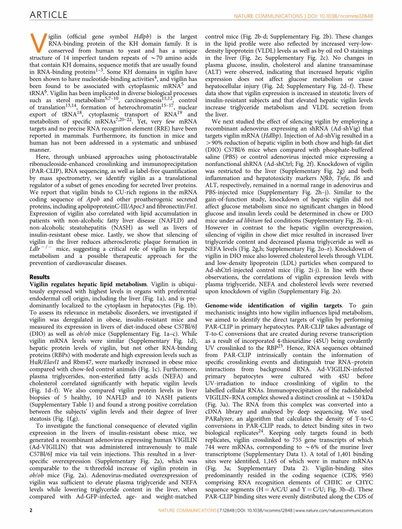
Vigilin (official gene symbol Hdlpb) is the largestRNA-binding protein of the KH domain family. It isconserved from human to yeast and has a unique
structure of 14 imperfect tandem repeats of B70 amino acidsthat contain KH domains, sequence motifs that are usually foundin RNA-binding proteins1–3. Some KH domains in vigilin havebeen shown to have nucleotide-binding activities4, and vigilin hasbeen found to be associated with cytoplasmic mRNA5 andtRNA6. Vigilin has been implicated in diverse biological processessuch as sterol metabolism5,7–10, carcinogenesis11,12, controlof translation13,14, formation of heterochromatin15–17, nuclearexport of tRNA18, cytoplasmic transport of RNA19 andmetabolism of specific mRNAs7,20–22. Yet, very few mRNAtargets and no precise RNA recognition element (RRE) have beenreported in mammals. Furthermore, its function in mice andhuman has not been addressed in a systematic and unbiasedmanner.
Here, through unbiased approaches using photoactivatableribonucleoside-enhanced crosslinking and immunoprecipitation(PAR-CLIP), RNA sequencing, as well as label-free quantificationby mass spectrometry, we identify vigilin as a translationalregulator of a subset of genes encoding for secreted liver proteins.We report that vigilin binds to CU-rich regions in the mRNAcoding sequence of Apob and other proatherogenic secretedproteins, including apolipoproteinC-III/Apoc3 and fibronectin/Fn1.Expression of vigilin also correlated with lipid accumulation inpatients with non-alcoholic fatty liver disease (NAFLD) andnon-alcoholic steatohepatitis (NASH) as well as livers ofinsulin-resistant obese mice. Lastly, we show that silencing ofvigilin in the liver reduces atherosclerotic plaque formation inLdlr� /� mice, suggesting a critical role of vigilin in hepaticmetabolism and a possible therapeutic approach for theprevention of cardiovascular diseases.
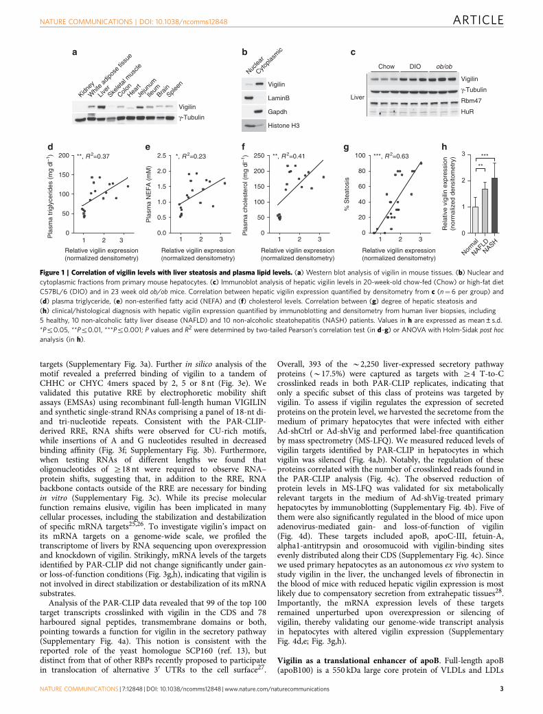
ResultsVigilin regulates hepatic lipid metabolism. Vigilin is ubiqui-tously expressed with highest levels in organs with preferentialendodermal cell origin, including the liver (Fig. 1a), and is pre-dominantly localized to the cytoplasm in hepatocytes (Fig. 1b).To assess its relevance in metabolic disorders, we investigated ifvigilin was deregulated in obese, insulin-resistant mice andmeasured its expression in livers of diet-induced obese C57Bl/6J(DIO) as well as ob/ob mice (Supplementary Fig. 1a–c). Whilevigilin mRNA levels were similar (Supplementary Fig. 1d),hepatic protein levels of vigilin, but not other RNA-bindingproteins (RBPs) with moderate and high expression levels such asHuR/Elavl1 and Rbm47, were markedly increased in obese micecompared with chow-fed control animals (Fig. 1c). Furthermore,plasma triglycerides, non-esterified fatty acids (NEFA) andcholesterol correlated significantly with hepatic vigilin levels(Fig. 1d–f). We also compared vigilin protein levels in liverbiopsies of 5 healthy, 10 NAFLD and 10 NASH patients(Supplementary Table 1) and found a strong positive correlationbetween the subjects’ vigilin levels and their degree of liversteatosis (Fig. 1f,g).
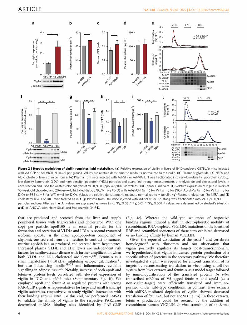
To investigate the functional consequence of elevated vigilinexpression in the livers of insulin-resistant obese mice, wegenerated a recombinant adenovirus expressing human VIGILIN(Ad-VIGILIN) that was administered intravenously to maleC57Bl/6J mice via tail vein injections. This resulted in a liver-specific overexpression (Supplementary Fig. 2a), which wascomparable to the Ethreefold increase of vigilin protein inob/ob mice (Fig. 2a). Adenovirus-mediated overexpression ofvigilin was sufficient to elevate plasma triglyceride and NEFAlevels while lowering triglyceride content in the liver, whencompared with Ad-GFP-infected, age- and weight-matched
control mice (Fig. 2b-d; Supplementary Fig. 2b). These changesin the lipid profile were also reflected by increased very-low-density lipoprotein (VLDL) levels as well as by oil red O stainingsin the liver (Fig. 2e; Supplementary Fig. 2c). No changes inplasma glucose, insulin, cholesterol and alanine transaminase(ALT) were observed, indicating that increased hepatic vigilinexpression does not affect glucose metabolism or causehepatocellular injury (Fig. 2d; Supplementary Fig. 2d–f). Thesedata show that vigilin expression is increased in steatotic livers ofinsulin-resistant subjects and that elevated hepatic vigilin levelsincrease triglyceride metabolism and VLDL secretion fromthe liver.
We next studied the effect of silencing vigilin by employing arecombinant adenovirus expressing an shRNA (Ad-shVig) thattargets vigilin mRNA (Hdlbp). Injection of Ad-shVig resulted in a490% reduction of hepatic vigilin in both chow and high-fat diet(DIO) C57Bl/6 mice when compared with phosphate-bufferedsaline (PBS) or control adenovirus injected mice expressing anonfunctional shRNA (Ad-shCtrl; Fig. 2f). Knockdown of vigilinwas restricted to the liver (Supplementary Fig. 2g) and bothinflammation and hepatotoxicity markers Nfkb, Tnfa, Il6 andALT, respectively, remained in a normal range in adenovirus andPBS-injected mice (Supplementary Fig. 2h–j). Similar to thegain-of-function study, knockdown of hepatic vigilin did notaffect glucose metabolism since no significant changes in bloodglucose and insulin levels could be determined in chow or DIOmice under ad libitum fed conditions (Supplementary Fig. 2k–n).However in contrast to the hepatic vigilin overexpression,silencing of vigilin in chow diet mice resulted in increased livertriglyceride content and decreased plasma triglyceride as well asNEFA levels (Fig. 2g,h; Supplementary Fig. 2o–r). Knockdown ofvigilin in DIO mice also lowered cholesterol levels through VLDLand low-density lipoprotein (LDL) particles when compared toAd-shCtrl-injected control mice (Fig. 2i-j). In line with theseobservations, the correlations of vigilin expression levels withplasma triglyceride, NEFA and cholesterol levels were reversedupon knockdown of vigilin (Supplementary Fig. 2s).
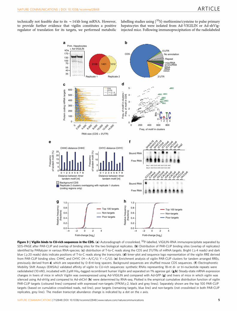
Genome-wide identification of vigilin targets. To gainmechanistic insights into how vigilin influences lipid metabolism,we aimed to identify the direct targets of vigilin by performingPAR-CLIP in primary hepatocytes. PAR-CLIP takes advantage ofT-to-C conversions that are created during reverse transcriptionas a result of incorporated 4-thiouridine (4SU) being covalentlyUV crosslinked to the RBP23. Hence, RNA sequences obtainedfrom PAR-CLIP intrinsically contain the information ofspecific crosslinking events and distinguish true RNA–proteininteractions from background RNA. Ad-VIGILIN-infectedprimary hepatocytes were cultured with 4SU beforeUV-irradiation to induce crosslinking of vigilin to thelabelled cellular RNAs. Immunoprecipitation of the radiolabeledVIGILIN–RNA complex showed a distinct crosslink at B150 kDa(Fig. 3a). The RNA from this complex was converted into acDNA library and analysed by deep sequencing. We usedPARalyzer, an algorithm that calculates the density of T-to-Cconversions in PAR-CLIP reads, to detect binding sites in twobiological replicates24. Keeping only targets found in bothreplicates, vigilin crosslinked to 755 gene transcripts of which744 were mRNAs, corresponding to B6% of the murine livertranscriptome (Supplementary Data 1). A total of 1,401 bindingsites were identified, 1,165 of which were in mature mRNAs(Fig. 3a; Supplementary Data 2). Vigilin-binding sitespredominantly resided in the coding sequence (CDS; 956)comprising RNA recognition elements of CHHC or CHYCsequence segments (H¼A/C/U and Y¼C/U; Fig. 3b–d). ThesePAR-CLIP binding sites were evenly distributed along the CDS of
ARTICLE NATURE COMMUNICATIONS | DOI: 10.1038/ncomms12848
2 NATURE COMMUNICATIONS | 7:12848 | DOI: 10.1038/ncomms12848 | www.nature.com/naturecommunications
targets (Supplementary Fig. 3a). Further in silico analysis of themotif revealed a preferred binding of vigilin to a tandem ofCHHC or CHYC 4mers spaced by 2, 5 or 8 nt (Fig. 3e). Wevalidated this putative RRE by electrophoretic mobility shiftassays (EMSAs) using recombinant full-length human VIGILINand synthetic single-strand RNAs comprising a panel of 18-nt di-and tri-nucleotide repeats. Consistent with the PAR-CLIP-derived RRE, RNA shifts were observed for CU-rich motifs,while insertions of A and G nucleotides resulted in decreasedbinding affinity (Fig. 3f; Supplementary Fig. 3b). Furthermore,when testing RNAs of different lengths we found thatoligonucleotides of Z18 nt were required to observe RNA–protein shifts, suggesting that, in addition to the RRE, RNAbackbone contacts outside of the RRE are necessary for bindingin vitro (Supplementary Fig. 3c). While its precise molecularfunction remains elusive, vigilin has been implicated in manycellular processes, including the stabilization and destabilizationof specific mRNA targets25,26. To investigate vigilin’s impact onits mRNA targets on a genome-wide scale, we profiled thetranscriptome of livers by RNA sequencing upon overexpressionand knockdown of vigilin. Strikingly, mRNA levels of the targetsidentified by PAR-CLIP did not change significantly under gain-or loss-of-function conditions (Fig. 3g,h), indicating that vigilin isnot involved in direct stabilization or destabilization of its mRNAsubstrates.
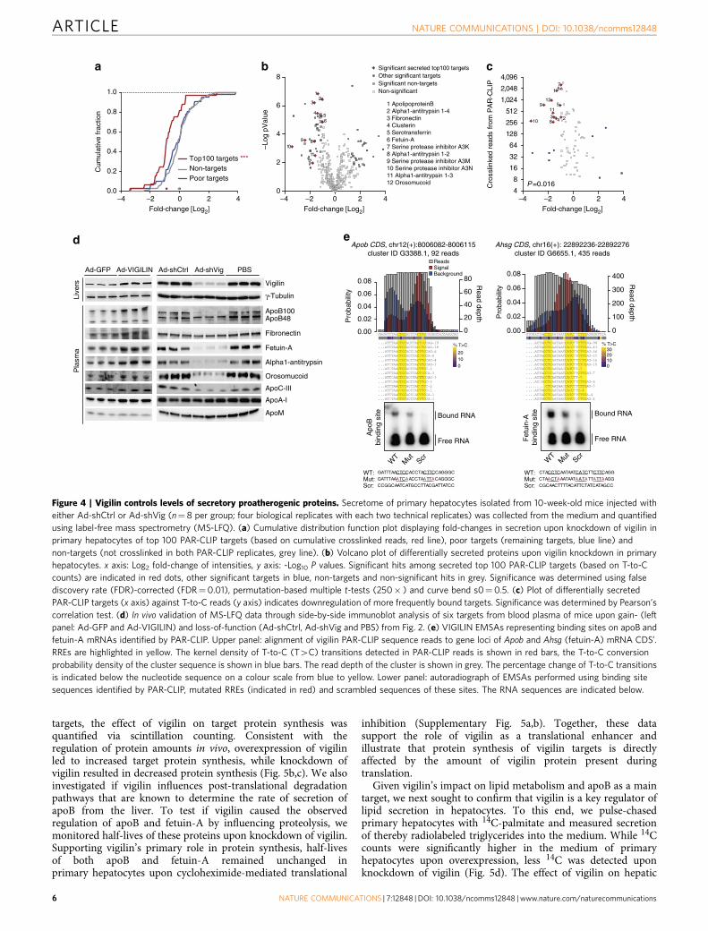
Analysis of the PAR-CLIP data revealed that 99 of the top 100target transcripts crosslinked with vigilin in the CDS and 78harboured signal peptides, transmembrane domains or both,pointing towards a function for vigilin in the secretory pathway(Supplementary Fig. 4a). This notion is consistent with thereported role of the yeast homologue SCP160 (ref. 13), butdistinct from that of other RBPs recently proposed to participatein translocation of alternative 30 UTRs to the cell surface27.
Overall, 393 of the B2,250 liver-expressed secretory pathwayproteins (B17.5%) were captured as targets with Z4 T-to-Ccrosslinked reads in both PAR-CLIP replicates, indicating thatonly a specific subset of this class of proteins was targeted byvigilin. To assess if vigilin regulates the expression of secretedproteins on the protein level, we harvested the secretome from themedium of primary hepatocytes that were infected with eitherAd-shCtrl or Ad-shVig and performed label-free quantificationby mass spectrometry (MS-LFQ). We measured reduced levels ofvigilin targets identified by PAR-CLIP in hepatocytes in whichvigilin was silenced (Fig. 4a,b). Notably, the regulation of theseproteins correlated with the number of crosslinked reads found inthe PAR-CLIP analysis (Fig. 4c). The observed reduction ofprotein levels in MS-LFQ was validated for six metabolicallyrelevant targets in the medium of Ad-shVig-treated primaryhepatocytes by immunoblotting (Supplementary Fig. 4b). Five ofthem were also significantly regulated in the blood of mice uponadenovirus-mediated gain- and loss-of-function of vigilin(Fig. 4d). These targets included apoB, apoC-III, fetuin-A,alpha1-antitrypsin and orosomucoid with vigilin-binding sitesevenly distributed along their CDS (Supplementary Fig. 4c). Sincewe used primary hepatocytes as an autonomous ex vivo system tostudy vigilin in the liver, the unchanged levels of fibronectin inthe blood of mice with reduced hepatic vigilin expression is mostlikely due to compensatory secretion from extrahepatic tissues28.Importantly, the mRNA expression levels of these targetsremained unperturbed upon overexpression or silencing ofvigilin, thereby validating our genome-wide transcript analysisin hepatocytes with altered vigilin expression (SupplementaryFig. 4d,e; Fig. 3g,h).
Vigilin as a translational enhancer of apoB. Full-length apoB(apoB100) is a 550 kDa large core protein of VLDLs and LDLs
a cb
Nuclea
r
Cytopla
smic
Vigilin
LaminB
Gapdh
Histone H3
Chow ob/obDIO
Kidney
Whit
e ad
ipose
tissu
e
Liver
Skelet
al m
uscle
Colon
Heart
Jejun
um
Ileum
BrainSple
en
γ-Tubulin
Vigilin
Liver
Vigilin
γ-Tubulin
HuR
Rbm47
Norm
al
NAFLD
NASH
0
1
2
3 ***
**
h
0.0
0.5
1.0
1.5
2.0
2.5P
lasm
a N
EF
A (
mM
)d e
0
20
40
60
80
100
% S
teat
osis
***, R 2=0.63f g
0
250
Pla
sma
chol
este
rol (
mg
dl–1
) **, R 2=0.41
1 2 30
50
100
150
200
50
100
150
200
Relative vigilin expression(normalized densitometry)
1 2 3
Relative vigilin expression(normalized densitometry)
1 2 3
Relative vigilin expression(normalized densitometry)
1 2 3
Relative vigilin expression(normalized densitometry)
Rel
ativ
e vi
gilin
exp
ress
ion
(nor
mal
ized
den
sito
met
ry)
Pla
sma
trig
lyce
rides
(m
g dl
–1) **, R 2=0.37 *, R 2=0.23
Figure 1 | Correlation of vigilin levels with liver steatosis and plasma lipid levels. (a) Western blot analysis of vigilin in mouse tissues. (b) Nuclear and
cytoplasmic fractions from primary mouse hepatocytes. (c) Immunoblot analysis of hepatic vigilin levels in 20-week-old chow-fed (Chow) or high-fat diet
C57BL/6 (DIO) and in 23 week old ob/ob mice. Correlation between hepatic vigilin expression quantified by densitometry from c (n¼ 6 per group) and
(d) plasma triglyceride, (e) non-esterified fatty acid (NEFA) and (f) cholesterol levels. Correlation between (g) degree of hepatic steatosis and
(h) clinical/histological diagnosis with hepatic vigilin expression quantified by immunoblotting and densitometry from human liver biopsies, including
5 healthy, 10 non-alcoholic fatty liver disease (NAFLD) and 10 non-alcoholic steatohepatitis (NASH) patients. Values in h are expressed as mean±s.d.
*Pr0.05, **Pr0.01, ***Pr0.001; P values and R2 were determined by two-tailed Pearson’s correlation test (in d–g) or ANOVA with Holm-Sidak post hoc
analysis (in h).
NATURE COMMUNICATIONS | DOI: 10.1038/ncomms12848 ARTICLE
NATURE COMMUNICATIONS | 7:12848 | DOI: 10.1038/ncomms12848 | www.nature.com/naturecommunications 3
that are produced and secreted from the liver and supplyperipheral tissues with triglycerides and cholesterol. With onecopy per particle, apoB100 is an essential protein for theformation and secretion of VLDLs and LDLs. A second truncatedisoform, apoB48, is the main apolipoprotein component ofchylomicrons secreted from the intestine. In contrast to humans,murine apoB48 is also produced and secreted from hepatocytes.Increased plasma VLDL and LDL levels are independent riskfactors for cardiovascular disease with further amplification whenboth VLDL and LDL cholesterol are elevated29. Fetuin-A is asmall hepatokine (E50 kDa) inhibiting ectopic calcification30,but also influencing insulin31–33 and inflammatory cytokinesignalling in adipose tissue30. Notably, increase of both apoB andfetuin-A protein levels correlated with elevated expression ofvigilin in DIO and ob/ob mice (Supplementary Fig. 4f). Weemployed apoB and fetuin-A as regulated proteins with strongPAR-CLIP signals as representatives for large and small transcriptvigilin substrates, respectively, to study vigilin’s interaction withtheir binding sites in vitro. To this end, we performed EMSAsto validate the affinity of vigilin to the respective PARalyzerdetermined mRNA binding sites identified by PAR-CLIP
(Fig. 4e). Whereas the wild-type sequences of respectivebinding regions induced a shift in electrophoretic mobility ofrecombinant, RNA-depleted VIGILIN, mutations of the identifiedRRE and scrambled sequences of these sites exhibited decreasedor no binding affinity by human VIGILIN.
Given the reported association of the yeast22 and vertebratehomologues34 with ribosomes and our observation thatvigilin positively regulates its targets post-transcriptionally,we hypothesized that vigilin influences protein production of aspecific subset of proteins in the secretory pathway. We thereforeinvestigated if vigilin was required for efficient translation of itstargets by reconstructing translation in vitro using a cell-freesystem from liver extracts and fetuin-A as a model target followedby immunopurification of the translated protein. In vitrotranscribed mRNAs of V5-tagged fetuin-A and apoM (as anon-vigilin-target) were efficiently translated and immuno-purified under wild-type conditions. In contrast, liver extractswith shRNA-mediated depletion of vigilin showed decreasedtranslation of fetuin-A, but not apoM (Fig. 5a). In these extracts,fetuin-A production could be rescued by the addition ofrecombinant human VIGILIN. In vitro translation of apoB was
ApoB48
ApoB100
ApoA-I
0
2
4
6
8
Cho
lest
erol
[(m
g dl
–1)
0
100
200
300
Pla
sma
chol
este
rol (
mg
dl–1
)
** *
0
2
4
6
8
100
1
2
3
4
Ad-GFPAd-VIGILIN
Ad-VIGILIN
1.5
1.0
0.5
0.0
Rel
. vig
ilin
expr
essi
on
(nor
mal
ized
den
sito
met
ry)
*** ***
Ad-shVig
ApoB1000
1
2
3
4**
Ad-GFP
Ad-VIG
ILIN
Ad-GFP
Ad-VIG
ILIN
Ad-GFP
Ad-VIG
ILIN
0
50
100
150
0
50
100
150*
9 12 15 18 22 25 454228 32 35 38
Ad-GFP Ad-VIGILIN
0
10
20
30
40
Trig
lyce
rides
(mg
dl–1
)
sh.Ctrlsh.Vig
8 11 14 17 20 23 423826 29 32 35
Ad-sh
Ctrl
Ad-sh
VigPBS
Ad-sh
Ctrl
Ad-sh
VigPBS
Ad-sh
Ctrl
Ad-sh
VigPBS
0
50
100
150
Pla
sma
trig
lyce
rides
(mg
dl–1
)
*****
Ad-shCtrl PBS0.0
0.5
1.0
1.5
2.0P
lasm
a N
EF
A (m
M)
* *
Ad-shCtrl
Ad-shCtrl
Ad-shCtrlAd-shVig
Ad-shVig
Ad-shVig
ApoB48
ApoA-I
a
Ad-VIGILIN
Ad-VIGILINAd-GFP
Ad-GFP
Ad-GFP
b c d e
f g h i j
0.0
0.2
0.4
0.6*
Fraction #
Fraction #
VLDL LDL HDL
VLDL LDL HDL
Vigilin
γ-Tubulin
Pla
sma
chol
este
rol (
mg
dl–1
)
Pla
sma
NE
FA (
mM
)
Pla
sma
trigl
ycer
ides
(m
g dl
–1)
Cho
lest
erol
(mg
dl–1
)Tr
igly
cerid
es(m
g dl
–1)
Rel
. vig
ilin
expr
essi
on(n
orm
aliz
ed d
ensi
tom
etry
)
Chow
DIO
γ-Tubulin
γ-Tubulin
Vigilin
Vigilin
Figure 2 | Hepatic modulation of vigilin regulates lipid metabolism. (a) Relative expression of vigilin in livers of 8–10-week-old C57BL/6 mice injected
with Ad-GFP or Ad-VIGILIN (n¼ 5 per group). Values are relative densitometric readouts normalized to g-tubulin. (b) Plasma triglyceride, (c) NEFA and
(d) cholesterol levels of mice from a. (e) Plasma from mice injected with Ad-GFP or Ad-VIGILIN was fractionated into very-low-density lipoprotein (VLDL),
low density lipoprotein (LDL) and high density lipoprotein (HDL) particles and quantified through measurements of triglyceride and cholesterol levels in
each fraction and used for western blot analysis of VLDL/LDL (apoB48/100) as well as HDL (apoA-I) markers. (f) Relative expression of vigilin in livers of
10-week-old chow-fed and 20-week-old high-fed diet C57BL/6 mice (DIO) with Ad-shCtrl (n¼ 6 for WT, n¼ 8 for DIO), Ad-shVig (n¼6 for WT, n¼8 for
DIO) or PBS (n¼ 3 for WT, n¼ 5 for DIO). Values are relative densitometric readouts normalized to g-tubulin. (g) Plasma triglyceride, (h) NEFA and (i)
cholesterol levels of DIO mice treated as in f. (j) Plasma from DIO mice injected with Ad-shCtrl or Ad-shVig was fractionated into VLDL/LDL/HDL
particles and quantified as in e. All values are expressed as mean±s.d. *Pr0.05, **Pr0.01, ***Pr0.001; P values were determined by student’s t-test (in
a–d) or ANOVA with Holm-Sidak post hoc analysis (in f–i).
ARTICLE NATURE COMMUNICATIONS | DOI: 10.1038/ncomms12848
4 NATURE COMMUNICATIONS | 7:12848 | DOI: 10.1038/ncomms12848 | www.nature.com/naturecommunications
technically not feasible due to its B14 kb long mRNA. However,to provide further evidence that vigilin constitutes a positiveregulator of translation for its targets, we performed metabolic
labelling studies using [35S]-methionine/cysteine to pulse primaryhepatocytes that were isolated from Ad-VIGILIN or Ad-shVig-injected mice. Following immunoprecipitation of the radiolabeled
CHHC-distance-CHHC
0 1 2 3 4 5 6 7 8
CHYC-distance-CHYC
Background CDSReplicate 2 clusters overlapping with replicate 1 clusters(coding regions only)
Fre
quen
cyof
tand
em m
otif
(%)
Fre
quen
cyof
tand
em m
otif
(%)
Distance between 4mertandem motif [nt]
0
5
10
15
20
25
0
5
10
15
Distance between 4mertandem motif [nt]
Repeat
No annotation5’UTR
CDS
3’UTR
intronrRNAtRNAsn/snoRNAmiscRNA
a b
–4 –2 0 2 40.0
0.2
0.4
0.6
0.8
1.0
Fold-change [log2]
Rel
ativ
e fr
eque
ncy
(fra
ctio
ns)
•••
g
2,00
004,
000
6,00
08,
000
10,0
00
12,0
00
14,0
00
0
100
200
300
400
500
600
RNA size (CDS + 3'UTR)
Pro
tein
cod
ing
mR
NA
targ
ets
CDS3'UTRT–> C ≥ 4T–> C ≥ 20
c
e f(p
olyU)1
8
(poly
C)18
(CU)x
9
(CCU)x
6
(CUU)x
6
(ACU)x
6
Seq1
(poly
A)18
(CAAx6
Bound RNA
Free RNA
(GUU)x
6
(GGU)x
6
(CAU)x
6
(GAU)x
6
(CAC)x
6
(GUA)x
6
(GCC)x
6
(AUU)x
6
(CCU)x
6
Bound RNA
Free RNA
170
13010070
554035
kDa
Prim. Hepatocytes+ Ad-VIGILIN
d
Replicate 1 Replicate 2
2138 10121401
Seq 1: UUC CUC UUC UUC CUC UUC
Seq 2: UUA CUU UAA UUA CUU UAA
•••
h
Seq2
Top 100 targets
Non-targets
Poor targets
–4 –2 0 2 40.0
0.2
0.4
0.6
0.8
1.0
Fold-change [log2]
Rel
ativ
e fr
eque
ncy
(fra
ctio
ns) Top 100 targets
Non-targets
Poor targets
0 1 2 3 4 5 6 7 8
CTTC
CATC
CCTC
CTCT
TCTC
CTCC
TCCT
TTCC
0 200 400 600 800
43210
1Bits
2
0
1
2
3
4
5
Freq. of motif in clusters
Fre
q. o
f mot
if in
clu
ster
s /
Fre
q. o
f mot
if in
CD
S
TAGC
CCTA
TTTG
AGTGTGCGAGGCACGG
CGGTTGTTGCTA
CCGT
AAGA
TTCA
CCTG
CCCC
ATCC
GCGTCAAG
GTAC
TTTA
ATCG
GGGG
CGTACTGG
CGATAGCGGAAA
AGGAGAAC
GGTCACCG
CTAC
CTAA
GGGC
GCACGGGT
CATA
TAAG
AATC
GGAC
TAGT
TTCT
ATAC
GACGGATT
ACAA
ACGAGGTATGCA
TGTC
AATT
GGAGGAAT
CCAC
TGGT
CAGAGCAT
GTGAGGCA
CGTC
GGAT
GAGGAGAG
AGCC
TGGC
ATTGCACG
ATTA
GGCC
GCGA
ATGG
TCAA
TGGA
CGCC
CCAG
TCTG
CAAT
AGGT
AACT
GCCC
CACC
TAAT
ACGC
ACAC
CGCA
CCGC
AGCA
ATTC
TCCG
TACC
CGAA
TTGC
TATC
CTGT
CAGCAAGG
TGCT
CAAC
AAAG
CAAA
AAGCTTGTGCCG
TTCG
AATG
TCTA
GAAG
AGCTATGT
ACCT
CGCT
AGTATTGA
TTTT
TTAGTCGA
ACTT
ATCA
TCGG
AACC
CACT
GATC
ATAT
TTAA
ACGT
CCTT
TCGT
CGGC
CCAT
ATGC
GCTC
GAGT
TATT
AGTC
TCCC
AAAT
TCAC
GTCGCGCGGACT
GTTG
CCCA
CATGGCTG
GCAGTGAT
GTCCCGGG
CTCG
CATT
ACTC
CTTA
GTAT
AATA
GGGACTAG
ATCT
GTTC
TCAG
AACA
AAAA
TACT
ATAA
CAGG
ATGAAAGT
GACCCGTT
TGAA
GTCT
CGAGGTAA
GGCT
AGAT
GACATGACATAG
CGTGCGGA
TCTT
AGAA
ACTG
TACG
CCAA
GCAAGGTG
ACAT
GTGG
CTAT
CGACGTGT
CTCA
GCCA
GGAAGTTA TGTG
ACTA
TCCA
GGCGGTCA
TAAA
TATG
GATGGTAG
TACA
GAGC
TGGG
ACCA
GCCT
GAGA
TATA
TAGGGCGCAACG
CCCT
CACA
CTGCCCCG
TCAT
CCGACTTG
GCTT
TTAC
CAGTACAGAGGG
TCGC
GTTT
TAAC
CCGG
ACCC
TTTC
TGTA
CTGA
ATTT
AAAC
TTGGTGCC
GCGG
CTTT
AGACGATATAGA
TTAT
AGTTTGAG
GGTTGTGC
Figure 3 | Vigilin binds to CU-rich sequences in the CDS. (a) Autoradiograph of crosslinked, 32P-labelled, VIGILIN–RNA immunoprecipitate separated by
SDS–PAGE after PAR-CLIP and overlap of binding sites for the two biological replicates. (b) Distribution of PAR-CLIP binding sites (overlap of replicates)
identified by PARalyzer in various RNA-species; (c) distribution of T-to-C reads along the CDS and 30UTRs of mRNA targets. Bright (Z4 reads) and dark
blue (Z20 reads) dots indicate positions of T-to-C reads along the transcripts. (d) kmer-plot and sequence logo representation of the vigilin RRE derived
from PAR-CLIP binding sites: CHHC and CHYC (H¼A/C/U; Y¼C/U). (e) Enrichment analysis of vigilin PAR-CLIP clusters for tandem arranged RREs,
previously derived from d, which are separated by 0–8 nt-long spacers. Background sequences are shuffled mouse CDS sequences. (f) Electrophoretic
Mobility Shift Assays (EMSAs) validated affinity of vigilin to CU-rich sequences: synthetic RNAs representing 18-nt di- or tri-nucleotide repeats were
radiolabeled (10 nM), incubated with 2 mM His6-tagged recombinant human Vigilin and separated on 1% agarose gel. (g,h) Steady-state mRNA expression
changes in livers of mice in which Vigilin was overexpressed using Ad-VIGILIN and compared with Ad-GFP (g) and livers of mice in which vigilin was
silenced using Ad-shVig and compared to Ad-shCtrl (h) were determined by RNA-seq. Plotted is the empirical cumulative distribution function of vigilin
PAR-CLIP targets (coloured lines) compared with expressed non-targets (FPKMZ2, black and grey lines). Separately shown are the top 100 PAR-CLIP
targets (based on cumulative crosslinked reads, red line), poor targets (remaining targets, blue line) and non-targets (not crosslinked in both PAR-CLIP
replicates, grey line). The median transcript abundance change is indicated by a dot on the x axis.
NATURE COMMUNICATIONS | DOI: 10.1038/ncomms12848 ARTICLE
NATURE COMMUNICATIONS | 7:12848 | DOI: 10.1038/ncomms12848 | www.nature.com/naturecommunications 5
targets, the effect of vigilin on target protein synthesis wasquantified via scintillation counting. Consistent with theregulation of protein amounts in vivo, overexpression of vigilinled to increased target protein synthesis, while knockdown ofvigilin resulted in decreased protein synthesis (Fig. 5b,c). We alsoinvestigated if vigilin influences post-translational degradationpathways that are known to determine the rate of secretion ofapoB from the liver. To test if vigilin caused the observedregulation of apoB and fetuin-A by influencing proteolysis, wemonitored half-lives of these proteins upon knockdown of vigilin.Supporting vigilin’s primary role in protein synthesis, half-livesof both apoB and fetuin-A remained unchanged inprimary hepatocytes upon cycloheximide-mediated translational
inhibition (Supplementary Fig. 5a,b). Together, these datasupport the role of vigilin as a translational enhancer andillustrate that protein synthesis of vigilin targets is directlyaffected by the amount of vigilin protein present duringtranslation.
Given vigilin’s impact on lipid metabolism and apoB as a maintarget, we next sought to confirm that vigilin is a key regulator oflipid secretion in hepatocytes. To this end, we pulse-chasedprimary hepatocytes with 14C-palmitate and measured secretionof thereby radiolabeled triglycerides into the medium. While 14Ccounts were significantly higher in the medium of primaryhepatocytes upon overexpression, less 14C was detected uponknockdown of vigilin (Fig. 5d). The effect of vigilin on hepatic
Fold-change [Log2]
Cum
ulat
ive
frac
tion
Read depth
0.00
0.02
0.04
0.06
0.08
0
100
200
300
400
0102030
% T>C
Pro
babi
lity
GTGGACTACCTCAATAATCATCTTCTTCAGGGATTCA
....ACTACCTCAATAACCATCTTCTTCAG~98
....ACTACCTCAATAATCACCTTCTTCAG~43
....ACTACCTCAACAATCATCTTCTTCAG~36
....ACTACCTCAATAATCATCCTCTTCAG~27
....ACTACCTCAATAATCATCTTCCTCAG~16
....ACTACCTCAATAATCATCTTCTCCAG~10
....ACTACCTCAATAACCATCTT~7
....ACTACCCCAATAATCATCTTCTTCAG~7
....ACTACCTCAATAATCACCTT~7
....ACCACCTCAATAATCATCTTCTTCAG~6
.........CTCAATAACCATCTTCTTCAG~5
....ACTACCTCAATAATCACCTTC~4
....ACTACCTCAATAACCATCTTCTTCA~4
....ACTACCTCAATAATCATCTCCTTCAG~4
GAGATTTAACTCCACCTACTTCCAGGGCACCAACCAG
...ATTTAACTCCACCTACTCCCAG~19
...ATTTAACTCCACCTACCTCCAG~18
...ATTTAACTCCACCCACTTCCAG~6
...ATTTAACTCCACCTACCTCCA~6
...ATTTAACCCCACCTACTTCCAG~4
...ATCTAACTCCACCTACTTCCAG~3
...ATCTAACTCCACCTACTTCC~3
...ATTTAACTCCACCTACTCCCA~3
...ATTCAACTCCACCTACTTCCAG~3
...ATTCAACTCCACCTACTTCC~3
...ATTTAACTCCACCTACCTCC~2
...ATTTAACCCCACCTACTTCC~1
...ATTTAACTCCACCCACTTCCA~1
...ATCTAACTCCACCTACTTCCA~1
0.00
0.02
0.04
0.06
0.08
0
20
40
60
80
01020
% T>CP
roba
bilit
y
Read depth
SignalBackground
Reads
WT
Mut Scr
WT
Mut Scr
e
GATTTAACTCCACCTACTTCCAGGGC
GATTTAAATCAACCTAATTACAGGGC
CCGGCAATCATGCCTTACGATTATCC
CTACCTCAATAATCATCTTCTTCAGG
CTAACTAAATAATAATATTATTAAGG
CGCAACTTTTACATTCTATCATAGCC
d
Bound RNA
Free RNA
Bound RNA
Free RNA
Apo
Bbi
ndin
g si
te
Fet
uin-
A
bind
ing
site
0
2
4
6
8
–Log
pV
alue
12
3
4 567
9 810
1112
Significant secreted top100 targetsOther significant targetsSignificant non-targetsNon-significant
12 Orosomucoid11 Alpha1-antitrypsin 1-310 Serine protease inhibitor A3N9 Serine protease inhibitor A3M8 Alpha1-antitrypsin 1-27 Serine protease inhibitor A3K6 Fetuin-A5 Serotransferrin4 Clusterin3 Fibronectin2 Alpha1-antitrypsin 1-41 ApolipoproteinB
–4 –2 0 2 4Fold-change [Log2]
–4 –2 0 2 4Fold-change [Log2]
–4 –2 0 2 40.0
0.2
0.4
0.6
0.8
1.0
•••
a b c7
4
8
4,096
2,048
1,024
512
256
128
64
32
16
1 6
8
129
10
11
34 2
5
P =0.016
Top100 targets ***Non-targetsPoor targets
Cro
sslin
ked
read
s fr
om P
AR
-CLI
P
Live
rs P
lasm
a
ApoB100ApoB48
ApoA-I
ApoM
ApoC-III
γ-Tubulin
Vigilin
Fibronectin
Fetuin-A
Alpha1-antitrypsin
Orosomucoid
Ad-shCtrl Ad-shVig PBSAd-GFP Ad-VIGILIN
Apob CDS, chr12(+):8006082-8006115cluster ID G3388.1, 92 reads
Ahsg CDS, chr16(+): 22892236-22892276cluster ID G6655.1, 435 reads
WT:Mut:Scr:
WT:Mut:Scr:
Figure 4 | Vigilin controls levels of secretory proatherogenic proteins. Secretome of primary hepatocytes isolated from 10-week-old mice injected with
either Ad-shCtrl or Ad-shVig (n¼8 per group; four biological replicates with each two technical replicates) was collected from the medium and quantified
using label-free mass spectrometry (MS-LFQ). (a) Cumulative distribution function plot displaying fold-changes in secretion upon knockdown of vigilin in
primary hepatocytes of top 100 PAR-CLIP targets (based on cumulative crosslinked reads, red line), poor targets (remaining targets, blue line) and
non-targets (not crosslinked in both PAR-CLIP replicates, grey line). (b) Volcano plot of differentially secreted proteins upon vigilin knockdown in primary
hepatocytes. x axis: Log2 fold-change of intensities, y axis: -Log10 P values. Significant hits among secreted top 100 PAR-CLIP targets (based on T-to-C
counts) are indicated in red dots, other significant targets in blue, non-targets and non-significant hits in grey. Significance was determined using false
discovery rate (FDR)-corrected (FDR¼0.01), permutation-based multiple t-tests (250� ) and curve bend s0¼0.5. (c) Plot of differentially secreted
PAR-CLIP targets (x axis) against T-to-C reads (y axis) indicates downregulation of more frequently bound targets. Significance was determined by Pearson’s
correlation test. (d) In vivo validation of MS-LFQ data through side-by-side immunoblot analysis of six targets from blood plasma of mice upon gain- (left
panel: Ad-GFP and Ad-VIGILIN) and loss-of-function (Ad-shCtrl, Ad-shVig and PBS) from Fig. 2. (e) VIGILIN EMSAs representing binding sites on apoB and
fetuin-A mRNAs identified by PAR-CLIP. Upper panel: alignment of vigilin PAR-CLIP sequence reads to gene loci of Apob and Ahsg (fetuin-A) mRNA CDS’.
RREs are highlighted in yellow. The kernel density of T-to-C (T4C) transitions detected in PAR-CLIP reads is shown in red bars, the T-to-C conversion
probability density of the cluster sequence is shown in blue bars. The read depth of the cluster is shown in grey. The percentage change of T-to-C transitions
is indicated below the nucleotide sequence on a colour scale from blue to yellow. Lower panel: autoradiograph of EMSAs performed using binding site
sequences identified by PAR-CLIP, mutated RREs (indicated in red) and scrambled sequences of these sites. The RNA sequences are indicated below.
ARTICLE NATURE COMMUNICATIONS | DOI: 10.1038/ncomms12848
6 NATURE COMMUNICATIONS | 7:12848 | DOI: 10.1038/ncomms12848 | www.nature.com/naturecommunications
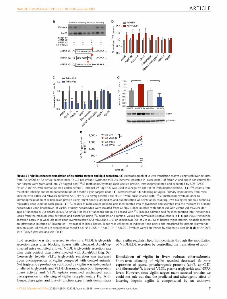
lipid secretion was also assessed in vivo in a VLDL triglyceridesecretion assay after blocking lipases with tyloxapol. Ad-shVig-injected mice exhibited a lower VLDL triglyceride secretion ratethan their control littermates injected with Ad-shCtrl (Fig. 5e).Conversely, hepatic VLDL triglyceride secretion was increasedupon overexpression of vigilin compared with control animals.Net triglyceride production controlled by vigilin was independentof altered triglyceride and VLDL clearance, since both lipoproteinlipase activity and VLDL uptake remained unchanged uponoverexpression or silencing of vigilin (Supplementary Fig. 5c,d).Hence, these gain- and loss-of-function experiments demonstrate
that vigilin regulates lipid homoeostasis through the modulationof VLDL/LDL secretion by controlling the translation of apoB.
Knockdown of vigilin in livers reduces atherosclerosis.Short-term silencing of vigilin revealed decreased de novoexpression of several proatherogenic proteins (apoB, apoC-IIIand fibronectin35), lowered VLDL, plasma triglyceride and NEFAlevels. However, since vigilin targets many secreted proteins wecould not rule out that the predicted anti-atherogenic effect oflowering hepatic vigilin is compensated by an unknown
0 1 2 30
500
1,000
1,500
2,000
Time (h)
VLD
L tr
igly
cerid
es(m
g dl
–1)
Ad-GFPAd-VIGILIN
Ad-shCtrlAd-shVig
* ***##
*
#
0.00.20.40.60.81.01.21.41.6
Nor
mal
ized
14C
cou
nts
** **
e
b
V5CDS-fetuin-A
rec. VIGILIN
mRNA #2:
mRNA #3:
mRNA #1:
Vigilin
γ-Tubulin
Ad-shVigAd-shCtrlAd-GFP Ad-VIGILIN
CDS-fetuin-A AAAAA ...V5
CDS-ApoM AAAAA ...V5
AAAAA ...
mRNA #1:mRNA #2:mRNA #3:
–
++–
– – +
++–
–++
++–
Fetuin-A
ApoM
dc
a Ad-shCtrl Ad-shCtrlAd-shVig Ad-shVig
ApoB
ApoC-II
I
Fetuin
-AOrm
1
Fibron
ectin
Alpha1
-ant
itryp
sin
ApoA-I
ApoM
1.5
1.0
0.5
Nor
mal
ized
35S
cou
nts
Ad-shCtrlAd-shVig
*** ***
*****
***
ApoB
ApoC-II
I
Fetuin
-AOrm
1
Fibron
ectin
Alpha1
-ant
itryp
sin
ApoA-I
ApoM
0.0
0.5
1.0
2.0
1.5
Nor
mal
ized
35S
cou
nts
Ad-GFPAd-VIGILIN
*
****
0.0
STOP
STOP
STOP
Figure 5 | Vigilin enhances translation of its mRNA targets and lipid secretion. (a) Autoradiograph of in vitro translation assays using fresh liver extracts
from Ad-shCtrl or Ad-shVig-injected mice (n¼ 2 per group). Synthetic mRNAs (scheme indicated in lower panel) of fetuin-A and apoM (as control for
non-target) were translated into V5-tagged and [35S]-methionine/cysteine radiolabelled protein, immunoprecipitated and separated by SDS–PAGE.
Fetuin-A mRNA with premature stop-codon before C-terminal V5-tag (#3) was used as a negative control for immunoprecipitation. (b,c) 35S counts from
metabolic labeling and immunoprecipitation of hepatic vigilin targets upon (b) overexpression (c) silencing of vigilin. Primary hepatocytes from mice
injected with either Ad-VIGILIN (control: Ad-GFP) or Ad-shVig (control: Ad-shCtrl) were pulse-chased with [35S]-methionine/cysteine prior to
immunoprecipitation of radiolabeled protein using target-specific antibodies and quantification via scintillation counting. Two biological and four technical
replicates were used for each group. (d) 14C counts of radiolabeled palmitic acid incorporated into triglycerides and secreted into the medium by primary
hepatocytes upon knockdown of vigilin. Primary hepatocytes were isolated from C57BL/6 mice injected with either Ad-GFP versus Ad-VIGILIN (for
gain-of-function) or Ad-shCtrl versus Ad-shVig (for loss-of-function) and pulse-chased with 14C-labelled palmitic acid for incorporation into triglycerides.
Lipids from the medium were extracted and quantified using 14C scintillation counting. Values are normalized relative counts in b–d. (e) VLDL triglyceride
secretion assay in 8-week-old mice upon overexpression (Ad-VIGILIN; n¼ 6) or knockdown (Ad-shVig; n¼ 6) of hepatic vigilin protein. Animals received
an intravenous injection of 500 mg kg� 1 tyloxapol to block lipases. Blood was collected at indicated time points and measured for plasma triglyceride
accumulation. All values are expressed as mean±s.d. *Pr0.05, **Pr0.01, ***Pr0.001; P values were determined by student’s t-test (in b–d) or ANOVA
with Tukey’s post hoc analysis (in e).
NATURE COMMUNICATIONS | DOI: 10.1038/ncomms12848 ARTICLE
NATURE COMMUNICATIONS | 7:12848 | DOI: 10.1038/ncomms12848 | www.nature.com/naturecommunications 7

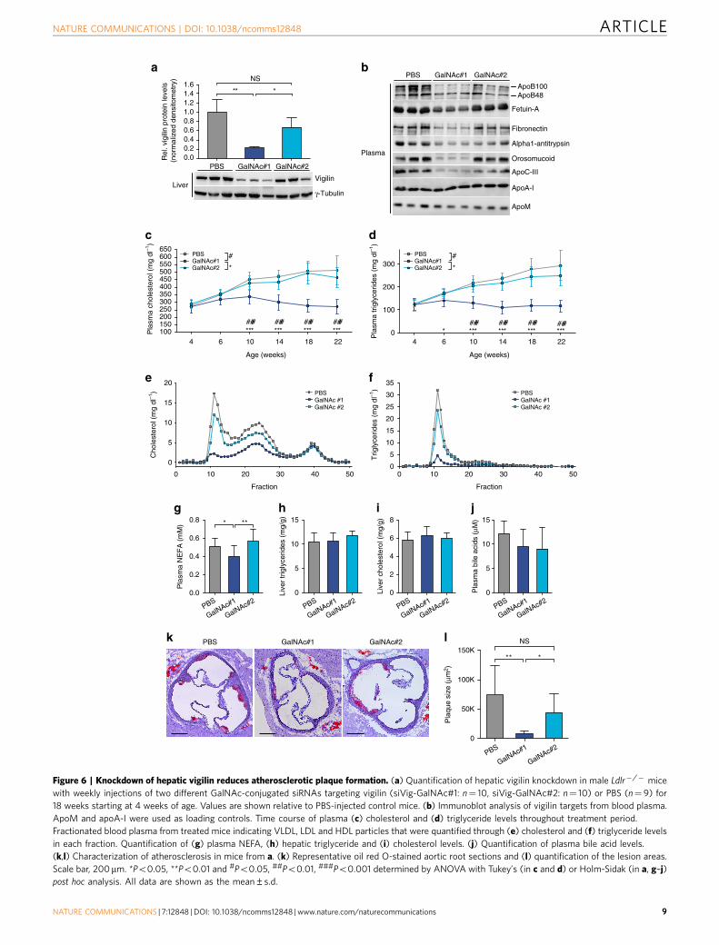
combination of proatherogenic vigilin targets. We thereforeevaluated the long-term effect of hepatic vigilin knockdown inatherosclerosis prone Ldlr� /� mice. Vigilin was silencedin the liver by two different siRNAs (siVig) that werechemically modified and covalently conjugated to multivalentN-acetylgalactosamine (GalNAc), a highly efficient ligand forclathrin-mediated endocytosis through the asialoglycoproteinreceptor (ASGPR). We tested two siVig-GalNAc conjugates(GalNAc#1 and GalNAc#2) for activity. GalNAc#1 was lesspotent than adenovirus delivered shRNA (Ad-shVig) and weeklysubcutaneous administration for 18 weeks resulted in a 70–80%reduction of hepatic vigilin. The second GalNAc-conjugatedsiRNA sequence (GalNAc#2) revealed no significant silencingactivity and was used to control for chemistry-related effects(Fig. 6a). Knockdown of vigilin using GalNAc#1 was specific forthe liver and showed no signs of liver toxicity (SupplementaryFig. 6a,b). We next measured the expression of vigilin targets inthe plasma of siVig-GalNAc-treated mice by immunoblotting.The vigilin targets fetuin-A, alpha1-antitrypsin, orosomucoid andthe proatherogenic apoB, apoC-III and fibronectin weredecreased in siVig-GalNAc#1-treated mice compared withcontrol animals with no changes in apoA-I and apoM (Fig. 6b).Plasma cholesterol and triglycerides were decreased in GalNAc#1treated mice compared with PBS and GalNAc#2-injected controlanimals (Fig. 6c,d). Furthermore, plasma NEFA were reduced andlipid profiling of FPLC-separated lipoprotein fractions inplasma of these mice revealed decreased VLDL and LDL levels(Fig. 6e–g). Liver triglyceride and cholesterol content as well asplasma bile acids were similar in vigilin knockdown and controlmice (Fig. 6h–j). Lastly, characterization of atherosclerosis inmice treated with siVig-GalNAc#1 revealed smaller lesions inH&E- and oil red O-stained aortic root sections (Fig. 6k,l).Together, these data demonstrate that long-term knockdown ofvigilin in the liver decreases VLDL and LDL levels and reducesatherosclerotic plaque formation in mice.
DiscussionThrough the combination of in vivo gain- and loss-of-functionstudies, PAR-CLIP as well as label-free quantification of secretedproteins by mass spectrometry, the present study investigated themolecular and physiological role of vigilin in the liver. Weidentify vigilin as a positive regulator of translation for mRNAscoding for a subset of proteins of the secretory pathway bybinding to an RRE consisting of a tandem repeat of CHHCseparated by 2–8 nt. However, only a fraction (B17.5%) of allliver-expressed secretory pathway proteins were targeted byvigilin. Since the identified RRE is also present in other abundantmRNAs that are not bound by vigilin, the specific regulatory roleof vigilin in protein translation may be dependent on additionalprotein interactions with the ribosome and other regulatoryfactors. Overall, these findings indicate a role of vigilin at the siteof protein synthesis and are in good accordance with the recentlyproposed function of the yeast homologue SCP160 as atranslational enhancer for proteins of the secretory pathway13.
Our data revealed apoB among the strongest targets ofhepatic vigilin. As the core protein of VLDL and LDL particles,apoB is of paramount importance for maintaining triglyceridebalance within the liver. Although regulation of apoB bypost-translational degradation pathways within the ER,post-ER36, and by autophagy37,38 is well established,translational regulatory mechanisms that govern apoBexpression are poorly understood39. Our study demonstratesthat vigilin is a major determinant of apoB translation and VLDLsecretion by hepatocytes without affecting VLDL clearance andtherefore a regulator of net triglyceride secretion by the liver.
Our current understanding of VLDL synthesis and regulationindicates that lipid availability constitutes the limiting factor forthe synthesis of functional apoB100, and thus, for the assembly ofsecretion-competent VLDL within the ER lumen40. We wouldtherefore expect a higher impact of vigilin on VLDL secretion inobese states, when lipids are abundant and less limiting. Hence,our results indicate that increased expression of vigilin enhancesapoB synthesis in conditions of lipid availability (i.e., in obesity)to further promote VLDL secretion. Indeed we noticed a largerreduction of VLDL and apoB amounts upon knockdown ofvigilin in DIO mice when compared with chow-fed animals(33% versus 51%, respectively), underlining the importance oflipid availability for apoB/VLDL secretion.
Short-term and strong (490%) silencing of vigilin (usingAd-shVig) in the liver was accompanied by mild steatosis,consistent with studies in which Apob was silenced usingshRNAs41. Long-term knockdown of vigilin via GalNAc-conjugated siRNAs did not result in a steatotic liver. Protectionagainst hepatic triglyceride accumulation is likely due to othervigilin targets and associated mechanisms, such as the reductionof apoC-III, which increases the catabolism of triglyceride richparticles and lowers plasma triglyceride levels in mice andhumans42–45. Long-term silencing of hepatic vigilin in Ldlr� /�
mice was also sufficient to lower VLDL and LDL levels andreduce atherosclerotic plaque formation. The anti-atherogeniceffects resulting from the inhibition of vigilin expression are likelymediated mainly by apoB. However we cannot exclude systemicsynergy of other vigilin targets such as apoC-III, fibronectin andpossibly others. Fetuin-A has been reported to inhibit insulin andproinflammatory cytokine signalling. Yet as a mineral carryingprotein, it also plays an important role in inhibiting systemiccalcification and therefore might have facultative effects onatherosclerosis46,47. It will therefore be important to addresspossible synergism of vigilin targets by performing simultaneousknockdowns of these targets in the liver in relevantatherosclerosis models.
Knockdown of vigilin resulted in substantially decreasedprotein levels of its targets without affecting mRNA levels. Incontrast, other highly expressed liver secreted proteins that werenot identified as targets in both PAR-CLIP replicates such asfetuin-B or the lipoprotein apoE, showed no significantperturbation upon vigilin knockdown, further substantiatingvigilin’s specificity for a distinct subset of metabolic transcripts.Hence, our findings support the emerging view of RBPsorganizing nascent RNA transcripts into functional groups thatare coordinately regulated, especially at the level of mRNAstability and translation48. Combinatorial binding of additionalRBPs to these mRNAs and their controlled proteasomaldegradation may provide a mechanism for multi-dimensionalregulation of protein fates and explain (1) the general poorcorrelation between the mRNA and protein pools in eukaryoticcells49,50 and (2) the low susceptibility of some targets towards avigilin knockdown ex and in vivo as observed for apoA-I. Targetsthat remained unchanged upon vigilin silencing may be subject tofurther regulatory mechanisms controlling their protein levels orbe due to secondary effects of the resulting phenotype.
Taken together, we provide first evidence that the mammalianvigilin is involved in translational regulation of mRNA targetsencoding for proteins of the secretory pathway, including theproatherogenic proteins apoB, apoC-III and fibronectin, andthereby serves as an important regulator of protein productionand lipid secretion from the liver. Moreover, we uncover aprimary role in lipid metabolism and hepatic steatosis in miceand humans to be mediated by an RNA-binding protein. Theincreased expression of vigilin in patients with hepatic steatosismay contribute to the overproduction and secretion of VLDL in
ARTICLE NATURE COMMUNICATIONS | DOI: 10.1038/ncomms12848
8 NATURE COMMUNICATIONS | 7:12848 | DOI: 10.1038/ncomms12848 | www.nature.com/naturecommunications
0
5
10
15
Pla
sma
bile
aci
ds (
μM)
PBS GalNAc#1 GalNAc#2R
el. v
igili
n pr
otei
n le
vels
(n
orm
aliz
ed d
ensi
tom
etry
)
***
0
5
10
15
Live
r tr
igly
cerid
es (
mg/
g)
0
50K
100K
150K** *
100
200
300
Pla
sma
trig
lyce
rides
(m
g dl
–1)
*** *** *******### #########
4 6 10 14 18 22100150200250300350400450500550600650
Age (weeks)
4 6 10 14 18 22
Age (weeks)
Pla
sma
chol
este
rol (
mg
dl–1
)
PBSGalNAc#1GalNAc#2 *
# PBSGalNAc#1GalNAc#2 *
#
***###
*** *** ***#########
g
ApoA-I
OrosomucoidPlasma
0
2
4
6
8
Live
r ch
oles
tero
l (m
g/g)
a b
c d
PBS GalNAc#1 GalNAc#2
ApoB100ApoB48
Fetuin-A
Alpha1-antitrypsin
l
jh
Fibronectin
PBS GalNAc#1 GalNAc#2
ApoM
ApoC-III
i
PBS
GalNAc#1
GalNAc#2
0
5
10
15
20
25
30
35T
rigly
cerid
es (
mg
dl–1
)
0 2010 30 40 50
0
5
10
15
20
GalNAc #2GalNAc #1PBS
GalNAc #2GalNAc #1PBS
Fraction
0 2010 30 40 50
Fraction
Cho
lest
erol
(m
g dl
–1)
e f
Vigilin
γ-TubulinLiver
Pla
que
size
(μm
2 )
k
0.8
0.6
0.4
0.2
0.0
Pla
sma
NE
FA
(m
M) * **
PBS
GalNAc#1
GalNAc#2PBS
GalNAc#1
GalNAc#2PBS
GalNAc#1
GalNAc#2PBS
GalNAc#1
GalNAc#2
NS
NS
0
1.61.41.21.00.80.60.40.20.0
Figure 6 | Knockdown of hepatic vigilin reduces atherosclerotic plaque formation. (a) Quantification of hepatic vigilin knockdown in male Ldlr�/� mice
with weekly injections of two different GalNAc-conjugated siRNAs targeting vigilin (siVig-GalNAc#1: n¼ 10, siVig-GalNAc#2: n¼ 10) or PBS (n¼ 9) for
18 weeks starting at 4 weeks of age. Values are shown relative to PBS-injected control mice. (b) Immunoblot analysis of vigilin targets from blood plasma.
ApoM and apoA-I were used as loading controls. Time course of plasma (c) cholesterol and (d) triglyceride levels throughout treatment period.
Fractionated blood plasma from treated mice indicating VLDL, LDL and HDL particles that were quantified through (e) cholesterol and (f) triglyceride levels
in each fraction. Quantification of (g) plasma NEFA, (h) hepatic triglyceride and (i) cholesterol levels. (j) Quantification of plasma bile acid levels.
(k,l) Characterization of atherosclerosis in mice from a. (k) Representative oil red O-stained aortic root sections and (l) quantification of the lesion areas.
Scale bar, 200mm. *Po0.05, **Po0.01 and #Po0.05, ##Po0.01, ###Po0.001 determined by ANOVA with Tukey’s (in c and d) or Holm-Sidak (in a, g–j)
post hoc analysis. All data are shown as the mean±s.d.
NATURE COMMUNICATIONS | DOI: 10.1038/ncomms12848 ARTICLE
NATURE COMMUNICATIONS | 7:12848 | DOI: 10.1038/ncomms12848 | www.nature.com/naturecommunications 9
obese, insulin-resistant subjects51,52. Given the high associationbetween NAFLD/NASH and cardiovascular diseases53,54,increased levels of vigilin in hepatic steatosis may thereforeprovide a potential link to increased risks for cardiovasculardiseases.
MethodsAnimal experiments. All animal models shown were male and on a C57BL/6 Nbackground and purchased from Janvier or Charles River. Mice used for isolationof primary hepatocytes were 8–10 weeks old. The ages of animals used forphysiological experiments are indicated in the respective figure legends. Mice werehoused in a pathogen-free animal facility at the Institute of Molecular HealthSciences at ETH Zurich. The animals were maintained in a temperature- andhumidity controlled room on a 12 h light–dark cycle (lights on from 6:00 to 18:00).Mice were either fed a standard laboratory chow, a high-fat diet (for DIO mice; fat,carbohydrate, protein content was 45, 35 and 20 kcal%, respectively) (ResearchDiets, D12451) or chow diet AIN76 supplemented with 0.02% cholesterol55
(for Ldlr� /� mice; Ssniff). All animal experiments were approved by theKantonale Veterinaramt Zurich.
Adenoviral infections. The sequence of human V5-tagged VIGILIN was clonedinto pVQAd CMV K-NpA (Viraquest) using the restriction sites BamHI and XhoI(NEB). Ad-GFP was based on the same vector backbone (including GFP) butlacked the insert transgene. shRNAs targeting vigilin (Supplementary Table 2) werecloned under a U6 promoter into the pVQAd AscI-NpA vector (Viraquest).Adenovirus production was performed at Viraquest, USA. All adenovirusesexpressed GFP from an independent promoter. Adenoviral infection of mice wasperformed by a single tail vein injection of 3� 109 plaque-forming units in a finalvolume of 0.2 ml diluted in PBS. Mice were sacrificed 7 (for gain-of-functionexperiments) or 10 days (for loss-of-function experiments) post injection.
Primary hepatocytes isolation. Mice were anaesthetized by intraperitonealinjection of 150 ml pentobarbital (Esconarkon US vet) pre-diluted 1:5 in PBS.The liver was perfused by cannulation of the hepatic portal vein with the caudalvena cava as a drain. The liver was perfused with pre- warmed Hank’s BalancedSalt Solution (Life Technologies) containing 0.5 mM EGTA followed bypre-warmed digestion medium (DMEM 1 g l� 1 glucose (Life Technologies),1% Penicillin–Streptomycin (Life Technologies), 15 mM HEPES (LifeTechnologies), 30 mg ml� 1 Liberase Research Grade medium Thermolysinconcentration (Roche)) each for four minutes with a flow rate of 3 ml min� 1. Theliver was surgically removed, hepatocytes released into 10 ml digestion media byshaking and supplemented with 15 ml ice-cold low glucose media (DMEM 1 g l� 1
glucose (Life Technologies), 1% Penicillin–Streptomycin (Life Technologies),10% heat-inactivated fetal bovine serum (Sigma-Aldrich), 1% GlutaMax (LifeTechnologies)) and filtered through a 100 mm Cell Strainer (BD). The suspensionwas then washed three times with 25 ml of ice-cold low glucose media at 50g and4 �C for 2 min. Hepatocytes were counted and plated at 4� 106 cells surface-treatedP10 plates (BD Primaria) in low glucose media. Three hours after plating, cellswere washed once with PBS and medium was changed to Williams E medium(or methionine-free DMEM for cell extracts used for in vitro translation; LifeTechnologies) supplemented with 1% Penicillin–Streptomycin (Life Technologies),1% GlutaMax (Life Technologies) and harvested 16 h (or 2 h for in vitrotranslation) after medium change56. All cells were incubated at 37 �C in ahumidified atmosphere containing 5% CO2.
Blood plasma collection and measurements. For measuring blood plasmainsulin, ALT, triglyceride, cholesterol and NEFA levels, blood was collected fromthe submandibular vein in non-heparinized capillary tubes. EDTA was added to afinal concentration of 5 mM as an anti-coagulant. Plasma was then separatedby centrifugation at 8,000g for 4 min. Measurements were performed usingcommercial kits. Plasma insulin was measured with the Rat Insulin ELISA Kit(Crystalchem). Plasma cholesterol (Roche), triglycerides (Roche), NEFA (Wako)and bile acids (Crystalchem) were measured by colorimetric assays, according tothe manufacturer’s instructions.
In vivo VLDL secretion assay. Gain-of-function and loss-of-function mousemodels were fasted for 6 h and injected intravenously with the lipase inhibitortyloxapol (500 mg kg� 1; Sigma) prior to blood collection at 0, 1, 2 and 3 h afterinjection. The collected blood samples were used for TG measurements and theVLDL-TG production rate was calculated from the slope of the plasma TG versustime curve.
Metabolic labelling and immunoprecipitation. Primary hepatocytes isolatedfrom gain-of-function and loss-of-function mouse models were washed twice withPBS and incubated for 2 h in methionine/cysteine free medium. Cells werethen pulsed for 2 h with 150 mCi of [35S-]methionine/cysteine (Expre35S35S;
PerkinElmer) per well and chased for 8 h in full hepatocytes medium after washingwith PBS. Metabolically labelled cells were harvested and lysed in 1�NP40 lysisbuffer (50 mM HEPES, pH 7.5, 150 mM KCl, 0.5 mM EDTA, 1 mM NaF,0.5% (v/v) NP40, 50mM DTT, complete EDTA-free protease inhibitor cocktail(Roche)) and incubated on ice for 10 min. Lysates were cleared by centrifugation at13,000g before the supernatant was collected and used for immunoprecipitation.Vigilin targets were immunoprecipitated with target-specific antibodies conjugatedto protein G Dynabeads (Life Technologies) over night at 4 �C. Ten microlitres ofprotein G magnetic particles were used per ml cell lysate. Magnetic beads werecollected on a magnetic rack and washed with 1�NP40 lysis buffer three timesbefore quantification using the scintillation counter.
In vivo VLDL clearance assay. Mice were injected intravenously with 100 mCi of[3H]glycerol and blood was collected 1 h post injection. Plasma was extracted andfractionated to obtain the radiolabeled VLDL fraction. Mice under study fromgain- and loss-of-function models were injected intravenously with 200,000 dpm of3H-labelled VLDL. The disappearance of radiolabeled VLDL was determined fromplasma samples drawn 30, 60, 90 and 120 min. after VLDL administration usingscintillation counting57.
Antibodies. The following antibodies were used in immunoblotting: rabbitanti-Hdlbp/vigilin (1:5,000) (Abcam, #ab109324), mouse anti-g-tubulin (1:10,000)(Sigma-Aldrich, #T6557), rabbit anti-laminB (1:5,000) (Cell Signaling, #9087S),rabbit anti-Gapdh (1:500) (Santa Cruz, #2118S), rabbit anti-Rbm47 (1:5,000)(Abcam, #ab167164), rabbit anti-HuR (1:500) (Santa Cruz, #sc-20694), rabbitanti-Histone H3 (1:5,000) (Cell Signaling, #4499S), rabbit anti-apoB (1:2,000)(Meridian, #K23300R), rabbit anti-apoA-I (1:10,000) (Meridian, #K23500R),rabbit anti-fibronectin (1:5,000) (Abcam, #ab2413), goat anti-fetuin-A (1:500)(Santa Cruz, #sc-9668), rabbit anti-alpha1-Antitrypsin (1:1,000) (Proteintech,#16382-1-AP), rabbit anti-orosomucoid (1:1,000) (Proteintech, #16439-1-AP),rabbit anti-apoM (1:2,000) (self-made, #aa140-159), goat anti-albumin(1:10,000) (Bethyl, #A90-134A), mouse anti-V5 (1:5,000) (Invitrogen, #R960-25),rabbit anti-HA (1:5,000) (Abcam, #ab9110).
PAR-CLIP in primary hepatocytes. For PAR-CLIP, primary hepatocytes fromAd-VIGILIN injected mice (for overexpression of V5-tagged human VIGILIN)were isolated and supplemented with 100 mM 4SU for 16 h before crosslinking.After decanting the growth medium, living cells were irradiated with 0.15 J cm� 2
of 365 nm UV light. Cells were harvested and lysed in NP40 lysis buffer (50 mMHEPES, pH 7.5, 150 mM KCl, 0.5 mM EDTA, 1 mM NaF, 0.5% (v/v) NP40, 50 mMDTT, complete EDTA-free protease inhibitor cocktail (Roche)). The cleared celllysates were treated with RNase T1 (at a final concentration of 1 U ml� 1 for 15 minat 22 �C). V5-tagged vigilin was immunoprecipitated with anti-V5 antibodiesbound to 10ml of Protein G magnetic particles per ml cell lysate prior to RNase T1treatment (Fermentas) at a final concentration of 100 U ml� 1 for 15 min at 22 �C.Beads were washed and resuspended in dephosphorylation buffer (50 mMTris-HCl (pH 7.9), 100 mM NaCl, 10 mM MgCl2, 1 mM DTT). Calf intestinalalkaline phosphatase (CIP, NEB) was added to a final concentration of 0.5 U ml� 1
to dephosphorylate the RNA. To label the crosslinked RNA, beads were washedand incubated with [g-32P]ATP to a final concentration of 0.5 mCi ml� 1 and T4PNK (NEB) to 1 U ml� 1 in one original bead volume for 30 min at 37 �C.Non-radioactive ATP was added to obtain a final concentration of 100 mM andincubated for another 5 min at 37 �C. The radiolabeled band corresponding to the155 kDa VIGILIN–RNA complex was separated by SDS–PAGE and electroeluted.The electroeluate was proteinase K digested at a final concentration of 1.2 mg ml� 1
and incubated for 30 min at 55 �C. The RNA was recovered by acidic phenol/chloroform extraction and ethanol precipitation. The recovered RNA was turnedinto a cDNA library using 30 and 50 adapter ligations that were carried out on a20 ml scale using 10.5 ml of the recovered RNA with chemically pre-adenylatedadapter oligodeoxynucleotides (30 adapter DNA, except for the riboadenylate rAppresidue: 50–30 rAppTCGTATGCCGTCTTCTGCTTGT) and 50 adapter RNA (50–30
GUUCAGAGUUCUACAGUCCGACGAUC) before reverse transcription andPCR amplification58. PCR products were cut out from a 3% NuSieve low-meltingpoint agarose gel, the PCR product eluted from gel pieces using the GelElute kit(Qiagen) and sequenced at the Rockefeller University Genomics Center using theSolexa technology. Reads were adapter extracted with cutadapt, clipped with lengthof at least 20 nts and mapped to the mm10 mouse genome with Bowtie 0.12.9(Bowtie parameters ‘-v 1 -m 10 –all –best –strata’), allowing for one mismatch.Processing and annotation of clusters to the ENCODE GRCm38 genomeannotation was performed using the PARalyzer software with default settings as inCorcoran et al.24 (http://www.genome.duke.edu/labs/ohler/research/PARalyzer/).Mapping statistics are given in Supplementary Data 1. The results from both PAR-CLIP replicates combined are listed in Supplementary Data 1 and 2. In addition,PAR-CLIP reads were mapped uniquely against the genome with 0 and 1mismatches and annotated with bedtools using the ENCODE GRCm38 genomeannotation to provide an independent measure of T-C transition, 1 mismatch and0 mismatch reads per transcript (Supplementary Data 2). Targets were ranked bythe sum of reads harbouring T-to-C conversions from both PAR-CLIP replicates.
ARTICLE NATURE COMMUNICATIONS | DOI: 10.1038/ncomms12848
10 NATURE COMMUNICATIONS | 7:12848 | DOI: 10.1038/ncomms12848 | www.nature.com/naturecommunications
Motif analysis. Kmer enrichment motif analysis was carried out calculating 4-merenrichments by sliding within a 20-nt long window along PAR-CLIP clusters andusing the shuffled (10,000 times) GRCm38 mouse protein-coding open readingframes as background sequences. Shuffled sequences were generated with theHMMER-3.0 suite. We further used the MEME suite, MEME (http://meme-suite.org/tools/meme) to define the motif of the top 759 protein-coding clusters(50UTR, CDS, 30 UTR) as defined by PARalyzer, which had at least 10 reads.
RNA isolation and quantification. RNA was extracted using Trizol (LifeTechnologies) according to the manufacturer’s instructions, except for a 30 minisopropanol precipitation at � 20 �C. RNA integrity was analysed on an Agilent2100 Bioanalyzer for all samples that were sequenced. RNA was subjected to DNaseI treatment with the DNA-free kit (Invitrogen), when necessary. RNA was reversetranscribed using the High Capacity cDNA Reverse Transcription Kit (AppliedBiosystems). Quantitative PCR was performed in an LC480 II Lightcycler (Roche)and using gene specific primers and Sybr Fast 2x Universal Master mix (Kapa).Results were normalized to 36B4 or Actb mRNA levels.
Illumina RNA sequencing. The quality of the isolated RNA was determined with aQubit (1.0) Fluorometer (Life Technologies) and a Bioanalyzer 2100 (Agilent).Only those samples with a 260/280 nm ratio between 1.8 and 2.1 and a 28S/18Sratio within 1.5 and 2.0 were further processed. The TruSeq RNA Sample Prep Kitv2 (Illumina) was used for cDNA library preparation. Quality and quantity of theenriched libraries were validated using Qubit (1.0) Fluorometer and the Caliper GXLabChip GX (Caliper Life Sciences). Libraries were normalized to 10 nM andsequenced on the Illumina HiSeq 2000 at the Functional Genomics Center Zurich.
Sequencing data analysis. Reads were quality checked with FastQC. Reads atleast 20 bases long, with a tail phred quality score greater than 20 were aligned tothe reference genome and transcriptome (FASTA and GTF files, respectively,downloaded from the UCSC, genome build mm10) with STAR56 with defaultsettings for single end reads. Distribution of the reads across genomic isoformexpression was quantified using the R package GenomicRanges59 fromBioconductor Version 3.0. Differentially expressed genes were identifiedusing the R package edgeR60 from Bioconductor Version 3.0.
Most abundant liver transcript isoforms were matched to the Uniprot databankfor signal peptides and transmembrane domains. The existence of signal peptidesand transmembrane domains were handcurated using Protter61.
Western blot analysis. Cells and tissues (using the Tissue Lyser II, Qiagen) werehomogenized with 3 volumes of RIPA lysis buffer (50 mM Tris-HCl pH 8, 150 mMNaCl, 1% NP40, 0.5% sodium deoxycholate, 0.1% SDS and 1 tablet cOmpleteEDTA-free protease inhibitor cocktail (Roche) per 50 ml buffer), incubated for10 min on ice and centrifuged for 10 min at 20,000g and 4 �C. Protein con-centrations were determined using the Bicinchoninic Acid Assay (Sigma-Aldrich).Equal protein amounts were boiled in Laemmli buffer (1.7% SDS, 5% glycerol,0.002% bromophenol blue, 60 mM Tris-HCl pH 6.8, 100 mM DTT) for 5 min at95 �C, separated by SDS–PAGE and transferred onto nitrocellulose membranes byelectroblotting in a wet chamber (Bio-Rad). The membranes were blocked for 1 hwith 5% non-fat dry milk TBS-0.1% Tween (Sigma-Aldrich), incubated with theprimary antibodies overnight at 4 �C, followed by three washes in TBS-0.1% Tweenand incubation with a horseradish peroxidase-conjugated secondary antibodies(Calbiochem) for 2–3 h. Blots were then developed by chemiluminescent detectionwith a Fujifilm analyzer (LAS-4000) and signals quantified using ImageJ.Uncropped scans of all of the immunoblots are shown in SupplementaryFigs 7 and 8.
Bacterial recombinant protein expression and purification. Three litre culturesof E. coli BL21 (DE3)pLysS competent cells transformed with the pETM30-vigilin-His6 construct were grown at 37 �C and 180 r.p.m. in Terrific Broth mediumcontaining 75mg ml� 1 Kanamycin until the OD600 reached 1.3. The culture wasthen incubated at 18 �C for 1 h and protein expression was induced with 0.2 mMisopropyl-D-thiogalactopyranoside (IPTG). Incubation was then continued at 18 �Cfor 12 hrs. Cells were harvested by centrifugation for 10 min at 6,000g and 4 �C. Allsubsequent procedures were performed at 4 �C. Bacterial pellets were resuspendedin chilled lysis buffer (50 mM Tris-HCl pH 7.5, 150 mM NaCl, 5 mM MgCl2,10% Glycerol, 1 mM b-Mercaptoethanol, 1 mM PMSF, 1 tablet EDTA-free proteaseinhibitor cocktail (Roche) per 50 ml, 1 mg ml� 1 Lysozyme (Sigma-Aldrich)5 mg ml� 1 DNase I (Roche) in a ratio of 1 g cell-wet weight to 1 ml lysis buffer).The lysates were further sonicated in a pre-chilled 50 ml tube (Falcon) to reduceviscosity (5 s on, 20 s off, for 2 min, Amplitude: 28%), and insoluble material wasremoved by centrifugation for 30 min at 20,000g. The resulting supernatant wasfiltered through 0.45 mm polyethersulfone filter membranes (Filtropur S 0.45,Sarstedt). The lysate was diluted in 50 ml HisTrap-Buffer (50 mM Tris-HCl pH 7.5,1 M NaCl, 5 mM MgCl2, 10% Glycerol, 30 mM Imidazole pH 8.5), adjusted bufferpH to 7.6 with concentrated HCl before loading onto a 5 ml HisTrap HP column(GE Healthcare Life Science), pre-equilibrated in HisTrap-Buffer and attached toan AKTA Explorer FPLC. Diluted lysate was passed through the column at
2 ml min� 1 and then gradually eluted with increasing concentrations ofHisTrap-Buffer containing 500 mM Imidazole, collecting 1 ml-sized fractions.The peak of fractions containing VIGILIN were determined by SDS–PAGE andCoomassie staining of the gel (typically at 150–200 mM imidazole). VIGILINcontaining fractions were pooled, diluted in 50 ml Heparin-Buffer (50 mMTris-HCl pH 7.6, 150 mM NaCl, 5 mM MgCl2, 10% Glycerol, adjusted bufferpH to 7.6) and loaded on a 5 ml HiTrap Heparin HP column (GE Healthcare LifeScience). RNA-depleted vigilin was eluted gradually using increasing concentra-tions of Heparin-Buffer containing 2 M NaCl and collected in 1 ml fractions. Eluatefractions were monitored by SDS–PAGE and Coomassie stainings. Fractionscontaining VIGILIN were pooled and dialyzed overnight using a 50 kDa MWCOPur-A-Lyzer (Sigma-Aldrich) into storage buffer (20 mM Tris-HCl pH 7.6,300 mM KCl, 5 mM MgCl2, 50% glycerol, 1 mM DTT, 1 mM PMSF, 1 tabletEDTA-free protease inhibitor cocktail (Roche) per 50 ml). Aliquots of VIGILINwere stored at � 80 �C. Protein concentrations were determined by intensity ofCoomassie staining in comparison to bovine serum albumin.
Electrophoretic mobility shift assays. Oligoribonucleotides were labelled with[g-32P]ATP and T4 polynucleotide kinase using standard conditions. A total of10 nM 32P-labelled RNA was incubated with 0–10 mM protein in 20 ml reactionscontaining 250 mM KCl, 5 mM MgCl2, 25 mM Tris-HCl pH 7.5, 10% glycerol,1 mg ml� 1 acetylated BSA (Ambion), 1.5 mM of yeast tRNA (Invitrogen).Reactions were incubated at 25 �C for 5 min and separated on 1.2% agarose gel for1 h at 130 V at room temperature using 1�TBE. Agarose gels were dried undervacuum gel dryers at 60 �C for 2 to 3 h, exposed to a phosphoimager screen.
Label-free mass spectrometry. Medium from primary hepatocytes was collected24 h after medium change and centrifuged at 14,000g to pellet insoluble remnants.Supernatants (60–80 ml) were precipitated with 1 volume of 20% TCA precipitationand washed twice with cold acetone. Dry pellets were dissolved in 45 ml buffer(10 mM Tris, 2 mM CaCl2, pH 8.2) and trypsinized with 5 ml of 100 ng ml� 1 trypsinin 10 mM HCl for 30 min at 60 �C. Samples were dried, dissolved in 20 ml 0.1%formic acid and transferred to an autosampler vial for LC/MS/MS. Two microliterwere injected. Label-free quantification of MS-data was performed by matchingraw data to the Mouse Swiss-Prot database using MaxQuant62. Statistical analysiswas then performed using Perseus (http://www.perseus-framework.org) afterfiltering out reverse hits, potential contaminants and entries identified by only onesite. Protein intensities were normalized to the median sample intensity. Statisticalsignificance was determined using a false discovery rate at 0.01, permutation-basedmultiple t-testing (250� ) and a curve bend at s0¼ 0.5.
Plasma fractionation. Lipoproteins from pooled plasma (200 ml total) werediluted in 1 mM EDTA–PBS and separated by FPLC using two Superose-6 FPLCcolumns in series (HR10/30) in 1 mM EDTA–PBS at 0.5 ml min� 1. Columns werecalibrated using high and low molecular weight standards (GE Healthcare).
Liver triglyceride and cholesterol content. Lipids from 50 mg liver wereextracted with 1 ml hexane:isopropanol (3:2) by homogenizing tissues using theTissueLyser II (Qiagen). Lysates were centrifuged at 20,000g for 3 min and thesupernatant was transferred to a fresh tube. The pellet was re-extracted with 0.5 mlhexane:isopropanol, spun again and the supernatants were combined. A volume of0.5 ml of 0.5 M Na2SO4 solution was added and the tubes mixed. The samples werecentrifuged for 3 min at full speed and the upper organic phase was transferred to afresh tube, avoiding contamination with the aqueous phase. The samples were spunagain and the upper phase was transferred to a fresh tube and evaporated overnightunder the fume hood. Lipids were dissolved in 1 ml of Triton X-100:methanol:butanol (1:1:3) mixture. Five microlitres were used for lipid quantifications.
Oil red O stainings. Frozen OCT-embedded liver pieces were stained with oil redO (ORO)63. Immediately after tissue collection livers were embedded in moldsfilled with OCT embedding matrix (CellPath) on dry ice and stored at � 80 �C.Tissues were cut with a cryostat into 10 mm thick sections. Sections were allowed toequilibrate for 10 min at room temperature. Sufficient ORO working solution(Abcam) was added to completely cover the sections and incubated for 10 min withthe ORO solution. Slides were rinsed carefully in a stain dish under running tapwater for 30 min. Slides were briefly dried and mounted with a water-solublemounting medium and coverslips on them.
In vitro translation assay. Full-length V5-tagged fetuin-A and apoM mRNAswere in vitro transcribed from pcDNA3.1 vectors using the mMESSAGEmMACHINE kit (Ambion). mRNAs were utilized for in vitro translation in 100mlreactions using methionine- and cysteine-free amino acid mix and nuclease-treatedextracts from primary hepatocytes isolated from mice injected with Ad-shCtrl orAd-shVig64. Proteins were co-translationally radiolabeled by addition of 50 mCi[35S]-methionine/cysteine (PerkinElmer) to the reaction. V5-tagged proteinproducts were immunoprecipitated with a V5-antibody conjugated to protein GDynabeads (Life Technologies), washed 10� with IP wash buffer (50 mM
NATURE COMMUNICATIONS | DOI: 10.1038/ncomms12848 ARTICLE
NATURE COMMUNICATIONS | 7:12848 | DOI: 10.1038/ncomms12848 | www.nature.com/naturecommunications 11
HEPES-KOH pH 7.5, 500 mM KCl, 0.05% NP40, 0.5 mM DTT, 1 tablet cOmpleteEDTA-free protease inhibitor cocktail (Roche) per 50 ml) separated by SDS–PAGEand visualized by autoradiography using x-ray films (Fuji).
Preparation of nuclear/cytoplasmic extracts. Primary hepatocytes were per-meabilized on ice in hypotonic lysis buffer (10 mM HEPES-KOH, pH 7.5, 1.5 mMMgCl2, 10 mM KCl, 0.5 mM EDTA, 0.1% NP40, 1 mM DTT, 1 tablet cOmpleteprotease inhibitor cocktail) per 50 ml buffer (Roche) for 30 s, vortexed briefly andimmediately centrifuged for 30 s at 8,000g and 4 �C. After centrifugation, thesupernatants (cytoplasmic extracts) were collected, and nuclear pellets were washedeight times in nuclear wash buffer (50 mM HEPES-KOH pH 7.5, 150 mM KCl,2 mM EDTA, 0.5% NP40, 1 mM DTT, 1 tablet cOmplete protease inhibitor cocktailper 50 ml buffer (Roche)) by resuspension and centrifugation at 8,000g for 30 s.Nuclear pellets were resuspended in RIPA buffer (50 mM Tris-HCl pH 8, 150 mMNaCl, 1% NP40, 0.5% sodium deoxycholate, 0.1% SDS and 1 tablet cOmpleteEDTA-free protease inhibitor cocktail (Roche) per 50 ml buffer).
Triglyceride secretion assay. Seeded primary mouse hepatocytes extracted frommice injected with adenovirus were pulsed with 1 mM of pre-warmed albuminbound [1-14C]-Palmitic Acid for 2 h and then washed three times with PBS.Williams E medium (Life Technologies) supplemented with 1% Penicillin–Streptomycin (Life Technologies), 1% GlutaMax (Life Technologies) was re-addedto the cells and harvested 4 h after medium change. Incorporation of palmitic acidinto triglycerides and subsequent secretion of radiolabeled triglycerides wasquantified by extraction of the lipid fraction from the medium followed by liquidscintillation counting.
GalNAc-conjugated siRNAs. siRNA-GalNAc conjugates were synthesized asdescribed previously65. The sequences and stabilizing nucleotide modifications ofthe siRNAs are shown in Supplementary Table 2. The GalNAc ligand wascovalently linked to the 30-end of the sense (S) strand of the siRNA by aphosphodiester linkage between the pyrrolidine scaffold and the S strand in allconjugate designs. A triantennary GalNAc ligand was synthesized to facilitatecovalent conjugation to siRNAs66. Appropriately protected triantennaryGalNAc monomer, which is compatible with solid-phase oligonucleotide synthesisand deprotection conditions, were synthesized using a trans-4-hydroxyprolinolmoiety. The triantennary GalNAc building block was synthesized fromD-(þ )-galactosamine, 2-amino-2-(hydroxymethyl)-1,3-propanediol (TRIS),trans-4-hydroxy-L-proline methyl ester, and a glass solid support. The GalNAcmonomer was then conjugated to sense strand of siRNA during solid-phasesynthesis and deprotected to obtain the desired conjugate. Sense and antisensestrands were synthesized on an ABI Synthesizer using commercially available50-O-(4,40-dimethoxytrityl)-20-deoxy-20-fluoro-, and 50-O-(4,4-dimethoxytrityl)-2-O-methyl-3-O-(2-cyanoethyl-N,N-diisopropyl) phosphoramidite monomers ofuridine, 4-N-acetylcytidine, 6-N-benzoyladenosine, and 2-N-isobutyrylguanosineusing standard solid-phase oligonucleotide synthesis and deprotection protocols.Phosphorothioate linkages were introduced by oxidation of phosphite utilizing0.1 M DDTT in pyridine. After deprotection, ion-exchange HPLC purificationfollowed by annealing of equimolar amounts of complementary sense andantisense strands provide the desired duplex sense and antisense strands weremixed and annealed by heating to 90 �C and slowly cooled to obtain the desiredsiRNAs. The siRNA samples were analysed by mass spectrometry, capillary gelelectrophoresis and for endotoxin and osmolality, and the purity was ascertained.
Quantification of atherosclerotic plaques. Atherosclerosis was quantified inaortic root cross-sections from fresh-frozen optimal cutting temperature medium(OCT)-embedded hearts. Lesion size is shown as means of 5 frozen sections(10 mm), each 50mm apart. Sections were stained with oil red O, hematoxylin andlight green for visualization of atherosclerotic lesions55. Lesion areas werequantified using LAS software, version 4.2. (Leica). Analysis of the aortic roots wasperformed blindly without knowledge of the treatments.
Statistical analysis. Numerical values are reported as average±s.d. unless statedotherwise. No statistical method was used to predetermine sample size, but samplesize was based on preliminary data and previous publications as well as observedeffect sizes. Outliers that were two standard deviations outside of the mean wereroutinely excluded from all analyses. Animals were sex- and age-matched. Animalstudies were performed without blinding of the investigator. We assessed data fornormal distribution and similar variance between groups using GraphPad Prism6.0 if applicable. Some data sets had a statistical difference in the variation betweengroups. If not mentioned otherwise in the figure legend, statistical significance(*Pr0.05, **Pr0.01, ***Pr0.001) was determined by unpaired two-tailed t-test,one-way ANOVA (when comparing Z3 groups) or two-way ANOVA (forrepeated measurements and time courses) with relevant post hoc tests (Holm-Sidakfor¼ 3 groups and Tukey’s for repeated time measurements and time courses).GraphPad Prism 6.0 software was used for statistical analysis of all data sets.
Data availability. The authors declare that the data supporting the findings of thisstudy are available within the article and its Supplementary Information Files.RNA-Seq data were deposited at Gene Expression Omnibus under GSE78084.Supplementary Data 1 and 2 containing two tables listing PARalyzer identifiedRNA target sites and genome-wide RNA and proteomic experiments can be foundwith this article online at.
References1. McKnight, G. L. et al. Cloning and expression of a cellular high density
lipoprotein-binding protein that is up-regulated by cholesterol loading of cells.J. Biol. Chem. 267, 12131–12141 (1992).
2. Schmidt, C. et al. Complete cDNA sequence of chicken vigilin, a novel proteinwith amplified and evolutionary conserved domains. Eur. J. Biochem. 206,625–634 (1992).
3. Burd, C. G. & Dreyfuss, G. Conserved structures and diversity of functions ofRNA-binding proteins. Science 265, 615–621 (1994).
4. Dejgaard, K. & Leffers, H. Characterisation of the nucleic-acid-binding activityof KH domains. Different properties of different domains. Eur. J. Biochem. 241,425–431 (1996).
5. Dodson, R. E. & Shapiro, D. J. Vigilin, a ubiquitous protein with 14 K homologydomains, is the estrogen-inducible vitellogenin mRNA 3’-untranslatedregion-binding protein. J. Biol. Chem. 272, 12249–12252 (1997).
6. Kruse, C. et al. Evidence for a novel cytoplasmic tRNA-protein complexcontaining the KH-multidomain protein vigilin. Biochem. J. 320(Pt 1): 247–252(1996).
7. Weber, V. et al. Purification and nucleic-acid-binding properties of aSaccharomyces cerevisiae protein involved in the control of ploidy. Eur. J.Biochem. 249, 309–317 (1997).
8. Cortes, A. et al. DDP1, a single-stranded nucleic acid-binding protein ofDrosophila, associates with pericentric heterochromatin and is functionallyhomologous to the yeast Scp160p, which is involved in the control of cellploidy. EMBO J. 18, 3820–3833 (1999).
9. Chen, J.-Y., Chen, J.-C. & Wu, J.-L. Molecular cloning and functional analysisof zebrafish high-density lipoprotein-binding protein. Comp. Biochem. Physiol.B, Biochem. Mol. Biol. 136, 117–130 (2003).
10. Brykailo, M. A., Corbett, A. H. & Fridovich-Keil, J. L. Functional overlapbetween conserved and diverged KH domains in Saccharomyces cerevisiaeSCP160. Nucleic Acids Res. 35, 1108–1118 (2007).
11. Molyneux, S. D. et al. Human somatic cell mutagenesis creates geneticallytractable sarcomas. Nat. Genet. 46, 964–972 (2014).
12. Yang, W. L. et al. Vigilin is overexpressed in hepatocellular carcinomaand is required for HCC cell proliferation and tumor growth. Oncol. Rep. 31,2328–2334 (2014).
13. Hirschmann, W. D. et al. Scp160p is required for translational efficiency ofcodon-optimized mRNAs in yeast. Nucleic Acids Res. 42, 4043–4055 (2014).
14. Mendelsohn, B. A. et al. Genetic and biochemical interactions between SCP160and EAP1 in yeast. Nucleic Acids Res. 31, 5838–5847 (2003).
15. Wang, Q., Zhang, Z., Blackwell, K. & Carmichael, G. G. Vigilins bind topromiscuously A-to-I-edited RNAs and are involved in the formation ofheterochromatin. Curr. Biol. 15, 384–391 (2005).
16. Wintersberger, U., Kuhne, C. & Karwan, A. Scp160p, a new yeast proteinassociated with the nuclear membrane and the endoplasmic reticulum,is necessary for maintenance of exact ploidy. Yeast 11, 929–944 (1995).
17. Huertas, D., Cortes, A., Casanova, J. & Azorın, F. Drosophila DDP1,a multi-KH-domain protein, contributes to centromeric silencing andchromosome segregation. Curr. Biol. 14, 1611–1620 (2004).
18. Kruse, C. et al. Export and transport of tRNA are coupled to a multi-proteincomplex. Biochem. J. 346(Pt 1): 107–115 (2000).
19. Gelin-Licht, R., Paliwal, S., Conlon, P., Levchenko, A. & Gerst, J. E.Scp160-dependent mRNA trafficking mediates pheromone gradient sensingand chemotropism in yeast. Cell Rep. 1, 483–494 (2012).
20. Baum, S., Bittins, M., Frey, S. & Seedorf, M. Asc1p, a WD40-domain containingadaptor protein, is required for the interaction of the RNA-binding proteinScp160p with polysomes. Biochem. J. 380, 823–830 (2004).
21. Frey, S., Pool, M. & Seedorf, M. Scp160p, an RNA-binding, polysome-associated protein, localizes to the endoplasmic reticulum of Saccharomycescerevisiae in a microtubule-dependent manner. J. Biol. Chem. 276,15905–15912 (2001).
22. Lang, B. D. & Fridovich-Keil, J. L. Scp160p, a multiple KH-domain protein, is acomponent of mRNP complexes in yeast. Nucleic Acids Res. 28, 1576–1584 (2000).
23. Hafner, M. et al. Transcriptome-wide identification of RNA-binding proteinand microRNA target sites by PAR-CLIP. Cell 141, 129–141 (2010).
24. Corcoran, D. L. et al. PARalyzer: definition of RNA binding sites fromPAR-CLIP short-read sequence data. Genome Biol. 12, R79 (2011).
25. Cunningham, K. S., Dodson, R. E., Nagel, M. A., Shapiro, D. J.& Schoenberg, D. R. Vigilin binding selectively inhibits cleavage of thevitellogenin mRNA 3’-untranslated region by the mRNA endonucleasepolysomal ribonuclease 1. Proc. Natl Acad. Sci. USA 97, 12498–12502 (2000).
ARTICLE NATURE COMMUNICATIONS | DOI: 10.1038/ncomms12848
12 NATURE COMMUNICATIONS | 7:12848 | DOI: 10.1038/ncomms12848 | www.nature.com/naturecommunications

26. Woo, H.-H. et al. Posttranscriptional suppression of proto-oncogene c-fmsexpression by vigilin in breast cancer. Mol. Cell Biol. 31, 215–225 (2011).
27. Berkovits, B. D. & Mayr, C. Alternative 3’ UTRs act as scaffolds to regulatemembrane protein localization. Nature 522, 363–367 (2015).
28. Moretti, F. A. et al. A major fraction of fibronectin present in the extracellularmatrix of tissues is plasma-derived. J. Biol. Chem. 282, 28057–28062 (2007).
29. Ren, J. et al. Long-term coronary heart disease risk associated with very-low-density lipoprotein cholesterol in Chinese: the results of a 15-Year ChineseMulti-Provincial Cohort Study (CMCS). Atherosclerosis 211, 327–332 (2010).
30. Schafer, C. et al. The serum protein alpha 2-Heremans-Schmid glycoprotein/fetuin-A is a systemically acting inhibitor of ectopic calcification. J. Clin. Invest.112, 357–366 (2003).
31. Auberger, P. et al. Characterization of a natural inhibitor of the insulin receptortyrosine kinase: cDNA cloning, purification, and anti-mitogenic activity. Cell58, 631–640 (1989).
32. Rauth, G. et al. The nucleotide and partial amino acid sequences of rat fetuin.Identity with the natural tyrosine kinase inhibitor of the rat insulin receptor.Eur. J. Biochem. 204, 523–529 (1992).
33. Srinivas, P. R. et al. Serum alpha 2-HS-glycoprotein is an inhibitor of thehuman insulin receptor at the tyrosine kinase level. Mol. Endocrinol. 7,1445–1455 (1993).
34. Kruse, C. et al. The multi-KH protein vigilin associates with free andmembrane-bound ribosomes. Cell. Mol. Life Sci. 60, 2219–2227 (2003).
35. Rohwedder, I. et al. Plasma fibronectin deficiency impedes atherosclerosisprogression and fibrous cap formation. EMBO Mol. Med. 4, 564–576 (2012).
36. Fisher, E. A. et al. The triple threat to nascent apolipoprotein B. Evidence formultiple, distinct degradative pathways. J. Biol. Chem. 276, 27855–27863 (2001).
37. Pan, M. et al. Presecretory oxidation, aggregation, and autophagic destructionof apoprotein-B: a pathway for late-stage quality control. Proc. Natl Acad. Sci.USA 105, 5862–5867 (2008).
38. Qiu, W. et al. Hepatic autophagy mediates endoplasmic reticulumstress-induced degradation of misfolded apolipoprotein B. Hepatology 53,1515–1525 (2011).
39. Fisher, E., Lake, E. & McLeod, R. S. Apolipoprotein B100 quality control andthe regulation of hepatic very low density lipoprotein secretion. J. Biomed. Res.28, 178–193 (2014).
40. Fisher, E. A. The degradation of apolipoprotein B100: multiple opportunities toregulate VLDL triglyceride production by different proteolytic pathways.Biochim. Biophys. Acta 1821, 778–781 (2012).
41. Maczuga, P. et al. Therapeutic expression of hairpins targeting apolipoproteinB100 induces phenotypic and transcriptome changes in murine liver. GeneTher. 21, 60–70 (2014).
42. Maeda, N. et al. Targeted disruption of the apolipoprotein C-III gene in miceresults in hypotriglyceridemia and protection from postprandialhypertriglyceridemia. J. Biol. Chem. 269, 23610–23616 (1994).
43. Ginsberg, H. N. et al. Apolipoprotein B metabolism in subjects with deficiencyof apolipoproteins CIII and AI. Evidence that apolipoprotein CIII inhibitscatabolism of triglyceride-rich lipoproteins by lipoprotein lipase in vivo. J. Clin.Invest. 78, 1287–1295 (1986).
44. TG and HDL Working Group of the Exome Sequencing Project, NationalHeart, Lung, and Blood Institute et al. Loss-of-function mutations in APOC3,triglycerides, and coronary disease. N. Engl. J. Med. 371, 22–31 (2014).
45. Pollin, T. I. et al. A null mutation in human APOC3 confers a favorable plasmalipid profile and apparent cardioprotection. Science 322, 1702–1705 (2008).
46. Westenfeld, R. et al. Fetuin-A protects against atherosclerotic calcification inCKD. J. Am. Soc. Nephrol. 20, 1264–1274 (2009).
47. Naito, C. et al. Facilitatory effects of fetuin-A on atherosclerosis. Atherosclerosis246, 344–351 (2016).
48. Keene, J. D. RNA regulons: coordination of post-transcriptional events. Nat.Rev. Genet. 8, 533–543 (2007).
49. de Sousa Abreu, R., Penalva, L. O., Marcotte, E. M. & Vogel, C. Global signaturesof protein and mRNA expression levels. Mol. BioSyst. 5, 1512–1526 (2009).
50. Vogel, C. et al. Sequence signatures and mRNA concentration can explaintwo-thirds of protein abundance variation in a human cell line. Mol. Syst. Biol.6, 400 (2010).
51. Adiels, M. et al. Overproduction of large VLDL particles is driven by increasedliver fat content in man. Diabetologia 49, 755–765 (2006).
52. Adiels, M., Olofsson, S.-O., Taskinen, M.-R. & Boren, J. Overproduction of verylow-density lipoproteins is the hallmark of the dyslipidemia in the metabolicsyndrome. Arterioscler. Thromb. Vasc. Biol. 28, 1225–1236 (2008).
53. Targher, G., Day, C. P. & Bonora, E. Risk of cardiovascular disease in patientswith nonalcoholic fatty liver disease. N. Engl. J. Med. 363, 1341–1350 (2010).
54. Loria, P. et al. Cardiovascular risk, lipidemic phenotype and steatosis. Acomparative analysis of cirrhotic and non-cirrhotic liver disease due to varyingetiology. Atherosclerosis 232, 99–109 (2014).
55. Teupser, D., Persky, A. D. & Breslow, J. L. Induction of atherosclerosis bylow-fat, semisynthetic diets in LDL receptor-deficient C57BL/6J and FVB/NJmice: comparison of lesions of the aortic root, brachiocephalic artery, and
whole aorta (en face measurement). Arterioscler. Thromb. Vasc. Biol. 23,1907–1913 (2003).
56. Dobin, A. et al. STAR: ultrafast universal RNA-seq aligner. Bioinformatics 29,15–21 (2013).
57. Aalto-Setala, K. et al. Mechanism of hypertriglyceridemia in humanapolipoprotein (apo) CIII transgenic mice. Diminished very low densitylipoprotein fractional catabolic rate associated with increased apo CIII andreduced apo E on the particles. J. Clin. Invest. 90, 1889–1900 (1992).
58. Spitzer, J. et al. PAR-CLIP (Photoactivatable Ribonucleoside-EnhancedCrosslinking and Immunoprecipitation): a step-by-step protocol to thetranscriptome-wide identification of binding sites of RNA-binding proteins.Methods Enzymol. 539, 113–161 (2014).
59. Lawrence, M. et al. Software for computing and annotating genomic ranges.PLoS Comput. Biol. 9, e1003118 (2013).
60. Robinson, M. D., McCarthy, D. J. & Smyth, G. K. edgeR: a Bioconductorpackage for differential expression analysis of digital gene expression data.Bioinformatics 26, 139–140 (2010).
61. Omasits, U., Ahrens, C. H., Muller, S. & Wollscheid, B. Protter: interactiveprotein feature visualization and integration with experimental proteomic data.Bioinformatics 30, 884–886 (2014).
62. Cox, J. & Mann, M. MaxQuant enables high peptide identification rates,individualized p.p.b.-range mass accuracies and proteome-wide proteinquantification. Nat. Biotechnol. 26, 1367–1372 (2008).
63. Mehlem, A., Hagberg, C. E., Muhl, L., Eriksson, U. & Falkevall, A. Imaging ofneutral lipids by oil red O for analyzing the metabolic status in health anddisease. Nat. Protoc. 8, 1149–1154 (2013).
64. Rakotondrafara, A. M. & Hentze, M. W. An efficient factor-depletedmammalian in vitro translation system. Nat. Protoc. 6, 563–571 (2011).
65. Rajeev, K. G. et al. Hepatocyte-specific delivery of siRNAs conjugated to novelnon-nucleosidic trivalent N-acetylgalactosamine elicits robust gene silencingin vivo. Chembiochem 16, 903–908 (2015).
66. Nair, J. K. et al. Multivalent N-acetylgalactosamine-conjugated siRNA localizesin hepatocytes and elicits robust RNAi-mediated gene silencing. J. Am. Chem.Soc. 136, 16958–16961 (2014).
AcknowledgementsWe thank B. Bazylak-Kaps and the Functional Genomics Center Zurich for technicalassistance. M.B.M. and S.G. were supported by a Boehringer Ingelheim Fonds PhDFellowship. This work was supported from the European Research Council (ERC) grant250210 ‘Metabolomirs’ (M.S.), National Center of Competence in Research (NCCR) onRNA Biology and Disease (to M.S.), the Howard Hughes Medical Institute (HHMI)(T.T.), and by Swiss National Science Foundation grant 310030B _147089/1 (M.H.H.)and 310030_141209 (M.S.). Research in the laboratory of T.T. was supported by NIHtransformative research award R01GM104962.
Author contributionsM.B.M. and M.S. conceived the project; M.B.M. performed experiments, analysed andinterpreted data and wrote the manuscript; S.G. performed bioinformatics analyses; D.T.analysed atherosclerotic lesions in aortic roots; M.M. and K.C. synthesized GalNAc-conjugated siRNAs; T.T. supervised PAR-CLIP and EMSA experiments. C.B. andM.H.H. recruited patients, collected clinical and blood chemistry data, and performed theliver biopsies. M.S. performed experiments, analysed and interpreted data, supervised theproject and wrote the manuscript.
Additional informationSupplementary Information accompanies this paper at http://www.nature.com/naturecommunications
Competing financial interests: M.S. and T.T. are members of the Scientific AdvisoryBoard and M.M. and K.C. are employees of Alnylam Pharmaceuticals. The other authorsdeclare no competing financial interests.
Reprints and permission information is available online at http://npg.nature.com/reprintsandpermissions/
How to cite this article: Mobin, M. B. et al. The RNA-binding protein vigilin regulatesVLDL secretion through modulation of Apob mRNA translation. Nat. Commun. 7:12848doi: 10.1038/ncomms12848 (2016).
This work is licensed under a Creative Commons Attribution 4.0International License. The images or other third party material in this
article are included in the article’s Creative Commons license, unless indicated otherwisein the credit line; if the material is not included under the Creative Commons license,users will need to obtain permission from the license holder to reproduce the material.To view a copy of this license, visit http://creativecommons.org/licenses/by/4.0/
r The Author(s) 2016
NATURE COMMUNICATIONS | DOI: 10.1038/ncomms12848 ARTICLE
NATURE COMMUNICATIONS | 7:12848 | DOI: 10.1038/ncomms12848 | www.nature.com/naturecommunications 13

1
Supplementary Figure 1: Additional metabolic parameters of obesity mouse
models and controls. (a) Body weight, (b) blood glucose and (c) insulin
resistance index of homeostatic model assessment (HOMA IR) of 20 week-old
chow fed (Chow), diet-induced obese (DIO) C57BL/6 mice, and 23 week old
ob/ob mice under study (n = 6 per group). (d) qPCR analysis of hepatic vigilin
transcript levels. Data are shown as relative expression levels. All values are
expressed as mean ± s.d. **P ≤ 0.01, ***P ≤ 0.001; p-values were determined
ANOVA with Holm-Sidak post hoc analysis.
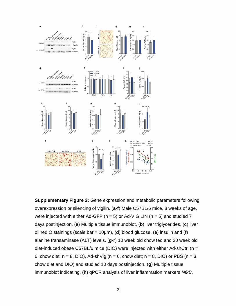
2
Supplementary Figure 2: Gene expression and metabolic parameters following
overexpression or silencing of vigilin. (a-f) Male C57BL/6 mice, 8 weeks of age,
were injected with either Ad-GFP (n = 5) or Ad-VIGILIN (n = 5) and studied 7
days postinjection. (a) Multiple tissue immunoblot, (b) liver triglycerides, (c) liver
oil red O stainings (scale bar = 10µm), (d) blood glucose, (e) insulin and (f)
alanine transaminase (ALT) levels. (g-r) 10 week old chow fed and 20 week old
diet-induced obese C57BL/6 mice (DIO) were injected with either Ad-shCtrl (n =
6, chow diet; n = 8, DIO), Ad-shVig (n = 6, chow diet; n = 8, DIO) or PBS (n = 3,
chow diet and DIO) and studied 10 days postinjection. (g) Multiple tissue
immunoblot indicating, (h) qPCR analysis of liver inflammation markers NfkB,
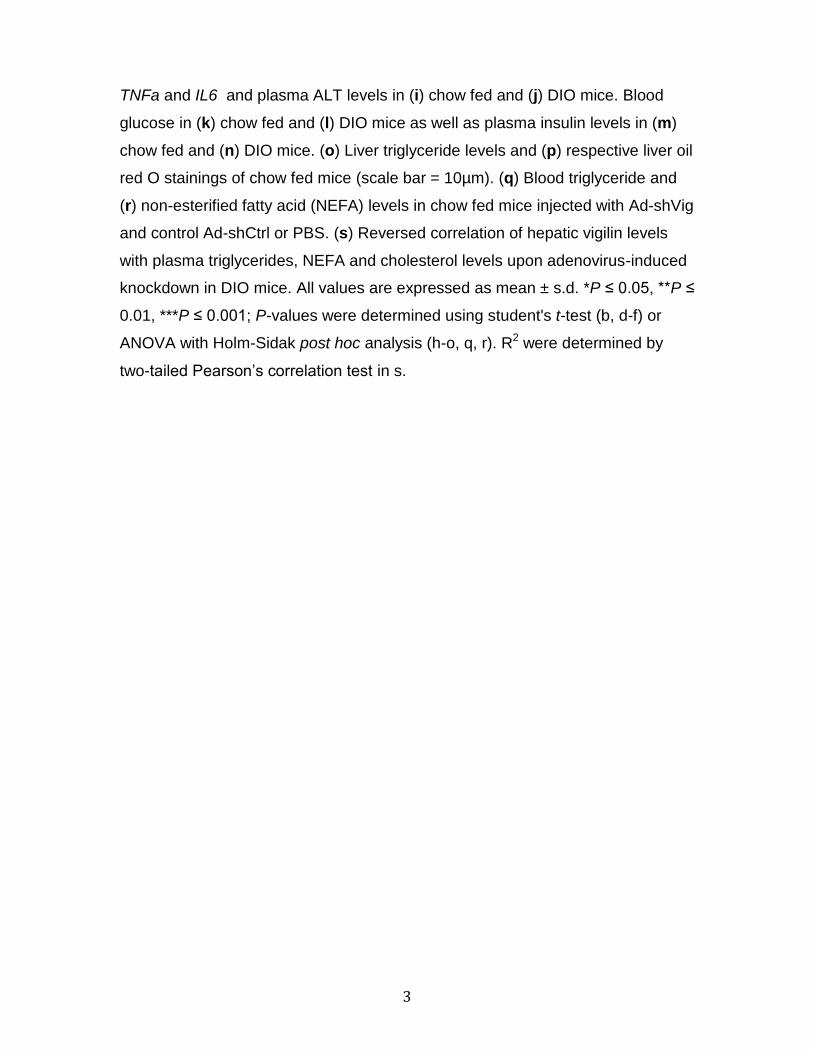
3
TNFa and IL6 and plasma ALT levels in (i) chow fed and (j) DIO mice. Blood
glucose in (k) chow fed and (l) DIO mice as well as plasma insulin levels in (m)
chow fed and (n) DIO mice. (o) Liver triglyceride levels and (p) respective liver oil
red O stainings of chow fed mice (scale bar = 10µm). (q) Blood triglyceride and
(r) non-esterified fatty acid (NEFA) levels in chow fed mice injected with Ad-shVig
and control Ad-shCtrl or PBS. (s) Reversed correlation of hepatic vigilin levels
with plasma triglycerides, NEFA and cholesterol levels upon adenovirus-induced
knockdown in DIO mice. All values are expressed as mean ± s.d. *P ≤ 0.05, **P ≤
0.01, ***P ≤ 0.001; P-values were determined using student's t-test (b, d-f) or
ANOVA with Holm-Sidak post hoc analysis (h-o, q, r). R2 were determined by
two-tailed Pearson’s correlation test in s.
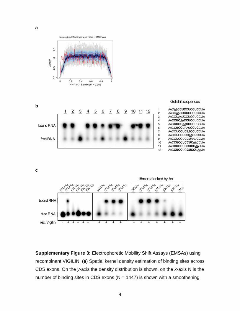
4
Supplementary Figure 3: Electrophoretic Mobility Shift Assays (EMSAs) using
recombinant VIGILIN. (a) Spatial kernel density estimation of binding sites across
CDS exons. On the y-axis the density distribution is shown, on the x-axis N is the
number of binding sites in CDS exons (N = 1447) is shown with a smoothening
5
bandwidth of 5. The signal of the clusters (red line) shows an even distribution
across the CDS exons within the probability range of the background distal
distribution (blue and grey, color shaded according to probability percentiles).
The black line indicates the average background probability distribution, dark
blue indicates 50th percentile smoothened background distribution, bright blue
indicates 75th percentile smoothened background distribution, grey lines indicate
sampled individual background probabilities. (b-c) Synthetic RNAs (10 nM) were
radiolabeled, incubated with 2 μM His6-tagged VIGILIN recombinant protein and
separated on a 1% agarose gel. (b) An 18-nt long RNA sequence containing 5x
CCU-repeats was systematically mutated at two adjacent sites with As
(underlined) and used for EMSAs. Shifts were only observed with sequences
containing a CYYC-2nt-CYYC RRE (in which Y=C/U). CYYC-2nt-CYYC RRE is
indicated in bold letters. (c) EMSA using CCU-trinucleotides with various repeats
(left and middle panel), and repeats with A-flanking sites indicate that sequences
≥18 nt containing ≥4 CCU repeats are required for efficient RNA binding.
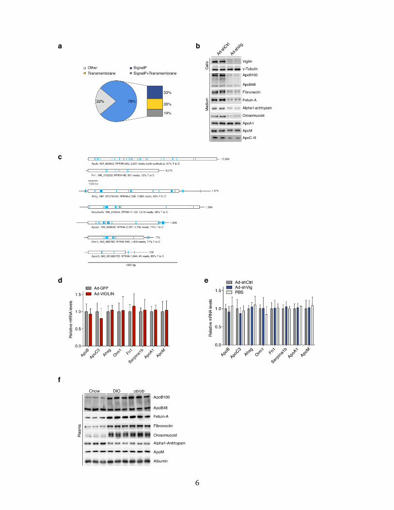
6
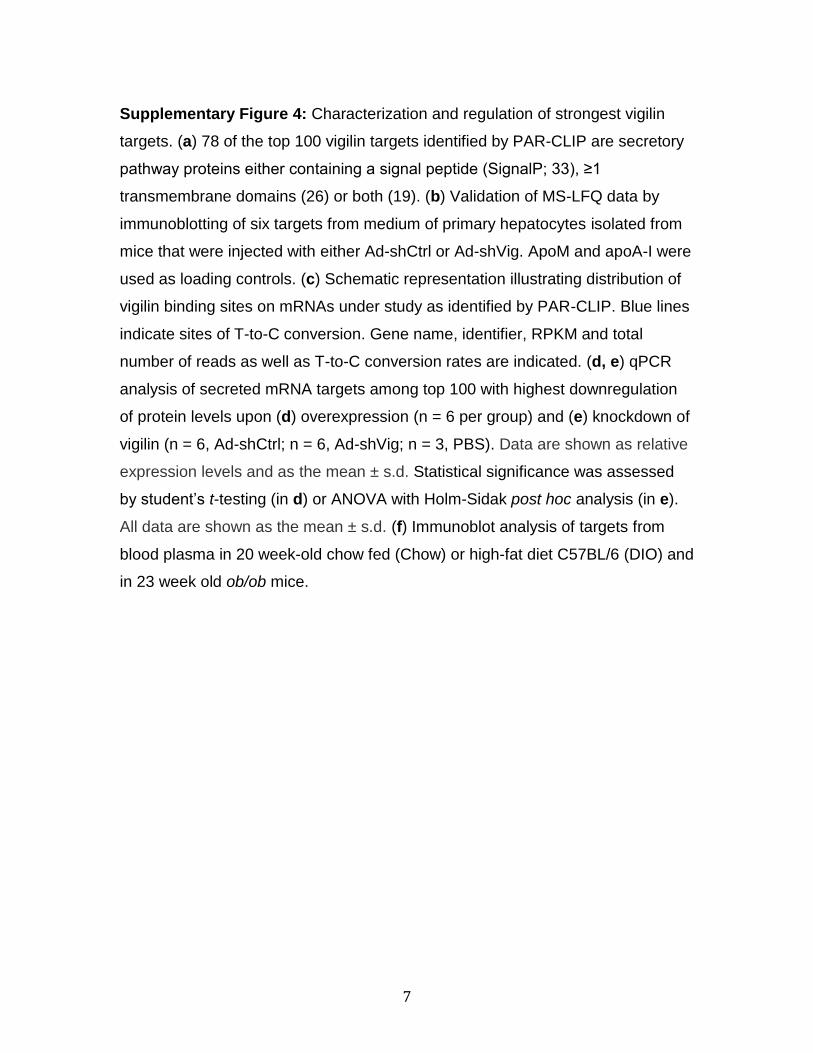
7
Supplementary Figure 4: Characterization and regulation of strongest vigilin
targets. (a) 78 of the top 100 vigilin targets identified by PAR-CLIP are secretory
pathway proteins either containing a signal peptide (SignalP; 33), ≥1
transmembrane domains (26) or both (19). (b) Validation of MS-LFQ data by
immunoblotting of six targets from medium of primary hepatocytes isolated from
mice that were injected with either Ad-shCtrl or Ad-shVig. ApoM and apoA-I were
used as loading controls. (c) Schematic representation illustrating distribution of
vigilin binding sites on mRNAs under study as identified by PAR-CLIP. Blue lines
indicate sites of T-to-C conversion. Gene name, identifier, RPKM and total
number of reads as well as T-to-C conversion rates are indicated. (d, e) qPCR
analysis of secreted mRNA targets among top 100 with highest downregulation
of protein levels upon (d) overexpression (n = 6 per group) and (e) knockdown of
vigilin (n = 6, Ad-shCtrl; n = 6, Ad-shVig; n = 3, PBS). Data are shown as relative
expression levels and as the mean ± s.d. Statistical significance was assessed
by student’s t-testing (in d) or ANOVA with Holm-Sidak post hoc analysis (in e).
All data are shown as the mean ± s.d. (f) Immunoblot analysis of targets from
blood plasma in 20 week-old chow fed (Chow) or high-fat diet C57BL/6 (DIO) and
in 23 week old ob/ob mice.
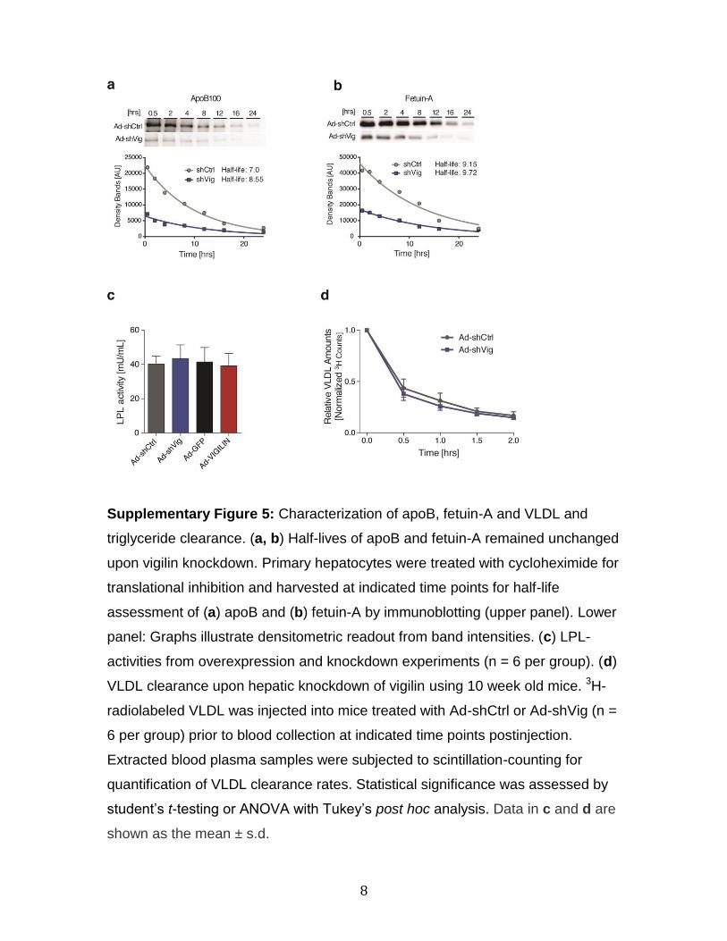
8
Supplementary Figure 5: Characterization of apoB, fetuin-A and VLDL and
triglyceride clearance. (a, b) Half-lives of apoB and fetuin-A remained unchanged
upon vigilin knockdown. Primary hepatocytes were treated with cycloheximide for
translational inhibition and harvested at indicated time points for half-life
assessment of (a) apoB and (b) fetuin-A by immunoblotting (upper panel). Lower
panel: Graphs illustrate densitometric readout from band intensities. (c) LPL-
activities from overexpression and knockdown experiments (n = 6 per group). (d)
VLDL clearance upon hepatic knockdown of vigilin using 10 week old mice. 3H-
radiolabeled VLDL was injected into mice treated with Ad-shCtrl or Ad-shVig (n =
6 per group) prior to blood collection at indicated time points postinjection.
Extracted blood plasma samples were subjected to scintillation-counting for
quantification of VLDL clearance rates. Statistical significance was assessed by
student’s t-testing or ANOVA with Tukey’s post hoc analysis. Data in c and d are
shown as the mean ± s.d.
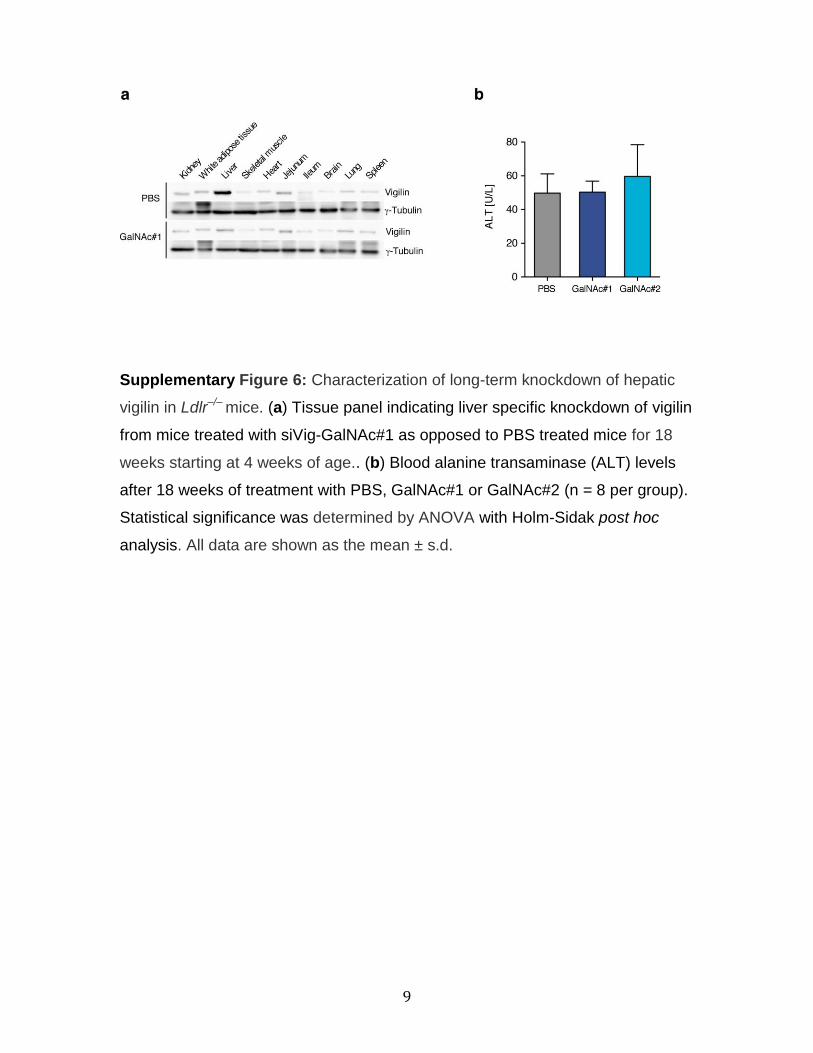
9
Supplementary Figure 6: Characterization of long-term knockdown of hepatic
vigilin in Ldlr–/– mice. (a) Tissue panel indicating liver specific knockdown of vigilin
from mice treated with siVig-GalNAc#1 as opposed to PBS treated mice for 18
weeks starting at 4 weeks of age.. (b) Blood alanine transaminase (ALT) levels
after 18 weeks of treatment with PBS, GalNAc#1 or GalNAc#2 (n = 8 per group).
Statistical significance was determined by ANOVA with Holm-Sidak post hoc
analysis. All data are shown as the mean ± s.d.
10

11
Supplementary Figure 7: Uncropped images of blots used in main figures. The
number of the corresponding figure is shown above the panels. Regions used for
figures are boxed. The position of the molecular mass markers is shown on the
left in kDa.
12

13
Supplementary Figure 8: Uncropped images of blots and gels used in
supplemental figures. Number of the corresponding figure is shown above the
panels. Regions used for figures are boxed. Position of the molecular mass
markers is shown on the left in kDa.
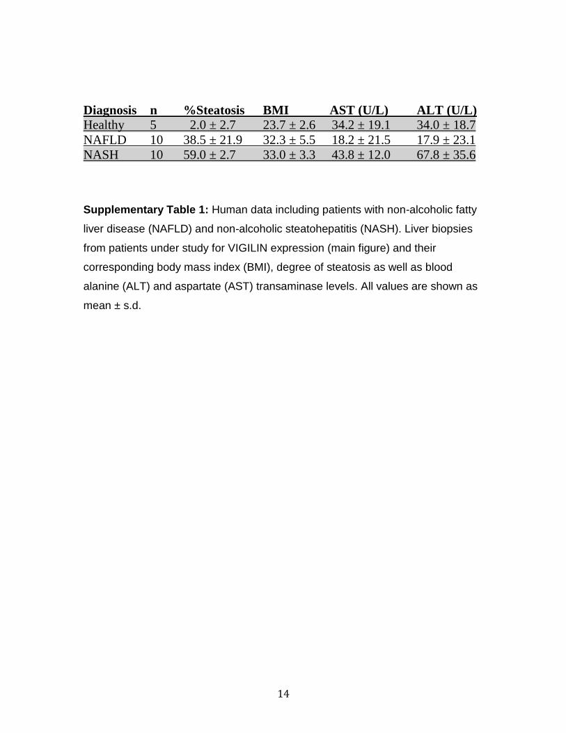
14
Diagnosis n %Steatosis BMI AST (U/L) ALT (U/L)
Healthy 5 2.0 ± 2.7 23.7 ± 2.6 34.2 ± 19.1 34.0 ± 18.7
NAFLD 10 38.5 ± 21.9 32.3 ± 5.5 18.2 ± 21.5 17.9 ± 23.1
NASH 10 59.0 ± 2.7 33.0 ± 3.3 43.8 ± 12.0 67.8 ± 35.6
Supplementary Table 1: Human data including patients with non-alcoholic fatty
liver disease (NAFLD) and non-alcoholic steatohepatitis (NASH). Liver biopsies
from patients under study for VIGILIN expression (main figure) and their
corresponding body mass index (BMI), degree of steatosis as well as blood
alanine (ALT) and aspartate (AST) transaminase levels. All values are shown as
mean ± s.d.
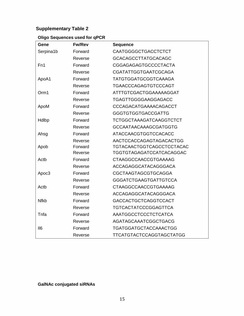
15
Supplementary Table 2
Oligo Sequences used for qPCR
Gene Fw/Rev Sequence
Serpina1b Forward CAATGGGGCTGACCTCTCT
Reverse GCACAGCCTTATGCACAGC
Fn1 Forward CGGAGAGAGTGCCCCTACTA
Reverse CGATATTGGTGAATCGCAGA
ApoA1 Forward TATGTGGATGCGGTCAAAGA
Reverse TGAACCCAGAGTGTCCCAGT
Orm1 Forward ATTTGTCGACTGGAAAAAGGAT
Reverse TGAGTTGGGGAAGGAGACC
ApoM Forward CCCAGACATGAAAACAGACCT
Reverse GGGTGTGGTGACCGATTG
Hdlbp Forward TCTGGCTAAAGATCAAGGTCTCT
Reverse GCCAATAACAAAGCGATGGTG
Ahsg Forward ATACCAACGTGGTCCACACC
Reverse AACTCCACCAGAGTAGACACTGG
Apob Forward TGTACAACTGGTCAGCCTCCTACAC
Reverse TGGTGTAGAGATCCATCACAGGAC
Actb Forward CTAAGGCCAACCGTGAAAAG
Reverse ACCAGAGGCATACAGGGACA
Apoc3 Forward CGCTAAGTAGCGTGCAGGA
Reverse GGGATCTGAAGTGATTGTCCA
Actb Forward CTAAGGCCAACCGTGAAAAG
Reverse ACCAGAGGCATACAGGGACA
Nfkb Forward GACCACTGCTCAGGTCCACT
Reverse TGTCACTATCCCGGAGTTCA
Tnfa Forward AAATGGCCTCCCTCTCATCA
Reverse AGATAGCAAATCGGCTGACG
Il6 Forward TGATGGATGCTACCAAACTGG
Reverse TTCATGTACTCCAGGTAGCTATGG
GalNAc conjugated siRNAs
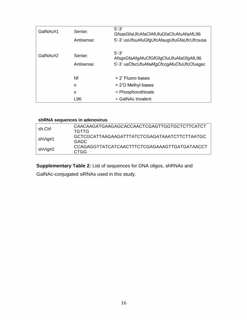
16
GalNAc#1 Sense: 5'-3' GfsasGfaUfcAfaCfAfUfuGfaCfcAfuAfaAfL96
Antisense: 5'-3' usUfsuAfuGfgUfcAfaugUfuGfaUfcUfcsusa
GalNAc#2 Sense: 5'-3' AfsgsGfaAfgAfuCfGfGfgCfuUfuAfaGfgAfL96
Antisense: 5'-3’ usCfscUfuAfaAfgCfccgAfuCfuUfcCfusgsc
Nf = 2’ Fluoro bases
n = 2’O Methyl bases
s = Phosphorothioate
L96 = GalNAc trivalent
shRNA sequences in adenovirus
sh.Ctrl CAACAAGATGAAGAGCACCAACTCGAGTTGGTGCTCTTCATCTTGTTG
shVig#1 GCTCGCATTAAGAAGATTTATCTCGAGATAAATCTTCTTAATGCGAGC
shVig#2 CCAGAGGTTATCATCAACTTTCTCGAGAAAGTTGATGATAACCTCTGG
Supplementary Table 2: List of sequences for DNA oligos, shRNAs and
GalNAc-conjugated siRNAs used in this study.