Embed Size (px)
Citation preview

$.’b HANDBUCH DER h4EDIZINISCI-IEN RADIOLOGIE Wq
ENCYCLOPEDIA OF MEDICAL RADIOLOGY
HERAUSCEGEBEN VON ,qoj,[]:;yO. OLSSON F. STRNAD H. VIETEN A. ZUPPINGER
LUND FRANKFURT/M. DUSSELDORF BERN
BAND 11/3
REDIGIERTVC)N
I A. ZUPPINGER, BERN und 0. HUG, MUNCHEN
SPRINGER-VERLAIG,BERLIN. HEtDE13ERG. NEW YORK wn(PRINTED IN CERMANY)
NICHT IM HANDEL
The radiation syndromes
By
E. P. Cronkite and T. M. Fliedner
.
The Meclicd Research Center
/4 “,#A/c# T/4AfUpton, L. L, New York

G. The radiation syndromes*
By
I
E. P. Cronkite** and T. 11. Fliedner***
With 9 figures
I. Introduction
Until 1945 the question of radiation syndromes produced by exposure of the wholebody or portions of the body to penetrating or poorly penetrating radiations was of littleconsequence except to the practicing radiologist. Following the use of nuclear bombs inJapan, the entire world became vitally concerned with the effects of whole body exposureto penetrating gamma radiation. Later, with the de~elopment of the atomic energy industriesand the application of nuclear energy in the production of electric power, injuries fromexposure to combined gamma and neutrons were seen folloning criticality accidents inthe processing of fissionable materials and in accidents involving nuclear reactors. Therehave been several deaths, and in the survivors the question of late effects of radiationconstitutes important medical-legal problems. These are not trivial problems. The questionsof monetary rewards for fractures, burns and other injuries are well established in thecourts. The question of rewards for a statistical possibility of getting a disease at somefuture date remains unanswered.
On March 1, 1954, the world was again forcibly reminded of the hazards of radiationin the modem world when a fall-out accident occurred following the experimental detona-tion of a thermonuclear device. The exposure of numerous native MarshaHese and Americanmilitary personnel to fission products involved both exposure to penetrating gammaradiation and exposure to superficial beta radiation from the fissionable materials thatwere in contact with the skin.
Thus, in view of the contemporary possible exposures of human beings to harmfulamounts of radiation, one can set up a general classification of radiation injuries to becovered in this chapter.
1. Classification
Radiation injuries can be divided into two general categories, early and late injuries.The early type results from brief, intense exposure; the late type either from exposureto large single doses, or from prolonged exposures of lower intensity. In this chapter thefollowing will be considered.
* Researchperformedat the BrookhavenNationalLaboratoryunder contract with the United StatesAtomic Energy commissionand at the Imtit.ut,fiir Himatologie J?reilmrgun&r contract with the Gesell-schaft fur StrahlenforschungmbH and the European.4tomicEnergyCommunity(EURATOM).
** MedicalDepartment,BrookhavenNationalLaboratory,Upton, L. I., N.Y., USA.*** Abt,ei]ungfur KIinischePhysiologic, Zentrumfur KlinischeGrundlagenforechung,Universitiit~rn,
Ulm (Donau).
,.—
—...

t,c..L. u ..... .... ....~........ .~\.L.A.
stance — tUIHJ$t:tk’ Of CdCium is its replllsil-e name — which is potential. whatever thatmeans, to the said rays. The consequence of which is that you can see other people’sbones with the naked eyc -- o~~the revolting indecency there is 11oneed to dwell. It wonldbe best to burn the works on these rays, execute the discoverer and whelln all cfilciumtungstate into the ocean. Let the fish contemplate each other’s bones if they like. but notus. ”
Oh roentgen, then the news is trueAnd not a trick or idle rumorThat bids us each beware of youAnd of your graveyard humorWe do not want. like Dr. SwiftTo take our flesh off and to pose inOur bones, or show each little riftAnd joint for you to poke your nose in.
– From PUNCH, January 25,1996.
Ill effects were usually blamed on such things as ultraviolet from the Crookes tubes,platinum particles, cathode rays, electrostatic discharges, heat and so forth. It was soonnoted that erythema was frequently ‘seen involving the hands, arms and faces of peopleworking with .X-ray apparatus, but it appeared that this was considered of scarcely anymore importance than sunburn. Thus, before long a tragic list of disastrous consequencesbegan to unroll and many of the early workers appeared as victims, often fatally injured.
One of the early reports of superficial radiation injury is that of Dr. KASSABIAY, whowrote of X-rays as an irritant in 1900 and described his own case: “About five monthsago the fingers, knuckles and dorsum of the left hand exhibited a general erythematouscondition. This continued about a month; the itching became intense, the skin becametough, glossy, edematous and yellow.” His condition became worse, and in 1903, he againwrote, “In order to effect a cure I have used every remedial agent mentioned in all thetextbooks — but nothing seemed any good.” In 1908 an area of ulceration showed malig-nant changes. In 1909, in spite of amputation, there were axillary metastasis and deathsoon followed. This story with minor variations could be told of many others indeed.
BOND, FLIEDNER and ARCHQIBEAU (1965)quote HOLTHIJSEX(1959) as saying of the336 early fatalities ascribed to radiation exposure, 251 died tiom cancer induced by re-peated radiation exposure of the skin, and 56 others died from blood dyscrasias such as ane-mia or leukemia. Of course, in this chapter we are more concerned with the depressiveeffects of radiation upon the blood forming tissues and the sequelae of prompt bone marrowdepression or aplasia. The harmful effects of whole body irradiation on animals were docu-mented early in the excellent studies of HEINECKE(190.3 to 1905). Radiation hemorrhagewas described by FERNAU d al. in 1913. FABRICIUS-MOLLERobserved the relationship ofthe hemorrhagic diathesis from X-ray exposure in guinea-pigs to the depressed plateletcount. In his classical initial study in 1922 he demonstrated that shielding of one leg bylead prevented the depression in platelets and thus the bleeding. Later ALLEN et al. (1948)ascribed radiation hemorrhage to heparinemia. This has been subsequently refutedby JACKSON et al. (1952) and CRONKITE and BRECHER (1952). In subsequent studiesCRONKITEet al. (1952) have demonstrated that the radiation hemorrhage can be preventedsolely by platelet transfusions. The importance of infection in pancytopenic states has longbeen appreciated by clinicians. The specific importance of infection in radiation pancyto-penia was emphasized by the studies of MILLER et al. (1951) in their classic studies onthe incidence of bacteriemia in mice sub jetted to total body X-irradiation and the improv-ed survival rate by the treatment if mice with antibiotics after exposure. The criticalrole of the bone mamow in the development of radiation injury was emphasized by a seriesof studies by JACOBSONet al. (1949—1950) in which it was shown that shielding of the spleen

—.. .
CY)Fro))) p~’netmting radiation
WI) Il”lIolcbodg expowre
The ~cute illness produced by total body irradiation may OCCUrin man frol~~exposureto gamma and/or nelltron radiation from a detonated atomic bonlb, high ~~nu~ltkexposurefrom fall-out from atomic bombs, from accidents with radioactive materiais, nuclearpower sources, or in radiotherapy either for malignancy or as a means of depressing anti-body response preparatory to organ transplantation.
PJ Partial body or markedly inhomogeneous e.cposures
An acute illness may result from partial body exposure to penetrating radiations asis commonly seen in therapeutic radiation for cancer, and might again be seen followingexposure in shielded criticalityy accidents and from radiation sources in radar de~-ices.
~) From poorly penetrating radiations
h this situation acute injury of the s~ln or other body integuments may result frombeta ray exposures of the skin as may occur in contact with fall-out radiation from mili-tary or experimental use of nuclear bombs or from accidents involving handling of radio-active materials. This injury is primarily limited to the surface of the skin.
b) Len:-twrn exposure
W).%gain, this must be sub-divided under injuries from penetrating radiation and thosefrom poorly penetrating radiation. From penetrating ra$lation one may de~-elop the follow-ing late effects: blood dyscrasias, namely leukemia; an increase in degenerative diseases;shortening of the life span: an increase in the incidence of cancer in general: retardationof growth and development in children; an increased incidence of cataracts; impairedfertility: and, of course, genetic effects in generations yet unborn. In this chapter thequestion of leukemia, shortening of the life span, the question of thyroid cancer, andretardation of growth and development in children will be considered brief Iy.
~) Poorly penetrating radiation primarily produces injury of the skin, and this willbe considered only in respect to exposure to fall-out or contact with radioacti~e materials.
y) Absorption of radioactive materials
This will be considered only briefly and in the context of absorption of fall-out materialsin the event nuclear warfare, laboratory radiation accidents and the results of past ab-sorption of radium and mesothorium.
IL Historical
Although the early history is well known, certain humorous and tragic aspects servewell to introduce the subject. In the early days there was some little apprehension con-cerning the newly found and mysterious rays. Unfortunately, the early fears were largelyabsurd and misdirected. Since they are rather humorous in the light of our knowledgetoday, it is of interest to refresh one’s memory. Scarcely anyone seemed to realize thatthere might be biological dangers. Instead, the public was treated to imbecilities. ThePall Mall (lazette considered X-rays revoltingly indecent. Punch poked fun at Roentgenand advised him to work on spoolis. .$ Xew Jersey legislature later was concerned aboutthe use of X-rays in opera glasses. In London X-ray proof underwear was offered for sale.Today the following from Punch, January 1896,is quite comic:
5012431. .

Uc-u.. . .. . . .. . . . - ..-, . . . .. ,crease the survival of irradiated ~llinlals. ‘I%ere followed a long period of time m ~rl~lcl~Itwas argued whether the beneficia] effect of spkm ic shieldiny. injection of splenic and bonemarrow cell suspensions was due to transplant at ion or a humoral stimulation of host cells.It is now clem th~t the beneficial effect is predominantly due to tr~~l~sl]l:llltatioll of hemo-poietic stem cells. The huge literature on this subject has conclusi~-e]y demol~stratecl therole of transplantation of the hemopoietic stem cells in protection against radiation in-jury. It is not clear when the hemopoietic effects of whole body irradiation were firstobserved in human beings. However, an early detailed report on the effects upon depressionof the blood in human beings without blood dycrasias by Roentgen ray therapy waspublished by MIXOT and SPURLINGin 19?4. A classical re~-iew of the effects of radiationon normal tissues of man and animals covering the period prior to 1942 was published byWARREN (1942).
The explosion of the nuclear bombs at Hiroshima and Nagasaki produced humanradiation pancytopenia on a massive scale. Everything that had been observed in animalswas abundantly observed in the Japanese. These observations are reported by OUGHTERSONand WARREN ( 1956). The potential hazardous effects of fall-out radiation were emphasizedfollowing the accidental exposure of a large number of human beings to fall-out radiationon March 1,1954,The effects of radiation from fall-out in human beings is described byCRONKITE, BOND and DUNHAM (1956).
Two other aspects of radiation injury are also of importance. First, there is the questionof severe injury to the nervous system either by whole body irradiation or exposure of thehead alone. This produces a rapidly fatal syndrome amply described by GERSTNER. —This has subsequently been observed in part in human beings exposed to huge amountsof radiation in criticality accidents (P~xTox et al., 1958). .&other means by which animalscan be killed is the acute intestinal radiation death that was initially described by QUAST-LER et (d. (1951).
IIL Definitions of human exposure
A semantic problem exists. In the past various descriptive terms have been used todescribe the types of exposure, such as acute, chronic, protracted, repetitive and contin-uous exposure, again sub-divided into various categories on the basis of the type of radia-tion, its energy or other specific characteristic. Much ambiguity existed in the use of thepreceding approach. Exposure conditions should therefore be defined according to themethod proposed by the United States National .kcademy of Sciences, h~ational ResearchCouncil Sub-Committee on Hematologic Effects of Radiation. These are:
1. Short-term exposure
Short-term includes total body exposures to radiation over a short period of time(e. g. in nuc[ear warfare, nucIear reactor or accelerator accidents) and exposure of limited butsubstantial body areas when the radiation is delivered either as a single dose or fraction-ated over a few days or weeks (e.g. in therapeutic radiation, diagnostic radiology or traceror therapeutic use of radioactive isotopes). In considering short-term exposure, a dosegreater than 50 rads is defined arbitrarily as a high dose, less than 50 rads a low dose.These definitions are for prospective and retrospective classification and study of radiationeffects.
2. Long-term exposure
Long-term exposure refers to continued or repeated exposure to radiation over longperiods of time — months or years. The possibility of such exposure is greatest in certain
5012U2..._..$_- —._—...—

)
1
Occtlpationa] groups and in persons having body burdens of radioactive isotoL)cs with
relatively 10I\g, effective half-lives. X-ray examinations repeated frequently over ;l long.’ time were also considered long-term exposure. <IS are exposures to cosmic radiation]}. uatl~-
rally occurring radioactive isotopes, and world wide fall-out.
Although the total dose of radiation is important in long-term exposure, it is moreuseful and convenient to indicate the degree of exposure in terms of dose per unit of time,usually cumulative dose per week.
I
1. Minimal weekly dose is less than LOOmilIirads.
2. Low weekly dose is 100 to 1000 millirads.13. High weekly dose is greater than 1000 millirads.
The minimal dose is less than the maxinlunl permissible dose (MPD) recommendedmost recent ly by the International Commission on Radiological Protection and the N“atio -nal Committee on Radiation Protection and Measurements. The dividing line betweenlow and high dose corresponds to the first MPD recommendations of these groups in effectbetween 1936 and 1948.
IV. Effect of physical factors on radiation response of mammals
,
Superficially it would appear unnecessary to discuss the role of physical factors uponthe response of the whole body to irradiation. However, problems are involved which aresignificantly different from those encountered in radiation therapy. Clearly the preferablemeasure of the amount of radiation is the absorbed dose (rad). This unit, as is well-knowm,is completely independent of the energy and type radiation. However, the expression ofbiological effects from whole body exposure or large parts of the body in terms of the radis not com.pletely satisfactory. The distribution of dose in respect to time and space isalso r. major determinant of response. It is necessary to know whether the dose is essen-tially instanteneous, fractionated, short-term or long exposure, because distribution ofdose with time also significantly influences the biological response. .$s will be pointed outlater in this chapter, the response, recovery and resistance to subsequent doses of radiationis significantly altered by prior exposure to radiation. Further, the spatial distribution ofthe absorbed dose anatomically in depth within an organ significantly influences the bio-logical response of the whole animal. The quality of the radiation is another meaningfulparameter to be considered. High energy protons, alpha, beta, gamma, X-ray and neutrons,all have properties which may influence the quantitative response per unit of absorbeddose. Generally tile relative biological effectiveness (RBE) per rad is a function of thelinear energy transfer (L13T). However, with very high proton energies and stripped nuclei,as may be encountered in space flight, one must also consider the possible influence ofspalatioll. A problem to which there is no satisfactory solution is the anatomic inhomo-geneities in the distribution of absorbed dose within the body. TVith most conditions ofhuman exposure the relative homogeneity of absorbed dose will be the exception rather thanthe rule. It is fair to say that after accidental exposures true homogeneity in absorbeddose has never been seen. The problem on unpredictable inhomogeneity in dose raises animportant practical difficulty in assessing the hazard of accidental exposure of man. Theinhornogeneities simply cannot be described by any single meaningful value for the dose dis-tribution received by the critical organs within the body, hence the biological responseis the key to management, not a physical dose estimate.
.Althougll exposure due to absorbed radioisotopes will rarely be observed, it doesconstitute a problem of difficult dosimetry. For practical purposes, it is almost impossibleto measure directlv the dose from internal emitters. Therefore, it is necessary to rely oncalculations of the-dose from estimations of the average concentration of the radioisotopeill~ol~-ed and the biological half-life of the substance. From the preceding two, one can
—
511U?U3

e.iw’) GL1tilllii[~ I,UI?cLLISO1’t.)(Xldc)Se ~J’o\ lUll I~ UIIC~6 LllL?l’e JS UllltOt VUlt~ 111Lll~ IJ~P(J*ILIUII~)tthe material within all of the orga.l~sinvolved. Since the distribution is alWa~S inllomoge-neous, one can only estimate the arerage dose and rarely, if ever, is one able to determineat ilnlicroscol]ic illlator~lical lew-eltl~er:~l~geillcl(~scfrc)nl the millinlllm to tl~elll:lxilll~~ill.llorthe influence of relatively uninjured tissues upon contiguous severely injured [issues.
The concept of theintegral dose is very useful inradiotherapy. Tllisrefers to the totalenergy absorbed within the object. This unit ofl~leastlrenlel~to fradiatiolli sof\-er~-1il~litedor no value considering the effects of radiation on the whole body of man or animals sinceit has been shown that gram roentgens or integral dose cannot be used to predict the de-gree of biological effect. This arises from the fact that the biological effect depends not onlyon the total dose absorbed, but also on the portion of the body receiving the dose. the re-lative homogeneity of the absorbed dose and, of most importance, the influence of non-irradiated or lesser irradiated hemopoietic tissue upon the recovery of the more heavily
— Fallout gamma150- _ Bomb gamma
~ Fission neutrans
lb ‘ 20 ‘ 30Depth in cm
Fig. 1. Depthdosecurvesfor threedifferenttypesof radiationto whichhumanbeingmightbe exposed
irradiated hemopoietic tissue. A very useful reference for a more technical and sophisticateddiscussion is the symposium on Physical Factors and their Modification of BiologicalEffects of Radiation (1964).
The relative biological effectiveness is analogous to the relative potency ratio for drugsin pharmacology. In radiation studies, a standard radiation, usually X-rays of prescribedcharacteristics, is used. The ratio of the dose in rads of X-rays to produce the same degreeof biological effect to the dose of the test radiation is known as the RBE of the test radi-ation. When the targets are very small, one can obtain a precise estimate of the absorbedenergy in each case and have a very scientific evaluation of the relative biological elec-tiveness. However, when one has larger targets, such as man, in a radiation field the problemis complicated by serious inhomogeneities in the distribution of absorbed dose. This isillustrated in Fig. 1. It will be noted that in the case of a diffuse field of gamma radiationcoming from all directions at the individual, the surface dose is somewhat higher than theair dose due to back scatter, and the dose throughout the body is relatively uniform untilthe other side of the body is reached, at which point the dose again increases due to scatterand low energy components that are present. In the case of the penetrating initial bombgamma radiation there is a very small increase in the absorbed dose in the first few centi-
5012434.

)11ofloge-mine, nor I
total~ited~incee de-onlye re-non-Ivily
~atedgical
lrugs-ibed?greeradi-lrbed;ffec-blemlis is*tion1 theuntilatterlomb?enti-
metres. and Lhell an attenuation with the unilateral irradiation, so that the exit dose isabout 45 ~~of the entrance dose. In the case of fission neutrons, the first collision, absorbeddose at the surface iS about 75 ~~ of the incident dose. The absorbed dose is rapidly atten-uated, reaehing a low value of about 10 “0 of the entrance dose at 1S cm. .Mthongh theradiation is unidirectional, it is greater on the back surface of the body due to scatteringfrom the air and entrance into the back side. These three examples of whole body exposurehave three completely unique and different depth dose curves. Clearly, per rad of expo-sure there is a si=~ificantly different biological eilect.
V. Qualitative and quantitative effects of radiation upon mammals
1. Dose mortidity responses
Dose mortality responses have been used for decades to express the effect of radiationand to observe factors that modify the radiation response. All of the dose mortality curveshave a characteristic sigmoidal appearance, as expressed in Fig. 2 b. Generally, in respectto mammals most of the mortality is completed by 30 days and the mortality is scored at
i
~ Unilateral1.0mev Xray
‘\_ Bilateral
100-1.0 mev Xray
1-
10 2’0 Jo 40
cm of Masonite
Fig. 2a. Depthdosecurvesfor 1.0 MeVX-ray whenall of the radiationis deliveredto one sideof thephantimandwhenhalfof the doseis deliveredto both sides(bilateral)
this time. Note that the upper and lower portions of the curves are very poorly defined.It is theoretically impossible to ascertain the dose below which no individual would die orabove which all individuals will die. However, these are in practice ascertained by expo-sure of large numbers of animals in the vicinity of zero and 100 o/. mortality. However,for comparative purposes the midpoint of the curve is used to characterize the entire doseresponse relationship. This dose is referred to as a median lethal dose, and is ordinarily
20 Handbuch(lermed.Hadiologie,Bd.11/3
.
-—
.&<
*’
+:.,
abhrevmtetiLD30. It IS a derlred dose aILU uoes not, nlean that WI llldlvldual animal
exposed to this dose will necess:lrily live or die. It indicates only that in a very large po-pulation one can expect half of those exposed to die within the stated time. USUally withmammals the range in dose between no dexths and 100 “o mortality is of the order of 2(KJto400 rads. Theoretically the lower end of the curve approaches the abscissa asynlptoticall>-and does not cross it until zero dose. In actuality it crosses the abscissa at a relatively highdose, creating effectively a threshold dose for mortality. A very useful means of expressingthe dose mortality is to plot it on probability paper, by which the sigmoidal curve is trans-formed into a line. Then one determines the slope of the curve and the LD50 tocharacterise the whole system.
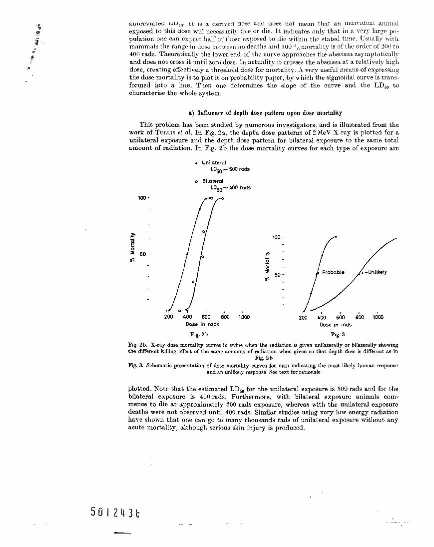
a) Influence of depth dose pattern upon dose mortality
This problem has been studied by numerous investigators, and is illustrated from thework of TULLISet al. In Fig. 2a, the depth dose patterns of f! MeV X-ray is plotted for aunilateral exposure and the depth dose pattern for bilateral exposure to the same totalamount of radiation. In Fig. 2b the dose mortality cum-es for each type of exposure are
. UnilateralL~O_ 500rads
o Bilateral
LD50 -400 rads
100-
a
[
o=-~sz Ijo..*
‘ /1o
e;00 400 660 8b0 I.&l
Dose in rads
100-
[/Probable .+ Unlikely
//
200 Go 660Dose in rads
8k l.&Ul
Fig. 2b Fig. 3
Fig. 2b. X-ray dose mortalitycurvesin m-inewhenthe radiationis given unilaterallyor bilaterallyshowingthe differentkillingeffect of the sameamountsof radiationwhengiven so that depth dose is differentas in
Wig.2bFig. 3. Schematicpresentationof dose mortalitycurvesfor man indicatingthe most likely humanresponse
andan unlikelyresponse.Seetext for rationale
plotted. Note that the estimated LD50 for the unilateral exposure is 500 rads and for thebilateral exposure is 400 rads. Furthermore, with bilateral exposure animals com-mence to die at approximately 200 rads exposure, whereas with the unilateral exposuredeaths were not observed until 400 rads. Similar studies using very low energy radiationhave shown that one can go to many thousands rads of unilateral exposure without anyacute mortality, although serious skin injury is produced.
\—

uiirnal:e po-.’ WithWI toticallj-y high‘essingtlrans-3W to
m the1 for a! totalre are
/’
?1y
lowingtssin
sponse
)r thecom-
osure!atio nt any
b
Earlier in this chapter. Fig. I, the dcpt[l dose curves for different types of radiationto which human beings might be exposed \vere plotted. othertypeso fa,ccidents in which
! hllmatl beings may be in~olved ilrf: critic~llit?- accidents that consist ofacomhination of
Imixed gamma and neutrons, and here there are also inhomogeneities in the absorbed dose.However, the influence of the inhomogeneities in depth dose illustrates in I?ig. 2a and bcannot be extrapolated for purposes of prediction to other types of exposure.
b) The median lethal dose for human beings
There are certain general facts that are pertinent to attempting to answer this vitalproblem. First, as shown by BOND and ROBERTSON( 1957 ), all small mammals have a rela-tively high LD~Owhen expressed as mid-line absorbed dose, and all large mammals (swine,burros, dogs etc.) have a relatively low LDiO. The slopes of most dose mortality curves
4 are roughly the same. In general the LD~O for large mammals is of the order of 250 radsfor X-rays with uniform dose distribution. and that for small species is approximatelydouble this value. Some investigators have considered primates to be more closely relatedto human beings. However, the LD60 of monkeys is similar to the small mammals ratherthan the large mammals. Large animals exposed under silimar geometric conditions haverather uniform dose mortality curves, perhaps partly because large ani reals, unlike smallanimals, provide their own constant maximum scatter of radiation. The experimentaldetermination of LD~Ois usually based on animals of a cIearly defined genetic bac kground,age and sex. In general females are a little more resistant to radiation than males. When oneis concerned with human populations, one is dealing with a spectrum of ages, sex and1races.
; Knowledge about the LD~Ofor man is clearly needed, both for evaluation of radiation,% accidents and from the standpoint of planning possible whole body radiation therapy for1 specific purposes. CRONKITEand BOND (1960) have approached the problem of estimation1
of the LD50 for man in the absence of any therapy for the injury in the followingmanner. A large number of human beings were exposed to 175 rads of gamma radiation
;from fall-out. As illustrated in Fig. 1, the depth dose below 1 cm is quite uniformfrom this type of radiation. These individuals had a severe hematologic depression and,were on the verge of developing purpura and had a severe Wanulocytopenia. From data onanimals, it can be assumed that an increment of 50 to 100 rads probably would have placedthese exposed human beings into the lethal dose range. one can therefore anchor the lowerend of the dose mortality curve for man in the vicinity of 200 rads. The next assumptionasserts that the slope of the dose mortality+ curve for large animals and for human beingswill be almost the same. With the preceding two assertions one can draw a dose mortalitycurve through the lower anchor with the slope similar to that of dogs, and obtains an esti-mated LD~O of about 360 rads for man. This is illustrated in Fig. 3. Other human dataare consistent with this estimate. one out of five of the Yugoslavs exposed to about350 rads in a nuclear accident died. The hematologic response of the individuals exposedto approximately 300 rads in another criticality accident was significantly greater thanthat in the Marshallese. Some of these individuals developed purpura, and the depressionin their granuloc yte count was also greater than in the Marshallese. Furthermore, extensivestudies on clinical radiation therapy exposing the entire body has resulted in a severehematologic depression of individual with metastatic cancer after exposure to 200 rads,and an occasional death in these diseased individuals has been observed (IMILLERet al.,1957).The doseof radiation received by the Japanese in Hiroshima and Nagasaki is undercontinuing study by the joint Japanese -.American Atomic Bomb Casualty Commission.With further refinements of their estimates, one may be able to ascertain the dose to asufficient number of human beings to get a rather precise estimate of the human LD~Oexposed to this type of radiation and given no therapy.~“.

%. (,( ’11I’(!nc}y:l,l Sysl!cl]ls — kll~ !.)t~bl> UL ull(lV~~i,iLLLULLL< L.tiLJ . . ..(--- ~ ‘- ~ -.
in the mammal
A very s~phisticateci ciiscussiou of the role of disturbance in cellular kinetics of cellrenewal systems as a basis for mammalian radiation lethality has been published by BOND.FLIEDNERand ARCHAMBEAU( 1965). For our purposes in this chapter only a brief summaryof the different types of organ systems within the body and their characteristics are needed.Mammalian organs consist of three general types on the basis of cell production. First,there are those organs in which there is essentially no production of new cells from cessationof growth to death. Examples of this type are the brain, cardiac muscle, skeletal muscleand the collagenous tissues. Second. there is the class of organs in which there is the cap a-
Extrusion Zone
Fig. 4.
Fig. 5.
.s+
1
rigrationd
FuthT
M!toslsstops
\
CellularDegener
ation (y
R1 24 hours 36 hours after 36 hours
Fig. 4 Fig. 5
Schematicof cell generativecycleindicatingthedifferenttimephasesbetweensuccessivemitoseswhichhaveradlobiologicalsignificance
Schematicpresentationof cell renewalin the small intestinalepitheliumsand the effectsof 2000radsof radiationupon proliferationof bowelepitheliumsleadingto ;he gastrointestinalsyndrome
bility of repair following injury, such as the liver, fibrous tissue, periosteum and blood ves-sels. Normally there is practically no cell turnover in these tissues, but upon injury theremaining tissues delete can burst into cell proliferation and repair the lossof cells or the separation of the tissues. Third, there are those tissues which are in a steadystate of cell proliferation, in which the production rate equals the death rate of cells.Examples of these tissues are the epitheliums of gastrointestinal tract, the skin, and thehemopoietic tissues. Cell proliferation is maintained in each of these cell lines by “the stemcell” which has the properties of self-replication and differentiation to exogenous influencesthat direct it down specific cellular lines. The cell cycle for a stem cell is illustrated inFig. 4. The cell cycle consists of the following periods commencing after mitosis. Firstthere is a rest period (Rl) prior to the commencement of DNA synthesis or chromosomalreplication. During the rest period, of course, other metabolic processes may be very active.During DNA synthesis the chromosomes are replicated to preserve the genetic code. Alsoduring this time there is a substantial amount of RNAsynthesis. L’pon the completion of thereplication of chromosomes there is another rest period termed R ~prior to mitosis. Whencells divide they can be assumed to be possessed of equal genetic capabilities. However,random influences impinge upon the stem cell pool and on an average induce half of the
5012WI

i
of celIBmn,m mary~eeded.
First,ssationmuscle? ca a-P
es Which
000rads
,od ves-my theLe losssteady)f cells.md theLestemluences~ted in<. Firstosomalactive.e. AIso1of theWhen
,wever,of the
progeny to differentiate in order to maintain the steady state equilibrium of the stem cellpool and specific cell lines. One can measure the relative rate of cell prolifemtion by count-
ing t]le nllxllbt.r of nlitotic fiylr~s. of Ce]]s tll:lt aye in I)hTA synthesis, or by stopping nlito~isat metaphase by stathokinetik agents such as colchicine, and observing the number ofcells that accumulate in rnetaphase. For a more detailed description of the characteristicsof cell renewal s~-stems one is referred to BOXD, FLI~D~~R and ARCHAnlBEAt~(1965).The cell cycle is of particular importance in radiation effects, since the sensitivity of cellsvaries with the stage of the cell cycle. In general. cells are most sensitive to radiation duringthe mitotic, the late RI or early phases of D~”.\ synthesis. In tissue culture after exposureto the same amount of radiation, 4 times the number of cells survive if exposed during lateDNA synthesis, and most of the RI and R ~ periods as compared to exposure insynchronized cultures during the early DATA synthetic phases. Mitosis is also very sensi-
tive, with about half as many cells surviving when exposed in mitosis as compared to lateRz and early DNA synthesis. A simple cell renewal system for the gastrointestinal tract isillustrated in Fig. 5. Norman y there is active proliferation of cells in the neck of thecrypts of LIEBERKUHN.Cells migrate out the villus in an orderly fashion and are extrudedat the tip. In the normal steady state it takes approximately 36 hours for cells to migrateto the tip of the villus.
Since the kinetics of cell proliferation in hemopoietic systems are described in consid-erable detail by BOND et ai. (1965), only a pertinent summary will be presented herein.
a) Erythropoiesis
Stem cells are acted upon by erythropoietin to direct them down the erythropoieticpathway. The approximate generation time of erythropoietic precursors in the proliferatingcompartment is 24 hours in man. The total transit time from the stem cell to the reti-culocyte is approximately 4 to 7 days. The mean life span of human red cells is 120 days.:h The steady state equilibrium is maintained by a feedback system that is sensitive to oxygentension and perhaps the total mass of the red cells in the peripheral blood. Decreased oxygentension increases the levels of erythropoietin which induces more stem cells down theerythropoietic pathway.
b) Granulocytopoiesis
Granulocytopoiesis is also reasonable well understood from the standpoint of the timeinvolved in the flow of cells from the stem cell to the mature granulocyte in the peripheralblood (CRONKITE et al., 1960;ELIEDNER et al.,’ 1965;CRONKITE and FLIEDNER, 1965).The factors that are responsible for the differentiation of stem cells into granulopoieticprecursors are not known. The transit time for cells from the myeloblast to the first non-dividing cell in man is about 6 days. The transit time through the maturing pool, that isfrom the metamyelocyte to the granulocyte, is 3 to 4 days. The total transit time fromthe stem cell to the mature granulocyte in the marrow is 9 to 10 days. (k-anulocytes dis-appear from the blood in a random fashion with a half-time of 6.6 hours (ATHENSet al.,1961 ). The random process is terminated by a senescent process at 30 hours (FLIED~ERet al., 1964).
c) Platelets
These are produced by the megakaryocytic system. The total transit time from themost immature megakaryocyte in the marrow to platelets in the peripheral blood has beendescribed as lying between 4 and 10 days (man, CRONKITE et al., 1961;rat, EBBE andSTOHLMAN,1965). The life span in the peripheral blood of man is of the order of 10days(LEEKSMA et al., 1956). Platelets are lost from the blood by a random process, and alsohave a finite life span that terminates the random 10SS.
.-.Nlmvj
_—_.—

d) Lymphopoi(’tk tissues
The lymphocyte prodllcing organs constitl[tc a unique system. me lylnl)llocytes may
conveniently be &\,ide~ into t~vo classes 011 the basis of their size (SIPE et (11., 1!)G6).The smaller ckws with volumes of 150 to 350 cubic microns is flu-ther sub-divided into twoclasses on the basis of life span. One is very short-lived and, at least in animal experiments,presumably is thymic in origin (EvERETT d al., 1905). The other has a much longer lifespan, witi~ some cells living in excess of a year (ROEINSOX et al., 1965). The larger lympho-cytes have a relatively short life span measured in hours to a few days. A fraction of thesmall blood lymphocytes have a unique migration pathway from the blood through thepostcapillary venules at the rnedullary cortical junction (GOWANS et al., 1963).
These cells migrate through this veuule into the substance of the lymph node, and thenrecycle out through the efferent lymph and into the blood. This recycling accounts for avery large fraction of the lymphocytic output of the lymphatic ducts. Since lymphocyteshave these wideIy varying life spans and migration patterns but are not unique in respectto morphology, it is difficult to study the kinetics of these cells adequately and separatelywith available techniques.
e) Bone marrow
The bone marrow is the site of the formation of red cells, granulocytes and platelets.Normally in man all myelopoiesis takes place within the cavities of bones. Thus it is con-tained within rigid walk which do not permit volume changes. The hemopoietic bonemarrow is composed principally of three cell renew-al systems: erythropoietic, granulo-poietic and thrombopoietic. The parameters of cell proliferation for these have been listedearlier. All three cell renewal systems are distributed throughout the extra-sinusoidal marrowspaces. Normally one never sees erythroblasts outside the marrow parenchyma. In addi-tion, it is rare to see granulocytic cells more immature than the band in the peripheralcirculation. It is possible that the mature granulocyt e spends some of its life span outsidethe blood vessels. However, there is no clear-cut evidence for re-entry of ganulocytes intothe blood vessels. There is no question that lymphocytes spend a substantial fraction oftheir life span outside the blood vessels as described under the recycling of lymphocytesearlier. The vascular structure of the bone marrow has been studied extensively. It isa closed system with a series of unique arteries and veins connected by a very fragilesinusoidal system which receives blood from the arterial capillaries and from which bloodempties into the venous side (FLIED~ER et al., 1956).It is believed that cells enter the bloodstream through the sinusoidal endothelium.
3. General cytological and histological effects of radiation on tissue
Radiation can produce immediate cellular death, suppression of the motility of cells,suppression of reproduction of cells, the induction of anomalies of cellular division, a re-tardation of growth and, in the germ cells, mutations. Whether mutations occur also insomatic cells maintaining the cellular renewal systems is open to question. What is observedmicroscopically in mammalian tissues following exposure to radiation is a function of thetime after irradiation, the dose of radiation, and the tissue involved. The sequence ofmicroscopic effects in the so-called radiosensitive tissues (those tissues that are continuallyrenewing themselves) is different from that observed in the radioresistant tissues (tissues,the cells of which are rarely replaced if at all from the time of birth, or after cessation ofgrowth).
There is nothing specific about the observable microscopic effects of radiation. There isno special type of necrobiosis. The cells degenerate according to the manner of the particu-lar cellular type or tissue. The picture associated with cellular death in histopathology hasbeen described as coagulative necrosis, Iiquefactive necrosis, granular degeneration, and
\. . . . . —..._

ytesmay
1., 1966).Iinto two L,eriments,onger lifer lympho-ion of therough the <
and thenunts for aaphocytesin respectseparately
platelets.~it is con-ietic bone
granulo-~een listedal marrowI,. IrI addi-peripheralan outside,cytes intofraction ofnphocytesvely. It isery fragilehich blood: the blood
,ue
ty of cells,ision, a re-cur also inis observedtion of theequence ofcontinually~es(tissues,:essation of
m. There ishe particu-hology hascation, and
cloudy parel~c@~atous degeneration. These phenomena are observed after irradiation.In ~ general sense, the more radioresistal~t tvpes of tissues. for example, slceletal muscle,~sseol]s. acli~wsc. aud glandular tissue, \Then exposed to sufficient amounts of radiation,undergo co%ul:~tive necrosis. It takes very large amounts of radiation to produce thesech:~~lges..>ltllf~t~gllthe cells themselves are still recognized, they undergo necrosis character-ized by poor staining, disappearance of cellular detail. homogeneous appearing c~to-plasm and disintegration of the nuclei. If the doses of radiation are sufficiently high, thenecrosis will be visible in a matter of hours. The injured tissue may be absorbed veryslowly following localized irradiation of mdioresistant tissues. Following whole bodyirradiation with these large doses, survival time is very short, and there is not sufficienttime for the whole sequence to unravel.
a) Central nervous system
Insight into the fact that there are a series of different modes of death following wholebody irradiation arose from the fact that there are different survival times as a functionof increasing dose of radiation. This is illustrated in Fig. 6. In the dose range of 200 toabout 1000 R, the mean survival time varies somewhat but is in the vicinity of 10 to
20.
Hemopoteticm
\’
Deaths; {
Gastmntestmal
% Deaths[
$ Central Nervousn~ 15- 1
Deaths1
100 2W 40 Ifio ‘‘iloko ‘ 106.000
Dose in rads
Fig. 6. Generalizedcurvefor meansurvivaltimeof decedentsas a functionof the dose.Positionof the curvevarieswith the species.~ote overlappingin deathshorn variousmodesand the plateaufor gastrointestinal
deaths
15days depending upon the species. As the dose of radiation is increased over 1000 rad,a stable survival time of about 3 to 4 days is seen, which corresponds to the gastrointesti-nal syndrome to be described later. This plateau in survival time is constant to roughly5—7000 R, with considerable variation between species. Again, the survival time com-mences to decrease, and with doses in excess of 10000 rad given in a short period of time,the survival time is of the order of 24 hours. As the dose is increased further, the survivaltime progressively becomes shorter, and afima]s die under the beam. The survival time
.

—
versus dose has been published for mice by CRO~~ITE (195 ~). .AS the survl~-aLI.lUWlLiu~below 3 to 4 days, central nervous system syrnptomatology and severe injury Of the centralnervous system may be observed hisrulo:iczlly. The central rw~~-otls systenl symlro[ne.when induced by whole body irradiation or irradiation of the head alorle with {miformdeposition of energy, is uniformly fxtai in a short period of time. only a brief summarywill be presented. Details of the patholoav and symptomatology are available from Gm’srr-~ER (1958), v.m CLEAVE (1962) and K&YEVS~II (1965).
a) The syrnptomdology of the CNLS syndrome
With extremely high doses of irradiation given at exceedingly high dose rates, respi-ratory difficulty commences, there may be a short period of agitation followed by markedapathy and death in a short period of time. When the radiation is given at slowerdose rates, there may be a period of agitation foUowed by apathy, disorientation. disturbedequilibrium, ataxia, diarrhea, vomiting, opisthotonus, prostration, convulsions followed bycoma and death. There is considerable variability in the sequence and the intensity ofthe various symptoms. In addition, there are also species differences. Part of the gastro-intestinal symptomatology is due to irradiation of the gastrointestinal tract, but a portionis also due to direct injury of the head. since it can be produced by head irradiation alone.
Human symptomatology that has been observed will be described later.
P) Histopathologicrzl changes in the central nervous system
One of the characteristic phenomena is the perivascular and parenchyrnatous granu-locytic infiltration of the meniuges, chorioid plexuses and brain. These infiammat oryinfiltrations are composed predominantly of granulocvtis and occur within a very fewhours and later are replaced by mononuclear cells and later macrophages. If the survivalis more than 4 to 5 hours, vasculitis is a constant finding. This consists of a perivasculargranulocytic infiltration which involves all layers of the blood vessels, and may extendinto the surrounding tissue. Veins and arteries of all sizes are equally in~-olved. Allparts of the cerebrum are involved. There are varying degrees of intensities, with the spinalcord and cerebellum apparently having less involvement. The intensity of the infiltrationis biphasic, with peaks at 8 and 48 hours (VOGEL et al., 1958).Edema becomes prominent.Hyperchromatic granule cells of the cerebellum and pyknosis of these cells are characte-ristic, dose-dependent finding follo~~ing whole body and head irradiation. The hyperchro-matism of these cells is uniform throughout the cerebellum of the monkey. Changes inthese cells appear as early as two hours, and are maximal at 24 hours.
In a general sense, with closes less than 500 rads to the brain, very little is seen. Ofcourse, in the late stages of the hemopoietic syndrome, when there is a severe depressionin the platelets and a generalized purpura, hemorrhage may be seen in the meninges andthe substance of the brain. With doses in excess of 1000 rads, one sees distinct effectsupon the brain that increase with dose. There are considerable differences in species sen-sitivityy. There is no generally agreed view as to the most sensitive cells in the brain. Somepathologists are inclined to consider the oligodendroglia as most sensitive. The permea-bility of the blood brain barrier is disturbed. Trypan blue injected intravenously stains thebrain. In summary, then, diffuse edema of the brain may be seen with perivascular in-filtration of leukocytes. The acute inflammatory response, the degeneration of neurons andlocal hemorrhage may result in prompt death of the animals. If they survive the first dayor so, later liquefaction and necrosis of the white substance of the brain may becomeprominent. Since there is no cellular turnover in the nervous system, all effects are dueeither to the inflammatory response and edema, or direct injury and necrosis of neurons.
When the irradiation is inhomogeneous in its deposition within the brain, as occurredin the Lockport incident (HOWUND et at., 196I ), there may be substantial nervous systemsymptomatology and direct. physical evidence of cerebella and cerebral injury, but pro-longed survival, as will be described later in this chapter.
. .

1(3 ILLLLS
centraldrome.uiformmrnaryGERST-
$, respi-markedslower
sturbed.wed bylsity ofgastro-portion~ alone.
; granu-~matory‘cry fewnn-vival,’ascularextend
red. AULespinaltitration)minent.haracte-perchro-anges in
seen. Of:pressionnges and% effects.cies sen-in. Somepermea-tains thecular in-!rons andfirst day- become: are duewrens.occurredts systembut pro-
111m~rke~l cul 11.1dfiL, <LL)l)<LK!l~L1} . L(J iLIi (JL LIL~ UC1lULb~~~blcb , LILC UUILU Ut5bL1U~~$I ~~ ~LL~
nervous s@en~ s~ml)tonl~tolog~-. aIId dies frequently from damage to the nervous systemfrom doses of r:l(~iatiun belon- 1(W M&(THOM.W and BROWN, 1961 ). There is no explanation~as yet why this rnamma!i:m species has such a, sensitive nervous system.
b) Gastroint{’stind systcm
In the sub-lethal range, less than 200 rads, there is a diminution in the mitotic indexof the small bowel (WILLIAXS et al., 1%5S)and a depression in the weight of the bowelthat is dose-dependent (C’ONARDet al., 1956). A slight amount of pyknosis and karyorrhexisin the crypts of LIEBER~UH~, and an occasional bizarre mitotic figure may be seen. Re-covery takes place promptly. The mitotic index returns to normal in a matter of a fewdays, and there may bean actual overshoot in both the mitotic index and the weight of thebowel.
In the mid-lethal dose range for mammals, effects are similar to those with sub-lethaldoses of radiation, except that the depression in the mitosis is greater, the duration of thedepression in the mitotic index is longer, and the decrease in the weight of the bowel isgreater. The return to a normal histologic appearance takes somewhat longer. There isalso an overshoot in both the mitotic index and the weight upon recovery (QU~STLER,1956).
In the mid-lethal dose range, there are certain changes that take place later in the bowelthat bear no direct relationship to the early injury. There are sequelae of the generalizedeffects of depression of bone marrow function and the resulting pancytopenia. About10–20 days after exposure, a tendency to Meed becomes prevalent, due to the markedthrombocytopenia. There may be numerous petechial hemorrhages scattered throughoutthe mucosa of the bowel. In some areas there maybe more extensive hemorrhage that willdissect the mucosa of the bowel from the underlying muscularis, and may even act as asite of commencement of intussusception, a point that has considerable clinical importance.If the animal survives the general effects of the depression of bone marrow function, thegastrointestinal tract will completely recover. In both the sub-lethal and mid-lethal rangethere is relatively little that can be observed with regard to the histologic changes in thestomach and the colon. When the doses of radiation are increased to the “supra-lethal”range, there are considerable differences in species sensitivity, with the rat being more sen-sitive than most mammalian species. In the general vicinity of 1000 to 3000 rads, the effecton the gastrointestinal tract is quite striking. There is complete cessation of mitosis in thecrypts of LIEBER~UHX. With doses up to 1600 rads this is temporary. However, if, as willbe described later, animals are kept alive by appropriate therapy, regeneration will occurif the dose of radiation is not in excess of about 1600 rads. The cells in the crypts undergotypical pyknosis, karyorrhexis and bizarre large cells with peculiar nucleoli make theirappearance. As the production of new cells diminishes and ceases completely, the cells onthe villi continue to migrate out to the extrusion zone at the tip of the villus. Thus, ascells are lost from the extrusion zone, the total number of cells covering the villi decreases.As the cells continue to move out, mature and become extruded into the lumen of thebowel, the epitheliums changes from columnar to cuboid to squamous. When the last fewstretched out squamous appearing cells are sloughed off into the bowel, generally aroundthe third to the fourth day in rodents, the bare villi are left exposed. The preceding areshown schematically in Fig. 5. At this time there is a serious 10SSof plasma into the bowel,and death occurs promptly within a few hours from this massive loss of fluid and electro-lytes unless antishock therapy is instituted, particularly with plasma, fluids and electro-lytes. Animals can be saved when the vascular beds collapse if death is prevented by heroicfluid therapy (CO~ARD et al., 1956). A striking series of events have been shown to takeplace in the small bowel after approximately 1200 rads of gamma radiation. Therapy pre-vents death from the gut injury. Between the 4th and 6th days regeneration commences

(BRECHERd d., 1958). Mixed between the bizarre abnormal cells are numerolls hyperchro-matic cells with much mit,otic activit~”. The crypts are reconstitllted, zl~d cells again hegil~.to migrate out orl to the vil]i and reconstitl~te the normal appearance Of- the bowelby the tenth to the twelfth day after exposure. The sequence of the degenerative effectson the bowel is shown in Fig. ~. 11’hether the recovery is abortive as in the case of hemo-poiesis is not established.
.\s recovery in the small bowel occurs, a similar sequence of degenerative events takesplace in the stomach of treated dogs. as seen earlier in the small bowel. Through the sixthday the stomach mucosa appears normal. However, there then appears an obvious necrosisof the cells in the necks of the gastric glands followed by a desquamation of epitheliums fromthe mucous surface of the stomach in a way comparable to that described before for thesmall intestinal tract. These observations demonstrate the essential role of time in additionto dose of radiation in any discussion of relative radiosensitivity. Even under conditions inwhich the dose to the gastrointestinal tract is identical in all parts, the sequence of eventstakes place much more rapidly in the small intestine than in the stomach. Thus, any esti-mate of radiosensitivity based on observation after a single dose of radiation, or only at asingle time after exposure, may be misleading.
c) The effect on lymphnodes,thymus and spleen
The maenitude of effect on these organs is a function of dose and time after exposure.Changes in the splenic and thymic weight of mice have been used practically to assay mixedgamma and neutron radiation. After the initial usual shoulder on the curve showing therelationship between the ratio of the control spleens to the irradiated spleen, the log of theratio of irradiated spleen and thymus to normal spleen and thymus is linearly related tothe dose of radiation. This has been a most useful biological dosimeter. Shortly after ex-posure of lymphatic tissue to even very small amounts of radiation down to the order of25—50 rads, one can see a moderate amount of pyknosis and karyorrhexis in the lympho-cytes during the first few hours after exposure. The nuclear debris is rapidly phagocytosedand disposed of. Mitosis returns and the lymph nodes, after small amounts of radiation, arereconstituted. TROWELL(1946) has used the fraction of lymphocytes that are pyknotic asa dosimeter also. In the spleen and thymus one sees similar nuclear debris. After largedoses of radiation, in the mid-lethal range, the destruction of the primary follicles of lymphnodes and spleen is quite striking and almost complete. Mitosis is essentially eradicated.By the third to the fifth day, at which time the maximum weight loss has taken place, theentire architecture of the lymph node is altered. The primary and secondary follicles havedisappeared, the sinuses in the lymph nodes are distended with clear lymph, and there isan apparent increase in the number of plasma and stroma cells of the node due to the atrophyof the small lymphocytes normally present. As time goes on, there is a slow return to nor-mal by an onset of mitosis and regeneration of the node. If the hemorrhagic phase of thewhole body radiation syndrome commences, bleeding takes place peripherally in thetissues. The red cells are absorbed into the Iymphatics and are in part phagocytosed by thereticulum cells of the sinuses as the bloody lymph flows past these phagocytic cells liningthe sinuses. The lymph nodes become red very soon as a result of the concentration of redcells. At later time intervals the nodes become brown as the hemoglobin is destroyed.
In the thymus a similar sequence of events may be observed. The dense cortical portionof the thymus rapidly atrophies, and there may be reversal in the usual tinctorial appear-ance of the dense blue staining cortex and a lighter staining medulla. t~pon regenerationthe normal architecture returns. In the spleen the picture varies depending upon the species.In the mouse, and to a lesser extent the rat, the spleen normally has myelopoiesis in the redpulp in addition to lymphocyte production in the follicles and white pulp. One of the earliestobservations is the inhibition of mitosis, followed by a depletion in erythropoiesis and mye-Iopoiesis in the red pulp. Lastly, the megakaryocytes disappear by five to six days after
\

hro- ‘ !
.akessixth:rosisfromr the{itionms invents- esti-~ata
osure.mixedlg theof theted toer ex-der ofmpho-ytosedm, are~tic asr largelymphicated.ce, the:s havehere istrophyto nor-! of thein theby the
s lining~ of red~d.portionappear-Lerationspecies.the redearliestId n~ye-ys after
E
exposure in the mid-leth:d range. Dllrillg this period Of tlrne there is W1atro[)hy Ot tlw .* L. Ll-
~o that practic;~]ly notllillg but the strotna cells and the central arteriolepighian follicles, ~is still visible. If the animal enters-the Ilen]omhaqic phase of the disease, there is a marked
erytllro-pil~lgacy tosis as bleeding takes place into the spleen. Macrophages are active inclearing up the hemorrhagic areas. TIN~minimum weight of the spleen usually is seen aro~lndthe fourth day after exposure.
d) BOIIPmarrow
In the bone marrow one can see, shortly after irradiation, pylmosis in the erythropoieticprecursors. Pyknosis in other bone marrow cell lines is extremely rare. Mitosis is reduced,as shown by FLIED~ER et al. (1959).The mitotic index of human beings exposed to anuclear accident has been shown to be significantly depressed by the fourth day afterexposure. The significant finding is the progressive disappearance of parenchyma with mi-nimal evidence of cell destruction. In the rigid bony container this is compensated bydilatation of the fragile sinusoids. These later rupture and intramedullary hemorrhage be-comes prominent (FLIED~ER et al., 1955).With the exception of the hemorrhage, most ofthe late atrophy can be explained on the basis that the differentiated, proliferating poolsof all cell lines continue to differentiate and are finally extruded into the blood. Since thestem cell pool has a DO of about 95 rads, doses in the lethal range (200—600 rads) severelydeplete this pool, and hence the replenishment of differentiated cell lines is drastically im-paired until the stem cell pool recovers its size and is receptive to differentiatinginfluences.
The preceding descriptions of the various tissues are predicated on homogeneous ab-sorption of energy throughout the body. If some areas have had significant shielding,so that more stem cells survive, there may be a very rapid regeneration of more heavilyirradiated areas of the hemopoietic tissues by migration, proliferation and differentiationof stem cells from the less severely injured areas.
e) Bloodvesselsand connectivetissues
Radiation affects the capillaries, veins and arteries. The doses necessary to producevisible effects are large, usually greater than 1500 rads. There is a swelling of capillaryendothelium. Bizarre forms of cells may appear. Changes usually take many days to weeksto appear. There is degeneration of the smooth muscle and connective tissue coats of theblood vessels, particularly arterioles. Endarteriitis and obliteration of vessels may be ob-vious, as are calcareous deposits at very late times. The vascular changes interfere withcirculation and produce all of the sequelae of impaired circ~lation. In the kidneys typicalnephrosclerosis may develop. All vascular changes take doses in excess of the minimumlethal dose for mammals and require many weeks to develop. There is a very extensiveliterature on these changes by W~RREX (1942).
4. BIootl counts
Observations on the concentration of blood cells in the peripheral blood reflect the in-jury to the production of new cells, the effect upon the life span in the peripheral blood,and any incrased death rate of cells within the peripheral blood. In a general sense then, ifall new production of cells is eliminated, the decrease in the peripheral blood, providi~gthere is no shortened life span, will reflect the normal life span and mode of 10SSfrom theperipheral blood. For example, the general case can be stated as follows. If producitonceases, there is no reservoir of mature cells outside the blood, and if the loss from theblood is a random process, then the cells will disappear from the peripheral blood exponen-tially with time simply as the normal half-life of the item in the blood. If there is no random10SSof the cell, and the other conditions listed above apply, then the disappearance from
501WI’5.—

the peripheral blood will be a linear process. The relative radiation sensitivity of the stemcells has been determined by MCCCLLOrH d al. (19{$?). It has been shown that, the DOis about !30radsl. .Accordingly, after doses in excess of 500 to 600 rads practically all newproduction from the stem cell level ceases until repletion of the stem cell compartment iscomplete and differentiation commences once again in each cell line. .Actually at an LD~Oit can be estimated from the dose effect curve that only about 1 of 500 stem cells wouldretain proliferative capabilities.
a) Eff?cts upon crythrocytcs
There is no significant effect UpOUerythrocytes in the peripheral blood until doses ofmany thousand rads are received. It has been shown in human beings by SCHIFFERet al.(1966)that human erythrocytes commence to have a shortened life span by the chromiumlabeling technique when doses in excessOf35000 rads are received. After doses in the lethaldose range (200–600 rads) are received by dogs, there is a shortened life span only duringthe hemorrhagic phase, and cells are subjected to an additional injury by circulating throughthe lymphatic spaces and lymph nodes. The normal human erythrocytes have a life span of120 days. Thus, after doses of radiation that completely eliminate new red cell production,there would be a decrease in the red cell mass of about 0.83 ?&per day: After doses of radia-tion in the mid-lethal dose range, as has been observed many times in human accidents,the reticulocytes decrease to very low T-alues or may, temporarily, disappear. During thisperiod of time there is a slow decrease in the red cell mass. If hemorrhage OCCUN,due tothrombocytopenia, the decrease is accelerated. The status of erythropoiesis can be easilyevaluated practically by counting the reticulocytes in the peripheral blood. The precedingis the sequence after homogeneous or near homogeneous exposure. After inhomogeneousexposure the sequence is different as will be described in a later section.
b) Granulocytcs
Gram.docytes are lost from the peripheral blood by a random process with a half-timeof 6.6 hours. However, there is a large reservoir of mature granulocytes in the bone marrowin addition to the non-dividing pool of maturing granulocytes from the metamyelocytethrough the band level that will continue to mature and be ultimately extruded into theperipheral blood. From the observations listed earlier on granulocytopoiesis, it has beenshown that the transit time through the maturation pOOl (metamyelocytes to granulo-cytes) is about four days. There is no evidence that the non-dividing, maturing cellsare significantly injured by radiation in the lethal dose range. Thus, if the proliferatinggranulocytic pool of cells were to be eradicated, maturation and entrance into the peri-pheral blood would continue. It would therefore take about four days before the last meta-myelocytes formed just before radiation would be transformed into segmented neutro-phils. With no further production, then, the granulocytes should co$mence to decrease inthe peripheral blood between the fourth and fifth days with a half-time of close to sevenhours. This has been observed, as will be illustrated later in the description of humancases.
Actually, one observes generally an initial granulocytosis. This is assumed to representa mobilization of the marginal leukocytes in the peripheral blood, and perhaps also somemobilization of the medullary reservoir. It clearly is not due to an accelerated productionof new cells.
After lower doses of radiation an initial granulocytosis is generally observed, followedby a more leisurely decrease in the granulocyte level, attaining in animals minimal valuesaround the tenth to the fifteenth day. Around this time there may be then a temporaryrise in the granulocytes that has been termed “the abortive rise”.
1DOis thatdosewhichreducesthe fractionsurrivingby 37% on the exponentialpart of the fractionsurviv-ing curve versusdose.
.
f
: DOnewnt isLDboOulcl
)s ofi al.Liumthalringughn ofion,dia-nts,thise tosilylingOus
imerow-ytetheeonllo -ellsingwi -ta-ro-t in‘en~an
mtme.on
‘edIes>ry
iv.
[ Cytological etfects of radl:ltion on human bone marrowand peripheralblood 317I
In all mammalian species irrespective. apparently, of the dose of radiation, the plateletsremain at ncmmal level or perhaps increase moderately for fonr to five days after exposure.Thereafter the platelet counts diminish rapidly, and the depth to which they fall depends
I upon the dose of radiation. In all mammals. except man, the minimum platelet level is●
attained 8 to 10 days after exposure (CROXIiITE and BRECHER, 1952). Below about theLD:O dose of radiation, the minimum level of platelets attained is dependent upon the doseof radiation (COHNand MIL~E, 1956). Below 100rads exposure there is a very minimal plate-let depression. In human beings, after mid-lethal and lower doses of radiation, the mini-mal platelet levels are attained around 28 to 30 days after exposure (CRO~KmE, BOXDand DUNHAM: 1956).When reproduction of rnegakaryocytes is completely stopped, thereis still an input of platelets into the blood for four to five days while the existing n]ega-karyocytes continue to mature and produce platelets. Perhaps fewer platelets per mega-karyocyte are produced (BOND, FLIEDNER and .Wicrmnmmiu, 1965)..%the megakaryocytesdisappear from the bone marrow, there is then a disappearance of platelets from the peri-pheral blood, consistent with their life span and a random loss. It is not known at what,level of exposure to radiation platelets are injured and disappear more rapidly.
5. Cytological effects of radiation on human bone marrow and peripheral blood
Pathologists have tended to say that study of the bone marrow and peripheral blood,except from the standpoint of changes in the concentration of cells and a relative hypo-plasia, yields no useful information. In general, it is very useful to consider two types ofmorphologic injury that can be observed in the blood and the bone marrow. First are thephenomena seen in cells which are directly injured, and second are the abnormalities thatoccur as a result of abnormal mitoses. This has been studied extensively by FLIEDNERet al.(196 1) in rats, and FLIEDNER et al. (1964)inaccidentally irradiated human beings. In ge-neral, in the bone marrow the directly injured cells are for the most part red cell precursors.Direct injury is also very prevalent in the lymphocytic tissues. The effects seen are pyk-nosis, karyolysis and karyorrhexis. These phenomena are observed in cells before theyhave had a chance to enter mitosis after exposure to radiation. The mitotically connectedcytologic abnormalities are observed later, after the irradiated cell has gone into or throughmitosis. The directly injured cells are very rapidly removed from the bone marrow andother tissues, and this type of cell injury is not seen beyond the first few hours, or at mostduring the first day. Mitotically connected abnormalities reach their maximum inciden-ce at approximately one day, but do no tdisappear from rat bone marrow until the thirdday and from human bone marrow- until nine days or so. The characteristic types of ab-normalities are chromatin dissociation with nuclear swelling, karyolysis, chromosomestickiness, chromosomal fragmentation, chromosomal bridges, tripolar or tretraploidmitosis, giant cells, binucleated cells, cytoplasmic chromatin clumps (karyomeres) andnuclear fragmentation. The preceding are illustrated by FLIEDNER et al. ( 1964).
In the peripheral blood abnormal myelocytic forms are seen, commencing about fourdays after exposure, and are present in the blood stream for about five to six days. The mostprevalent is a giant neutrophil or metamyelocyte. In addition, one sees an occasional lym-phocyte with a nuclear satellite. The latter may persist for very long periods of time(FLIEDNER et al., 1964).
Although animal studies have been performed, it is unfortunate that a systematicstudy of cytological abnormalities in human bone mamow and blood at various time inter-vals after exposure has not been made. It may be very rewarding to study systematicallythe appearance of cells in the blood and bone marrow during the period of the abortive rise.It would be of particular interest to determine the DNA content prior to and during theabortive rise.
#
50 W-M

31s E. P. CROWCITEMd T. M. FI.IEDSER:The radiation syndronles
After all obvious injuries to cells in blood and marrow have disappeared, one can stilldetect injury by culturing marrow or blood, and detect mitotic abnormalities (B END~Rand GOOCH, 1962).
VI. Physiopathological effects
As a result of the destruction of tissue, the interference of mitosis in the ordinaryrenewal of many tissues described earlier, and the absorption of the products of destructionof the tissue (purines, pyrimidines, vasoactive peptides etc.), a sequence of events is ob-served that is reasonably well established. The symptomatology varies in intensity withthe dose of radiation and, in the case of partial body irradiation, the area of the bodyexposed. The description of the physiophatological effects can be divided int o four parts:vital signs, symtomatology, increased susceptibility to infection and the results of hemo-poietic depression. The following are discussed in much greater detail by ELLINGER.
1. The vital signs
The temperature remains constant following irradiation, except under two conditions.After massive doses of radiation, of the order of thousands of rads, there is general collapseand a rise and later fall in the body temperature. Also, after a few days, with doses in themid-lethal range, there may be an increase in the temperature as a result of the develop-ment of infections. The pulse rate is variable, depending upon the dose of radiation andpsychological factors. After the higher doses of radiation there is a tachycardia. The respi-ration is usually unchanged. After even moderate doses of radiation, there is a minor de-crease in the blood pressure, presumably due to the release of histamine-like substancesfrom the destruction of tissues. After massive doses of radiation. there is a precipitousfall of blood pressure. This may occur as a result of either irradiation of the abdomen aloneor the head alone. The decrease in the blood pressure is precipitous, and a shock-like statedevelops which may be followed by death promptly unless heroic therapeutic measuresof appropriate drug and fluid preplacement are instituted promptly. This is particularlywell documented in the fatal radiation syndrome from an accidental nuslear excursiondescribed by K~R~s and STA~BURY( 1965). This patient received a neutron dose of approxi-mately 2200 rads and a gamma ray dose of 6600 rads for a total of 8800 rads, which isprobably 10 to 20 times the mean lethal dose for a man. Within minutes after exposure thepatient complained of severe abdominal cramps, headache, he vomited and was incontinentof diarrheal stool. Initially his blood pressure was elevated, as was the temperature. Within4 hours the blood pressure was falling and the pulse rati was rising. From then untildeath about 48 hours after exposure, blood pressure as maintained only by continuousintravenous infusion of Levarterenol and methylprednisolone. In addition to the hypo-tension there was also moderate disorientation and difficulty with speech.
2. Symptomatology
Symptomatology, observed is definitely a function of the dose of radiation. In allmammals studied, after very large doses of radiation in excess of 5—6000 rads, there aresigns and symptoms referable to the central nervous system that develop within minutesor hours. This symptom complex following massive doses of radiation to either the heador the whole body has been termed “the central nervous system syndrome”. TO date it hasalways been 100 % fatal when due to homogeneous absorption of radiation in the brain.The level of radiation that it takes to produce this very acute nervous system death inhuman beings is not established. The symptomatology of the nervous system syndromeis described in more detail under this title. After lower doses of radiation, the sympto-matology may be very variable. Changes in pulse rate are closely related to physiologicaleffects. Headache, irritability, anorexia, nausea and vomiting are variable. In the

Incrcasd sus<vptlbility to infection319
stilliD13R
maryctions ob -withbodyarts:emo:R. “
ions.apse1the310p-amd
espi-. de-
,ncesltousdone;tatemreslarlyrsionroxi-ch is? thenent{thinuntilUousypo-
1 all+are
tm eshead~hasrain.th in“ometpto-gical
the
upper part of the lethal dose range these are quite prevalent. As the dose diminishes belowthe LD50, the severity of all diminishes, and in the high sub-let M range around 175—200rads in human beings perhaps only a little nausea, transient vomiting and diarrhea will
,- be observed.,1
3. Increased susceptibility to infection
This is a complex subject covered in detail by BOND, &LVERM.iN and CRONKITE( 1954)and summarized herein. one of the major causes of death is infection in al[ animals and inhuman beings. This was observed frequently after the nuclear bombing of Hiroshima andNagasaki. The relationship of radiation injury to the various defenses against infectionhas been extensively studied. Defenses against infection can be divided into stationaryand mobile forces. The stationary defenses are the surfaces of the body, the fixed phago-cytic elements in the connective tissues, the connective tissues which react to localinvasion, and the fixed microphage elements in the various filters of the body (spleen,liver and lymph nodes). The mobile defenses are the circulating leukocytes and antibodies(natural and acquired) which can attack at the point of infection. of great importance tothe acquired antibody defenses is the ability to renew synthesis of specific antibodies onre-introduction of the antigen, or the so-called anamnestic response. Infection has beenvery well established as one of the lethal sequelae of whole body irradiation in the lethaland supra-lethal ranges, when the survival time is greater than 3 days (MILLER et al.,1951).However, death occurs also in irradiated germ-free animals, even though the sur-vival time was prolonged by a moderate amount. This fact and other data discussed latershows that infection is not the only cause of death in the lethal range. It has been possibleto control infection in irradiated animals with appropriate antibiotics (MILLER et al.,1951).If bacteria to which there is no known effective antibiotic invade, there is obviouslyno benefit. The bacteria that kill in irradiation injury frequently are the intestinal bacterianormally present. The normal physiology of intestinal bacteria is of interest. It is knownthat bacteria penetrate the normal intestinal wall, since one can culture orally introducedbacteria with distinctive color (S. marcescens) from mesenteric lymph nodes. In irradiatedanimals the organisms are able to pass through the mesenteric lymph nodes, and can becultured from the spleen. The correlation of low granulocyte count with probability of deathhas been amply confirmed by many. However, the replacement of separated neutrophilsin the dog (CRONKITEet al., 1954, 1955) did not increase the survival rate, it only modified.the histological picture. However the application of granulocyte transfusions in granulo-penic human beings with leukemia has been definitely beneficial (FREIREICH et al.).In addition to the granulopenia, irradiated leukocytes develop an impairment in theirability to phagocytize bacteria after irradiation. This does not develop immediately, butafter a period of days, coinciding in time with the abortive rise. Thus, it is not clear whetherthis is due directly to an effect of radiation on the granulocyte or to the fact that cellsremaining with time are progressively older and may be cytologically abnormal. In addi-tion, the migratory ability of leukocytes in in vitro systems is impaired at the same time asphagocytosis. The opsonic and phagocytic indices are depressed. The bactericidal powerof the blood is clearly decreased after exposure to radiation. The reticulo-endothelial system(RES) immediately after radiation has an intact capability to phagocytize injected bac-teria. However, the reticulo-endothelial system is not able to kill the phagocytized bac-teria, and they may break out into the blood stream at a later date, producing overwhelm-ing bacterial sepsis, unless there has been a recovery of granulocytopoiesis. The reasons forthe inability of the reticulo-endothelial system to kill the ingested bacteria are not known.However, the time of re-establishment of the bacteremia more or less parallels the depres-sion in granulocytes and implies that there may be an interaction between the number ofcirculating granulocytes and the power of the fixed macrophages in the reticulo-endothelialsystem to kill ingested bacteria. In addition to the genera] diminution in the bactericidal
~power of the serum from irradiated animals, there is apparently a bona fide dinlinution in
....

320 E. P. CRONKITE .~nd T. M. FLIEDWR:The radiation syncirornes ~
the properdin concentration of irradiated serum at the time when animals are most suscept-ible to infection. The experimental background for the preceding is reviewed (BOND d al.,1954;BOND and CRO~~ITM, 1957). In summary then:
The increased susceptibility to infection in irradiated mammals is reasonably wellunderstood. In most species, infection is a dominant cause of death in the lethal range.Obliteration of infection by means of the germ-free state does not eliminate death fromother causes, primarily hemorrhagic phenomena. Protection by the mucosa of thenormal bowel is relative, since bacteria can penetrate the normal bowel. The station-ary defenses are altered. Changes in the morphological appearance and in the permea-bility of the connective tissues and skin following irradiation are know-n. The importanceof these changes in defense against infection are not known. The ability of fixed reticulo-endothelial cells to phagocytose bacteria appears to be relatively normal following wholebody irradiation, but the ability to retain and kill ingested bacteria appears to be im-paired. Granulocytes are diminished in number, and evidence that they are functionallydeficient is accumulating. New antibody production and in particular the secondaryresponse for acquired antibody production are drastically inhibited. Lastly, the titer ofnatural antibodies (properdin) is reduced. Thus, there is little wonder that irradiatedanimals in the lethal range are exquisitely sensitive to the development of infection, evenby the commensal organisms usually living in harmony on the surfaces of the body andwithin the gut.
4. Defects in hemostasis
Bleeding is a result of numerous facets, and the causes depend upon the dose of radia-tion and the time after exposure. After very high doses of radiation of many thousandsof rads, there is apparently sufficient injury and disturbance to the capillary beds thatresult particular y in the gastrointestinal tract in stasis, apparent thrombosis and localbleeding of varying degrees. After very high levels of radiation, similarly one may alsosee capillary bleeding in cerebral blood vessels. After doses of radiation in the range of200 to 1000 rads, a very definite tendencv to bleed develops, providing that the animalslive sufficiently long. Historically. the m~st critical observations on the pathogenesis ofbleeding were made by FABRICIUS-MOLLER,who correlated plateled levels with bleeding andnoted that lead shielding of a leg during irradiation prevented later bleeding and thethrombopenia was much less marked. The French workers, in particular L~CASSAG~Eet a?.,emphasized the probable importance of thrombocytopenia in the development of thebleeding tendency. ALLEN etc-d.claimed that heparinemia was a major cause of bleeding inaddition to the thrombocytopenia in irradiated dogs, and that the thrombopenia actuallysensitizes to heparin. Of the latter there is no doubt. Unfortunately, the concept of increas-ed amounts of circulating heparin was readily accepted, probably because positive treat-ment by antiheparin agents, such as protamine or toluidine blue would neutralize theheparin and thus, in part at least, control the bleeding. unfortunately, heparinemia hassubsequently been proved not to be a cause of bleeding, and the anti-heparin agents ofno value in treatment of radiation hemorrhage (JACKSON et al., 1952). The defects in hemo -stasis that lead to bleeding are well documented. A progressive thrombopenia developsthat is time- and dose-dependent. The thrombopenia leads to a quantitative defi-ciency in clot retraction, prothrombin utilization and capillary integrity. Lastly, at verylow platelet levels with virtually no prothrombin conversion, the whole blood clottingtime becomes remarkably prolonged. The bleeding tendency may take many forms, frommerely a microscopic diapedesis of red cells into the connective tissue with absorption ofthe red cells into the lymphatic and their concentration in the filtering apparatus oflymph nodes. This very mild bleeding tendency may escalate to diffuse macroscopic pur-pura. When infectious processes become prominent, there may be ulcerations into smallblood vessels with severe local bleeding that may result in exsanguination and death ofthe subject. Trauma and sepsis also increase the bleeding tendency.

scept-et al.,
J wellrange.
fromf theAtion-rmea-“tanceiculo-whole,e im -mallyndaryter oflistedeven
r and
“adia-sands~thatlocal
r alsoLgeofimalssis ofg andI theet al.,,f theng inually:reas-meat-e thea hasLts ofemo-elopsde fi-very
ktingfromon ofus ofpur-
3mallth of
The most direct evidence on the role of the platelet has been the proof that platelettransfusion will prevent bleeding or stop bleeding that has already commenced (CRox-KITE d al., 1954). It is of particular pertinence that the platelets must be viable and cir-culate in order to control the bleeding tendency. Lyophilized or frozen platelets are ofno value in controlling the bleeding tendency (JACKSON et al., 1%57;FLXEDXER et al.,1957).The platelet level at which bleeding may occur spontaneously has been studiedby LANIERTON et al. in rats. At platelet levels above 40000 per cubic millimetre, bleedingand anemia did not occur. In human beings exposed to fall-out with platelets as low as35000 per mm3, bleeding was not observed except for menorrhagia (CRONKITE, BONDand DUNHAM, 1956). As mentioned earlier, there is a significant difference in the rateat which platelets fall in man and in other mammals.
VII. Radiation syndrome due to short-term exposure
to penetrating radiation
The following condensed summary on syndromes is based on reports of human radia-tion injury (BRUCER; CRONKITE, BOND, DL-XHAM, 1956;GERSKOVA and B-AISOGOLOV:HMTERLI~ and MARI~ELLI; HEMPELXL\NXet al.; HOW-LAXDet al.; INGRAMet01,; LE ROY;MATHE et al.; OUGHTERSEX and WARREX: PEXDIC; ROSSI et al.; SHIPMAN et al.;K.*RAS and ST~~BURY). It is highly unlikely that one will see human radiation injury inwhich there is a very uniform deposition of energy throughout the entire body followingcriticality and laboratory accidents. However, exposure of human beings in fall-out fieldswill result in quite uniform deposition of energy throughout the body one cm below thesurface. It is useful pedagogically to describe a hierarchy of death related to dose and timeafter exposure to radiation, in which there is a relatively uniform whole body, short-termhigh dose exposure. The relation of snrvi~-al time to dose was considered earlier. .Modesof death have been established by experimentation with reasonable degrees of homo-geneityy of exposure particularly in the smaller mammals. The studies are believed to beparticularly pertinent because they implicate various organ systems. Furthermore, thesame syndrome can be produced by regional exposure of the implicated organ systems.For example, after very high doses of radiation deli~-ered either to the head or the wholebody, a characteristic syndrome is observed that has been termed “the central nervoussystem syndrome” because of marked symptoms of brain dysfunction that are consistentwith necrologic damage. W’hen radiation is delivered to the abdomen alone or to the wholebody in the range of roughly 700 to 3000 rads, a very characteristic symptomatology de-velops. The symptoms are related to the gastrointestinal tract, and thus have been term-ed “the gastrointestinal syndrome”. In animals it is characterized by a stable survivaltime, particularly in mice and rats, of about 3 to -1 days. The survival time for othermammals varies somewhat but is also stable over a wide range of doses. After lower dosesof radiation, in the range of 200 to 1000rads, a mild, transient gastrointestinal syndromeoccurs, and a typical hemopoietic syndrome develops, which is characterized by sequelaeof pancytopenia due to aplasia of the bone marrow-. In most mammals, the killing processof the hemopoietic syndrome is completed by 30 days. However, in contrast, in man thekilling process is not completed until about 60 days after exposure. Even after appa-rent recovery, hidden radiation injury (somatic mutations ?) may manifest itself laterin the form of neo@asia, particularly leukemia, cataracts, shortened life span, etc.
1. The centrai nervous system syndrome
This syndrome has been very well described in monkeys by LANGHAM et al. in 1956.The full ~lown cerebral form seen in animals has not been observed in human beings.
21 Hsndlmchder med.Radiologic,R1. 11/3
5012451m ‘-----”--”---”‘“-’-”’-’----.
———---

32.. E. P. CRONKXTnand T. M. FI.IEDXER: The radiation syndronws
It was not observed in Hiroshima or Nagasaki bacause persons who where close enoughto the hypocenter to receive such high doses necessarily experienced lethal thermal burnsor mechanical injuries as well, and thus were lost during the confusion of the first threepost-attack days wit bout examination and recording. However, nervous system sym-ptoms were observed in a criticality accident at the Los Alamos scientific laboratory byS~PMm (1961) and by K~RXS and ST~NBURY(1965). From animal observations, one canattempt to predict and describe for man the acute cerebral form of disease. Weakness,irritability, ataxia, disorientation, drowsiness, and listlessness, will develop rapidly andmay be present immediately. This will proceed within a very short time to severe apathy,prostration and lethargy. There will be progressive loss of physical and mental activity.With very high doses, probably in excess of 8–10 000 rads, there will probably be dis-orientation and unawareness of reality almost immediably. In addition, after exposureto these very high doses, a disturbance of the motor system can be anticipated. Seizuresmay occur either in the form of generalized muscle tremors or ataxic movements, with thelikelihood of full-blown epileptoid convulsions of the grand-real type. If individuals sur-vive the convulsive phase, they will be prostrate, somnolent, and will expire within 2 to3 days following exposure. Animal studies have shown clearly that progressive cerebralsymptoms result from widespread inflammatory foci that begin to develop within onehour after irradiation. These non-bacterial, radiation-induced reactions simulate meningi-tis, encephalitis and vasculitis, and are soon associated with oedema of the brain.
2. The gastrointestinal syndrome
This syndrome can appropriately be divided into the non-fatal type that is observedin the mid-lethal and sub-lethal dose range. Here there is temporary anorexia, nausea,vomiting, and perhaps a little diarrhea. This subsides within a few hours to a day or so.In the mid-lethal range, gastrointestinal symptomatology may return during the period oftime of marked infectious processes and hemorrhage. This phase in man occurs 4 to 6weeks after exposure, and is primarily due to the ulceration and hemorrhage into thebowel rather than from the direct radiation injury to the mucosa of the gastrointestinaltract. This will be described in more detail under the hemopoietic syndrome. In the fatalform of gastrointestinal injury there are considerable differences in species sensitivity,with the rat being more sensitive, particularly in the younger ages, than other mamma-lian species. The histologic effects of high doses of radiation in the range of 1000 to 3000rads has been described earlier. As discussed in an earlier part of the chapter, followingdoses of irradiation in excess of roughly 700 rads, the cells in the crypts undergo pyknosis,karyorrhexis, and mitosis ceases. With no new cell production the cells on the villicontinue to migrate out to the extrusion zone and are lost at the tip of the villus. The totalnumber of cells covering the villi decreases. Finally, there are many denuded, swollen,stubby villi from which a serious loss of plasma into the bowel commences. Although itis probably an over-simplification one tends to connect severe diarrhea, hemoconcentra-tion and shock followed by death with the anatomical disturbances that are observed.This is clearly an over-simplification because it takes of the order of 2 to 3 days for thedenudation to be significant, and with the higher doses of radiation there is a persistentvomiting and diarrhea apparently as result of autonomic disturbances having theirorigin within the emetic center of the brain and elsewhere in the nervous system. How-ever, at least in dogs, when the dose of radiation does not exceed roughly 1500 rads, thebowel will recover histologically and symptoms subside when there is adequate plasmaand fluid replacement to prevent the shock-like death from hemoconcentration and peri-pheral vascular collapse.
Clinically, the following sequence may be observed after doses in excess of 700 rad.Nausea occurs within a few minutes, and vomiting may be severe by 60 minutes after
\

,L@urnshree5ym -Y by: canness,and
khy ,vit y.dis-
)sure:uresi thesur-2 to
~bralone
ingi -
rvedzsea,r so.~d Ofto6) thetinalfatalTity,lma-1000wing.osis,villi;otalIlen,;h it~tra-ved.~thetentiheir{ow-, the,sma3eri-
rad.,fter
, exposure. Tile vonlitillg lnay increase itl freque[lcy. a]ld diarrhea may becon~e se~-ere. Theinitial symptomatology after lower doses mat- subside. However, after very high doses,the signs and symptoms are persistent al~d c&til~ue until death. In the erent that theinitial symptonmtology subsides, there mav be a period of 1 to 3 days during which thereis a sense of relative well-being, following which. around the third to the eight day a gravedeterioration will occur. Severe nausea, ~-omiting and diarrhea again develop, and deathoccurs promptly as a result of a tremendous loss of plasma into the gastrointestinal tract.This severe gastrointestinal syndrome and its pathologic effects were described by HE~-PEL~M~N et al. (1952) in a patient fatally irradiated at one of the nuclear accidents at theLos Alamos scientific laboratory. A very severe form of the gastrointestinal syndrome wasdescribed by K~RAS and STANBCRY ( 1965) after a critical excursion. A very mild, transient,non -fatal form of the gastrointestinal syndrome w-as observed in the Marshallese nativesexposed to sub-lethal doses of radiation from fallout (CRONKITE, BOND and DLXHAM,1956 ). In the preceding discussion on the pathogenesis and sympt omatology observed,emphasis has been placed upon the local inj m-y to the gastrointestinal tract. This is clearlyan important facet, but not the only facet of the problem. As alluded to earlier, there isclearly a component mediated through the central nervous system, resulting in auto-nomic stimulation of motility in the gastrointestinal tract. In addition, it has been shownthat ligation of the bile duct in the irradiated rat or cannulation and diversion of the bileprevents diarrhea (SULLIVAN, 1962). The role of bile in producing diarrhea has been re-lated to an action of the bile salts on the irradiated bowel epitheliums, since the diarrheaoccurs at a time when there are marked histological disturbances of the gastrointestinalepitheliums, and when depression of the absorptive function of the intestine is maximal.Ordinarily, bile salts are primarily absorbed in the distal ileum. Thus, bile salts increasein concentration in the colon. It is possible — though not proved — that the increasedbtconcentration of bile salts in the colon may produce the diarrhea. Another contributingfact that may have some clinical importance is that, in some animals at least, there is asubstantial loss of sodium in the urine at least in the rat (JACKSON et al., 1958).
Infection probably also plays a role. at least in some species of mammals, in thegastrointestinal syndrome. This is most clearly brought out in the studies that show- ittakes approximately 1.6times as much radiation to produce the five day deaths in germ-free mice as it does in conventional animals (WmSO~, 1963). In addition, antibiotics andbone marrow transplantation that delay or prevent infectious processes also favorably,decrease the portability of the gastrointestinal syndrome. Although infection plays arole in the gastrointestinal syndrome and the resulting mortality in the dose range fromroughly 600 to 1200 rads after higher doses of radiation greater than 2000 rads antibioticsand bone marrow transplantation do not increase the survival rate. Death ensues from thecombined neurological disturbance of the gastrointestinal tract and the severe injuryto the mucosa that results in the severe diarrhea, loss of plasma, disturbances in electro-lyte metabolism, vascular collapse and death.
3. Hemopoietic syndromes
,;,
ix
The disturbances in hemopoiesis that lead to the clinical syndromes have been de-scribed earlier. In this section, the general phenomena which may be observed after expo-sure of human beings in the lethal dose range will be summarized. First, there may besome overlap with a transient disturbance due to influence on the nervous system and thegastrointestinal tract as described earlier. The basic cause of the hemopoietic syndromesis hypoplasia to aplasia of the bone marrow. The simplest forms are seen when there isuniform deposition of energy throughout the entire hemopoietic system. The time sequencewill be significantly altered by partial body exposure or marked inhomogeneities. Thereare three facets to the hemopoietic syndrome. First to be observed are usually the infee-
21*
..
SO /2453 —.. .—----

tious aspects that generally come WI earlier and tnay be see~~as early as the seventh dayafter exposure. The reasons for the increased susceptibility to infection have been enunler-.ated in an earlier section. The infections ma]- be due to any bacteria. Generally, theinfections result from invasion of the epithelial surfaces of the body by the commensalorganisms that n~rnlallv li~.e 011the ~llrf:kce of tile body. Of coL1rse,ally traumatic injury,thermal burn, or burns “due to su}~erficial ulceration of the skin from beta burns producedby fall-out material in contact with the skin provide portals of entry for bacteria. In addi-tion, hemorrhagic areas in the bowel may result in superficial ulceration and hence fur-ther portals of entry for bacteria. The infectious processes will be much more severe anddevelop more rapidly wth overwhelming sepsis and prompt death if true pathogens in-vade. In addition, sites of chronic infection in the urinary tract or latent tuberculouslesions may be reactivated. The granulocyte depression is primarily responsible for theincreased susceptibility to infection. This occurs earlier than the thrombopenia which isresponsible primarily for hemorrhage. One may therefore see fatal infection before sig ni-ficant hemorrhagic manifestations develop.
In man, as described earlier, hemorrhage is due primarily to thrombocytopenia.After doses up to the mid-lethal dose range it takes approximately 30 days for the plate-lets to reach the minimum levels. until the platelets fall below roughly 25000 per mm3,no bleeding is observed. The bleeding has been manifested in so-me individuals as onlya mild cutaneous purpura. Sometimes it is manifested as a very severe epistaxis. Theremay be severe bleeding into the lungs that causes death. Bleeding may also be prominentaround the larynx and result in respiratory obstruction. Another severe complication ofbleeding may be the induction of intussusception of the bowel by hematomata with theconcomitant acute abdominal episode that necessitates abdominal exploration under veryhazardous conditions. Fatal hemorrhages may also take place from bacterial ulcerationsof the bowel, and result in exsanguination. In addition, one may rarely have a fatalcerebral or myocardial hemorrhage. In man, hemorrhagic manifestations are rarely seenprior to the fourth week, and subsided by the end of the sixth to the seventh week in theJapanese.
Anemia may also be a major problem. This develops much more slowly for reasonsdescribed earlier. With complete cessation of blood production one will have a decreasein the red cell mass of 0.93 % per day. When hemorrhage occurs, two factors contributeto the acceleration of the development of the anemia.
First is the overt loss of blood from the body, and second is the shortened life spanof red cells that recycle from the tissues throught the lymphatic. In the presence ofsevere bacterial infection, there may also be a severe hemolytic component.
4. Clinical evaluation of human beings exposed to radiation
There has been a strong desire on the part of physicians considering human radiationinjury to depend upon estimates of radiation exposure for evaluation of the severity ofthe injury. One of the major objectives of this entire chapter is to demonstrate that, al-though precise estimates of radiation dose and — more important — the distribution ofdose within the body are important from the standpoint of later statistical evaluationof radiation hazards, dose per se is not really essential for the management of radiationinjury. Accordingly, from a pr~chical standpoint, many years ago a practical clinicalclassification of radiation injury was proposed by CRONKITE (1951). Initially this classifi-cation was proposed as a means of assisting in the management of mass radiation casual-ties in the event of nuclear warfare. It is still useful from a clinical standpoint. This classi -fication consists of three general categories: survival improbable, survival possible, andsurvival probable. More recently FLKEDNER(1964) has analysed all available hematologicdata that has been collected on human radiation exposures, and presented the hematologicpicture for the above three categories.
NH2W-I

,sul’vl\;{1Illlpr(hi)lc y-,
daynler-
thems.alInry,ucedLddi-fur-and
s in-11OUS“ the>11isigni-
enia.late-~m3,
onlyherenentm ofLthevery;ionsfatalseen~the
sonseasebute
spane of
~tiony of>,al-m of.tion,tionlicalwiti-+ual-assi -andogicogic
a) Survi\-al inlprolmhlc
Individuals in the survival improbable group will experience prompt-intractable nau-sea, vomiting and diarrhea. Unless, as discussed earlier, there is drastic fluid and plasmareplacement to compensate for the loss of fluid into the gastrointestinal tract, these in-dividuals will die within a matter of a few days. Even with fluid replacement, they willyet experience sequelae of bone marrow aplasia and pancytopenia, and probably have100 % mortality at a later date. The blood pictures for individuals in this category arescanty. In Fig. 7 the sequence in the grannlocytes, Iymphoc ytes and plateIets is plotted.Note that there was a striking initial granulocytosis present in two of the three casesshown, sometime between the first and the third day. Ml of the patients had very lowgranuloc yt e counts by the fifth day after exposure. L-nfortunat ely. it was impossible to
2-
1
L 8 P,,
Granuloqtes
,1 ,’?, Q,,,
Lymphocytes
,,, -,, ,, q,
‘- u. ,,,,,,,,,,,,,,,5 10 15
Days after Irradiation
Fig. 7. Gmnulocyte, lymphocyte and platelet counts in survival improbable category. Cmes idealized fromFLIEDXER(1964)
get blood counts in the Hiroshima casualties prior to the fifth day after exposure. How-ever, in individuals dying in the first two weeks, the leukocyte counts between the fifthand the twelfth days were below 500 per mms (JACOBSetal.). The lymphocytes in these threecases had dropped to very low levels within houra after exposure, In one case, lympho-cytes disappeared by 6 hours. Platelet counts were also very low by the eighth to the tenthday after exposure in the individuals in whom they were studied. Thus. with sufficientlyhigh doses platelets diminish in human beings apparently as rapidly as they do in theother mammals. The cases which die within the first 3—4 days after irradiation haveusually severe gastrointestinal symptomatology which is presumably due to a combi-nation of central nervous system and gut injury. The relative importance of each varieswith time after exposure. In the first few hours the gastrointestinal symptomatology ismore likely central in origin and at later intervals the result of direct injury to the gut l]e-comes more prominent. Ill addition, the most severe have been complicated by severeradiation injury to the cutaneous tissues. It is of interest in the patient presented byKARAS and STA~BURY ( 1965) dying 49 hours after exposure. that lymphocytes had dis-
.[,

appeared from the blood by 15 hours after exposure. During the period from exposureto death, the granulocyte count continually climbed until it reached almost 50000,3!)hours after exposure and lohours before death. Tl~is patient had severe gastrointestinalsymptomatology until death. The patients observed by HEMPELMANNet m?. ( 1957) andOUGHTERSENand WARREN dying 7–16 days after exposure also had severe gut symptoms.At autopsy histopathologic examination of the bowel indicated a loss of the duodenalepitheliurn and complete erosion of the epitheliums of ileum and jej unum. In the ulcer-ated portion of the jejunum, bacteria reportedly had invaded the intestinal wall. Thus,it appears, with the scanty information that is available, that the gastrointestinal syn-drome reported in man maybe accentuated by the granulocytic insufficiency and infection.Thus, prompt and continuous nausea. vomiting and diarrhea followed by virtual disap-pearance of neutrophils within 5 to 6 days, and a steady decline of platelets to low valuesby the 8th to the 10th day, are very poor prognostic signs. Survival in these cases mustbe considered improbable with all types of available therapy at the present time.
b) Survival possible
In general, the sumival possible goup ~vill consist of individuals in whom the nauseaand =romiting is relatively brief, subsiding within a period of 1 to 2 days, and followed bya period of well-being. These individuals will later suffer primarily from the hemopoieticsyndrome. These are the patients who hal-e a spontaneous statistical chance of survivalthat has been described by the classic sigmoid dose mortality curve. From data discussedearlier on the probable lethal dose for human beings, it has been postulated in the absenceof therapy that the most likely human LD~Ois in the vicinity of 360 rads. Patients in thesum-ival possible group, after the subsidence of the initial symptoms, will show a typicalseries of changes de veloping in the peripheral blood, and ultimate granulocytopenia. throm-bocytopenia and persistent Iymphopenia. The patients on whom the blood picture in thiscategory are based represent individuals Who probably were exposed to relatively uniformwhole body radiation in the range of 200 to 45o rads. There were marked variations in thecircumstances of the exposure, the dose rate, radiation quality and energy. and a certaindegree of additional complications were present. Despite these differences, a rather uni-form hematological picture was observed. In Fig. 8, the probable sequence of leuko-cytes, lymphocytes and platelets is shown for this group. The graphs am redrawn, show-ing roughly the average response of the 17 individuals that FLIEDNER (1964) analysed inmore detail. There is an initial granulocytosis occuring in most individuals within thefirst 2 to 4 days after exposure. Thereafter, there is a sharp decline which, if extrapolated,would reach zero values by 8 to 10 days. However, this decline is terminated by either arebound or a plateau. The rebound or plateau lasts about 10 to 12 days. The precedingrise or plateau has been termed “the abortive rise”. Around the 22nd day a final leuko-penia commences. The granulocytes continue to decrease downwards, attaining the mini-mum values after about 34 days. A slow recovery in the granulocyte count commencesafter about the 36th day. However, in some patients this is a very S1OWprocess. In con-trast, to the granulocyte count, the Iymphocvtes show a much less variable course. It isof interest and importance to note that the lymphocytes do not reach zero lerels in anyof the cases as in the category “survival improbable”. Within 3 to 4 days the lymphocytesreach their minimum and remain at this level for at least b weeks. Thereafter the lynlpho-cyte counts slowly recover, but may not reach normal values for weeks or many months.
In contrast to the patients in the “survival ‘‘ improbable group, there is a much slowerdiminution in the platelet counts. The minimal values are not attained until about 28 to39 days after exposure. Actually, there is a period during the first few days after exposurein which there is only a slight decrease or a constant platelet level. A thrombocytosis, as hasbeen observed in animals, has not as yet been observed in the human beings. Actually,the sharp drop in platelet count does not commence until about 20 days. After the nadir
Nwwib.— .. . .

Lre i is reached around the 30t,h day, a slow recovery commences39 to normal.lalcd1s.
Laln..rus,‘n-}n.Lp-les “Ist
jeabyticvalsedIcethecalJm-hismutheaintni-ko-)w-Lintheed,r aingko-ini -ceson-t ismy‘tesho-\hs.wer3 tourehaslly,Ldir
.
3!27
with many weeks to return
i,* Although extensive studies of the bone marrow have not been performed on humani beings exposed in the survival possible category, it is believed that a usef[d procedure in. this dose range is the serial determination of the mitotic index of the bone marrow. In
normal individuals, the mitotic index of normal human bone marrow between the hours~ of 10.00 a.m. and 1.00 p.m. is approximately 9 per 100 cells (FLIED~ER et al., 1959). In
the casualties that occurred following exposure in the criticality accident at Oak Ridge,ithe mitotic index was studied and found to be significantly decreased in the exposure of50 to 200 rads. By the 4th day after exposure, in the more heavily exposed individuals,
I
/
15-
●Granulocytes
~E 10-
E
ba. 5-
$10x
In=
832
1
‘E3
E
g8
I
Lymphocytes
Normal______ --- _____ ____ _____
*-
‘-”G~
Platelets
10 20 30 40 50 h
Days after i rradiatian
Fig. 8. Grarmlocyte,lymphocyte and platelet counts in patientsin survival possiblecategory, Open circlerepresentupper and lower limits of observeddata and the solid points the mid trend. Curvesidealizedhorn
FLIEDXER(1964)
the mitotic index reached almost zero. Furthermore, there was an apparent dose depend-ency in the depression. Thus, it is believed that the bone marrow aspirations performedpromptly after an assumed exposure and at daily intervals for a period of one weekwould be most useful in detecting exposure to radiation in the 50 to 300 rads range. Inaddition to straightforward enumeration of the mitotic figures, qualitative changes arealso evident. These changes consist of binucleated cells, mitotic bridges, fragments, sticki-ness and clumping of the chromosomes which can be quantitated. If a complete aplasiaof the marrow has recurred, then examination of the marrow should be able to detectbeginning regeneration, which is a most favorable prognostic sign.
A typical case of radiation injury in the survival possible category is summarized formOUGHTERSO~and WARREN. .4 25-year-old soldier was about 0.6 miles from the hypocenterin Hiroshima. He complained of malaise on the first day, but recovered. Between days4 and 8 he worked on a drill field and marched 9.3 miles. Epilation began on day 8. Onday 22, petechiae appeared. He was admitted to d hospital on day 26, with a sharp tempera-
501”2051

:]~s E. P. CROXKITE Jnd T. 11. FLIEDXER: ‘CIm rmliatlon syudronles
ture rise on day 27, reaching ~0.3: C cm day 28, with subseql~ent gradual lysis. Tl~epetechiae began to clear 011day 34, and the patient was discharged on day 59. Many otherJapanese soldiers had malaise, nausea and vomiting on the first day of exposure. Theyrecovered from the initial synlpton~atology, did hard physical worl< for 2 to 3 weeks andthen developed purpura and severe infections. The mortality in this group was high.
c) ‘iWvival prohablc
This group consists of individuals who have had either no initial symptoms or mild orfleeting ones, disappearing within a few hours. Udess these individuals have sequelae ofmarrow depression, these patients will show no further subjective effects of irradiation,and obviously constitute no therapeutic problem. However, it is clear from many animalexperimental studies that individuals in this category will be more susceptible to patho-genic organisms if present. Typical blood picture seen in this category is illustrated inFig. 9. The granulocytes show an initial decrease during the first 8 days after exposure.
Granulocytes
x
:3= Lymphocytesv
2“
,
=’e’s1’0 2il 3i 4; & 6~
Days after Irradiation
Fig. 9. &anulocyte, lyruphocyti and plateletcounts in survival probable ca~gory. Open circlesrepresenthighestand lowest valuesobserved
However, they do not drop below 2000 per mm~. Thereafter, the declin elevek off andapproaches a late minimum after about 40 Uays. During the first two weeks there ispractically no change on the platelet counts. Thereafter, the platelet counts commenceto fall and reach the minimal values around 28 days af~r exposure. Thereafter, there isa recovery in the platelet count which takes a matter of months.
The lymphocyte counts show a marked initial drop after exposure, the minimalvalues being attained within 2 to 3 days, with considerable variation from patient to patient.The depression remains constant for several weeks.
In general it has often been stated that the blood count has relatively little prognosticvalue, In respect to the lymphocyte, this is probably true, since doses up to about 300rads produce almost a maximal depression in the lymphocytes, and further increase in
.--— ,-

‘heLereynd
orof
m,lalio- ,in
re.
ent
ndis
meis
‘lidnt.
tic;00in
dose, unless it is extremely high. do not del~ress this level further. However. the nentro-phil count is extremely useful from a proylostic standpoint,, as has beeu demonstratedin dogs, where there is a clear-cut difference in the rate of depression of the granulocytecount at 100 ‘io, 80 ~~, and 100,0 mortality respectively (CRONKITEand BRECHER, 1955).From the animal observations and the human observations summarized before, it can beseen that on clinical and laboratory gro~lnds one call fairly well categorize patients intothe relative degrees of radiation injury. This is clearly the first step in the managementof radiation injury.
The preceding casualty sorting and general considerations about prognosis are relevantto therapy. In reality, one is confronted Irith the treatment of a patient who presentsbasically the same problems as the management of any other patient with pancytopenia.However, the irradiated patient presents the additional challenge that the aplastic stateof the bone marrow may be reversible. T1lus. if the patient can be carried through thecritical period, he may recover, in contrast to the many cases of idiopathic or drug-induced bone marrow aplasia that are rarely reversible.
5. influence of inhomogeneities of dose of radiation and partial body exposure
The fht human radiation casualties from a criticality accident were characterized bymarked inhomogeneities of the absorbed dose (HE~PELMA~~ et al., 1952). In some of theindividuals, many thousands of rads were received by the hands with almost total destruc-tion of the epitheliums. The superficial skin over the abdomen and the face also receivedtremendous amounts of radiation, with the absorbed dose grading off towards the backand down to the legs that had some shielding. very severe gastrointestinal injury wasobserved in two of the individuals. In the fatal cases, there was severe injury to the gas-trointestinal tract, and marked necrosis of the skin of the hands and the face, with promi-nent oedema and erythema of the side of the body facing the criticality event. In addi-tion, there was severe hemopoietic depression.
In the last criticalityy accident at LOS Alamos scientific laboratory (S~rvc~X et al.),the dose to the head and the thorax w-as measured in thousands of roentgens. Immedia-tely after exposure, the individual was disorientated, amnestic, and shortly thereafter wentinto collapse and was prevented from immediate death by extensive fluid and electro-lyte replacement. He died within a short period of time, and at autopsy there was seriousinjury to the myocardium in addition to the central nervous system.
In the Lockport incident, a group of individuals had removed the lead shield from aKlystron tube that was not functioning properly. The device was emitting unfiltered 200 kVX-rays. The details of this incident are described by HOWLA~D et al. (1961)and INGRATWet al. The dose distribution to the head of one individual showed marked inhomogeneitiesin the absorbed dose through the head. The initial necrologic symptoms were minimal,but later severe necrologic symptoms developed, demonstrating that necrologic injuryper se is not necessarily fatal unless the absorbed dose is uniform throughout the headand greater than the minimum necessary to produce the necrologic syndrome. In theLockport accident, vomiting was present initia]ly and subsided in 4 patients out of 7within the first day. Nausea was present in 5 of the patients and subsided more gradually,only after one week in the most seriously injured man. Headache was a serious problemin the Lockport incident. The type of pain experienced in the most heavily irradiatedcasualties, whose doses to the head were estimated to be about 1500 rads, was that ofa deep pain located in the center of the head, unlike any headache experienced before.This is probably due to the relatively high inhonlogeneous dose to the brain. In the moresevere cases, small petechial showers and a few vesicular oral lesions were observed duringthe 4th week. The marked inhomogeneity and tl~e component of the soft X-rays mayhave contributed to capillary injury in the Lockport cases. A clinical and hematologic

330 E. P. CIWXKrrE MI(I T. 11. I?LIEIJSIX:The rwliation s.vmhmnws
course of the more severe Lockport patients that received of the order of L500 rads tothe head and 300 rads to the trunk ditiered significantly from syndromes described earlier.The symptomatology appeared to come in waves for weeks and months after exposure.These late reactions were preceded in one patient by transient erythema during the first7 days, between days 13 and 19, and between days 24 and W. However, on day 38, thispatient became febrile, somnolent, and mentally depressed, with conjunctivitis, photo-phobia, and pain on movements of eyes. @ day 44 another febrile attack was observed,with a new wave of somnolence and a moderate ataxia. With this, transient paresthesiaof the right arm and left hand were present, and mild reflex changes were also noted.These neurological signs continued over a period of 12 days, the somnolence for 8 to 10days. The neurological picture was compatible with diffuse involvement of the centralnervous system. The Lockport cases are particularly confusing because of the sustaineddepression in the Mood counts and the failure of the striking recovery around the 30th day inplatelets observed in other human whole body injury. The reticulocytes showed a tendencyto rise continually in both of the more severly exposed Lockport patients. This has notbeen observed in more uniform whole body exposure. The reasons for the discrepanciesin the blood changes between more or less uniform whole body exposure and the X-irra-diation to the trunk with a higher exposure of the head are not clear. From animal experi-ments in which partial body shielding produces a speedier recovery, one would expectinhomogeneous exposure such as in the Lockport incident to see more rapid recovery.Why erythropoiesis continued with a striking increase in the reticulocyte count from thevery beginning with a continuous failure for a prolonged period of time in granulocyto-poiesis and thrombocytopoiesis, is obscure. clearly, much more information is needed onthe influence of inhomogeneous exposure of human beings.
a) Patilal body exposure
There is little direct information on human beings except from radiotherapy. There isconsiderable information on partial body exposure on animals, part of which will be sum-marized. Irradiation of the abdomen only produces gastrointestinal symptomatology.However, there are differences in survival time when the whole body is irradiated ascompared to irradiation of the abdomen only, the exteriorized bowel or only a portionof the bowel. There is an e&ension of 2 to 3 days in survival time when only a portionof the abdomen or its contents are irradiated (BOND et al., 1950;SULLIVAN et al.,1959).In addition, there is also apparently a delay in the denudation of the epitheliumsof the irradiated bowel when only the abdomen or a portion of the bowel is irradiated.The longer survival time when the abdomen or the exteriorized bowel is irradiated is inpart explained on the basis that a portion of the bowel is shielded, and thus fluid and elec-trolytes are better maintained. The increasing survival time with exposure of smallersegments of bowel is consistent with this concept. Selective irradiation of the oropharynxin the mouth has led to a syndrome described by QUASTLER etal. as “oral death”.
Another source of information on the influence of partial body irradiation is derivedfrom the aftermaths of excessive therapeutic radiation. ‘his has been summarized byCRONKITE (1966). The following is a condensation of the preceding reference. Large singledoses in excess of 2000 racls or larger doses given over a longer period of time to thekidney will produce a radiation nephritis. Within a period of 6 to 13 months, or shorter inchildren, changes in renal function are observed. Chemical and cytological findings ofnephritis are what one would expect. Hypertension and its sequekw follow.
Myopathy is produced by irradiation in single doses of around 1600rads. Fractionationof the irradiation necessitates larger amounts of radiation. 5000 rads delivered over a6 to 9 week period has resulted in severe inflammatory processes within muscles. Endar-teriitis obliterans and the subsequent necrosis of muscle and connective tissue are charac-teristic.
50U?WI

“ads toearlier.Josure.~e first18, thisphoto-served,thesiasnoted.~to 10centralstainedday in
ndencyLasnot~anciesX-irra-
P+x eri -expect;overy.)m thelocyto-ded on
here is3 sum-;Ology.ted asbortion~ortionei! al.,leliumlisted,~is ind elec-mallerLarynx
erivede.1 bysingleto therter inlgs of
kationwer ahdar-mrac -
.Pneumonitis can be produced by single doses of roughly 2000 rads. The chnicid sym-
ptoms are dyspnea, cough, pain and an increased temperature probably related to pneu-monia. The clinical and pathologic severity is correli~ted with the volume and area ofthe lungs exposed.:
T. Impaired bone growth and osteonecrosis. This has long been recognized as a concomi-—tant of heavy reontgen therapy over growing bones in children. With fractionated dosesi
. it is due primarily to progressive vascular obliteration. Bone necrosis inevitably follows:. in due course. The bones of growing children are a particular problem. In the Marshallese
only 175 rads to the whole body resultet in impaired skeletal growth in some of the4i children (CONARD et al., 1963).
6. Therapy of the acnte radiation syndromes
.!
i
Therapy of all diseases is based on proved clinical principles that apply to the treat-ment of signs and symptoms that are present in the course of the disorder. As mentionedearlier, the diagnosis of radiation injury and its clinical management are not dependentupon an accurate knowledge of the radiation doses received. As a matter of fact, it maybe simpler to manage radiation injury without any initial knowledge of dosimetry, sinceall initial estimates so far have been greatly in excess of the ultimate established dose.This tendency for the physicist to over-estimate initially is understandable, but may bevery disturbing to the physician responsible and the patient if he happens to know it(and he almost certainly will).
In general sense, then, one is confronted with the following problems. First, there issymptomatic relief of the central nervous system and gastrointestinal symptoms. Second,there may be severe pain and discomfort due to intestinal cramps. Third, there may beanxiety and a depression. Fourth, there are problems concerning fluid and electrolytereplacement and the maintenance of reasonable blood pressure and pulse. Fifth, thereis the question of direct therapy or pre~-ention of infectious complications, thrombopenicpurpura and anemia. These general problems may occur to varying degrees in any of themajor divisions of the radiation syndrome.
In respect to the central nervous system syndrome produced by very large amountsof radiation, there is little that one can do other than give sedation to control symptoms.In the case of the gastrointestinal syndrome, the intestinal hypermobility and ~omitingthat comes on early is probably related more to the nervous system than direct injuryof the gastrointestinal tract. In all probability this can be controlled to a large extentby dithenhydramine in 100 mg doses given intramuscularly. Probably the most effica-cious agent for pain and irritabilityy is morphine in 12mg subcutaneous doses. Since, aspointed out earlier, there is probably a synergism between the injury to the gastrointesti-nal tract and a rapidly developing granulocytopenia producing local infection of thebowel, one should consider the desirability of sterilization of the intestinal tract with h’eo-mycin in 1 g doses every 4 hours orally. In the very severe cases of radiation injury,intravenous methylprednisolone and levarterenol may be of some help in maintainingan adequate blood pressure. In desperate cases of hypotension, one may try metaraminoland hypertensinogen.
However, it is highly probable that if the condition is so severe in the first few hoursafter irradiation that the preceding type of therapy is needed for the combination of neu-rological and gastrointestinal symptomatology, no therapy will increase the probabilityof survival. However, it is mandatory to keep the patient comfortable and treat eachsymptom complex irrespective of the prognosis. The management of the hemopoieticsyndrome involves prevention of infection, treatment of infection when it occurs, pre-vention of bleeding and its control if it occurs, and prevention of anemia by appropriatetransfusion, and considering the possibility of marrow transplantation to restore hemo -poietic function. I
ii W2H)I

a) Detinitc thwapmtic outliut aud guiding principles~+
a) The cardinal therapeutic principle is to do nothing witl~~ut a clear-cur c1inical I??m-
indication. Except for the severe forms of radiation injury as discussed earlier, there is .2.
no urgency. However, if there is a question of neutron exposure, a sample of Mood for deter-.?-x
mination of the activation of sodium must be obtained immediately. From the latter, T.3
an estimate of the thermal neutron dose can be made. Whole body counter measurement-.*“$
of induced radioactive sodium is better.
/3) A history and physical examination is particularly pertinent, and must be detailed ~~with special reference to detecting the possibility of prior chronic infections of any type. ~It is highly desirable to obtain immediately any earlier hospital records of the patient.
y) Inform responsible medical, administrative, health physics and legal authoritiespromptly. In dealing with radiation accidents, one obviously is primarily concerned im-mediately wit h the welfare of the p~t ient, but because of the great public interest in theproblem one cannot avoid inquiries from the press and diverse curiously interested peopleand agencies.
6) Hospitalize the individuals as rapidly as reasonable until the degree of exposurehas been ascertained.
t) Follow closely the peripheral ganulocyte, lymphocyte, platelet counts and hema-tocrits. Blood counts should be performed at daily intervals for one week and thereafter3 times a week for at least -1 weeks. Then, the interval for blood study should be deter-mined on the basis of the prior sequence. .4 bone marrow aspiration is essential as soonas possible after exposure, and at 1.2 and 4 days after irradiation. Thereafter, a marrowaspiration should be performed I to 2 times a week, in order to observe for the possibilityof spent aneous regeneration. With these studies and the general clinical appearance, onecan determine the survival category in which an individual should be placed by the 4thor 5th day, utilizing the principles described earlier in this chapter.
~) Weigh daily and record fluid intake and output with intermittent electrolytestudies on the blood.
q) Institute the highest quality of nursing care. Particular attention should be paidto oral and skin hygiene. Portals of entry for bacteria are frequently the oral mucosa,abrasions of the skin, or around the anogenital area. Strict asepsis is mandatory with allprocedures involving a cutaneous puncture.
0) If it is believed that the short-term exposure has been in excess of 200 rads, or ifthe patients conform to the surviral possible group, reverse isolation should be insti-tuted promptly in order to prevent to introduction of pathogens into the patient’s envi-ronment. Perform throat and nasal cultures on the staff. Staphylococcal carriers shouldnot be permitted to participate in the care of irradiated individuals.
t) In addition to the regular recording of vital signs, blood pressure and pulse shouldbe watched carefully in the event that there are serious gastrointestinal symptoms. Inparticular, pay attention to the fluid and electrolyte balance, and correct as necessarywith the appropriate solutions or plasma.
z) If signs of infection develop, such as a sudden spike in fever, or the development ofulcerations when the leukocyte count is below 1500 per mm3, give antibiotics in largedoses. Antibiotics are rarely indicated prophylactically. It is recommended that anti-biotic doses be 3 times those that are ordinarily used. If the temperature is not controlledwith the antibiotic employed, switch to another antibiotic. If the temperature rises again,switch to still another antibiotic, of course, the choice of antibiotic will be determinedin part by the bacterial cultures, but do not wait for the sensitivities to be determined, sincethe introduction of true pathogens may result in rapid, progressive overwhelming infec-tion in the pancytopenic state. With commensal organisms, the infections do not progressas rapidly, but it takes time to get bacterial cultural confirmation of the infecting organism.

licalre is2ter-tter,[lent
~iledype.It.
itiesim-the
ople
sure
nla-Lfter:ter-:Oon“rowilityOne4th
Jyte
paidosa,
1 all
m iftSti-nvi-~uld
mld. In~ary
lt Ofwgemti-llledain,nedincefec-resssm.
Use oral antifungal antibiotics when giving broad spectrum antibiotics. sulfonamides havebeen used with success in pancyteopenias from other causes, and shou]cl a,l~~taysbe gi~-endue consideration.
. 2) Watch the platelet count carefully and observe the patient for signs of bleeding.Watch for the presence of hematuria. cutaneous petechiae or retinal bleeding. If there isany indication of bleeding give platelets in a single, whole blood transfusion until thehematocrit is at a reasonable level. Blood must be fresh (less than + hours old). When thehematocrit is elevated to satisfactory levels. give freshly separated platelets in a sing] etransfusion in amounts equivalent to those found in approximately one third of the bloodvolume of the patient. Follow the patient closely, and retransfuse as indicated by theclinical picture. Generally speaking, platelet transfusions will not be necessary at morefrequent intervals than 3 to 5 days. It is not necessary to maintain a normal platelet count.The probability of bleeding, with platelets in excess of 40000, is very rare indeecl. In fact.bleeding is rarely obser}’ed until platelets fall below 25000 per mm3.
p) In summary, when indicated, push antibiotic therapy vigorously to control infectionand platelet transfusion to stop bleeding. .-lLo\-e all, try to prevent serious infection byreserve isolation and prevent gastrointestinal bleeding by bed rest and careful attentionto the bowels, since fatal hemorrhage has been initial by straining at stool in the pancyto-penic state. Combined antibiotic and transfusion therapy has significantly increased thesurvival rate of dogs exposed up to all LDQ5.100dose of radiation by SORENSEXet al. (1960).At supralethal doses this regimen has been of no avail. One can anticipate that asimilar regimen would be equally as effective in man exposed to biologically comparabledoses of radiation. Many indi}riduals have now survived after even fairly high doses ofradiation without any therapy. Thus, unless signs of hemorrhage of infection, or extremedepression of peripheral blood counts appear. no treatment is needed. In the Marshalleseexposed to high sub-lethal doses of radiation. in whom an epidemic of upper respiratoryinfections developed, practically no therapy was indicated or needed. In the 5 individualsexposed to neutron and gamma radiation in the industrial ~’-l2 accident at Oak Ridge,therapy, was limited to treatment of infection in a single individual who responded well.In these individuals, peripheral blood count depression was severe and in the Oak Ridgeaccident some loss of hair and some evidence of bleeding were also noted.
The use of antibiotics prophylactically has been recommended by others. However,it is felt that this is definitely contraindicated, for this may result in unnecessarydevelopment of resistant bacteria within the irradiated individual prior to the onset ofinfection, thus shortening the period of time over which the antibiotics may sustain life,and thereby also shortening the period of time during which spontaneous regeneration inthe bone marrow may take place. However, with the overwhelming doses with a verysevere gastrointestinal syndrome, consideration to the use of non-absorbable antibioticsto sterilize the intestine and prevent local infection accentuating the gastrointestinalsyndrome should be given. Perhaps it is of academic interest, since individuals exposedto such doses of radiation probably have no statistical chance of survival any way.
If the dose of radiation is high, in excess of 500 rads, or if the individuals are deteriorat-ing with a very rapid decline in the granulocyte and the platelet counts, one of course mustconsider the possibility of attempting homologous bone marrow transplantation. Theselection of patients for homotransplantation of bone marrow is very difficult. Certainly,if the dose of radiation has been proved without doubt to be definitely in excess of 600 rack(probability of spontaneous survival nil) one should consider homologous transplantationof bone marrow. However, one cannot be very enthusiastic about the probability of asuccessful homologous bone marrow transplantation for two significant reasons. First, inanimals the dose of radiation must be very high in order to suppress immunity sufficientlyfor the foreign tissue to settle out and grow: and second, the number of cells required toprotect animals is so large that a comparable amount could only be obtained from a single

:334 E. P. CRON~ITUJnd T. M. ~l. tKDxHR: TIItI radiution syndromes
human donor by multiple aspiration which necessitates general anesthesia. However,situations may wellarise in which one must give serious consideration toattenlpting trans-plantation of marrow. The biological background of marrow transplantation has beenpresented by LOUTIT and MICKLEM(1966).
The situation may be summarized somewhat as follows. Homologous bone marrowcontains cells which have the capability of repopulating bone marrow and restoring immu-nological competence by donor cells. The former is desired and the latter is to be avoided.The establishment of immunological competence by donor cells permits these cells tolaunch a graft veraus host disease (secondary disease or allogenic disease). The foreign,immunologically competent cells attack kidneys liver, lymph nodes, skin, and intestinaltract, producing severe symptomatology and death in a large fraction of treated animals.This has been shown by MATHE et al. to be formidable problem also in man. Followingany attempted homotransplantation in man, one desires a temporary take of the hemo-poietic tissue to save a life. It is hoped that this will be followed by a slow recovery of thehost’s immunologically competent system, which would then reject the transplanted bonemarrow. In parallel with this, one wishes the host’s bone marrow to recover. This has beenapparently observed from time to time in experimental animals by chance alone. It is notclear as to how one can deliberately attain the desired result in man. For certain possiblesituations one should give serious thought to the preservation of bone marrow for autolo-gous transfusion. It has been established by ALPEN et al. and THOMASet al. that autologoustransfusions significantly increase the survival rate of fatally irradiated dogs. KURNICKet al. have evidence that suggests preserved autologous marrow- is of %-alue in humanbeings with marrow depressed from extensive chemotherapy. Since marrow can be pre-served satisfactorily one should consider the possible conditions where one might havegreater probability ies of severe radiation injury and consider the desirahilit y of preparingand preserving one’s own marrow for possible future use. How-e\-er, in this case one isconfronted with the problem of submitting the individual to a small but definite hazardto life by general anesthesia in obtaining enough of his o~~mmarrow to the small but de-finite possibility of being involved in an accident at a later time and then needing the pre-served marrow.
The entire problem is when and how to transplant bone marrow and obtain the desiredresults described above, or, in the event that a permanent take of the marrow is needed, tosuppress secondary disease. There is no doubt that if one gets a permanent take of the trans-planted marrow, the individuals would have died had the marrow not been transplanted.Now the question is how to suppress the secondary disease. These complicated problemsdo not have satisfactory answers at the present time, and one is referred to the WorldHealth Organisation monograph on “Diagnosis and Treatment of Acute Radiation In-jury” and “La Greffe des Cellules H4matopoi6tiques Allog6nique” for more details.
7. Prophylactic therapy
No-one will disagree that in any operation where hazard is involved, every possibleprecaution must be taken at the planning stage. Work with radiation is no exception, andthe remarkable rarity of accidents in modern radiation work is a tribute to the plannersin the nuclear energy industry of all countries. lluch of the credit is due to the health physi-cists.
The first and necessary funcbion of the health physicist is to advise and help his col-leagues in other scientific and engineering disciplines to design; test and operate their variousinstallations with safety. Clearly, the first time an assembly is operated is the most haz-ardous time. However, experience has clearly indicated that the hazards also continue be-cause of natural capability of humans to err. Even in the case of often repeated successfuloperations, accidents do occur. This was demonstrated clearly in the Oak Ridge accident atLos Alamos accidents. Accordingly, one of the most important functions in prevention
50LNM—-. .—.

Bet:l exposure of th(, skin fro!n f~tll-ollt X].i
‘er,ns-een
‘0wnu-ed.to
m,nalals.ingno- ‘theoneeennotible)lo-OuslCKIanwe-ave.inge isardde-me-
red[, toms-;ed.?msmldIn-
ibleandIersysi-
col-Ouslaz -be-;fult at
of accidents is the continuing enthusiastic vigikmce of the health physicist and his Col-leagues. This clearly is an unpleasant police f~lnction, and one to which industrial psychol-ogists should well give significant attention. There is no substitute for planning adequateprotective measures and continuing enthusiastic vigilance on the part of those who mustcontinually observe potentially hazardous operations to see that they remain safe in func-tioning.
The question of chemical protection against radiation has been intensively studied,both for a basic understanding of the mechanism of action of radiation, and with the hopethat some practical prophylactic measure might be developed that could protect indivi-duals operating in a radiation area. In fact, it has been shown that there are many agentswhich will increase resistance of animals to radiation by about a factor of 2. The two mosteffective procedures are the induction of se~ere hypoxia, and the administration of highconcentrations of certain sulf hydro compounds. The former clearly is too hazardous initself, and the second also has serious pharmacological effects, that prevents, at the presenttime, practical use of these agents. of course, basic studies are continuing in this importantfield, but it is regrettable that nothing as yet of a practical nature has arisen that can headministered prior to exposure and will protect individuals against radiation.
VIII. Beta exposure of the skin from fall-out
This general problem has been covered in great detail by CONARD et cd. The problemwill only be summarized briefly here in. Generally it was considered that beta radiationfrom fission products would not constitute any hazard to human beings. However,in 1954 the importance of this hazard became apparent when widespread radiation lesionsof the skin developed in a large group of people accidentally exposed to fall-out radiationin Marshall Islands following the experimental detonation of a large nuclear device. Inaddition to exposure of some 239 Marshallese people and 28 Americans, there were 23 Ja-panese fishermen exposed on the fishing boat. L~p to this time some beta burns had beenobserved on the backs of animals near the atomic energy testing site. In addition, exposureof the hands of several individuals who had carelessly handled fission products, samplesfrom a detonation, resulted in the development of severe lesions.
The lesions of the skin induced by fall-out are primarily due to the beta radiation fromthe fission products adhering to the particular fall-out material, and are thereforereferred to as beta burns. Actually, in the fall-out accident, the radioactive material fellout of the sky, resembling small snow flakes. In this instance, the fall-out material consistedof calcium oxide from the incineration of the calcium carbonate of the coral islands. How-ever, the chemical and physical make-up of fall-out will vary according to the type of ter-rain or soil over which the detonation occurs. All fall-out is particulate in nature, but thesize and other characteristics of the particles will depend on the physical and chemicalproperties of the soil. The calcium oxide of the fall-out material in this accident partlydissolved in the perspiration of the skin and brought the adsorbed radioactive particles invery close contact.
The beta emitters of the fall-out material have a wide range of energies from a few kVto a few MeV. The depth of penetration is limited by the most energetic component. Whenin direct contact with the skin 50% of the most energetic component will be absorbed within1000 microns. Thus injury will be relatively superficial. However, severe burns necessitat-ing skin grafting have been produced by accidental contamins,tion of hands by fissionproducts (KXOWLTON et al.).
During the first 24—48 hours after the fall-out descended on the Marshallese there wasa varying degree of itching, burning and tingling of the skin along with lacrymation. Howmuch of this was due to or accentuated by the calcium oxide and its caustic action onmoist skin is not known. These symptoms subsided and for a period of about 2 weeksthere were no symptoms or signs referable to the skin. At this time small highly pigmented

.. ..
macules appeared accompal~ied by illtel~se itching and burning. These lesions were locatedon the exposed skin and were most commonly seen on the neck, face, cubital fossa, andfeet. These coalesced into larger plaques and finally superficial desquamation of theepi-dermis occured. In a few instances there was severe edema, pain, and deeper ulcerationon the dorsum of the feet and in one patient the back of the ears. All lesions ultimatelyhealed. The ones in which there was deeper ulceration left residual scars and de-pigmentedareas.
In addition to the slzin lesions there were varying degrees of epilation. .M1hair regrewwith normal color and texture. Treatment was limited to local antibiotic ointments andcleanliness when ulceration appeared.
The people who had beta burns hare ken carefully observed. To date, 16 yeara afterthe accident, no cases of cutaneous cancer have developed. Scars are present. Biopsies haveshown mild typical late radiation effects in blood vessels and connective tissue howeverno trophlc ulcers have developed. Perhaps radiation injury of the epitheliurn is an insuf-ficient injury to produce epitheliomata and the secondary trophic disturbances due toulceration and its sequelae are required to produce radiation induced cancer of theskin.
IX. Recovery from radiation injury and influence of prior whole
body exposure on response to later exposure
In general the first order model for exponential recovery from radiation injury formu-lated by BLAIR in 1950 has been widely accepted and used to estimate hazards and re-sidual injury at various times after prior exposure. The experimental basis for this modelis the extensive study of the behaviour of mice previously exposed to radiation of variousdoses and later challenged to determine changes in the LD50. AS a result of accumulationof data on radiation effects on mammals in general and the accidental exposure of humanbeings to fall-out considerable doubt w-asthrown on the validity of the general applicationof the exponential recovery concept. ALPEN (1966) began some years ago to systematicallystudy the mortality and hematological response of a wide spectrum of mammals (mice toswine) after the first and subsequent exposures to radiation. First the LD~Ofor mammalsfalls into two categories one for small mammals (rat, mouse, hamster, and rabbit ) with anLD50 of S89–941 rads and one for large mammals (dog, burro, goat, sheep and swine)with an LD50 of 251—3’76 rads. The monkey is midway between and the guinea pig hasan LD50 of 255 rads rather than the 900 rads one might expect in a smaU animal.
The hematological responses in the early phases are relatively similar with some differ-ences in the rapidity of the development of the reduced counts. There are striking differ-ences in the recovery patterns which depend upon the species and not the dose ofradiation.
When all species were studied for radiation recovery following the administration of2/3 of the LD,O it was found that only the mouse gave an exponential recovery followingexposure. Even with the mouse there are significant deviations during the first few hoursfollowing the conditioning dose. The recovery patterns for the mammals are quoted from
ALPE~:
a) “a temporary delay in the commencement of the decrease in radiosensitivity termedthe recovery plateau”
b) “a two component decrease in radiosensitivity made up of an early fast phaseand a later slower phase”
c) “a recrudescence of radiosensitivity such that the animal is more radiosensitivefollowing a period of early recovery”
d) “a period of radioresistance of ‘over recovery’ of varying duration and character”.
,-.

edndpi-ionelyted
ew
md
Ierlverer ‘
f;U -to
the
de
nu-
re-]del10UStionnan
allye tonals1anine)has
ffer-ffer-3 of
n of.vingoursfrom
.med
h) ase
iitive
r“.
References >37
None of the amimalshad a]l four of the char~cteristicsli steclabove. Each species hasits own unique pattern. The response pattern of man of course is not, known but there isno cogent reason to believe that man is different from all other mammalian species. .-c -cordingly these general principles should be incorporated into one’s thinking when tryingto evaluate repetitive whole body exposure of human beings.
References
ALLEN, J. G., JACOBSON, L. 0.. et al.: Heparinemia –an antlcoagolant in the blood of dogs with he-morrhagic tendency after total body exposure toroentgen rays. J. Exp. Med. 87. 71-s.5 (194S).
— .~LPEXv,E. A.. BAum S. J.: Modlficntion of X-radiation lethality by autologous marrow infusionin dogs. Blood I:L 1168 (1958).
— The comparison of hematological responses andradiation recovery in several mammalian species.International Atomic Energy Agency Symposiumon Effects of Radiation. Vienna 1966.
ATHENS. J. IV.. R.\AB, S. O.. HAAB, O. P.. M~uEB.A. M., ASHEXBRWCKER. H.. CARTNTUGHT.G. F..WINTROBE. M. M.: Lmdiokinetic Studies III.J. Clin. Inv. 40. 159–164 (1961).
BENDER. M. A.. GOOCH.P. C.: Persistent chromosomeaberrations in irradiated human subjects. Rad.Res. 16.44 (1962).
BLAIR, H. A.: Recovery from radiation injury inmice and its effect on LDfio for durations of expo-sure up tQ several weeka. L’SAEC Report UR-312.University of Rochester. Rochester. X.Y’., 1954.
BOND, V. P.. CROXKITE. E. P.: Effects of radiation onmammals. Ann. Rev. Physiol. 1%299–32S (1957).
— ROBERTSOX, J. S.: Vertebrate radlobiology (lethalactions and associated effects). Ann. Rev. Nucl.Sci. 7, 13>162 (1957).
— SILVERMU, M., CRONKITE. E. P.: Pathogenesisand pathology of post-irradiation infection. Rad.Res. 1. 3s9-400 (1954).
— FLIEDNER, T. M., ARCH.IXBEAU,J. O.: MammalianRadiation Lethality, A Disturbance in cellularKinetics. — A. I. B. S. Monograph, Academic Press.New York and London, 1965.
— SWIFT, M. M., ALLEX, A. C., FISFiLER, M. C.:Sensitivity of abdomen of rat to x-rays. Amer. J.Physiol. 161, 323-330 (1950).
BRECHER. G., CRONKITE, E. P., COX~RD, R. A.,SMITH, W. W.: Gastric lesions in experimentalanimals following single exposures to ionizingradiations. Amer. J. Path. 34.105-119 (1958).
BRUCER,M.: The acute radiation syndrome. A medkalreport on the Y. 12 accident, June 16, 19.58,USAEC Report ORIXS. Oak Ridge. Term., 1959.
COHN,S. H., MKLXE,W. L.: The effects of combinedadministration of 90 Sn and external radiation.U.S. Naval Radiological Defense LaboratoryUSNRDL-TR-89, San Francisco, Calif.. 1956.
CONARD,R. A.. CRONKITE,E. P.. BOSD, V, P.: Fall-Out Radiation: Effects on the Skin. Chapter 12.BEHREYS. C. F., KING, E. R.: Atomic Medicine.Baltimore, Md.: Williams and TVilkins, 1964.
— — BRECHER. G., STIiOME. C. P. A.: Experimentaltherapy of the gastrointestinal syndrome produced
H H:uidlmcti,Icr mol. lta[liologic.,[M. 11/:1
by lethal doses of ionizing rdation. J. Appl.Physiol. 9,227-233 (1956).
CON~RD, R. A., MEYER. L. M., SCTOW, W. W.,310Lo~EY, W. C., C.AXSOX, B.. HICKrNG. A..RICKLON, E.: Medical sur~-eyof the llarshallesenine years after exposure to fall-out ra&ation.BrookhavenNatl. L&b.Report SO. 7766, Upton,N.Y., 1963.
CRONKITE.E. P.: The diagnosis.prognosisand treat-ment of radiation injury produced by atomicbombs. Radlologv 30.661-669 (1951).
— Radiationinjury-inman.Chapter5 in:SCHW.ARTZ.E. E.: Biological Basis of Radiotherapy. Phila-delphia:J. B. Lippincott,1966.
— BRECHER,G.: Defectsin hemostasisproducedbywhole body irradiation.5th Annual Conferenceon Blood Coagulationand Allied Subjects. XewYork: Trans. Josiah Macy Foundation. 1952.
— FLIED~ER, T. M.: Gramdocytopoiesis. New Engl.J. Med. 270.1347-1352 (1964).
— BOXD, V. P., FLIEDXER, T. M.. K.ULMAXX. S. A.:The use of 3 H-thymidine in the study of hemo-poietic cell proliferation. Ciba Foundation Sym-posium Hemopoiesis. London: Churchill and Co.,1960.
— BRECHER, G.: The protective effect of granulo-cytes in rdlation injury. Ann. A-.Y. Acad. Sci.59.815-833 (1955).
— JACOBS, G.. BRECHER, G., DILLARD, G. H. L.:The hemorrhagic phase of the acute radiationsyndrome due to whole body expos ire. Amer. J.Roentgenol. 67, 796-803 (19>2).
— BRECHER,G., WILBTJR,K. M.: Development anduse of a canine blood donor colony. I. Leukocyteand platelet transfusions in irradiation aplasiaof the dog. Military Surgeon 111. 359–365 (1954).
— BOND, V. P., Dnwrsr, C. L.: Some effects ofionizing radiation on human beings. U.S. Govern-ment Printing Office, TID 5358, Washington,D. C., 1956.
— — Dia=mosisof radiation injury. An analysis ofthe human lethal dose of radiation. U.S. ArmedForces Med. J. 11, 249–260, 1960.
— — FLIEDNEB, T. M.. P.k~LLI. D. A., AD. QIIK,E. R.: Studies on the origin. production and de-struction of platelets, pp. 39:5--609. Intern. Symp.Henry Ford Hospital, Detroit. Mich.. 1961.
EBBE. S., STOKLMAX.JR.. F.: Megakaryocytopoiesisin the rat. Blood 26.20-35 (196.5).
ELLINGER.F.: Medical Radiation Biology. Springfield,Illinois: Charles C. Thomas, 1957.
EVERETT, N. B., COFrRE~, R. tV., RI~KL, JY. O.:Recirculation of lymphocytes. -inn. N-. Y. Acad.Sci. 11:1.887(1964).
,-

:13s E. P, CRONKITJ; ,Ind T. 31. FLIEDSER: The mdiation syndrnmcs
FAMUCXLW-310LLEIL..J.: Experimental studies of thehemorrhagic diathcsis from x-ray sickness. Copen-hagen: I&\-in and Munksgaard I?orlag, 1922.
FERXAU. ScHR.\NE~. ZAILz~cm: L’ber die i~-irliungvon induzierter Radwaktivitlit. W1en. Min.Wschr. 26.94 (1913).
FLIEDXER. T. M., SA~DKtiHLER, S., STODTNtEtSTER.R.:Die Knochenmarkstruktur bei Ratten nach Be-strahlung mit schnellen Elektronen. Z. Zellforsch.Mikroskop. Anat. 43.195-205 (1955).
— — — Research on the architecture of the vas-cular bed of the bone marrow. Z. Zellfotsch.Mikrosliop. .Anat. 45, 328-338 (19.56).
— SOR~XSOX, D. K.. BOND, V. P., CROXKLTE.E. P.. JACKSON-,D. P.. ADAIIIK, E.: Con]pwa-tive effectiveness of fresh and lyophilized plateletsin controlling irradiation hemorrhage. Proc. Sot.Exp. Biol. Med. 99. 731–733 (1958).
— CROXIiITE,E. P., BOXD, V. P., RUBIM. J. R.,AJDREWS,G.: The mitotic index of human bonemarrow in healthy individuals and irradiated hu-man beings. .+cta Haemat. W. 65-68 (1959).
— BoxD. V. P., CRONKITE,E. P.: Structural cyto-logical and autoradiographic changes in the bonemarrow follo\ringtotal body irradiation. Amer. J.Pathol. 38, 599%623(1961),
— AXDREWS,G.. CRO~~ITE. E. P., BoxD. V. P.:Earlv and late cytological effects of whole bodyirra~lation on human bone marrow. Blood 23.471-487 (1964).
— Himatologische Befunde beim akuten Strahlen-syndrom. Straldentherapie. Sonderbande 36. 1964.
— CROXIWTE, E. P., ROBERTSON, J. S.: Granulocy-topoiesis I. Senescence and random loss of neutro-philic granulocytes in human beings. Blood 24,4@._414 (1964).
— — hm+~x, S. A., BOND, V. P.: Granuloceyto-poiesis II. Emergence and pattern of labeling ofneutrophilic granulocytes in human beings. Blood24, 683-700 (1964).
FREIREICH, E. J., LEVI~, R. H., WLIA~G, J.. CAR-BOXE, P. P., BRONSOK, W., MORSE, E. E.: Thefunction and fate of transfused leukocytes in leu-kopenic recipients. Ann. N. Y. Acad. Sci. I I:],1081-1089 (1964).
GEIWKOVA, A. K.. BAISOGOLOV,G. D.: Two cases ofacute radiation disease in man. Proc. Intern. Conf.Peaecful Uses of .ltimic Energy, United Xations.Geneva 3, 35--44 (1956).
GERSTNER, H. B.: Acute radiation syndrome in man.U.S. Armed Forces Med. J. 9,313-354 (1958).
Gow.ms, J. L.: The effect of continuous reinfusionof lymph and lymphocytes from the thoracic duct.Brit. J. Exp. Path. 30, 67 (1957).
HMTERLIK, R. J.. MABI~ELLI, L. D.: Physical dosi.metry and clinical observations on four humanbeings involved in an accidental critical assemblyexcursion. Proc. Int. Conf. Peaceful Uses ofAtomic Energy. United Nations. Geneva 2.25-34 (1955).
HEIXECKE, H.: ~ber die Einwirkung der R6ntgen.strahlen auf Tiere. Miinchner med. VVschr. 1,2090-2092 (1903).
HEMPELMANN, L. H., LESCO, H., HOFFMAXX. J. G.:The acute radiation syndrome; a study of nine
cases and a revie~v of the problem. .in[i. Int. Med.36. 279–510 (19$2).
HOLTNCSEN, H.. MEYER. H.. MOLISELTX. W. M.:
EhrenlJuch der Rontgenologen und Radiologenidler Nationen. Munich and Berlin: Verlag LTrbunund Schwarzenberg, 1959.
HOWLAND. J. W.. INGRAM. M.. MERNMSEN. H.:The LockPort incident. Accidental partial bodyexposure in: Diagnosis and Therapy of AcuteRadiation Injury, pp. 11-26. World Health Or-ganization. Geneva, 1961.
IN~KAWM.. HOWLAND.J. W., HASSEN,C. H.:Sequentialmanifestationsof acute radiation in-jury versus “acute radiation syndrome.’ stereo-type. Ann. X. Y. Acad. Sci. 114.356-367 (1964).
International Commission on Radiologic Protection.Pergamon Press. London. England. Health Phy-sics 11. 1–20 (1959).
J.ACKSON. D. P.. CROXKITE. E. P., LE ROY. G. V.,HALPERN, B.: Further studies on the nature of thehemorrhagic phase of radiation injury. J. Lab.Clin. Med. 39.449-461 (1952).
JACKSOX. K. L.. REODES. R.. ENTENiIMN, C.:Electrolyte excretion in the rat after severe in-testinal damage by x-irradiation. Rad. Res. 0,361–373 (1958).
JACOBS. G., LYXCH. F. X.. CROX~ITE. E. P., BOND.V. P.: Human radiation injury – a correlation ofleukocyte depression with mortality- in the Japa-nese exposed to the atomic bomb. Military Med.1211.732-739(19ti3).
J~COBSOX. L. O.. M-mm. E. K.. GASTOX, E. O..ROBSON.J. J.. ZIRKLE. R. E.: The role of the spleenin radiation injury. Proc. Sot. Exp. Biol. Med. 71J.740-742 (1949j. -
— Recovery from radiation injury. Blood 6, 769–770(1951). -
.J.kMMET. H. P.: Treatment of victims of the zeroenergy reactor accident at Vinca. Diagnosis andTreatment of Radiation Injury. World HealthOrganization. Geneva. 1961.
KAEAS, J. S., STANBURY, J. B.: Fatal radiation syn-drome from an accidental nuclear excursion. h’.Eng. J. Med. 272, 75>761 (1965).
KXOWLTON, N. P., LEIFER. E.. HOGNESS, J. R.,HEMPELMANN,L. H., BLAXEY, L. A.. GLLL, D. C.,OAKES, W., SHAFER, C. L.: Beta ray burns of theskin. J. A.M.A. 141, 239-246 (1949).
KRAYEVSKII, N. .-L: Studies on the Pathology ofRadiation Disease. London: Pergamon Press, 1965.
KURNICK, N. B.: Autologous bone marrow in thetreatment of severe iatrogenic myelo-suppression.Diagnosis and Treatment of Radiation Injury,World Health Organization, Geneva, 1961.
LACASSAGNE, A., LATTES, J., LAVEDAX, J.: $tudeexp6rimentale des effets biologiques du poloniumintroduit clans l’arganesine. J. radiol. et electrol. 9,1-14 (1925).
LAMERTON, L. F., BAXTER. C. F.: An experimentalstudy of radiation induced anemia with referenceto shielding procedures and platelet changes.Brit. J. Radiol. 28. 87–94 (1955).
LANGKAM, W., WOODWARD, K. T., ROTHERMEL,
S. M., HARRIS, P. S., LTXHBAL,GH,C. C., STORER,J. B.: Studies on the effect of rapidly delivered,
d.
L:
mm
L:iyte)r-
{...in-!o-+).,n.y-
f.,heLb.
. .J. .
m-8,
:D,of)a-.d.
J..en;0.
70
:rondIth
m-Y.
R.,c.,he
of(M.hem.
rY~
mlIcees.
massive doses of g aroma rays cm mammals. Rad.Res. 5. 40&4W (1956).
LEEKSMA, C. H. W.. COII~~. tJ. A.: Determinationof life span of human platelets. J. C’lin. Invest. :13.96-1-969 ( 1956).
LE ROY, G. V.: Hematology of Atomic Bomb Casual-ties. Arch. Internal Med. 86, 691–710 (19.50).
LORENZ, E., COIWDON, C.. UPHOFF, D.: Modificationof acute irmdiation injury in mice and guineu pigsby bone marrow injections. Radiology 58. S63 (1952).
MICKLEM, H. S., LOUTIT, J. F., FORD. C. E.: TissueGrafting and Radiation. ABIS Monograph.Academic Press. Ne\v York and London, 1966.
M~NXICK, J. A., LOCHTE. H. L., ASHLEY. C. A..THOMAS,E. D., FERREBEE,J. W.: i!wtogmftsof bone marrow in dogs after lethal total bodyradiation. Blood 15.255 (1960).
M~THE, G.. JAMMET. H. P.. PENDKC,B.. SCHWARZEK-BERG, L., DuPL~~, J, F., MAUPIN, B., LATARJET,R., L+URIEN, M. J., KALIC, D.. DJUKIC, L.:Transfusions et greffes de moelle osseuse homo-Iogue chez des humains irradi& a haute dosesaccidentalement. Rev. Fran$. Etudes Clin. Biol.4, 226-23S (1959).
MCCWLLOCH. E. A.. TILL. J. E.: The sensitivity ofcells from normal mouse bone marrow to gammaradiation in uitro and in vito. Radiat. Res. 16.822–832 (1962).
MILLEB. C. P.. H.umfoND. C. W., TOMPKINS, M.: Tberole of infection in radiation injury-. J. Lab. C1in.Med. 38. 331–343 (1951).
MILLER. L. S., FLETCHER. G. H.. GERST~ER. H. B.:Systemic and clinical effects induced in 263 cancerpatients by whole bocy x-ray with air doses of15–200 r. School of Aviation Med. LL’MF ReportNo. 57–92, Brooks .Iir Force Base, San Antonio.Texas, L1.S.A.. 1957.
MI~OT. G. R.. SPURLING, R. G.: The effects on theblood of irradiation. Am. J. Med. Sci. 16.215-240(1924).
National Committee on Radiation Protection andMeasurements Permissible Dose Handbook 59.U.S. Government Printing Office. Washington,D. C.. 1954.
National Academy of Sciences, National ResearchCouncil: Effect of ionizing radiation on the humanhemopoietic system. Publication No. 87.5, Washing-ton, D. C., 1961.
OUGHTERSEN, A. W., WARREN, S.: Medical Effectsof the Atomic Bomb on Japan. New York: McGrawHill Book Co., 1946.
PAXTON. H. C., BAKER, R. D., MARAMAN, W. J.:Nuclear critical accident at Los Alamos ScientificLaboratory. USAEC Report MS-2293, Los Alamos.New Mexico, 1959.
PENDIC. B.: The zero reactor accident at Vinca.Diagnosis and Treatment of Radiation Injury.World Health Organization, Geneva. 1961.
Physical Factors and Modification of Radii~tion In-jury-Symposium. Ann. N’.l-. Acad. Sci. 114,1-716 (1964).
QU~STLER. H.. L~NZL, E. F., KELLER. M. E., Os-BORNE. J. JV.: Acute intestinal radiation death.Studies on roentgen death in mice HI. Amer. J.Physiol. 1C4, 546–556 (1951).
~~●
QU.iSTLINKH., ALWTIX. M. K., MILLER. M.: Oral ra-diation dcwth. Rad. Res. 7. 33S -333 [19~t$}.
— The nature of intestinal rndiation death. R,K1. Res.4, 303-320(1956).
ROREWON. S. H.. BRECHEIU G., LOWRIE. I. S.,HALEY. J. E.: Leukocyte labeling in rats duringand after continuous 3 H-thy midine infusion inrats. Implications for lymphocyte longevity.Blood 26. 281–296 (1965).
ROSSI, E., THORNCMTE.A. H.. LAIZSOX. F. C.: Acuteradiation syndrome caused by accidental exposureto cobalt 60. J. Lab. Clin. Med. 5% 655–666(1962).
SCHIFFER. L. M.. ATIiI~S. H. L., CHAXAXA. A. D.,CBOXKITE. E. P.. GREEXBER~. M. L.. JOHXSOX.H. A., ROBE~TSOX. J. S., STRYCKMAXS. P. A.:Extracorporeal irradiation of the blood in humans.Effects upon erythrocyte survival. Blood. ~y.pp. 831-843, 1966.
SHIPWiN. T. L.: A fatal radiation fatality resultingfrom massive over-exposure to neutrons and gammarays. Diagnosis and Treatment of Rtdiation In-jury, pp. 113-133. World Health Organization.Geneva. 1961.
SKPE, C. R.. CHANAXA. A. D., CROXIIITE. E. P.,JOEL, D., SCHIFFER, L. M.: Studies on lyn~pho-poiesis. \’II. Size distribution of bovine thoracicduct lymphocytes duct Proc. Sot. Exp. Biol. Med.1?3. 158–161, 1966.
SORENSEN, D. K.. BoxD. V. P., CROSIUTE. E. P.,PERM~N. V.: An effective therapeutic regimen forthe hemopoietic phase of the acute radiation syn-drome in dogs. Rad. Res. l:], 669-685 (1960).
SULLIVAN, M. F.: Dependence of radiation diarrheaon the presence of bile in the intestine. A“ature195. 1217–1218 (1962).
— MARKS. S.. HACKETT. P. L.. THOMPSOX. R. C.:X-imadiation of the exteriorized or in situ intestineof the rat. Radiat. Res. 11. 653–666 (1959).
TERMINM, T.. TOLMACH. L. J.: Variations in severalresponses of HeLa cells to x-irradiation during thedivision cycle. Biophysics. J. 3, 11-33 (1963).
THOMAS, R. E.. BROWS, D. G.: Response of burrosto neutron — gamma radiation. Health Physics 6.19–26 (1961).
TULLIS. J. L., G%mmmts. R. W., MORCAX.J. E.:Mortality in swine and dose distribution studiesin phantoms exposed to supervoltage radiation.Amer. J. Roentgenol. 57, 620-627 (19.52).
VAN CLEAVE. C. D.: Irradiation and the XervousSystem. New York: Rowman and Littlefield, 1962.
VOGEL, F. S.. HOAK, C. G., SLOPER, J. C.. HAYMAKER,W.: The induction of acute morphological changesin the central nervous system of monkeys bycobalt 60 irradiation. .J. Neuropatb. Exptl. Xeu-rol. 17. 138–150 (19.58).
WARREN. S.: Effects of radiation on normal tissue.Arch. Path. 34.443, 562.249, 917.1070 (1942).
WILLIAMS. R. B.. TOAL, J. N.. l$7HKTE.J.. C.iRPEXTEILH. M.: Effect of total body x-radiation on smallbowel epitheliums. I. Changes in rate of cell di-vision. J. N-atl. Cancer Inst. 21. 17+8 (1958).
WILSON. B. R.: Survival studies of whole body x-irradiated germ-free ~~ice. Rad. Res. 20,477433 (1963).
Nw!wJq