Embed Size (px)
Citation preview
Research ArticleThe Protective Effect of Low Dose of LipopolysaccharidePretreatment on Endotoxin-Induced Uveitis in Rats IsAssociated with Downregulation of CSF-1 andUpregulation of LRR-1
Yunli Ling,1 Jing Wang,1 Shuo Yu,2 Wenjie Li,3 and Hong Lu 1
1Department of Ophthalmology, Beijing Chao-Yang Hospital, Capital Medical University, No. 8 South Gongren Tiyuchang Road,Chaoyang District, Beijing 100020, China2Department of Ophthalmology, Peking University Third Hospital, Beijing 100083, China3Department of Ophthalmology, Lanzhou University First Hospital, Lanzhou 730000, China
Correspondence should be addressed to Hong Lu; [email protected]
Received 31 December 2019; Revised 26 May 2020; Accepted 5 June 2020; Published 1 July 2020
Academic Editor: Subramanya Hegde
Copyright © 2020 Yunli Ling et al. This is an open access article distributed under the Creative Commons Attribution License,which permits unrestricted use, distribution, and reproduction in any medium, provided the original work is properly cited.
Purpose. To observe the effect of low dose of lipopolysaccharide (LPS) pretreatment on the expression of CSF-1 and LRR-1 in ratswith endotoxin-induced uveitis (EIU), and to explore the possible role of TLR4.Method. EIU was induced by a single subcutaneousinjection of 200μg LPS. For the endotoxin tolerance group, the induction of EIU was preceded by a daily subcutaneous injection of0.1mg/kg LPS for five days. Clinical scores were graded at 24 h after EIU under a slit lamp microscope. HE stain was performed toobserve the histopathology. The concentrations of IL-17, INF-γ, and IL-6 in aqueous humor were quantified with enzyme-linkedimmunosorbent assay. Real-time PCR, Western blot, and immunofluorescence analysis were used to determine the expression ofNF-κB P65 and the activation of CSF-1, LRR-1. Results: Low dose of LPS pretreatment produced a suppressive effect by significantlyreducing the inflammatory reaction of anterior segment as measured by slit lamp and histopathology. It also significantly reducedthe concentrations of IL-17, INF-γ, and IL-6 in aqueous humor and the expression of CSF-1 and NF-κB P65, while increased theexpression of LRR-1 compared to the EIU group. Conclusions. Low dose of LPS pretreatment can ameliorate endotoxin-induceduveitis in rats. This protection may be associated with upregulation of LRR-1 and downregulation of CSF-1, which is regulatedby TLR4 signaling pathway.
1. Introduction
Uveitis represents a group of conditions characterised byintraocular inflammation. The term uveitis includes iritis,cyclitis, and choroiditis; however, it now encompassesinflammation of adjacent intraocular structures such as theretina, vitreous, and optic nerve [1]. Endotoxin-induced uve-itis (EIU) is an efficient animal model to study pathologicalmechanisms associated with the disease [2]. It is mainlymanifested as signs of acute anterior uveitis, such as ciliarycongestion, iris blood vessels dilatation, anterior chamberexudate, pupil occlusion, and fibrous membrane formation.The expression of Toll-like receptor (TLR) 4, MyD88, and
Nuclear factor-κB (NF-κB) P65 in rats’ iris ciliary body tis-sues change with time and are closely related to the degreeof inflammation [3, 4]. The activation of NF-κB P65 medi-ated by TLR4 signaling pathway is the key of acute anterioruveitis [5].
As a cell wall component of gram-negative bacteria, lipo-polysaccharide (LPS) can induce a strong inflammatoryresponse through the TLR4 signaling pathway, with a seriesof inflammatory factors, like interferon-γ (INF-γ),interleukin-6 (IL-6), and interleukin-17 (IL-17) releasing.However, a low dose of LPS stimulation cannot induce thestable expression of proinflammatory mediators. The capac-ity of a cell to respond to LPS challenge is reduced. The body
HindawiJournal of Immunology ResearchVolume 2020, Article ID 9314756, 11 pageshttps://doi.org/10.1155/2020/9314756
becomes tolerant to subsequent exposure to a lethal dose ofLPS and tissue damage caused by inflammatory reaction issignificantly reduced; this phenomenon is called endotoxintolerance (ET) [6]. Accumulating evidences [7, 8] show thata low dose of LPS pretreatment can relieve the intraocularinflammation in EIU rats. This protective effect is associatedwith changes of PI3K/AKT pathway and upregulation ofIRAK-M, which is mediated by TLR4 signaling pathway.The specific mechanism is still unclear, but the phenomenonof endotoxin tolerance is identified as a state of generalizeddampening of inflammatory pathways. A substantial bodyof researches shows that a low dose of LPS leads to a shiftaway of macrophages from a proinflammatory responsetoward an anti-inflammatory response [9].
The occurrence of uveitis is related to the local infiltrationof neutrophils and the apoptosis of peripheral blood lympho-cytes. Cell apoptosis may be conducive to the elimination ofinflammation, while abnormal apoptosis may lead to pro-longed inflammation and easy recurrence [10]. Macrophagecolony-stimulating factor (M-CSF), also known as colony-stimulating factor-1 (CSF-1), is playing a significant role inregulating the differentiation of internal and external granu-locytes or monocytes and promoting the maturation of gran-ulocytes. It is considered as one of the important markers ofnonspecific cellular immune response in the body’s anti-inflammatory process [11]. Althoughmacrophages and othermononuclear cell lines are also regulated by other growth fac-tors, the most important regulator is CSF-1. Recent studieshave shown that downregulated CSF-1 can block cell cycleand induce apoptosis [12]. It is not clear whether downregu-lated CSF-1 is involved in reducing uveitis, which this studyintends to explore. Leucine-rich repeats (LRRs) are the maincomponents of the extracellular domain of TLR4, amongwhich leucine-rich repeat protein-1 (LRR-1) plays an impor-tant role in immune tolerance. Therefore, this studyattempted to investigate whether CSF-1 and LRR-1 areinvolved in endotoxin tolerance in uveitis through theTLR4 pathway and their exact mechanisms. The results ofthis study provide a new idea for the targeted therapy ofuveitis.
2. Materials and Methods
2.1. Animals and Reagents.Male SPFWistar rats (8-10 weeksold, weighing 180-200 g) were purchased from the Vital RiverLaboratory Animal Technology Co., Ltd. (Beijing, China)and maintained in an air-conditioned room with 12 hlight/12 h dark cycles. Food and water were unlimited. Allexperimental procedures were in accordance with the Insti-tute for Laboratory Animal Research guidelines (Guide forthe Care and Use of Laboratory Animals).
Lipopolysaccharide (V. cholerae, classical Biotype, sero-type Ogawa) was provided by the Lanzhou Biological Prod-uct Research. Rabbit CSF-1 antibody was purchased fromLifeSpan BioSciences, Inc., USA. Rabbit LRR-1 antibodywas purchased from Wuhan Proteintech Group, Inc. RabbitNF-κB p65 antibody was purchased from Abcam Co., Ltd.,UK. HRP-marked goat anti-rabbit IgG antibody and CY3-marked goat anti-rabbit fluorescence IgG antibody were pur-
chased from Wuhan Servicebio technology, Co., Ltd. Rever-tAid First Strand cDNA Synthesis Kit was purchased fromThermoFisher Scientific Co., Ltd., USA. FastStart UniversalSYBR Green Master(Rox) was purchased from F.Hoffmann-La Roche Ltd., Switzerland. Rat INF-γ ELISAkit, Rat IL-6 ELISA kit, and Rat IL-17 ELISA kit were pur-chased from Cusabio.
2.2. Animal Model and Experimental Groups. Endotoxin-induced uveitis (EIU) was induced by a single subcutaneousinjection of 200μg LPS dissolved in 0.1ml sterile saline(1mg/kg) as previously described [13]. All animals were ran-domly divided into three groups: normal control (NC) group,endotoxin-induced uveitis (EIU) group, and endotoxin toler-ance (ET) group. In the ET group, endotoxin tolerance wasinduced by daily subcutaneous injection of 0.1mg/kg LPSfor five days [14]. The other two groups of rats were treatedwith sterilized saline in the same manner as pretreatment.On day 6, the animals in the ET and EIU groups received asingle subcutaneous injection of 200μg LPS to induce EIU.The NC group received subcutaneous injection of 0.1ml ster-ile saline.
2.3. Clinical Evaluation. Rat eyes were examined under a slitlamp at 24 h after injection of 200μg LPS or sterile saline. Theseverity of the ocular inflammation was evaluated accordingto the scoring criteria (Table 1) [15].
2.4. Histopathological Examination. The rats were killed afterclinical evaluation. Both eyes were enucleated and placed in10% neutral buffered formalin solution for 24 hours. Theeye specimens were dehydrated in a graded ethanol seriesand embedded in paraffin. Sagittal sections (4-5μm thick),cut near the optic nerve head, were stained with hematoxylinand eosin. As previously described [16], anterior chambertissues were scored for severity of inflammation as follows:0: normal tissue; 1: dilated iris vessels and thickened irisstroma with exudate, protein, and/or a few scattered inflam-matory cells in the anterior chamber; 2: infiltration of inflam-matory cells into the stroma of the iris and/or ciliary body,with a moderate number of inflammatory cells within theanterior chamber; 3: heavy infiltration of inflammatory cellswithin the iris stroma and ciliary body and a heavy infiltra-tion of inflammatory cells within the anterior chamber; and4: heavy exudation of cells in dense protein aggregation inthe anterior chamber and inflammatory cell deposits on thecorneal endothelium.
2.5. Real-Time PCR Analysis. Iris ciliary body tissues (ICB)were separated under a stereomicroscope and total RNAwas extracted with Trizol reagent. Signal-stranded cDNAwas synthesized according to the kit instructions. Transcrip-tion levels of CSF-1, LRR-1, and NF-κB p65 were analyzed byreal-time PCR performed in the SLAN fluorescence quantita-tive PCR detection system (Shanghai Hongshi MedicalEquipment Co., Ltd.) with FastStart Universal SYBR GreenMaster (Rox). All samples were run in triplicate. The CTvalues of the genes of interest were first normalized with β-actin from the same sample, and ΔΔ CT method was used
2 Journal of Immunology Research
to quantify mRNA expression levels. The primer sequencesused in the present study were listed in Table 2.
2.6. Western Blot. ICB were separated under a stereomicro-scope. Rinsed with PBS for 3 times and lysed with RIPAextraction reagent to extract total proteins. Oscillated in theice water for 30 minutes, centrifuged for 10 minutes, andthen collected supernatant fluid. BCA method was used tomeasure protein concentration. SDS-PAGE electrophoresis,film transfer, and closure were performed. Incubated withanti-CSF-1, anti-LRR-1, and anti-NF-κB p65 at 4°C for 3hours, respectively. Rinsed with TBST at room temperaturefor 3 times. Incubated with HRP-marked goat anti-rabbitIgG secondary antibody at a dilution of 1 : 3000 for 30minutes. Rinsed with TBST again. ECLA and ECLB wereused for chemiluminescence. Detected and analyzed the grayvalues of protein strips. All values were normalized with β-actin as loading control. Each sample was collected from fiverats (10 eyes).
2.7. Immunofluorescence Analysis. After antigen repair, ICBsections were rinsed with PBS and sealed with BSA for 30
minutes. Incubated with anti-CSF-1 (at a dilution of 1 : 50),anti-LRR-1 (at a dilution of 1 : 100), and anti-NF-κB p65 (ata dilution of 1 : 500) at 4°C for the night, respectively. Rinsedwith PBS for 3 times and incubated with the CY3-markedgoat anti-rabbit fluorescence IgG antibody (at a dilution of1 : 300) at dark room temperature for 50 minutes. Rinsedagain and stained nuclear with DAPI. Incubated at darkroom temperature for 10 minutes. Rinsed with PBS for 3times and sealed the film. Observed and collected the imageunder a fluorescence microscope (Nikon Eclipse C1, Nikon,Japan). The images were captured, and the pairs of imageswere superimposed for colocalization analysis using imagemanagement software (Adobe Photoshop CS3. 10.0; AdobeSystems, Mountain View, CA).
2.8. The Concentrations of INF-γ, IL-6, and IL-17 in AqueousHumor. Collected aqueous humor at 24h after injection of200μg LPS or sterile saline. Methods: anesthetized the ratsand cleaned the conjunctival sac, the aqueous humorsamples were collected from both eyes of all rats using a30-gauge needle attached to a 1ml syringe under a micro-scope [17]. The concentrations of INF-γ, IL-6, and IL-17in the samples were determined by enzyme-linked immu-nosorbent assay.
2.9. Statistical Analysis. Quantitative data were analyzed withone-way analysis of variance (ANOVA) (SPSS 19.0; SPSSInc., Chicago, IL). Values of P < 0:05 were considered statis-tically significant.
3. Results
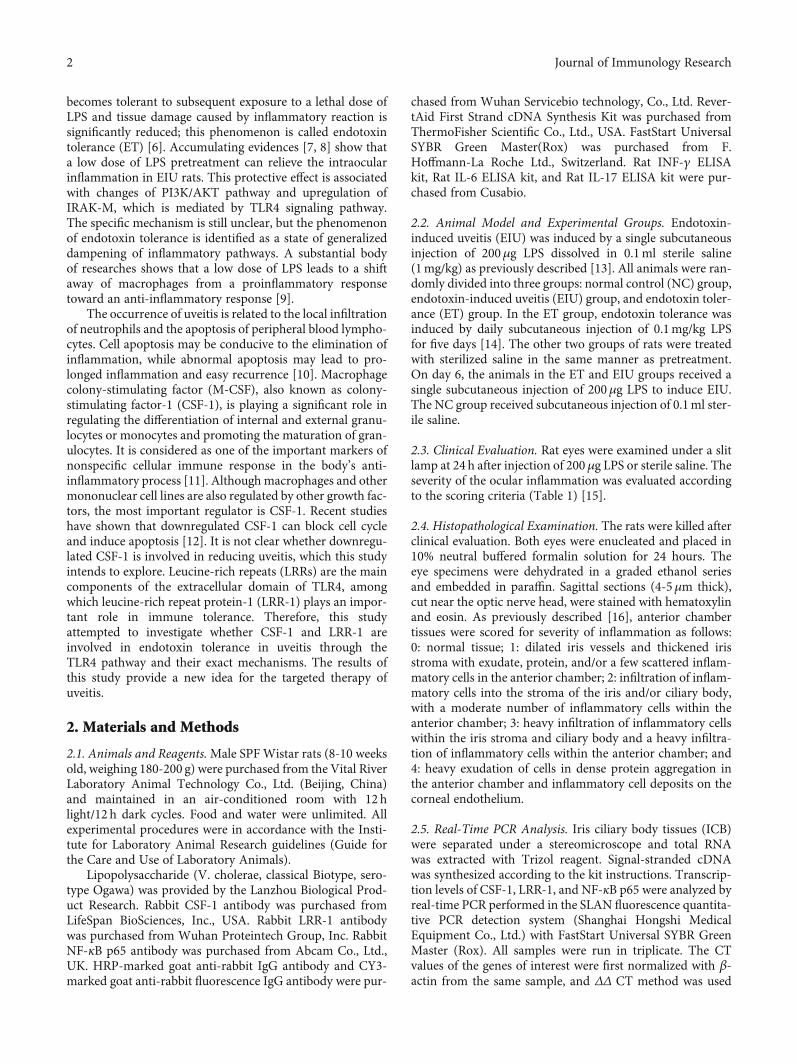
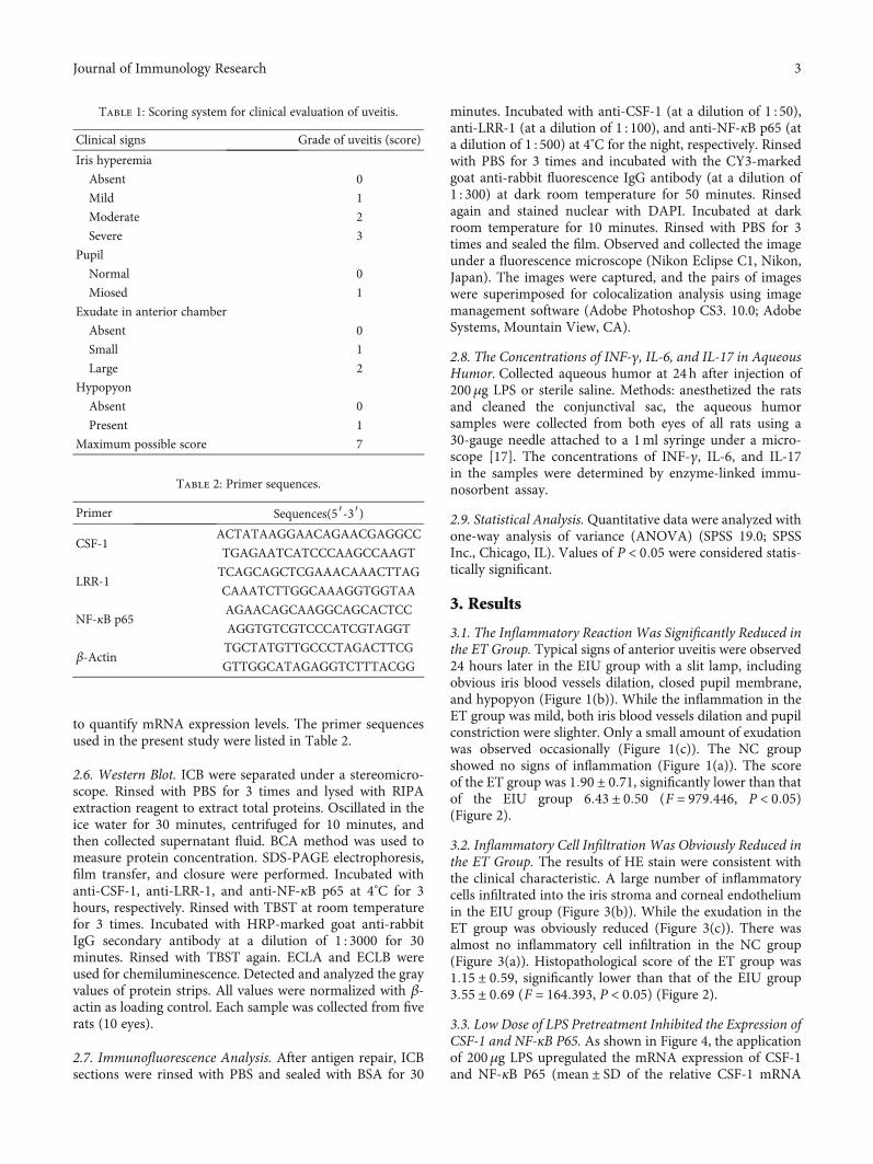
3.1. The Inflammatory Reaction Was Significantly Reduced inthe ET Group. Typical signs of anterior uveitis were observed24 hours later in the EIU group with a slit lamp, includingobvious iris blood vessels dilation, closed pupil membrane,and hypopyon (Figure 1(b)). While the inflammation in theET group was mild, both iris blood vessels dilation and pupilconstriction were slighter. Only a small amount of exudationwas observed occasionally (Figure 1(c)). The NC groupshowed no signs of inflammation (Figure 1(a)). The scoreof the ET group was 1:90 ± 0:71, significantly lower than thatof the EIU group 6:43 ± 0:50 (F = 979:446, P < 0:05)(Figure 2).
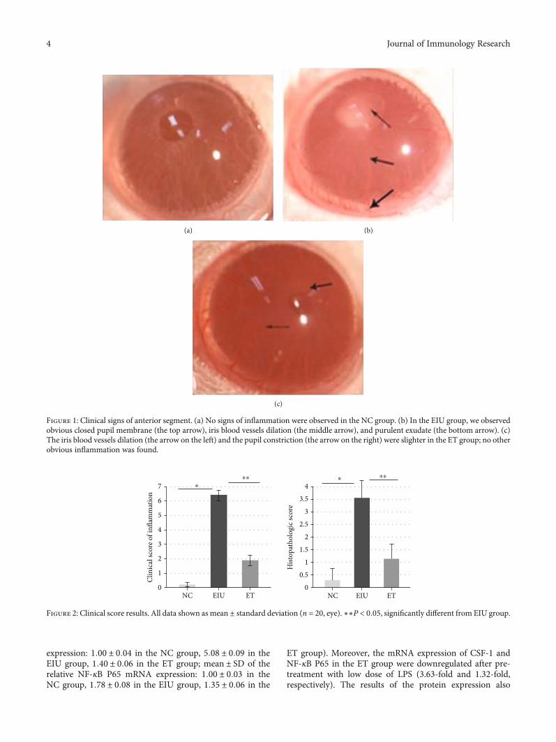
3.2. Inflammatory Cell Infiltration Was Obviously Reduced inthe ET Group. The results of HE stain were consistent withthe clinical characteristic. A large number of inflammatorycells infiltrated into the iris stroma and corneal endotheliumin the EIU group (Figure 3(b)). While the exudation in theET group was obviously reduced (Figure 3(c)). There wasalmost no inflammatory cell infiltration in the NC group(Figure 3(a)). Histopathological score of the ET group was1:15 ± 0:59, significantly lower than that of the EIU group3:55 ± 0:69 (F = 164:393, P < 0:05) (Figure 2).
3.3. Low Dose of LPS Pretreatment Inhibited the Expression ofCSF-1 and NF-κB P65. As shown in Figure 4, the applicationof 200μg LPS upregulated the mRNA expression of CSF-1and NF-κB P65 (mean ± SD of the relative CSF-1 mRNA
Table 1: Scoring system for clinical evaluation of uveitis.
Clinical signs Grade of uveitis (score)
Iris hyperemia
Absent 0
Mild 1
Moderate 2
Severe 3
Pupil
Normal 0
Miosed 1
Exudate in anterior chamber
Absent 0
Small 1
Large 2
Hypopyon
Absent 0
Present 1
Maximum possible score 7
Table 2: Primer sequences.
Primer Sequences(5′-3′)
CSF-1ACTATAAGGAACAGAACGAGGCC
TGAGAATCATCCCAAGCCAAGT
LRR-1TCAGCAGCTCGAAACAAACTTAG
CAAATCTTGGCAAAGGTGGTAA
NF-κB p65AGAACAGCAAGGCAGCACTCC
AGGTGTCGTCCCATCGTAGGT
β-ActinTGCTATGTTGCCCTAGACTTCG
GTTGGCATAGAGGTCTTTACGG
3Journal of Immunology Research
expression: 1:00 ± 0:04 in the NC group, 5:08 ± 0:09 in theEIU group, 1:40 ± 0:06 in the ET group; mean ± SD of therelative NF-κB P65 mRNA expression: 1:00 ± 0:03 in theNC group, 1:78 ± 0:08 in the EIU group, 1:35 ± 0:06 in the
ET group). Moreover, the mRNA expression of CSF-1 andNF-κB P65 in the ET group were downregulated after pre-treatment with low dose of LPS (3.63-fold and 1.32-fold,respectively). The results of the protein expression also
(a) (b)
(c)
Figure 1: Clinical signs of anterior segment. (a) No signs of inflammation were observed in the NC group. (b) In the EIU group, we observedobvious closed pupil membrane (the top arrow), iris blood vessels dilation (the middle arrow), and purulent exudate (the bottom arrow). (c)The iris blood vessels dilation (the arrow on the left) and the pupil constriction (the arrow on the right) were slighter in the ET group; no otherobvious inflammation was found.
0NC EIU ET NC EIU ET
1
2
3
4
5
6
7
Clin
ical
scor
e of i
nflam
mat
ion
0
0.5
1
1.5
2
2.5
3
3.5
4
Hist
opat
holo
gic s
core
⁎⁎⁎
⁎ ⁎⁎
Figure 2: Clinical score results. All data shown asmean ± standard deviation (n = 20, eye). ∗∗P < 0:05, significantly different from EIU group.
4 Journal of Immunology Research
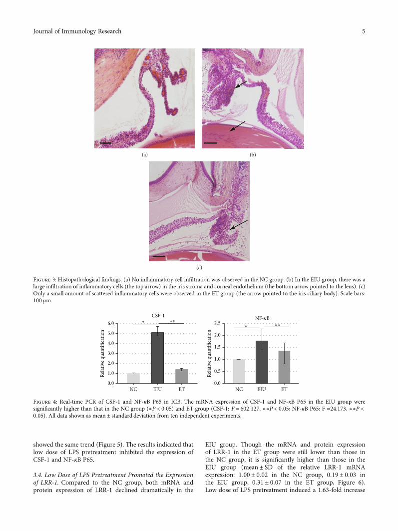
showed the same trend (Figure 5). The results indicated thatlow dose of LPS pretreatment inhibited the expression ofCSF-1 and NF-κB P65.
3.4. Low Dose of LPS Pretreatment Promoted the Expressionof LRR-1. Compared to the NC group, both mRNA andprotein expression of LRR-1 declined dramatically in the
EIU group. Though the mRNA and protein expressionof LRR-1 in the ET group were still lower than those inthe NC group, it is significantly higher than those in theEIU group (mean ± SD of the relative LRR-1 mRNAexpression: 1:00 ± 0:02 in the NC group, 0:19 ± 0:03 inthe EIU group, 0:31 ± 0:07 in the ET group, Figure 6).Low dose of LPS pretreatment induced a 1.63-fold increase
(a) (b)
(c)
Figure 3: Histopathological findings. (a) No inflammatory cell infiltration was observed in the NC group. (b) In the EIU group, there was alarge infiltration of inflammatory cells (the top arrow) in the iris stroma and corneal endothelium (the bottom arrow pointed to the lens). (c)Only a small amount of scattered inflammatory cells were observed in the ET group (the arrow pointed to the iris ciliary body). Scale bars:100μm.
0.0
1.0
2.0
3.0
4.0
5.0
6.0
Rela
tive q
uant
ifica
tion
0.0
0.5
1.0
1.5
2.5
2.0
Rela
tive q
uant
ifica
tion
NC EIU ET NC EIU ET
⁎⁎⁎CSF-1
⁎ ⁎⁎NF-𝜅B
Figure 4: Real-time PCR of CSF-1 and NF-κB P65 in ICB. The mRNA expression of CSF-1 and NF-κB P65 in the EIU group weresignificantly higher than that in the NC group (∗P < 0:05) and ET group (CSF-1: F = 602:127, ∗∗P < 0:05; NF-κB P65: F =24.173, ∗∗P <0:05). All data shown as mean ± standard deviation from ten independent experiments.
5Journal of Immunology Research
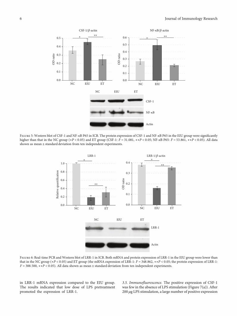
in LRR-1 mRNA expression compared to the EIU group.The results indicated that low dose of LPS pretreatmentpromoted the expression of LRR-1.
3.5. Immunofluorescence. The positive expression of CSF-1was few in the absence of LPS stimulation (Figure 7(a)). After200μg LPS stimulation, a large number of positive expression
0.0
0.1
0.2
0.3
0.4
0.5
OD
ratio
NC EIU ET
NC EIU ET
NC EIU ET0.0
0.1
0.2
0.3
0.4
0.5
0.6
OD
ratio
CSF-1/𝛽-actin
CSF-1
NF-κB
Actin
⁎⁎⁎ ⁎
⁎⁎
NF-κB/𝛽-actin
Figure 5: Western blot of CSF-1 and NF-κB P65 in ICB. The protein expression of CSF-1 and NF-κB P65 in the EIU group were significantlyhigher than that in the NC group (∗P < 0:05) and ET group (CSF-1: F = 31:081, ∗∗P < 0:05; NF-κB P65: F = 53:861, ∗∗P < 0:05). All datashown as mean ± standard deviation from ten independent experiments.
0.0
0.2
0.4
0.6
0.8
1.0
Rela
tive q
uant
ifica
tion
0.0
0.1
0.2
0.3
0.4
OD
ratio
⁎LRR-1
⁎⁎
LRR-1/𝛽-actin
LRR-1
Actin
NC EIU ET
NC EIU ET
NC EIU ET
⁎
⁎⁎
Figure 6: Real-time PCR andWestern blot of LRR-1 in ICB. Both mRNA and protein expression of LRR-1 in the EIU group were lower thanthat in the NC group (∗P < 0:05) and ET group (the mRNA expression of LRR-1: F = 348:862, ∗∗P < 0:05; the protein expression of LRR-1:F = 388:500, ∗∗P < 0:05). All data shown as mean ± standard deviation from ten independent experiments.
6 Journal of Immunology Research
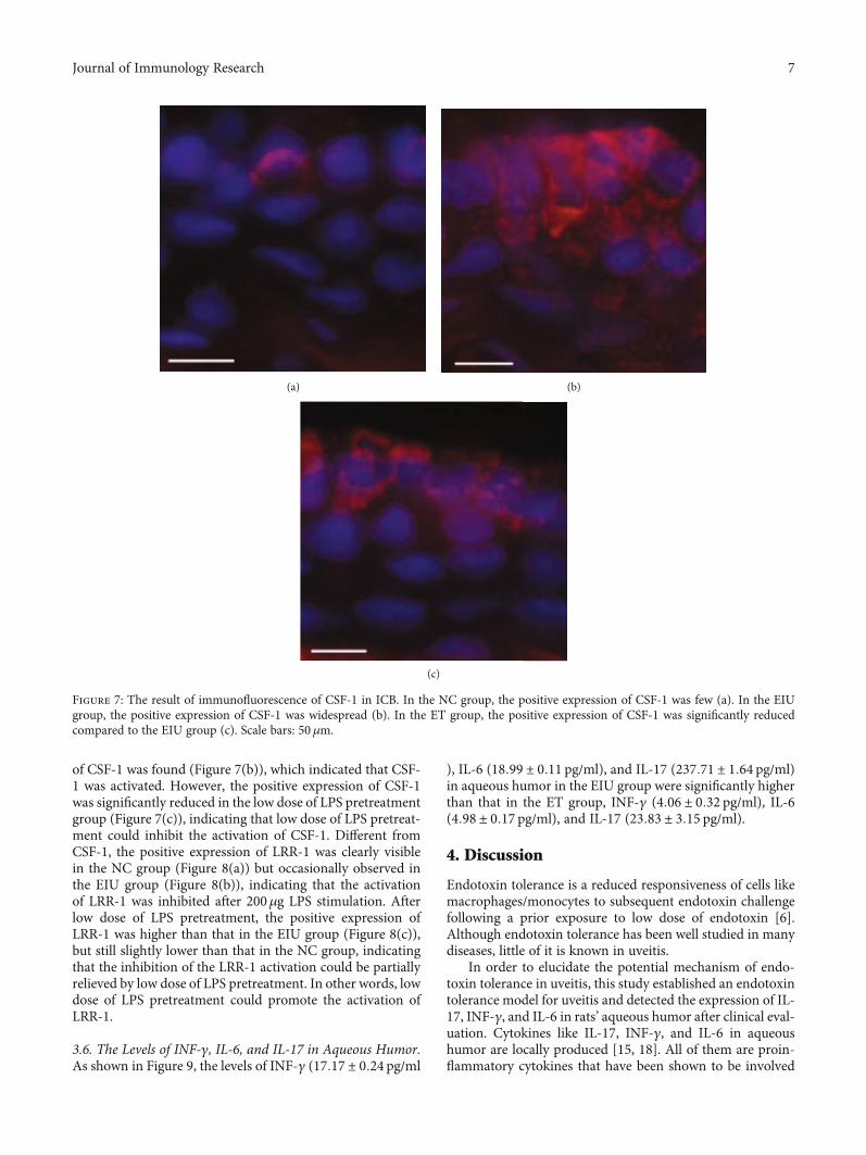
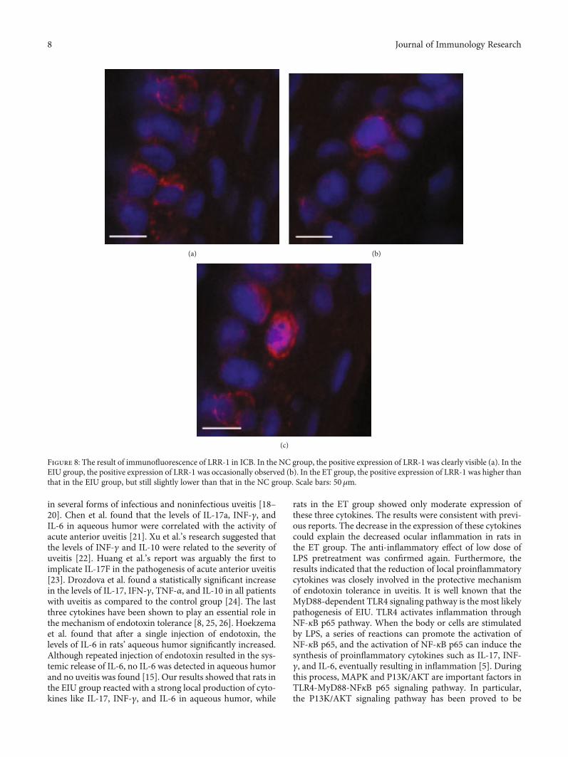
of CSF-1 was found (Figure 7(b)), which indicated that CSF-1 was activated. However, the positive expression of CSF-1was significantly reduced in the low dose of LPS pretreatmentgroup (Figure 7(c)), indicating that low dose of LPS pretreat-ment could inhibit the activation of CSF-1. Different fromCSF-1, the positive expression of LRR-1 was clearly visiblein the NC group (Figure 8(a)) but occasionally observed inthe EIU group (Figure 8(b)), indicating that the activationof LRR-1 was inhibited after 200μg LPS stimulation. Afterlow dose of LPS pretreatment, the positive expression ofLRR-1 was higher than that in the EIU group (Figure 8(c)),but still slightly lower than that in the NC group, indicatingthat the inhibition of the LRR-1 activation could be partiallyrelieved by low dose of LPS pretreatment. In other words, lowdose of LPS pretreatment could promote the activation ofLRR-1.
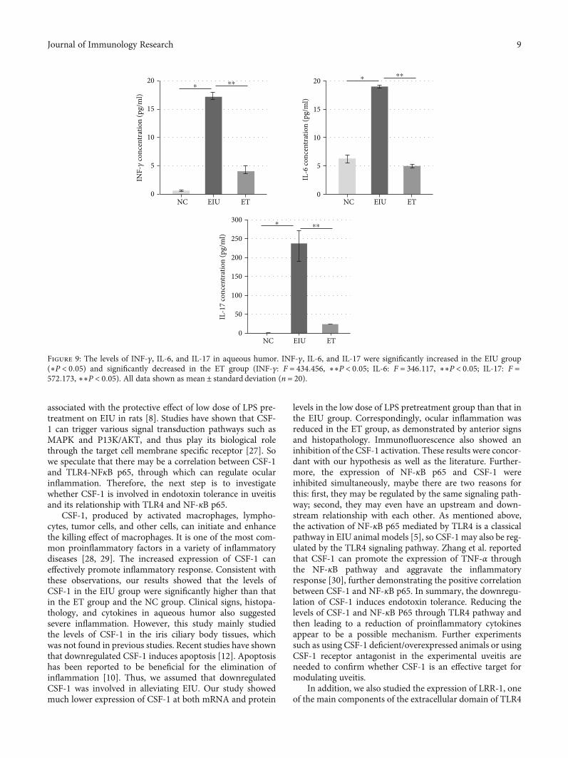
3.6. The Levels of INF-γ, IL-6, and IL-17 in Aqueous Humor.As shown in Figure 9, the levels of INF-γ (17:17 ± 0:24 pg/ml
), IL-6 (18:99 ± 0:11 pg/ml), and IL-17 (237:71 ± 1:64 pg/ml)in aqueous humor in the EIU group were significantly higherthan that in the ET group, INF-γ (4:06 ± 0:32 pg/ml), IL-6(4:98 ± 0:17 pg/ml), and IL-17 (23:83 ± 3:15 pg/ml).
4. Discussion
Endotoxin tolerance is a reduced responsiveness of cells likemacrophages/monocytes to subsequent endotoxin challengefollowing a prior exposure to low dose of endotoxin [6].Although endotoxin tolerance has been well studied in manydiseases, little of it is known in uveitis.
In order to elucidate the potential mechanism of endo-toxin tolerance in uveitis, this study established an endotoxintolerance model for uveitis and detected the expression of IL-17, INF-γ, and IL-6 in rats’ aqueous humor after clinical eval-uation. Cytokines like IL-17, INF-γ, and IL-6 in aqueoushumor are locally produced [15, 18]. All of them are proin-flammatory cytokines that have been shown to be involved
(a) (b)
(c)
Figure 7: The result of immunofluorescence of CSF-1 in ICB. In the NC group, the positive expression of CSF-1 was few (a). In the EIUgroup, the positive expression of CSF-1 was widespread (b). In the ET group, the positive expression of CSF-1 was significantly reducedcompared to the EIU group (c). Scale bars: 50 μm.
7Journal of Immunology Research
in several forms of infectious and noninfectious uveitis [18–20]. Chen et al. found that the levels of IL-17a, INF-γ, andIL-6 in aqueous humor were correlated with the activity ofacute anterior uveitis [21]. Xu et al.’s research suggested thatthe levels of INF-γ and IL-10 were related to the severity ofuveitis [22]. Huang et al.’s report was arguably the first toimplicate IL-17F in the pathogenesis of acute anterior uveitis[23]. Drozdova et al. found a statistically significant increasein the levels of IL-17, IFN-γ, TNF-α, and IL-10 in all patientswith uveitis as compared to the control group [24]. The lastthree cytokines have been shown to play an essential role inthe mechanism of endotoxin tolerance [8, 25, 26]. Hoekzemaet al. found that after a single injection of endotoxin, thelevels of IL-6 in rats’ aqueous humor significantly increased.Although repeated injection of endotoxin resulted in the sys-temic release of IL-6, no IL-6 was detected in aqueous humorand no uveitis was found [15]. Our results showed that rats inthe EIU group reacted with a strong local production of cyto-kines like IL-17, INF-γ, and IL-6 in aqueous humor, while
rats in the ET group showed only moderate expression ofthese three cytokines. The results were consistent with previ-ous reports. The decrease in the expression of these cytokinescould explain the decreased ocular inflammation in rats inthe ET group. The anti-inflammatory effect of low dose ofLPS pretreatment was confirmed again. Furthermore, theresults indicated that the reduction of local proinflammatorycytokines was closely involved in the protective mechanismof endotoxin tolerance in uveitis. It is well known that theMyD88-dependent TLR4 signaling pathway is the most likelypathogenesis of EIU. TLR4 activates inflammation throughNF-κB p65 pathway. When the body or cells are stimulatedby LPS, a series of reactions can promote the activation ofNF-κB p65, and the activation of NF-κB p65 can induce thesynthesis of proinflammatory cytokines such as IL-17, INF-γ, and IL-6, eventually resulting in inflammation [5]. Duringthis process, MAPK and P13K/AKT are important factors inTLR4-MyD88-NFκB p65 signaling pathway. In particular,the P13K/AKT signaling pathway has been proved to be
(a) (b)
(c)
Figure 8: The result of immunofluorescence of LRR-1 in ICB. In the NC group, the positive expression of LRR-1 was clearly visible (a). In theEIU group, the positive expression of LRR-1 was occasionally observed (b). In the ET group, the positive expression of LRR-1 was higher thanthat in the EIU group, but still slightly lower than that in the NC group. Scale bars: 50μm.
8 Journal of Immunology Research
associated with the protective effect of low dose of LPS pre-treatment on EIU in rats [8]. Studies have shown that CSF-1 can trigger various signal transduction pathways such asMAPK and P13K/AKT, and thus play its biological rolethrough the target cell membrane specific receptor [27]. Sowe speculate that there may be a correlation between CSF-1and TLR4-NFκB p65, through which can regulate ocularinflammation. Therefore, the next step is to investigatewhether CSF-1 is involved in endotoxin tolerance in uveitisand its relationship with TLR4 and NF-κB p65.
CSF-1, produced by activated macrophages, lympho-cytes, tumor cells, and other cells, can initiate and enhancethe killing effect of macrophages. It is one of the most com-mon proinflammatory factors in a variety of inflammatorydiseases [28, 29]. The increased expression of CSF-1 caneffectively promote inflammatory response. Consistent withthese observations, our results showed that the levels ofCSF-1 in the EIU group were significantly higher than thatin the ET group and the NC group. Clinical signs, histopa-thology, and cytokines in aqueous humor also suggestedsevere inflammation. However, this study mainly studiedthe levels of CSF-1 in the iris ciliary body tissues, whichwas not found in previous studies. Recent studies have shownthat downregulated CSF-1 induces apoptosis [12]. Apoptosishas been reported to be beneficial for the elimination ofinflammation [10]. Thus, we assumed that downregulatedCSF-1 was involved in alleviating EIU. Our study showedmuch lower expression of CSF-1 at both mRNA and protein
levels in the low dose of LPS pretreatment group than that inthe EIU group. Correspondingly, ocular inflammation wasreduced in the ET group, as demonstrated by anterior signsand histopathology. Immunofluorescence also showed aninhibition of the CSF-1 activation. These results were concor-dant with our hypothesis as well as the literature. Further-more, the expression of NF-κB p65 and CSF-1 wereinhibited simultaneously, maybe there are two reasons forthis: first, they may be regulated by the same signaling path-way; second, they may even have an upstream and down-stream relationship with each other. As mentioned above,the activation of NF-κB p65 mediated by TLR4 is a classicalpathway in EIU animal models [5], so CSF-1 may also be reg-ulated by the TLR4 signaling pathway. Zhang et al. reportedthat CSF-1 can promote the expression of TNF-α throughthe NF-κB pathway and aggravate the inflammatoryresponse [30], further demonstrating the positive correlationbetween CSF-1 and NF-κB p65. In summary, the downregu-lation of CSF-1 induces endotoxin tolerance. Reducing thelevels of CSF-1 and NF-κB P65 through TLR4 pathway andthen leading to a reduction of proinflammatory cytokinesappear to be a possible mechanism. Further experimentssuch as using CSF-1 deficient/overexpressed animals or usingCSF-1 receptor antagonist in the experimental uveitis areneeded to confirm whether CSF-1 is an effective target formodulating uveitis.
In addition, we also studied the expression of LRR-1, oneof the main components of the extracellular domain of TLR4
0
5
10
15
20
0
5
10
15
20
0
50
100
150
200
250
300
IL-6
conc
entr
atio
n (p
g/m
l)
INF-𝛾
conc
entr
atio
n (p
g/m
l)
⁎ ⁎⁎⁎ ⁎⁎
IL-1
7 co
ncen
trat
ion
(pg/
ml)
⁎ ⁎⁎
NC EIU ET NC EIU ET
NC EIU ET
Figure 9: The levels of INF-γ, IL-6, and IL-17 in aqueous humor. INF-γ, IL-6, and IL-17 were significantly increased in the EIU group(∗P < 0:05) and significantly decreased in the ET group (INF-γ: F = 434:456, ∗∗P < 0:05; IL-6: F = 346:117, ∗∗P < 0:05; IL-17: F =572:173, ∗∗P < 0:05). All data shown as mean ± standard deviation (n = 20).
9Journal of Immunology Research
[31]. Up to now, though 375 LRR proteins have been identi-fied, the function of most of them is still unknown [32]. LRR-1 is one of them. Jang et al. found that overexpression ofLRR-1 inhibited the activation of NF-κB [33]. In the presentstudy, we found similar results; both the gene and proteinexpression of LRR-1 were significantly increased in the lowdose of LPS pretreatment group, and the immunofluores-cence results also showed an activation of LRR-1, while theexpression of NF-κB P65 was inhibited. The results suggestedthat the upregulation of LRR-1 was involved in endotoxintolerance in uveitis. LRR-1 has been shown to negatively reg-ulate the signaling pathway [33]. However, the currentresearch on LRR-1 is limited, and the specific mechanismof its involvement in endotoxin tolerance is worthy of furtherstudy.
In conclusion, low dose of LPS pretreatment has a protec-tive effect on endotoxin-induced uveitis in rats. This protec-tion is related to upregulation of LRR-1 anddownregulation of CSF-1, which may be mediated by TLR4signaling pathway. CSF-1 is expected to be a new therapeutictarget for uveitis.
Data Availability
The data used to support the findings of this study areincluded within the article.
Conflicts of Interest
The authors declare that there is no conflict of interestregarding the publication of this paper.
Acknowledgments
The authors thank for Lanzhou Institute of Biologic Productsfor providing LPS in this study. The earlier version of thispaper has been presented as abstract in 2019 ARVO AnnualMeeting, Canada, April 28-May 2, 2019. However, more rel-evant experiments were performed and added to this paperafter the meeting. This study was supported by the NationalNatural Science Foundation of China (No: 81471575 and81273246), the Natural Science Foundation of Gansu Prov-ince (No: 17JR5RA225).
References
[1] U. Krishna, D. Ajanaku, A. K. Denniston, and T. Gkika, “Uve-itis: a sight-threatening disease which can impact all systems,”Postgraduate Medical Journal, vol. 93, no. 1106, pp. 766–773,2017.
[2] U. C. S. Yadav and K. V. Ramana, “Endotoxin-induced uveitisin rodents,”Methods in Molecular Biology, vol. 1960, pp. 161–168, 2019.
[3] S. Yang, H. Lu, J. Wang, X. Qi, X. Liu, and X. Zhang, “Theeffect of toll-like receptor 4 on macrophage cytokines duringendotoxin induced uveitis,” International Journal of MolecularSciences, vol. 13, no. 6, pp. 7508–7520, 2012.
[4] J. Wang, H. Lu, X. Hu et al., “Nuclear factor translocation andacute anterior uveitis,”Molecular Vision, vol. 17, pp. 170–176,2011.
[5] S. Li, H. Lu, X. Hu, W. Chen, Y. Xu, and J. Wang, “Expressionof TLR4-MyD88 and NF-κB in the Iris during Endotoxin-Induced Uveitis,” Mediators of Inflammation, vol. 2010, Arti-cle ID 748218, 7 pages, 2010.
[6] D. Liu, S. Cao, Y. Zhou, and Y. Xiong, “Recent advances inendotoxin tolerance,” Journal of Cellular Biochemistry,vol. 120, no. 1, pp. 56–70, 2018.
[7] S. Yu, X. Liu, N. Zhang et al., “Protection of lipopolysaccharide(LPS) preconditioning against endotoxin-induced uveitis(EIU) in rats is associated with overexpression of interleukin-1 receptor-associated kinase M (IRAK-M),” Ocular Immunol-ogy and Inflammation, vol. 26, no. 6, pp. 943–950, 2017.
[8] N. Zhang, S. Yu, X. Liu, and H. Lu, “Low Dose of Lipopolysac-charide Pretreatment Preventing Subsequent Endotoxin-Induced Uveitis Is Associated with PI3K/AKT Pathway,” Jour-nal of Immunology Research, vol. 2017, Article ID 1273940, 7pages, 2017.
[9] P. E. Collins and R. J. Carmody, “The regulation of endotoxintolerance and its impact on macrophage activation,” CriticalReviews in Immunology, vol. 35, no. 4, pp. 293–323, 2015.
[10] J. Li, P. Yang, J. Lin, H. Zhou, and X. Huang, “Apoptosis of theperipheral blood lymphocytes during endotoxin-induced uve-itis in SD rats,” Yan Ke Yan Jiu, vol. 20, no. 2, pp. 123–125,2002.
[11] S. N. Schlink, K. M. Lager, S. L. Brockmeier et al., “Enhance-ment of innate immunity with granulocyte colony-stimulating factor did not mitigate disease in pigs infected witha highly pathogenic Chinese PRRSV strain,”Veterinary Immu-nology and Immunopathology, vol. 179, pp. 70–76, 2016.
[12] X. Li, S. Kong, and Y. Cao, “miR‐1254 inhibits progression ofglioma in vivo and in vitro by targeting CSF-1,” Journal of Cel-lular and Molecular Medicine, vol. 24, no. 5, pp. 3128–3138,2020.
[13] W. Chen, X. Hu, L. Zhao, S. Li, and H. Lu, “Expression of toll-like receptor 4 in uvea-resident tissue macrophages duringendotoxin-induced uveitis,” Molecular Vision, vol. 15,pp. 619–628, 2009.
[14] Y. Endo, M. Shibazaki, K. Yamaguchi et al., “Enhancement bygalactosamine of lipopolysaccharide (LPS)-induced tumournecrosis factor production and lethality: its suppression byLPS pretreatment,” British Journal of Pharmacology, vol. 128,no. 1, pp. 5–12, 1999.
[15] R. Hoekzema, P. I. Murray, M. A. van Haren, M. Helle, andA. Kijlstra, “Analysis of interleukin-6 in endotoxin-induceduveitis,” Investigative Ophthalmology & Visual Science,vol. 32, no. 1, pp. 88–95, 1991.
[16] R. G. Tilton, K. Chang, J. A. Corbett et al., “Endotoxin-induceduveitis in the rat is attenuated by inhibition of nitric oxide pro-duction,” Investigative Ophthalmology & Visual Science,vol. 35, no. 8, pp. 3278–3288, 1994.
[17] K. Kitazawa, C. Sotozono, N. Koizumi et al., “Safety of anteriorchamber paracentesis using a 30-gauge needle integrated witha specially designed disposable pipette,” The British Journal ofOphthalmology, vol. 101, no. 5, pp. 548–550, 2017.
[18] H. Takase, Y. Futagami, T. Yoshida et al., “Cytokine profile inaqueous humor and sera of patients with infectious or nonin-fectious uveitis,” Investigative Ophthalmology & Visual Sci-ence, vol. 47, no. 4, pp. 1557–1561, 2006.
[19] M. C. E. Guedes, L. M. Borrego, and R. D. Proença, “Roles ofinterleukin-17 in uveitis,” Indian Journal of Ophthalmology,vol. 64, no. 9, pp. 628–634, 2016.
10 Journal of Immunology Research
[20] K. G.-J. Ooi, G. Galatowicz, V. L. Calder, and S. L. Lightman,“Cytokines and chemokines in uveitis: is there a correlationwith clinical phenotype?,” Clinical Medicine & Research,vol. 4, no. 4, pp. 294–309, 2006.
[21] W. Chen, B. Zhao, R. Jiang et al., “Cytokine expression profilein aqueous humor and sera of patients with acute anterior uve-itis,” Current Molecular Medicine, vol. 15, no. 6, pp. 543–549,2015.
[22] Y. Xu, W. Chen, H. Lu et al., “The expression of cytokines inthe aqueous humor and serum during endotoxin-induced uve-itis in C3H/HeN mice,” Molecular Vision, vol. 16, pp. 1689–1695, 2010.
[23] J. C.-C. Huang, M. Schleisman, D. Choi et al., “Preliminaryreport on interleukin-22, GM-CSF, and IL-17F in the patho-genesis of acute anterior uveitis,” Ocular Immunology andInflammation, vol. 25, pp. 1–8, 2019.
[24] E. A. Drozdova, E. V. Yadykina, E. A. Mezentseva, and K. V.Nikushina, “Cytokine profile changes in children with juvenileidiopathic arthritis-associated uveitis,” Vestnik oftal'mologii,vol. 133, no. 1, pp. 27–31, 2017.
[25] H. Mashimo, N. Ohguro, S. Nomura, N. Hashida, K. Nakai,and Y. Tano, “Neutrophil chemotaxis and local expression ofinterleukin-10 in the tolerance of endotoxin-induced uveitis,”Investigative Ophthalmology & Visual Science, vol. 49, no. 12,pp. 5450–5457, 2008.
[26] B. Matta, P. Jha, P. S. Bora, and N. S. Bora, “Tolerance tomelanin-associated antigen in autoimmune uveitis is mediatedby CD4+CD25+ T-regulatory cells,” The American Journal ofPathology, vol. 173, no. 5, pp. 1440–1454, 2008.
[27] C. V. Jones and S. D. Ricardo, “Macrophages and CSF-1:implications for development and beyond,” Organogenesis,vol. 9, no. 4, pp. 249–260, 2014.
[28] A. Kumari, O. Silakari, and R. K. Singh, “Recent advances incolony stimulating factor-1 receptor/c-FMS as an emergingtarget for various therapeutic implications,” Biomedicine &Pharmacotherapy, vol. 103, pp. 662–679, 2018.
[29] S. Bonelli, X. Geeraerts, E. Bolli et al., “Beyond the M-CSFreceptor-novel therapeutic targets in tumor-associated macro-phages,” The FEBS Journal, vol. 285, no. 4, pp. 777–787, 2018.
[30] W. Jia, Z. Wenjing, H. Xiangzhen, Z. Qiqi, and H. Huiyu,“Effect of Macrophage Colony-Stimulating Factor Overex-pression on Osteoclast-related Cytokine Factors,” Journal ofOral Science Research, vol. 35, no. 11, pp. 1089–1093, 2019.
[31] A. Ng and R. J. Xavier, “Leucine-rich repeat (LRR) proteins:integrators of pattern recognition and signaling in immunity,”Autophagy, vol. 7, no. 9, pp. 1082–1084, 2014.
[32] A. C. Y. Ng, J. M. Eisenberg, R. J. W. Heath et al., “Humanleucine-rich repeat proteins: a genome-wide bioinformatic cat-egorization and functional analysis in innate immunity,” Pro-ceedings of the National Academy of Sciences, vol. 108,Supplement_1, pp. 4631–4638, 2011.
[33] L. K. Jang, Z. H. Lee, H. H. Kim, J. M. Hill, J. D. Kim, and B. S.Kwon, “A novel leucine-rich repeat protein (LRR-1): potentialinvolvement in 4-1BB-mediated signal transduction,” Mole-cules and Cells, vol. 12, no. 3, pp. 304–312, 2001.
11Journal of Immunology Research

![Open Access Single low-dose lipopolysaccharide preconditioning ...€¦ · models of ischemic stroke and traumatic brain injury (TBI).[1] The neuroprotective mechanisms of LPS remain](https://img.dokumen.tips/doc/110x75/6034fee4b26f840c261f96ad/open-access-single-low-dose-lipopolysaccharide-preconditioning-models-of-ischemic.jpg)