Embed Size (px)
Citation preview

© The Rockefeller University Press, 0021-9525/2001/01/385/16 $5.00The Journal of Cell Biology, Volume 152, Number 2, January 22, 2001 385–400http://www.jcb.org/cgi/content/full/152/2/385 385
The Positioning and Dynamics of Origins of Replication
in the Budding Yeast Nucleus
✪
Patrick Heun,* Thierry Laroche,* M.K. Raghuraman,
‡
and Susan M. Gasser*
*Swiss Institute for Experimental Cancer Research, CH-1066 Epalinges/Lausanne, Switzerland; and
‡
Department of Genetics, University of Washington, Seattle, Washington 98195
Abstract.
We have analyzed the subnuclear position ofearly- and late-firing origins of DNA replication in intactyeast cells using fluorescence in situ hybridization andgreen fluorescent protein (GFP)–tagged chromosomaldomains. In both cases, origin position was determinedwith respect to the nuclear envelope, as identified by nu-clear pore staining or a NUP49-GFP fusion protein. Wefind that in G1 phase nontelomeric late-firing origins areenriched in a zone immediately adjacent to the nuclearenvelope, although this localization does not necessarilypersist in S phase. In contrast, early firing origins are ran-domly localized within the nucleus throughout the cellcycle. If a late-firing telomere-proximal origin is excisedfrom its chromosomal context in G1 phase, it remains
late-firing but moves rapidly away from the telomerewith which it was associated, suggesting that the posi-tioning of yeast chromosomal domains is highly dynamic.This is confirmed by time-lapse microscopy of GFP-tagged origins in vivo. We propose that sequences flank-ing late-firing origins help target them to the peripheryof the G1-phase nucleus, where a modified chromatinstructure can be established. The modified chromatinstructure, which would in turn retard origin firing, is bothautonomous and mobile within the nucleus.
Key words: DNA replication • nuclear organization •in vivo green fluorescent protein imaging • origins ofreplication • nuclear envelope
Introduction
One characteristic feature of eukaryotic genomes, as op-posed to bacterial genomes, is that each chromosome con-tains multiple origins of replication capable of initiatingDNA replication at different times during S phase. Inmammalian cells, the timing of replication of a given chro-mosomal domain often correlates with the transcriptionalactivity of nearby genes. More precisely, it was shown thatthe late-replicating Giemsa Dark bands contain tissue-spe-cific genes that are repressed in many tissues, while Gi-emsa-light, or R-bands, replicate early and contain mostubiquitously expressed housekeeping genes (Holmquist,1987). Similarly, in yeast, origins of replication in the tran-scriptionally silent subtelomeric regions fire either late ornot at all, although origin recognition complex and othercomponents of the pre-Replicative Complex appear to as-semble at these origins normally (Santocanale and Diffley,1996). Thus, despite correlations between a lack of tran-scriptional inactivity and a delay in origin firing, little isknown about the molecular mechanisms that establish andregulate this latter event.
In addition to having a precise temporal pattern for ori-gin activation, DNA replication has been shown to occurat a limited number of discrete foci within the nucleus inboth yeast and higher eukaryotic cells (Nakamura et al.,1986; Pasero et al., 1997). Studies with synchronized mam-malian cells suggest that the pattern of active replicationfoci changes in an ordered and reproducible fashion dur-ing S-phase progression (Nakayasu and Berezney, 1989;van Dierendonck et al., 1989; Fox et al., 1991; Manders et
al., 1992; O’Keefe et al., 1992). Consistently, using a prolif-
erating cell nuclear antigen–green fluorescent protein (GFP)
1
fusion protein to identify replication foci in situ, it was shownthat the transition from one pattern to the next is not due tothe movement of foci, but to their gradual assembly and dis-assembly at discrete sites (Leonhardt et al., 2000). The re-producible positioning of early- and late-replicating fociled to the proposal that origin location within the nucleusmight not only correlate with, but also regulate, the time atwhich origins fire. Indeed, euchromatic regions are foundthroughout the nucleus and replicate early, while mid- tolate-S phase foci tend to reflect heterochromatic regions
✪
The online version of this article contains supplemental material.Address correspondence to Susan M. Gasser, Swiss Institute for Exper-
imental Cancer Research (ISREC), Chemin des Boveresses 155, CH-1066Epalinges/Lausanne, Switzerland. Tel.: 41 21 692 5886. Fax: 41 21 6526933. E-mail: [email protected]
1
Abbreviations used in this paper:
3-D, three-dimensional; ARS, autono-mously replicating sequence; GFP, green fluorescent protein; IF/FISH,immunofluorescence/in situ hybridization.
on Septem
ber 11, 2005 w
ww
.jcb.orgD
ownloaded from
http://www.jcb.org/cgi/content/full/152/2/385/DC1Supplemental Material can be found at:

The Journal of Cell Biology, Volume 152, 2001 386
that localize either at the nuclear periphery or near nucle-oli (Ma et al., 1998).
The spatial positioning of late-replicating DNA withinthe mammalian nucleus seems to be determined in earlyG1 phase (Dimitrova and Gilbert, 1999). Using a pulse-chase technique in which isolated mammalian nuclei arestimulated to synthesize DNA in a
Xenopus
egg extract, itwas shown that a characteristic subnuclear pattern was es-tablished for late-replicated chromosomal regions roughly2 h after the completion of anaphase (Dimitrova and Gil-bert, 1999). The colocalization of signals from a first andsecond round of replication suggests that both the timingand the characteristic subnuclear localization of a late-rep-licating region is retained through mitotic division. How-ever, since very few mammalian origins of replication havebeen mapped, the subnuclear positioning of specific ori-gins during cell-cycle progression could not be addressedin these studies of cultured cells.
Budding yeast has provided an excellent model systemto identify the molecular mechanisms that control originactivation, thanks to a detailed understanding of the se-quences and proteins responsible for origin function (re-viewed in Campbell and Newlon, 1991; Newlon, 1997). Inyeast, as in mammalian cells, each origin of replication hasa characteristic time of activation in S phase (reviewed inFangman and Brewer, 1992). Two mechanisms for keep-ing late origins from firing early have been suggested: thefirst proposes that chromatin structure regulates origin ac-tivation by limiting accessibility to common factors, andthe second proposes that timing reflects the action of aspecific molecular regulator. The two mechanisms are notmutually exclusive. For instance, compact nucleosomal ar-rays may limit or modify access to the origin complex forcomponents of the replication machinery or for specificinitiation-promoting factors. One such limiting factormight be the cyclin B/Cdk1 kinase, for it has been shownthat Cyclin B5 (Clb5p) is required for the firing andproper timing of both early and late origins, while Clb6pactivates early origins only (Donaldson et al., 1998b). It isnot known whether these complexes associate with late-firing origins early in S phase or only when initiation actu-ally occurs. In addition to Clb5/Cdc28p, a second Ser/Thrkinase, encoded by
CDC7
, is necessary for both early andlate origin firing (Bousset and Diffley, 1998; Donaldson etal., 1998a). This complex appears to be bound to at least asubset of origins in late G1 (Pasero et al., 1999).
In addition to cell-cycle controlling kinases, the con-served checkpoint kinases encoded by
MEC1
and
RAD53
play a role in regulating the firing of late origins when rep-lication fork progression is blocked by hydroxyurea (HU)or methylmethanesulfonate (Santocanale and Diffley,1998; Shirahige et al., 1998). Mutation of the Rad53 kinaseor a component of the origin recognition complex resultsin the premature firing of late origins even in the absenceof HU, suggesting that this kinase may also be constitu-tively involved in delaying the activation of certain originsuntil late in S phase (Shirahige et al., 1998). Rad53p inter-acts both genetically and physically with Dbf4p, the regu-latory subunit of the Cdc7 kinase (Dohrmann et al., 1999),and, in HU-arrested cells, Dbf4p becomes hyperphos-phorylated in a Rad53-dependent manner (Weinreich andStillman, 1999). The modification of Dbf4p results in its
release from chromatin, suggesting that Rad53p helps reg-ulate origin firing through this kinase complex (Pasero etal., 1999; Weinreich and Stillman, 1999).
Evidence that chromosomal context plays an importantrole in replication timing in budding yeast comes from theanalysis of timing of specific origin activation (reviewedin Fangman and Brewer, 1992). These authors found thatorigins near yeast telomeres generally fire late in S phase,while those near centromeres fire early (McCarroll andFangman, 1988). The silent chromatin found at yeast telo-meres also exerts a position effect, ensuring that originsin subtelomeric repeats are repressed in a manner depen-dent on structural proteins of silent chromatin [silent in-formation regulators (SIR) 2, 3, and 4; Stevenson andGottschling, 1999]. Although the timing of activation of agiven origin in its genomic location is highly reproducible,this timing does not depend on the minimal core se-quence of the origin, which contains the autonomouslyreplicating sequence (ARS) consensus. More specifically,the transfer of a minimal origin domain (or ARS) to aplasmid, confers an early replication status on the epi-some, even though the origin may be late firing in itschromosomal context. Thus, it appears that the activationof origins in early S phase is the default state. Further-more, DNA sequences that flank the late-firing origin onchromosome XIV (ARS1412) can render a plasmid-borne origin late firing (Friedman et al., 1996). It hasbeen suggested that these “late elements” act by nucleat-ing a specific chromatin structure that prevents initiationin early S phase, although the genes flanking ARS1412are not transcriptionally silent. An alternative, but notmutually exclusive, mechanism for ensuring late initiationwould be to sequester or target the origin to a specificsubcompartment of the nucleus, which would delay initia-tion. Such a mechanism is consistent with the correlationfound in many organisms between transcriptionally re-pressed chromatin, late-firing origins, and their localiza-tion near the nuclear periphery.
As in mammalian cells, the establishment of a late-firingorigin in budding yeast requires events that occur betweenmitosis and the subsequent G1 phase (Raghuraman et al.,1997). This may correlate either with the formation of a re-pressed chromatin structure following DNA replication(Taddei et al., 1999), with the assembly of the pre–regula-tory complex, or with establishment of a G1-specific nu-clear organization, as proposed by Dimitrova and Gilbert(1999). To investigate the importance of the nuclear posi-tioning on origin timing in yeast, we have localized early-and late-activated origins by a combined immunofluores-cence/in situ hybridization (IF/FISH) protocol in fixedcells and by the lac operator/GFP-lac repressor recogni-tion system in live cultures. In this paper, we demonstrateby time-lapse microscopy that in G1-phase nuclei there isa significant enrichment of late-activated origins in a zoneimmediately adjacent to the nuclear envelope, despitetheir oscillatory subnuclear movement. Using site-specificrecombination to excise a chromosomal origin, we presentdata suggesting that local chromatin structure is dominantover nuclear positioning in the regulation of origin timingin S phase, although the establishment of a late replicationstate of chromatin in G1 phase correlates with its transientenrichment at the nuclear periphery.
on Septem
ber 11, 2005 w
ww
.jcb.orgD
ownloaded from

Heun et al.
Positioning of Early- and Late-firing Origins of Replication in Yeast
387
Materials and Methods
Yeast Strains, Plasmids and Growth Conditions
Diploid strain GA-116 (
MAT
a/
MAT
a
cdc4-3/cdc4-3 ade1/ADE1 ade2/ade2-R8 met/Met ura1/URA1 tyr1/TYR1 his7/HIS7 lys2/LYS2 can1-11/CAN1
; formerly 212-10, gift from G. Simchen, The Hebrew University ofJerusalem, Jerusalem, Israel) was grown 14 h at 23
8
C to
z
5
3
10
6
cells/mlin YPD, synchronized in late G1 for approximately one generation (105min) at 36
8
C, and subjected to IF/FISH. YPD medium supplemented with40 mg/liter adenine, synthetic medium lacking uracil for selection of p12and p12ARS plasmids (Friedman et al., 1996) were prepared and standardgenetic techniques were used as described (Rose et al., 1990). p12 andp12ARS were introduced into GA-1190 (
MAT
a/
MAT
a
cdc4-3
/
cdc4-3ade2
/
ade2 leu2/leu2 ura3/ura3 trp1/trp1
) by lithium acetate transforma-tion, and cells were grown as described above. Haploid strain GA-1205 (
MAT
a,
cdc7-1, bar1 his6 leu2-3,112 trp1-289 ura3-52
, formerlyR3ZXVR2, described in Raghuraman et al., 1997) contains a 30-kbARS501-origin excision cassette defined by two target sites for the “R” re-combinase from
Zygosaccharomyces rouxii
, and three copies of the re-combinase gene integrated in tandem under the control of the GAL1UAS at the
LEU2
locus. The conditions are modified from Raghuramanet al. (1997) as follows: cells were grown 14 h in 2% lactose, 3% glycerolcontaining YP-medium to 0.5
3
10
6
cells/ml and arrested in G1 by treat-ment with
a
-factor (40 nM) for approximately one generation (3.2 h). Toexcise the ARS501 cassette in G1, the culture was split in half and galac-tose was added to 2% to one portion, while the other half served as theuninduced control. After 4 h, 2% glucose was added to both, and, 30 minlater, cultures were centrifuged, washed, and resuspended in YPD. Cul-tures were then shifted to 37
8
C and treated with pronase (50
m
g/ml) to de-grade
a
-factor. Cells accumulated at the
CDC7
execution point (G1/Sboundary) were subjected to IF/FISH. For excision at the nocodazoleblock, cells were grown and synchronized with
a
-factor as describedabove, and were released from
a
-factor and arrested as large-budded cellsin the presence of 10
m
g/ml nocodazole. The culture was split, 2% galac-tose was added to one half, and after 4 h, 2% glucose was added to bothsubcultures. Synchronization at the G1/S boundary was achieved by a shiftto 37
8
C, as above, except pronase was omitted. Samples were collectedthroughout the procedure and analyzed by flow cytometry to confirm cell-cycle arrest (data not shown).
A SacI/BamHI fragment containing the NUP49GFP fusion was in-serted into pRS406 from pUN100-NUP49GFP (provided by V. Doye, In-stitut Curie, Paris, France; described in Belgareh and Doye, 1997), to al-low integration of the NUP49-GFP gene into GA-180 (W303-1a) by genereplacement (Rose et al., 1990). NheI-cut AFS135 (provided by A.Straight, Harvard University, Cambridge, MA) carrying the lac repressor-GFP fusion under the
HIS3
promotor was integrated into the
HIS3
locusin this same strain creating GA-1320. For lac repressor site integrations,PCR products of roughly 1 kb containing genomic DNA from regions sev-eral kilobases from a given origin (see below) were cloned into pAFS52such that the region flanks the 256 multimerized lac operator repeats (seeStraight et al., 1996). Unique sites were used to linearize the plasmids forintegration, and insertion into the correct genomic loci was verified byPCR. GA-1323 contains the insert 10-kb upstream of ARS1413, 240 kbfrom the left telomere of ChrXIV. GA-1325 contains the insert near ori-gin ChrIV-908, positioned
z
905 kb from the left telomere of ChrIV.Tagged strains were grown 14 h at 30
8
C, in SD-histidine medium to 5
3
10
6
cells/ml, and were spread on thin 1.4% agarose patches in SD-histidine(with 4% glucose) on slides with a circular depression (Merck Eurolab)for microscopy.
In Situ Hybridization Probes
Probes for FISH are labeled by nick-translation with Digitonin-dUTP(Roche Diagnostics), Alexa 488-5-dUTP, or Alexa 546-14-dUTP (Molec-ular Probes) as described in Gotta et al. (1999), with the following excep-tions: the optimal amount of DNaseI (Amersham Pharmacia Biotech) wasdetermined empirically for each batch (typically 1–10 ng/ml) to obtainDNA fragments in the range of 200–500 bp. Free label was removed di-rectly after the nick-translation reaction by the use of spin columns (HighPure PCR Product Purification Kit; Roche Diagnostics).
The source of DNA templates used to make probes and the chromo-somal position of the corresponding homologous sequences were as fol-lows (chromosomal positions are expressed in kb downstream of the cor-responding left telomere): XbaI fragments of plasmids p12 and p13
(Friedman et al., 1996) were ligated into XbaI cut pGEM3Zf(
2
), creatingpARS1412-8844 and pARS1412-2400, and these were used for theARS1412 probe (centered at position 204 kb on ChrXIV). pARS1413-4219, pARS1413-3929, and pARS1413-2684 served as template for theARS1413 probe (centered at position 250 kb on ChrXIV). pEL42H10contains the subtelomeric Y
9
element and a short TG
1-3
repeat (providedby E.J. Louis, Oxford University, Oxford, UK; described in Gotta et al.,1996) and plasmid J10A (provided by C.S. Newlon, University of Medi-cine and Dentistry of New Jersey, Nutley, NJ) for
HML
(centered at posi-tion 20 kb from the left telomere of ChrIII; Newlon et al., 1991). pBR322was used as probe to hybridize to the plasmid backbone of p12 andp12ARS. Four cosmids were used for centromeric FISH, pUKG047(CENXI, a gift from H. Scherthan, Kaiserslauten, Germany), and c8270,c9780, and 14-20 (70921 and 71205; American Type Culture Collection;CENXIII, CENVIII, and CENXIV, respectively, gifts from P. Philippsen,Basel, Switzerland).
For all other probes, appropriate primers were chosen to amplify ge-nomic DNA regions of 6–8 kb using genomic DNA as a template. PCR-amplified probes extend to either side of the indicated origin, with thecenter of the probe at the chromosomal position relative to the left telo-mere indicated in parentheses. For the chromosomal origins: ARS1 (462 kb,or
z
17 kb downstream of CEN4); chromosome V, ARS501 (548 kb; Fer-guson et al., 1991); chromosome VI, ARS603 (65 kb), ARS606 (168 kb),and ARS607 (200 kb; described in Friedman et al., 1997; Shirahige et al.,1993), the timing, position, and efficiency of firing were characterized byMeselson-Stahl CsCl-density gradient experiments on synchronized cells(McCarroll and Fangman, 1988) and by two-dimensional gels (Brewer andFangman, 1987). Origins ChrIV-210, ChrIV-257, ChrIV-908, ChrIV-1153,ChrX-305, and ChrX-613 were identified as part of a genome-wide surveyof replication timing in yeast. For these origins, the number after the dashindicates the position of the probe center from the left telomere of thegiven chromosome. The timing of these origins has been confirmed byCsCl-density gradient experiments (Table I). Probes to localize Tel V-R(562 kb) and the 2-
m
m circle were also obtained by PCR of geno-mic DNA.
Combined Immunofluorescence and In Situ Hybridization on Yeast Cells
Primary antibodies used include monoclonal antibodies against the yeastnuclear pore protein p62 (Mab414; Babco) and Spc98 (anti–p90, a gift ofJ. Kilmartin, Medical Research Council, Cambridge, UK) to stain thespindle pole body. Secondary antibodies were Cy5™ or Cy3™-conjugatedGoat anti–mouse IgG (Jackson ImmunoResearch Laboratories) and fluo-rescein-derivatized sheep anti–digoxigenin F(ab) fragments (Roche Diag-nostics). Secondary antibodies are preabsorbed against fixed yeast sphero-plasts before use, and no cross reactivity among these reagents has beendetected.
Cells were prepared for immunofluorescence and FISH following thestandard fixation method described in Gotta et al. (1999), except that cellswere fixed in growth medium for 10 min at 30
8
C in 4% paraformaldehydebefore spheroplasting. Fixed cells were washed three times and the con-version to spheroplasts was done with 300 U/ml of lyticase and 0.6–1.2 mg/ml of Zymolyase (20T) for
z
15 min. The arrest of
cdc4-3
and
cdc7-1
strains at the restrictive temperature results in more fragile cell walls and
Table I. The Timing of Firing of Selected Origins
Early origins R.I. Late Origins R.I.
ARS305 0
ARS603 0.71ARS607 0.02 ARS1413 0.76ChrIV-908 0.07 ARS1412 0.77ChrIV-1153
2
0.05 ChrX-305 0.88ChrX-613 0.21 ChrIV-210 0.67ARS1 0.27 ChrIV-257 0.87ARS606 0.28
R11 1
The timing of activation of six early- and six late-activated origins is expressed as areplication index as determined by CsCl density transfer experiments (R.I.; Friedmanet al., 1996). The R.I. ranges from the very early replicated ARS305 to the very latereplicating DNA fragment R11 or
RAP1
(
5
1, both shown in bold). The last numberseparated by a dash indicates the distance in kilobases from the left telomere.
on Septem
ber 11, 2005 w
ww
.jcb.orgD
ownloaded from

The Journal of Cell Biology, Volume 152, 2001 388
requires use of less lyticase. For alternative preparations (see Fig. 1), cellswere fixed in the presence of 0.1% Triton X-100 or fixed after exposure todetergent as described (Klein et al., 1992). To visualize DNA, we usePOPO-3, YOYO-3, or TOTO-3 (Molecular Probes) as indicated.
Confocal images were obtained as described in Gotta et al. (1996), us-ing an Axiovert 100 microscope (Carl Zeiss, Inc.) equipped with the LaserScanning Microscope 410 or 510 (Carl Zeiss, Inc.), with a 63
3
or 100
3
Plan-Apochromat objective (1.4 oil). Nuclei were reconstituted in threedimensions by typically taking 30 stacks of images along the z axis (100-nm steps) and performing computational deconvolution using Imaris
®
andHuygens
®
Software (Bitplane).
Quantitation of Fluorescent Signals
Distance-to-edge Measurements.
FISH signals were localized relative to thenuclear periphery by the use of concentric circles that either define five re-gions within the nucleus of fixed area or two zones of equal area. For thelatter, an internal circle of radius 0.71 divides the surface area into twoequal parts. A second method for scoring perinuclear localization was per-formed by image processing as follows. The antinuclear pore signal wascomputationally expanded to occupy a zone that contains 50% of the sur-face area of the nucleus, while leaving the FISH signals unmodified. FISHsignals overlapping
.
50% with the enlarged pore zone were scored as pe-ripheral. Finally, to analyze the position of the ARS501-excision cassetteor GFP-tagged origins relative to either the telomere proximal locus onchromosome V or to the nuclear periphery, the line profile tool of the 510Confocal software version 2.5 was used. If the minimal distance betweenthe FISH signal and the nuclear pore was
,
0.29
3
, the nuclear radius, itwas scored as peripheral. In three-dimensional reconstituted nuclei, thepresence of signals within two equal volumes defining a peripheral and aninternal sphere was quantified. FISH signals were scored on orthogonalrepresentations of the nucleus. Centromere FISH signals were scored asclustered when found within a circle containing 16% of the nuclear sur-face area. Standard
x
2
tests were performed to determine the statisticalsignificance of differences between the FISH signal distribution in the nu-cleus and a random distribution. Following the convention to avoid fre-quencies less than five in any given group, frequencies in zones 4 and 5(see Fig. 3 a) have been combined for the
x
2
test.Quantitation of colocalization between different FISH signals was car-
ried out on computer graphic representations of the confocal images ofdiploid cells in which at least two signals of one probe are detected. Thethreshold for contour tracing using Adobe Photoshop 4.0 software was setat 80 (from a gray scale maximum of 255) after normalization of each filterchannel independently to give the same maximum signal. Overlap of
.
25% of the less abundant signal in a nucleus with an equal or moreabundant signal was considered as colocalization. Chromatic aberration iscorrected before image capture in all analyses by aligning the red andgreen channel signals from 0.1 and 0.2
m
m Tetraspeck Microspheres (Mo-lecular Probes).
In Vivo Imaging of Replication Origins
Cells with GFP-tagged origins (ChrIV-908 or ARS1413) were grown andmounted in media-containing agar as described above, and images werecaptured as cells progressed through the cell cycle at room temperature(25
8
C). Image capture was performed on an Olympus IX70 microscopewith a Plan-Apochromat 60
3
/1.4 oil objective, a GFP filter (B51/DCLP500/LP515; Chroma), and 1.5
3
magnification using the Till Vision
®
imaging software (Till Photonics). Using monochromatic light at 475 nmto visualize GFP fluorescence, stacks of 14 images with a step size of 300nm were taken along the z axis of the nuclei. To maximize resolution, onlynuclei in which the GFP-tagged origin was detected in one of the threecentral focal planes were scored. Cell shape and bud size was determinedusing transmission light images. Categorization used the following criteria:G1, unbudded cell with undistorted round nuclear pore staining; early S,cells containing very small buds; mid S, cells with small to medium sizedbuds; late S/G2, cells with large buds and an undistorted round nucleus atthe bud neck; M, cells with large buds and an elongated nuclear envelopestaining entering the daughter cell. Images were pseudocolored andmerged using Adobe Photoshop 4.0.
Online Supplementary Material
For time-lapse analysis, cells with GFP-tagged origins were grown andmounted in media-containing agar as described above. Focal stacks ofthree images (500-nm step size) were captured every minute during 5 h on
cells carrying the tagged origin ChrIV-908 (50-ms exposure time) using anOlympus IX70 microscope with a Plan-Apochromat 60
3
/1.4 oil objective,a GFP filter (B51/DCLP500/LP515; Chroma), and 1.5
3
magnification us-ing the Till Vision
®
imaging software. For each time point, a single picturewas taken using infrared light to monitor the cellular shape. For shorttime-lapse photography, the LSM 510 (Carl Zeiss, Inc.) was used (one im-age every 612 ms for ChrIV-908, or every 336 ms for ARS1413) over a pe-riod of 100 s. The focal plane of the image was adjusted manually to main-tain the tagged origin in focus. Available at http://www.jcb.org/cgi/content/full/152/2/385/DC1.
Results
To test the hypothesis that late replicating regions are tar-geted to specific subcompartments of the nucleus, we havelocalized early- and late-activated origins by combined IF/FISH in fixed diploid budding yeast cells. We have chosento examine the position of origins before initiation, toavoid potential movements that may occur during replica-tion. We therefore arrest cells late in G1 using the temper-ature-sensitive
cdc4-3
mutation, which stabilizes the Clb/Cdk1 inhibitor p40
SIC1
and arrests cells at the G1/S transi-tion. The culture was then immediately fixed with formal-dehyde and processed for in situ hybridization. To comple-ment this approach, our analysis was extended to livingcells and all phases of the cell cycle by the use of directGFP tagging of chromosomal domains (see below).
Yeast Nuclear Structure Is Preserved after Combined IF/FISH Analysis
This study required that we optimize protocols for efficientin situ hybridization of DNA in yeast cells such that thethree-dimensional (3-D) architecture of the nucleus wasfully preserved. We have found that nuclear integrity is bestmonitored by confocal microscopy sectioning and 3-D re-constitution of the immunofluorescence signal from nuclearpore staining (Mab414, see Materials and Methods). Tocompare the degree to which different fixation techniquespreserve nuclear organization, we combined antinuclearpore immunofluorescence with FISH using a subtelomericY
9
sequence, which is a conserved element found on
z
75%of the telomeres of strain GA-1190 (derived from A364abackground; Louis et al., 1994, Fig. 1, a–c, green). We havepreviously shown by FISH, IF, and live GFP-fluorescence(Palladino et al., 1993; Gotta et al., 1996; Martin et al., 1999;Laroche et al., 2000) that telomeres are localized in
,
10clusters near the nuclear envelope in interphase yeast cells,although when cells are fixed in the presence of detergents apreparation that is commonly used for yeast FISH or IFanalysis (Klein et al., 1992; Weiner and Kleckner, 1994;Guacci et al., 1994; Loidl et al., 1998), subnuclear organiza-tion is lost and many dispersed telomeric signals can be de-tected. In situ hybridization is highly efficient on thesespread nuclei, yet reliable data on the 3-D organization ofchromosomal domains cannot be obtained. In contrast, effi-cient fixation before detergent treatment maintains the in-tegrity of the nuclear pore staining, the clustering of telo-meres, and allows us to monitor the diameter of the sphericalnucleus in x–y, x–z and y–z planes after serial confocal im-age capture and reconstitution in 3-D (Fig. 1 a). Throughoutthis study, we use the combined nuclear pore IF/FISHmethod shown here, monitoring nuclear diameter andheight for every cell (Ø
,
2.5
m
m for a diploid nucleus) asan internal control for structural preservation.
on Septem
ber 11, 2005 w
ww
.jcb.orgD
ownloaded from

Heun et al.
Positioning of Early- and Late-firing Origins of Replication in Yeast
389
Because our aim is to probe for the position of specificDNA regions, it was important to demonstrate that the ge-nome itself is evenly distributed throughout the nuclearvolume, when optimal nuclear shape is preserved. To dothis, pore staining was combined with the DNA stainYOYO-3. Intensity measurements reveal a fairly uniformdistribution of DNA throughout the nuclear volume (Fig.1, d–e, green is DNA; red is antinuclear pore), except for aregion of weaker green staining that we could identify asthe nucleolus by anti–Nop1 staining (data not shown).Note that in some nuclei a DNA loop projects into the nu-cleolus, apparently reflecting the tandem rDNA repeatson chromosome XII (Guacci et al., 1994). A scan profileshows that the periphery of the genomic DNA fluores-cence typically extends to the midpoint of the nuclear en-velope staining (Fig. 1, e and f), allowing us to use the mid-dle of the antipore ring to define the outer limit ofgenomic DNA distribution.
Late Activated Origins Are Enriched at theNuclear Periphery
Short DNA probes were designed to recognize sequencesflanking six late-firing origins, six early-firing origins, andthe subtelomeric Y
9
element that contains a silent origin(Fig. 2 a, and Table I). The regions chosen were based onpublished information (Shirahige et al., 1993; Friedman etal., 1996, 1997) and on DNA microarray data that charac-terized the timing of replication and approximate originpositions on all 16 yeast chromosomes (Raghuraman,M.K., B. Brewer, and W. Fangman, personal communica-
tion). FISH probes are obtained by nick-translation ofplasmid- or PCR-based DNA templates in the presence ofderivatized nucleotides, designed to recognize from 6 to 8kb of genomic DNA (see Materials and Methods). Diploid
cdc4-3
yeast cells (GA-116) were synchronized in late G1by shifting a culture in an exponential growth phase to re-strictive temperature for one generation time (
z
105 min).Conditions optimized for the maintenance of a sphericalnuclear structure were used to label both nuclear poresand the indicated origins (Figs. 1 a and 2, b–f). Serial con-focal microscopy images (z-scans) were performed in eachexperiment, followed by 3-D reconstruction to evaluatethe quality of the nuclear preservation. For each probe, weperformed distance-to-edge measurements of the FISHsignals on 50–100 nuclei selected on the basis of having abright undistorted nuclear pore staining that was eitherround or oval shaped. Typically, the efficiency of in situhybridization is such that 40% of the cells with intact nu-clear pore staining display at least one FISH signal on anequatorial focal section.
Fig. 2 shows confocal images of the midsection of yeastnuclei selected from five different reactions with digoxige-nin-labeled DNA probes specific for telomeres (Y
9
; b) fortwo late activated origins (ARS1412 in c and ChrIV-210,an origin located 210 kb from the left telomere of chromo-some IV, in l d) and for two early activated origins(ChrIV-908, which lies 908 kb from the left telomere ofchromosome IV, and ARS607 in e and f, respectively).These are visualized as green foci, while the nuclear porestaining is red.
Figure 1. Three-dimensional re-constitution of confocal scansmonitor nuclear integrity aftercombined IF/FISH labeling.(a–b) Double labeling of diploidbudding yeast cells (GA-1190)by fluorescence in situ hybrid-ization with a digoxigenin-derivatized subtelomeric repeatprobe (Y9, green) and nuclearpore immunostaining (Mab414,red). (a) Cells synchronized atthe G1/S border were fixed inthe absence of detergents beforeprocessing for FISH. They re-main structurally intact asjudged by the presence of threeto eight clusters of the subtelo-meric Y9 element signals and theirperipheral localization. Confo-cal sections were taken alongthe z axis of the nucleus andwere reconstituted and decon-volved in 3-D (Imaris®). Threedifferent views of the reconsti-
tuted nucleus are shown in the lower part of each panel, corresponding to the x–y, x–z, and y–z plane. (b) Cells were fixed in the pres-ence of 0.1% Triton X-100 (see Materials and Methods). The nuclei are flattened (compare x–z with y–z planes) as nuclear organizationis gradually destroyed. (c) Cells were fixed after exposure to 0.1% Triton X-100, a technique called nuclear spreading (Klein et al.,1992). In the samples shown in c, nuclear pore staining is routinely lost, probably due to solubilization of the pore protein. In this case,to visualize the nucleus, DNA was stained with POPO-3 (shown in red). (d and e) Double staining of cells that were fixed as in a. In thiscase, the DNA is visualized with YOYO-3 (green) and the nuclear pore is red (Mab414, see above). (f) A line profile histogram showsthe DNA distribution along a line for the nucleus shown in e combined with the nuclear pore staining. a–c were collected on an LSM410 confocal microscope and d–e on an LSM 510 confocal microscope (Carl Zeiss, Inc.). Scale bars: 2 mm.
on Septem
ber 11, 2005 w
ww
.jcb.orgD
ownloaded from

The Journal of Cell Biology, Volume 152, 2001 390
We quantify the localization of FISH signals in relation tothe nuclear periphery with three different methods (see Ma-terials and Methods). In the first, the surface of a 2-D equa-torial nuclear section is divided into five different zones byconcentric circles of
z
0.25
m
m in width, a value that is sig-nificantly larger than the resolution limit in the x–y plane inhigh resolution confocal microscopy images (
z
180 nm inx–y; Webb, 1999). The frequency of a randomly localizedsignal in any given zone would be proportional to the sur-face area, which is indicated by a straight line on each graph(Fig. 3 a). In the second method, the nucleus is divided intwo zones of equal surface area on 2-D sections and signalsare scored as either internal (I) or peripheral (P). The thirdmethod scores for signals within inner and outer shells ofequal volumes in 3-D space-filling models reconstitutedfrom stacks of confocal images through individual nuclei(Fig. 3 c, see Materials and Methods).
Confirming previous results, all three methods demon-strate a pronounced bias of the telomere-associated Y
9
se-quences and their associated ARS elements for the nuclearperiphery. Applying the five-zone method on midfocal sec-tions, we obtain 74
6
7.6% of the Y
9
signals in the most pe-ripheral zone, zone 1, which contains 36% of the nuclearsurface area (Fig. 3 a;
n
5
264 signals; 66 nuclei). Similarly,the nontelomeric late-firing origin ARS1412 is preferen-tially detected in the most peripheral region (51
6
5.4% in
zone 1; Fig. 3 a;
n
5
106 signals; 76 nuclei), while the earlyorigin ChrIV-908 is not (30
6
3%, Fig. 3 a;
n
5
83 signals; 64nuclei). A
x
2
analysis performed on these results confirmsthat the distribution of ARS1412 is significantly nonrandom(
P
,
0.01), with an enrichment at the nuclear periphery. Incontrast, the distribution of the ChrIV-908 early firing ori-gin among the five zones is not significantly different fromrandom (
P
5
0.1). The frequency with which telomeric sig-nals are detected at the nuclear periphery is even higherthan the ARS1412 with statistical significance to
P
,
0.001.All three modes of evaluation led to a similar conclusionwith respect to late origins and telomeres.
Additional early- and late-firing origins were scored on30–120 2-D images, and the distance-to-edge measurementswere classified into the five concentric zones, allowing us totake advantage of the better optical resolution in the x–yplane (
z
180 nm in x–y, compared with
z500 nm along thevertical z axis in the confocal images; Webb, 1999). The sum-mary of the values for six late and six early origins are pre-sented in Fig. 3 d. To compensate for the different surfaceareas of each zone, we have subtracted from each the valueexpected for a distribution proportional to the surface area.Each bar represents the results from two to three indepen-dent experiments for each DNA probe, and the standard de-viation is indicated. A statistically significant nonrandomdistribution was obtained for four of the six late-firing ori-
Figure 2. In situ hybridization of early- (E) andlate- (L) firing origins of replication. (a) Originsfor which the timing of replication has been de-termined by density gradient fractionation(Shirahige et al., 1993; Friedman et al., 1996)were chosen and appropriate FISH probes wereprepared as described in Materials and Methods.Early and late origins are indicated in green andred, respectively (see Table I). The Y9 subtelo-meric repeat element is not only found at thetelomere of chromosome IV, but is at 70–80% ofyeast telomeres (Louis et al., 1994). (b–g) IF/FISH has been performed on diploid GA-116(cdc4-3/cdc4-3) cells that were synchronized inlate G1 by a shift to restrictive temperature.Shown are representative examples of confocalimages of the mid-cell plane showing nucleistained for antipore (red) and hybridized withfluorescent probes (green) for the subtelomericY9 element, late-activated origins (ARS1412 andChrIV-210), and early-activated origins (ChrIV-908 and ARS607). (g) Distance-to-edge mea-surements were performed by categorizing eachFISH signal into one of five zones delineated byconcentric circles. Images were collected on anLSM 410 confocal microscope. Scale bars: 2 mm. on S
eptember 11, 2005
ww
w.jcb.org
Dow
nloaded from

Heun et al. Positioning of Early- and Late-firing Origins of Replication in Yeast 391
gins (P , 0.05), while all the early-firing origins were scoredas random. When the values obtained for the six late-firingorigins in zone 1 were pooled and their frequency comparedwith either that of early origins in zone 1 or with a randomdistribution profile, the enrichment of late origins in the out-ermost zone was highly significant (P 5 0.001).
Flanking Sequences Determine both the Timing and the Peripheral Location of a Plasmid-borne Late Firing Yeast Origin
Studies to determine elements responsible for the late ac-tivation of origins suggest that the flanking DNA playsan important role in establishing and maintaining late ini-
Figure 3. Late replication ori-gins are enriched near the nu-clear periphery. FISH signalswere quantified on equatorialconfocal sections of diploidstrain GA-116, for ARS1412,ChrIV-908, and Y9-containingtelomeres (Fig. 2, b, c, and e)relative to the zones indicated ina–c. ARS1412 and the subtelo-meric Y9 elements are late repli-cating, while ChrIV-908 is early.(a) Distance to edge is measuredby the use of five zones, anddata are represented in a bargraph as a percentage of signalsper zone, each of which has adifferent area. The line corre-sponds to theoretical values forrandomly distributed signals andis proportional to the surfacearea of each zone. From left toright (in brackets: n 5 signals,nuclei): ARS1412 (106, 76),ChrIV-908 (83, 64), and Y9 (264,66). Each experiment has beenperformed at least twice and er-ror bars represent standard de-viations. Analysis using a stan-dard x2 test reveals a statisticallysignificant nonrandom localiza-tion for ARS1412 (P , 0.05)and Y9 FISH probes (P ,0.001), but not for ChrIV-908(P 5 0.1). (b) The nuclei are di-vided into two zones of equalsurface area: peripheral and in-ternal. FISH signal localizationwas quantified on a computergraphic representation of 2-Dconfocal images in which the nu-clear pore staining was enlargedto create a peripheral zone thatcomprises 50% of the originalnuclear surface. FISH signalsfound within this zone werescored as peripheral. ARS1412(43, 42), ChrIV-908 (59, 44), andY9 (149, 44). ARS1412 and Y9
signals were scored using nuclei from multiple experiments. (c) 3-D reconstituted nuclei (as in Fig. 1 a) are divided in two spaces of sim-ilar volume and the frequency of FISH signals is scored. ARS1412 (5, 5), ChrIV-908 (10, 5), and Y9 (22, 7). (d) The localization of sixlate-activated (dark columns) and six early-activated (light columns) origins has been quantified by distance-to-edge measurements asdescribed in a. For each zone, the theoretical random signal distribution, based on surface area, has been subtracted from the corre-sponding signal frequency. A value of 0 corresponds to random distribution of signals; numbers . and , 0 indicate enrichment and de-pletion, respectively. Columns from left to right (brackets: n 5 signals, nuclei): ARS1412 (106, 76), ARS1413 (143, 94), ChrIV-210 (112,78), ChrIV-257 (111, 77), ARS603 (64, 54), ChrX-305 (189, 123) ARS1 (43, 32), ChrIV-908 (83, 64), ChrIV-1153 (123, 86), ARS606 (83,66), ARS607 (47, 37), and ChrX-613 (81, 63). A x2 test demonstrates that four out of six late origins and none of the early origin FISHsignals differ significantly from a random distribution to P , 0.05. When taken together, the localization of the pooled late origins inzone 1 is significantly nonrandom and is significantly different from the localization of the pooled early origins (P , 0.001).
on Septem
ber 11, 2005 w
ww
.jcb.orgD
ownloaded from

The Journal of Cell Biology, Volume 152, 2001 392
tiation (Friedman et al., 1996). Notably, a plasmid bear-ing only the core of the late-firing origin ARS1412(p12ARS) is early replicating, while the same plasmidcarrying an additional 16 kb of flanking DNA (p12) repli-cates almost as late as the origin in its genomic location.Based on our previous results, we asked whether these“late DNA elements,” which appear to determine thetiming of replication, also determine nuclear localization
of the plasmids. Both plasmids are detected by FISH indiploid cdc4-3 cells blocked in late G1 phase. Althoughthe early replicating p12ARS is randomly localized (37 60.5% in zone 1, Fig. 4 a), we again observed an enrich-ment in peripheral regions of the nucleus for the late-rep-licating plasmid p12 (49 6 4.8% in zone 1, Fig. 4 b). Thiswas scored for a large number of signals (n 5 300 total)and the x2 analysis again confirms that the peripheral en-richment of the late-replicating plasmid is highly signifi-cant (P , 0.01). These results are consistent with a modelin which flanking sequences, or proteins bound to them,are responsible for targeting the late-replicating plasmidto the nuclear periphery.
Do the flanking sequences also position the plasmid ad-jacent to the genomic ARS1412 origin? To examine thisquestion, we differentially labeled probes specific for thelate-replicating plasmid p12, the genomic ARS1412 locus,and the adjacent late-replicating ARS1413 origin. Usingdifferent combinations of probes, we first determined theminimal distance we can optically resolve between twodifferentially labeled loci on the same chromosome. Bymeasuring the resolution between the centers of FISH sig-nals of different pairs of probes, we found that the mini-mal distance between two probes that allows reproduciblesignal separation is z50 kb (data not shown). Using theLSM410 confocal microscope (Zoom 3, 633/1.4 oil objec-tive; Carl Zeiss, Inc.), this represents an optical separationof three pixels or roughly 250 nm. Analysis of the resolu-tion of signals from two adjacent chromosomal origins,ARS1412 and ARS1413 (separated by 50 kb), shows atleast 25% signal overlap in 70% of the cells (Fig. 4 c andTable II; see Materials and Methods). In contrast, late-replicating plasmid p12 was in close proximity to the ge-nomic ARS1413 in only 16% of the cells (Fig. 4 d). Thisresult argues that late origin flanking sequences do nottarget a chromosome-specific subnuclear position, al-though the late-replicating plasmid is indeed enriched inthis perinuclear zone.
Figure 4. The late replicating plasmid p12 is enriched at the nu-clear periphery, but does not colocalize with genomic ARS1412.(a and b) The plasmid p12ARS carries z1.5 kb of the coreARS1412 sequence, which fires late in its genomic location but be-comes activated early on p12ARS (Friedman et al., 1996). Theplasmid p12 bears an additional 17 kb of flanking sequences up-and downstream of the core ARS1412 and maintains a late activa-tion in S phase. Subnuclear localization based on distance-to-edgemeasurements was quantified for p12ARS and p12, which wereprobed in diploid cdc4-3 cells that were carrying the appropriateplasmid and were blocked in G1 by a shift to restrictive tempera-ture. Quantification of the FISH signal distribution was per-formed as described in Fig. 3 a. Numbers are based on two experi-ments for each plasmid and error bars represent standarddeviations. p12ARS (n 5 81 signals, 58 nuclei), p12 (n 5 219 sig-nals, 143 nuclei). A x2 test shows that p12 is significantly enrichedin the nuclear periphery to P , 0.01, while p12ARS is randomlydistributed (P 5 0.5). (c and d) Shown are representative confocalimages of diploid cdc4-3 cells (GA-1190), blocked in late G1, andlabeled with pairs of FISH probes and antinuclear pore (blue). Inc, FISH probes recognize the genomic ARS1412 (green) andARS1413 (red) loci, which colocalize. In d, cells carrying the latereplicating plasmid p12 were probed for the plasmid (green) andthe genomic ARS1413 (red). The signals were quantified for colo-calization and the result is presented in Table II. Images were col-lected on an LSM 410 confocal microscope. Scale bar: 2 mm.
Table II. Nontelomeric Late Origins Do Not Colocalizewith Telomeres
Probe 1(Dig)
Probe 2(Alexa-546) Colocalization
n (signal pairs,nuclei)
ARS1412 ARS1413 70% 33, 27p12 ARS1413 16% 49, 36ARS1412 Y’ 32% 41, 32Y9 ARS1413 33% 71, 56Y9 ChrIV-257 30% 27, 20Y9 ARS603 27% 30, 23HML Y’ 68% 69, 60CEN8 Y’ 27% 60, 42Y9-tel Y’-tel 88% 34, 23
Colocalization of FISH signals representing subtelomeric regions (Y9), the indicatedlate-firing ARS elements, the centromeric region from chromosome VIII (CEN8), anda region flanking the HML locus on the left arm of chromosome III: data are taken fromexperiments shown in Figs. 4, c and d, and 5, a–d, and from additional experiments inwhich the fluorescent markers were switched such that the same probe was not alwayslabeled with Dig-dUTP (see column labeled Probe 1/Dig) or with Alexa-546-dUTP(Probe 2/Alexa-546). For quantifying signals, midcell focal planes were examined forthe presence of at least one signal of each labeled probe. Cells with more than two ofeach were omitted from evaluation, unless the more abundant signal was thesubtelomeric Y9 probe. Cells displaying two signals from each probe were scored astwo signal pairs. Overlap of signal areas .25% above a given threshold was definedas colocalization (see Materials and Methods).
on Septem
ber 11, 2005 w
ww
.jcb.orgD
ownloaded from

Heun et al. Positioning of Early- and Late-firing Origins of Replication in Yeast 393
Telomeres and Late Firing Origins Do Not Colocalize at the Nuclear Periphery
Since both internal late-activated origins and telomeresare enriched at the nuclear periphery in G1, we askedwhether they localize to common sites by using codetec-tion of differentially labeled FISH probes. As a controlfor multiple signals that we do not expect to colocalize,we first quantified the frequency with which two probesthat recognize the centromere on chromosome VIII(CEN8) and the Y9 subtelomeric region overlap by $25%(Fig. 5 d, Table II). Using this criterion, 27% of the cen-tromere signals were found to coincide with Y9 signals. Incontrast, 88% colocalization was obtained using two dif-ferently labeled Y9 probes. This latter value indicates theexperimental maximum for identical signals, while 27%most likely reflects the stochastic coincidence. When non-telomeric, late firing origins were tested in combinationwith the same Y9 probe, we found that only 33% of thesignals coincide, a background equivalent to that ob-tained with CEN8. Similar frequencies of overlap werecalculated for the late firing origins in combination withthe Y9 probe (see probes ARS603, 1412, 1413, andChrIV-257; Fig. 5, a and c, and Table II), indicating thatin general, nontelomeric, late origins are not associatedwith telomeres, despite the fact that both are enriched ina perinuclear zone. Recent findings suggest that telo-meres associate with the nuclear envelope through yKu(Laroche et al., 1998) and the large coiled-coil proteinsMlp1/2 (Galy et al., 2000). The lack of coincidence of Y9elements and late origins, suggests that the latter associ-ate with the nuclear periphery in a manner distinct fromthat which tethers telomeres.
Interestingly, a probe specific for a 10-kb DNA se-quence 3-kb upstream of the silent mating type locusHML on chromosome III colocalizes efficiently with theY9 probe (68%; Fig. 5, Table II). As the left telomere ofchromosome III does not contain a Y9 element in thestrain we use (data not shown), the coincidence of signalsis not due to a lack of resolution along one chromosomearm, but probably reflects the clustering of different re-gions that are repressed in a SIR-dependent manner.Colocalization may also result from interactions betweenthe left telomere of chromosome III (12 kb from HML)with telomeres of other chromosomes.
A Nonperipheral Localization for Early Replicating Centromeres and 2-mm Circles
We next asked whether the characteristic pattern of sub-nuclear localization of early- and late-firing origins appliesto other DNA elements. We have chosen centromeresand the extrachromosomal 2-mm circle, both of which rep-licate early in S phase and have unusually stable segrega-tion properties (Zakian et al., 1979; McCarroll and Fang-man, 1988). Centromere organization is of particularinterest because FISH studies performed on nuclearspreads have suggested that they cluster close to the spin-dle pole body (SPB), which itself is embedded in the nu-clear membrane in yeast (Jin et al., 1998). Our experi-ments are performed on diploid cdc4-3 cells blocked inlate G1 phase, using four different probes recognizing thecentromeres of chromosomes VIII, XI, XIII, and XIV(see Materials and Methods). We clearly observe the clus-tering of centromeres in a subcompartment of the nucleus,scored here as 16% of the surface area (Fig. 6 a, circle).
Figure 5. Late replicating origins and telomeresdo not colocalize at the periphery. Representa-tive confocal images of the midsection of fixedyeast cells hybridized with FISH probes forthe subtelomeric Y9 element and either (a)ARS1412, (b) the silent mating type locus HML,or (c) ChrIV-210. In d controls for maximal colo-calization and stochastic coincidence are shown.For the latter, detection of a Y9 probe (red) wascombined with the centromere of chromosome 8(CEN8, green). For maximal colocalization val-ues, cells were probed with identical Y9 DNAslabeled with dig-dUTP (green) or Alexa546-dUTP (red). The different combinations ofprobes were quantified for colocalization and re-sults are shown in Table II. Images were col-lected on an LSM 410 confocal microscope. Scalebar: 2 mm.
on Septem
ber 11, 2005 w
ww
.jcb.orgD
ownloaded from

The Journal of Cell Biology, Volume 152, 2001 394
Moreover, codetection of the CEN-probe FISH with anti–SPB immunofluorescence (anti–Spc98, see Materials andMethods) shows that the clustering occurs around thespindle pole body, forming a ring-like structure (Fig. 6, aand b; see also Jin et al., 2000). However, centromere sig-nals do not coincide with the SPB (Hayashi et al., 1998)and distance-to-edge measurements indicate that the cen-tromeres are enriched in zones 2 and 3, but not zone 1,which is most peripheral (Fig. 6, a and c). This distributionof centromeres is significantly nonrandom (P , 0.05), un-like most genomic early origins.
The 2-mm circle is a 6-kb endogenous, early-replicat-ing plasmid that distributes symmetrically betweenmother and daughter cells despite its lack of a cen-tromere. It overcomes the maternal inheritance bias bythe use of a partitioning system that involves a cis-act-ing stability locus STB, and the 2-mm circle-encodedproteins Rep1p and Rep2p (Hadfield et al., 1995). Im-munolocalization studies of these two proteins that bindthe STB locus show a staining of about four to eight fociin the nucleus, although the 2-mm circle is usually main-tained at a high copy number of 30–100 per cell (Scott-Drew and Murray, 1998). We performed FISH experi-ments with a probe for the 2-mm circle and found themto be clustered in 4–10 foci in late G1 phase (Fig. 6, dand e). Strikingly, the 2-mm clusters are virtually ex-cluded from the nuclear periphery with a very strongenrichment in internal nuclear zones 3 and 4 (Fig. 6 f).x2 analysis indicates that this internal localization ishighly significant (P , 0.001).
Late Firing ARS501 Distributes Randomly Away from Tel V-R When Excised after Establishment of itsLate-firing State
It has been shown for a telomere-proximal late-firing ori-gin that the establishment of the late replication programin yeast is cell-cycle dependent (Raghuraman et al., 1997).Using site-specific recombination to separate the late ori-gin ARS501 from its associated telomere in vivo, excisionof an origin at the G2/M transition was found to correlatewith a switch from late to early firing, while excision in G1allowed maintenance of the late-firing state. Thus, late ini-tiation appeared to be determined by events that occur be-tween mitosis and “Start” in mid-G1 phase. Using this sys-tem, we could ask whether or not the excised origin movesaway from its adjacent telomere on chromosome V, oncethe late replication state is established in G1. With thisanalysis, we also determine whether a late-firing excisedcircle retains its perinuclear localization or is free to movethroughout the nucleoplasm.
The yeast strain used for this study carries the tempera-ture-sensitive cdc7-1 allele and direct repeats flankingARS501 (GA-1205), enabling the excision of a 30-kb circleupon induction of a site-specific recombinase and subse-quent synchronization immediately before initiation ofDNA replication. Cells were first arrested either at G2/Mwith nocodazole or at Start in G1 with the pheromone afactor, and the recombinase was induced by the additionof galactose. After a 4-h incubation, release from the ar-rest points was achieved by transferring cells to inhibitor-free glucose-containing medium at 378C. This results in
Figure 6. The localization ofyeast centromeres and the 2-mmcircle. Centromeres cluster closeto the spindle pole body in lateG1, but do not localize to theextreme nuclear periphery. Dip-loid GA-1190 cells with thecdc4-3 allele synchronized inlate G1 were fixed and sub-jected to IF/FISH. (a) Four dif-ferent centromeres, those ofChrVIII, XI, XIII, and XIV,have been detected by FISHand the signals were analyzedfor cluster formation and nu-clear localization. Centromereprobes, green; nuclear pore, red.(Inset) Centromere probes,green; anti–p90 (Spc98, whichlocalizes to the spindle polebody), red; DNA stain (TOTO-3;Molecular Probes), blue. (b)Quantification of the clusteringof centromere signals. The crite-ria for scoring clustering wasthat the labeled centromeres fall
within a circle containing 16% of the nuclear surface at the midsection focal plane (a). (c) Distance-to-edge measurements for the cen-ters of the centromere cluster are displayed in five different nuclear zones (n 5 546 signals, 208 nuclei). (d) The early replicating, en-dogenous 2-mm plasmid clusters in internal regions of the nucleus. Cells were probed for the 2-mm circle (red) and the subtelomeric Y9element. Nuclear pore, blue. (e) Quantification of the number of 2-mm foci found in 2-D sections of the nuclei. (f) Distance-to-edgemeasurements (n 5 541 signals, 109 nuclei). A x2 test revealed a significant nonrandom distribution for centromeres (P , 0.05) and ahighly significant enrichment of the 2-mm signals at the nuclear interior (P , 0.001). Images were collected on an LSM 410 confocal mi-croscope. Scale bars: 2 mm.
on Septem
ber 11, 2005 w
ww
.jcb.orgD
ownloaded from

Heun et al. Positioning of Early- and Late-firing Origins of Replication in Yeast 395
two populations of cells synchronized just before initiationof DNA replication: one carries an ARS501 circle excisedin midmitosis and the other excised in mid-G1 (see Mate-rials and Methods). Cells were fixed and subjected to nu-clear pore immunofluorescence and FISH with probesrecognizing either ARS501 (red) or a nonexcised 5-kb se-quence 14-kb closer to the right telomere of chromosomeV (Fig. 7 a, green).
As expected, in control cells in which the recombinase isnot induced, colocalization of the two probes is observedin .90% of the cells (Table III, and Fig. 7, a–c), and thesesignals were found predominantly at the nuclear peripheryeven at the G1/S boundary (Fig. 7, f and g, inset, 2gal). In-terestingly, in cells that had expressed recombinase, thetwo signals were separated regardless of whether excisiontook place in G1 phase or in mitosis (Fig. 7, d–g, 1gal).We have scored the distance between the excised circleand Tel V-R and find that the distances between the sig-
Figure 7. Once the late timingis established, ARS501 requiresneither telomere proximity nora peripheral position to remainlate firing. Haploid buddingyeast cells with the tempera-ture-sensitive cdc7-1 allele andthe excisable 30-kb cassettebearing ARS501 (GA-1205;Raghuraman et al., 1997) wereblocked with nocodazole atG2/M (c and e) or with a factorat Start in G1 (b and d). The site-specific recombinase was in-duced by addition of galactose.After a 4-h incubation, cellswere released into glucose-con-taining medium at 378C, whichrepresses the recombinase andsynchronizes cells at the G1/Sboundary (see Materials andMethods). Cells were fixed andsubjected to nuclear pore im-munofluorescence (blue), FISHwith ARS501 (red), and Tel V-R(green). In a, we show ascheme of the FISH probes rec-ognizing 7 kb on the excisableARS501 circle (red) and an ad-jacent 5-kb probe that is 14 kbcloser to Tel V-R (green). The30-kb ARS501 cassette isflanked by recombination sitesspecifically recognized by the R
recombinase from Z. rouxii (Raghuraman et al., 1997). (b–e) Codetection of the two FISH probes has been performed as describedabove and representative confocal images of equatorial sections through hybridized nuclei are shown. In cells with preserved nuclearpore staining, subnuclear localization of the 30-kb ARS501 cassette was quantified for its position relative to either the telomereproximal locus on chromosome V or to the nuclear periphery using the line profile tool of LSM510 Confocal Software. Distancemeasurements between the maxima of two signals were categorized in groups from 0–99 nm 5 100, 100–199 5 200 etc., and ex-pressed in bar graphs in f (nnonexcised; excised 5 42, 37) and g (nnonexcised; excised 5 24, 22). The general position of the ARS501 cassettewithin the nucleus was analyzed by computing the ratio between the minimal distance from the pore of the FISH signal and the ra-dius of the nucleus. Values ,0.29 were scored as peripheral as in Fig. 3 b (see Materials and Methods). Percentages of peripheral(dark grey) or internal (light grey) localization are presented as bar graphs. Peripheral localization is expressed in percent in f(nnonexcised; excised 5 48, 66) and g (nnon-excised; excised 5 57, 48), insets. Analysis using a standard x2 test revealed that the localization ofARS501 is significantly nonrandom before excision (P , 0.001), but not after (P 5 0.35). Images were collected on an LSM 510 con-focal microscope. Scale bar: 2 mm.
Table III. Localization of an Excised Subtelomeric Origin
Colocalization of ARS501signals with Tel V-R
Localization of ARS501n
(nuclei scored)% peripheral % internal
a-factor 2gal 90% 73 27 42; 48a-factor 1gal 11% 56 44 37; 66nocodazole 2gal 92% 81 19 24; 57nocodazole 1gal 18% 56 44 22; 48
Subnuclear localization of the 30-kb ARS501 cassette was quantified for its positionrelative to either the telomere proximal locus on Chromosome V or to the nuclearperiphery, as identified by antinuclear pore. Data were obtained from experiments likethose described in Fig. 7, b–e, in which the ARS501 cassette was excised. Distancemeasurements between the maxima of two signals were performed using the lineprofile tool of LSM510 Confocal Software. Signals #300 nm apart were scored ascolocalizing. For subnuclear localization of the ARS501 signal, the nucleus wasdivided in two zones of similar area: peripheral and internal (Fig. 3 b, and see Materialsand Methods). Right column: ncolocalization, nperipheral internal. Analysis using a standard x2
test revealed that the localization of ARS501 was significantly different from randomlocalization before (P , 0.001) but not after excision.
on Septem
ber 11, 2005 w
ww
.jcb.orgD
ownloaded from

The Journal of Cell Biology, Volume 152, 2001 396
nals range from 0.1 to 2.1 mm, spanning the nuclear diame-ter (Fig. 7, d–g). Thus, although association with the telo-mere in G1 is necessary to establish the late chromatinstate, the 30-kb circle migrates rapidly from the telomerefrom which it was excised, regardless of whether it will fireearly or late in S phase. By scoring the frequency withwhich the signals are found at the nuclear periphery, wedetect no significant bias for a particular subnuclear posi-tion at the cdc7 arrest point, after excision (Fig. 7, f and g,insets, and Table III; 56% of signals are peripheral). Statis-tical analyses confirm that the ARS501 signal is signifi-cantly peripheral at its chromosomal locus (P , 0.001),whereas its distribution is random when excised. In con-clusion, excision either in mitosis or in G1 phase afterestablishment of the timing pattern releases the telomere-adjacent origin and its chromatin from the nuclear periph-ery. This also indicates that, once a late state has beenestablished, association with the nuclear envelope is nolonger necessary to maintain delayed initiation.
Origin Localization Is Dynamic and Changes during the Cell Cycle
The FISH analysis of the recombinase-sensitive telomere-proximal origin suggests that it can diffuse rapidly from TelV-R upon excision. To confirm this in living cells, we have
tagged origins fluorescently to follow their localization invivo, making use of a lac operator/GFP-lac repressor sys-tem (Robinett et al., 1996). A repeat of 256 lac operatorsites has been integrated in haploid wild-type cells (GA-1320) at the late origin cluster on chromosome XIV adja-cent to ARS1413, or next to the early-activated origin onchromosome IV, 908 kb away from the left telomere (Fig.8, a and b). These cells carry both a GFP-tagged copy of thenuclear pore protein Nup49p and the GFP-lac repressor fu-sion protein. The binding of the lac repressor to the multi-merized lac sites results in a strong signal that can easily bedistinguished from the weaker GFP-nuclear envelope fluo-rescence, allowing us to monitor the relative position of thetagged origins with respect to the nuclear periphery. Con-trol experiments confirm that the GFP tagging does not sig-nificantly alter the timing of replication of the modified re-gion (F. Neumann, personal communication).
Cells growing exponentially were analyzed by fluores-cence microscopy for origin positioning, and the distancebetween the origins and the nuclear pore fluorescencewere measured on 2-D midcell confocal sections. Cellswere categorized as G1, early S, mid-S, late S/G2, or Mphase based on their cellular and nuclear morphologymonitored by both transmission and fluorescence micros-copy (see Materials and Methods). By classifying the dis-
Figure 8. Dynamics of origins invivo: a nontelomeric late originbecomes increasingly nonpe-ripheral in S and G2 phases. Or-igin localization has been fol-lowed in living cells by the use ofthe lac operator/lac repressorsystem. A repeat of 256 lac op-erator sites has been integratedin wild-type budding yeast cells(GA-1320), either at the late or-igin cluster on chromosomeXIV, z10 kb upstream ofARS1413 (a), or z3 kb up-stream of the early activated ori-gin ChrIV-908 (d, see Materialsand Methods). These cells alsocarry a GFP-Nup49p fusion pro-tein to monitor the nuclear pe-riphery. Log phase cells (,106
cells/ml) were concentrated by ashort centrifugation and trans-ferred to agar lacking histidineon a microscope slide. Transmis-sion and fluorescence micros-copy pictures were taken on anOlympus microscope coupled tothe TillVision imaging system.In nuclei where the GFP-taggedorigin and the nuclear equatorcoincide in the same focal plane,
origin position from the midpore signal was determined using the line profile tool of LSM510 Confocal Software. Distances were classi-fied peripheral or internal using the 50% methods described in Figs. 3 b and 7. Cell-cycle stages were based on both cellular and nuclearshape and categorized as G1, early S, mid S, lateS/G2, and M phase (see Materials and Methods). Shown are results for the late-origincluster on chromosome XIV (c; n 5 161 nuclei) and the early activated origin ChrIV-908 (f; n 5 165 nuclei). (b and e) One cell for eachstage is shown as merged transmission/fluorescence images (top) and the fluorescence alone (bottom). A x2 test demonstrates no signif-icant difference from random localization for any origin at any cell-cycle phase, except for the late origin cluster on chromosome XIV inG1 (P , 0.001). Bar 5 2 mm.
on Septem
ber 11, 2005 w
ww
.jcb.orgD
ownloaded from
Heun et al. Positioning of Early- and Late-firing Origins of Replication in Yeast 397
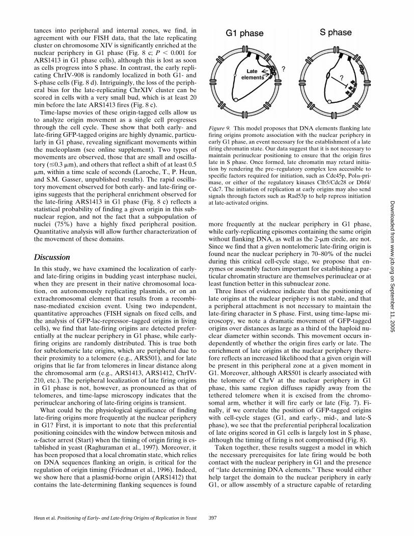
tances into peripheral and internal zones, we find, inagreement with our FISH data, that the late replicatingcluster on chromosome XIV is significantly enriched at thenuclear periphery in G1 phase (Fig. 8 c; P , 0.001 forARS1413 in G1 phase cells), although this is lost as soonas cells progress into S phase. In contrast, the early repli-cating ChrIV-908 is randomly localized in both G1- andS-phase cells (Fig. 8 d). Intriguingly, the loss of the periph-eral bias for the late-replicating ChrXIV cluster can bescored in cells with a very small bud, which is at least 20min before the late ARS1413 fires (Fig. 8 c).
Time-lapse movies of these origin-tagged cells allow usto analyze origin movement as a single cell progressesthrough the cell cycle. These show that both early- andlate-firing GFP-tagged origins are highly dynamic, particu-larly in G1 phase, revealing significant movements withinthe nucleoplasm (see online supplement). Two types ofmovements are observed, those that are small and oscilla-tory (#0.3 mm), and others that reflect a shift of at least 0.5mm, within a time scale of seconds (Laroche, T., P. Heun,and S.M. Gasser, unpublished results). The rapid oscilla-tory movement observed for both early- and late-firing or-igins suggests that the peripheral enrichment observed forthe late-firing ARS1413 in G1 phase (Fig. 8 c) reflects astatistical probability of finding a given origin in this sub-nuclear region, and not the fact that a subpopulation ofnuclei (75%) have a highly fixed peripheral position.Quantitative analysis will allow further characterization ofthe movement of these domains.
DiscussionIn this study, we have examined the localization of early-and late-firing origins in budding yeast interphase nuclei,when they are present in their native chromosomal loca-tion, on autonomously replicating plasmids, or on anextrachromosomal element that results from a recombi-nase-mediated excision event. Using two independent,quantitative approaches (FISH signals on fixed cells, andthe analysis of GFP-lac-repressor–tagged origins in livingcells), we find that late-firing origins are detected prefer-entially at the nuclear periphery in G1 phase, while early-firing origins are randomly distributed. This is true bothfor subtelomeric late origins, which are peripheral due totheir proximity to a telomere (e.g., ARS501), and for lateorigins that lie far from telomeres in linear distance alongthe chromosomal arm (e.g., ARS1413, ARS1412, ChrIV-210, etc.). The peripheral localization of late firing originsin G1 phase is not, however, as pronounced as that oftelomeres, and time-lapse microscopy indicates that theperinuclear anchoring of late-firing origins is transient.
What could be the physiological significance of findinglate-firing origins more frequently at the nuclear peripheryin G1? First, it is important to note that this preferentialpositioning coincides with the window between mitosis anda-factor arrest (Start) when the timing of origin firing is es-tablished in yeast (Raghuraman et al., 1997). Moreover, ithas been proposed that a local chromatin state, which relieson DNA sequences flanking an origin, is critical for theregulation of origin timing (Friedman et al., 1996). Indeed,we show here that a plasmid-borne origin (ARS1412) thatcontains the late-determining flanking sequences is found
more frequently at the nuclear periphery in G1 phase,while early-replicating episomes containing the same originwithout flanking DNA, as well as the 2-mm circle, are not.Since we find that a given nontelomeric late-firing origin isfound near the nuclear periphery in 70–80% of the nucleiduring this critical cell-cycle stage, we propose that en-zymes or assembly factors important for establishing a par-ticular chromatin structure are themselves perinuclear or atleast function better in this subnuclear zone.
Three lines of evidence indicate that the positioning oflate origins at the nuclear periphery is not stable, and thata peripheral attachment is not necessary to maintain thelate-firing character in S phase. First, using time-lapse mi-croscopy, we note a dramatic movement of GFP-taggedorigins over distances as large as a third of the haploid nu-clear diameter within seconds. This movement occurs in-dependently of whether the origin fires early or late. Theenrichment of late origins at the nuclear periphery there-fore reflects an increased likelihood that a given origin willbe present in this peripheral zone at a given moment inG1. Moreover, although ARS501 is clearly associated withthe telomere of ChrV at the nuclear periphery in G1phase, this same region diffuses rapidly away from thetethered telomere when it is excised from the chromo-somal arm, whether it will fire early or late (Fig. 7). Fi-nally, if we correlate the position of GFP-tagged originswith cell-cycle stages (G1, and early-, mid-, and late-Sphase), we see that the preferential peripheral localizationof late origins scored in G1 cells is largely lost in S phase,although the timing of firing is not compromised (Fig. 8).
Taken together, these results suggest a model in whichthe necessary prerequisites for late firing would be bothcontact with the nuclear periphery in G1 and the presenceof “late determining DNA elements.” These would eitherhelp target the domain to the nuclear periphery in earlyG1, or allow assembly of a structure capable of retarding
Figure 9. This model proposes that DNA elements flanking latefiring origins promote association with the nuclear periphery inearly G1 phase, an event necessary for the establishment of a latefiring chromatin state. Our data suggest that it is not necessary tomaintain perinuclear positioning to ensure that the origin fireslate in S phase. Once formed, late chromatin may retard initia-tion by rendering the pre–regulatory complex less accessible tospecific factors required for initiation, such as Cdc45p, Pola-pri-mase, or either of the regulatory kinases Clb5/Cdc28 or Dbf4/Cdc7. The initiation of replication at early origins may also sendsignals through factors such as Rad53p to help repress initiationat late-activated origins.
on Septem
ber 11, 2005 w
ww
.jcb.orgD
ownloaded from

The Journal of Cell Biology, Volume 152, 2001 398
the initiation event, or both (Fig. 9). Once established, themodified late chromatin structure appears to be autono-mous and mobile. Our data further suggest that a periph-eral localization alone is not sufficient to confer delayedinitiation, since early replicating origins are found withequal efficiency at internal sites as at the nuclear peripheryusing both FISH and GFP fluorescence.
The dynamic character of late origin localization con-trasts with the more stable association of telomeres withcomponents of the nuclear envelope (Gotta et al., 1996;Laroche et al., 1998), and suggests that there are at leasttwo pathways for achieving perinuclear association. This isconsistent with the stronger enrichment of subtelomericY9 elements at the extreme nuclear periphery as comparedwith other late origins (Fig. 3 a), and with the lack of colo-calization between late-firing origins and telomeres (Fig.5, and Table II).
What is the nature of this “late firing chromatin” statethat appears to be assembled at the nuclear periphery?Here it helps to draw distinctions between three types oflate origins. The first includes origins that are immediatelyadjacent to telomeres (Y9 or X elements). In these, thelate firing character appears to result from the presence ofSIR complexes that propagate along the nucleosomal fiberand restrict access of the DNA to large enzymes (e.g.,RNA polymerases, DAM methyltransferase). Such originsshow a SIR-dependent repression of firing (Stevensonand Gottschling, 1999). The second class of late-firing ori-gin is more internal, but still telomere-proximal, such asARS501 (z28 kb from the right telomere of ChrV). Al-though its late activation clearly depends on the presenceof a nearby telomere (Ferguson and Fangman, 1992), it isnot affected by sir mutations (Stevenson and Gottschling,1999). ARS501 may nonetheless have an unusual nucleo-somal ordering typical for chromosomal ends, since deple-tion of histone H4 affects gene expression of a large num-ber of genes up to 20 kb from yeast telomeres, while sirmutations only affect genes immediately adjacent to theTG repeat (Wyrick et al., 1999). The third type includes in-ternal late-firing origins such as ARS1412 and ARS1413,which are far from telomeres and fire late in a SIR-inde-pendent manner. Despite the fact that these origins areflanked by actively transcribed genes, they may still havecharacteristic patterns of histone modification that markthem for late initiation or for contact with the nuclear pe-riphery in G1, without silencing transcription.
The presence of tightly positioned nucleosomes in theregions flanking late origins may favor the formation of acompact or folded chromatin structure that could also de-lay initiation. Such patterns have been observed at the re-combination enhancer on ChrIII, which regulates recom-bination between the MAT locus with HM loci at adistance (Weiss and Simpson, 1998). The resulting higher-order folding of the chromatin fiber in this region could in-terfere sterically with one or more components of the ma-chinery required for initiation. Candidates for excludedfactors include Cdc45p, Pola-primase, RFA, RFC, or reg-ulatory kinases such as the Clb5/Cdc28 or Dbf4/Cdc7 com-plexes. Alternatively, the prereplication complex itselfmay have a different composition at the late firing origins,again possibly due to its assembly near the nuclear periph-ery. Finally, unidentified components that modulate initia-
tion may be present at late-firing origins, or known com-ponents could be modified in a characteristic manner (e.g.,ubiquitination, phosphorylation, methylation, or acetyla-tion). Since it is unlikely that we will be able to biochemi-cally purify early- and late-firing origins in significantamounts, distinguishing between these models will requirethe use of genetics and chromatin immunoprecipitationanalyses in mutant cells.
How do our findings relate to the control of early- andlate-DNA replication in mammalian cells? It is well-docu-mented that heterochromatin replicates late and has acharacteristic perinuclear and/or perinucleolar position inmammalian cells (O’Keefe et al., 1992; Li et al., 1998; Maet al., 1998; Croft et al., 1999). The formation of this char-acteristic pattern of chromatin distribution has beenshown to be established in early G1, roughly 2 h after mi-tosis (Dimitrova and Gilbert, 1999), and correlates wellwith the observation described in this paper. Althoughseveral studies suggest that DNA regions move little dur-ing interphase in mammalian cells (confined to ,0.3 mm;Shelby et al., 1996; Abney et al., 1997; Marshall et al.,1997; Zink et al., 1998; Manders et al., 1999), large-scalechromosomal movements have been observed during in-terphase in Drosophila (Csink and Henikoff, 1998). Themore highly compacted nature of mammalian heterochro-matin may render it less dynamic. Moreover, mammaliannuclei have elements of nuclear substructure that are ab-sent or modified in yeast, such as the nuclear lamina (Moiret al., 2000), NuMA (Harborth and Osborn, 1999), and thetetratricopeptide repeat (TPR) filaments that appear toextend into the nuclear interior (Cordes et al., 1997; Bangset al., 1998; Strambio-de-Castillia et al., 1999). In contrastto higher eukaryotic cells, the equivalent of the TPR fibers(Myosin-like proteins 1 and 2) are perinuclear in buddingyeast (Galy et al., 2000).
Most studies on late mammalian origins have examinedthe late replication of satellite or repeated DNA elements,which are probably most comparable with subtelomericregions in yeast. It remains to be seen whether there arelate-firing origins in transcriptionally active regions of themammalian genome, and whether these have a unique po-sition in the interphase nucleus. An intriguing study byBridger et al. (2000) has shown that the late-replicatingchromosome 18 is largely peripheral in the nuclei of cul-tured human cells, although it loses this position when thecells entered quiescence. Upon restimulation of growth,the chromosome remains late-replicating, despite loss ofits peripheral location. This is highly reminiscent of ourobservations in yeast, which suggest that the peripheral as-sociation is not necessary in S phase, but may be needed toestablish the late-firing state in G1. Parallels such as thesesuggest that studies in yeast on the character of late-firingorigins will provide useful paradigms for identifying thecause of late replication in more complex nuclei.
The data on origin timing were generously provided from a manuscript inpreperation by M.K. Raghuraman, D. Collingwood, W. Winzeler, S. Hunt,L. Wodicka, A. Conway, D. Lockhart, R.W. Davis, B. Braurer, and W.Fangman. We thank Marc Gasser and Anya Wilson for their technical as-sistance, Frank Neumann for determining the timing of replication ontagged ARS1413 and ARS1, and Dr. J.-P. Antonietti (University of Lau-sanne, Lausanne, Switzerland) for help with statistical analyses. We thankDrs. C. Newlon, E. Louis, V. Doye, G. Simchen, and M. Peter (ISREC) for
on Septem
ber 11, 2005 w
ww
.jcb.orgD
ownloaded from

Heun et al. Positioning of Early- and Late-firing Origins of Replication in Yeast 399
strains and plasmids. We thank Dr. Philippe Pasero (Centre National de laRecherche Scientifique, Montpellier, France) for suggesting this projectand for helpful discussions, and Drs. Bernard Duncker, Moira Cockell,and Joachim Lingner (ISREC) for critically reading the manuscript.
This study was supported in part by a predoctoral fellowship from Boeh-ringer Ingelheim Foundation (P. Heun), and the Roche Foundation. TheGasser laboratory is funded by grants from the Swiss National ScienceFoundation and the Swiss Cancer League.
Submitted: 15 September 2000Revised: 22 November 2000Accepted: 29 November 2000
References
Abney, J.R., B. Cutler, M.L. Fillbach, D. Axelrod, and B.A. Scalettar. 1997.Chromatin dynamics in interphase nuclei and its implications for nuclearstructure. J. Cell Biol. 137:1459–1468.
Bangs, P., B. Burke, C. Powers, R. Craig, A. Purohit, and S. Doxsey. 1998.Functional analysis of Tpr: identification of nuclear pore complex associa-tion and nuclear localization domains and a role in mRNA export. J. CellBiol. 143:1801–1812.
Belgareh, N., and V. Doye. 1997. Dynamics of nuclear pore distribution in nu-cleoporin mutant yeast cells. J. Cell Biol. 136:747–759.
Bousset, K., and J.F. Diffley. 1998. The Cdc7 protein kinase is required for ori-gin firing during S phase. Genes Dev. 12:480–490.
Brewer, B.J., and W.L. Fangman. 1987. The localization of replication originson ARS plasmids in S. cerevisiae. Cell. 51:463–471.
Bridger, J.M., S. Boyle, I.R. Kill, and W.A. Bickmore. 2000. Re-modelling ofnuclear architecture in quiescent and senescent human fibroblasts. Curr.Biol. 10:149–152.
Campbell, J.L., and C.S. Newlon. 1991. Chromosomal DNA replication. In TheMolecular Biology of the Yeast Saccharomyces cerevisiae: Genome Dynam-ics, Protein Synthesis and Energetics.Vol. 1. J.R. Broach, J. Pringle, and E.Jones, editors. Cold Spring Harbor Laboratory Press, Cold Spring Harbor,NY. 41–146.
Cordes, V.C., S. Reidenbach, H.R. Rackwitz, and W.W. Franke. 1997. Identifi-cation of protein p270/Tpr as a constitutive component of the nuclear porecomplex-attached intranuclear filaments. J. Cell Biol. 136:515–529.
Croft, J.A., J.M. Bridger, S. Boyle, P. Perry, P. Teague, and W.A. Bickmore.1999. Differences in the localization and morphology of chromosomes in thehuman nucleus. J. Cell Biol. 145:1119–1131.
Csink, A.K., and S. Henikoff. 1998. Large-scale chromosomal movements dur-ing interphase progression in Drosophila. J. Cell Biol. 143:13–22.
Dimitrova, D.S., and D.M. Gilbert. 1999. The spatial position and replicationtiming of chromosomal domains are both established in early G1 phase.Mol. Cell. 4:983–993.
Dohrmann, P.R., G. Oshiro, M. Tecklenburg, and R.A. Sclafani. 1999. RAD53regulates DBF4 independently of checkpoint function in Saccharomycescerevisiae. Genetics. 151:965–977.
Donaldson, A.D., W.L. Fangman, and B.J. Brewer. 1998a. Cdc7 is requiredthroughout the yeast S phase to activate replication origins. Genes Dev. 12:491–501.
Donaldson, A.D., M.K. Raghuraman, K.L. Friedman, F.R. Cross, B.J. Brewer,and W.L. Fangman. 1998b. CLB5-dependent activation of late replicationorigins in S. cerevisiae. Mol. Cell. 2:173–182.
Fangman, W.L., and B.J. Brewer. 1992. A question of time: replication originsof eukaryotic chromosomes. Cell. 71:363–366.
Ferguson, B.M., B.J. Brewer, A.E. Reynolds, and W.L. Fangman. 1991. A yeastorigin of replication is activated late in S phase. Cell. 65:507–515.
Ferguson, B.M., and W.L. Fangman. 1992. A position effect on the time of rep-lication origin activation in yeast. Cell. 68:333–339.
Fox, M.H., D.J. Arndt-Jovin, T.M. Jovin, P.H. Baumann, and M. Robert-Nicoud. 1991. Spatial and temporal distribution of DNA replication sites lo-calized by immunofluorescence and confocal microscopy in mouse fibro-blasts. J. Cell Sci. 99:247–253.
Friedman, K.L., B.J. Brewer, and W.L. Fangman. 1997. Replication profile ofSaccharomyces cerevisiae chromosome VI. Genes Cells. 2:667–678.
Friedman, K.L., J.D. Diller, B.M. Ferguson, S.V. Nyland, B.J. Brewer, andW.L. Fangman. 1996. Multiple determinants controlling activation of yeastreplication origins late in S phase. Genes Dev. 10:1595–1607.
Galy, V., J.C. Olivo-Marin, H. Scherthan, V. Doye, N. Rascalou, and U. Nehr-bass. 2000. Nuclear pore complexes in the organization of silent telomericchromatin. Nature. 403:108–112.
Gotta, M., T. Laroche, A. Formenton, L. Maillet, H. Scherthan, and S.M. Gas-ser. 1996. The clustering of telomeres and co-localization with Rap1, Sir3,and Sir4 proteins in wild-type Saccharomyces cerevisiae. J. Cell Biol. 134:1349–1363.
Gotta, M., T. Laroche, and S.M. Gasser. 1999. Analysis of nuclear organizationin Saccharomyces cerevisiae. Methods Enzymol. 304:663–672.
Guacci, V., E. Hogan, and D. Koshland. 1994. Chromosome condensation andsister chromatid pairing in budding yeast. J. Cell Biol. 125:517–530.
Hadfield, C., R.C. Mount, and A.M. Cashmore. 1995. Protein binding interac-tions at the STB locus of the yeast 2 microns plasmid. Nucleic Acids Res. 23:995–1002.
Harborth, J., and M. Osborn. 1999. Does NuMA have a scaffold function in theinterphase nucleus? Crit. Rev. Eukaryot. Gene Expr. 9:319–328.
Hayashi, A., H. Ogawa, K. Kohno, S.M. Gasser, and Y. Hiraoka. 1998. Meioticbehaviours of chromosomes and microtubules in budding yeast: relocaliza-tion of centromeres and telomeres during meiotic prophase. Genes Cells.3:587–601.
Holmquist, G.P. 1987. Role of replication time in the control of tissue-specificgene expression. Am. J. Hum. Genet. 40:151–173.
Jin, Q., J. Fuchs, and J. Loidl. 2000. Centromere clustering is a major determi-nant of yeast interphase nuclear organization. J. Cell Sci. 113:1903–1912.
Jin, Q., E. Trelles-Sticken, H. Scherthan, and J. Loidl. 1998. Yeast nuclei dis-play prominent centromere clustering that is reduced in nondividing cellsand in meiotic prophase. J. Cell Biol. 141:21–29.
Klein, F., T. Laroche, M.E. Cardenas, J.F. Hofmann, D. Schweizer, and S.M.Gasser. 1992. Localization of RAP1 and topoisomerase II in nuclei and mei-otic chromosomes of yeast. J. Cell Biol. 117:935–948.
Laroche, T., S.G. Martin, M. Gotta, H.C. Gorham, F.E. Pryde, E.J. Louis, andS.M. Gasser. 1998. Mutation of yeast Ku genes disrupts the subnuclear orga-nization of telomeres. Curr. Biol. 8:653–656.
Laroche, T., S.G. Martin, M. Tsai-Pflugfelder, and S.M. Gasser. 2000. The dy-namics of yeast telomeres and silencing proteins through the cell cycle. J.Struct. Biol. 129:159–174.
Leonhardt, H., H.P. Rahn, P. Weinzierl, A. Sporbert, T. Cremer, D. Zink, andM.C. Cardoso. 2000. Dynamics of DNA replication factories in living cells. J.Cell Biol. 149:271–280.
Li, G., G. Sudlow, and A.S. Belmont. 1998. Interphase cell cycle dynamics of alate-replicating, heterochromatic homogeneously staining region: precisechoreography of condensation/decondensation and nuclear positioning. J.Cell Biol. 140:975–989.
Loidl, J., F. Klein, and J. Engebrecht. 1998. Genetic and morphological ap-proaches for the analysis of meiotic chromosomes in yeast. Methods CellBiol. 53:257–285.
Louis, E.J., E.S. Naumova, A. Lee, G. Naumov, and J.E. Haber. 1994. Thechromosome end in yeast: its mosaic nature and influence on recombina-tional dynamics. Genetics. 136:789–802.
Ma, H., J. Samarabandu, R.S. Devdhar, R. Acharya, P.C. Cheng, C. Meng, andR. Berezney. 1998. Spatial and temporal dynamics of DNA replication sitesin mammalian cells. J. Cell Biol. 143:1415–1425.
Manders, E.M., H. Kimura, and P.R. Cook. 1999. Direct imaging of DNA in liv-ing cells reveals the dynamics of chromosome formation. J. Cell Biol. 144:813–821.
Manders, E.M., J. Stap, G.J. Brakenhoff, R. van Driel, and J.A. Aten. 1992. Dy-namics of three-dimensional replication patterns during the S-phase, ana-lyzed by double labelling of DNA and confocal microscopy. J. Cell Sci. 103:857–862.
Marshall, W.F., A. Straight, J.F. Marko, J. Swedlow, A. Dernburg, A. Belmont,A.W. Murray, D.A. Agard, and J.W. Sedat. 1997. Interphase chromosomesundergo constrained diffusional motion in living cells. Curr. Biol. 7:930–939.
Martin, S.G., T. Laroche, N. Suka, M. Grunstein, and S.M. Gasser. 1999. Relo-calization of telomeric Ku and SIR proteins in response to DNA strandbreaks in yeast. Cell. 97:621–633.
McCarroll, R.M., and W.L. Fangman. 1988. Time of replication of yeast cen-tromeres and telomeres. Cell. 54:505–513.
Moir, R.D., T.P. Spann, R.I. Lopez-Soler, M. Yoon, A.E. Goldman, S. Khuon,and R.D. Goldman. 2000. Review: the dynamics of the nuclear lamins duringthe cell cycle—relationship between structure and function. J. Struct. Biol.129:324–334.
Nakamura, H., T. Morita, and C. Sato. 1986. Structural organizations of repli-con domains during DNA synthetic phase in the mammalian nucleus. Exp.Cell Res. 165:291–297.
Nakayasu, H., and R. Berezney. 1989. Mapping replicational sites in the eucary-otic cell nucleus. J. Cell Biol. 108:1–11.
Newlon, C.S. 1997. Putting it all together: building a prereplicative complex.Cell. 91:717–720.
Newlon, C.S., L.R. Lipchitz, I. Collins, A. Deshpande, R.J. Devenish, R.P.Green, H.L. Klein, T.G. Palzkill, R.B. Ren, S. Synn, et al. 1991. Analysis of acircular derivative of Saccharomyces cerevisiae chromosome III: a physicalmap and identification and location of ARS elements. Genetics. 129:343–357.
O’Keefe, R.T., S.C. Henderson, and D.L. Spector. 1992. Dynamic organizationof DNA replication in mammalian cell nuclei: spatially and temporally de-fined replication of chromosome-specific alpha-satellite DNA sequences. J.Cell Biol. 116:1095–1110.
Palladino, F., T. Laroche, E. Gilson, A. Axelrod, L. Pillus, and S.M. Gasser.1993. SIR3 and SIR4 proteins are required for the positioning and integrityof yeast telomeres. Cell. 75:543–555.
Pasero, P., D. Braguglia, and S.M. Gasser. 1997. ORC-dependent and origin-specific initiation of DNA replication at defined foci in isolated yeast nuclei.Genes Dev. 11:1504–1518.
Pasero, P., B.P. Duncker, E. Schwob, and S.M. Gasser. 1999. A role for theCdc7 kinase regulatory subunit Dbf4p in the formation of initiation-compe-tent origins of replication. Genes Dev. 13:2159–2176.
on Septem
ber 11, 2005 w
ww
.jcb.orgD
ownloaded from

The Journal of Cell Biology, Volume 152, 2001 400
Raghuraman, M.K., B.J. Brewer, and W.L. Fangman. 1997. Cell cycle-depen-dent establishment of a late replication program. Science. 276:806–809.
Robinett, C.C., A. Straight, G. Li, C. Willhelm, G. Sudlow, A. Murray, and A.S.Belmont. 1996. In vivo localization of DNA sequences and visualization oflarge-scale chromatin organization using lac operator/repressor recognition.J. Cell Biol. 135:1685–1700.
Rose, M.D., F. Winston, and P. Hieter. 1990. Methods in Yeast Genetics. ColdSpring Harbor Laboratory Press, Cold Spring Harbor, NY. 198 pp.
Santocanale, C., and J.F. Diffley. 1996. ORC- and Cdc6-dependent complexesat active and inactive chromosomal replication origins in Saccharomyces cere-visiae. EMBO (Eur. Mol. Biol. Organ.) J. 15:6671–6679.
Santocanale, C., and J.F. Diffley. 1998. A Mec1- and Rad53-dependent check-point controls late-firing origins of DNA replication. Nature. 395:615–618.
Scott-Drew, S., and J.A. Murray. 1998. Localisation and interaction of the pro-tein components of the yeast 2 mu circle plasmid partitioning system suggesta mechanism for plasmid inheritance. J. Cell Sci. 111:1779–1789.
Shelby, R.D., K.M. Hahn, and K.F. Sullivan. 1996. Dynamic elastic behavior ofalpha-satellite DNA domains visualized in situ in living human cells. J. CellBiol. 135:545–557.
Shirahige, K., Y. Hori, K. Shiraishi, M. Yamashita, K. Takahashi, C. Obuse, T.Tsurimoto, and H. Yoshikawa. 1998. Regulation of DNA-replication originsduring cell-cycle progression. Nature. 395:618–621.
Shirahige, K., T. Iwasaki, M.B. Rashid, N. Ogasawara, and H. Yoshikawa. 1993.Location and characterization of autonomously replicating sequences fromchromosome VI of Saccharomyces cerevisiae. Mol. Cell. Biol. 13:5043–5056.
Stevenson, J.B., and D.E. Gottschling. 1999. Telomeric chromatin modulatesreplication timing near chromosome ends. Genes Dev. 13:146–151.
Straight, A.F., A.S. Belmont, C.C. Robinett, and A.W. Murray. 1996. GFP tag-ging of budding yeast chromosomes reveals that protein–protein interac-
tions can mediate sister chromatid cohesion. Curr. Biol. 6:1599–1608.Strambio-de-Castillia, C., G. Blobel, and M.P. Rout. 1999. Proteins connecting
the nuclear pore complex with the nuclear interior. J. Cell Biol. 144:839–855.Taddei, A., D. Roche, J.B. Sibarita, B.M. Turner, and G. Almouzni. 1999. Du-
plication and maintenance of heterochromatin domains. J. Cell Biol. 147:1153–1166.
van Dierendonck, J.H., R. Keyzer, C.J. van de Velde, and C.J. Cornelisse. 1989.Subdivision of S-phase by analysis of nuclear 5-bromodeoxyuridine stainingpatterns. Cytometry. 10:143–150.
Webb, R.H. 1999. Theoretical basis of confocal microscopy. Methods Enzymol.307:3–20.
Weiner, B.M., and N. Kleckner. 1994. Chromosome pairing via multiple inter-stitial interactions before and during meiosis in yeast. Cell. 77:977–991.
Weinreich, M., and B. Stillman. 1999. Cdc7p-Dbf4p kinase binds to chromatinduring S phase and is regulated by both the APC and the RAD53 check-point pathway. EMBO (Eur. Mol. Biol. Organ.) J. 18:5334–5346.
Weiss, K., and R.T. Simpson. 1998. High-resolution structural analysis of chro-matin at specific loci: Saccharomyces cerevisiae silent mating type locusHMLalpha. Mol. Cell. Biol. 18:5392–5403.
Wyrick, J.J., F.C. Holstege, E.G. Jennings, H.C. Causton, D. Shore, M. Grun-stein, E.S. Lander, and R.A. Young. 1999. Chromosomal landscape of nu-cleosome-dependent gene expression and silencing in yeast. Nature. 402:418–421.
Zakian, V.A., B.J. Brewer, and W.L. Fangman. 1979. Replication of each copy ofthe yeast 2 micron DNA plasmid occurs during the S phase. Cell. 17:923–934.
Zink, D., T. Cremer, R. Saffrich, R. Fischer, M.F. Trendelenburg, W. Ansorge,and E.H. Stelzer. 1998. Structure and dynamics of human interphase chro-mosome territories in vivo. Hum. Genet. 102:241–251.
on Septem
ber 11, 2005 w
ww
.jcb.orgD
ownloaded from


![Index [dnareplication.cshl.edu]dnareplication.cshl.edu/content/free/chapters/index.pdfDNA replication initiator endonuclease, 800 origins, 804 replication proteins, 9-10 Rep protein](https://img.dokumen.tips/doc/110x75/601ceb5e8974bf48b64622df/index-dna-replication-initiator-endonuclease-800-origins-804-replication-proteins.jpg)