Embed Size (px)
Citation preview

204
THE PLEUROPNEUMONIA-LIRE ORGANISMS:FURTHER COMPARATIVE STUDIES AND A
DESCRIPTIVE ACCOUNT OF RECENTLYDISCOVERED TYPES
BY EMMY KLIENEBERGER
From the Bacteriological Department, Lister Institute, London
(With Plates II-IV)
REPORTS on the isolation of pleuropneumonia-like organisms have beenfrequent during the last few years, and there can be no doubt that we aredealing here with a widely spread group of organisms. In 1935 the writerreported the occurrence in cultures of Streptobacillus moniliformis of a pleuro-pneumonia-like organism, LI, recoverable therefrom in a pure state andinterpreted the association as one of symbiosis. Since then she has found andplaced on record several new species of pleuropneumonia-like organismsoccurring independently and not in apparent symbiosis with bacteria. Newinformation, collected in both these fields during the past year, forms thesubject of this communication and is discussed under the following headings:
A. The relationship of Ll to S. moniliformis: symbiont or variant?B. A new strain resembling S. moniliformis isolated from abscesses in the
necks of guinea-pigs.C. Other probable associations from which the symbiont has not so far
been obtained in pure condition.D. Pleuropneumonia-like organisms occurring independently in lesions in
rats or mice:(a) L3—bronchiectatic lesions in rats. (See p. 210.)(6) L4—-abscesses, arthritis and swollen glands in rats.(c) L5—-"rolling disease" in mice.(d) L6—from brains of mice inoculated intracerebrally with blood of
splenectomized mice containing Eperythrozoon coccoides.(e) "M55"—arthritis in mice.
E. Further studies on the filterable organisms isolated from sewage,soil, etc.
A. THE RELATIONSHIP OF L l TO S. MONILIFORMIS: SYMBIONT OK VARIANT?
The four L l strains described by the writer in the study of 1938 havebeen maintained and studied for a further period. Attempts to induce themto revert to S. moniliformis have been made by the method employed byDienes (1938), who believes that the L1 is a growth variant of the bacillus andnot a symbiont. Dawson & Hobby (1939a, b) bring forward evidence pointing,in their view, in the same direction. Small agar blocks covered with colonies
available at https://www.cambridge.org/core/terms. https://doi.org/10.1017/S0022172400027765Downloaded from https://www.cambridge.org/core. IP address: 65.21.228.167, on 20 Dec 2021 at 03:01:09, subject to the Cambridge Core terms of use,
EMMY KLIENEBBRGER 205
of the writer's four strains have not, however, reproduced streptobacilli ifincubated in Levinthal broth enriched with serum as Dienes claimed, but withan L1 growth freshly isolated from S. moniliformis streptobacillary elementsmade their appearance several times under these conditions. The writer istherefore of opinion that Dienes has not dealt with sufficiently purified strains.He purified his strains only five times, while it proved necessary in the writer'sexperiments to pick isolated colonies 30-50 times in succession before a pureL1 strain was established.
It should be emphasized here that Dienes (1939 a) and the writer agree inthe main points regarding the relationship of the LI and S. moniliformis; butthere are some minor points of disagreement which have led the two authorsto different explanations of the whole phenomenon.
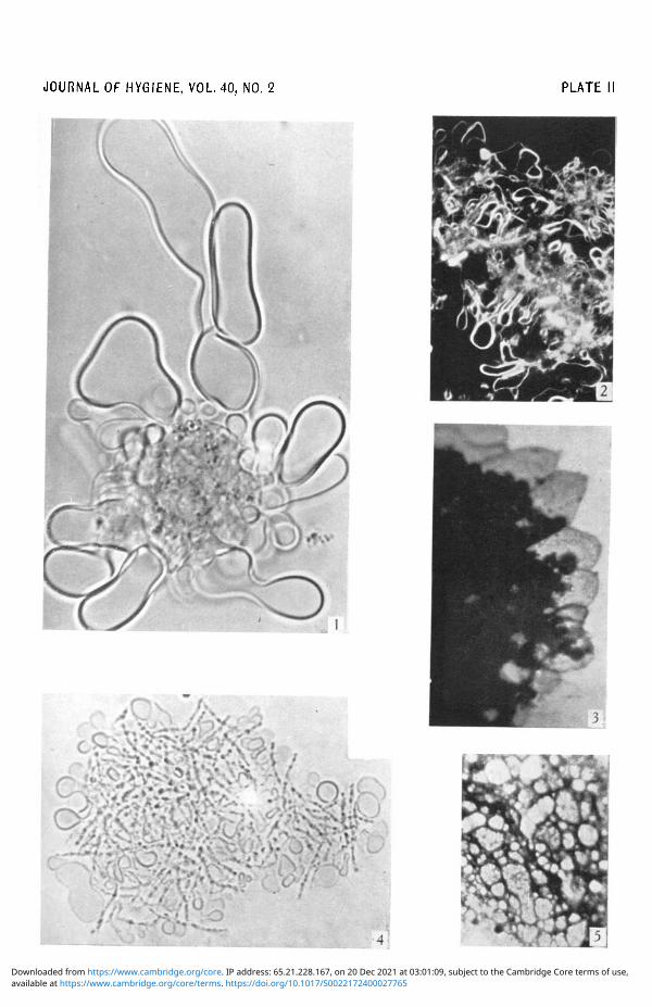
While Dienes describes the globular bodies as transformed bacilli, the writer finds thatin very young colonies of her strains the globular and "balloon-like" forms develop asseparate forms detached from the bacilli (see PL II, fig. 4). In this photograph (fig. 4) ofa living colony which developed in a drop of medium under a coverslip, the single globulescan be distinctly seen apart from the granular bacillary chains. On the other hand, froma picture such as that reproduced in Fig. 20 of Dienes's paper (1939a) it cannot be concludedthat the swollen forms are of bacillary origin, because with all the methods so far describedit is not possible to decide if a bacillus is simply swollen or if such a soft globular body asthat found in pure L1 growth is lying on top of or surrounding a bacillus. There seems tobe a further discrepancy between Dienes's and the writer's observations. On his plates,seeded from a 24 hr. old S. moniliformis culture, he observed the bacilli to develop first,while the LI formed a kind of secondary growth; he interpreted this observation assupporting his opinion that the LI is produced by the bacilli as a new growth phase. Incontrast to this the writer found that if a 2 days old culture was spread on a plate of "specialmedium"1 the LI started to develop after 2-3 hr. of incubation while the bacilli commencedto grow only after a lag period of 5-6 hr. Both elements, bacilli and L1 forms, were alwaysfound in the young colonies, while Dienes observed that his young colonies consisted chieflyof bacilli. This difference in the ratio between the numbers of bacilli and L1 forms in youngcolonies may have been caused by differences of medium or of inoculum; the writer, however,has found that bacillary elements and LI forms are always mixed up in 8. moniliformiscolonies, no matter if very young or old growth is studied.
It should be mentioned here that the so-called filamentous "network", described by thewriter in 1934 for pleuropneumonia and agalaetia and in 1935 for the LI organism, wasa misinterpretation of a genuine picture which these organisms often present, for if theglobules are tightly packed and if only their contours show up (unstained or stained) a kindof honeycomb structure is presented which in the above-mentioned papers was wronglyinterpreted as a network. The interpretation of the structures seen was complicated by thefact that the contours of peripheral globules are often only partly visible in the living aswell as in the stained specimen, thus producing a picture which can easily be taken for ameshwork with filamentous extensions. Careful observation of the living young colony willoften show that the second part of a half-visible globule becomes visible in later development,thus proving the globular nature of the structure seen. This would explain why Dienes didnot find the extended "network" structures described and depicted by the present authorin her earlier papers. The writer also agrees with Dienes in the observation that the slightesttearing or distortion applied to colonies of pleuropneumonia-like organisms may produce
1 Ox heart infusion peptone Levinthal agar (Levinthal & Fernbach, 1922), pH 7-8-8-0 enrichedwith 30% of horse serum (Klieneberger, 1938).
J. Hygiene 40 14
available at https://www.cambridge.org/core/terms. https://doi.org/10.1017/S0022172400027765Downloaded from https://www.cambridge.org/core. IP address: 65.21.228.167, on 20 Dec 2021 at 03:01:09, subject to the Cambridge Core terms of use,
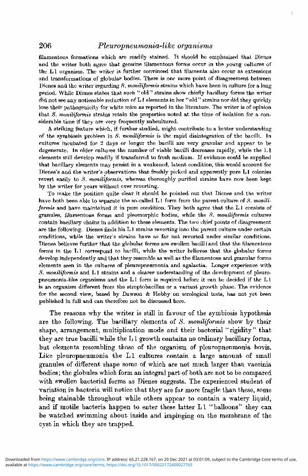
206 Pleuropneumonia-like organismsfilamentous formations which are readily stained. It should be emphasized that Dienesand the writer both agree that genuine filamentous forms occur in the young cultures ofthe LI organism. The writer is further convinced that filaments also occur as extensionsand transformations of globular bodies. There is one more point of disagreement betweenDienes and the writer regarding 8. moniliformis strains which have been in culture for a longperiod. WhEe Dienes states that such "old" strains show chiefly bacillary forms the writerdid not see any noticeable reduction of LI elements in her "old" strains nor did they quicklylose their pathogenicity for white mice as reported in the literature. The writer is of opinionthat S. moniliformis strains retain the properties noted at the time of isolation for a con-siderable time if they are very frequently subcultured.
A striking feature which, if further studied, might contribute to a better understandingof the symbiosis problem in 8. moniliformis is the rapid disintegration of the bacilli. Incultures incubated for 2 days or longer the bacilli are very granular and appear to bedegenerate. In older cultures the number of viable bacilli decreases rapidly, while the L1elements still develop readily if transferred to fresh medium. If evidence could be suppliedthat bacillary elements may persist in a weakened, latent condition, this would account forDienes's and the writer's observations that freshly picked and apparently pure L1 coloniesrevert easily to <S. moniliformis, whereas thoroughly purified strains have now been keptby the writer for years without ever reverting.
To make the position quite clear it should be pointed out that Dienes and the writerhave both been able to separate the so-called L1 form from the parent culture of S. monili-formis and have maintained it in pure condition. They both agree that the LI consists ofgranules, filamentous forms and pleomorphic bodies, while the 8. moniliformis culturescontain bacillary chains in addition to these elements. The two chief points of disagreementare the following. Dienes finds his L1 strains reverting into the parent culture under certainconditions, while the writer's strains have so far not reverted under similar conditions.Dienes believes further that the globular forms are swollen bacilli and that the filamentousforms in the LI correspond to bacilli, while the writer believes that the globular formsdevelop independently and that they resemble as well as the filamentous and granular formselements seen in the cultures of pleuropneumonia and agalactia. Longer experience with8. moniliformis and L1 strains and a clearer understanding of the development of pleuro-pneumonia-like organisms and the L1 form is required before it can be decided if the L1is an organism different from the streptobacillus or a variant growth phase. The evidencefor the second view, based by Dawson & Hobby on serological tests, has not yet beenpublished in full and can therefore not be discussed here.
The reasons why the writer is still in favour of the symbiosis hypothesisare the following. The bacillary elements of S. moniliformis show by theirshape, arrangement, multiplication mode and their bacterial "rigidity" thatthey are true bacilli while the L1 growth contains no ordinary bacillary forms,but elements resembling those of the organism of pleuropneumonia bovis.Like pleuropneumonia the LI cultures contain a large amount of smallgranules of different shape some of which are not much larger than vacciniabodies; the globules which form an integral part of both are not to be comparedwith swollen bacterial forms as Dienes suggests. The experienced student ofvariation in bacteria will notice that they are far more fragile than these, somebeing stainable throughout while others appear to contain a watery liquid,and if motile bacteria happen to enter these latter LI "balloons" they canbe watched swimming about inside and impinging on the membrane of thecyst in which they are trapped.
available at https://www.cambridge.org/core/terms. https://doi.org/10.1017/S0022172400027765Downloaded from https://www.cambridge.org/core. IP address: 65.21.228.167, on 20 Dec 2021 at 03:01:09, subject to the Cambridge Core terms of use,

EMMY KLIENEBERGER 207PI. II, fig. 2, shows a darkground picture of LI globules. PI. II, fig. 1, shows a living
colony of L1 grown in semi-solid medium in which granules and enormous bodies are tobe seen. PI. II, fig. 3, shows a corresponding colony edge, stained. The membrane itself mayappear very refractile and fairly thick in some globules, while it appears very thin in others;it can be drawn out into filaments; in most cases the membrane contains granules resemblingthose forming a large part of the whole growth. Some of the globules and filamentousappendices resemble in fact myelin structures, and a microchemical test has recently suggestedthat the LI and the <S. moniliformis cultures may contain a considerable amount ofsterols. Some of the globular elements in a well-grown culture are doubly refractile.Photographs of stained preparations of some of the main types of pleuropneumonia-likeorganisms are reproduced in PI. II, fig. 3 (L1 organism), PL II, fig. 5 (organism of agalactia),PL III, figs. 13 and 14 (sewage organism "A"), PL III, fig. 15 (Asterococcus canis II), PL IV,fig. 19 (L 5 organism), fig. 20 (L 6 organism). These photographs demonstrate the characteristicstructures common to all pleuropneumonia-like organisms.
Besides possessing a similar morphology, LI and the organisms of thepleuropneumonia group show also the same colony type, characterized by acentral granular part embedded in the agar medium and a flatter peripheralzone. There is further the regular filterability of L1 and the pleuropneumonia-like organisms through some of the coarser niters such as the Berkefeld Vcandle. It seems a reasonable conclusion from these data that the L1 is itselfa pleuropneumonia-like organism and consequently unlikely to prove a variantgrowth form of the streptobacillus. There is the further argument that if theLI is a variant form of S. moniliformis, the now numerous strains of theL series occurring independently should be derived from streptobacillarymother strains, but evidence of the existence of such is not forthcoming.
B. A NEW STRAIN RESEMBLING S. MONILIFORMIS ISOLATED PROM ABSCESSESIN THE NECKS OF GUINEA-PIGS
A culture resembling S. moniliformis was isolated by the writer in thewinter of 1938-9, and a note of its occurrence was submitted to the Patho-logical Society of Great Britain and Ireland in July 1939 and to the ThirdInternational Congress of Microbiology in September 1939 (Klieneberger,1939 b, c). Among the Institute's guinea-pig stocks were often found individualspresenting large abscesses in the neck. As a rule these abscesses ran a chroniccourse, but eventually they either burst and cleared up or they regressed anddisappeared. The "chronic streptococcus cervical lymphadenitis" of theguinea-pig recently described by Seastone (1939) seems to be of differentaetiology, for streptococci were never found in the pus of the swollen lymphglands of our guinea-pigs. The caseous pus did not reveal any formed elementsof bacterial nature in an ordinary Gram preparation, but if fixed with methylalcohol and stained with a weak Giemsa solution over-night or if treated byone of the methods for the demonstration of elementary bodies small bacillaryforms and thread-like elements were discovered. On ordinary media this pusyielded no growth; but in rich serum broth and on the writer's special mediumgrowth was obtained in 3-6 days aerobically and in 2-3 days anaerobically.While growth appeared as lumps and flakes in the liquid medium, irregular
14-2
available at https://www.cambridge.org/core/terms. https://doi.org/10.1017/S0022172400027765Downloaded from https://www.cambridge.org/core. IP address: 65.21.228.167, on 20 Dec 2021 at 03:01:09, subject to the Cambridge Core terms of use,

208 Pleuropneumonia-like organisms
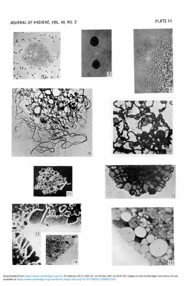
dense colonies grew up on the solid medium in which by low magnificationsome globular elements were discovered (PL III, fig. 6). If higher magnificationsare used the direct agar method reveals that the whole colony is interspersedwith globules of different size and shape. Giemsa-stained colonies show delicatebacillary threads abundantly interlaced at the edges (PI. Ill, fig. 9). Betweenthe tangled bacillary chains stained and unstained globules can be seen.A Gram-stained smear of a culture shows Gram-negative clumps of minutebacilli. The structure of the guinea-pig culture resembles in all details that ofS. moniliformis, the only morphological difference being that the bacillarythreads are more delicate and more entangled. The guinea-pig culture differsfurther from S. moniliformis in that it grows at first better anaerobically thanaerobically. Evident differences can be demonstrated by animal experiment:S. moniliformis has, as we know, no pathogenic effects at all on guinea-pigsand rabbits; it is usually not infective for rats though it inhabits theirnasopharynx; the only small laboratory animal for which it is highly infectiveis the mouse in which it produces arthritis and septicaemia. Prof. WilsonSmith (Sheffield) informed me that he had also independently isolated a culturesimilar to the guinea-pig strain here described and had studied its pathogenicityextensively. On this work he will doubtless report in due course. The writerhas so far been interested chiefly in the morphology of the "guinea-pigculture". Because of its resemblance to S. moniliformis it seemed of interestto investigate its possible symbiotic nature. For this purpose old cultures,kept in the incubator for days, or even weeks, were spread on plates of specialmedium, as described by the writer in 1936. On some plates small coloniescame up in which bacilli were no longer seen. From these colonies a growth,different from the parent culture and resembling pleuropneumonia, wasobtained. Subcultures at first grew with difficulty and developed on the semi-solid medium only under agar cover. Gradually the growth became betterestablished and a purification process was started. From each subsequentculture a piece of agar was cut out containing as few colonies as possible.It was hardly ever possible to pick a single colony. The agar piece was movedover the surface of a new plate. After the first eight passages some coloniesreproduced the whole parent culture, but from others the pleuropneumonia-like culture could be carried on. When this process had been continued thirtytimes another reversion to the parent culture occurred, but it was againpossible to pick pleuropneumonia-like colonies. After twenty passages, duringwhich no reversion took place, this culture was unfortunately lost during thedisorganization occasioned by the outbreak of war. The clearing process willagain be started with a new strain, and it is hoped that a pure pleuropneumonia-like culture will be obtained. The lost pleuropneumonia-like strain from the'' guinea-pig culture'' formed a roundish colony with a dark granular centre and aperipheral zone showing a lace-like structure (PI. Ill, fig. 7). PI. Ill, fig. 8, showsa colony edge at a higher magnification, clearly revealing the globular structureof the peripheral zone. The colony resembled closely that of the L1 organism.
available at https://www.cambridge.org/core/terms. https://doi.org/10.1017/S0022172400027765Downloaded from https://www.cambridge.org/core. IP address: 65.21.228.167, on 20 Dec 2021 at 03:01:09, subject to the Cambridge Core terms of use,

EMMY KLIENEBERGER 209
C. OTHER PROBABLE ASSOCIATIONS PROM WHICH THE SYMBIONT HAS NOT
SO FAR BEEN OBTAINED IN PURE CONDITION
During the past year another apparently symbiotic culture was isolatedby the writer. It was accidentally found on a plate which had been spreadwith material from the skin of a pig inoculated with swine-pox. The coloniesof this organism consist of Gram-positive cocci interspersed with large,balloon-like forms. This culture grows well on ordinary media and developsat 37° C. as well as at room temperature. The globular forms resemble thoseof the L1 organism, but attempts to isolate a pleuropneumonia-like microbefrom it have failed. The development of peculiar large forms has also beenobserved in Fusobacterium nucleatum (syn. Bacterium fundilifor me), in whichattempts to separate a pleuropneumonia-like organism have likewise beenunsuccessful. PI. Ill, fig. 11, shows a darkground picture of a young aerobicserum broth culture of F. nucleatum containing many globular forms. As thecultures grow older more bacillary forms develop, and at the same timeabundant gas production starts in the liquid medium. On the surface of platesincubated anaerobically numerous globular forms develop. Small coloniesoften consist chiefly of these forms. It is possible to obtain globular coloniesin which bacillary forms are no longer demonstrable, but if transferred theyeventually produce a mixture of globular forms and bacilli. PI. Ill, fig. 10,shows both forms grown on solid medium (agar fixation Giemsa technique),while fig. 12 shows the edge of a globular colony in which bacilli cannot bedetected. Similar phenomena were observed by Dienes (19396) in cultures ofa Flavobacterium, of Haemophilus influenzae and B. fundiliforme (syn. Fuso-bacterium nucleatum). It is difficult to decide if these cultures from whichDienes and the writer have not been able to obtain the apparent LI-likeelements in pure culture do really contain an LI-like symbiont or growthvariant as S. moniliformis. The mere observation of swollen bodies or globularforms is not sufficient evidence to prove the existence of a filterable, pleuro-pneumonia-like symbiont or growth phase in these cultures. Swollen formshave been described in all kinds of cultures, but the separation of the LIgrowth from the parent culture has so far only been successful in S. moniliformisand the "guinea-pig strain".
D. PLEUROPNEUMONIA-LIKE ORGANISMS OCCURRING INDEPENDENTLY
IN LESIONS IN RATS AND MICE
In previous papers investigations on the organisms of pleuropneumonia,agalactia, Asterococcus canis I and II (Shoetensack) and the LI, L3, L4organisms have been recorded by the writer (Klieneberger, 1938). The moreorganisms of the pleuropneumonia group that come to light, the more necessaryit seems to find differences which may serve for classification purposes.Therefore studies on the growth, morphology, serology, occurrence andpathogenicity of the newly found L5 and L6 organisms (Findlay, Kliene-
available at https://www.cambridge.org/core/terms. https://doi.org/10.1017/S0022172400027765Downloaded from https://www.cambridge.org/core. IP address: 65.21.228.167, on 20 Dec 2021 at 03:01:09, subject to the Cambridge Core terms of use,
210 Pleuropneumonia-like organismsberger, MacCallum & Mackenzie, 1938, 1939; Findlay, Mackenzie, MacCallum& Klieneberger, 1939) have been carried out, and new information has beencollected about the L 3 organism. A number of strains from sewage, soil andwater have also been included in these studies. These new types have beencompared with those already described (Klieneberger, 1935, 1936, 1938,1939a, 6).
(a) £3—bronchiectatic lesions in ratsThree different organisms of the pleuropneumonia group have been isolated
from rats by the writer. The LI, regularly associated with S. moniliformis, wasonce found independently in the rat lung. The L3 has been isolated byKlieneberger & Steabben (1937) from lung lesions of rats, and some new dataregarding its association with the pulmonary disease are recorded in thisJournal, p. 223, by the same authors.
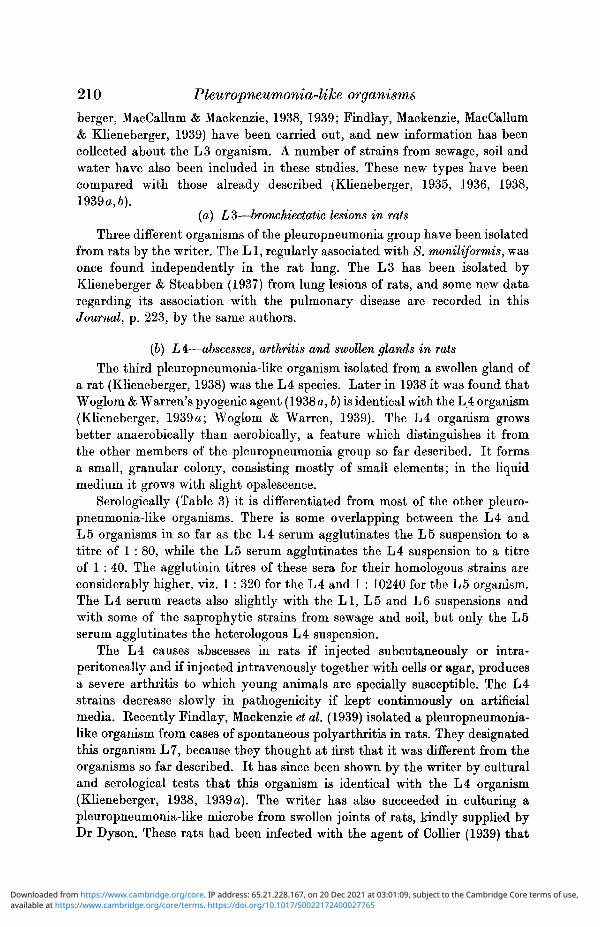
(b) L4:—abscesses, arthritis and swollen glands in ratsThe third pleuropneumonia-like organism isolated from a swollen gland of
a rat (Klieneberger, 1938) was the L4 species. Later in 1938 it was found thatWoglom & Warren's pyogenic agent (1938 a, b) is identical with the L4 organism(Klieneberger, 1939 a; Woglom & Warren, 1939). The L4 organism growsbetter anaerobically than aerobically, a feature which distinguishes it fromthe other members of the pleuropneumonia group so far described. It formsa small, granular colony, consisting mostly of small elements; in the liquidmedium it grows with slight opalescence.
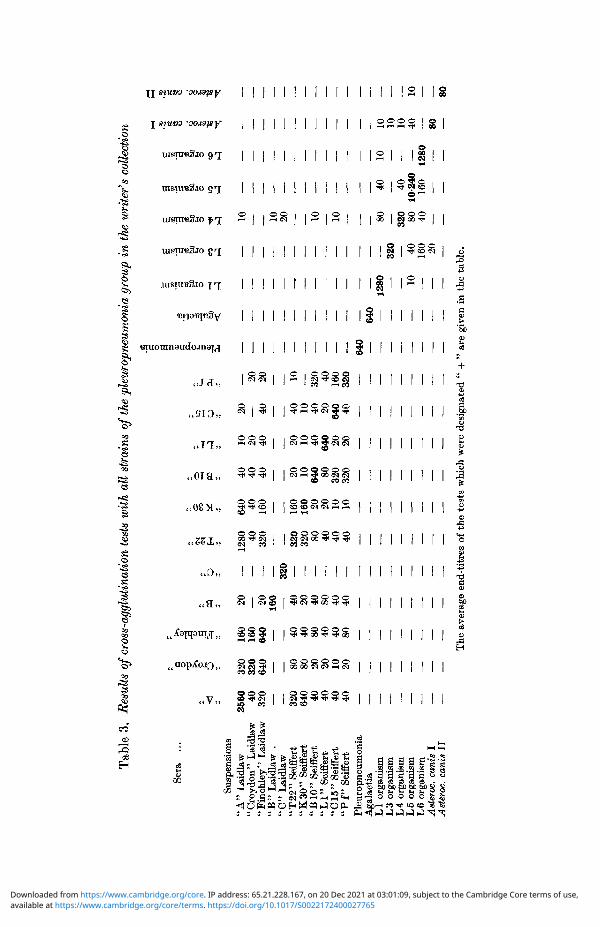
Serologically (Table 3) it is diiferentiated from most of the other pleuro-pneumonia-like organisms. There is some overlapping between the L4 andL5 organisms in so far as the L4 serum agglutinates the L5 suspension to atitre of 1 : 80, while the L5 serum agglutinates the L4 suspension to a titreof 1 : 40. The agglutinin titres of these sera for their homologous strains areconsiderably higher, viz. 1 : 320 for the L4 and 1 : 10240 for the L5 organism.The L4 serum reacts also slightly with the LI, L5 and L6 suspensions andwith some of the saprophytic strains from sewage and soil, but only the L5serum agglutinates the heterologous L4 suspension.
The L4 causes abscesses in rats if injected subcutaneously or intra-peritoneally and if injected intravenously together with cells or agar, producesa severe arthritis to which young animals are specially susceptible. The L4strains decrease slowly in pathogenicity if kept continuously on artificialmedia. Kecently Findlay, Mackenzie et al. (1939) isolated a pleuropneumonia-like organism from cases of spontaneous polyarthritis in rats. They designatedthis organism L7, because they thought at first that it was different from theorganisms so far described. It has since been shown by the writer by culturaland serological tests that this organism is identical with the L4 organism(Klieneberger, 1938, 1939 a). The writer has also succeeded in culturing apleuropneumonia-like microbe from swollen joints of rats, kindly supplied byDr Dyson. These rats had been infected with the agent of Collier (1939) that
available at https://www.cambridge.org/core/terms. https://doi.org/10.1017/S0022172400027765Downloaded from https://www.cambridge.org/core. IP address: 65.21.228.167, on 20 Dec 2021 at 03:01:09, subject to the Cambridge Core terms of use,

EMMY KLIENEBERGER 211
had been introduced into this country with rats from Batavia sent directlyby Collier to Dr Dyson. It was not possible to investigate if this strain wasidentical with the L 4, for, unfortunately, it was lost at the outbreak of warand the infected rats were destroyed. Rhodes & van Rooyen (1939) do notmention in their recently published paper on an "infective disease affectinglimbs and tails of rats" if they have attempted cultivation on the writer's"special medium". From their description of the pathology of the disease itseems that they have been dealing with a condition similar to the L 4 infection.
The differences between the three pleuropneumonia-like organisms so farfound in rats are outlined in Table 1.
Table 1. Organisms from rats
Growth in liquidColony (low mag. ofmicroscope)
Source in rat
Pathogenicity for rats
LILarge clumpsVery coarse and large
S. moniliformis, lungonce
Serology, see Table 3 Special type
L3Small clumpsFairly coarse andfairly large
Lung lesions
Causes probably"broncho-pneumonia"
Special type
L4OpalescentGranular and small
Abscesses, arthritis,swollen glands
Causes abscesses,arthritis, lympha-denitis
Special type, oveilapping with L5
(c) L5—"rolling disease" in mice
The first pleuropneumonia-like organism found in mice was the L5. Itwas isolated from the brains of mice exhibiting nervous symptoms called"rolling disease" (Findlay et al. 1938). This characteristic disease has sofar been found three times in mice. It was first noted by Findlay in 1933 thatmice intracerebrally injected with yellow fever virus developed the nervoussymptoms of "rolling disease". He succeeded in separating the agent of"rolling disease" from the yellow fever strain. In 1937 Findlay found thatduring the course of routine passages of lymphocytic choriomeningitis virussome mice again developed the nervous symptoms. He was then again ableto separate the agent of "rolling disease", from the virus and to carry it onin mice. It was proved by Findlay et al. (1938) that the cause of the diseasewas a pleuropneumonia-like organism which was designated L 5. In America,Sabin (1938) described the same symptoms exhibited by mice which had beenintracerebrally injected with a strain of toxoplasma. Findlay et al. (1938)isolated a similar organism from dried mouse brain kindly supplied byDr Sabin, and showed that the same L5 species was responsible for thenervous disease of the mice in New York and London. Sabin himself wasalso able to isolate the pleuropneumonia-like organism from his mice. Theseobservations show that during the course of intracerebral passage of differentagents in mice the same latent organism became active. The L5 organismseems to have a special affinity for the brain. Not only did it occur threetimes during the course of intracerebral passages of different materials, but
available at https://www.cambridge.org/core/terms. https://doi.org/10.1017/S0022172400027765Downloaded from https://www.cambridge.org/core. IP address: 65.21.228.167, on 20 Dec 2021 at 03:01:09, subject to the Cambridge Core terms of use,

212 Pleuropneumonia-like organisms
it multiplied abundantly if injected into the brain and remained alive therefor a considerable time. This distinguishes the L5 organism from othermembers of the group such as the LI, L3 and L4 which do not multiply ifinjected intracerebrally into mice and cannot be recovered from the brainssome days after the injection.
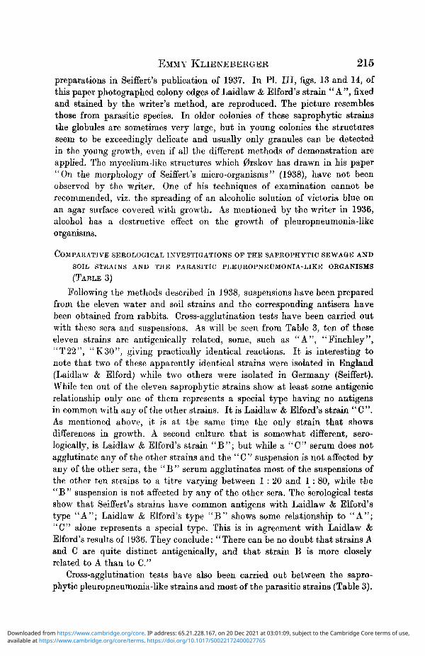
The cultural and morphological features of the L 5 organism also distinguishit from other species. The round colony of the L5 organism (PL IV, fig. 16)shows a well-marked, small, dark centre and a large peripheral zone in whichdelicate globular structures can usually be detected by a magnification of1 : 200, but not by 1 : 80. By high magnification (oil immersion lens) thefairly regular globular structure can be easily demonstrated in unstained andstained colonies. PL IV, fig. 19, shows a stained, highly magnified colony ofthe L5 organism. The thick central part of the colony has stained verydeeply, while the thin peripheral layer of globules has stained delicately.Different L5 immune sera prepared in rabbits gave a positive agglutinationreaction with the American and English L5 strains. The reaction was obtainedin the unusually high titre of 1 : 10240 (Table 3). Though the L5 sera agglu-tinated the homologous suspensions in these high dilutions, most heterologousspecies were not agglutinated at all by these sera, with the exception of theLl , L4 and L6 suspensions which showed a positive reaction in the dilutionsof 1 : 40 (L1, L4) and of 1 : 160 (L6). There was also positive cross agglutinationbetween the Ll , L4 and L6 sera and the L5 suspensions, but the titres ofthese positive reactions compared with that of the homologous sera andstrains were low (Table 3). These tests together with the animal experiments,the morphology and the colonial type, support the view that the L5 is aspecific type possessing, however, antigens related to those of Ll, L4 and L6.
(d) L 6—from brains of mice inoculated intracerebrally with blood ofsplenectomized mice containing Eperythrozoon coccoides
During work with splenectomized animals carried out in collaborationwith Findlay, Klieneberger, MacCallum & Mackenzie (1939) a second pleuro-pneumonia-like organism was recovered from mice. It was isolated six times fromthe brains of mice which had been injected intracerebrally with blood of splen-ectomized mice containing Eperythrozoon coccoides. The first suggestion thatEperythrozoon and this new organism, L 6, was one and the same thing has notbeen verified. It was not possible to recover L6 regularly from mice injected inthe way mentioned and all attempts to isolate it from the Eperythrozoon bloodfailed completely.
The L6 colony can be distinguished from that of the L5. It is usuallylarger and more irregular, and is further characterized by the presence ofvery large globules which can be seen by low magnification (1 : 80) and whichgive the surface of the colony a coarse structure (PL IV, fig. 17). The pictureof the more highly magnified colony edge is shown in a darkground photograph(PL IV, fig. 18; x490) and stained (PL IV, fig. 20; x600). The L5 growth
available at https://www.cambridge.org/core/terms. https://doi.org/10.1017/S0022172400027765Downloaded from https://www.cambridge.org/core. IP address: 65.21.228.167, on 20 Dec 2021 at 03:01:09, subject to the Cambridge Core terms of use,

EMMY KLIENEBERGER 213
is opalescent in liquid medium while the L6 often forms little clumps whichare chiefly composed of fairly large globules. The opalescent growth of theL5 is chiefly composed of small granules and small globules appearing as ringsin the darkground preparation. The L6 serum seems specific and does notagglutinate any of the other pathogenic species of the pleuropneumonia group.In contrast to this the L6 suspension is influenced to a certain degree by L3,L4 and L5 sera.
(e) "M55"—arthritis in miceIn addition to the new types, L5 and L6, a pleuropneumonia-like organism
was unexpectedly isolated from a joint of a mouse by Dr H. Jahn, workingin the writer's laboratory. This mouse had been used for an experiment withS. moniliformis but instead of this organism or the L1 an unknown pleuro-pneumonia-like organism was cultivated from the swollen joint. This neworganism, "M55", which has not been tested serologically and therefore notyet been assigned a number in the L series, causes arthritis and may be relatedto Sabin's organism from mouse arthritis.
The L5, L6 and "M55" types are the new pleuropneumonia-like organismsisolated from mice during the past year. Table 2 outlines the characteristicsof these organisms from mice.
Table 2. Organisms from mice
Growth in liquidL5
OpalescentL6 "M55"
In small clumps and Slightly opalescentopalescent
Colony (low mag. of Round, small, dark Flat, irregular, with Round, small, centremicroscope)
Source in mice
centre
Brain in "rolling dis-
Pathogenicity for mice Causes "rolling dis-ease "
Serology, see Table 3 Special type, but anti-genically related toLI, L4andL6
some large globules
Brain after intracere-bral injection ofEperythrozoon blood
Special type, but in-fluenced by L3, L4and L5 sera
and peripheral zonegranular
Swollen joint
Causes arthritis
Not vet examined
E. FURTHER STUDIES ON THE FILTERABLE ORGANISMS ISOLATEDFROM SEWAGE, SOIL, ETC.
Altogether eleven strains isolated from sewage, soil, etc. were studied.The writer obtained five strains isolated by Laidlaw & Elford (1936) fromthe National Collection of Type Cultures, which have been kept there since1936. Dr G. Seiffert kindly sent six other strains to the writer in 1937. Thedesignation of these eleven strains was as follows:
Strain "A" Isolated by Laidlaw & Elford in 1936it T> )J
)J -*-* j) 55 55
Finchley"Croydon"
available at https://www.cambridge.org/core/terms. https://doi.org/10.1017/S0022172400027765Downloaded from https://www.cambridge.org/core. IP address: 65.21.228.167, on 20 Dec 2021 at 03:01:09, subject to the Cambridge Core terms of use,
214 Pleuropneumonia-like organisms
Strain " B10 " Isolated by Seiffert in 1937
" T 1 "
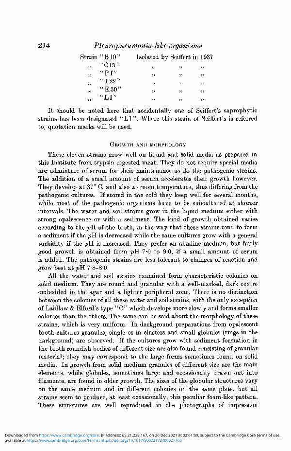
It should be noted here that accidentally one of Seiffert's saprophyticstrains has been designated " L I " . Where this strain of Seiffert's is referredto, quotation marks will be used.
GROWTH AND MORPHOLOGY
These eleven strains grow well on liquid and solid media as prepared inthis Institute from trypsin digested meat. They do not require special medianor admixture of serum for their maintenance as do the pathogenic strains.The addition of a small amount of serum accelerates their growth however.They develop at 37° C. and also at room temperature, thus differing from thepathogenic cultures. If stored in the cold they keep well for several months,while most of the pathogenic organisms have to be subcultured at shorterintervals. The water and soil strains grow in the liquid medium either withstrong opalescence or with a sediment. The kind of growth obtained variesaccording to the pH of the broth, in the way that these strains tend to forma sediment if the pK is decreased while the same cultures grow with a generalturbidity if the pH is increased. They prefer an alkaline medium, but fairlygood growth is obtained from pH. 7-0 to 9-0, if a small amount of serumis added. The pathogenic strains are less tolerant to changes of reaction andgrow best at pK 7-8-8-0.
All the water and soil strains examined form characteristic colonies onsolid medium. They are round and granular with a well-marked, dark centreembedded in the agar and a lighter peripheral zone. There is no distinctionbetween the colonies of all these water and soil strains, with the only exceptionof Laidlaw & Elford's type " C " which develops more slowly and forms smallercolonies than the others. The same can be said about the morphology of thesestrains, which is very uniform. In darkground preparations from opalescentbroth cultures granules, single or in clusters and small globules (rings in thedarkground) are observed. If the cultures grow with sediment formation inthe broth roundish bodies of different size are also found consisting of granularmaterial; they may correspond to the large forms sometimes found on solidmedia. In growth from solid medium granules of different size are the mainelements, while globules, sometimes large and occasionally drawn out intofilaments, are found in older growth. The sizes of the globular structures varyon the same medium and in different colonies on the same plate, but allstrains seem to produce, at least occasionally, this peculiar foam-like pattern.These structures are well reproduced in the photographs of impression
available at https://www.cambridge.org/core/terms. https://doi.org/10.1017/S0022172400027765Downloaded from https://www.cambridge.org/core. IP address: 65.21.228.167, on 20 Dec 2021 at 03:01:09, subject to the Cambridge Core terms of use,
EMMY KLIENEBERGER 215
preparations in Seiffert's publication of 1937. In PI. Ill , figs. 13 and 14, ofthis paper photographed colony edges of Laidlaw & Elford's strain "A", fixedand stained by the writer's method, are reproduced. The picture resemblesthose from parasitic species. In older colonies of these saprophytic strainsthe globules are sometimes very large, but in young colonies the structuresseem to be exceedingly delicate and usually only granules can be detectedin the young growth, even if all the different methods of demonstration areapplied. The mycelium-like structures which 0rskov has drawn in his paper"On the morphology of Seiffert's micro-organisms" (1938), have not beenobserved by the writer. One of his techniques of examination cannot berecommended, viz. the spreading of an alcoholic solution of victoria blue onan agar surface covered with growth.. As mentioned by the writer in 1936,alcohol has a destructive effect on the growth of pleuropneumonia-likeorganisms.
COMPARATIVE SEROLOGICAL INVESTIGATIONS OF THE SAPROPHYTIC SEWAGE AND
SOIL STRAINS AND THE PARASITIC PLEUROPNEUMONIA-LIKE ORGANISMS
(TABLE 3)
Following the methods described in 1938, suspensions have been preparedfrom the eleven water and soil strains and the corresponding antisera havebeen obtained from rabbits. Cross-agglutination tests have been carried outwith these sera and suspensions. As will be seen from Table 3, ten of theseeleven strains are antigenically related, some, such as "A", "Finchley","T22", "K30", giving practically identical reactions. It is interesting tonote that two of these apparently identical strains were isolated in England(Laidlaw & Elford) while two others were isolated in Germany (Seiffert).While ten out of the eleven saprophytic strains show at least some antigenicrelationship only one of them represents a special type having no antigensin common with any of the other strains. It is Laidlaw & Elford's strain "C".As mentioned above, it is at the same time the only strain that showsdifferences in growth. A second culture that is somewhat different, sero-logically, is Laidlaw & Elford's strain " B " ; but while a "C" serum does notagglutinate any of the other strains and the "C" suspension is not affected byany of the other sera, the "B " serum agglutinates most of the suspensions ofthe other ten strains to a titre varying between 1 : 20 and 1 : 80, while the" B " suspension is not affected by any of the other sera. The serological testsshow that Seiffert's strains have common antigens with Laidlaw & Elford'stype "A"; Laidlaw & Elford's type " B " shows some relationship to "A";"C" alone represents a special type. This is in agreement with Laidlaw &Elford's results of 1936. They conclude: "There can be no doubt that strains Aand C are quite distinct antigenically, and that strain B is more closelyrelated to A than to C."
Cross-agglutination tests have also been carried out between the sapro-phytic pleuropneumonia-like strains and most of the parasitic strains (Table 3).
available at https://www.cambridge.org/core/terms. https://doi.org/10.1017/S0022172400027765Downloaded from https://www.cambridge.org/core. IP address: 65.21.228.167, on 20 Dec 2021 at 03:01:09, subject to the Cambridge Core terms of use,
Tab
le 3
. R
esul
ts o
f cr
oss-
aggl
utin
atio
n te
sts
wit
h al
l st
rain
s of
the
ple
urop
neun
ioni
a gr
oup
in t
he w
rite
r's
coll
ecti
on
Ser
a ..
.
Sus
pens
ions
"A
" L
aidl
aw
2560
"Cr o
yd
on
" L
aidl
aw
40"F
inch
ley
" L
aidl
aw
320
"B
" L
aidl
aw
. —
"C
" L
aidl
aw"T
22
" Se
iffe
rt"
K3
0"
Seif
fert
"BI O
" Se
iffe
rt"
LI "
Se
iffe
rt"C
15
"Sei
f fer
t"P
f"
Seif
fert
Ple
urop
neum
onia
Aga
lact
iaL
I or
gani
smL
3 or
gani
smL
4 or
gani
smL
5 or
gani
smL
6 or
gani
smA
sler
oc.
cani
s I
Ast
troc
. ca
nis
II
§ I"
320
320
640
inchl
160
160
640
PQ 20 20 160
O —
1280
—
40—
32
0
« 5
_H
JHC
H ^
1a 2 3 so 3 • H
a .a s W3
o eo
a .2 § CO bo s
a s 'a bi s
640 40 160
40 40 40
10 20 40
20
—
—
4020
—
—
—
—
20
—
_ _
io
—
—
—
__
__
32
0—
—
—
—
—
—
320
640 40 40 40 40
80 80 20 20 10 20
40 40 80 40 40 80
40 20 40 80 40 40
320
320 80 40 40 40
160
160 20 20 10 10
20 10 640 80 320
320
10 2020 10 40 640 20 20
40 10 40 20 640 40
10
—
—
—
320 40 160
320
__
__
10
—
—
—
—
__
__
10
—
—
—
—
640
—
—
——
64
0 —
—
—
—
1280
—
—
—
—
320
80
4010
The
ave
rage
end
-tit
res
of t
he t
ests
whi
ch w
ere
desi
gnat
e d "
+ "
are
giv
en i
n th
e ta
ble.
available at https://www.cambridge.org/core/terms. https://doi.org/10.1017/S0022172400027765Downloaded from https://www.cambridge.org/core. IP address: 65.21.228.167, on 20 Dec 2021 at 03:01:09, subject to the Cambridge Core terms of use,

EMMY KLIENEBERGER 217
These reactions gave completely negative results with the one exception ofthe L4 serum which gave a slight positive result with five water and soilstrains including "C"; but the titres of these reactions were very low (1 : 10 forfour strains and 1 : 20 for strain "C") and none of the sera prepared withsaprophytic strains affected the L4 suspensions. It should be mentioned thatthe L4 organism is the one growth type of all the pathogenic strains whichshows a certain resemblance to the water and soil strains by its very granularcolony and by producing chiefly small elements.
None of the saprophytic strains produces lesions in laboratory animals,even if agar is added to the culture injected.
The morphological and serological examinations of allthepleuropneumonia-like organisms in the writer's possession show that with the one exception ofLaidlaw & Elford's strain "C" the other ten strains from water and soil arerelated organisms, while the pathogenic strains can be divided up into speciesdifferent both from one another and from the saprophytic organisms.Agglutinin-absorption tests have not yet been practised.
DISCUSSION
There exists a group of microbes of which the organisms of pleuropneumoniaof cattle and agalactia of sheep are the prototypes. Their morphology,development and possible life cycle have been widely discussed by differentauthors. Though agreement concerning their nature and development has notyet been reached and we are therefore not able to assign systematic rank tothem, it is possible for the student of these organisms, whenever a new typecomes to light, to recognize it as a member of the pleuropneumonia group.The author is of opinion that we are dealing with a distinct family whosemembers resemble each other closely, but differ widely from other knownmicrobes. An attempt is here made to outline plainly the characteristics ofthe family without attempting to give a definition of their possible nature ora description of their still hypothetical life cycle.
All members of the pleuropneumonia group form characteristic colonies,which grow up slowly and after some days reach a diameter varying between0-1 and 0-6 mm., the latter size being reached only by some giant colonies andonly in a few species. The colonies show usually a darker central zone embeddedin the medium and a lighter peripheral zone. If studied under the microscope,actually the only proper means for collecting information about the colonies,at magnifications of 30-200 they show either a granular or a coarser, globularstructure which distinguishes them from bacterial colonies. Nearly all of themdevelop a brownish tinge if they are isolated enough and if incubated for aprotracted period.
In an ordinary smear preparation fixed with heat or alcohol and stainedwith aniline dies such as fuchsin, methylene blue, gentian violet or by Gram'smethod formed elements such as bacilli, cocci, commas or spirochaetes cannotbe detected. Such a slide shows a cloudy, indistinct, faintly stained material
available at https://www.cambridge.org/core/terms. https://doi.org/10.1017/S0022172400027765Downloaded from https://www.cambridge.org/core. IP address: 65.21.228.167, on 20 Dec 2021 at 03:01:09, subject to the Cambridge Core terms of use,
218 Pleuropneumonia-like organisms
which looks as if derived from the culture medium. If staining methods areapplied which serve for the demonstration of virus particles some granulesmay or may not be found, but no picture showing large numbers of smallelements like the elementary bodies of certain viruses indicates the presenceof numerous elements of a micro-organism. Yet each colony of any pleuro-pneumonia-like organism consists of numerous very small and fairly largeelements. Many of them can be demonstrated in a carefully carried outimpression preparation, fixed with alcohol after drying, and stained withGiemsa (Ledingham, 1933). The agar fixation method (Klieneberger, 1934) isa modified impression technique. Its advantage is that whole small coloniescan be fixed and stained in situ without considerable dislocation of singleelements so that a fairly true picture of the actual growth is obtained. Aneven simpler and rather conclusive method, though not revealing the finestdetails, is the direct agar microscopy (0rskov, 1927). All magnificationsincluding oil immersion systems can be used to study the growth on top ofan agar plate or better on a slide culture. Dienes (1939 a) used a direct agarmicroscopy in combination with staining methods, which furnished him withexcellent preparations. These methods, together with the darkground obser-vations, show that all pleuropneumonia-like organisms produce a large amountof very small forms which are of slightly different sizes and shapes. These"granules" often stain deeply as if consisting of concentrated material. It hasbeen stated by most authors that the organisms of pleuropneumonia andagalactia form filaments, because granules with filaments attached have oftenbeen observed; but the actual germination of granules has so far not beenfollowed up satisfactorily. Filamentous formations occur in all members ofthe pleuropneumonia group. They are frequent in pleuropneumonia, agalactiaand the L1 organism and rare in others, such as the saprophytic strains andthe L4 organism; but occasionally they are present in all members of thefamily. They occur not only in connexion with granules, but also free and inconnexion with globules or large bodies, which represent the third type ofelement composing the growth of pleuropneumonia-like organisms. Two kindsof globules or bodies seem to be present. One kind stains well, sometimes verydeeply, purple or bluish with Giemsa solution while the other seems to containa liquid. Consequently often only its contours can be seen or stained; butsometimes the surface shows up, very lightly stained. If many of theseglobules are present in a colony a honeycomb or cellular structure is producedwhich can be mistaken for a meshwork of filaments. This mistake of inter-pretation has been made in the earlier papers of the present writer on'pleuropneumonia, agalactia and the LI. The so-called "network structures"are in reality produced by the contours of globules densely packed together.In the same way the ring forms, so often mentioned in studies on pleuro-pneumonia and related organisms, must be interpreted as contours of globules.If they turn over in darkground preparations they always show ring formsand therefore must be regarded as outlines of corpuscles or globules.
available at https://www.cambridge.org/core/terms. https://doi.org/10.1017/S0022172400027765Downloaded from https://www.cambridge.org/core. IP address: 65.21.228.167, on 20 Dec 2021 at 03:01:09, subject to the Cambridge Core terms of use,
EMMY KLIENEBERGER 219
The different elements present in pleuropneumonia-like organisms can bewell demonstrated, but how they grow up and develop is not yet clearlyunderstood. Nevertheless, the members of the group are so well characterizedby their morphology and colony type that it seems reasonable to regard themas belonging to a distinct family different from all other Schizomycetales.
Organisms of the pleuropneumonia group have been found in symbiosiswith bacteria. In two cases a separation of the pleuropneumonia-like organismfrom the parent culture has been achieved, viz. in the case of Streptobacillusmoniliformis and in the case of the so-called "guinea-pig culture", obtainedfrom cervical lymphadenitis in these rodents.
Organisms of the pleuropneumonia group have been found independentlyand not in association with bacteria in materials such as sewage and soil,showing that these microbes are able to lead a saprophytic existence. Mostof the organisms so far found in such materials are related and are representedby Laidlaw & Elford's type "A".
A number of different types of pleuropneumonia-like organisms have beenfound in lesions of animals. In most cases they have been shown to causeinfection in these animals under certain experimental conditions such as, forexample, when agar is mixed with a suspension of the organisms. The sameis true also of the organism of pleuropneumonia (Mettam & Ford, 1939;Daubney, 1936). In addition to the organisms of pleuropneumonia of cattleand agalactia of sheep the following independent pathogenic types have beendescribed since 1933: Asterococcus canis type I and type II (Shoetensack, 1934),the L3 organism in pulmonary lesions of rats (Klieneberger & Steabben, 1937),the L4 organism in swollen glands, abscesses and swollen joints of rats(Klieneberger, 1938, Findlay, Mackenzie et al. 1939; Woglom & Warren, 1939),the L5 organism causing "rolling disease" in mice (Findlay et al. 1938; Sabin,1939 a), the L6 organism in brains of mice after intracerebral injection ofblood of splenectomized mice (Findlay, Klieneberger et al. 1939), and lastlyan organism causing swollen joints in mice (Sabin, 19396, Klieneberger,this paper). These filterable organisms of the pleuropneumonia group are offrequent occurrence in rats and mice and their presence must undoubtedlycomplicate virus research in these animals.
SUMMARY
1. The four different L1 strains in the writer's possession isolated between1933 and 1936 have been maintained for a further period without revertingto Streptobacillus moniliformis. The supposition that the LI is a pleuro-pneumonia-like organism and lives in symbiosis with a bacterium in culturesof S. moniliformis is still maintained by the writer and the reasons for itare given.
2. From a culture isolated from lesions in guinea-pigs and pathogenic forthese animals called the "guinea-pig strain", an Ll-like organism of thepleuropneumonia group has been separated, but not yet been maintained for
available at https://www.cambridge.org/core/terms. https://doi.org/10.1017/S0022172400027765Downloaded from https://www.cambridge.org/core. IP address: 65.21.228.167, on 20 Dec 2021 at 03:01:09, subject to the Cambridge Core terms of use,

220 Pleuropneumonia-like organisms
a long enough period to ensure irreversibility. The morphology of the ''guinea-pig strain" shows that it is closely related to *S. moniliformis though it canbe distinguished from the latter by its cultural and pathogenic properties.The morphology of two other cultures, viz. a saprophytic coccus from theskin of a pig and the organism known as Fusobacterium niccleatum, have beendescribed as possibly representing similar symbiotic associations of bacteriaand pleuropneumonia-like organisms.
3. Cultural and serological differences between the L4 causing arthritis,swollen glands and abscesses in rats, and the two other pleuropneumonia-likeorganisms from rats, LI and L3, are recorded. It has been shown that theorganisms occurring in the brains of mice, L5 and L6, and in the joint of amouse, "M55", are of aetiological significance for the condition in which theyoccur. At the same time they differ in their colony type, morphology andserological features.
4. Morphological and serological studies of the saprophytic organisms ofthe pleuropneumonia group isolated by Laidlaw & Elford and Seiffert fromwater and soil show that Seiffert's organisms are closely related to Laidlaw& Elford's type "A". In agreement with Laidlaw & Elford it was found thattheir type " B " is slightly different from "A" serologically, while "C" isdistinct from "A" and " B " and may be regarded as a special type.
These saprophytic types are not antigenically related to the parasiticvarieties.
My thanks are due to Sir John Ledingham for his continued interest in thiswork and his greatly appreciated criticism. I am indebted to Dr C. F. Robinowfor a number of photographs and should like to express my thanks for hisco-operation in this matter.
REFERENCESCOLLIER, W. A. (1939). J . Path. Bad. 48, 579.DAUBNEY, R. (1936). J. comp. Path. 48, 83.DAWSON, M. H. & HOBBY, G. (1939a). Abstr. of Proc. Third Int. Congr. Microbiol., N.Y.,
p. 21.(19396). Trans. Ass. Amer. Phys. 54, 329.
DIENBS, L. (1938). Proc. Soc. exp. Bid., N.Y., 39, 365.(1939a). J. infect. Dis. 65, 24.
• (19396). Proc. Soc. exp. Biol., N.Y., 42, 636.FARRELL, E., LORDI, G. H. & VOGEL, J. (1939). Arch, intern. Med. 64, 1.FINDLAY, G. M., KLIENEBERGER, E., MACCALLUM, F. 0. & MACKENZIE, R. D. (1938).
Lancet, 2, 1511.(1939). Trans. R. Soc. trop. Med. Hyg. 33 , 6.
FINDLAY, G. M., MACKENZIE, R. D., MACCALLUM, F. 0. & KLIENEBERGER, E. (1939).
Lancet, 2, 7.KLIBNEBERGER, E. (1934). J. Path. Bad. 39, 409.
(1935). J. Path. Bad. 40, 93.—— (1936). J. Path. Bad. 42, 587.
available at https://www.cambridge.org/core/terms. https://doi.org/10.1017/S0022172400027765Downloaded from https://www.cambridge.org/core. IP address: 65.21.228.167, on 20 Dec 2021 at 03:01:09, subject to the Cambridge Core terms of use,
EMMY KLIENEBERGER 221KLIENEBERGER, E. (1938). J. Hyg., Gamb., 38, 458.
(1939a). J. Hyg., Camb., 39, 260.(19396). J. Path. Bad. 49, 451.(1939c). Abstr. of Proc. Third Int. Congr. Microbiol, N.Y., p. 20.
KLIENEBERGER, E. & STEABBEN, D. B. (1937). J. Hyg., Camb., 37, 143.LAIDLAW, P. P. & ELFORD, W. J. (1936). Proc. ray. Soc. B, 120, 292.LEDINGHAM, J. C. G. (1933). J. Path. Bad. 37, 393.LEVTNTHAL, W. & FERNBACH, H. (1922). Z. Hyg. InfektKr. 96, 456.METTAM, R. W. M. & FORD, J. (1939). J. comp. Path. 52, 15.0RSKOV, J. (1927). Ann. Inst. Pasteur, 48, 308.
(1938). ZU. Bait. I. Abt. Orig. 141, 230.RHODES, A. J. & VAN ROOYEN, C. E. (1939). J. Path. Bad. 49, 577.SABIN, A. B. (1938). Science, 88, 575.
(1939a). Science, 89, 228.(19396). Science, 90, 18.
SEASTONE, C. V. (1939). J. exp. Med. 70, 347.SEIPFERT, G. (1937 a). ZU. Bakt. I. Abt. Orig. 139, 337.
(19376). ZU. Bakt. I. 140, Beiheft, 168.SHOETENSACK, H. M. (1934). Kitasato Arch. 11,277.
(1936a). Kitasato Arch. 13, 145.(19366). Kitasato Arch. 13, 269.
WOGLOM, W. H. & WARREN, J. (1938a). Science, 87, 370.(19386). J. exp. Med. 68, 513.(1939). J. Hyg., Camb., 39, 266.
EXPLANATION OF PLATES II-IVPLATE II
Fig. 1. L\ organism, grown on a coverslip in a mixture of 1 part rabbit plasma, 1 part spleenextract and 3 parts of special liquid medium; 4 days 37° C. The globular edge of this colonyshows extremely large forms. Photographed alive by Dr Robinow; mag. 1134.
Fig. 2. LI organism, grown on a coverslip in special liquid medium; 3 days 37° C. Many globularforms. Photographed alive, darkground illumination; mag. 900.
Fig. 3. L1 organism, edge of stained colony, grown on solid medium, showing large globules withtheir delicate contours at the edge. Agar fixation method, Bouin-Giemsa; mag. 600.
Fig. 4. S. moniliformis, grown on a coverslip in special liquid medium, 3 days 37° C. The bacillarychains containing granular structures and the large globules lying apart can be distinctlyrecognized. Photographed alive by Dr Robinow; mag. 1134.
Fig. 5. Organism of agalactia, part of stained colony showing globules of different size. Agarfixation method, Flemming-Giemsa; mag. 600.
PLATE IIIFig. 6. "Guinea-pig strain" colony grown up from pus spread on the surface of special medium
after 6 days of incubation. Some big globules can be seen. Photographed alive by DrRobinow; mag. 90.
Fig. 7. Ll-like organism isolated from the "guinea-pig culture". The large, dark, granularcentres of the pleuropneumonia-like colonies and the lighter, peripheral zones with theirlace-like structures are to be seen. Photographed alive by Dr Robinow; mag. 90.
Fig. 8. Part of one of these same L 1-like colonies (see fig. 7) higher magnified; its globular formscan be well distinguished. Photographed alive by Dr Robinow; mag. 486.
Fig. 9. Colony of "guinea-pig strain" stained. The bacillary filaments, large unstained globulesand small stained bodies show up. Agar fixation method, Bouin-Giemsa; mag. 900.
Fig. 10. Fusobacterium nucleatum grown anaerobieally on serum agar; a mixture of bacillarychains and globular forms can be seen. Agar fixation method, Bouin-Giemsa; mag. 900.
J. Hygiene 40 15
available at https://www.cambridge.org/core/terms. https://doi.org/10.1017/S0022172400027765Downloaded from https://www.cambridge.org/core. IP address: 65.21.228.167, on 20 Dec 2021 at 03:01:09, subject to the Cambridge Core terms of use,

222 Phuropneumonia-like organismsFig. 11. Fusobacterium nucleatum grown aerobically in serum broth for 15 hr. The globular forms
and a few bacillary chains can be seen. Photographed alive, darkground illumination;mag. 900.
Fig. 12. Fusobacterium nucleatum, edge of an anaerobically grown colony showing only globularforms. Agar fixation method, Bouin-Giemsa; mag. 900.
Fig. 13. Sewage organism "A". Stained colony edge. Agar fixation method, Bouin-Giemsa;mag. 600.
Fig. 14. The same, but different colony edge; mag. 900.Fig. 15. Asterococcus canis, Shoetensack, type II. Stained colony edge, showing large globules,
differently stained. Agar fixation method, Bouin-Giemsa; mag. 600.
PLATE IVFig. 16. £5 organism, colonies on solid medium, 3 days 37° C. The small, marked dark centre
and the delicately structured, peripheral zone can be recognized. Photographed alive byDr Robinow; mag. 90.
Fig. 17. L6 organism, colonies on solid medium, 4 days 37° C. The globular surface structureshows up. Photographed alive by Dr Robinow; mag. 90.
Fig. 18. Ld organism, edge of a colony grown on solid medium, darkground illumination, photo-graphed without application of a coverslip by Dr Robinow; mag. 490.
Fig. 19. L5 organism, stained, the marked dark centre and the delicate, globular fringe arecharacteristic for the colonies of this organism. Agar fixation method, Bouin-Giemsa;mag. 600.
• Fig. 20. £6 organism, stained colony edge showing a coarser globular structure than the colonyof the L5 organism (fig. 19). Agar fixation method, Bouin-Giemsa; mag. 600.
(Received for -publication 25. i. 1940—Ed.)
available at https://www.cambridge.org/core/terms. https://doi.org/10.1017/S0022172400027765Downloaded from https://www.cambridge.org/core. IP address: 65.21.228.167, on 20 Dec 2021 at 03:01:09, subject to the Cambridge Core terms of use,
JOURNAL OF HYGIENE, VOL. 40, NO. 2 PLATE
. 4 <
available at https://www.cambridge.org/core/terms. https://doi.org/10.1017/S0022172400027765Downloaded from https://www.cambridge.org/core. IP address: 65.21.228.167, on 20 Dec 2021 at 03:01:09, subject to the Cambridge Core terms of use,
JOURNAL OF HYGIENE, VOL. 40, NO. 2 PLATE
available at https://www.cambridge.org/core/terms. https://doi.org/10.1017/S0022172400027765Downloaded from https://www.cambridge.org/core. IP address: 65.21.228.167, on 20 Dec 2021 at 03:01:09, subject to the Cambridge Core terms of use,

JOURNAL OF HYGIENE, VOL. 40, NO. 2 PLATE IV
17
19
20
available at https://www.cambridge.org/core/terms. https://doi.org/10.1017/S0022172400027765Downloaded from https://www.cambridge.org/core. IP address: 65.21.228.167, on 20 Dec 2021 at 03:01:09, subject to the Cambridge Core terms of use,