Embed Size (px)
Citation preview
on July 9, 2018http://rsob.royalsocietypublishing.org/Downloaded from
rsob.royalsocietypublishing.org
ResearchCite this article: Yurchenko T, Sevcıkova T,
Strnad H, Butenko A, Elias M. 2016 The plastid
genome of some eustigmatophyte algae
harbours a bacteria-derived six-gene cluster for
biosynthesis of a novel secondary metabolite.
Open Biol. 6: 160249.
http://dx.doi.org/10.1098/rsob.160249
Received: 30 August 2016
Accepted: 31 October 2016
Subject Area:genomics/microbiology/biochemistry/
biotechnology/bioinformatics
Keywords:Eustigmatophyceae, horizontal gene transfer,
plastid genome, secondary metabolism,
sugar phosphate cyclase superfamily,
UbiA superfamily
Author for correspondence:Marek Elias
e-mail: [email protected]
Electronic supplementary material is available
online at https://dx.doi.org/10.6084/m9.fig-
share.c.3573258.
& 2016 The Authors. Published by the Royal Society under the terms of the Creative Commons AttributionLicense http://creativecommons.org/licenses/by/4.0/, which permits unrestricted use, provided the originalauthor and source are credited.
The plastid genome of someeustigmatophyte algae harbours abacteria-derived six-gene cluster forbiosynthesis of a novel secondarymetabolite
Tatiana Yurchenko1,2, Tereza Sevcıkova1, Hynek Strnad3, Anzhelika Butenko1
and Marek Elias1,2
1Faculty of Science, Department of Biology and Ecology, Life Science Research Centre, and2Faculty of Science, Institute of Environmental Technologies, University of Ostrava, Chittussiho10, 710 00 Ostrava, Czech Republic3Institute of Molecular Genetics of the ASCR, v. v. i., Prague, Czech Republic
ME, 0000-0003-0066-6542
Acquisition of genes by plastid genomes (plastomes) via horizontal gene
transfer (HGT) seems to be a rare phenomenon. Here, we report an interest-
ing case of HGT revealed by sequencing the plastomes of the
eustigmatophyte algae Monodopsis sp. MarTras21 and Vischeria sp. CAUP
Q 202. These plastomes proved to harbour a unique cluster of six genes,
most probably acquired from a bacterium of the phylum Bacteroidetes,
with homologues in various bacteria, typically organized in a conserved
uncharacterized putative operon. Sequence analyses of the six proteins
encoded by the operon yielded the following annotation for them: (i) a
novel family without discernible homologues; (ii) a new family within the
superfamily of metallo-dependent hydrolases; (iii) a novel subgroup of the
UbiA superfamily of prenyl transferases; (iv) a new clade within the sugar
phosphate cyclase superfamily; (v) a new family within the xylose isomer-
ase-like superfamily; and (vi) a hydrolase for a phosphate moiety-
containing substrate. We suggest that the operon encodes enzymes of a path-
way synthesizing an isoprenoid–cyclitol-derived compound, possibly an
antimicrobial or other protective substance. To the best of our knowledge,
this is the first report of an expansion of the metabolic capacity of a plastid
mediated by HGT into the plastid genome.
1. BackgroundEustigmatophytes are a small yet expanding class of stramenopile algae
(ochrophytes), nowadays perceived as a particularly interesting target for bio-
technological research and exploitation thanks to the ability of some of the
members to produce high amounts of lipid substances as energy and carbon
reserves, promising for biofuel production [1,2]. This has driven research on
eustigmatophytes into the genomics era, resulting in a number of completed
genome sequencing projects [3–6]. They, however, targeted only members of
the genus Nannochloropsis, recently divided into two separate (but related)
genera: Nannochloropsis (sensu stricto) and Microchloropsis [7]. These projects
have also yielded sequences of organellar (both plastid and mitochondrial) gen-
omes, which are now available for nearly all species of the traditionally
circumscribed genus Nannochloropsis [3,8,9].
However, recent surveys revealed a surprising phylogenetic diversity of
the class Eustigmatophyceae [10,11]. This prompted us to initiate a more
rsob.royalsocietypublishing.orgOpen
Biol.6:160249
2
on July 9, 2018http://rsob.royalsocietypublishing.org/Downloaded from
comprehensive investigation of eustigmatophytes at the
genomic level. The first fruit of these efforts was sequencing
the plastid genome (plastome for short) of Trachydiscus min-utus [12], a member of a recently recognized major
eustigmatophyte clade, Goniochloridales, which is deeply
diverged from the group comprising ‘traditional’ eustigmato-
phytes including Nannochloropsis [10,13]. More recently, we
reported results of a comparative analysis of eustigmatophyte
mitochondrial genomes, including newly sequenced gen-
omes of three phylogenetically diverse species, T. minutus,
Vischeria sp. CAUP Q 202 and Monodopsis sp. MarTras21
[14]. In this study, we describe complete plastome sequences
of Vischeria sp. CAUP Q 202 and Monodopsis sp. MarTras21
(for a schematic tree showing the relative phylogenetic pos-
ition of the different eustigmatophytes discussed in this
study see figure 1). As discussed below, analyses of these
two genomes uncovered an unexpected evolutionary event
that nominates the respective eustigmatophytes to a position
of highly interesting candidates for further biochemical
investigations with promise towards biotechnological
applications.
Plastomes have evolved from the genome of the cyano-
bacterial progenitor that gave rise to plastids through the
process of primary endosymbiosis dated between 1.7 and
0.9 billion years ago [15]. The primary mode of genome evol-
ution in plastids was reduction, partly outright gene loss,
partly relocation of the genes to the nuclear genome of the
host eukaryotic cell via the process of endosymbiotic gene
transfer [16]. As a result, the most gene-rich extant plastomes,
found in red algae (rhodophytes), contain only up to around
250 protein-coding genes [17]. However, an opposite process,
i.e. gene gain, has also been documented as a factor shaping
the plastome gene repertoire. This includes not only emer-
gence of extra genes by gene duplication [18,19], but
notably also horizontal gene transfer (HGT) from foreign
genomes.
The number of known instances of plastid genes gained
by HGT is growing, but is still quite limited. The first
documented HGT cases affecting plastid genomes were
replacements of original cyanobacterial genes: of the
RuBisCO operon by a Proteobacteria-derived operon in red
algae [20] and of the rpl36 gene in haptophytes and crypto-
phytes by an isoform of the same gene gained from a
bacterial donor [21]. Nevertheless, HGT can result in gains
of new genes not normally present in plastomes. Interest-
ingly, the majority of known foreign genes in plastid
genomes of various algal groups encode proteins operating
on DNA or RNA, including DNA polymerases, recombi-
nases, integrases, reverse transcriptases or maturases
[18,22–27]. The significance of these genes for the functioning
of the plastid genome and plastid as a whole remains unclear,
but many of these acquisitions apparently relate to the
activity of various mobile genetic elements, including group
II introns [24,28], transposons [26] and plasmids [27]. Plas-
tomes of several angiosperms were shown to harbour
segments derived from mitochondrial genomes [29,30], but
the functional significance of these elements is also unknown.
Much rarer are the examples of HGT-derived plastid
genes conferring a clear metabolic function. We are aware
of only one such case, specifically the presence of the cluster
of the leuC and leuD genes in the plastome of the red alga
Gracilaria tenuistipitata var. liui [31]. These genes encode the
large and small subunits of 3-isopropylmalate dehydratase,
an enzyme involved in leucine biosynthesis, and were appar-
ently moved to the plastome of the G. tenuistipitata lineage
by HGT from a eubacterium. In contrast to other red
algae, G. tenuistipitata may lack a nuclear genome-encoded
plastid-targeted 3-isopropylmalate dehydratase, and the
authors suggested that this loss has been complemented by
the acquisition of the leuC and leuD genes by the plastome.
However, to the best of our knowledge, there has been no
documented case of an acquisition of genes by a plastid
genome that would expand the metabolic capacity of the
respective plastid.
We were, therefore, highly surprised to find out that the
newly sequenced plastid genomes of Vischeria sp. CAUP Q
202 and Monodopsis sp. MarTras21 contain a novel six-gene
cluster encoding proteins homologous to various enzymes
from bacteria. Orthologues of these genes could not be ident-
ified in other plastomes, including those previously reported
from eustigmatophytes (i.e. those from T. minutus and Nanno-chloropsis/Microchloropsis spp.). This finding points to a
peculiar case of HGT from bacteria to the plastid genome of
a eustigmatophyte lineage that has endowed these algae
with a new metabolic pathway. Our sequence and phyloge-
netic analyses described below indicated that the gene
cluster, a putative novel operon, is widespread in many
groups of bacteria and that at least five of the proteins
encoded by the operon represent novel enzymes of different
(super)families. While it is impossible to make a specific pre-
diction about the function of these enzymes and the operon
as a whole, the data available suggest that the eustigmato-
phytes and bacteria endowed with the operon may have
the capacity to synthesize a novel secondary metabolite that
may prove to be a practically interesting bioactive compound.
2. Material and methods2.1. Sequencing and assembly of plastid genomes of
Vischeria sp. CAUP Q 202 and Monodopsis sp.MarTras21
Obtaining draft genome sequence data from Vischeria sp.
CAUP Q 202 and Monodopsis sp. MarTras21 was described
previously [14]. Briefly, 454 and Illumina reads (in the case
of Vischeria sp.) or Illumina reads only (in the case of Mono-dopsis sp.) generated from total DNA isolated from the
respective algal cultures were assembled to obtain a mixed
assembly of the nuclear and organellar genomes. Scaffolds
representing the plastid genome were identified by tblastn
[32] using as queries protein sequences encoded by the pre-
viously sequenced plastid genome of the eustigmatophyte
T. minutus. Two and three such scaffolds were found in the
Vischeria sp. and the Monodopsis sp. assemblies, respectively.
Assuming the common presence of inverted repeats in plas-
tomes and using the original sequencing reads to carry out
a targeted reassembly, we obtained complete circular-map-
ping sequences of both plastid genomes.
Both assemblies were validated by the visual inspection
of reads mapped onto the assembled consensus. To this
end, Illumina genomic reads for Vischeria sp. CAUP Q 202
and Monodopsis sp. MarTras21 were subjected to trimming
and quality filtering using CLC GENOMICS WORKBENCH
v. 8.0.3 (CLC Inc, Aarhus, Denmark) with the following

Trachydiscus minutus
Vischeria sp. CAUP Q 202
Monodopsis sp. MarTras21
Microchloropsis salina
Nannochloropsis oculata
trnRacpPebooperon
acpPpsbZ
ycf36
rbcRtrnLtsf
ebooperon
psbW
Goniochloridales
Eustigmatales
- gene gain- gene loss- pseudogenization
Figure 1. Schematic phylogeny of eustigmatophytes showing the position of species with sequenced plastid genomes. The taxa with plastomes sequenced in thisstudy are highlighted in bold. For the genera Nannochloropsis and Microchloropsis, only the type species are shown for simplicity, although plastomes have beensequenced for a number of other closely related species or strains. The topology of the tree reflects a robustly resolved phylogeny based on the 18S rRNA gene [14].Evolutionary events impacting the gene content of eustigmatophyte plastid genomes are mapped onto the tree based on the most parsimonious interpretation ofthe pattern of the gene presence/absence ( provided in the electronic supplementary material, table S1).
rsob.royalsocietypublishing.orgOpen
Biol.6:160249
3
on July 9, 2018http://rsob.royalsocietypublishing.org/Downloaded from
settings: regions with Phred quality less than 20 were
trimmed, no more than one N was allowed in the remaining
sequence, then TruSeq adapter trimming and a minimum
length threshold of 75 nt were applied. Filtered reads were
mapped to the assembled plastid genome sequences using
BOWTIE2 v. 2.2.5 [33] with ‘–end-to-end’ and ‘–very-sensitive’
options. Visual inspection of the resulting read mappings in
INTEGRATIVE GENOMICS VIEWER [34] validated both assemblies
and led to a correction of a few indel errors in homopoly-
meric tracks.
2.2. Annotation of the Vischeria sp. CAUP Q 202 andMonodopsis sp. MarTras21 plastid genomes
An initial annotation of the two new plastome sequences was
obtained using MFannot (http://megasun.bch.umontreal.
ca/cgi-bin/mfannot/mfannotInterface.pl). Prediction of
individual genes was checked by comparison to homologous
sequences (primarily from other eustigmatophytes), which
led us to revise the definition of the actual initiation codons
of some of the genes. 50 and 30 ends of genes for non-
coding RNAs (rRNAs and tRNAs) were likewise checked
and adjusted by comparison to orthologous genes from
other eustigmatophytes. Predicted intergenic regions were
translated in all six frames and the conceptual translations
were used as queries in blastp searches [32] against the
non-redundant (nr) protein sequence database at the
National Center for Biotechnology Information (NCBI;
http://blast.ncbi.nlm.nih.gov/Blast.cgi). In parallel, the poss-
ible presence of protein-coding genes known to reside in
previously sequenced eustigmatophyte plastid genomes,
but not predicted by MFannot in the two newly sequenced
genomes, was tested by tblastn searches against the respect-
ive plastid genome assemblies using the respective protein
sequences from other eustigmatophytes. This led to the
identification of some short genes missed by MFannot. A pre-
viously missed ssrA gene was identified in all
eustigmatophyte plastid genomes thanks to the comparison
of the plastid gene repertoire in different ochrophytes; its
borders were delimited according to a published annotation
of the gene in diatom plastomes. We also identified and
incorporated into the annotation an intron-interrupted leu-
tRNA gene that is conserved in eustigmatophyte plastomes
[12] yet was missed by MFannot. Circular genome maps
were generated with OGDRAW v.1.2 [35]. A list of genes ident-
ified in plastomes of Vischeria sp. CAUP Q 202, Monodopsissp. MarTras21 and selected other eustigmatophytes and
ochrophytes is provided in the electronic supplementary
material, table S1.
2.3. Identification of ebo gene homologues andanalyses of bacterial ebo operons
Protein sequences encoded by the six genes of the novel ebooperon from Vischeria (for the definition of the operon, see
§3.2) were used as queries for blastp of the NCBI nr protein
sequence database. A total of 100 best hits for each query
were downloaded and arrayed according to their taxonomic
provenance, yielding 148 different species or strains as the
main set for subsequent analyses. For each taxon in the list,
the presence of homologues (orthologues) of all six ebogenes was checked. In cases where these homologues were
not present in the initial set, we carried out dedicated
blastp or tblastn searches against the genome of the respect-
ive species or evaluated by blastp or phylogenetic analyses
the genes neighbouring in the genome the initially identified
ebo genes. In several cases, some ebo genes were not rep-
resented by protein sequence records owing to annotation
errors. Some ebo gene sequences proved to be partial owing
to incomplete genome assembly or interrupted owing to
frame-shifts. PSI-blast searches [32] were used to check
those organisms for which some ebo genes could not be ident-
ified by the approach described above, but their genuine
absence was confirmed in most cases. The relative genomic
position of the ebo genes identified for each species (strain)
was established using an in-house Python program. The elec-
tronic supplementary material, table S2, provides a list of the
148 taxa analysed, together with sequence identifiers of their
rsob.royalsocietypublishing.orgOpen
Biol.6:160249
4
on July 9, 2018http://rsob.royalsocietypublishing.org/Downloaded from
Ebo proteins and the spatial arrangement of ebo genes in their
genomes (i.e. the architecture of their ebo operons).
A broader analysis was carried out to investigate the
origin of eboF homologues in eukaryotes other than eustigma-
tophytes. To assemble a maximally comprehensive set of
eukaryotic eboF sequences, we searched by blastp or tblastn
not only sequence databases at NCBI, but also two extensive
resources of assembled transcriptomes of diverse microbial
eukaryotes and plants, the Marine Microbial Eukaryote
Transcriptome Sequencing Project (MMETSP; http://data.
imicrobe.us/project/view/104 [36]) and the 1000 Plants
project (OneKP, https://sites.google.com/a/ualberta.ca/
onekp/). In addition, eboF homologues were also identified
in the transcriptomes of Phaeothamnion confervicola and Schizo-cladia ischiensis (http://www.research.kobe-u.ac.jp/rcis-ku-
macc/e.p.folder/e.i.folder/download.html [37]). Deduced
sufficiently complete protein sequences of obvious eboF hom-
ologues were kept for further analysis. To detect possible
closest bacterial relatives of the eukaryotic eboF genes, we
searched the NCBI nr protein database with blastp using as
a query a representative EboF sequence for each main eukary-
otic group. A total of 100 best hits for each query were
compared to the set of EboF homologues from the 148 bac-
terial species (strains) selected in the eustigmatophyte-
centred analysis described above (electronic supplementary
material, table S2) and non-redundant sequences were
retained for a subsequent phylogenetic analysis combining
all (eukaryotic and bacterial) EboF sequences thus identified
(see the electronic supplementary material, table S3 for the
complete list).
2.4. Phylogenetic analysesAll phylogenetic analyses reported in this study were based
on the alignments of protein sequences built using MAFFT
v7 [38] and further processed by removing unreliably aligned
or too divergent regions using the GBLOCKS 0.91b program
http://molevol.cmima.csic.es/castresana/Gblocks_server.html
[39]) with the settings keeping the maximal number of pos-
itions in the final alignment. The alignments were analysed
using the maximum-likelihood (ML) method as implemented
in RAxML-HPC BLACKBOX (7.3.2) [40] at the CIPRES Portal
(http://www.phylo.org/sub_sections/ portal/, Cyberinfras-
tructure for Phylogenetic Research, San Diego
Supercomputing Center [41]). The LG4X substitution model
[42] was used for analyses of individual protein families,
except for the analysis of the large dataset covering the
whole sugar phosphate cycle superfamily (see §3.4.2),
which was analysed using the less computationally demand-
ing LG model. The rapid bootstrapping algorithm (with the
optimal number of bootstrap replicates chosen by the pro-
gram) was employed to assess the robustness of the tree
topologies. A multigene phylogenetic analysis was carried
out to improve the resolution of the analysis. Individual
Ebo proteins from eustigmatophytes, Bacteroidetes, Cyano-
bacteria and Leptospira spp. were aligned (in the rare cases
with two paralogues of some ebo genes, picking only the
one integrated in the operon or behaving more consistently
with the ebo genes from the same species in single-gene
trees) and concatenated using FASCONCAT [43]. An ML infer-
ence was done as above, but employing the general time
reversible (GTR) model, empirical base frequencies, and con-
sidering individual Ebo proteins as separate partitions. All
phylogenetic trees were displayed and adjusted using ITOL
(http://itol.embl.de/; [44]), with the final graphical proces-
sing done using INKSCAPE 0.91 (Free Software Foundation
Inc., Boston, USA). Details specific for individual phyloge-
netic analyses are described below and in the legends to
respective figures.
2.5. Analyses of the identity of Ebo proteinsFunctional annotation of ebo genes was primarily attempted
by the inspection of sufficiently close homologues retrieved
in searches against the NCBI nr protein database with repre-
sentative Ebo proteins. In addition to blastp, PSI-blast was
used for some Ebo proteins using a conservative inclusion
E-value threshold of 1 � 1025. The Kyoto Encyclopedia of
Genes and Genomes (KEGG) database [45] was also searched
with blastp (http://www.genome.jp/tools/blast/) to find
possible Ebo protein homologues with experimentally vali-
dated biochemical function. Representative Ebo proteins
were additionally compared to collections of profile Hidden
Markov Models (HMMs) in the Pfam 30.0 (http://pfam.
xfam.org/ [46]) and the SUPERFAMILY 1.75 (http://
supfam.org/SUPERFAMILY/ [47]) databases. In cases
where Pfam and/or SUPERFAMILY searches did not retrieve
significantly similar hits (EboA, EboE), we used HHpred
(https://toolkit.tuebingen.mpg.de/hhpred [48]) to identify
possible remote homologues using the more sensitive
HMM–HMM comparison. HHalign (https://toolkit.tuebin-
gen.mpg.de/hhalign [48]) was used for testing possible
homology between EboA and EboG (see §3.2) by direct com-
parison of multiple alignments of EboA and EboG sequences
(the alignments were built with MAFFT using sequences col-
lected from the NCBI nr protein database with PSI-blast).
The relationship of EboB and EboC proteins to other
members of broader protein superfamilies they proved to
belong to was investigated using the cluster analysis
implemented in the CLANS (CLuster ANalysis of Sequences)
program (https://toolkit.tuebingen.mpg.de/clans [49]). For
EboC, the procedure followed a recently reported analysis
of the UbiA superfamily [50], which did not consider EboC
proteins. Representative members of previously defined
UbiA superfamily subgroups (listed in [50]) were used as
queries for blastp searches against the NCBI nr database
filtered for 70% maximum sequence identity (nr70). Up to
100 best hits (satisfying the E-value threshold of 1 � 1025)
were taken for each query. All sequences were combined
with EboC homologues (electronic supplementary material,
table S2) and the cluster analysis was carried out. For EboB,
seed alignment sequences of all 14 families in the Amidohy-
drolase clan (CL0034) in the Pfam database were taken,
combined with eustigmatophyte and bacterial EboB
sequences (electronic supplementary material, table S2), and
subjected to a cluster analysis as described above.
EboA proteins, which could not be assigned to any
known conserved protein domain or family, were investi-
gated for the possible presence of transmembrane regions
using TMHMM Server v. 2 (http://www.cbs.dtu.dk/ser-
vices/TMHMM/) and of N-terminal signal peptides using
the SignalP 4.1 Server (http://www.cbs.dtu.dk/services/Sig-
nalP/). For the sugar phosphate cyclase (SPC) superfamily, to
which EboD belongs (§3.4.2), an extended phylogenetic
analysis was performed. EboD homologues collected by us
in the previous step (electronic supplementary material

rsob.royalsocietypublishing.orgOpen
Bio
5
on July 9, 2018http://rsob.royalsocietypublishing.org/Downloaded from
table S2) were combined with other members of the super-
family from the same species identified by blastp and with
sequences used in two previously published phylogenetic
analyses of the superfamily [51,52]. Redundant sequences
were removed, resulting in a set of 584 sequences that were
aligned and submitted to phylogenetic inference as described
above. The position of EboF within the Pfam family PF01663
(see §3.4.3) was checked by aligning a representative set of
EboF sequences to the seed alignment of the family as avail-
able in the Pfam database (http://pfam.xfam.org/family/
PF01663) using the ‘add’ function of MAFFT (http://mafft.
cbrc.jp/alignment/server/add_sequences.html); the align-
ment was processed and a phylogenetic tree was inferred
as generally described above.
l.6:1602493. Results and discussion3.1. Basic features of the plastid genomes of Vischeria
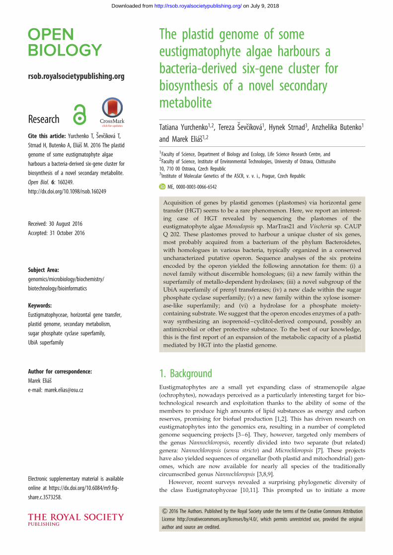
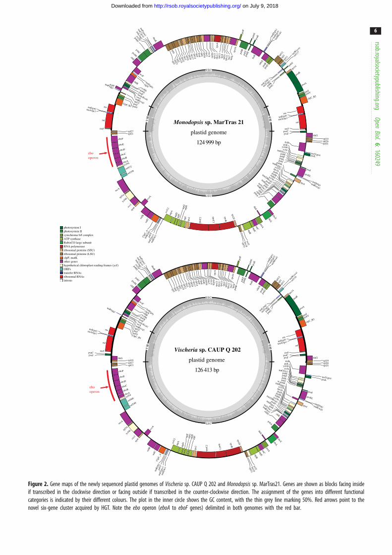
sp. CAUP Q 202 and Monodopsis sp. MarTras21Both newly sequenced plastid genomes are circular-mapping
molecules with typical inverted repeats (IRs) separating the
short and the long single-copy regions (figure 2). The size
of the genomes and the number, repertoire and order of
genes are highly similar to those of the previously sequenced
plastid genomes of other eustigmatophytes, especially Nanno-chloropsis and Microchloropsis species (table 1; electronic
supplementary material, table S1; and data not shown).
Most genes could be identified as orthologues of common
plastid genes except for four ORFs conserved in eustigmato-
phyte plastomes but lacking discernible homologues in other
organisms. In addition, six extra genes were identified in the
analysed plastomes as detailed below. All previous reports
on eustigmatophyte plastomes, including our own study
[3,8,9,12], missed the presence of the ssrA gene specifying
transfer-messenger RNA (tmRNA). The ssrA gene is situated
in a previously unannotated region between two tRNA genes
and this genomic position is shared with some other ochro-
phytes. We additionally identified unannotated ssrA genes
in plastomes from phaeophytes, whereas the chrysophyte
Ochromonas sp. CCMP1393 and the xanthophyte Vaucherialitorea appear to lack this gene (or it has diverged beyond rec-
ognition in these taxa; electronic supplementary material,
table S1).
Except for the varying size of the inverted repeats (i.e. a
different copy number of some genes; table 1), the differences
in the gene repertoire of individual eustigmatophytes primar-
ily result from differential gene loss. Previously, the genes
acpP (coding for acyl carrier protein), lysR (i.e. rbcR; encoding
a transcription regulator of the RuBisCO operon) and tsf(encoding translation elongation factor Ts) were identified
in the plastome of T. minutus, but were found missing from
plastomes of Nannochloropsis (incl. Microchloropsis) [12]. Our
present study documents the absence of lysR and tsf also
from the Vischeria and Monodopsis plastomes, suggesting
their loss early in the evolution of Eustigmatales. However,
the presence of an acpP gene in the Monodopsis plastome,
but not that of Vischeria, indicates that there were at least
two independent losses of acpP in the eustigmatophyte
evolution (since Monodopsis is specifically related to Nanno-chloropsis/Microchloropsis; figure 1). The most notable losses
impacting terminal eustigmatophyte taxa concern the
Vischeria plastome. Specifically, it proved to lack the psbZgene encoding a subunit of photosystem II (electronic sup-
plementary material, table S1). The region corresponding to
another photosystem II gene, psbW, is present in the Vischeriaplastome and looks mostly intact, but a single-nucleotide del-
etion (confirmed with inspection of reads mapped to the
region) has caused a frame-shift mutation in the coding
sequence that probably makes the gene non-functional
(hence it is considered a pseudogene in table 1 and electronic
supplementary material, table S1). Neither of the plastid gene
losses mentioned above are unique to eustigmatophytes, as
the respective genes are also missing from some other ochro-
phyte plastomes we analysed (electronic supplementary
material, table S1). Future analyses of nuclear genomes will
reveal whether these are genuine losses or whether nuclear
genes exist to encode homologous or functionally analogous
plastid-targeted proteins.
The most striking finding yielded by our analyses is that
the eustigmatophyte plastomes have been sculpted not only
by gene losses, but also by gene gains. In the previously
sequenced plastomes of T. minutus, Nannochloropsis spp. and
Microchloropsis spp., the genes ycf54 and rpl21 are direct neigh-
bours [3,8,9,12], but the plastomes of Vischeria sp. CAUP Q 202
and Monodopsis sp. MarTras21 surprisingly proved to exhibit a
cluster of six putative genes inserted between ycf54 and rpl21(figure 2). Blast searches against the non-redundant protein
and nucleotide sequence databases at the NCBI revealed that
neither of these genes has a homologue in any of the plastid
genomes sequenced so far, but homologues could be readily
identified in various bacteria. This suggested that the whole
cluster was most probably acquired by an ancestor of Vischeriaand Monodopsis by HGT from a bacterial source. In the rest
of this report, we provide a detailed analysis of this novel
gene cluster in order to elucidate its evolutionary origin and
biological significance.
3.2. A novel six-gene operon shared by plastidgenomes of Vischeria sp. CAUP Q 202 andMonodopsis sp. MarTras21 and many bacteria
To illuminate the origin and function of this gene cluster, we
carried out a more detailed investigation of the homologous
genes in bacteria. Using blastp, we retrieved and analysed
100 best hits in the NCBI nr protein database for each of
the six proteins encoded by the cluster in the Vischeria plas-
tome (we did not repeat the same procedure for
Monodopsis, as its sequences are highly similar to those
from Vischeria). All these hits came from various bacterial
species representing seven different phyla, with the phylum
Bacteroidetes being most represented followed by the
phylum Cyanobacteria. The hits for the six different queries
often came from the same bacterial species and closer inspec-
tion revealed that they are, in most cases, encoded by genes
that are physically close to each other in the genome of the
respective bacterium. This suggested the existence of gene
clusters similar to that in the Vischeria and Monodopsis plastid
genomes. Therefore, we systematically looked for homol-
ogues of all six genes in every bacterial species present at
least once among the best 100 hits for any of the original
queries (altogether 148 species/strains) and defined their
relative position within the genome (details provided in the
electronic supplementary material, table S2). Crucially, this
trnW
(cca)
orf1
17
trnL(ta
g)
trnT(tg
t)
psbZ
psbK
petG
rps14
psaB
psaA
petJpsbV
clpC_B2
rns
trnI(gat)
trnA(tgc)
rnl
rrn5psaCpsaJ
thiGrpl19rpl20rpl35
ilvB
trnY(gta)psaL
petLycf49ycf4trnG(tcc)psbEpsbFpsbLpsbJ
psaIgroEL
trnQ(ttg)trnR(acg)
psbW
rps4
trnH(gtg)ycf19rps16petNpetMorf60ycf36
trn(f)M(cat)psaDtrnS(tga)psbI
trnV(tac)
trnR(tct)chlLchlN
psbA
rbcSrbcL
thiS
petAtatCatpEatpBycf3rps18
rpl33
rps20
rpoB
rpoC1
rpoC
2
rps2atpI
atpHatpG
atpFatpDat
pA
trnE(
ttc)
ycf1
2
trnG
(gcc
)
psbY
trnK
(ttt)
Clp
C_A
sufC
sufB
ccsA
rps6
ccs1
ycf46rpl34
secA
orf198orf472
ycf54eboAeboB
eboC
eboD
eboE
eboF
rpl21rpl27
rrn5
rnl
trnA(tgc)
trnI(gat)rns
ClpC-B1 psbV
petJrpl32
trnP(tgg)
acpP
trnR(ccg)
ssrA
trn(k)I(cat)
psaF
psbX
ycf66
chlB clpN
trnN(gtt)
acsF
chlI
psaMpsbC
psbDycf34
trnL(tag)trnC(gca)
trnF(gaa)
dnaK
rpl3rpl4
rpl23rpl2
rps19rpl22
rps3rpl16
rpl29rps17rpl14rpl5rps8rpl6rpl18rps5secY
rpl3
6rp
s13
rps1
1rp
oArp
l13
rps9
rpl3
1rp
s12
rps7
tufA
rps1
0
petF
psbB
psbT
psbN
psbH
psaE
ftsH
trnD
(gtc
)trn
S(gc
t)trn
M(c
at)
cbbX
petB
petD
rpl1
2rp
l1
rpl11
Monodopsis sp. MarTras 21
plastid genome
124 999 bp
intronsribosomal RNAstransfer RNAsORFshypothetical chloroplast reading frames (ycf)other genesclpP, matKribosomal proteins (LSU)ribosomal proteins (SSU)RNA polymeraseRubisCO large subunitATP synthasecytochrome b/f complexphotosystem IIphotosystem I
IRA
IRB
LS C
SSC
ebooperon
trnW
(cca)
orf1
16
trnL(ta
g)
trnT(tg
t)
psbK
petG
rps14
psaB
psaA
petJpsbV
clpC_B2
rns
trnI(gat)
trnA(tgc)
rnl
rrn5psaCpsaJ thiG
rpl19rpl20rpl35
ilvB
trnY(gta)psaL
petLycf49ycf4trnG(tcc)
psbEpsbFpsbLpsbJ
psaI
groEL
trnQ(ttg)trnR(acg)
psbW
rps4
trnH(gtg)ycf19rps16petNpetMorf60ycf36trn(f)M(cat)psaDtrnS(tga)psbI
trnV(tac)trnR(tct)
chlL
chlN
psbA
rbcSrbcL
thiS
petAtatCatpEatpBycf3rps18
rpl33
rps20rpoB
rpoC1
rpoC
2
rps2
atpIatpHatpGatpF
atpDat
pA
trnE
(ttc
)
ycf1
2
trnG
(gcc
)
psbY
trnK
(ttt)
Clp
C_Asu
fC
sufB
ccsA
rps6cc
s1
ycf46
rpl34
secA
orf246
orf486ycf54eboA
eboBeboC
eboD
eboE
eboF
rpl21rpl27rpl19
thiGpsaJpsaC
rrn5
rnl
trnA(tgc)
trnI(gat)rns
ClpC_B1
psbV
petJrpl32
trnP(tgg)
trnR(ccg)
ssrAtrn(k)I(cat)
psaF
psbX
ycf66
chlB
clpN
trnN(gtt)
acsF
chlI
psaMpsbC
psbDycf34
trnL(tag)trnC(gca)
trnF(gaa)
dnaK
rpl3rpl4
rpl23rpl2
rps19rpl22
rps3rpl16
rpl29rps17rpl14rpl5rps8rpl6rpl18rps5secYrpl36
rps1
3rp
s11
rpoA
rpl1
3rp
s9rp
l31
rps1
2rp
s7tu
fArp
s10
petF
psbB
psbT
psbN
psbH
psaE
ftsH
trnD
(gtc
)trn
S(gc
t)trn
M(c
at)
cbbX
petB
petD
rpl1
2rp
l1rp
l11
Vischeria sp. CAUP Q 202
plastid genome
126 413 bp
IRA
IRB
LS C
SSC
ebooperon
Figure 2. Gene maps of the newly sequenced plastid genomes of Vischeria sp. CAUP Q 202 and Monodopsis sp. MarTras21. Genes are shown as blocks facing insideif transcribed in the clockwise direction or facing outside if transcribed in the counter-clockwise direction. The assignment of the genes into different functionalcategories is indicated by their different colours. The plot in the inner circle shows the GC content, with the thin grey line marking 50%. Red arrows point to thenovel six-gene cluster acquired by HGT. Note the ebo operon (eboA to eboF genes) delimited in both genomes with the red bar.
rsob.royalsocietypublishing.orgOpen
Biol.6:160249
6
on July 9, 2018http://rsob.royalsocietypublishing.org/Downloaded from
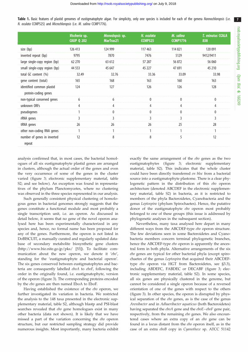
Table 1. Basic features of plastid genomes of eustigmatophyte algae. For simplicity, only one species is included for each of the genera Nannochloropsis (i.e.N. oculata CCMP525) and Microchloropsis (i.e. M. salina CCMP1776).
Vischeria sp.CAUP Q 202
Monodopsis sp.MarTras21
N. oculataCCMP525
M. salinaCCMP1776
T. minutus CCALA838
size (bp) 126 413 124 999 117 463 114 821 120 091
inverted repeat (bp) 9795 7870 7476 5129 9412/9411
large single-copy region (bp) 62 270 63 612 57 287 56 872 56 060
small single-copy region (bp) 44 553 45 647 45 227 47 691 45 210
total GC content (%) 32.49 32.76 33.36 33.09 33.98
gene content (total) 165 168 163 160 163
identified common plastid
protein-coding genes
124 127 126 126 128
non-typical conserved genes 6 6 0 0 0
unknown ORFs 4 4 4 4 4
pseudogenes 1 0 0 0 0
rRNA genes 3 3 3 3 3
tRNA genes 26 26 26 25 28
other non-coding RNA genes 1 1 1 1 1
number of genes in inverted
repeat
12 8 8 5 12
rsob.royalsocietypublishing.orgOpen
Biol.6:160249
7
on July 9, 2018http://rsob.royalsocietypublishing.org/Downloaded from
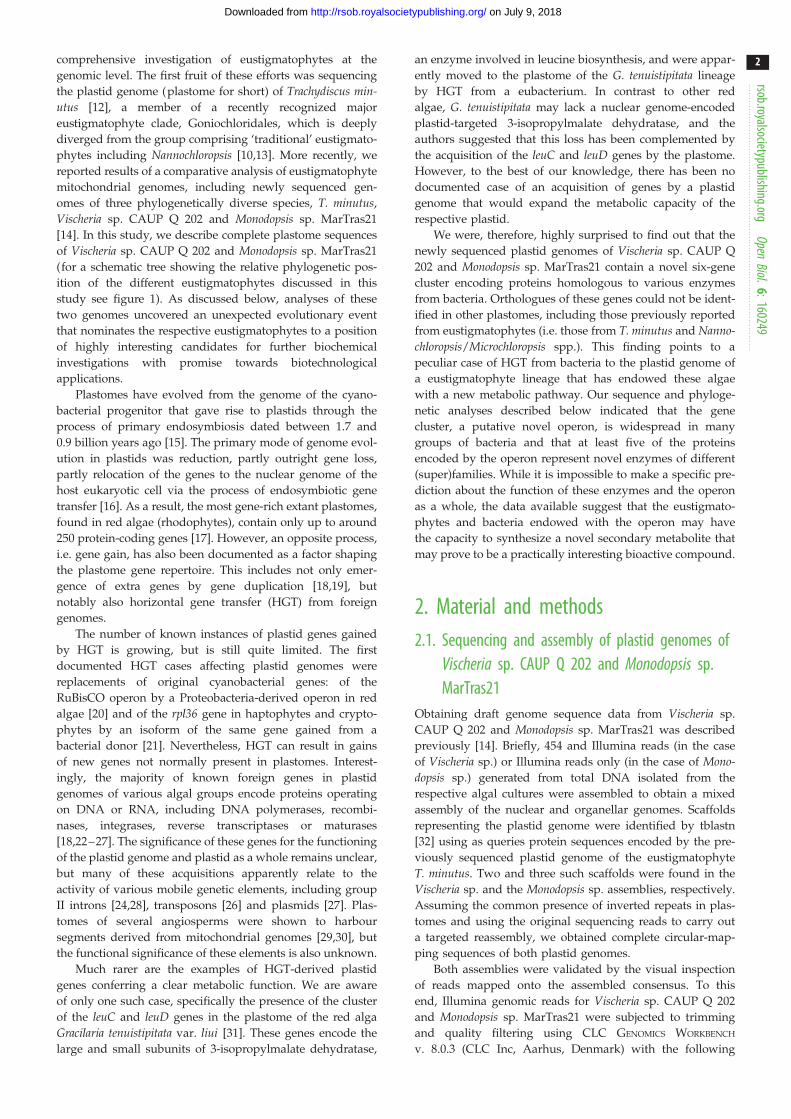
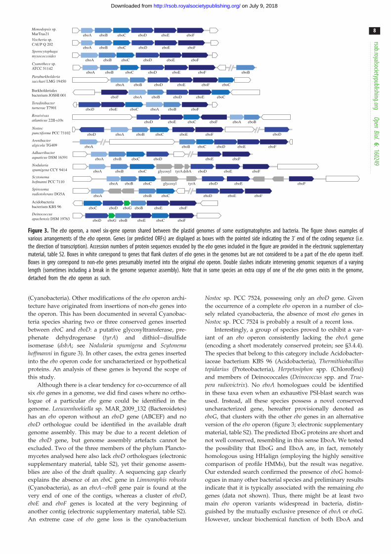
analysis confirmed that, in most cases, the bacterial homol-
ogues of all six eustigmatophyte plastid genes are arranged
in clusters, although the actual order of the genes and even
the very occurrence of some of the genes in the cluster
varied (figure 3; electronic supplementary material, table
S2; and see below). An exception was found in representa-
tives of the phylum Planctomycetes, where no clustering
was observed in the three species represented in our analysis.
Such generally consistent physical clustering of homolo-
gous genes in bacterial genomes strongly suggests that the
genes constitute a functional module and most probably a
single transcription unit, i.e. an operon. As discussed in
detail below, it seems that no gene of the novel operon ana-
lysed here has been experimentally characterized in any
species and, hence, no formal name has been proposed for
any of the genes. Furthermore, the operon is not listed in
DoBISCUIT, a manually curated and regularly updated data-
base of secondary metabolite biosynthetic gene clusters
(http://www.bio.nite.go.jp/pks/ [53]). To facilitate com-
munication about the new operon, we denote it ‘ebo’,
standing for the ‘eustigmatophyte and bacterial operon’.
The six genes conserved between eustigmatophytes and bac-
teria are consequently labelled eboA to eboF, following the
order in the originally found, i.e. eustigmatophyte, version
of the operon (figure 3). The corresponding proteins encoded
by the ebo genes are then named EboA to EboF.
Having established the existence of the ebo operon, we
further investigated its variation in bacteria. We restricted
the analysis to the 148 taxa presented in the electronic sup-
plementary material, table S2, although blastp and PSI-blast
searches revealed that ebo gene homologues exist in many
other bacteria (data not shown). It is likely that we have
missed a part of the variation concerning the ebo operon
structure, but our restricted sampling strategy did provide
numerous insights. Most importantly, many bacteria exhibit
exactly the same arrangement of the ebo genes as the two
eustigmatophytes (figure 3; electronic supplementary
material, table S2). This indicates that the whole cluster
could have been directly transferred en bloc from a bacterial
source into a eustigmatophyte plastome. There is a clear phy-
logenetic pattern in the distribution of this ebo operon
architecture (denoted ABCDEF in the electronic supplemen-
tary material, table S2) in bacteria, as it is restricted to
members of the phyla Bacteroidetes, Cyanobacteria and the
genus Leptospira (phylum Spirochaetes). Hence, the putative
donor of the eustigmatophyte ebo operon most probably
belonged to one of these groups (this issue is addressed by
phylogenetic analyses in the subsequent section).
Nevertheless, many taxa analysed here depart in many
different ways from the ABCDEF-type ebo operon structure.
The few deviations seen in some Bacteroidetes and Cyano-
bacteria members concern terminal phylogenetic branches,
hence the ABCDEF-type ebo operon is apparently the ances-
tral form in both phyla. Alternative arrangements of the six
ebo genes are typical for other bacterial phyla (except spiro-
chaetes of the genus Leptospira that acquired their ABCDEF-
type ebo operon via HGT from Bacteroidetes, see §3.3),
including ABDEFC, FABDEC or DECABF (figure 3; elec-
tronic supplementary material, table S2). In some species,
all six genes are physically clustered in the genome, but
cannot be considered a single operon because of a reversed
orientation of one of the genes with respect to the others
(figure 3). In other species, the operon is disrupted by phys-
ical separation of the ebo genes, as is the case of the genus
Arenibacter and in Adhaeribacter aquaticus (both Bacteroidetes)
having separated the eboA gene and the eboE–eboF gene pair,
respectively, from the remaining ebo genes. We also encoun-
tered cases where an extra copy of an ebo gene can be
found in a locus distant from the ebo operon itself, as in the
case of an extra eboB copy in Cyanothece sp. ATCC 51142
eboF
eboF
eboD eboFeboEglycosyl dsbAtyrA
eboD eboFeboEglycosyl tyrA
eboB
eboCeboD eboEeboF
eboBeboF eboAeboD
eboD
eboA eboB eboC eboD eboE eboF
eboA
eboA eboB eboC eboD eboE eboF
eboA eboB eboC eboD eboE eboF
eboA eboB eboC eboD eboE eboF
eboD eboG eboB eboE eboC
eboC eboD eboG eboB eboE
eboD eboA eboB eboC eboE eboF
eboA eboB eboD eboE eboF eboC
eboA eboB eboC
eboA eboB eboC
eboA eboB eboC eboD eboE eboF
eboA eboB
eboD eboE eboC eboA eboB eboF
eboE eboC
eboA eboB eboC eboD eboE eboF
eboB eboC eboD eboE eboF
Monodopsis sp.MarTras21
Vischeria sp.CAUP Q 202
Sporocytophagamyxococcoides
Nostocpunctiforme PCC 73102
Deinococcusapachensis DSM 19763
Acidobacteriabacterium KBS 96
Paraburkholderiasacchari LMG 19450
Nodulariaspumigena CCY 9414
Scytonemahofmanni PCC 7110
Cyanothece sp.ATCC 51142
Burkholderialesbacterium JOSHI 001
Teredinibacterturnerae T7901
Roseivivaxatlanticus 22II-s10s
Arenibacteralgicola TG409
Adhaeribacteraquaticus DSM 16391
Spirosomaradiotolerans DG5A
Figure 3. The ebo operon, a novel six-gene operon shared between the plastid genomes of some eustigmatophytes and bacteria. The figure shows examples ofvarious arrangements of the ebo operon. Genes (or predicted ORFs) are displayed as boxes with the pointed side indicating the 3’ end of the coding sequence (i.e.the direction of transcription). Accession numbers of protein sequences encoded by the ebo genes included in the figure are provided in the electronic supplementarymaterial, table S2. Boxes in white correspond to genes that flank clusters of ebo genes in the genomes but are not considered to be a part of the ebo operon itself.Boxes in grey correspond to non-ebo genes presumably inserted into the original ebo operon. Double slashes indicate intervening genomic sequences of a varyinglength (sometimes including a break in the genome sequence assembly). Note that in some species an extra copy of one of the ebo genes exists in the genome,detached from the ebo operon as such.
rsob.royalsocietypublishing.orgOpen
Biol.6:160249
8
on July 9, 2018http://rsob.royalsocietypublishing.org/Downloaded from
(Cyanobacteria). Other modifications of the ebo operon archi-
tecture have originated from insertions of non-ebo genes into
the operon. This has been documented in several Cyanobac-
teria species sharing two or three conserved genes inserted
between eboC and eboD: a putative glycosyltransferase, pre-
phenate dehydrogenase (tyrA) and dithiol–disulfide
isomerase (dsbA; see Nodularia spumigena and Scytonemahoffmanni in figure 3). In other cases, the extra genes inserted
into the ebo operon code for uncharacterized or hypothetical
proteins. An analysis of these genes is beyond the scope of
this study.
Although there is a clear tendency for co-occurrence of all
six ebo genes in a genome, we did find cases where no ortho-
logue of a particular ebo gene could be identified in the
genome. Leeuwenhoekiella sp. MAR_2009_132 (Bacteroidetes)
has an ebo operon without an eboD gene (ABCEF) and no
eboD orthologue could be identified in the available draft
genome assembly. This may be due to a recent deletion of
the eboD gene, but genome assembly artefacts cannot be
excluded. Two of the three members of the phylum Plancto-
mycetes analysed here also lack eboD orthologues (electronic
supplementary material, table S2), yet their genome assem-
blies are also of the draft quality. A sequencing gap clearly
explains the absence of an eboC gene in Limnoraphis robusta(Cyanobacteria), as an eboA–eboB gene pair is found at the
very end of one of the contigs, whereas a cluster of eboD,
eboE and eboF genes is located at the very beginning of
another contig (electronic supplementary material, table S2).
An extreme case of ebo gene loss is the cyanobacterium
Nostoc sp. PCC 7524, possessing only an eboD gene. Given
the occurrence of a complete ebo operon in a number of clo-
sely related cyanobacteria, the absence of most ebo genes in
Nostoc sp. PCC 7524 is probably a result of a recent loss.
Interestingly, a group of species proved to exhibit a var-
iant of an ebo operon consistently lacking the eboA gene
(encoding a short moderately conserved protein; see §3.4.4).
The species that belong to this category include Acidobacter-
iaceae bacterium KBS 96 (Acidobacteria), Thermithiobacillustepidarius (Proteobacteria), Herpetosiphon spp. (Chloroflexi)
and members of Deinococcales (Deinococcus spp. and True-pera radiovictrix). No eboA homologues could be identified
in these taxa even when an exhaustive PSI-blast search was
used. Instead, all these species possess a novel conserved
uncharacterized gene, hereafter provisionally denoted as
eboG, that clusters with the other ebo genes in an alternative
version of the ebo operon (figure 3; electronic supplementary
material, table S2). The predicted EboG proteins are short and
not well conserved, resembling in this sense EboA. We tested
the possibility that EboG and EboA are, in fact, remotely
homologous using HHalign (employing the highly sensitive
comparison of profile HMMs), but the result was negative.
Our extended search confirmed the presence of eboG homol-
ogues in many other bacterial species and preliminary results
indicate that it is typically associated with the remaining ebogenes (data not shown). Thus, there might be at least two
main ebo operon variants widespread in bacteria, distin-
guished by the mutually exclusive presence of eboA or eboG.However, unclear biochemical function of both EboA and
rsob.roy
9
on July 9, 2018http://rsob.royalsocietypublishing.org/Downloaded from
EboG (see §3.4.4) makes it impossible to deduce whether the
two ebo operon variants govern biosynthesis of different final
products or whether they impact other functional aspects of
the operon, such as its regulation.
alsocietypublishing.orgOpen
Biol.6:160249
3.3. The evolutionary origin of the eustigmatophyte ebooperon
The analyses presented above establish the presence of the ebooperon in the Vischeria and Monodopsis plastid genomes as an
obvious case of horizontal gene (operon) transfer from a bac-
terium. The salient remaining evolutionary questions are
what were the recipient and the donor of this transfer.
As to the first question, correlating the occurrence of the
operon with the eustigmatophyte phylogeny [10,14] needs
to be considered. Vischeria and Monodopsis both belong to
the order Eustigmatales, whereas the second major eustigma-
tophyte group, the clade Goniochloridales represented by the
plastome sequence from T. minutus, lacks the ebo operon
(figure 1). The most parsimonious scenario thus is that the
ebo operon was acquired only after the split of Goniochlori-
dales and Eustigmatales. Within the latter group, Vischeriaand Monodopsis represent two of the three principal lineages,
specifically the Eustigmataceae group and the family Mono-
dopsidaceae [10]. Thus, the ebo operon must have been
acquired before the split of these two lineages. However, so
far we lack plastid genome data from the third main lineage
of the order Eustigmatales, the so-called Pseudellipsoidiongroup. Moreover, the branching order of the three main
Eustigmatales lineages has not been robustly resolved yet
[10]. Hence, it is presently unclear whether the acquisition
of the operon preceded the radiation of the whole Eustigma-
tales group or whether it occurred within a particular
Eustigmatales lineage. Importantly, the absence of the ebooperon from all sequenced plastid genomes of the genera
Nannochloropsis and Microchloropsis, together constituting a
sister group to the Monodopsis lineage [7], implies a secondary
loss of the operon from that group (figure 1).
To answer the question on the nature of the putative bac-
terial donor, we first constructed phylogenetic trees for each
of the six encoded proteins. Specifically, we analysed align-
ments of the two eustigmatophyte sequences along with
homologues from the bacterial species (strains) selected as
described above (electronic supplementary material, table
S2). The trees were generally poorly resolved, perhaps
owing to the limited length of the Ebo proteins and, hence,
a limited phylogenetic signal in their sequences (figure 4
and electronic supplementary material, figure S1). In all six
cases, the two eustigmatophyte sequences clustered together
with maximal bootstrap support, confirming that the ebooperon was acquired only once in eustigmatophytes, but
the position of the eustigmatophyte branch varied between
the trees. In three trees (for EboA, EboC and EboE) the eustig-
matophyte branch was nested among sequences from the
phylum Bacteroidetes, and in one tree (EboF) it was sister
to Bacteroidetes sequences, but with low bootstrap support
values in all cases. In the remaining two trees (EboB and
EboD), the eustigmatophyte branch was found sister to Cya-
nobacteria, but again with low bootstrap support. Such
conflicting or unclear results are commonly observed in phy-
logenetic analyses concerning ancient HGT events, and,
indeed, they rarely point with confidence to a particular
bacterial taxon as a source of bacteria-derived eukaryotic
genes (e.g. [54,55]).
Nevertheless, the phylogenetic trees for individual Ebo
proteins did provide additional insights into the evolutionary
history of the operon. The EboB tree shows with maximal
bootstrap support a bipartition separating taxa with eboAon the one side and eboG on the other side (electronic sup-
plementary material, figure S1b). This supports the notion
of the existence of two principal variants of the operon. The
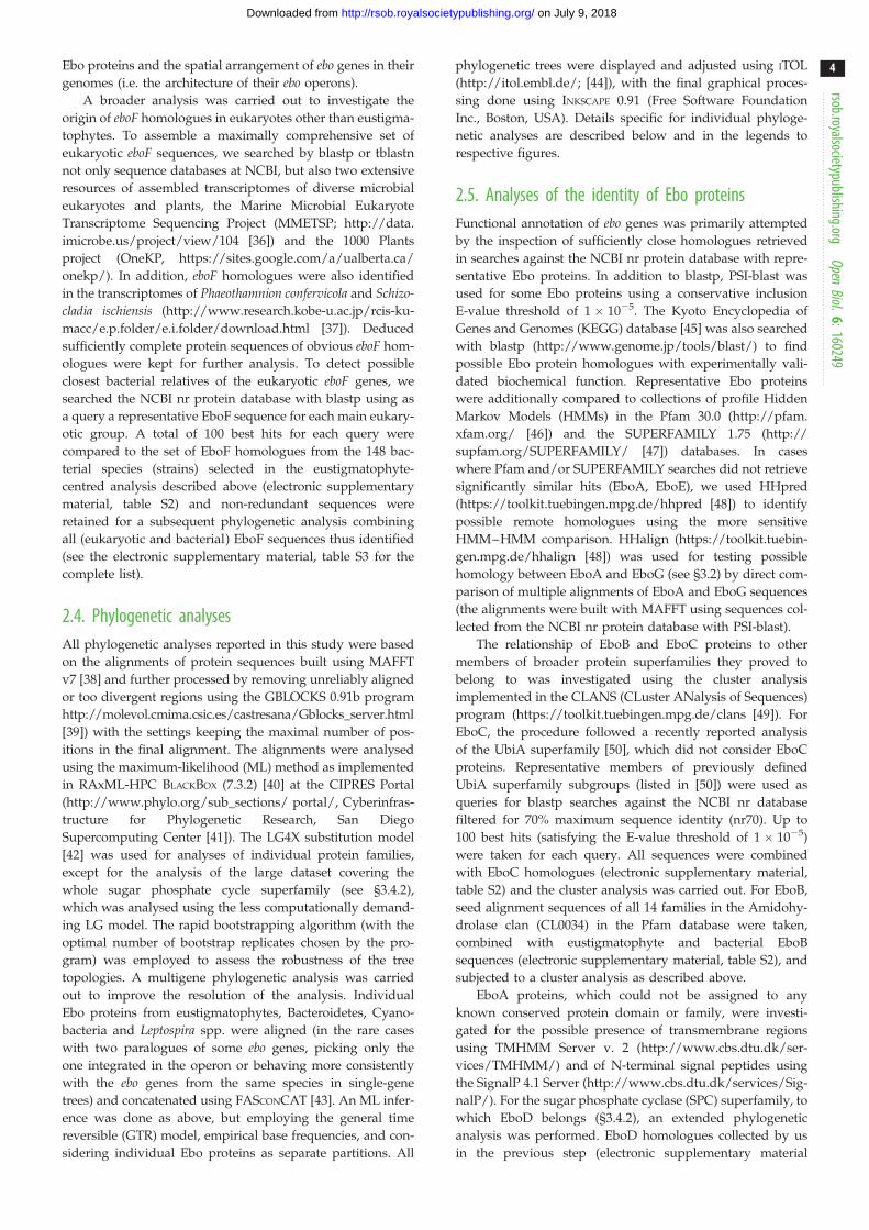
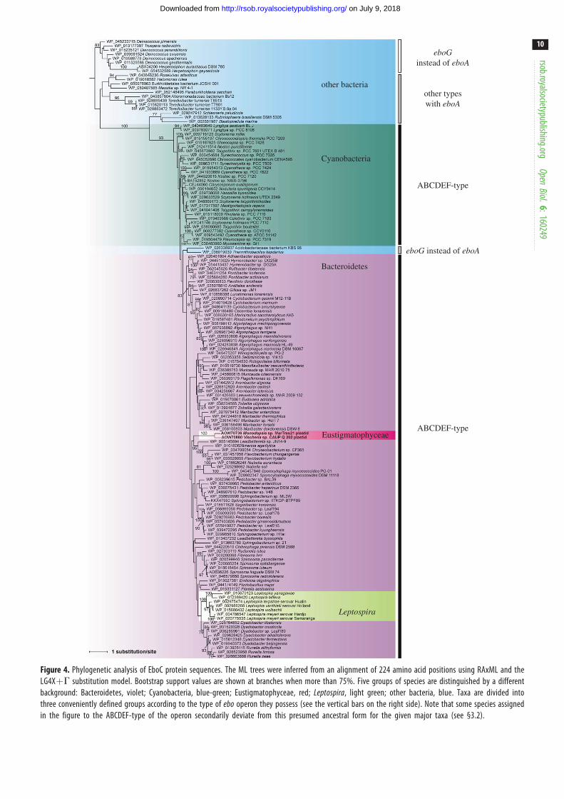
EboC phylogeny (figure 4) suggests a more complex scenario.
The tree topology is generally consistent with separating taxa
according to the presence of eboA or eboG, and further accord-
ing to the ebo operon architecture ancestral for the respective
taxa—the ABCDEF-type versus other arrangements. How-
ever, two taxa, Acidobacteriaceae bacterium KBS 96 and
Thermithiobacillus tepidarius, disturb this simple picture by
being nested (with bootstrap support of 93%) within the
ABCDEF-type group. The most probable explanation is repla-
cement of the original eboC gene in these taxa with a
homologue from a bacterium representing the ABCDEF-
type group. Indeed, separation of taxa (ancestrally) with the
ABCDEF-type ebo operon from other operon arrangements
(including Acidobacteriaceae bacterium KBS 96 and Ther-mithiobacillus tepidarius) is retained (with moderate bootstrap
support of 78%) in the EboE tree (electronic supplementary
material, figure S1d ). EboA and EboF trees (electronic
supplementary material, figure S1a,e) are topologically con-
sistent with this split as well, although without good
statistical support, while the EboD tree has a mixed topology
in this respect, yet the discrepancies have low statistical sup-
port (electronic supplementary material, figure S1c).
Altogether, these phylogenetic analyses suggest that the ebooperons in eustigmatophytes, Bacteroidetes, Cyanobacteria
and the genus Leptospira are all derived from a common
ABCDEF-type operon architecture and are evolutionarily
separated from ebo operons from other taxa.
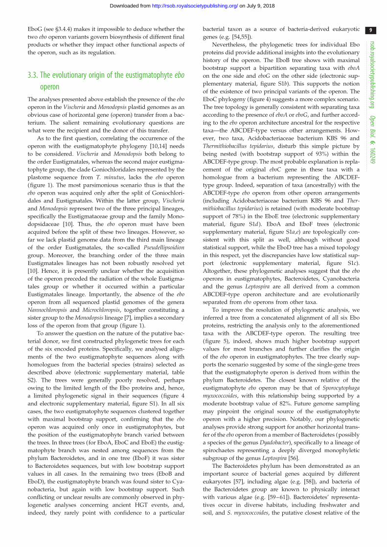
To improve the resolution of phylogenetic analysis, we
inferred a tree from a concatenated alignment of all six Ebo
proteins, restricting the analysis only to the aforementioned
taxa with the ABCDEF-type operon. The resulting tree
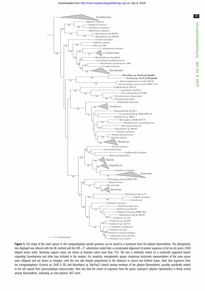
(figure 5), indeed, shows much higher bootstrap support
values for most branches and further clarifies the origin
of the ebo operon in eustigmatophytes. The tree clearly sup-
ports the scenario suggested by some of the single-gene trees
that the eustigmatophyte operon is derived from within the
phylum Bacteroidetes. The closest known relative of the
eustigmatophyte ebo operon may be that of Sporocytophagamyxococcoides, with this relationship being supported by a
moderate bootstrap value of 82%. Future genome sampling
may pinpoint the original source of the eustigmatophyte
operon with a higher precision. Notably, our phylogenetic
analyses provide strong support for another horizontal trans-
fer of the ebo operon from a member of Bacteroidetes (possibly
a species of the genus Dyadobacter), specifically to a lineage of
spirochaetes representing a deeply diverged monophyletic
subgroup of the genus Leptospira [56].
The Bacteroidetes phylum has been demonstrated as an
important source of bacterial genes acquired by different
eukaryotes [57], including algae (e.g. [58]), and bacteria of
the Bacteroidetes group are known to physically interact
with various algae (e.g. [59–61]). Bacteroidetes’ representa-
tives occur in diverse habitats, including freshwater and
soil, and S. myxococcoides, the putative closest relative of the
eboGinstead of eboA
eboG instead of eboA
other typeswith eboA
ABCDEF-type
other bacteria
Cyanobacteria
Bacteroidetes
Eustigmatophyceae
Leptospira
ABCDEF-type
Figure 4. Phylogenetic analysis of EboC protein sequences. The ML trees were inferred from an alignment of 224 amino acid positions using RAxML and theLG4XþG substitution model. Bootstrap support values are shown at branches when more than 75%. Five groups of species are distinguished by a differentbackground: Bacteroidetes, violet; Cyanobacteria, blue-green; Eustigmatophyceae, red; Leptospira, light green; other bacteria, blue. Taxa are divided intothree conveniently defined groups according to the type of ebo operon they possess (see the vertical bars on the right side). Note that some species assignedin the figure to the ABCDEF-type of the operon secondarily deviate from this presumed ancestral form for the given major taxa (see §3.2).
rsob.royalsocietypublishing.orgOpen
Biol.6:160249
10
on July 9, 2018http://rsob.royalsocietypublishing.org/Downloaded from
Flexithrix dorotheae
Pedobacter antarcticus
Mesoflavibacter zeaxanthinifaciens
Flectobacillus major
Mariniradius saccharolyticus AK6
Pedobacter sp. Leaf194
Flavobacterium chungangense
Flagellimonas sp. DK169
Monodopsis sp. MarTras21 plastid
Algoriphagus machipongonensis
Hymenobacter sp. DG25A
Lunatimonas lonarensis
Zobellia galactanivorans
Chitinophaga pinensis DSM 2588
Eudoraea adriatica
Pedobacter sp. Leaf176
Hymenobacter sp. DG25B
Robiginitalea biformata
Pontibacter korlensis
Pedobacter kyungheensis
Vischeria sp. CAUP Q 202 plastid
Winogradskyella sp. PG-2
Chryseobacterium sp. CF365
Fibrella aestuarina
Pedobacter heparinus DSM 2366
Anditalea andensis
Segetibacter koreensis
Adhaeribacter aquaticus
Cyanobacteria
Cyclobacterium
Algoriphagus
Arenibacter
Maribacter
Runella
Dyadobacter
Leptospira
Spirosoma
Pedobacter borealis
Sporocytophaga myxococcoides DSM 11118
Niabella soli
Leeuwenhoekiella sp. MAR 2009 132
Pedobacter ginsenosidimutans
Pedobacter sp. BAL39
Leadbetterella sp. JN14-9
Rufibacter tibetensis
Fibrisoma limi
Muricauda lutaonensis
Zobellia uliginosa
Dyadobacter tibetensis
Rudanella lutea
Emticicia oligotrophica
Rhodonellum psychrophilum
Aquimarina agarilytica
Cecembia lonarensis
Pedobacter sp. V48Sphingobacterium sp. ML3W
Sphingobacterium sp. 21
Gillisia sp. JM1
Niabella aurantiaca
Pedobacter sp. Leaf216
Flavobacterium hydatis
Sporocytophaga myxococcoides PG-01
Sphingobacterium sp. H1ai
Muricauda sp. MAR 2010 75Sediminicola sp. YIK13
Leadbetterella byssophila
Pontibacter actiniarum
100
100
100
100
84
99
100
100
100
90
92
99
100
100
100
100
100
100
100
100
100
100
100
100
100
99
76
100
100
100
100
99
100
94
95
100
100
95
100
82
100
100
100
100
100
100
100
100
100
100
100
76
100
87
100
100
0.1 substitutions/site
Figure 5. The origin of the novel operon in the eustigmatophyte plastid genomes can be traced to a bacterium from the phylum Bacteroidetes. The phylogenetictree displayed was inferred with the ML method and the GTRþG substitution model from a concatenated alignment of protein sequences of all six ebo genes (1838aligned amino acids). Bootstrap support values are shown at branches when more than 75%. The tree is arbitrarily rooted on a maximally supported branchseparating Cyanobacteria and other taxa included in the analysis. For simplicity, monophyletic groups comprising exclusively representatives of the same genuswere collapsed and are shown as triangles, with the two side lengths proportional to the distances to closest and furthest leaves. Note that sequences fromthe eustigmatophytes Vischeria sp. CAUP Q 202 and Monodopsis sp. MarTras21 branch among members of the phylum Bacteroidetes, possibly specifically relatedto the ebo operon from Sporocytophaga myxococcoides. Note also that the cluster of sequences from the genus Leptospira ( phylum Spirochaetes) is firmly nestedamong Bacteroidetes, indicating an inter-phylum HGT event.
rsob.royalsocietypublishing.orgOpen
Biol.6:160249
11
on July 9, 2018http://rsob.royalsocietypublishing.org/Downloaded from
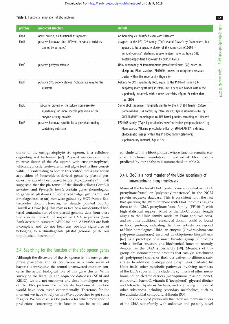
Table 2. Functional annotation of Ebo proteins.
protein predicted function details
EboA novel protein, no functional assignment no homologues identified even with HHsearch
EboB putative hydrolase (but different enzymatic activities
cannot be excluded)
assigned to the PF01026 family (‘TatD-related DNase’) by Pfam search, but
appears to be a separate cluster of the same clan (CL0034 –
‘Amidohydrolase’; electronic supplementary material, figure S3);
‘Metallo-dependent hydrolase’ by SUPERFAMILY
EboC putative prenyltransferase UbiA superfamily of intramembrane prenyltransferases [50] based on
blastp and Pfam searches (PF01040); proved to comprise a separate
cluster within the superfamily (figure 6)
EboD putative SPC, sedoheptulose 7-phosphate may be the
substrate
belongs to SPC superfamily [66], equal to the PF01761 family (‘3-
dehydroquinate synthase’) in Pfam, but a separate branch within the
superfamily putatively with a novel specificity (figure 7) rather than
true DHQS
EboE TIM-barrel protein of the xylose isomerase-like
superfamily, no more specific prediction of the
enzyme activity possible
Some EboE sequences marginally similar to the PF01261 family (‘Xylose
isomerase-like TIM barrel’) by Pfam search; ‘Xylose isomerase-like’ by
SUPERFAMILY; homologous to TIM-barrel proteins according to HHsearch
EboF putative hydrolase specific for a phosphate moiety-
containing substrate
PF01663 family (‘Type I phosphodiesterase/nucleotide pyrophosphatase’) by
Pfam search; ‘Alkaline phosphatase-like’ by SUPERFAMILY; a distinct
phylogenetic lineage within the PF01663 family (electronic
supplementary material, figure S2)
rsob.royalsocietypublishing.orgOpen
Biol.6:160249
12
on July 9, 2018http://rsob.royalsocietypublishing.org/Downloaded from
donor of the eustigmatophyte ebo operon, is a cellulose-
degrading soil bacterium [62]. Physical association of the
putative donor of the ebo operon with eustigmatophytes,
which are mostly freshwater or soil algae [63], is thus concei-
vable. It is interesting to note in this context that a case for an
acquisition of Bacteroidetes-derived genes by plastid gen-
omes has already been raised before. Moszczynski et al. [64]
suggested that the plastomes of the dinoflagellates Ceratiumhorridum and Pyrocystis lunula contain genes (homologous
to genes in plastomes of some other algal groups but not
dinoflagellates so far) that were gained by HGT from a Bac-
teroidetes donor. However, as already pointed out by
Dorrell & Howe [65], this may in fact be a misidentified bac-
terial contamination of the plastid genome data from these
two species. Indeed, the respective DNA sequences (Gen-
Bank accession numbers AF490364 and AF490367) are both
incomplete and do not bear any obvious signatures of
belonging to a dinoflagellate plastid genome (2016, our
unpublished observation).
3.4. Searching for the function of the ebo operon genesAlthough the discovery of the ebo operon in the eustigmato-
phyte plastomes and its occurrence in a wide array of
bacteria is intriguing, the central unanswered question con-
cerns the actual biological role of this gene cluster. While
surveying the literature and sequence databases (NCBI and
KEGG), we did not encounter any close homologue of any
of the Ebo proteins for which its biochemical function
would have been tested experimentally. Therefore, for the
moment we have to rely on in silico approaches to get some
insights. We first discuss Ebo proteins for which more specific
predictions concerning their function can be made, and
conclude with the EboA protein, whose function remains elu-
sive. Functional annotation of individual Ebo proteins
predicted by our analyses is summarized in table 2.
3.4.1. EboC is a novel member of the UbiA superfamily ofintramembrane prenyltransferases
Many of the bacterial EboC proteins are annotated as ‘UbiA
prenyltransferase’ or ‘polyprenyltransferase’ in the NCBI
protein sequence database. This is consistent with the fact
that querying the Pfam database with EboC proteins assigns
them to the ‘UbiA prenyltransferase family’ (PF01040) with
high statistical support. Most of the EboC protein length
aligns to the UbiA family model in Pfam and vice versa,
and no other additional conserved domain could be found
in EboC proteins, indicating that they entirely correspond
to UbiA homologues. UbiA, an enzyme (4-hydroxybenzoate
polyprenyltransferase) involved in ubiquinone biosynthesis
[67], is a prototype of a much broader group of proteins
with a similar structure and biochemical function, recently
denoted as the UbiA superfamily [50]. Members of this
group are intramembrane proteins that catalyse attachment
of (poly)prenyl chains or their derivatives to different sub-
strates. In addition to ubiquinone biosynthesis mediated by
UbiA itself, other metabolic pathways involving members
of the UbiA superfamily include the synthesis of other mem-
brane-bound electron carriers (menaquinone, plastoquinone),
chlorophyll, haem O, vitamin E (tocopherol), glycerol diether
and tetraether lipids in Archaea, and a growing number of
other substances including secondary metabolites, such as
the antimicrobial compound shikonin [50].
It has been noted previously that there are many members
of the UbiA superfamily with unknown and possibly novel
COX10
COQ2UbiA
archaeal UbiA
DPPRsynthase
DGGGPsynthase
chlorophyllsynthase
UbiAD1
MenA
homogentisate PT(HPT, HST, HGGT)
EboC
AuaA
Af
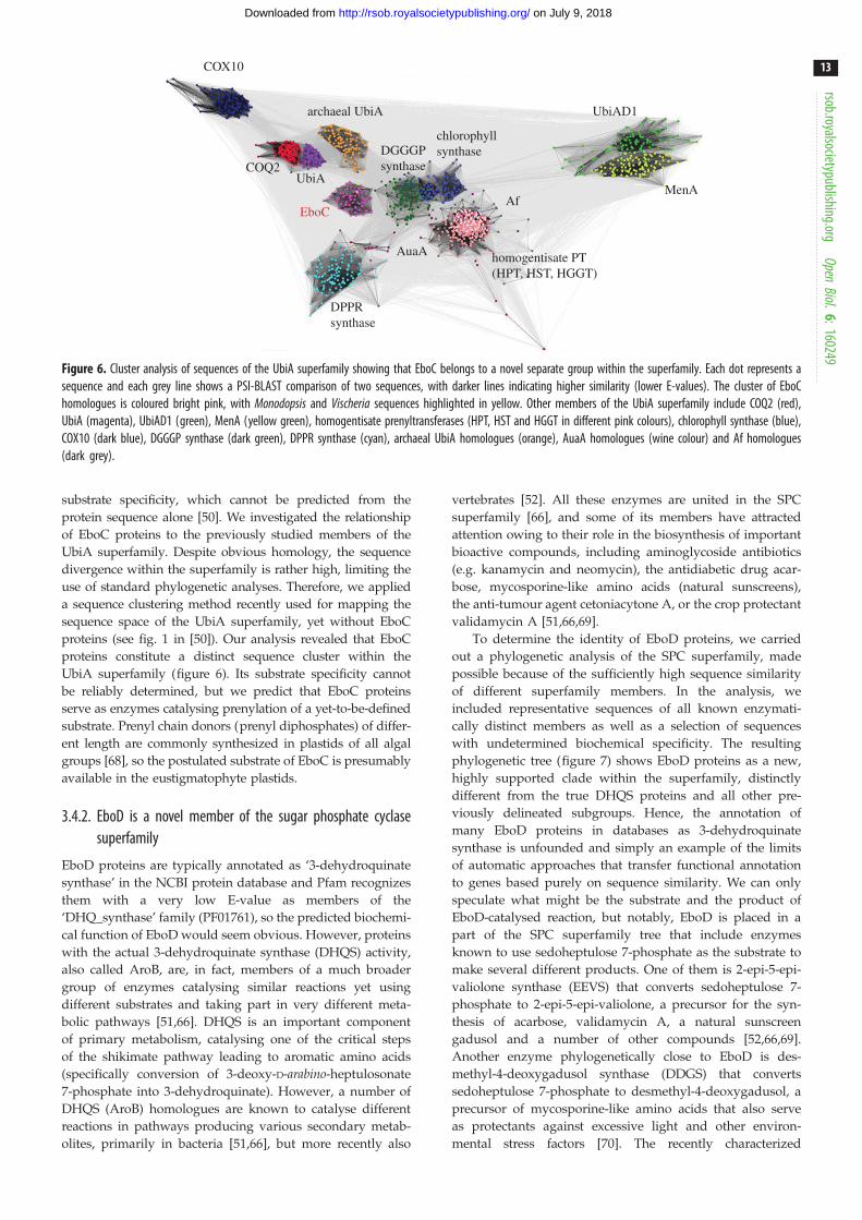
Figure 6. Cluster analysis of sequences of the UbiA superfamily showing that EboC belongs to a novel separate group within the superfamily. Each dot represents asequence and each grey line shows a PSI-BLAST comparison of two sequences, with darker lines indicating higher similarity (lower E-values). The cluster of EboChomologues is coloured bright pink, with Monodopsis and Vischeria sequences highlighted in yellow. Other members of the UbiA superfamily include COQ2 (red),UbiA (magenta), UbiAD1 (green), MenA (yellow green), homogentisate prenyltransferases (HPT, HST and HGGT in different pink colours), chlorophyll synthase (blue),COX10 (dark blue), DGGGP synthase (dark green), DPPR synthase (cyan), archaeal UbiA homologues (orange), AuaA homologues (wine colour) and Af homologues(dark grey).
rsob.royalsocietypublishing.orgOpen
Biol.6:160249
13
on July 9, 2018http://rsob.royalsocietypublishing.org/Downloaded from
substrate specificity, which cannot be predicted from the
protein sequence alone [50]. We investigated the relationship
of EboC proteins to the previously studied members of the
UbiA superfamily. Despite obvious homology, the sequence
divergence within the superfamily is rather high, limiting the
use of standard phylogenetic analyses. Therefore, we applied
a sequence clustering method recently used for mapping the
sequence space of the UbiA superfamily, yet without EboC
proteins (see fig. 1 in [50]). Our analysis revealed that EboC
proteins constitute a distinct sequence cluster within the
UbiA superfamily (figure 6). Its substrate specificity cannot
be reliably determined, but we predict that EboC proteins
serve as enzymes catalysing prenylation of a yet-to-be-defined
substrate. Prenyl chain donors (prenyl diphosphates) of differ-
ent length are commonly synthesized in plastids of all algal
groups [68], so the postulated substrate of EboC is presumably
available in the eustigmatophyte plastids.
3.4.2. EboD is a novel member of the sugar phosphate cyclasesuperfamily
EboD proteins are typically annotated as ‘3-dehydroquinate
synthase’ in the NCBI protein database and Pfam recognizes
them with a very low E-value as members of the
‘DHQ_synthase’ family (PF01761), so the predicted biochemi-
cal function of EboD would seem obvious. However, proteins
with the actual 3-dehydroquinate synthase (DHQS) activity,
also called AroB, are, in fact, members of a much broader
group of enzymes catalysing similar reactions yet using
different substrates and taking part in very different meta-
bolic pathways [51,66]. DHQS is an important component
of primary metabolism, catalysing one of the critical steps
of the shikimate pathway leading to aromatic amino acids
(specifically conversion of 3-deoxy-D-arabino-heptulosonate
7-phosphate into 3-dehydroquinate). However, a number of
DHQS (AroB) homologues are known to catalyse different
reactions in pathways producing various secondary metab-
olites, primarily in bacteria [51,66], but more recently also
vertebrates [52]. All these enzymes are united in the SPC
superfamily [66], and some of its members have attracted
attention owing to their role in the biosynthesis of important
bioactive compounds, including aminoglycoside antibiotics
(e.g. kanamycin and neomycin), the antidiabetic drug acar-
bose, mycosporine-like amino acids (natural sunscreens),
the anti-tumour agent cetoniacytone A, or the crop protectant
validamycin A [51,66,69].
To determine the identity of EboD proteins, we carried
out a phylogenetic analysis of the SPC superfamily, made
possible because of the sufficiently high sequence similarity
of different superfamily members. In the analysis, we
included representative sequences of all known enzymati-
cally distinct members as well as a selection of sequences
with undetermined biochemical specificity. The resulting
phylogenetic tree (figure 7) shows EboD proteins as a new,
highly supported clade within the superfamily, distinctly
different from the true DHQS proteins and all other pre-
viously delineated subgroups. Hence, the annotation of
many EboD proteins in databases as 3-dehydroquinate
synthase is unfounded and simply an example of the limits
of automatic approaches that transfer functional annotation
to genes based purely on sequence similarity. We can only
speculate what might be the substrate and the product of
EboD-catalysed reaction, but notably, EboD is placed in a
part of the SPC superfamily tree that include enzymes
known to use sedoheptulose 7-phosphate as the substrate to
make several different products. One of them is 2-epi-5-epi-
valiolone synthase (EEVS) that converts sedoheptulose 7-
phosphate to 2-epi-5-epi-valiolone, a precursor for the syn-
thesis of acarbose, validamycin A, a natural sunscreen
gadusol and a number of other compounds [52,66,69].
Another enzyme phylogenetically close to EboD is des-
methyl-4-deoxygadusol synthase (DDGS) that converts
sedoheptulose 7-phosphate to desmethyl-4-deoxygadusol, a
precursor of mycosporine-like amino acids that also serve
as protectants against excessive light and other environ-
mental stress factors [70]. The recently characterized

WP 013376023|Stigmatella aurantiaca
WP 006330699|Mesorhizobium sp. STM 4661
WP 006197469|Nodularia spumigena
WP 021013499|Serratia sp. ATCC 39006
WP 041039713|Tolypothrix campylonemoides
WP 039714189|Scytonema millei
WP 019045670|Nocardia asteroides
WP 001156090|Helicobacter pylori
WP 039717832|Scytonema millei
CEJ47810|Chrysosporum ovalisporum
WP 038081034|Tolypothrix bouteillei
WP 010588262|Schlesneria paludicola
1DQS A|Aspergillus nidulans
WP 054536910|Herpetosiphon geysericola
WP 005477027|Streptomyces bottropensis
WP 012411972|Nostoc punctiforme
WP 015118261|Rivularia sp. PCC 7116
WP 017317005|Mastigocladopsis repens
WP 019489068|Calothrix sp. PCC 7103
WP 015155923|Chroococcidiopsis thermalis
WP 048868121|Scytonema tolypothrichoides
WP 028988951|Thermithiobacillus tepidarius
WP 002650691|Blastopirellula marina
ABX02918|Herpetosiphon aurantiacus DSM 785
WP 043843740|Roseivivax atlanticus
KYC43254|Scytonema hofmannii PCC 7110
76
100
87
100
100
87
100
100
94
88
95
91
100
89
100
90
100
92
100
bacteria
bacteria
bacteria
bacteria
bacteria
bacteria
bacteria
vertebrates
algae
DDGS
DDGS?
EboD
aminoDHQS
EVS
bacteria and Archaea
bacteria, plants and algae
DOIS
Cyanobacteria
0.1 substitutions/site
knownand putative
EEVS
knownand putative
DHQS
Figure 7. EboD represents a new lineage within the SPC superfamily. The phylogenetic tree displayed was inferred with the ML method using the LGþG sub-stitution model and a protein sequence of alignment of 147 amino acids. For simplicity, most monophyletic branches of the same putative enzymatic activity withmore than two members were collapsed as in figure 5, bootstrap support values are shown only when more than 75%. The tree was rooted at a position suggestedby a previously published analysis using glycerol dehydrogenase (CglD) from Escherichia coli as an outgroup [52]. Main subgroups of the superfamily characterized bydifferent enzymatic activities are indicated. Note that most proteins in the tree have not been characterized biochemically, so the functional assignment suggested bythe tree topology has to be taken with caution (for example, the clade including confirmed EVS enzymes would apparently be considered as putative DHQS withoutthe actual data to the contrary). AminoDHQS, aminodehydroquinate synthase; DDGS, desmethyl-4-deoxygadusol synthase; DDGS?, a clade of uncharacterized proteinsrelated to DDGS, but distinctly different to possibly exhibit a different enzymatic activity; DHQS, 3-dehydroquinate synthase; DOIS, 2-deoxy-scyllo-inosose synthase;EEVS, 2-epi-5-epi-valiolone synthase; EVS, 2-epi-valiolone synthase.
rsob.royalsocietypublishing.orgOpen
Biol.6:160249
14
on July 9, 2018http://rsob.royalsocietypublishing.org/Downloaded from
rsob.royalsocietypublishing.orgOpen
Biol.6:160249
15
on July 9, 2018http://rsob.royalsocietypublishing.org/Downloaded from
reaction converting sedoheptulose 7-phosphate is catalysed
by 2-epi-valiolone synthase (EVS) and leads to 2-epi-valio-
lone, a putative precursor of hitherto uncharacterized final
secondary metabolites [51].
Taking into account the phylogenetic position of EboD
within the SPC superfamily and the known substrate speci-
ficity of its relatives, we posit that EboD catalyses
conversion of sedoheptulose 7-phosphate into a cyclic hydro-
carbon with multiple hydroxy groups, i.e. a cyclitol (similar
to, if not identical with, 2-epi-5-epi-valiolone or another pro-
duct mentioned above). In most organisms, the source of
sedoheptulose 7-phosphate is the pentose phosphate path-
way, but this substance is also an intermediate of the
Calvin cycle, which in eukaryotes is located to plastids.
Hence, the EboD proteins in eustigmatophytes would have
an access to their postulated substrate in the plastid.
3.4.3. EboF is a putative hydrolase specific for a phosphatemoiety-containing substrate
EboF proteins in the NCBI database are typically annotated
as ‘phosphodiesterase’ or ‘alkaline phosphatase family
protein’. The former is consistent with the results of searches
against the Pfam database, which assigned EboF proteins
with high confidence to the family PF01663, annotated as
‘Type I phosphodiesterase/nucleotide pyrophosphatase’.
The latter annotation perhaps reflects the fact that this
family belongs to the Pfam clan CL0088 named ‘Alkaline
phosphatase-like’. SUPERFAMILY database searches also
assigned EboF proteins to the ‘Alkaline phosphatase-like’
superfamily, but not into a particular subordinate family,
consistent with the absence of a solved protein structure for
any close EboF homologue. We performed a phylogenetic
analysis of a subset of EboF proteins with the sequences
used to build the seed alignment of the PF01663 family in
Pfam. The analysis revealed that EboF sequences (including
five from the seed alignment) make a cluster well separated
from the other sequences analysed (electronic supplementary
material, figure S2), supporting the notion that EboF constitu-
tes a separate lineage within the Type I phosphodiesterase/
nucleotide pyrophosphatase family. The experimentally
studied members of the family catalyse hydrolysis of
phosphoanhydride or phosphodiester bonds in various sub-
strates, including nucleotides and their derivatives,
lysophospholipids or choline phosphate esters [71]. It is,
therefore, conceivable that EboF catalyses hydrolysis of a
phosphate moiety-containing substrate, but a more specific
prediction cannot be made by in silico analyses alone.
3.4.4. Only general functional predictions can be made for EboBand EboE, and little can be said about the EboA protein
Most EboB proteins are annotated as ‘hydrolase TatD’ in the
NCBI database, consistent with our searches against the Pfam
database, which assigned EboB sequences to the Pfam family
PF01026 annotated as ‘TatD related DNase’. However, the E-
values for this assignment were relatively high (from as high
as 1 � 1025 to 1 � 10221 at best for different EboB proteins),
with none of the sequences used to build the seed alignment
of the family being particularly similar to EboB. Searching the
SUPERFAMILY database assigned EboB sequences with high
confidence to the metallo-dependent hydrolases superfamily,
which roughly corresponds to the Amidohydrolase
superfamily (i.e. clan CL0034 including the PF01026 family)
defined in the Pfam database. The SUPERFAMILY search
did not assign EboB to any particular family because of the
E-value threshold, but the ‘TatD Mg-dependent DNase-like’
family was identified as the most similar. These observations
suggest that EboB is a member of the metallo-dependent
hydrolase (or amidohydrolase) superfamily, possibly
affiliated to the TatD DNase family. To get a better view of
the position of EboB within this broader protein grouping,
we performed a cluster analysis using CLANS. It showed
that EboB sequences group together as a well-defined cluster
separated from the TatD DNase family, yet still closest to it
among all families of the Pfam clan CL0034 (electronic sup-
plementary material, figure S3). Therefore, in our view,
EboB cannot be considered as a true member of the TatD-
related DNase family, and the functional annotation of the
latter cannot be applied to the former. It is likely that EboB
does function as a hydrolase, which is the typical biochemical
activity of members of the metallo-dependent hydrolase
(amidohydrolase) superfamily. Nevertheless, it should be
noted that some members of the superfamily catalyse differ-
ent reactions, including decarboxylation [72] or isomerization
[73].
Similarly, little can be deduced by in silico approaches
concerning the biochemical function of EboE. Two different
specific annotations (other than ‘hypothetical proteins’) are
attached to EboE records in the NCBI nr protein database,
‘xylose isomerase’ or ‘AP endonuclease’. These annotations
are apparently related to the fact that for a few EboE
sequences, Pfam search gave marginally significant hits to
the Pfam family PF01261, labelled ‘AP_endonuc_2’ and
referred to as ‘Xylose isomerase-like TIM barrel’ in a more
detailed annotation (see http://pfam.xfam.org/family/
PF01261.22). Searching the SUPERFAMILY database
assigned EboE to the Xylose isomerase-like superfamily,
although not to a particular family, as the match to the
most similar family defined in the database, Endonuclease
IV, exhibited too high E-values. Consistent with these results,
HHsearch retrieved various TIM-barrel proteins as best and
significant hits for EboE. Altogether, this suggests that
EboE is a TIM-barrel protein representing the Xylose isomer-
ase-like (super)family. However, this protein group is rather
diverse in regard to a specific biochemical activity. It includes
isomerases of various sugars, epimerases, endonucleases,
dehydratases and even putative oxygenases (see the list of
the family members with solved three-dimensional structures
in the Pfam database, http://pfam.xfam.org/family/
PF01261.22#tabview=tab9). Hence, at the moment, the sub-
strate(s) and the type of enzymatic reaction catalysed by
EboE cannot be inferred.
The most enigmatic among all Ebo proteins is EboA. Most
EboA homologues in the NCBI database are annotated as
hypothetical proteins, except those from various Actinobac-
teria (identified by iterative PSI-blast searches) annotated as
‘sugar phosphate isomerase’ or ‘xylose isomerase’. However,
these entries turned out to be hybrid proteins consisting of a
C-terminal region corresponding to EboA fused to an
N-terminal region representing a member of the xylose
isomerase-like superfamily (data not shown). No assignment
to a previously defined conserved family or domain was
made for EboA proteins by searching the Pfam or the SUPER-
FAMILY databases. No significant similarity to other proteins
was detected even when the highly sensitive HHpred search
rsob.royalsociet
16
on July 9, 2018http://rsob.royalsocietypublishing.org/Downloaded from
based on HMM–HMM comparisons was used. EboA pro-
teins have no predicted transmembrane domains or an
N-terminal signal peptide, indicating that they are probably
cytosolic (or stromal in case of eustigmatophyte plastids)
proteins. Direct biochemical characterization is necessary to
unveil what the EboA proteins do in the cells.
ypublishing.orgOpen
Biol.6:160249
3.5. Independent acquisition of eboF homologues byseveral different eukaryotic lineages
While checking the evolutionary origin of the eustigmato-
phyte ebo operon, we were interested to investigate whether
it might have been acquired independently by other eukary-
otic organisms. Blastp searches against eukaryotic proteins in
the NCBI nr database retrieved significantly similar (in the
case of EboA and EboE) or only distantly related, apparently
non-orthologous (in the case of EboB, EboC, EboD; data not
shown), hits. The only exception was EboF, for which four
closely related eukaryotic hits were found, coming from the
red alga Chondrus crispus, the plant parasite Plasmodiophorabrassicae (Rhizaria), the brown alga Ectocarpus siliculosus and
a partial protein sequence from the amoebozoan Vermamoebavermiformis (electronic supplementary material, table S3). The
authenticity of the V. vermiformis sequence could not be ver-
ified, as it is derived from a transcriptome sequencing
project and bacterial contamination cannot be ruled out.
However, the remaining three protein sequences corre-
sponded to genes in nuclear genome sequences of the
respective species. Inspection of the corresponding genomic
loci confirmed the presence of eukaryotic introns or that the
flanking genes were of a clearly eukaryotic provenance
(data not shown). This observation prompted us to look for
eukaryotic eboF homologues more systematically. The elec-
tronic supplementary material, table S3 lists putative
eukaryotic eboF genes that we identified in genome or tran-
scriptome sequences of various eukaryotes, specifically
different protists. The corresponding predicted protein
sequences do not exhibit apparent N-terminal targeting
sequences (such as signal or transit peptides), suggesting
they most probably function in the cytosol.
To illuminate the evolutionary history of the eukaryotic
eboF genes, we carried out a broad phylogenetic analysis of
EboF protein sequences (figure 8). It suggested that the eboFgene was acquired by at least four different eukaryotic
lineages from different bacterial donors. In one case, the
eboF gene was gained as a part of the whole ebo operon by
the plastome of eustigmatophytes. The other instance is
an apparent HGT event from a bacterium to a nuclear
genome of a red alga that seems to have occurred in the
Florideophyceae class before radiation of the subclass
Rhodymeniophycidae. Probably, an independent acquisition
occurred in the sister lineage of Florideophyceae, i.e. the class
Bangiophyceae, documented by the presence of an eboFhomologue in the nuclear genome of Pyropia yezoensis.
Interestingly, eboF homologues were encountered in tran-
scriptome sequences of the members of a third red algal
class, Stylonematophyceae. These form part of a separate,
broader EboF clade that also includes sequences from
two unrelated eukaryotic groups, the so-called PX clade of
ochrophyte algae (Phaeophyceae, Schizocladia ischiensis,
Phaeothamnion confervicola, Vaucheria litorea) and the rhizarian
P. brassicae (figure 8). An EboF sequence from strain RCC2339
(an unidentified organism that appears to be a green alga
representing the ‘prasinophyte’ clade VIIB) may also be
affiliated with this broader eukaryotic clade, but statistical
support for this is lacking and the authenticity of this
sequence is uncertain, as it is derived from a large transcrip-
tome project known to be plagued with contaminations [74].
The final eukaryotic EboF sequence in our analysis came from
a transcriptome assembly of the liverwort Frullania sp.
(figure 8). This is a sole eboF homologue identified in all
plant sequence resources investigated, so we consider it a
likely contamination from a bacterium.
Although the interpretation of the occurrence of eboFhomologues in different eukaryotes as results of HGT
events from bacteria is undisputed, our analysis did not
reveal the exact source lineages for these transfers. As dis-
cussed above for the eboF genes in eustigmatophyte
plastomes, this is a common situation in the field of HGT
research. The phylogenetically disparate composition of the
‘main’ eukaryotic EboF clade may look intriguing, but a simi-
lar pattern has frequently been observed for eukaryotic
metabolic genes acquired from bacteria. For example, genes
for squalene-tetrahymanol cyclase exhibit an extremely
patchy distribution in phylogenetically diverse eukaryotes,
yet all these genes constitute a strongly supported exclusively
eukaryotic clade [75]. One possible explanation for such cases
is an ancient acquisition of the gene by an early eukaryote
and extensive multiple losses from most descendants of this
ancestor. Alternatively, more recent acquisition of the gene
by a particular eukaryotic lineage followed by further disse-
mination among eukaryotes via HGT between different
eukaryotic lineages is also conceivable. In some cases, selec-
tive forces for such spreading may be readily envisaged.
For example, the distribution of the aforementioned squa-
lene-tetrahymanol cyclase is obviously related to multiple
independent emergences of an anaerobic lifestyle in different
eukaryotes. At the moment, discussing such scenarios is not
possible for EboF, as its exact biochemical function remains
unknown (see §3.4.3). However, as the non-eustigmatophyte
eukaryotes with eboF apparently lack the other five ebo genes,
our analysis indicates that EboF does not functionally depend
on the other Ebo proteins.
3.6. Is the ebo operon involved in the synthesis ofscytonemin?
While surveying the literature for previous reports on ebogenes, we encountered a work that touches upon the possible
biochemical role of the ebo operon. Soule and colleagues
studied the genomic context of a cluster of six genes denoted
scyA to scyF and conserved in some cyanobacteria, which had
been implicated in the production of the natural sunscreen
scytonemin [76]. They noted that the genes downstream of
this cluster are also generally conserved in the identity and
relative position between the cyanobacteria studied. Impor-
tantly for our case, six of these genes correspond to eboAthrough eboF. Soule et al. assigned functional annotation to
some of them, specifically ‘hydrolase’ for eboB, ‘UbiA’ for
eboC, ‘AroB’ for eboD and ‘phosphodiesterase’ for eboF. This
is partly congruent with our analyses above, but note that
in the case of eboC and eboD the annotation provided by
Soule et al. is misleading, as we have shown EboB is not a
UbiA orthologue, but a novel enzyme with unknown

Prasinophyceae clade VIIB Strain RCC2339
Gloeocapsa sp. PCC 7428
Rhodosorus marinus UTEX LB 2760
Sporocytophaga myxococcoides PG-01
Monodopsis sp. MarTras21 plastid
Frullania sp. (possible contamination)
Sporocytophaga myxococcoides DSM 11118
Vischeria sp. CAUP Q 202 plastid
Rubinisphaera brasiliensis DSM 5305
Burkholderiales bacterium JOSHI 001
Plasmodiophora brassicae
Streptomyces sp. NRRL WC-3742
Rhodothermaceae bacterium RA
Schlesneria paludicola
Herpetosiphon aurantiacus DSM 785
Bangiopsis sp. CCMP199
Persicobacter sp. CCB-QB2
Blastopirellula marina
Phaeothamnion confervicola
Pyropia yezoensis
Vaucheria litorea CCMP2940
Persicobacter sp. JZB09
Phycisphaerae bacterium SG8 4
Thermithiobacillus tepidarius
Schizocladia ischiensis
Herpetosiphon geysericola
Acidobacteriaceae bacterium KBS 96
Verrucomicrobia
Proteobacteria
Cyanobacteria
Rhodomeniophycidae
deinococci
Proteobacteria
Phaeophyceae
Bacteroidetes
100
100
100
88
100
86
76
91
100
100
100
87
80
98
97
8996
96
0.1 substitutions/site
Figure 8. Multiple gains of the eboF gene by eukaryotes via HGT from bacteria. The phylogenetic tree displayed was inferred with the ML method and theLG4XþG substitution model from an alignment of EboF protein sequences from eukaryotes and their most similar homologues from bacteria (380 alignedamino acids). Bootstrap support values are shown at branches when more than 75%. Eukaryotic taxa are printed in bold (except the sequence from the liverwortFrullania sp., which is a likely bacterial contamination, see §3.5). The two branches crossed by a double slash were shortened to half of their actual length to makethe figure more compact. Accession numbers of all the sequences used to infer the tree are provided in the electronic supplementary material, table S3.
rsob.royalsocietypublishing.orgOpen
Biol.6:160249
17
on July 9, 2018http://rsob.royalsocietypublishing.org/Downloaded from
rsob.royalsocietypublishing.orgOpen
Biol.6:160249
18
on July 9, 2018http://rsob.royalsocietypublishing.org/Downloaded from
substrate specificity in the UbiA superfamily, and EboD is not
an AroB (i.e. 3-dehydroquinate synthase) orthologue, but a
novel lineage within the same superfamily probably with a
different catalytic activity from AroB (DHQS). Hence, we
do not support the conjecture of Soule et al. that their scyto-
nemin cluster-associated ‘AroB’ is involved in the
biosynthesis of aromatic amino acids.
The enzymes responsible for catalysing the initial reac-
tions of scytonemin biosynthesis have been identified and
are encoded by scyABC genes [77,78], but the enzymes
involved in the last two reactions, specifically the penultimate
oxidation and the final dimerization step of a monomeric scy-
tonemin moiety, remain uncharacterized. Recently, Ferreira &
Garcia-Pichel [79] tested the possibility that products of the
scyDEF in the cyanobacterium Nostoc punctiforme may be
the missing enzymes, but their results ruled out this hypoth-
esis. Thus, the authors speculated that the final reactions of
scytonemin biosynthesis might possibly be catalysed by pro-
teins encoded by the ‘satellite five-gene cluster’ (Npun_F5232
to Npun_F5236). This cluster, in fact, corresponds to the eboA,
eboB, eboC, eboE and eboF genes in N. punctiforme. In contrast
to the majority of other cyanobacteria with the scytonemin
cluster, N. punctiforme has retained only an eboD gene
(‘AroB’ sensu Soule et al.) directly downstream of the scyregion, whereas the remaining five ebo genes are present else-
where in the genome together with a second eboD paralogue
located in the reverse orientation upstream of the eboA gene
(figure 3). Interestingly, it was previously shown that
expression of the satellite cluster in N. punctiforme is induced
by UV exposure [80], which supports the hypothesis about its
functional link to scytonemin production.
However, scytonemin is restricted to Cyanobacteria
[79,81], whereas our analyses have established that the ebooperon is present in many different bacterial groups. Hence,
if there is any role of the operon in scytonemin biosynthesis,
it would mean that the involved Ebo proteins either have a
broader specificity allowing interaction with different sub-
strates, or that the specificity has differentiated between
Cyanobacteria and other groups such that the ebo operon is
involved in the production of different compounds in differ-
ent organisms. In addition, the molecular structure of
scytonemin and the nature of the reactions Ferreira &
Garcia-Pichel [79] hypothesized to be possibly catalysed by
enzymes of the ebo operon do not obviously fit the enzymatic
activities predicted for at least some of the Ebo proteins, such
as EboC, EboD and EboF. Therefore, we believe that direct
involvement of these proteins in scytonemin synthesis is unli-
kely and they must be involved in catalysing the synthesis of
a separate compound. However, we cannot exclude the possi-
bility of their indirect role, e.g. in synthesizing a cofactor
employed by bona fide scytonemin–synthesis enzymes.
3.7. The ebo operon most probably mediates thesynthesis of an isoprenoid – cyclitol-derivedcompound
The presence of a new member of the SPC superfamily—
EboD, possibly using sedoheptulose 7-phosphate as a sub-
strate—suggests that the ebo operon may be involved in the
synthesis of a cyclitol-derived secondary metabolite. Cycli-
tol-derived compounds are an extremely diverse group of
chemicals and the metabolic pathways mediating their
production differ accordingly. Interestingly, at least one pre-
viously characterized example includes a reaction catalysed
by a member of the UbiA superfamily, which also includes
the EboC protein (see §3.4.1). This specifically concerns the
product called BE-40644, isolated from the actinobacterium
Actinoplanes sp. A40644 and shown to have an inhibitory
effect on the human thioredoxin system [82]. It is synthesized
from sedoheptulose 7-phosphate by first converting it to 2-
epi-5-epi-valiolone by the enzyme EEVS encoded by one of
the genes of the BE-40644 synthesis gene cluster [66,83].
After a series of further modifications, a particular intermedi-
ate is prenylated (farnesylated) presumably by a
prenyltransferase encoded by another gene of the same clus-
ter [84], which, in fact, is a member of the UbiA superfamily
(other than EboC; data not shown). Therefore, we posit that
EboD and EboC similarly cooperate to make a hybrid isopre-
noid–cyclitol substance.
The presence of EboF homologues in various eukaryotes
independently of the other five ebo genes (see §3.5) suggests
that the putative phosphate moiety-containing substrate of
this predicted hydrolase (see §3.4.3) may be a commonly
occurring metabolite, not a specific product of the action of
Ebo proteins. Indeed, although SPC superfamily enzymes
use a phosphorylated sugar as a substrate, the phosphate
moiety is extracted from the carbohydrate skeleton during
the reaction, and the resulting cyclitol molecule lacks a phos-
phate group [51,66]. Similarly, a prenyl diphosphate is a
typical substrate of UbiA superfamily enzymes, but the
diphosphate moiety is liberated in the reaction and does
not become a part of the prenylated product [50]. EboF
thus functions either in a separate branch of the hypothetical
pathway that makes an intermediate subsequently condensed
with a product of the branch including the EboD (and poss-
ibly also EboC) step, or it works upstream of EboD or EboC.
Which enzyme would mediate the condensation step
assumed by the first possibility? EboB and EboE are pre-
dicted to exhibit hydrolytic, isomerization, dehydration or
decarboxylation activities (see §3.4.4), so they do not seem
likely to catalyse a step that would join together products
of converging branches of our hypothetical biosynthetic path-
way. We cannot exclude the possibility that the enigmatic
EboA is responsible for catalysing such a condensation step.
However, given the existence of an alternative version of
the ebo operon with the apparently unrelated EboG in place
of EboA, we assume that these small rapidly evolving pro-
teins are more likely to serve as regulatory subunits of
another Ebo protein or are involved in the terminal step of
the pathway leading to alternative products depending on
the presence of EboA or EboG.
These considerations suggest that the pathway in ques-
tion may have only one step in which two separate organic
molecules are fused together—the one catalysed by the puta-
tive prenyltransferase EboC. Then EboF would work
upstream of EboD or EboC. Above we suggested that sedo-
heptulose 7-phosphate, a direct product of the pentose or
the Calvin cycle, is a likely substrate of EboD, but we
cannot dismiss the possibility that another phosphorylated
sugar is used instead. This alternative substrate then could
be produced by EboF, e.g. by dephosphorylating a bisphos-
phate precursor or liberating the sugar phosphate by
hydrolysing a phosphodiester or phosphoanhydride bond
in a larger molecule. A role of EboF in preparing a substrate
for EboC is also conceivable. Although most UbiA
rsob.royalsocietypublishing.orgOpen
Biol.6:160249
19
on July 9, 2018http://rsob.royalsocietypublishing.org/Downloaded from
superfamily members use prenyl diphosphates as their sub-
strate, at least one member, decaprenylphosphoryl-5-
phosphoribose (DPPR) synthase, uses a prenyl phosphate
(specifically decaprenyl phosphate [50]), which may be true
also for EboC. Because prenyl chains of different length are
originally synthesized as diphosphates [85], a specific phos-
phatase is needed to produce a prenyl phosphate (such as
has been described, e.g. for the synthesis of undecaprenyl
phosphate [86]). EboF could thus hypothetically be a prenyl
diphosphate phosphatase.
The position of steps catalysed by EboB and EboE in the
pathway encoded by the ebo operon remains similarly
hypothetical. These enzymes may be involved in the syn-
thesis of the putative EboC substrate by specifically
modifying a prenyl chain produced by a common prenyl syn-
thesis pathway, or they may mediate formation of the EboD
substrate (in the case the substrate is in fact not the appar-
ently most likely candidate, sedoheptulose 7-phosphate).
Alternatively, they may be responsible for modifications of
the cyclitol produced by EboD, or they may operate down-
stream of the EboC-catalysed step by further modifying the
putative isoprenoid–cyclitol compound. Indeed, the syn-
thesis of BE-40644, the isoprenoid–cyclitol-derived
metabolite mentioned above, includes dehydration of the
cyclitol unit and final cyclization of the farnesyl chain via
intramolecular rearrangement (isomerization) [66,84]. Such
enzymatic steps are reminiscent of reactions catalysed by
superfamilies accommodating EboB and EboE (see §3.4.4).
Although our reasoning on the role of the ebo operon and
individual Ebo proteins includes some uncertainty, we
believe it provides a good basis for future direct biochemical
investigations.
4. ConclusionIn this study, we have described a peculiar case of a HGT
event of a whole operon from a bacterium of the phylum
Bacteroidetes into the plastid genome of an early eustigmato-
phyte alga before the split of the Vischeria and Monodopsislineages. A molecular clock analysis based on the 18S
rRNA gene phylogeny of ochrophyte algae estimated the
divergence of the genera Vischeria and Monodopsis to have
most probably occurred around 120 Ma [87]. Hence, the ebooperon is apparently functional and confers a selective
advantage to its bearers, as it has survived intact in eustigma-
tophyte plastomes for such a long time. Nevertheless, the ebo
operon is secondarily missing from plastomes of the genus
Nannochloropsis (including Microchloropsis), indicating that it
did not become essential for eustigmatophytes and specific
conditions or evolutionary circumstances enabled its loss
(figure 1). Defining the causes behind the phylogenetic distri-
bution of the ebo operon in eustigmatophytes and bacteria
must wait until the biochemical pathway executed by the
six conserved Ebo proteins is studied by direct in vitro or
in vivo approaches, and its final product is characterized.
Our sequence analyses of individual Ebo proteins provide a
good starting point for these future investigations. In analogy
with secondary metabolite gene clusters described before, we
speculate that this unknown product might have interesting
bioactive properties, serving, for example, as an antimicrobial
substance or a protectant against adverse environmental
factors.
While researchers have mastered artificial methods for
engineering plastid genomes to introduce novel metabolic
capacities into the plastid [88], such a case of natural engin-
eering we have discovered in eustigmatophytes, to our
knowledge, has not been reported before. It makes these
eustigmatophytes an even more exciting target for dedicated
research than ever before.
Data accessibility. The newly sequenced plastomes were deposited atGenBank with accession numbers KX839261.1 (Vischeria sp. CAUPQ 202) and KX839260.1 (Monodopsis sp. MarTras21). The datasetssupporting this article have been uploaded as part of the electronicsupplementary material.
Authors’ contributions. T.Y. performed most of the bioinformatics andphylogenetic analyses, prepared figures and tables and contributedto the writing. T.S. participated in the annotation of newly sequencedplastid genomes and phylogenetic analyses. H.S. performed theassembly of raw NGS data. A.B. participated in obtaining and vali-dating complete plastid genome sequences. M.E. designed thestudy, participated in data analyses and interpretation, and draftedmost of the manuscript. All authors read and approved the final text.
Competing interests. We declare we have no competing interests.
Funding. This project has received funding from the European Union’sHorizon 2020 research and innovation programme under the MarieSklodowska-Curie grant agreement No 642575. Further support forthis work came from the Czech Science Foundation (grant13-33039S), the project LO1208 of the National Feasibility ProgrammeI of the Czech Republic, and EU structural funding OperationalProgramme Research and Development for Innovation, project no.CZ.1.05/2.1.00/19.0388.
Acknowledgements. We thank Vyacheslav Yurchenko and two anon-ymous reviewers for their valuable comments on the manuscript.We are also thankful to Prof. Ferran Garcia-Pichel for providing ahighly useful reflection on the ebo operon in Cyanobacteria.
References
1. Rezanka T, Petrankova M, Cepak V, Pribyl P, Sigler K,Cajthaml T. 2010 Trachydiscus minutus, a newbiotechnological source of eicosapentaenoic acid.Folia Microbiol. (Praha) 55, 265 – 269. (doi:10.1007/s12223-010-0039-0)
2. Ma XN, Chen TP, Yang B, Liu J, Chen F. 2016 Lipidproduction from Nannochloropsis. Mar. Drugs 14,61. (doi:10.3390/md14040061)
3. Radakovits R, Jinkerson RE, Fuerstenberg SI, Tae H,Settlage RE, Boore JL, Posewitz MC. 2012 Draftgenome sequence and genetic transformation of
the oleaginous alga Nannochloropis gaditana. Nat.Commun. 3, 686. (doi:10.1038/ncomms1688)
4. Vieler A et al. 2012 Genome, functional geneannotation, and nuclear transformation of theheterokont oleaginous alga Nannochloropsisoceanica CCMP1779. PLoS Genet. 8, e1003064.(doi:10.1371/journal.pgen.1003064)
5. Wang D et al. 2014 Nannochloropsis genomes revealevolution of microalgal oleaginous traits. PLoSGenet. 10, e1004094. (doi:10.1371/journal.pgen.1004094)
6. Corteggiani Carpinelli E et al., 2014 Chromosomescale genome assembly and transcriptomeprofiling of Nannochloropsis gaditana in nitrogendepletion. Mol. Plant. 7, 323 – 335. (doi:10.1093/mp/sst120)
7. Fawley MW, Jameson I, Fawley KP. 2015 Thephylogeny of the genus Nannochloropsis(Monodopsidaceae, Eustigmatophyceae), withdescriptions of N. australis sp. nov. andMicrochloropsis gen. nov. Phycologia 54, 545 – 552.(doi:10.2216/15-60.1)
rsob.royalsocietypublishing.orgOpen
Biol.6:160249
20
on July 9, 2018http://rsob.royalsocietypublishing.org/Downloaded from
8. Wei L et al. 2013 Nannochloropsis plastid andmitochondrial phylogenomes reveal organellediversification mechanism and intragenusphylotyping strategy in microalgae. BMC Genomics14, 534. (doi:10.1186/1471-2164-14-534)
9. Starkenburg SR, Kwon KJ, Jha RK, McKay C, JacobsM, Chertkov O, Twary S, Rocap G, Cattolico RA. 2014A pangenomic analysis of the Nannochloropsisorganellar genomes reveals novel genetic variationsin key metabolic genes. BMC Genomics 15, 212.(doi:10.1186/1471-2164-15-212)
10. Fawley KP, Elias M, Fawley MW. 2014 The diversityand phylogeny of the commercially important algalclass Eustigmatophyceae, including the new cladeGoniochloridales. J. Appl. Phycol. 26, 1773 – 1782.(doi:10.1007/s10811-013-0216-z)
11. Nakayama T, Nakamura A, Yokoyama A, Shiratori T,Inouye I, Ishida K. 2015 Taxonomic study of a neweustigmatophycean alga, Vacuoliviride crystalliferumgen. et sp. nov. J. Plant Res. 128, 249 – 257.(doi:10.1007/s10265-014-0686-3)
12. Sevcıkova T et al. 2015 Updating algal evolutionaryrelationships through plastid genome sequencing:did alveolate plastids emerge throughendosymbiosis of an ochrophyte? Sci. Rep. 5, 10134.(doi:10.1038/srep10134)
13. Pribyl P, Elias M, Cepak V, Lukavsky J, Kastanek P.2012 Zoosporogenesis, morphology, ultrastructure,pigment composition, and phylogenetic position ofTrachydiscus minutus (Eustigmatophyceae,Heterokontophyta). J. Phycol. 48, 231 – 242. (doi:10.1111/j.1529-8817.2011.01109.x)
14. Sevcıkova T, Klimes V, Zbrankova V, Strnad H,Hroudova M, Vlcek C, Elias M. 2016 A comparativeanalysis of mitochondrial genomes ineustigmatophyte algae. Genome Biol. Evol. 8, 705 –722. (doi:10.1093/gbe/evw027)
15. McFadden GI. 2014 Origin and evolution of plastidsand photosynthesis in eukaryotes. Cold Spring Harb.Perspect. Biol. 6, a016105. (doi:10.1101/cshperspect.a016105)
16. Archibald JM. 2015 Genomic perspectives on thebirth and spread of plastids. Proc. Natl Acad. Sci.USA 112, 10 147 – 10 153. (doi:10.1073/pnas.1421374112)
17. Lang BF, Nedelcu AM. 2012 Plastid genomes ofalgae. In Genomics of chloroplasts and mitochondria.Advances in photosynthesis and respiration (eds RBock, V Knoop), pp. 59 – 87. Dordrecht, TheNetherlands: Springer.
18. Ruck EC, Nakov T, Jansen RK, Theriot EC, AlversonAJ. 2014 Serial gene losses and foreign DNAunderlie size and sequence variation in the plastidgenomes of diatoms. Genome Biol. Evol. 6, 644 –654. (doi:10.1093/gbe/evu039)
19. Tajima N et al. 2016 Sequencing and analysis of thecomplete organellar genomes of Parmales, a closelyrelated group to Bacillariophyta (diatoms). Curr.Genet. 62, 887 – 896. (doi:10.1007/s00294-016-0598-y)
20. Delwiche CF, Palmer JD. 1996 Rampant horizontaltransfer and duplication of rubisco genes in
eubacteria and plastids. Mol. Biol. Evol. 13, 873 –882.
21. Rice DW, Palmer JD. 2006 An exceptional horizontalgene transfer in plastids: gene replacement by adistant bacterial paralog and evidence thathaptophyte and cryptophyte plastids are sisters.BMC Biol. 4, 31. (doi:10.1186/1741-7007-4-31)
22. Khan H, Parks N, Kozera C, Curtis BA, Parsons BJ,Bowman S, Archibald JM. 2007 Plastid genomesequence of the cryptophyte alga Rhodomonassalina CCMP1319: lateral transfer of putative DNAreplication machinery and a test of chromist plastidphylogeny. Mol. Biol. Evol. 24, 1832 – 1842. (doi:10.1093/molbev/msm101)
23. Brouard JS, Otis C, Lemieux C, Turmel M. 2008Chloroplast DNA sequence of the green algaOedogonium cardiacum (Chlorophyceae): uniquegenome architecture, derived characters shared withthe Chaetophorales and novel genes acquiredthrough horizontal transfer. BMC Genomics 9, 290.(doi:10.1186/1471-2164-9-290)
24. Brembu T, Winge P, Tooming-Klunderud A,Nederbragt AJ, Jakobsen KS, Bones AM. 2014 Thechloroplast genome of the diatom Seminavisrobusta: new features introduced through multiplemechanisms of horizontal gene transfer. Mar.Genomics 16, 17 – 27. (doi:10.1016/j.margen.2013.12.002)
25. Kim JI, Yoon HS, Yi G, Kim HS, Yih W, Shin W. 2015The plastid genome of the cryptomonad Teleaulaxamphioxeia. PLoS ONE 10, e0129284. (doi:10.1371/journal.pone.0129284)
26. Leliaert F, Lopez-Bautista JM. 2015 The chloroplastgenomes of Bryopsis plumosa and Tydemaniaexpeditiones (Bryopsidales, Chlorophyta): compactgenomes and genes of bacterial origin. BMCGenomics 16, 204. (doi:10.1186/s12864-015-1418-3)
27. Lee J, Kim KM, Yang EC, Miller KA, Boo SM,Bhattacharya D, Yoon HS. 2016 Reconstructing thecomplex evolutionary history of mobile plasmids inred algal genomes. Sci. Rep. 6, 23744. (doi:10.1038/srep23744)
28. Khan H, Archibald JM. 2008 Lateral transfer ofintrons in the cryptophyte plastid genome. NucleicAcids Res. 36, 3043 – 3053. (doi:10.1093/nar/gkn095)
29. Straub SC, Cronn RC, Edwards C, Fishbein M, ListonA. 2013 Horizontal transfer of DNA from themitochondrial to the plastid genome and itssubsequent evolution in milkweeds (Apocynaceae).Genome Biol. Evol. 5, 1872 – 1885. (doi:10.1093/gbe/evt140)
30. Ma PF, Zhang YX, Guo ZH, Li DZ. 2015 Evidence forhorizontal transfer of mitochondrial DNA to theplastid genome in a bamboo genus. Sci. Rep. 5,11608. (doi:10.1038/srep11608)
31. Janouskovec J, Liu SL, Martone PT, Carre W, LeblancC, Collen J, Keeling PJ. 2013 Evolution of red algalplastid genomes: ancient architectures, introns,horizontal gene transfer, and taxonomic utility ofplastid markers. PLoS ONE 8, e59001. (doi:10.1371/journal.pone.0059001)
32. Altschul SF, Madden TL, Schaffer AA, Zhang J,Zhang Z, Miller W, Lipman DJ. 1997 Gapped BLASTand PSI-BLAST: a new generation of proteindatabase search programs. Nucleic Acids Res. 25,3389 – 3402. (doi:10.1093/nar/25.17.3389)
33. Langmead B, Salzberg SL. 2012 Fast gapped-readalignment with Bowtie 2. Nat. Methods 9, 357 –359. (doi:10.1038/nmeth.1923)
34. Thorvaldsdottir H, Robinson JT, Mesirov JP. 2013Integrative Genomics Viewer (IGV): high-performance genomics data visualization andexploration. Brief. Bioinform. 14, 178 – 192. (doi:10.1093/bib/bbs017)
35. Lohse M, Drechsel O, Kahlau S, Bock R. 2013OrganellarGenomeDRAW–a suite of tools forgenerating physical maps of plastid andmitochondrial genomes and visualizing expressiondata sets. Nucleic Acids Res. 41, W575 – W581.(doi:10.1093/nar/gkt289)
36. Keeling PJ et al. 2014 The Marine MicrobialEukaryote Transcriptome Sequencing Project(MMETSP): illuminating the functional diversity ofeukaryotic life in the oceans through transcriptomesequencing. PLoS Biol. 12, e1001889. (doi:10.1371/journal.pbio.1001889)
37. Yamagishi T, Muller DG, Kawai H. 2014 Comparativetranscriptome analysis of Discosporangiummesarthrocarpum (Phaeophyceae), Schizocladiaischiensis (Schizocladiophyceae), andPhaeothamnion confervicola(Phaeothamniophyceae), with special reference tocell wall-related genes. J. Phycol. 50, 543 – 551.(doi:10.1111/jpy.12190)
38. Katoh K, Standley DM. 2013 MAFFT multiplesequence alignment software version 7:improvements in performance and usability. Mol.Biol. Evol. 30, 772 – 780. (doi:10.1093/molbev/mst010)
39. Castresana J. 2000 Selection of conserved blocksfrom multiple alignments for their use inphylogenetic analysis. Mol. Biol. Evol. 17, 540 – 552.
40. Stamatakis A. 2014 RAxML version 8: a tool forphylogenetic analysis and post-analysis of largephylogenies. Bioinformatics 30, 1312 – 1313.(doi:10.1093/bioinformatics/btu033)
41. Miller MA, Pfeiffer W, Schwartz T. 2010 Creating theCIPRES Science Gateway for inference of largephylogenetic trees. In Proc. Gateway ComputingEnvironments Workshop (GCE), New Orleans, LA,USA, pp. 1 – 8. IEEE. (doi:10.1109/GCE.2010.5676129)
42. Le SQ, Dang CC, Gascuel O. 2012 Modeling proteinevolution with several amino acid replacementmatrices depending on site rates. Mol. Biol. Evol.29, 2921 – 2936. (doi:10.1093/molbev/mss112)
43. Kuck P, Meusemann K. 2010 FASconCAT: convenienthandling of data matrices. Mol. Phylogenet. Evol.56, 1115 – 1118. (doi:10.1016/j.ympev.2010.04.024)
44. Letunic I, Bork P. 2016 Interactive tree of life (iTOL)v3: an online tool for the display and annotation ofphylogenetic and other trees. Nucleic Acids Res. 44,W242 – W245. (doi:10.1093/nar/gkw290)
rsob.royalsocietypublishing.orgOpen
Biol.6:160249
21
on July 9, 2018http://rsob.royalsocietypublishing.org/Downloaded from
45. Kanehisa M, Sato Y, Kawashima M, Furumichi M,Tanabe M. 2016 KEGG as a reference resourcefor gene and protein annotation. Nucleic AcidsRes. 44, D457 – D462. (doi:10.1093/nar/gkv1070)
46. Finn RD et al. 2016 The Pfam protein familiesdatabase: towards a more sustainable future.Nucleic Acids Res. 44, D279 – D285. (doi:10.1093/nar/gkv1344)
47. de Lima Morais DA, Fang H, Rackham OJ, Wilson D,Pethica R, Chothia C, Gough J. 2011 SUPERFAMILY1.75 including a domain-centric gene ontologymethod. Nucleic Acids Res. 39, D427 – D434. (doi:10.1093/nar/gkq1130)
48. Soding J. 2005 Protein homology detection byHMM – HMM comparison. Bioinformatics 21, 951 –960. (doi:10.1093/bioinformatics/bti125)
49. Frickey T, Lupas A. 2004 CLANS: a Java applicationfor visualizing protein families based on pairwisesimilarity. Bioinformatics 20, 3702 – 3704. (doi:10.1093/bioinformatics/bth444)
50. Li W. 2016 Bringing bioactive compounds intomembranes: the UbiA superfamily ofintramembrane aromatic prenyltransferases. TrendsBiochem. Sci. 41, 356 – 370. (doi:10.1016/j.tibs.2016.01.007)
51. Asamizu S, Xie P, Brumsted CJ, Flatt PM, MahmudT. 2012 Evolutionary divergence of sedoheptulose7-phosphate cyclases leads to several distinct cyclicproducts. J. Am. Chem. Soc. 134,12 219 – 12 229.(doi:10.1021/ja3041866)
52. Osborn AR et al.2015 De novo synthesis of asunscreen compound in vertebrates. eLife 4, e05919.(doi:10.7554/eLife.05919)
53. Ichikawa N, Sasagawa M, Yamamoto M, Komaki H,Yoshida Y, Yamazaki S, Fujita N. 2013 DoBISCUIT: adatabase of secondary metabolite biosynthetic geneclusters. Nucleic Acids Res. 41, D408 – D414. (doi:10.1093/nar/gks1177)
54. Andersson JO. 2005 Lateral gene transfer ineukaryotes. Cell. Mol. Life Sci. 62, 1182 – 1197.(doi:10.1007/s00018-005-4539-z)
55. Katz LA. 2015 Recent events dominate interdomainlateral gene transfers between prokaryotes andeukaryotes and, with the exception ofendosymbiotic gene transfers, few ancient transferevents persist. Phil. Trans. R. Soc. B 370, 20140324.(doi:10.1098/rstb.2014.0324)
56. Smythe L, Adler B, Hartskeerl RA, Galloway RL,Turenne CY, Levett PN; International Committee onSystematics of Prokaryotes Subcommittee on theTaxonomy of Leptospiraceae. 2013 Classification ofLeptospira genomospecies 1, 3, 4 and 5 asLeptospira alstonii sp. nov., Leptospira vanthielii sp.nov., Leptospira terpstrae sp. nov. and Leptospirayanagawae sp. nov., respectively. Int. J. Syst. Evol.Microbiol. 63, 1859 – 1862. (doi:10.1099/ijs.0.047324-0)
57. Hirt RP, Alsmark C, Embley TM. 2015 Lateral genetransfers and the origins of the eukaryote proteome:a view from microbial parasites. Curr. Opin.Microbiol. 23, 155 – 162. (doi:10.1016/j.mib.2014.11.018)
58. Bowler C et al. 2008 The Phaeodactylum genomereveals the evolutionary history of diatom genomes.Nature 456, 239 – 244. (doi:10.1038/nature07410)
59. Kodama M, Doucette GK, Green DH. 2006Relationships between bacteria and harmful algae.In Ecology of harmful algae (eds E Graneli, JTTurner), pp. 243 – 255. Berlin, Germany: Springer.
60. Yoon J, Oku N, Kasai H. 2015 Wenyingzhuangiagracilariae sp. nov., a novel marine bacterium of thephylum Bacteroidetes isolated from the red algaGracilaria vermiculophylla. Antonie Van Leeuwenhoek107, 1607 – 1613. (doi:10.1007/s10482-015-0456-9)
61. Ramanan R, Kim BH, Cho DH, Oh HM, Kim HS. 2016Algae – bacteria interactions: evolution, ecology andemerging applications. Biotechnol. Adv. 34, 14 – 29.(doi:10.1016/j.biotechadv.2015.12.003)
62. Krieg NR, Ludwig W, Euzeby J, Whitman WB. 2010Phylum XIV. Bacteroidetes phyl. nov. In Bergey’sManualw of Systematic Bacteriology (eds NR Krieg,JT Staley, DR Brown, BP Hedlund, BJ Paster, NLWard, W Ludwig, BW Whitman), pp. 25 – 469.New York, NY: Springer.
63. Ott DW, Oldham-Ott CK, Rybalka N, Friedl T. 2015Xanthophyte, Eustigmatophyte, and RaphidophyteAlgae. In Freshwater algae of North America. Ecologyand classification (eds JD Wehr, RG Sheath, JPKociolek), pp. 485 – 536. New York, NY: AcademicPress.
64. Moszczynski K, Mackiewicz P, Bodyl A. 2012Evidence for horizontal gene transfer fromBacteroidetes bacteria to dinoflagellate minicircles.Mol. Biol. Evol. 29, 887 – 892. (doi:10.1093/molbev/msr276)
65. Dorrell RG, Howe CJ. 2015 Integration of plastidswith their hosts: Lessons learned fromdinoflagellates. Proc. Natl Acad. Sci. USA 112,10 247 – 10 254. (doi:10.1073/pnas.1421380112)
66. Wu X, Flatt PM, Schlorke O, Zeeck A, Dairi T,Mahmud T. 2007 A comparative analysis of thesugar phosphate cyclase superfamily involved inprimary and secondary metabolism. ChemBioChem8, 239 – 248. (doi:10.1002/cbic.200600446)
67. Cheng W, Li W. 2014 Structural insights intoubiquinone biosynthesis in membranes. Science343, 878 – 881. (doi:10.1126/science.1246774)
68. Lohr M, Schwender J, Polle JE. 2012 Isoprenoidbiosynthesis in eukaryotic phototrophs: a spotlighton algae. Plant Sci. 185 – 186, 9 – 22. (doi:10.1016/j.plantsci.2011.07.018)
69. Kean KM, Codding SJ, Asamizu S, Mahmud T,Karplus PA. 2014 Structure of a sedoheptulose7-phosphate cyclase: ValA from Streptomyceshygroscopicus. Biochemistry 53, 4250 – 4260.(doi:10.1021/bi5003508)
70. Balskus EP, Walsh CT. 2010 The genetic andmolecular basis for sunscreen biosynthesis inCyanobacteria. Science 329, 1653 – 1656. (doi:10.1126/science.1193637)
71. Stefan C, Jansen S, Bollen M. 2005 NPP-typeectophosphodiesterases: unity in diversity. TrendsBiochem. Sci. 30, 542 – 550. (doi:10.1016/j.tibs.2005.08.005)
72. Xu S, Li W, Zhu J, Wang R, Li Z, Xu GL, Ding J. 2013Crystal structures of isoorotate decarboxylases reveala novel catalytic mechanism of 5-carboxyl-uracildecarboxylation and shed light on the search forDNA decarboxylase. Cell Res. 23, 1296 – 1309.(doi:10.1038/cr.2013.107)
73. Bouvier JT, Groninger-Poe FP, Vetting M, Almo SC,Gerlt JA. 2014 Galactaro d-lactone isomerase:lactone isomerization by a member of theamidohydrolase superfamily. Biochemistry 53,614 – 616. (doi:10.1021/bi5000492)
74. Elias M, Klimes V, Derelle R, Petrzelkova R, TachezyJ. 2016 A paneukaryotic genomic analysis of thesmall GTPase RABL2 underscores the significance ofrecurrent gene loss in eukaryote evolution. Biol.Direct 11, 5. (doi:10.1186/s13062-016-0107-8)
75. Xu F, Jerlstrom-Hultqvist J, Kolisko M, Simpson AG,Roger AJ, Svard SG, Andersson JO. 2016 On thereversibility of parasitism: adaptation to a free-livinglifestyle via gene acquisitions in the diplomonadTrepomonas sp. PC1. BMC Biol. 14, 62. (doi:10.1186/s12915-016-0284-z)
76. Soule T, Palmer K, Gao Q, Potrafka RM, Stout V,Garcia-Pichel F. 2009 A comparative genomicsapproach to understanding the biosynthesis of thesunscreen scytonemin in cyanobacteria. BMCGenomics 10, 336. (doi:10.1186/1471-2164-10-336)
77. Balskus EP, Walsh CT. 2008 Investigating the initialsteps in the biosynthesis of cyanobacterial sunscreenscytonemin. J. Am. Chem. Soc. 130,15 260 – 15 261.(doi:10.1021/ja807192u)
78. Balskus EP, Walsh CT. 2009 An enzymaticcyclopentyl[b]indole formation involved inscytonemin biosynthesis. J. Am. Chem. Soc.131,14 648 – 14 649. (doi:10.1021/ja906752u)
79. Ferreira D, Garcia-Pichel F. 2016 Mutational studiesof putative biosynthetic genes for the cyanobacterialsunscreen scytonemin in Nostoc punctiforme ATCC29133. Front. Microbiol. 7, 735. (doi:10.3389/fmicb.2016.00735)
80. Soule T, Garcia-Pichel F, Stout V. 2009 Geneexpression patterns associated with the biosynthesisof the sunscreen scytonemin in Nostoc punctiformeATCC 29133 in response to UVA radiation.J. Bacteriol. 191, 4639 – 4646. (doi:10.1128/JB.00134-09)
81. Garcia-Pichel F, Castenholz RW. 1991 Characterizationand biological implications of scytonemin, acyanobacterial sheath pigment. J. Phycol. 27, 395 –409. (doi:10.1111/j.0022-3646.1991.00395.x)
82. Torigoe K, Wakasugi N, Sakaizumi N, Ikejima T,Suzuki H, Kojiri K, Suda H. 1996 BE-40644, a newhuman thioredoxin system inhibitor isolated fromActinoplanes sp. A40644. J. Antibiot. (Tokyo) 49,314 – 317. (doi:10.7164/antibiotics.49.314)
83. Kawasaki T, Kuzuyama T, Furihata K, Itoh N, Seto H,Dairi T. 2003 A relationship between themevalonate pathway and isoprenoid production inactinomycetes. J. Antibiot. (Tokyo) 56, 957 – 966.(doi:10.7164/antibiotics.56.957)
84. Mahmud T, Flatt PM, Wu X. 2007 Biosynthesis ofunusual aminocyclitol-containing natural products.

rsob.royalsocietypu
22
on July 9, 2018http://rsob.royalsocietypublishing.org/Downloaded from
J. Nat. Prod. 70, 1384 – 1391. (doi:10.1021/np070210q)
85. Wang KC, Ohnuma S. 2000 Isoprenyl diphosphatesynthases. Biochim. Biophys. Acta 1529, 33 – 48.(doi:10.1016/S1388-1981(00)00136-0)
86. Manat G, Roure S, Auger R, Bouhss A, Barreteau H,Mengin-Lecreulx D, Touze T. 2014 Deciphering the
metabolism of undecaprenyl-phosphate: thebacterial cell-wall unit carrier at the membranefrontier. Microb. Drug Resist. 20, 199 – 214. (doi:10.1089/mdr.2014.0035)
87. Brown JW, Sorhannus U. 2010 A molecular genetictimescale for the diversification ofautotrophic stramenopiles (Ochrophyta):
substantive underestimation of putative fossilages. PLoS ONE 5, e12759. (doi:10.1371/journal.pone.0012759)
88. Daniell H, Lin CS, Yu M, Chang WJ. 2016 Chloroplastgenomes: diversity, evolution, and applications ingenetic engineering. Genome Biol. 17, 134. (doi:10.1186/s13059-016-1004-2)
b
lish ing.orgOpenBiol.6:160249