Embed Size (px)
Citation preview
Aberystwyth University
Visualisation of plastid degradation in sperm cells of wheat pollenPrimavesi, Lucia F.; Wu, Huixia; Mudd, Elisabeth A; Day, Anil; Jones, Huw D
Published in:Protoplasma
DOI:10.1007/s00709-015-0935-x
Publication date:2017
Citation for published version (APA):Primavesi, L. F., Wu, H., Mudd, E. A., Day, A., & Jones, H. D. (2017). Visualisation of plastid degradation insperm cells of wheat pollen. Protoplasma, 254(1), 229-237. https://doi.org/10.1007/s00709-015-0935-x
Document LicenseCC BY-NC
General rightsCopyright and moral rights for the publications made accessible in the Aberystwyth Research Portal (the Institutional Repository) areretained by the authors and/or other copyright owners and it is a condition of accessing publications that users recognise and abide by thelegal requirements associated with these rights.
• Users may download and print one copy of any publication from the Aberystwyth Research Portal for the purpose of private study orresearch. • You may not further distribute the material or use it for any profit-making activity or commercial gain • You may freely distribute the URL identifying the publication in the Aberystwyth Research Portal
Take down policyIf you believe that this document breaches copyright please contact us providing details, and we will remove access to the work immediatelyand investigate your claim.
tel: +44 1970 62 2400email: [email protected]
Download date: 28. Jan. 2022
12/05/2016 e.Proofing
http://eproofing.springer.com/journals/printpage.php?token=vLXtNn5vJl9XcwdLq7kEv6RHVdyIxHZtaBVGbjRDHNUzXBmEVvVvsA 1/20
Visualisation of plastid degradation insperm cells of wheat pollen
Lucia F. Primavesi
Huixia Wu
Elisabeth A. Mudd
Anil Day
Huw D. Jones
Phone +44 (0) 1582 938722Email [email protected]
Rothamsted Research, Harpenden, Hertfordshire, AL5 2JQ UKAQ2
Michael Smith Building, Faculty of Life Sciences, University ofManchester, Oxford Road, Manchester, M13 9PT UK
Present Address: Dow AgroSciences LLC, 9330 ZionsvilleRoad, Indianapolis, IN, 46268 USA
Present Address: IBERS, University ofAberystwyth, Gogerddan, Aberystwyth, Ceredigion, SY23 3EE UK
Abstract
Like most angiosperms, wheat (Triticum aestivum) shows maternalinheritance of plastids. It is thought that this takes place by cytoplasmicstripping at fertilisation rather than the absence of plastids in spermcells. To determine the fate of plastids during sperm cell development,plastidtargeted green fluorescent protein was used to visualise theseorganelles in nuclear transgenic wheat lines. Fewer than thirty small 1–2μm plastids were visible in early uninucleate pollen cells. Thesedramatically increased to several hundred larger (4 μm) plastids duringpollen maturation and went through distinct morphological changes.
1
1,3
2
2
1,*,4
1
2
3
4
12/05/2016 e.Proofing
http://eproofing.springer.com/journals/printpage.php?token=vLXtNn5vJl9XcwdLq7kEv6RHVdyIxHZtaBVGbjRDHNUzXBmEVvVvsA 2/20
Only small plastids were visible in generative cells (n = 25) and youngsperm cells (n = 9). In mature sperm cells, these green fluorescentprotein (GFP)tagged plastids were absent. This is consistent withmaternal inheritance of plastids resulting from their degradation inmature sperm cells in wheat.
KeywordsWheatPollenGFPPlastidSperm cellPlastid networksMaternal inheritance
Handling Editor: Benedikt Kost
Electronic supplementary material
The online version of this article (doi: 10.1007/s007090150935x )contains supplementary material, which is available to authorized users.
IntroductionAll plastids present in a plant arise, by division, from those originallyinherited from the egg and/or sperm cell at fertilisation. Some angiospermspecies inherit plastids from both male and female gametes; however,others obtain them from only the egg cell with no contribution of plastidsfrom the paternal parent (Corriveau and Coleman 1988 ; Mogensen 1996 ;Nagata 2010 ). Transplastomic technologies (Svab et al. 1990 ), where thetransgene is inserted into the plastid genome rather than the nucleargenome, exploits this concept to reduce the risk of pollenmediated transferof recombinant DNA from crop to crop or from crop to closely relatedweeds (Daniell et al. 1998 ; Ruf et al. 2007 ; Svab and Maliga 2007 ).
Previous studies on the inheritance of variegation (Briggle 1966 ; Pao andLi 1946 ) have shown that wheat (Triticum aestivum) plastids arematernally inherited. Mature wheat pollen is tricellular consisting of avegetative cell containing starchfilled amyloplasts and two sperm cells.
12/05/2016 e.Proofing
http://eproofing.springer.com/journals/printpage.php?token=vLXtNn5vJl9XcwdLq7kEv6RHVdyIxHZtaBVGbjRDHNUzXBmEVvVvsA 3/20
During pollen development, the singlecelled haploid microsporeundergoes mitosis to produce two new cells: the vegetative and generativecells (pollen mitosis I). The generative cell later divides again to form twosperm cells (pollen mitosis II). One sperm cell goes on to fertilise the eggcell and form the new zygote, the other sperm cell fuses with the centralcell to eventually form the endosperm.
Several mechanisms have been identified to explain plastid maternalinheritance (Birky 1995 ; Mogensen 1996 ) which may result in variouspatterns of plastid distribution and persistence through pollen mitoses,generative/sperm cell development and subsequent fertilisation (Hagemannand Schroder 1989 ). For example, paternal plastids may be unequallydistributed during the first pollen mitosis such that they are excluded fromthe generative cell (Lycopersicontype inheritance). Plastids may beselectively degenerated during maturation of the generative cell (Solanumtype) so that they are not present at the second mitosis and are thus absentfrom the subsequent sperm cells. Plastids may be excluded from one spermcell (sperm dimorphism) or both at the second pollen mitosis. Plastids mayalso be excluded from the zygote during fertilisation.
In wheat, it is thought that plastid maternal inheritance occurs bycytoplasmic stripping at fertilisation (Triticum type) where the cytoplasm(and hence organelles) of the sperm cell stay outside the egg cell atfertilisation (Bock 2007 ; Greiner et al. 2015 ; Hagemann and Schroder1989 ). This conclusion was based on earlier observations using electronmicroscopy where plastids were visible in the generative cell (Hu et al.1979 ) and the sperm cells (Hagemann et al. 1985 ) yet they were nottransmitted to the zygote (Briggle 1966 ; Pao and Li 1946 ). Thus, it wasassumed that the sperm cell nucleus was somehow separated from the othercellular contents during the process of fertilisation. Cytoplasmic strippinghas also been observed in barley (Mogensen 1988 ) where sperm cellcytoplasmic contents have been detected on the exterior of the egg byelectron microscopy, although no direct evidence currently exists forwheat.AQ3
In this study, we tagged plastids with green fluorescent protein (GFP) toexamine their distribution and behaviour in wheat pollen and particularlyin developing and mature sperm cells with the aim of understanding the
12/05/2016 e.Proofing
http://eproofing.springer.com/journals/printpage.php?token=vLXtNn5vJl9XcwdLq7kEv6RHVdyIxHZtaBVGbjRDHNUzXBmEVvVvsA 4/20
mechanism of maternal inheritance of plastids in T. aestivum.
Materials and methods
Plant materialHomozygous transgenic wheat (Triticum aestivum var. Cadenza) linesexpressing plastidtargeted GFP (Nterminal transit sequence from maizeferredoxin III) and nontargeted GFP (no transit sequence) were createdand tested as described in Primavesi et al. ( 2008 ). Pollen from sevenindependent transgenic lines was examined. Plants were grown in aglasshouse with 20 °C day / 15 °C night temperatures with 16 h day length.Natural light was supplemented with SonT lamps when below300 μmol.m .s but not above 400 μmol.m .s .
Preparation of samplesPollen samples were harvested, stored briefly in a humid environment andexamined almost immediately after harvest. For fresh mature pollen,anthers were harvested on the day when the ear had just reached anthesis.Selected anthers were yellow, still retained within the floret and freely shedpollen but showed no signs of dehydration. Immature pollen was obtainedby selecting green anthers at different heights within the florets. Threeanthers from each floret examined were placed in a drop of raffinosemedium (Primavesi et al. 2008 ) on a glass slide and chopped quickly intoapproximately 1mm lengths to release the pollen grains. Most antherdebris was quickly removed with forceps before the addition of thecoverslip. Incubation of the pollen in raffinose medium eventually causedthe pollen grains to burst in a manner that expelled the cell contentsincluding the generative cell or sperm cells into the medium. These cellsthen persisted in a viable state for several hours before eventually bursting.Pollen was germinated on microscope slides prepared with a thin layer ofraffinose medium containing 0.7 % agar.
MicroscopyPrepared tissue samples were viewed with a Zeiss 780 LSM invertedconfocal laser scanning microscope equipped with Argon 488 nm andHeNe 633 nm lasers. and Zen2010 software. For CLSM, cells were imagedwith ×40/1.0 C Apo or ×63/1.2 C Apo objectives and recorded at 1024 × 1024 pixels per image. GFP fluorescence was collected in the range 510–
−2 −1 −2 −1

12/05/2016 e.Proofing
http://eproofing.springer.com/journals/printpage.php?token=vLXtNn5vJl9XcwdLq7kEv6RHVdyIxHZtaBVGbjRDHNUzXBmEVvVvsA 5/20
532 nm and falsecoloured green. Overlays were captured at the same timeas single images. Images in TIFF format were cropped using AdobePhotoshop CS5 version 12.1. To estimate of the number of plastids withingenerative and sperm cells, the cells were rapidly optically sectioned inincrements of 1 μm. The stack of images was then projected into one singleimage and the number of GFPlabelled spots counted. Mitchondria werevisualised with Mitotracker Deep Red (Life Technologies) according to themanufacturer’s instructions. Fluorescence was collected in the range 647–686 nm and falsecoloured red. DAPI (Thermo Fisher) was used to stainnuclei in sperm cells according to the manufacturer’s instructions.Fluorescence was collected in the range 410–585 nm and falsecolouredblue.
Results
Wheat pollen plastids show highly variable morphologyduring pollen developmentDiscrete GFPtagged plastids (Primavesi et al. 2008 ) were easily visibleby confocal laser scanning microscopy in mature viable pollen grains inthe plastidtargeted lines. Figure 1 shows the morphological changes inpollen cell plastids through pollen development starting from theuninucleate stage and ending at the completely mature stage which hasmany starchfilled amyloplasts.
Fig. 1
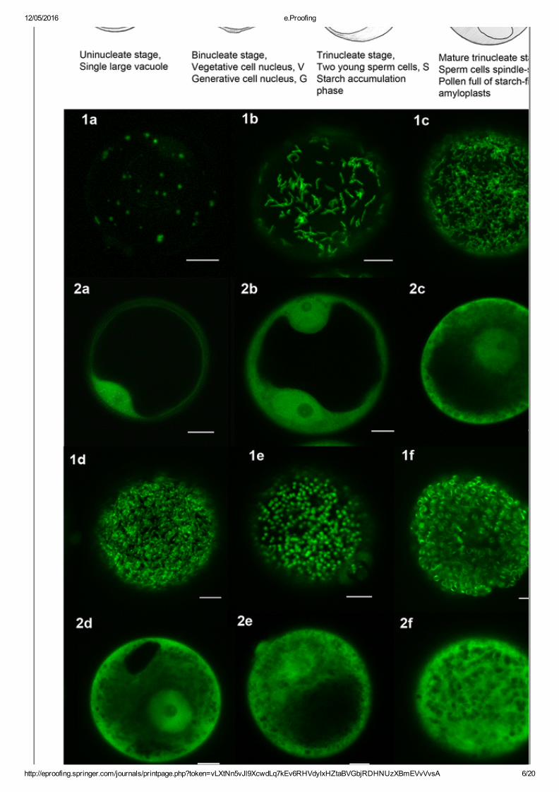
Plastids in developing pollen 1a–f GFPlabelled plastids. 2a–f UntargetedGFP (vacuole often clearly delineated). 1a and 2a uninucleate stage, plastidsvisible as small GFP labelled dots. 1b,c and 2b, c binucleate stage(vegetative nucleus and generative cell nucleus clearly visible in 2b).Plastids now visible as elongated structures in increased numbers. 1d, e and2d, e trinucleate stage. Plastids form an indistinct networklike structure (1c,d) before becoming clearly visible as discrete dots again (1e). 1f and 2fmature trinucleate stage, starch filled pollen. Plastids present as discreteexpanded structures. Scale bars represent 10 μm
12/05/2016 e.Proofing
http://eproofing.springer.com/journals/printpage.php?token=vLXtNn5vJl9XcwdLq7kEv6RHVdyIxHZtaBVGbjRDHNUzXBmEVvVvsA 6/20
12/05/2016 e.Proofing
http://eproofing.springer.com/journals/printpage.php?token=vLXtNn5vJl9XcwdLq7kEv6RHVdyIxHZtaBVGbjRDHNUzXBmEVvVvsA 7/20
At the uninucleate stage (single microspore cell with large vacuole), thereare relatively few, small plastids distributed throughout the cytoplasm(around 26 plastids, 1–2 μm, Fig. 1a ). As the pollen cell develops, themicrospore cell undergoes mitosis to form the vegetative cell and thesmaller generative cell. At this stage, the plastids have increased in numberand become thin elongated structures about 3–4μm long (Fig. 1b ). Opticalsections deeper into the pollen grain showed elongated plastidssurrounding dark areas corresponding to the vegetative nucleus and thecentral vacuole (not shown). By the late binucleate stage, the plastidsappear to form an extensive network throughout the cytoplasm (Fig. 1c ). Itwas not clear if they were actually connected to each other because theywere also highly dynamic and subject to rapid photobleaching. After thebinucleate stage, the generative cell undergoes mitosis to produce twosperm cells, the trinucleate stage. At this point, the channels of the ‘plastidnetwork’ appear to thicken (Fig. 1d ) and then become discrete sphericalplastids (Fig. 1e ). As the pollen grain finally matures, the spheres expand,filling up with starch, to a maximum size of 4 μm. There were severalhundred plastids distributed throughout the cytoplasm which itselfsurrounded a large vacuole (Fig. 1f ). At this point, GFP was often visibleas a band confined around the middle of the plastid. At all stages,untargeted GFP fluorescence patterns are shown for comparison (Fig. 1panels 2a–f). We observed the same changes in the morphologies andnumbers of plastids in pollen using three different plastidtargeted transitpeptides (rubisco SSU, ferredoxin III, ftsZ) (data not shown). Progressionfrom the unicellular stage shown in Fig. 1a to mature pollen in Fig. 2ftook around 72 h. During this relatively short time period, large changes inthe morphologies and abundance of GFPtagged plastids were observed.These included familiar circular forms (Fig. 1a, e–f ) as well as novelforms (Fig. 1b–d ) not previously described in pollen. Continuousmonitoring of GFPtagged plastids during the pollen development allowedus to uncover these dynamic features of pollen plastids, which would notbe easily revealed by classical electron microscopic approaches.
Fig. 2
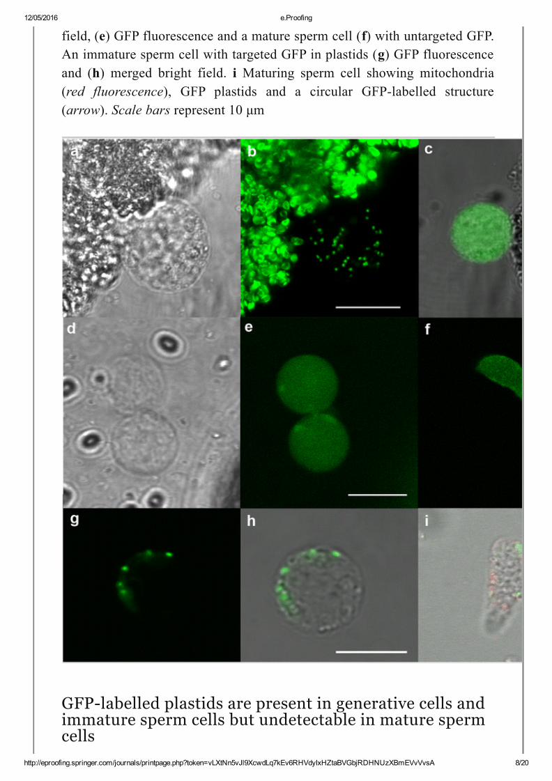
Plastids in generative and sperm cells (a) bright field and (b) GFPlabelledplastids in a generative cell. c Overlay of bright field and GFP fluorescencefrom nontargeted GFP generative cell. Two young sperm cells (d) bright
12/05/2016 e.Proofing
http://eproofing.springer.com/journals/printpage.php?token=vLXtNn5vJl9XcwdLq7kEv6RHVdyIxHZtaBVGbjRDHNUzXBmEVvVvsA 8/20
field, (e) GFP fluorescence and a mature sperm cell (f) with untargeted GFP.An immature sperm cell with targeted GFP in plastids (g) GFP fluorescenceand (h) merged bright field. i Maturing sperm cell showing mitochondria(red fluorescence), GFP plastids and a circular GFPlabelled structure(arrow). Scale bars represent 10 μm
GFPlabelled plastids are present in generative cells andimmature sperm cells but undetectable in mature spermcells
12/05/2016 e.Proofing
http://eproofing.springer.com/journals/printpage.php?token=vLXtNn5vJl9XcwdLq7kEv6RHVdyIxHZtaBVGbjRDHNUzXBmEVvVvsA 9/20
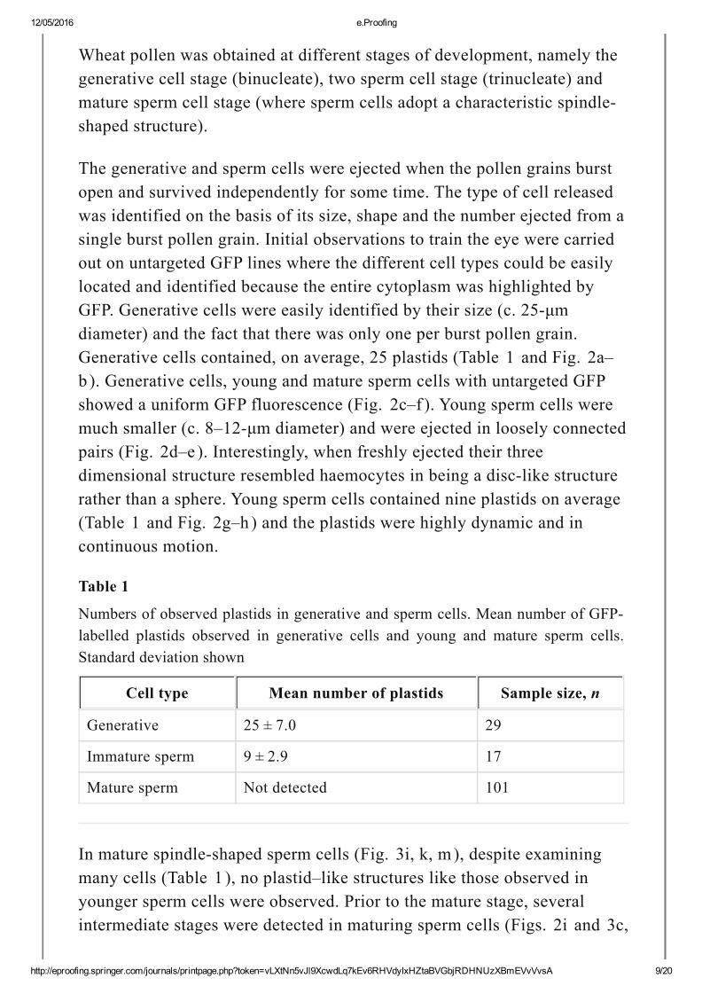
Wheat pollen was obtained at different stages of development, namely thegenerative cell stage (binucleate), two sperm cell stage (trinucleate) andmature sperm cell stage (where sperm cells adopt a characteristic spindleshaped structure).
The generative and sperm cells were ejected when the pollen grains burstopen and survived independently for some time. The type of cell releasedwas identified on the basis of its size, shape and the number ejected from asingle burst pollen grain. Initial observations to train the eye were carriedout on untargeted GFP lines where the different cell types could be easilylocated and identified because the entire cytoplasm was highlighted byGFP. Generative cells were easily identified by their size (c. 25μmdiameter) and the fact that there was only one per burst pollen grain.Generative cells contained, on average, 25 plastids (Table 1 and Fig. 2a–b ). Generative cells, young and mature sperm cells with untargeted GFPshowed a uniform GFP fluorescence (Fig. 2c–f ). Young sperm cells weremuch smaller (c. 8–12μm diameter) and were ejected in loosely connectedpairs (Fig. 2d–e ). Interestingly, when freshly ejected their threedimensional structure resembled haemocytes in being a disclike structurerather than a sphere. Young sperm cells contained nine plastids on average(Table 1 and Fig. 2g–h ) and the plastids were highly dynamic and incontinuous motion.
Table 1
Numbers of observed plastids in generative and sperm cells. Mean number of GFPlabelled plastids observed in generative cells and young and mature sperm cells.Standard deviation shown
Cell type Mean number of plastids Sample size, n
Generative 25 ± 7.0 29
Immature sperm 9 ± 2.9 17
Mature sperm Not detected 101
In mature spindleshaped sperm cells (Fig. 3i, k, m), despite examiningmany cells (Table 1 ), no plastid–like structures like those observed inyounger sperm cells were observed. Prior to the mature stage, severalintermediate stages were detected in maturing sperm cells (Figs. 2i and 3c,
12/05/2016 e.Proofing
http://eproofing.springer.com/journals/printpage.php?token=vLXtNn5vJl9XcwdLq7kEv6RHVdyIxHZtaBVGbjRDHNUzXBmEVvVvsA 10/20
f ), where a single unidentified structure containing GFP was alsoobserved. This structure was detected with plastids present, (Fig. 2i ) but inlater stages, it remained even in the absence of detectable plastids andcontained much fainter GFP fluorescence (Fig. 3i ). Compared to earlierstage plastids, this structure was larger, static and circular in cross section.In contrast, for transgenic lines containing the untargeted GFP,fluorescence was retained in mature sperm cells (Fig. 2f ). To confirm thatthe GFPlabelled structure in Figs. 2i and 3i was not simply the nucleus,DAPI staining of mature sperm cells was performed. As expected, theDAPIstained nucleus is much larger and appears to completely fill the cell(Supplementary Fig 1 .)
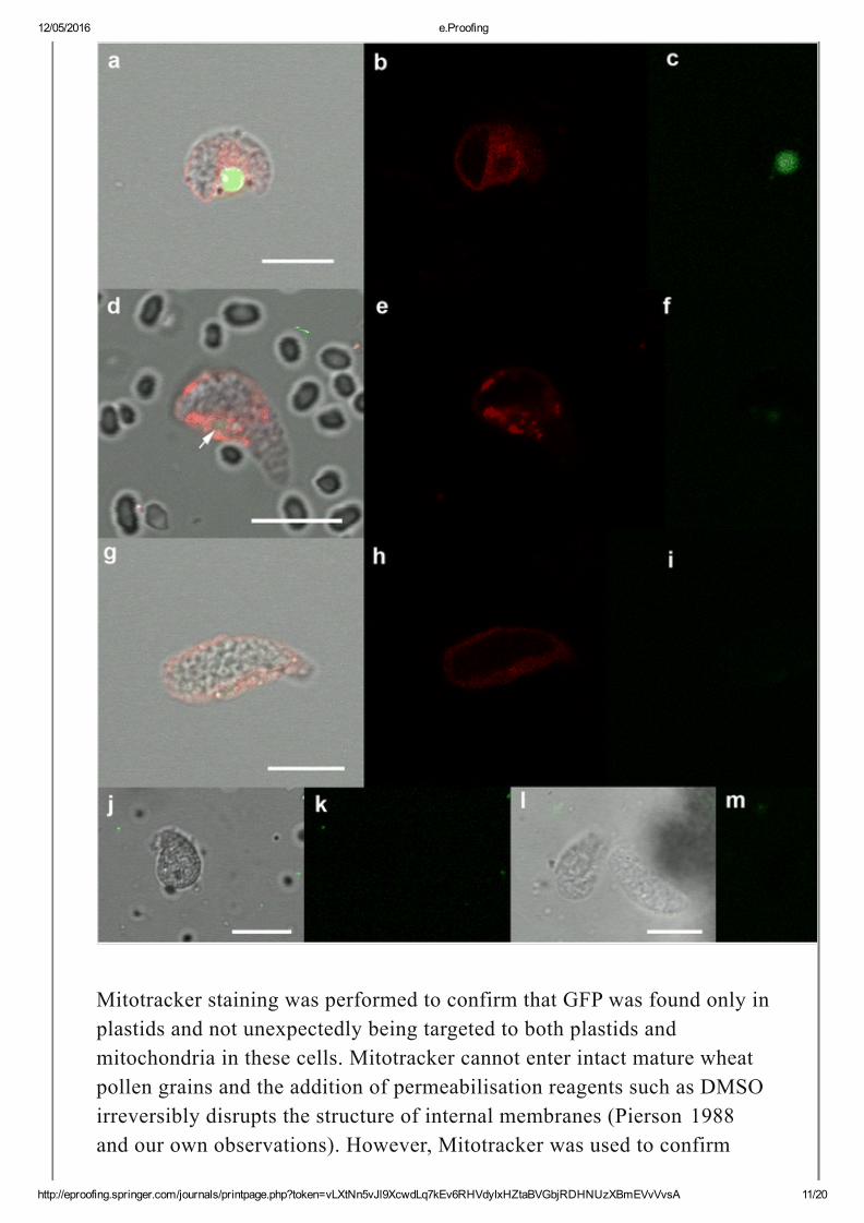
Fig. 3
Plastids throughout sperm cell maturation. a, d Overlay images of maturingsperm cells, (d) arrow points to faint circular GFP structure. b, eCorresponding fluorescence from Mitotracker, and (c, f) GFP. (g–i) Overlay,Mitotracker and GFP fluorescence from a mature sperm cell. j, k and l, mTwo more examples of mature sperm cells showing overlay and GFPchannels. Scale bars represent 10 μm
12/05/2016 e.Proofing
http://eproofing.springer.com/journals/printpage.php?token=vLXtNn5vJl9XcwdLq7kEv6RHVdyIxHZtaBVGbjRDHNUzXBmEVvVvsA 11/20
Mitotracker staining was performed to confirm that GFP was found only inplastids and not unexpectedly being targeted to both plastids andmitochondria in these cells. Mitotracker cannot enter intact mature wheatpollen grains and the addition of permeabilisation reagents such as DMSOirreversibly disrupts the structure of internal membranes (Pierson 1988and our own observations). However, Mitotracker was used to confirm
12/05/2016 e.Proofing
http://eproofing.springer.com/journals/printpage.php?token=vLXtNn5vJl9XcwdLq7kEv6RHVdyIxHZtaBVGbjRDHNUzXBmEVvVvsA 12/20
specific targeting of GFP to plastids by staining mitochondria ingerminated pollen tubes (Supplementary Fig 2 ). In addition, simultaneousstaining with Mitotracker to label mitochondria showed that GFP andMitotracker labelled distinct and separate structures in sperm cells(Fig. 3b, e, h ).
DiscussionBy using plastidtargeted GFP it was possible to examine living plastids inwheat pollen vegetative cells and observe the extensive morphologicalchanges that they undergo throughout pollen development. We observedthat through pollen development the number of plastids in the vegetativecell increases from a low number (approx. 30–40) of small plastids (1–2 μm) to several hundred plastids (around 4μm diameter) at maturity. Theplastids pass through a distinct morphological stage where they form thinelongated tubelike structures (binucleate stage). Such elongated plastidshave previously been observed in young green wheat leaves during anearly step in chloroplast division (Leech et al. 1981 ) and also inArabidopsis thaliana pollen plastids (Fujiwara et al. 2010 ). After thisstage, the plastids appear to form a very dynamic rapidly moving andcomplex network. In this study it was not possible to determine whetherthis complex network provided a mechanism to join the contents ofmultiple plastids. Elegant evidence from (Schattat et al. 2012 ) showed thatplastids connected by stromules do not exchange stromal contents.Apparent complex networks of plastids have been observed in a number ofdifferent species and cells (Pyke 2010 ; Osteryoung and Pyke 2014 ). Thefunctional significance of these networks is not fully understood (Schattatet al. 2015 ) although wheat pollen has welldefined developmental stagesand might be a particularly good system to study these networks. It is notclear what is happening to the plastids at this stage or why they adopt thesevaried shapes although amoeboid or pleomorphic amyloplasts have beenobserved in the root tips of Phaseolus (Newcomb 1967 ), in epithelial cellsof resin ducts in pine needles (Dvorak and Stokrova 1993 ) and in Alnusactinorhizal root nodules (Gardner et al. 1989 ) which are all nonphotosynthetic cells. GFPtagged plastids appear as spherical structures atearly and late stages of pollen development (Fig. 1a, e ). Spherical GFPtagged plastids were also observed in mature wheat (Fig. 1f and Primavesiet al. 2008 ) and Arabidopsis pollen (Matsushima et al. 2011 ). In contrast,mitochondria appear more oval in mature Arabidopsis pollen (Matsushima
12/05/2016 e.Proofing
http://eproofing.springer.com/journals/printpage.php?token=vLXtNn5vJl9XcwdLq7kEv6RHVdyIxHZtaBVGbjRDHNUzXBmEVvVvsA 13/20
et al. 2011 ).
We were also able to detect small GFPlabelled plastids present in thereproductive cells. Generative cells contained an average of 25 very smallplastids (1–2 μm). This is similar to meristem cells which contain 10–20proplastids per cell (Cran and Possingham 1972 ; Lyndon and Robertson1976 ). Young sperm cells contained an average of nine very small plastidsper cell. Previously, no overall count of the actual plastid numbers in thesecells has been obtained from electron microscopy (Hagemann et al. 1985 ).Since the number of plastids decreases from generative cell to sperm cell,it is likely that the generative cell plastids are simply distributed to the twosperm cells such that each sperm cell ends up with roughly half theplastids.
At no point during development and maturation did the generative/spermcell plastids increase in size as they do in the vegetative pollen cell whenthey accumulate starch, nor did the numbers significantly increase and norwas there any obvious change in their morphology. Our observations areconsistent with division of plastids being arrested in the generative cell andtheir number per cell reducing further following formation of the twosperm cells. Once inside the sperm cell, it is possible that the plastidssimply remain as proplastids and are protected in some way from thesignals which cause the vegetative cell plastids to elongate, divide andaccumulate starch. It was only when the sperm cell had undergonematuration and become spindleshaped and the pollen grain had reachedthe correct state for pollination that any change was observed in the spermcell plastids. At this stage, the typical small dynamic GFPlabelled plastidwas never observed in sperm cells. This loss of GFPtagged plastids wasobserved in several independent transgenic lines. In contrast, sperm cellswith untargeted GFP continued to show GFP fluorescence right throughdevelopment. No dimorphism with regard to plastid number or presencewas observed in young or mature sperm cells. An earlier electronmicroscopic study on barley pollen showed limited or no differences in thecontent of mitochondria in sperm pairs (Mogensen and Rusche 1985 ).
It had been assumed from earlier evidence in cereals (Hagemann andSchroder 1989 ; Schroder and Hagemann 1986 ) that the cytoplasmicstripping mode of maternal inheritance must occur in T. aestivum (Triticumtype). This assumes that plastids are present in sperm cells and are then
12/05/2016 e.Proofing
http://eproofing.springer.com/journals/printpage.php?token=vLXtNn5vJl9XcwdLq7kEv6RHVdyIxHZtaBVGbjRDHNUzXBmEVvVvsA 14/20
somehow excluded from the egg cell at fertilisation when the sperm cellcytoplasm is left on the outside of the egg cell while the sperm nucleusgoes on to fuse with that of the egg. Our study provides evidence thatpaternal plastids are clearly present in young sperm cells but at a pointvery close to sperm cell maturity (very close to the point of pollen release)the plastids are degraded. We conclude, that the detection of plastids inpollen sperm cells by electron microscopy is not sufficient to deduce theircontinued presence in mature sperm. This requires monitoring the fate ofplastids during sperm development, which is more easily achieved byvisualising GFPtagged plastids (this work) than by electron microscopicanalysis. It is also possible that environmental factors play a role in themaintenance of plastids in sperm cells. This was addressed by placingtillers with developing pollen continuously at 15 or 5 °C for a weekfollowed by 23 °C. The cold temperatures used did not appear to affect thefate of plastids in sperm cells, which were degraded in mature pollen(results not shown). Cytoplasmic stripping for organelles other thanplastids is not ruled out by this study, but we provide evidence that theplastids are degraded prior to fertilisation.
In this study, we observe the disappearance of intact GFPlabelled plastids.The fate of these plastids might be related to the observation that duringthe sperm maturation process, a single small area of GFP fluorescence wasoften observed and which later disappeared from mature sperm. Thefluorescence in this structure was contained in a static circular/sphericalstructure unlike the variable shape of the dynamic plastids in earlier pollenstages. The fluorescence was sometimes quite bright, but as the spermmatured, it became greatly diminished. We speculate that the structure is acandidate for an organelle involved in the terminal degradation of plastids,possibly a lytic vacuole. Several pathways have been described for thedestruction of plastids including stromal and thylakoid proteins (Chiba etal. 2003 ; Michaeli et al. 2014 ; Wang and Blumwald 2014 ). The presenceof chloroplasts within the vacuole has been observed by electronmicroscopy studies on senescing leaves of wheat (Wittenbach et al. 1982 )and Phaseolus vulgaris (Minamikawa et al. 2001 ). Plastid degradation issupported by the observation that plastid nucleoids are detected by DAPIstaining in wheat generative cells (Miyamura et al. 1987 ) but not in spermcells (Corriveau and Coleman 1988 ). Two further observations areconsistent with destruction of plastids and their genomes during pollendevelopment. First, wheat plants regenerated from pollen by anther culture
12/05/2016 e.Proofing
http://eproofing.springer.com/journals/printpage.php?token=vLXtNn5vJl9XcwdLq7kEv6RHVdyIxHZtaBVGbjRDHNUzXBmEVvVvsA 15/20
show a very high level of albinism and large deletions in their plastid DNA(Day and Ellis 1984 ). Second, escape of plastid DNA to the nucleus iselevated in the sperm cells of tobacco pollen (Sheppard et al. 2008 ).
This study provides evidence that plastids do not persist in mature wheatsperm cells, and that they may be selectively degraded in the germ lineduring the final stages of sperm cell maturation. We propose the plastidinheritance pattern in wheat is actually more like the Solanum type whereplastids are not present in the sperm cells (Greiner et al. 2015 ; Hagemannand Schroder 1989 ), although in the case of wheat, they are present in thegenerative cell and destroyed during maturation of sperm cells.
AcknowledgmentsRothamsted Research receives support from the Biotechnological andBiological Sciences Research Council (BBSRC) of the UK as part of the20:20 Wheat® Programme.Work was supported by grant number GM114215. We thank CarolineSparks for her invaluable help and also Richard Parkinson and FionaGilzean for their assistance in growing the wheat plants.
Electronic supplementary materialBelow is the link to the electronic supplementary material.
ESM 1
(DOCX 1977 kb)
ReferencesAQ4
Birky CW (1995) Uniparental inheritance of mitochondrial andchloroplast genes—mechanisms and evolution. Proc Natl Acad Sci U SA 92:11331–11338. doi: 10.1073/pnas.92.25.11331
Bock R (2007) Structure, function and inheritance of plastid genomes.In: Cell and Molecular Biology of Plastids. Topics in Current Genetics,vol 19. SpringerVerlag, Berlin
12/05/2016 e.Proofing
http://eproofing.springer.com/journals/printpage.php?token=vLXtNn5vJl9XcwdLq7kEv6RHVdyIxHZtaBVGbjRDHNUzXBmEVvVvsA 16/20
Briggle LW (1966) Inheritance of a variegated leaf pattern in hexaploidwheat. Crop Sci 6:43–45
Chiba A, Ishida H, Nishizawa NK, Makino A, Mae T (2003) Exclusionof ribulose1, 5bisphosphate carboxylase/oxygenase from chloroplastsby specific bodies in naturally senescing leaves of wheat. Plant CellPhysiol 44:914–921. doi: 10.1093/Pcp/Pcg118
Corriveau JL, Coleman AW (1988) Rapid screening method to detectpotential biparental inheritance of plastid DNA and results for over 200angiosperm species. Am J Bot 75:1443–1458. doi: 10.2307/2444695
Cran DG, Possingham JV (1972) Variation of plastid types in spinach.Protoplasma 74:345–356. doi: 10.1007/Bf01282537
Daniell H, Datta R, Varma S, Gray S, Lee SB (1998) Containment ofherbicide resistance through genetic engineering of the chloroplastgenome. Nat Biotechnol 16:345–348. doi: 10.1038/Nbt0498345
Day A, Ellis THN (1984) Chloroplast DNA deletions associated withwheat plants regenerated from pollen—possible basis for maternalinheritance of chloroplasts. Cell 39:359–368. doi: 10.1016/00928674(84)90014X
Dvorak J, Stokrova J (1993) Structure of the needles in the early phasesof development in PinusPonderosa Lawson, P. Et Lawson, C. withspecial reference to plastids. Ann BotLondon 72:423–431. doi:10.1006/anbo.1993.1128
Fujiwara MT et al (2010) Dynamic morphologies of pollen plastidsvisualised by vegetativespecific FtsZ1GFP in Arabidopsis thaliana.Protoplasma 242:19–33. doi: 10.1007/s0070901001197
Gardner IC, Abbas H, Scott A (1989) The occurrence of ameboidplastids in the actinorhizal rootnodules of AlnusGlutinosa (L) Gaertn.Plant Cell Environ 12:205–211. doi: 10.1111/j.13653040.1989.tb01934.x
Greiner S, Sobanski J, Bock R (2015) Why are most organelle genomes
12/05/2016 e.Proofing
http://eproofing.springer.com/journals/printpage.php?token=vLXtNn5vJl9XcwdLq7kEv6RHVdyIxHZtaBVGbjRDHNUzXBmEVvVvsA 17/20
transmitted maternally? Bioessays 37:80–94. doi:10.1002/bies.201400110
Hagemann R, Schroder MB (1989) The cytological basis of the plastidinheritance in angiosperms. Protoplasma 152:57–64. doi:10.1007/Bf01323062
Hagemann R, Schroder MB New results about the presence of plastidsin generative and sperm cells of Gramineae. In, Pudoc, Wagenigen,1985. Sexual reproduction in seed plants, ferns and mosses. pp 53–55
Hu S, Zhu C, Xu L, J. S (1979) Ultrastructure of male gametophyte inwheat 1. The formation of generative and vegetative cells. ActaBotanica Sinica 21:208–214
Leech RM, Thomson WW, PlattAloia KA (1981) Observations on themechanism of chloroplast division in higher plants. New Phytol 87:1–9.doi: 10.1111/j.14698137.1981.tb01686.x
Lyndon RF, Robertson ES (1976) Quantitative ultrastructure of peashoot apex in elation to leaf initiation. Protoplasma 87:387–402. doi:10.1007/Bf01624007
Matsushima R, Tang LY, Zhang L, Yamada H, Twell D, Sakamoto W(2011) A conserved, Mg2 dependent exonuclease degrades organelleDNA during Arabidopsis pollen development. Plant Cell 23:1608–1624.doi: 10.1105/tpc.111.084012
Michaeli S, Honig A, Levanony H, PeledZehavi H, Galili G (2014)Arabidopsis ATG8INTERACTING PROTEIN1 is involved inautophagydependent vesicular trafficking of plastid proteins to thevacuole. Plant Cell 26:4084–4101. doi: 10.1105/tpc.114.129999
Minamikawa T, Toyooka K, Okamoto T, HaraNishimura I, NishimuraM (2001) Degradation of ribulosebisphosphate carboxylase byvacuolar enzymes of senescing French bean leaves:immunocytochemical and ultrastructural observations. Protoplasma218:144–153. doi: 10.1007/Bf01306604
+
12/05/2016 e.Proofing
http://eproofing.springer.com/journals/printpage.php?token=vLXtNn5vJl9XcwdLq7kEv6RHVdyIxHZtaBVGbjRDHNUzXBmEVvVvsA 18/20
Miyamura S, Kuroiwa T, Nagata T (1987) Disappearance of plastid andmitochondrial nucleoids during the formation of generative cells ofhigher plants revealed by fluorescence microscopy. Protoplasma141:149–159. doi: 10.1007/Bf01272897
Mogensen HL (1988) Exclusion of male mitochondria and plastidsduring syngamy in barley as a basis for maternal inheritance. Proc NatlAcad Sci U S A 85:2594–2597. doi: 10.1073/pnas.85.8.2594
Mogensen HL (1996) The hows and whys of cytoplasmic inheritance inseed plants. Am J Bot 83:383–404. doi: 10.2307/2446172
Mogensen HL, Rusche ML (1985) Quantitative ultrastructural analysisof barley sperm.1. Occurrence and mechanism of cytoplasm andorganelle reduction and the question of sperm dimorphism. Protoplasma128:1–13. doi: 10.1007/BF01273229
Nagata N (2010) Mechanisms for independent cytoplasmic inheritanceof mitochondria and plastids in angiosperms. J Plant Res 123:193–199.doi: 10.1007/s102650090293x
Newcomb EH (1967) Fine structure of proteinstoring plastids in beanroot tips. J Cell Biol 33:143–163. doi: 10.1083/Jcb.33.1.143
Osteryoung KW, Pyke KA (2014) Division and dynamic morphology ofplastids. Annu Rev Plant Biol 65(65):443–472. doi: 10.1146/annurevarplant050213035748
Pao WK, Li HW (1946) Maternal inheritance of variegation in commonwheat. J Am Soc Agron 38:90–94
Pierson ES (1988) Rhodaminephalloidin staining of Factin in pollenafter dimethylsulfoxide permeabilization. Sex Plant Reprod 1:83–87
Primavesi LF, Wu H, Mudd EA, Day A, Jones HD (2008) Visualisationof plastids in endosperm, pollen and roots of transgenic wheatexpressing modified GFP fused to transit peptides from wheat SSURubisCO, rice FtsZ and maize ferredoxin III proteins. Transgenic Res17:529–543. doi: 10.1007/s1124800791267
12/05/2016 e.Proofing
http://eproofing.springer.com/journals/printpage.php?token=vLXtNn5vJl9XcwdLq7kEv6RHVdyIxHZtaBVGbjRDHNUzXBmEVvVvsA 19/20
Pyke KA (2010) Plastid division AoB PLANTS 2010:plq016plq016doi: 10.1093/aobpla/plq016
Ruf S, Karcher D, Bock R (2007) Determining the transgenecontainment level provided by chloroplast transformation. Proc NatlAcad Sci U S A 104:6998–7002. doi: 10.1073/pnas.0700008104
Schattat MH et al (2012) Differential coloring reveals that plastids donot form networks for exchanging macromolecules. Plant Cell 24:1465–1477. doi: 10.1105/tpc.111.095398
Schattat MH, Barton KA, Mathur J (2015) The myth of interconnectedplastids and related phenomena. Protoplasma 252:359–371. doi:10.1007/s0070901406664
Schroder MB, Hagemann R (1986) Ultrastructural studies on plastids ofgenerative and vegetative cells in Liliaceae.6. Patterns of plastiddistribution during generative cellformation in Aloe secundiflora andAloe jucunda. Acta Bot Neerl 35:243–248
Sheppard AE et al (2008) Transfer of plastid DNA to the nucleus iselevated during male gametogenesis in tobacco. Plant Physiol 148:328–336. doi: 10.1104/pp.108.119107
Svab Z, Maliga P (2007) Exceptional transmission of plastids andmitochondria from the transplastomic pollen parent and its impact ontransgene containment. Proc Natl Acad Sci U S A 104:7003–7008. doi:10.1073/pnas.0700063104
Svab Z, Hajdukiewicz P, Maliga P (1990) Stable transformation ofplastids in higher plants. Proc Natl Acad Sci U S A 87:8526–8530. doi:10.1073/pnas.87.21.8526
Wang SH, Blumwald E (2014) Stressinduced chloroplast degradationin Arabidopsis is regulated via a process independent of autophagy andsenescenceassociated Vacuoles. Plant Cell 26:4875–4888. doi:10.1105/tpc.114.133116
Wittenbach VA, Lin W, Hebert RR (1982) Vacuolar localization of

12/05/2016 e.Proofing
http://eproofing.springer.com/journals/printpage.php?token=vLXtNn5vJl9XcwdLq7kEv6RHVdyIxHZtaBVGbjRDHNUzXBmEVvVvsA 20/20
proteases and degradation of chloroplasts in mesophyll protoplasts fromsenescing primary wheat leaves. Plant Physiol 69:98–102. doi:10.1104/Pp.69.1.98