Embed Size (px)
Citation preview

The Pingry Community Research (PCR) Journal
A Journal of Scientific Research
Volume 4: Spring 2016

ContentsPage 1: The Wolbachia Project: Discover the Microbes Within
By Sean Wang ‘17, Mariel Sander ‘16, Parth Patel ‘17, Lily Cao ‘17, Ethan Chung ‘18 3: Protein Purification of Salmonella Typhimurium Transcription Factors HilC and HilD By Matthew Newman ‘16, Arjun Patel ‘16, Charles Papandreou ‘17, Jackie Chang ‘18, Lindsay Rispoli ‘18 Jessica Day, F. Jon Kull, Alexandra Lo gerfo, Morgan D’Ausilio 5: The Effects of Meditation on Stress and Cognitive Ability in Teenagers By Kathryn Abbot ‘16, Caroline Terens ‘16 7: Evaluating the Ability for Mealworms to Digest Plastics By Brandon Li ‘17 9: Increased Tau and Amyloid-Beta in “Alzheimer’s Induced” Drosophila Me lanogaster Through Oxygen Deprivation By Hannah Curtis ‘16, Andrew Epifano ‘17 10: The Effects of Menstruation on Cognitive Memory Function By Amaka Nnaeto ‘16 11: Optimization of Landfill Gases for Biomethane Energy Production Kira Bartnick ‘16, Chris Browne ‘16, Danielle LeGrand ‘16, Aidan Zola ‘16 13: Investigating the eIF4E -4EBP1 Signaling Pathway Philip Geter, Andrew Verdesca ‘15, Morgan D’Ausilio, Alexandra Lo gerfo, Katie Coyne ‘16, Isabella Zanobini ‘16, Ben Zhou ‘17, Jess Li ‘18 15: The Effects of Chrysin on Liver Detoxification and Metabolism of Acet aminophen Ryan Lane ‘16, Alina Jan ‘16 17: Pharmacologic reduction of anxiety in Danio rerio Michael James ‘16, Sydney Stein ‘16, Owen Storms ‘16 19: Investigating the Effect of Amygdalin on Melanoma Cell Proliferation Caroline Marone ‘17, Heba Syed ‘17 20: Creating Point Mutations in BRCA2 Emily Kwon ‘16, Yanni Angelides ‘16, Luke De, Ryan Jensen 22: The Effect of Frequency on Anxiety in Danio rerio Brian Grimaldi ‘16, Samantha Palazzolo ‘16

For Materials and Methods sections, please visit www.pingry.org/pcr
Cover Photo: Paul Hebert - Functional Genomics Thickens the Biological Plot. Gewin V, PLoS Biology Vol. 3/6/2005, e219. doi:10.1371/journal.pbio.0030219
THE PCR STAFF
Editor in Chief:Brad Hong ‘16
Layout Editor:Kartikeya Sharma ‘17
Copy Editors:Yelena Salvador ‘17Andrew Epifano ‘17Namita Davey ‘18Ketaki Tavan ‘19
Faculty Advisor:Mr. David Maxwell

1
The Wolbachia Project: Discover the Microbes WithinSean Wang ‘17, Mariel Sander ‘16, Parth Patel ‘17, Lily Cao ‘17, Ethan Chung ‘18
Wolbachia has a variety of different features that make it a point of interest for researchers in the realms of population control and disease vectors. There is a large genetic diversity within strains of Wolbachia; as a result, there is a need for catalog-ing all the different sequences present in nature. The Wolbachia Project was developed to allow students
ABSTRACTto learn basic lab protocols while simultaneously getting involved with ongoing research. While we have been able to detect the presence of Wolbachia and insect DNA within our samples, the lack of consistency and reliability has become the primary focus of this year’s research.
INTRODUCTION Wolbachia is a symbiotic bacterium present in approximately one-fifth of the world’s insect popula-tion (3). It is transmitted only from females to their offspring and facilitates its own survival by chang-ing the gender ratio of an insect population through three main methods. The first is feminization, or the changing of male insects to females (3). The second is parthenogenesis, which allows female insects to reproduce without fertilization (3). The third main method used by the bacterium is killing males (3). As a result, the Wolbachia bacterium has several practi-cal applications; the bacterium can potentially be used to control disease vector populations like mosquitoes by interfering with the vector’s reproduction capabili-ties (4). Wolbachia is also linked with river blindness (onchocerciasis) as the bacterium infects nematodes which parasitize humans. The true scope of the Wolbachia bacterium is not yet known. As a result, scientists are attempting to compile a database of all known strains of Wolbachia by bringing the Wolbachia Project to high schools across the country (2). Our branch of the Wolbachia Project has two aims: to introduce lab techniques to high school students and to catalogue strains of Wolbachia bacterium. Through a five-lab course, we can help students gain basic lab techniques and un-derstand how their research contributes to a larger program. The Project’s curriculum is broken up into five components: insect identification, extraction of insect and Wolbachia DNA, amplification of insect and Wolbachia DNA, analysis for presence/absence of insect and/or Wolbachia DNA, and sequencing/bioinformatics (2). In insect identification, we find the
taxonomic order of an insect (e.g. the order of a but-terfly is lepidoptera) which will be used for catalog-ing purposes. Once we extract insect and Wolbachia DNA, we will amplify certain portions of the DNA through polymerase chain reaction (PCR), which replicates DNA at a rapid rate. We will then test for the presence of insect and Wolbachia DNA through gel electrophoresis. By working on the Wolbachia Project, students will become familiar with basic lab skills like pipetting, setting up a PCR protocol, and running gel electrophoresis. If experimental samples test positive for Wolbachia DNA, they are sent to the Marine Biological Laboratory (MBL) for sequenc-ing. These sequences will be added to the MBL’s local database and a student will have submitted their own DNA sequence. Students can then run an evolutionary taxonomy analysis and BLAST sequencing to see how their sequence relates to previously found sequences or organisms. DISCUSSION We have been performing the protocol to en-sure that the project can be successfully run. However, we have found difficulty in obtaining accurate bands from our samples. Some bands that were not expected appeared, while others that were expected did not ap-pear, possibly due to contamination or an error while performing protocol. Thus, we have continued to revise protocol primarily in the PCR step by changing the thermocycling temperatures and the reaction com-position. Due to the results indicating that there are er-rors within the experiment, we have yet to send in our DNA samples for sequences. Rather, we are focusing on improving our results and improving the reliability of the results.

2Obtaining greater consistency in the gels would enable us to send in PCR samples of our experimental insect samples for DNA sequencing. We could then analyze our DNA sequences through the bioinformatics por-tion of the Wolbachia Project and have our sequences inputted into the Marine Biological Lab’s internal database. Once we obtain more consistent results, we plan on expanding the program to the freshman and sophomore student body so they can also participate in the Wolbachia Project.
REFERENCES1. Wolbachia Biology. (n.d.). Retrieved February 15, 2016, from University of Rochester website: https://www.rochester.edu/College/BIO/labs/WerrenLab/WerrenLab-WolbachiaBiology.html 2. Bordenstein, S. R. (2007, Fall). Discover the Mi-crobes Within!: The Wolbachia Project. Focus on Microbiology Education, 14(1). Retrieved from http://www.mbl.edu/education/files/2014/04/bordenstein_fome_20071.pdf 3. Werren, J. H., Baldo, L., & Clark, M. E. (2008). Wolbachia: master manipulators of invertebrate biol-ogy. Nat Rev Micro, 6(10), 741–751. Retrieved from http://dx.doi.org/10.1038/nrmicro1969(4)Bourtzis, K. (2008). Transgenesis and the Manage-ment of Vector-Borne Disease. In S. Aksoy (Ed.), (pp. 104–113). New York, NY: Springer New York. http://doi.org/10.1007/978-0-387-78225-6_9
ACKNOWLEDGEMENTSSpecial thanks to David Maxwell, Alexandra Logerfo, Luke De, Morgan D’Ausilio, Sarah Bordenstein, Hege Lizarralde, Jack Werren, and Phillip Geter.
Between September to mid-December (2015), we were able to run through the protocols efficiently; however, the presence of bands was lacking due to the lack of a marking agent (i.e. SYBRSAFE) or issues with the PCR protocol (e.g. too high annealing tem-peratures). After corresponding with professors in late December, we began to obtain a number of bands (see figure 1) by adjusting the PCR reaction concentrations and the PCR protocol temperatures (e.g. changing an-nealing or elongation temperatures). However, revis-ing the protocol as a reaction using Taq Polymerase Master Mix (a liquid) would increase the reaction volume to 50ul which would indicate that we used too little primers or DNA. Finding the proper combi-nation of PCR reaction concentrations and protocol temperatures have become our main areas of focus for improving the protocol. In addition, our review of literature suggests DNA extraction samples shearing due to thawing and stability over periods of time as other plausible reasons for our data inconsistencies. We plan to continue trying to improve our results in the Nasonia controls as well as the insect primers (i.e. obtaining the insect bands regularly).
Figure 1. We were able to obtain Wolbachia bands (bottom band) across the gel. In our ladybug sample, we were able to obtain an insect band (top band) as well. However, there are no insect bands within the bee or nasonia samples. In addition, we ob-tained a Wolbachia band in the Nasonia - control which was not expected which could be due to contamination.

Protein Purification of Salmonella Typhimurium Transcription Factors HilC and HilD
Matthew Newman ‘16, Arjun Patel ‘16, Charles Papandreou ‘17, Jackie Chang ‘18, Lindsay Rispoli ‘18 Jessica Day, F. Jon Kull, Alexandra Logerfo, Morgan D’Ausilio
INTRODUCTION S. Typhimurium causes salmonellosis, specifi-cally mammalian gastroenteritis. An estimated 93.8 million cases of gastroenteritis due to Salmonella species occur globally each year (8). Symptoms vary from diarrhea and abdominal cramps to septic infec-tions. In general, nontyphoidal salmonellosis alone kills over 100,000 people each year. Infection begins with the ingestion of contaminated food or water (5). Many of the genes required for intestinal penetration, invasion of host cells, and intestinal and diarrheal disease are carried on a 40-kb region at centisome 63,
called Salmonella pathogenicity island 1 (SPI1) (8,12). SPI1 encodes for a protein secretion system, termed the type III secretion system (T3SS), which modulates host cellular functions by delivering effector proteins to the host cytoplasm (8). The transcription factor HilA is the major regulator of the salmonella toxin and is located on SPI1. HilA is regulated by HilC and HilD, also located on SPI1. HilC and HilD increase expression of HilA by opposing down-regulating activity. Inhibiting HilC and HilD would also inhibit HilA, thus inhibiting the whole virulence cascade of S. Typhimurium.
Salmonella enterica subsp. enterica serovar Ty-phimurium (S. Typhimurium) is a Gram-negative enteric pathogen that is a leading cause of bacterial food-borne illness worldwide (13). This has led scien-tists to more closely examine the virulence cascade of S. enterica. The cascade includes two AraC superfam-ily transcription factors, HilC and HilD, which regu-late the primary toxicity factors (4). Structural analy-sis carried out in the Kull Laboratory at Dartmouth College revealed the regulatory mechanism of ToxT, an AraC family member involved in the virulence cascade of Vibrio cholerae (9).The crystal structure of ToxT showed that DNA binding is inhibited by a small
fatty acid (9). Comparison of ToxT and S. enterica transcription factor sequences suggests that HilC and HilD (also members of the AraC protein superfamily) may also be inhibited by lipid binding. To investigate whether or not HilC and HilD can be inhibited in a manner similar to ToxT inhibition, we are work-ing to express, purify, and ultimately solve the three dimensional structures of HilC and HilD. Solving the structures of these transcription factors would help to reveal the mechanism of transcriptional regulation in S. enterica and in turn could lead to development of novel treatments for Salmonella related illnesses.
ABSTRACT
Figure 1: A fatty acid bound to ToxT’s ligand binding pocket, therefore inhibiting the tran-scription factor’s ability to bind to DNA.
Figure 2 : The regulation pathway of HilC, HilD, RtsA and HilA, as well as HilC and HilD’s interactions with HilA. RtsA regu-lates the expres-sion of invading flagellar genes in Salmonella enterica serovar Typhimurium and is thought to use the same pathway of expression and HilC and HilD.
3

4DISCUSSION Once the proteins HilC and HilD are isolated and expressed, we can send them to be structured. The crystallization process utilizes the hanging drop vapor diffusion method. A synchrotron radiation machine blasts x-ray beams at the protein crystals. A supercom-puter generates an electron density map, which shows possible structures of crystallized protein based on the diffraction patterns collected from the synchrotron. From this data, scientists can determine the structure of the protein. Determining the structures of HilC and HilD would allow us to examine the possibility of lipid inhibition of the S. enterica virulence cascade, which could in turn lead to preventive treatments for salmonellosis.
Figure 14 : X-ray crystal-lography allows researchers to conclude a general structure of the crystallized protein.
Figure 15: An example of an electron density map that helps to solve the structure of the protein.
REFERENCES1. De Silva, Rukman S., et al. “Crystal Structure of the Vibrio cholerae Quorum-Sensing Regulatory Protein HapR.” Journal of Bacteriology 189.15 (2007): n. pag. Print.2. De Silva, Rukman S., et al. “Crystal Structure of the Virulence Gene Activator AphA from Vibrio cholerae Reveals It Is a Novel Member of the Winged Helix Transcription FactorSuperfamily.” Journal of Biological Chemistry 280.14 (2005): n.
pag. Print.3. Dessau, Moshe A., and Yorgo Modis. “Protein Crystalli-zation for X-ray Crystallography.” Journal of Visualized Experi-ments (2011): n. pag. Print.4. Ellermeier, Craig D., Jeremy R. Ellermeier, and James M. Slauch. “HilD, HilC and RtsA constitute a feed forward loop that controls expression of the SPI1 type three secretion system regulator hilA in Salmonella enterica serovar Typhimurium.” Molecular Microbiology 57.3 (2005): n. pag. Print.5. Fàbrega, Anna, and Jordi Vila. “Salmonella enterica Serovar Typhimurium Skills To Succeed in the Host: Virulence and Regulation.” Clinical Microbiology Reviews 26.2 (2013): n. pag. Print.6. Hartley, James L., Gary F. Temple, and Michael A. Brasch. “DNA Cloning Using In Vitro Site-Specific Recombina-tion.” Genome Research 10.11 (2000): n. pag. Print.7. Hedhammar, My, Amelie Eriksson Karlström, and So-phia Hober. “Chromatographic methods for protein purification.” Royal Institute of Technology: n. pag. Print.8. Lim, Sangyong, et al. “Analysis of HilC/D-dependent invF promoter expression under different culture conditions.” Microbial Pathogenesis 52 (2012): n. pag. Print.9. Lowden, Michael J., et al. “Structure of Vibrio cholerae ToxT reveals a mechanism for fatty acid regulation of virulence genes.” PNAS 107.7 (2010): n. pag. Print.10. Miller, V. L., and J. J. Mekalanos. “A novel suicide vec-tor and its use in construction of insertion mutations: osmoregula-tion of outer membrane proteins and virulence determinants in Vibrio cholerae requires toxR.” Journal of Bacteriology 170.6 (1988): n. pag. Print.11. Mitchell, S. F., and J. R. Lorsch. “Protein Affinity Purification using Intein/Chitin Binding Protein Tags.” Methods Enzymol (2014): n. pag. Print.12. Olekhnovich, Igor N., and Robert J. Kadner. “DNA-Binding Activities of the HilC and HilD Virulence Regulatory Proteins of Salmonella enterica Serovar Typhimurium.” JOUR-NAL OF BACTERIOLOGY 184.15 (2002): n. pag. Print.13. Petrone, Brianna L., Anne M. Stringer, and Joseph T. Wade. “Identification of HilD-Regulated Genes in Salmonella enterica Serovar Typhimurium.” Journal of Bacteriology 196.5 (2014): n. pag. Print.14. Taylor, Jennifer, et al. “The crystal structure of AphB, a virulence gene activator from Vibrio cholerae, reveals residues that influence its ?response to oxygen and pHmmi_7919.” Mo-lecular Microbiology 83.3 (2012): n. pag. Print.15. Yang, Ji, Marija Tauschek, and Roy M. Robins-Browne. “Control of bacterial virulence by AraC-like regulators that re-spond to chemical signals.” Trends in Microbiology 19.3 (2011): n. pag. Print.
ACKNOWLEDGEMENTSJessica Day, Dr. John Kull, Morgan D’Ausilio, Kull Lab, Luke De, Denise Brown-Allen, David Maxwell

The Effects of Meditation on Stress and Cognitive Ability in TeenagersKathryn Abbot ‘16, Caroline Terens ‘16
The purpose of this experiment was to test the ef-fects of meditation on stress and cognitive ability. We split 16 human subjects into four groups, two experimental groups and two controls. Heart rate, memory, blood pressure, and reaction time were
ABSTRACTtaken before and after the experimental phase. Al-though our results were statistically insignificant, we observed patterns that suggest that meditation has positive effects on stress and cognitive ability, which can lead to further research.
INTRODUCTION Meditation is a practice used to reduce stress (1). High levels of stress can inhibit cogni-tive ability and take away from a person’s mental capability and performance (2). Stress affects people of all ages and professions, but the ef-fects of stress in students, both male and female, of ages 17-18 are analyzed in this study (3). We chose this age category because it is an important developmental and transitional phase and most relevant to our community. Previous research tested the short term ef-fects of meditation either before or after induced stress in males between the ages of 23-30. A con-clusion was made that “the practice of meditation reduced the physiologic stress responses without taking away the beneficial effect of stress, namely, improved memory score.” We conducted this study to see if medita-tion produces the same beneficial effects on male and female teenagers who naturally have higher levels of stress due to hormonal changes. 16 sub-jects, 8 female and 8 male, practiced either medi-tation or normal activity before or after an in-duced stress test. There were 4 distinct groups in order to compare the effects of meditation before and after stress compared to continuing with nor-mal life. The control of the study was the subjects who did not meditate. Both prior to and following the meditation and induced stress test, vthe sub-jects’ cognitive ability was tested to provide the data necessary for comparison.
RESULTS
5
Figure 1: The average change in heart rate in beats per minute of each group from pre-Phase 1/Phase 2 to post-Phase 1/Phase 2.
Figure 2: The average change in memory level of each group from pre-Phase 1/Phase 2 to post-Phase 1/Phase 2.

6
DISCUSSION In the pre-test questionnaire, most subjects reported normal sleep patterns from the night before ranging from 6 to 8 hours. Subjects who each rated their stress on a scale of 1-10 reported stress from 2 to 8 due to various reasons including schoolwork, social life, college, family, and lack of sleep. Current anxiety levels and alertness varied for each subject. The Trier Stress Test induced stress due to fears of unpreparedness when being put on the spot, public speaking, and arithmetic testing under pressure. Heart rate generally either decreased or remained the same post-meditation and stress in Groups 1 and 3, while either increasing or remaining the same post-waiting and stress in Groups 2 and 4. Based on the patterns seen in the graphs, it is possible that medita-tion can improve a subject cognitive ability by im-proving memory and reaction time. There is a possibil-ity that stress and anxiety can also improve reaction time as seen in the subjects’ results, which could be further studied. In the post-questionnaire, subjects who medi-tated after induced stress (Group 3) reported the most significant decrease in their levels of stress and anxi-ety. All subjects felt more or just as alert as they did beforehand. An ANOVA test (one-way analysis of vari-ance) was used to determine the statistical significance of our results. Our p-values were as follows:
p-valueHeart rate 0.4Memory 0.4Reaction 0.8
The p-values were too high for our results to be statistically significant. However, the patterns from the graphs suggest that the groups that meditated (1 and 3) show more relaxing results and better cognitive per-formance skills than do the groups that waited (2 and 4), and the group that meditated after stress showed more favorable results than the group that meditated before stress. Although our data was insignificant, we can now extend our research to whether meditation is effective in reducing heart rate and increasing memory before and after stress-inducing tasks. Further research could also be done to test whether meditation is more effective before or after stress.
REFERENCES1. Arnsten, A. F. T. (2009). Stress signalling path-ways that impair prefrontal cortex structure and func-tion. Nat Rev Neurosci, 10(6), 410–422. Retrieved from http://dx.doi.org/10.1038/nrn26482. Mohan, Amit, MD, Ratna Sharma, PhD, and Ramesh L. Bijlani, MD. “Effect of Meditation on Stress-Induced Changes in Cognitive Functions.” 2. Moore, Adam, and Peter Malinowski. “Medi-tation, Mindfulness, and Cognitive Flexibility.” Con-sciousness and Cognition 18 (2009): 176-86. Print.
ACKNOWLEDGEMENTSDavid Maxwell, Colleen Kirkhart, Joy Livak, Deirdre O’Mara, Participants, The Pingry School
Figure 3: The average change in reaction time in milliseconds of each group from pre-Phase 1/Phase 2 to post-Phase 1/Phase 2.

7Evaluating the Ability for Mealworms to Digest Plastics
Brandon Li ‘17ABSTRACT
Previous studies have identified bacteria in the diges-tive systems of various insects that have the ability to break down different kinds of plastic. Mealworms, also known as Tenebrio Molitor, are able to break down Styrofoam into biodegradable material. Because of this trait, I tested the extent to which mealworms
were able to digest other kinds of plastics by check-ing their growth length and mass wise twice a week, over a period of 30 days. The results indicate that most mealworms were able to survive off of plastic in the experiment; however further experimentation may be required to confirm these results.
INTRODUCTION Plastic plays a huge role in our world today and is used for many everyday products. Although it is inexpensive and durable, the use of plastic leads to the problem of overconsumption; our current global consumption rate of plastic is 299 million tons annu-ally (1) . A substantial portion of the plastic we throw away ends up in landfills or accumulates in natural habitats. These actions have caused a myriad of prob-lems for many ecosystems because some plastics are not biodegradable (2). For instance, in the marine environment, there have been numerous accounts of species ingesting and entangling themselves in plastic, ultimately leading to death (3). Some scientists even speculate that the ingestion of plastic debris leads to the transfer of toxic chemicals to wildlife (4). Recent studies have shown the ability of multiple species of insects to chew and digest certain plastics, through the use of specialized bacteria in their guts such as Enterobacter asburiae YT1, Bacillus sp. YP1, and Exiguobacterium YT2 (1,5,6). The former two bacteria strains assisted waxworms (Plodia inter-punctella) in metabolizing PE films, while the latter bacterium aided mealworms (Tenebrio molitor) in di-gesting polystyrene, better known as Styrofoam. These bacteria have only recently been discovered, and are still poorly understood.
My objective was to determine the effect of the digestive system of the yellow mealworm (Tenebrio molitor) on different plastics. The growth and sur-vival of the mealworms depended solely on a diet of plastic that determined their ability to metabolize and break down the materials. If a mealworm was able to survive, it would indicate the ability of its intestinal bacteria to break down the plastic food source.RESULTS At the end of the 30-day period, there was an average survival rate of 5.83 for the mealworms in the containers with food sources, while there was a 100% mortality rate with the mealworms in the negative control. Excluding the dead mealworms and pupae, there were 3 mealworms for the positive con-trol, 6 for PETE, and 4 for HDPE, PVC, PP, and PS. Over the course of the experiment, there were several instances of fluctuation in mass and length of meal-worms; however, there was a general trend of mass and length increase among the mealworms, except for a single mealworm in PVC, which decreased in mass and length at the conclusion of the experiment. Of the mealworms that became pupae, several were able to mature into beetles, which requires energy from food in order to do so successfully.

8
As shown in the data, there is overlap of variability among the mealworms, with PETE show-ing the greatest variance in mass change, while PVC shows the greatest variability in length change.DISCUSSION The goal of the experiment was to determine the ability of mealworms to survive and grow off of a diet solely of plastic. These observations would indicate the mealworms’ ability to metabolize plastic, which would prove they could break down the materi-als into possibly biodegradable substances, as shown with Styrofoam. The data regarding the mass and length change indicates that differences in the results are not sta-tistically significant; however, the variability in the data indicates that the results may be inconclusive. It should be noted that almost 6 mealworms out of the 7 survived on each separate food source, while the mealworms without food all died. These results indicate that the mealworms may have been able to actually digest and utilize energy obtained to not only grow, but to also mature. The discrepancies in data may also result from the bran the mealworms were fed before the experi-ment, even though plastic was their designated food for a month afterwards. It also may have been difficult for the mealworms to drink water, as they usually rely on obtaining moisture from food instead of droplets from a pipette. This procedure may have contributed to the deaths of the mealworms in the negative con-trol, as the others could have moisture mixed with their food. Although the data indicates that most of the mealworms were able to survive and grow based on the diets they were given, there also may have not been enough data gathered, as any deviations from the typical behavior would account for 14% of the data for any group of worms. The data most likely is inconclusive, as there are many anomalies. A larger amount of mealworms tested would allow for more consistent data. A better method was needed to obtain the length of the meal-worms, as they constantly moved while being mea-sured against a ruler. Also an important note would have been to control the age and size of the worms. This could reduce the different growth rates at dif-ferent ages of mealworms. A possible, better way to conduct this experiment may be to examine the plastic content of the feces of the mealworms to see the amount of plastic metabolized.
REFERENCES1Yang, Y., Yang, J., & Wu, W.-M. (2015). Biodegrada-tion and Mineralization of Polystyrene by Plastic-Eat-ing Mealworms: Part 1. Chemical and Physical Char-acterization and Isotopic Tests. Environmental Science and Technology.2 PlasticsEurope. PlasticsThe Facts 2014/2015An Analysis of European Latest Plastics Production, Demand and Waste Data; PlasticsEurope: Brussels, Belgium, 2014; www.plasticseurope.org/ documents/document/20150227150049-final_plastics_the_facts_ 2014_2015_260215.pdf. 3Gregory M. R. 2009 Environmental implications of plastic debris in marine settings—entanglement, inges-tion, smothering, hangers-on, hitch-hiking and alien invasions. Phil. Trans. R. Soc. B 364, 2013–2025.4Mato Y., Isobe T., Takada H., Kanehiro H., Ohtake C., Kaminuma T. 2001 Plastic resin pellets as a trans-port medium for toxic chemicals in the marine envi-ronment. Environ. Sci. Technol. 35, 318–324.5Yang, J., Yang, Y., & Wu, W.-M. (2014). Evidence of polyethylene biodegradation by bacterial strains from the guts of plastic-eating waxworms. Environmental Science and Technology.6Yang, Y., Yang, J., & Wu, W.-M. (2015). Biodegrada-tion and Mineralization of Polystyrene by Plastic-Eat-ing Mealworms: Part 2. Role of Gut Microorganisms. Environmental Science and Technology.
ACKNOWLEDGEMENTSA special thanks to Mr. David Maxwell, Ms. Alexan-dra Logerfo, and The Pingry School for all of their support.

9
Increased Tau and Amyloid-Beta in “Alzheimer’s Induced” Drosophila Mela-nogaster Through Oxygen Deprivation
Hannah Curtis ‘16, Andrew Epifano ‘17
ABSTRACTThe purpose of this project is to test the effects of oxygen deprivation on Tau and Amyloid-beta protein levels in Alzheimer’s-induced Drosophila. The concen-trations of these proteins will be quantified in wild-
type as well as mutated Drosophila. The data may help create important connections for future drug develop-ment.
INTRODUCTION Amyloid-beta proteins (Aβ) are intracellular neurofibrillary protein lesions that are present in Al-zheimer’s and other neurodegenerative disease such as Progressive Supranuclear Palsy (3). Aβ is a member of a family of polypeptides that are prone to disease-asso-ciated amyloid formation. Other members of this fam-ily include tau, which is involved in tauopathies and AD (1). Studies have shown that oxygen deprivation increases the development of Amyloid-beta protein (Aβ), a common marker of Alzheimer’s. An excess of Amyloid-beta protein results in more oxidative stress within the neurons; this development perpetuates the negative effects of Alzheimer’s (1). Our objective is to genetically cross drosophila to create a model of Alzheimer’s. These models can be used to test the po-tential effects of oxidative stress on Tau and Amyloid-beta protein levels in Drosophila and to help predict the effects of similar conditions in human patients.RESULTSThe following crosses were created:First Mutant Fly: elav-Gal4; +; +Second Mutant Fly: +; abeta40; +Third Mutant Fly: +; alz3; alz8/TM6BFourth Mutant Fly: +; 51D/cy0; +cy0 = Curly Wing balancer TM6B = Hairy Shoulder balancer. We analyzed the differences between a carrier of alz8 gene and a non-carrier through the balancer chromosome, TM6B. A fly with the gene had hairer shoulders than those with-out the genes. We analyzed each mutant group. The flies are presently being sustained.FUTURE EXPERIMENTATION To deprive the flies of oxygen, we will create two separate groups: no exposure (control), intermit-tent exposure, and extended exposure. Flies of no
exposure are not exposed to any nitrogen gas. Flies of intermittent exposure will be exposed to a mixture of 5% nitrogen gas (in oxygen) for 4 minute intervals 3 times a day. Fies of extended exposure will be exposed to a concentration of 7.5% nitrogen gas in oxygen for 7 minutes 3 times a day. Each non-control fly group will undergo 5 days of exposure. Today, the cause of Alzheimer’s is still being disputed. Alzheimer’s research is being carried out and no confirmed singular cause exists. However, Scientist Damian Crowther and others have found that oxygen deprivation can lead to an increase in amyloid-beta proteins, accelerating the effect of Alzheimer’s.REFERENCESFavrin, Giorgio, Dan Bean, Elizabeth Bilsland, H. Boyer, B. E. Fischer, and S. Russell. “Identification of Novel Modi-fiers of Aβ Toxicity by Transcriptomic Analysis in the Fruit-fly.” ResearchGate. Scientific Reports, Dec. 2013. Web. 04 Apr. 2016. 3. Heidary, Gena, and Mark E. Fortini. “Identification and Characterization of the Drosophila Tau Homolog.” Identi-fication and Characterization of the Drosophila Tau Homo-log. Mechanisms of Development, Oct. 2001. Web. 05 Apr. 2016.Ott, Stanislav, Nikolas Dziadulewicz, and Damian C. Crowther. “Iron Is a Specific Cofactor for Distinct Oxida-tion- and Aggregation-dependent A Toxicity Mechanisms in a Drosophila Model.” ResearchGate. Disease Models and Mechanisms, 19 Nov. 2015. Web. 04 Apr. 2016. Rival, Thomas, Richard M. Page, Dhianjali S. Chandr-aratna, Timothy J. Sendall, Edward Ryder, Beinan Liu, Huw Lewis, Thomas Rosahl, Robert Hider, L. M. Camargo, Mark S. Shearman, Damian C. Crowther, and David A. Lo-mas. “Fenton Chemistry and Oxidative Stress Mediate the Toxicity of the β-amyloid Peptide in a Drosophila Model of Alzheimer’s Disease.” The European Journal of Neurosci-ence. Blackwell Publishing Ltd, 29 Apr. 2009. Web. 04 Apr. 2016.ACKNOWLEDGEMENTSDamian Crowther, Dr. Colleen Kirkhart, Mr. Luke De, Mr. David Maxwell, and the Pingry School.

The Effects of Menstruation on Cognitive Memory FunctionAmaka Nnaeto ‘16
INTRODUCTION Menstruation is a bodily function that binds all women together. All healthy, viable females undergo menstruation for many years of their lives, making it one of the inevitable facets of womanhood. Yet though it is experienced by all females, menstruation is rarely discussed and is frequently seen as inappropriate. This apparent demonization of the female body’s natural function makes menstruation a controversial topic; the taxation of sanitary towels, which are currently under the luxury tax in 40 states, is frequently challenged and scrutinized. When menstruation occurs, there is a fluctua-tion in hormone levels, possibly influencing bodily functions outside the reproductive system (2). The effects of menstruation have been extensively explored in menopausal women but not in younger, menstruat-ing women (3). The effects of estrogen on cognitive function have also been explored. In studies involving middle-aged female mice, it was found that the mice’s object recognition increased when given various doses of estrogen supplements and the mice’s levels of nerve growth factor and hippocampal proteins increased when given high doses (4). Studies have shown that higher estrogen levels have similar effects in human women, with higher performance in verbal fluency, fine motor skills, and perceptual speed (1). The purpose of this study is to examine the ef-fects of hormonal changes during the menstrual cycle
on cognitive memory function. In order to do this, memory tests were administered on females ranging from ages 15-18 at two distinct points in their men-strual cycle. This study was conducted in order to de-termine possible correlations between hormone levels during menstruation levels and cognitive function.
RESULTS The average value across all 12 participants was 7.25 when menstruating and 7.42 when estrogen levels were estimated at their highest. This is a 2.3% increase in average score when estrogen levels are highest. However, the p-value for the data collected was .5505, rendering the increase statistically insignifi-cant. The study yielded no statistically significant data. This is possibly due to the fact that the subject pool was very small. When using such a small subject pool, the possibility of uncontrollable variables influ-encing the outcome of the experiment is higher. It is also possible that the data collected has no correlation, meaning that menstruation has no correlation with cognitive memory function. Looking at the results, there are many notice-able trends. Those who scored a 9 while they were menstruating remained consistent when they took the test again. This could possibly mean that those who have naturally high cognitive memory function are not
Figure 2: Results from participants, shown in ascending order
10

as affected by change in estrogen hormone level. To further this study, one could repeat the experiment with participants every month in order to figure out whether the results gathered were consistent or anomalies. After gathering these results one could do further research on participants who consistently have large discrepancies between their two scores, looking to see if there are outside factors at play.
REFERENCES1. Luine, Victoria N. “Hormones and Behavior.” Elsevier 66.4 (2014): 603-15. Science Direct. Web. 15 Feb. 2016.2. “Menstruation”. Encyclopædia Britannica. Encyclopædia Britannica Online. Encyclopædia Bri-
tannica Inc., 2016. Web. 15 Feb. 2016 <http://www.britannica.com/science/menstruation>.3. Potsma, Albert, et al. Sex Differences for Selective Forms of Spacial Memory. Rept. no. 1. N.p.: Bran and Cognition, 2003. Researchgate. Web. 7 Oct. 2015. 4. S.M. Fernandez, K.M. Frick Chronic oral es-trogen affects memory and neurochemistry in middle-aged female mice Behav. Neurosci., 118 (2004), pp. 1340–1351ACKNOWLEDGEMENTSThank you to the 12 Pingry students that participated in my study and offered their time to help. Thank you to Mr. Maxwell, Mrs. O’Mara, and Dr. Kirkhart.
Optimization of Landfill Gases for Biomethane Energy Production Kira Bartnick ‘16, Chris Browne ‘16, Danielle LeGrand ‘16, Aidan Zola ‘16
ABSTRACTLandfills produce high levels of methane gas, which is one of the most dangerous greenhouse gases. How-ever, methane can be burned to produce heat, making methane a source of energy alternative to fossil fuels (1). Here, we investigate at what temperature certain types of garbage are likely to produce the highest amount of methane gas. By trapping the gas produced
in 2 liter bottles and using gas displacement to quan-tify our results, we found that paper and food yield the highest levels of methane per gram of waste. These re-sults demonstrate that in order to build a more efficient landfill, the environmental impacts of the breakdown of certain types of trash must be first taken into ac-count.
INTRODUCTION Landfills produce large quantities of methane gas. When one ton of methane is produced, it traps 72 times more heat in the atmosphere than one ton of carbon dioxide. However, methane can be burned to produce heat, making methane an extremely underuti-lized source of energy. Much research has been done to optimize the capture and containment of landfill gases, and in 2012, a study investigated the optimiza-tion of biomethane production in crop waste biomass (rice, corn, wheat, and sugarcane) (2). In order to produce more energy from landfill gases, we will find correlations between outside variables and methane production in an effort to optimize the amount of methane produced and trapped in landfills. We will create an all-encompassing cross-section of a landfill, and then alter the type of garbage being tested as well as the temperature of the garbage to attempt to create larger quantities of biomethane that will later be used as biofuel. It is important to minimize the effects of harm-ful gases on the climate since allowing large quantities
of methane to escape into the environment causes ir-reparable damage to the atmosphere. Turning methane into carbon dioxide is a potential way to lessen the ef-fects of landfill gases. Using methane as fuel also pro-vides an alternative to fossil fuels and other fuels that are harvested in a harmful manner (3). By determining the optimal environment for methane production in a landfill model, we are able to find possibilities that can be executed on a larger scale to mitigate the damage done by landfills.
RESULTS Our experiments have revealed at a 95% confidence level that both paper and food produce the largest volume of gas per gram of waste, as shown in Fig. 2. The amount of gas produced by food is heav-ily dependent on the temperature the waste is kept at. Yard trimmings produced the next largest volume of gas, and wood produced the lowest volume of gas, as shown in Fig 2. As expected, the temperature the waste was
11
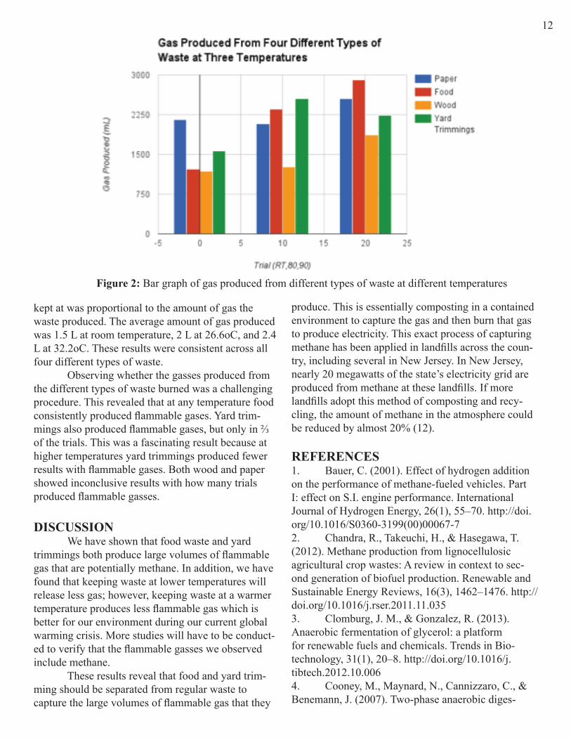
kept at was proportional to the amount of gas the waste produced. The average amount of gas produced was 1.5 L at room temperature, 2 L at 26.6oC, and 2.4 L at 32.2oC. These results were consistent across all four different types of waste. Observing whether the gasses produced from the different types of waste burned was a challenging procedure. This revealed that at any temperature food consistently produced flammable gases. Yard trim-mings also produced flammable gases, but only in ⅔ of the trials. This was a fascinating result because at higher temperatures yard trimmings produced fewer results with flammable gases. Both wood and paper showed inconclusive results with how many trials produced flammable gasses.
DISCUSSION We have shown that food waste and yard trimmings both produce large volumes of flammable gas that are potentially methane. In addition, we have found that keeping waste at lower temperatures will release less gas; however, keeping waste at a warmer temperature produces less flammable gas which is better for our environment during our current global warming crisis. More studies will have to be conduct-ed to verify that the flammable gasses we observed include methane. These results reveal that food and yard trim-ming should be separated from regular waste to capture the large volumes of flammable gas that they
Figure 2: Bar graph of gas produced from different types of waste at different temperatures
produce. This is essentially composting in a contained environment to capture the gas and then burn that gas to produce electricity. This exact process of capturing methane has been applied in landfills across the coun-try, including several in New Jersey. In New Jersey, nearly 20 megawatts of the state’s electricity grid are produced from methane at these landfills. If more landfills adopt this method of composting and recy-cling, the amount of methane in the atmosphere could be reduced by almost 20% (12).
REFERENCES1. Bauer, C. (2001). Effect of hydrogen addition on the performance of methane-fueled vehicles. Part I: effect on S.I. engine performance. International Journal of Hydrogen Energy, 26(1), 55–70. http://doi.org/10.1016/S0360-3199(00)00067-72. Chandra, R., Takeuchi, H., & Hasegawa, T. (2012). Methane production from lignocellulosic agricultural crop wastes: A review in context to sec-ond generation of biofuel production. Renewable and Sustainable Energy Reviews, 16(3), 1462–1476. http://doi.org/10.1016/j.rser.2011.11.0353. Clomburg, J. M., & Gonzalez, R. (2013). Anaerobic fermentation of glycerol: a platform for renewable fuels and chemicals. Trends in Bio-technology, 31(1), 20–8. http://doi.org/10.1016/j.tibtech.2012.10.0064. Cooney, M., Maynard, N., Cannizzaro, C., & Benemann, J. (2007). Two-phase anaerobic diges-
12

tion for production of hydrogen-methane mixtures. Bioresource Technology, 98(14), 2641–51. http://doi.org/10.1016/j.biortech.2006.09.0545. DEFALCO, M., GIACONIA, A., MARRELLI, L., TARQUINI, P., GRENA, R., & CAPUTO, G. (2009). Enriched methane production using solar ener-gy: an assessment of plant performance. International Journal of Hydrogen Energy, 34(1), 98–109. http://doi.org/10.1016/j.ijhydene.2008.09.0856. Hallenbeck, P. C., & Ghosh, D. (2009). Ad-vances in fermentative biohydrogen production: the way forward? Trends in Biotechnology, 27(5), 287–97. http://doi.org/10.1016/j.tibtech.2009.02.0047. Klingstedt, F., Karhu, H., Neyestanaki, A. K., Lindfors, L.-E., Salmi, T., & Väyrynen, J. (2002). Bar-ium Promoted Palladium Catalysts for the Emission Control of Natural Gas Driven Vehicles and Biofuel Combustion Systems. Journal of Catalysis, 206(2), 248–262. http://doi.org/10.1006/jcat.2001.35058. Lehtomäki, A., Huttunen, S., & Rintala, J. A. (2007). Laboratory investigations on co-digestion of energy crops and crop residues with cow manure for methane production: Effect of crop to manure ratio. Resources, Conservation and Recycling, 51(3), 591–609. http://doi.org/10.1016/j.resconrec.2006.11.004
9. Meng, X., Yang, J., Xu, X., Zhang, L., Nie, Q., & Xian, M. (2009). Biodiesel production from oleagi-nous microorganisms. Renewable Energy, 34(1), 1–5. http://doi.org/10.1016/j.renene.2008.04.01410. Regueiro, L., Carballa, M., Alvarez, J. A., & Lema, J. M. (2012). Enhanced methane produc-tion from pig manure anaerobic digestion using fish and biodiesel wastes as co-substrates. Bioresource Technology, 123, 507–13. http://doi.org/10.1016/j.biortech.2012.07.10911. (Bauer, 2001; Chandra, Takeuchi, & Hasega-wa, 2012; Clomburg & Gonzalez, 2013; Cooney, May-nard, Cannizzaro, & Benemann, 2007; DEFALCO et al., 2009; Hallenbeck & Ghosh, 2009; Klingstedt et al., 2002; Lehtomäki, Huttunen, & Rintala, 2007; Meng et al., 2009; Regueiro, Carballa, Alvarez, & Lema, 2012)12. E, A. P. “Overview of Greenhouse Gases.” Methane Emissions. EPA, 15 Apr. 2016. Web. 29 Apr. 2016.
ACKNOWLEDGEMENTSWe thank Mr. Maxwell, Ms. Logerfo, Mr. De, Avni Memani, the Chemistry Department, and the Pingry School for their efforts to make this work possible.
13
Investigating the eIF4E -4EBP1 Signaling PathwayPhilip Geter, Andrew Verdesca ‘15, Morgan D’Ausilio, Alexandra Logerfo, Katie Coyne ‘16,
Isabella Zanobini ‘16, Ben Zhou ‘17, Jess Li ‘18INTRODUCTION Breast cancer is one of the most pervasive and deadly forms of cancer, with over 200,000 diagnosed cases in the United States in 2013 (5). Approximately 70% of breast cancers can be classified as estrogen-receptor positive, characterized by the presence of the estrogen receptor (ER+) (3) (6). Although the drug tamoxifen has been developed specifically to treat ER+ breast cancer, approximately 33% of patients develop resistance to tamoxifen during the five-year course of treatment (4). Previous studies have impli-cated the proteins eIF4E and 4EBP1 in the develop-ment of this resistance. In this experiment, we seek to further investigate and test the relationship between these two proteins, and the larger implications of this interaction in tamoxifen resistant breast cancer. Eukaryotic Initiation Factor 4E (eIF4E) is a key regulator of selective translation in mammalian cells. In tamoxifen-resistant breast cancer cells, eIF4E is believed to be hyperactive, and is responsible for the selective translation of certain oncogenic mRNAs
(such as c-myc and cyclin D1) by binding to the m7GTP (5’) cap of these mRNAs and recruiting the small subunit of the ribosome (1) (2) (8) (Topisirovic & Sonenberg, 2011). The protein eukaryotic Initia-tion Factor 4E Binding Protein 1 (4EBP1) sequesters eIF4E.
When sequestered by 4EBP1, eIF4E is un-able to translate these oncogenic factors (8). 4EBP1 is itself regulated by mTORC1 (molecular Target of Rapamycin Complex 1) (8). In vivo, mTORC1 phos-phorylates 4EBP1’s 114th phenylalanine residue, which is believed to result in a conformational change

that decreases its binding affinity for eIF4E, prevent-ing it from being properly sequestered by 4EBP1. In our project, we aim to further character-ize the relationship between eIF4E, 4EBP1, and mTORC1. We aim to mutate 4EBP1 using site-direct-ed mutagenesis at three sites on the protein; serine 83, serine 112, and threnine 70. All of these sites have been implicated as phosphorlyation sites, and we will mutate them into valines, which cannot be phosphory-lated (7). The goal of preventing phosphorylation of 4EBP1 is to prevent a conformational change that would prevent sequestration of 4EBP1 by eIF4E (7). Our project aims to investigate the 4EBP1-eIF4E sig-naling pathway through inducing mutations of 4EBP1 that are designed to prevent phosphorylation. RESULTS This year, we aimed to purify and amplify the genes for eIF4E and 4EBP1 in order to investigate their interaction. Last year, we obtained cNDA coding for wild type eIF4E cloned into the pBABE mamma-lian expression vector and wild type 4EBP1 cloned into the pMUD mammalian expression vector from
the Schneider Laboratory at NYU. Last year, we had prepared glycerol stocks of both plasmids for future usage. We streaked plates with the glycerol stocks in order to grow E coli colo-nies expression the pBABE eiF4E and pMUD 4EBP1 genes. The innocluate was extracted from the cells and then purified via a plasmid DNA miniprep, following a standard Qiagen Miniprep protocol. A PCR reaction using custom designed prim-ers was performed on the purified plasmid in order to purify the eIF4E and 4EBP1 genes. We have thus far successfully cloned and purified the genes coding for wild type 4EBP1 and eIF4E.DISCUSSION AND FUTURE STEPS First, we aim to ligate both 4EBP1 and eIF4E into the pTXB1 bacterial expression vector. We then plan to introduce 6 different mutations into 4EBP1 by performing site-directed mutagenesis (which consists of a PCR reaction using specifically designed primers that contain the desired mutation.) Then, we plan to separately express mutated 4EBP1, wild-type 4EBP1 and eIF4E in E coli and purify the protein. Finally, we
14

plan to test the relative binding affinity between eiF4E and wild-type 4EBP11 as compared to eIF4E and mu-tated 4EBP1.
Additionally, we may use mammalian expres-sion vector pBABE to express those three proteins in Chinese Hamster Ovary cells and determine the effect of this mutation on cell proliferations in vivo. REFERENCES1. Carraway, H., & Hidalgo, M. (2004). New targets for therapy in breast cancer: Mammalian target of rapamycin (mTOR) antagonists. Breast Cancer Research , 6 (5), 219-224.2. Carroll, M., & Borden, K. L. (2013). The On-cogene eIF4E: Using Biochemical Insights to Target Cancer. Journal of Interferon & Cytokine Research , 33 (5), 227-238.3. Huang, B. et al (2014, February 11). Differen-tial expression of estrogen receptor α, β1, and β2 in lobular and ductal breast cancer. Proceedings of the
National Academy of Sciences for the United States of America, 111(5).4. Jiang, Q., Zheng, S., & Wang, G. (2013). De-velopment of new estrogen receptor-targeting thera-peutic agents for tamoxifen-resistant breast cancer. Future Medicinal Chemistry , 5 (9), 1023-1035.5. Litzenburger, B. C., & Brown, P. H. (2014). Advances in Preventive Therapy for Estrogen-Recep-tor-Negative Breast Cancer. Current Breast Cancer Reports , 6, 96-109.6. Manavathi, B., Dey, O., Gajulapalli, V. N., Bhatia, R. S., Bugide, S., & Kumar, R. (2013). De-railed Estrogen Signaling and Breast Cancer: An Authentic Couple. Endocrine Reviews , 34 (1), 1-32.7. Peter, Daniel, Cátia Ingreja, Lara Wohlbold, Catrin Weiler, Linda Ebertsch, Oliver Weichenrieder, and Elisa Izaurralde. “Molecular Architecture of 4E-BP Translational Inhibitors Bound to EIF4E.” Molecu-lar Cell 57 (2015): 1074-087. Elsevier. Web. 15 Apr. 2015.8. Sun, S.-Y., Rosenberg, L. M., Wang, X., Zhou, Z., Yue, P., Fu, H., et al. (2005). Pathways by Ra-pamycin-Mediated Mammalian Target of Rapamycin Inhibition. Cancer Research, 65.ACKNOWLEDGEMENTSThank you to Philip Geter, pHD candidate at NYU, for mentoring this team. Thank you to Andrew Verd-esca for his past leadership on the project. Thank you to Morgan D’Ausilio, and Alexandra Logerfo for su-pervising and guiding the project. Thank you to Luke De, David Maxwell, and the rest of the science depart-ment at Pingry for their ongoing support
15
The Effects of Chrysin on Liver Detoxification and Metabolism of Acetamino-phen
Ryan Lane ‘16, Alina Jan ‘16ABSTRACT
Zebra danio provide an effective model when study-ing different disease states and disorders, as they share many genetic similarities to humans (6). Liver disease currently affects 30 million individuals in America alone, and is caused by a variety of instigators, includ-ing medication overdose and interchemical reactions (2). Having a useable and thoroughly understood model of liver disease would be highly advantageous for further investigatory research and breakthroughs in not only liver disease treatment, but also preventa-
tive action. We investigated the effectiveness of the zebra danio liver model through exposure to toxic levels of acetaminophen delivered via a water solu-tion introduced into their environment. After 1 and 5 days of repeated exposure to varying concentrations of acetaminophen, we found through dissection that there was little to no difference visually, leaving these results to be inconclusive. Further research will reveal if zebra danio experience an increased platelet count upon induced acute inflammation of the liver.

INTRODUCTION The liver is an essential organ that processes nutrients absorbed by the intestines, regulates the basic composition of blood, and is essential to the metabo-lism of toxic wastes such as drugs and alcohol. There-fore, the ability of the liver to function properly is vital to human health and survival. The buildup of toxins and other antigenic substances within the liver not only hinders the liver’s ability to detoxify the blood, but also can cause hepatic encephalopathy if sustained for long periods of time. Acetaminophen is a medication proven to cause liver damage due to toxicity at high doses. The accumulation of NAPQI, acetaminophen’s metabolite, overwhelms the glutathione pathway. Only a small percentage of the substrate can be processed, which causes damage to the liver (3). Additionally, alcohol and other medications can significantly increase the damage through specific chemical interactions such as the P-450 pathway, and ultimately have been shown to slow down the processing capabilities of the liver (3). A flavonoid found in high concentrations in honey known as Chrysin has been shown to inhibit aromatase enzyme activity (5). Previous studies have shown that it may also protect against hypercholester-olemia and decreased serum hepatic marker enzyme levels in rats injected with a lipoprotein lipase (4). Chrysin may also be used as a pharmaceutical that allegedly hinders the toxin processing capacity of the liver. Using known liver toxins, we examined the chemical interaction between chrysin and acetamino-phen in order to determine overall drug safety and possible side effects.
RESULTS The goal of our protocol was to determine if there were any discernible changes in the physical appearance of the zebrafish liver upon liver damage caused by acetaminophen, which is said to cause acute hepatitis-like symptoms. During our dissections, we had difficulty distinguishing a healthy liver from a damaged liver. For that reason, our results are incon-clusive. We cannot say with certainty that there was any visible inflammation. CONCLUSION At this stage, our results show no clear signs of visible inflammation upon dissection after single day or repeated doses of varying concentration of acetaminophen. Though surrounding tissue and organs are seemingly less healthy and less pink in compari-son to the control specimen, the livers themselves do not show large amounts of damage as hypothesized. By spinning down blood samples, we will be able to determine varying levels of plasma and hematocrit as affected by acetaminophen exposure and subsequent liver damage. For further research using known liver toxins such as acetaminophen, we plan to examine the chemical interaction between chrysin and acet-aminophen in order to determine overall drug safety and possible side effects otherwise unknown. Further experimentation using real time qt-pcr could yield bet-ter results in quantifying the inflammation of the fish. REFERENCES1) Gupta, Tripti, and Mary C. Mullins. “Dissec-tion of Organs from the Adult Zebrafish.” Journal of Visualized Experiments JoVE 37 (2010): n. pag. Web.2) Vliegenthart, A. D Bastiaan, Carl S. Tucker,
Figure 1: Control fish (A), low dose of .325 g/L acetaminophen after 1 day (B), high dose of .645 g/L acetaminophen after 1 day (C), after 5 days of dosing respectively for 3 hours each day (D, E)
16

Jorge Del Pozo, and James W. Dear. “Zebrafish as Model Organisms for Studying Drug-induced Liver Injury.” (n.d.): n. pag. British Journal of Clinical Phar-macology. BlackWell Publishing Ltd. Web. 16 Feb. 2016.3) Lee, Dennis, MD. “Tylenol Liver Damage Symptoms, Causes, Treatment - How Is Acetamino-phen Processed (metabolized) in the Body? - Medi-cineNet.” MedicineNet. Ed. Jay W. Marks. Medi-cineNet, Inc., n.d. Web. 01 Mar. 2016.4) Anandhi, Ramalingam et al. “Antihypercho-lesterolemic and Antioxidative Effects of an Extract
of the Oyster Mushroom, Pleurotus Ostreatus, and Its Major Constituent, Chrysin, in Triton WR-1339-in-duced Hypercholesterolemic Rats.” Journal of Physi-ology and Biochemistry 69.2 (June 2013): 313-23. Springer Link. 27 Oct. 2012. Web.5) Gambelunghe, Cristiana et al. “Effects of Chrysin on Urinary Testosterone Levels in Human Males.” Journal of Medicinal Food 6.4 (July 2004): n. pag. Web. 01 Mar. 2016.
Pharmacologic reduction of anxiety in Danio rerioMichael James ‘16, Sydney Stein ‘16, Owen Storms ‘16
17
ABSTRACTSAME-e is known to be effective in the treatment of depression and osteoarthritis, but reports of anxiolytic effects of the drug have come to our attention. Using zebrafish as a model for humans, we will be testing the effect of different doses of SAM-e on anxiety in hopes to show that there is in fact an anxiolytic effect
on the fish. Our data does not suggest that there is any correlation between SAM-e and decreased anxiety in zebrafish, but with further experimentation we hope to decrease inconsistencies in experimental design and conditions.
INTRODUCTION Anxiety disorders are the most common form of mental illness in the United States, affecting ap-proximately 18% of the adult population (1). Popular anxiety treatments include selective seratonin reuptake inhibitors (SSRIs), which treat anxiety symptoms by blocking the reabsorption of seratonin in the brain (2) However, anti-anxiolytic drugs such as SSRIs often have side effects that include insomnia and weight gain (1). SAM-e is a naturally occurring element in the human body typically consumed and processed by the liver. SAM-e is currently on the market as a supple-ment used as a natural treatment for depression (6) and osteoarthritis (7). SAM-e has also reported anti-anxio-lytic side effects (8) . Since many anxiety drugs such as SSRI’s are also used to combat depression (8) , we hypothesized that SAM-e would be a viable natural option for anxiety treatment. Zebrafish are a suitable candidate for SAM-e experimentation because they share a great degree of genetic similarity with humans (4). Using caffeine, a known anxiolytic to Zebrafish (Bencan), to instill anxiety in the fish, we looked at the effect of three different human-dependent doses of SAM-e on the anxiety levels of the fish using the black-and-white box method.
DISCUSSION Unfortunately, after conducting three experi-ments with six fish each, our results were inconclu-sive. With the 160 mg trial, which is equivalent to a human dose of 800 mg, our results did not confirm our hypothesis (Fig. 1a). The same was true for the 320 mg trial, which is equivalent to a human dose of 2400 mg (Fig. 1b). With the 240 mg trial, or 2000 mg human dose, the results looked more like we expected them to. When SAM-e was administered to fish who were given caffeine, there was a significant decrease in anxiety from the fish who only received caffeine (Fig. 1c). Unfortunately, even these results cannot be deemed valid because of the unusual spike in anxiety seen in the fish that were just administered SAM-e. We attribute this lack of consistency in our results to unstable experimental conditions. During testing fish were subjected to loud noises due to having used shared lab space for experiments. This could be a reason the fish were more anxious than expected, and the SAM-e did not have the expected result. In the future, a more controlled environment could be used to conduct experiments in hopes of getting more valid results.

REFERENCES1. American and Depression Association of America, Facts and Statistics. September 2014 http://www.adaa.org/about-adaa/press-room/facts-statistics2. Bencan, Z., Sledge, D., & Levin, E. D. (2009). Buspirone, chlordiazepoxide and diazepam effects in a zebrafish model of anxiety. Pharmacology, Bio-chemistry, and Behavior, 94(1), 75–80. doi:10.1016/j.pbb.2009.07.0093. Cachat, J., Stewart, A., Grossman, L., Gaik-wad, S., Kadri, F., Chung, K. M., … Kalueff, A. V. (2010). Measuring behavioral and endocrine responses to novelty stress in adult zebrafish. Nature Protocols, 5(11), 1786–99. doi:10.1038/nprot.2010.1404. Egan, R. J., Bergner, C. L., Hart, P. C., Cachat, J. M., Canavello, P. R., Elegante, M. F., … Kalueff, A. V. (2009). Understanding behavioral and physiological phenotypes of stress and anxiety in zebrafish. Behav-ioural Brain Research, 205(1), 38–44. doi:10.1016/j.bbr.2009.06.0225. Grossman, L., Utterback, E., Stewart, A., Gai-kwad, S., Chung, K. M., Suciu, C., … Kalueff, A. V. (2010). Characterization of behavioral and endocrine effects of LSD on zebrafish. Behavioural Brain Re-search, 214(2), 277–84. doi:10.1016/j.bbr.2010.05.0396. Howland, Robert H. “Dietary Supplement Drug Therapies For Depression”. J Psychosoc Nursment Health Serv 50.6 (2012): 13-16. Web. 16 Feb. 2016.7. Maximino, C., de Brito, T. M., Colmanetti, R., Pontes, A. A. A., de Castro, H. M., de Lacerda, R. I. T., … Gouveia, A. (2010). Parametric analyses of anxiety in zebrafish scototaxis. Behavioural Brain Research, 210(1), 1–7. doi:10.1016/j.bbr.2010.01.0318. Maximino, C., Marques de Brito, T., Dias, C. A. G. de M., Gouveia, A., & Morato, S. (2010). Scoto-taxis as anxiety-like behavior in fish. Nature Protocols, 5(2), 209–16. doi:10.1038/nprot.2009.229. Maximino, C., da Silva, A. W. B., Gouveia, A., & Herculano, A. M. (2011). Pharmacological analy-
Figure 1: SAM-e experiments using a caffeine control, three experimental groups of caffeine and SAM-e, a SAM-e control, and a drug-free control with 160 mg (a), 320 mg (b), and 240 mg (c).
sis of zebrafish (Danio rerio) scototaxis. Progress in Neuro-Psychopharmacology & Biological Psychiatry, 35(2), 624–31. doi:10.1016/j.pnpbp.2011.01.00610. Norton, W. H. J., Folchert, A., & Bally-Cuif, L. (2008). Comparative analysis of serotonin receptor (HTR1A/HTR1B families) and transporter (slc6a4a/b) gene expression in the zebrafish brain. The Journal of Comparative Neurology, 511(4), 521–42. 11. Papakostas G, (2009). Evidence for S-adeno-syl-L-methionine (SAM-e) for the treatment of major depressive disorder. J Clin Psychiatry, issue 2009 Retrived from: http://www.psychiatristcom/JCP/article/Pages/2009/v70s05/v70s0504.aspx 12. Sackerman, J., Donegan, J. J., Cunningham, C. S., Nguyen, N. N., Lawless, K., Long, A., … Gould, G. G. (2010). Zebrafish Behavior in Novel Environ-ments: Effects of Acute Exposure to Anxiolytic Com-pounds and Choice of Danio rerio Line. International Journal of Comparative Psychology / ISCP ; Spon-sored by the International Society for Comparative Psychology and the University of Calabria, 23(1), 43–61. Retrieved from http://www.pubmedcentral.nih.gov/articlerender.fcgi?artid=2879659&tool=pmcentrez&rendertype=abstract13. THE SHERWIN-WILLIAMS COMPANY KRYLON PRODUCTS GROUP (2015). Safety Data Sheet of KRYLON Interior/Exterior Paint. https://www.pts-tools.com/MSDS/LP40K1910.pdf14. Wong, K., Elegante, M., Bartels, B., Elkhayat, S., Tien, D., Roy, S., … Kalueff, A. V. (2010). Analyz-ing habituation responses to novelty in zebrafish (Da-nio rerio). Behavioural Brain Research, 208(2), 450–7. doi:10.1016/j.bbr.2009.12.023
ACKNOWLEDGEMENTSDavid Maxwell, Luke De, Maya Huffman, Amy Stein
18

19Investigating the Effect of Amygdalin on Melanoma Cell Proliferation
Caroline Marone ‘17, Heba Syed ‘17ABSTRACT
Amygdalin, a natural compound found in plants, raw nuts, and the pits of many fruits, has been used as an alternative treatment for cancer patients worldwide for over a century (3). However, its anti-tumor potential is widely debated. We conducted an in vitro study exam-
ining the effects of amygdalin in concentrations rang-ing from 1.25-10 mg/mL on Mus musculus melanoma cells (B16-F10), and our results showed that the use of amygdalin may have a significant effect on melanoma cell proliferation.
INTRODUCTION Complementary and Alternative Medicine is a class of unconventional medical treatments that are quite popular among cancer patients. Alternative medicine, as its name suggests, is used as a direct substitute for traditional therapies. Despite such wide-spread popularity, there is a lack of full understanding for many of the natural compounds used as alternative treatments (1). The compound amygdalin gained prominence in the latter half of the twentieth century as one of the most popular alternative cancer treatments (3). By 1978, 70,000 U.S. cancer patients had taken amygda-lin (5). However, as of 2016, the FDA has not ap-proved amygdalin in the United States for legal use, and evidence of the compound’s benefits is controver-sial. Although amygdalin has not been shown to have any significant effects in clinical studies and there is minimal clinical data to support its benefits (5), sev-eral laboratory studies attest to the compound’s anti-tumor potential. Advocates of amygdalin support the use of what they believe to be a natural cancer cure, while others remain strongly opposed because of the compound’s supposed inefficiency and even potential toxicity. Overall, evidence-based research remains sparse and the extent of amygdalin’s effects have not been clearly elucidated (3). We are conducting an in vitro study to investi-gate the influence of amygdalin in varying concentra-tions on mouse melanoma cells (B16-F10). Melanoma is a type of skin cancer with rates that have been rising for over thirty years. According to the American Cancer Society, in 2016, approximately 76,380 new melanoma cases will be diagnosed and 10,130 people will die of the cancer (6). In this study, we hope to provide evidence that supports whether or not amygdalin exhibits any notable effects on melanoma cell proliferation.
RESULTS Each plate started with approximately 497,500 melanoma cells. 24 hours after the initial amygdalin application, the control groups had an average total cell count of approximately 833,333 cells per plate. The plates with 1.25 mg/mL amygdalin had approxi-mately 725,000 cells; 2.5 mg/mL amygdalin had ap-proximately 700,000 cells; 5 mg/mL amygdalin had approximately 633,333 cells; and 10 mg/mL amygda-lin had approximately 683,333 cells. The general trend in the data was that as the concentration of amygdalin increased, the cell count decreased. The average proliferation over 24 hours for the control group was 336,000 cells; 1.25 mg/mL amyg-dalin was 227,500 cells; 2.5 mg/mL amygdalin was 202,500 cells; 5 mg/mL amygdalin was 135,833; and 10 mg/mL amygdalin was 185,833 cells. Standard er-ror was calculated using ANOVA statistical analysis.
Figure 1. Average proliferation of B16-F10 cells treated with increasing amygdalin concentrations over 24-hour period.DISCUSSION After looking at our results, we plan to contin-ue this study over the next year. Our preliminary data suggests that amygdalin may have a significant effect on melanoma cell proliferation, but at this point the re-sults are inconclusive. Although our data shows slight decreases in melanoma cell proliferation with in-creased amygdalin concentrations, an ANOVA statisti-cal analysis calculated a p-value of 0.22, which means

20that the data is not statistically significant. The next step in our study would be to repeat the same trials (with melanoma cells and the same five concentrations of amygdalin) at least three to six more times (for two/three times as much data as we have now). This would provide more data and therefore lead to a more con-clusive and precise p-value. We would then be able to decisively determine whether or not amygdalin affects B16-F10 melanoma cell proliferation. If we find that there is a significant statistical effect, future studies can then test amygdalin’s effect over longer periods of time, its effect on different types of cancer cells, and the relative toxicity of amygdalin on cancerous cells versus healthy cells. However, if we find that amygda-lin is inefficient in controlling cell proliferation, that would be the conclusion of our study. Our data also showed that although there were slight differences between the 2.5 mg/mL, the 5 mg/mL, and the 10 mg/mL amygdalin concentrations, when standard error is taken into account, it can be concluded that they all allow very similar values of cell proliferation, especially 5 mg/mL and 10 mg/mL. This suggests that there may be a limit to the effective-ness of amygdalin, and therefore increasing the con-centration to anything more than 10 mg/mL would not have any significant effect. REFERENCES1. “Defining Complementary and Alternative
Medicine.” Everyday Health. N.p., n.d. Web. 22 Feb. 2016. <http://www.everydayhealth.com/alternative-health/the-basics.aspx>.2. “Laetrile/Amygdalin (PDQ).” National Cancer Institute. N.p., n.d. Web. 22 Feb. 2016. <http://www.cancer.gov/about-cancer/treatment/cam/hp/laetrile-pdq>.3. Makarevic, Jasmina, et al. “Amygdalin Blocks Bladder Cancer Cell Growth In Vitro by Diminishing Cyclin A and cdk2.” PLOS ONE: n. pag. Print.4. Milazzo, Stefania, Stephane Lejeune, and Edzard Ernst. “Laetrile For Cancer: a Systematic Review of the Clinical Evidence.” Supportive Care in Cancer: n. pag. Print.5. Moss, Ralph W. “Tijuana Cancer Clinics in the Post-NAFTA Era.” Integrative Cancer Therapies: n. pag. Print.6. “What Are The Key Statistics About Melano-ma Skin Cancer?” American Cancer Society. N.p., n.d. Web. 25 Mar. 2016. <http://www.cancer.org/cancer/skincancer-melanoma/detailedguide/melanoma-skin-cancer-key-statistics>. ACKNOWLEDGEMENTSThank you to Dr. Aisha Hasan and Memorial Sloan Kettering for providing us with the cells, culture me-diums and guidance on our project. Thank you to Mr. Maxwell and the Pingry Science Department.
Creating Point Mutations in BRCA2Emily Kwon ‘16, Yanni Angelides ‘16, Luke De, Ryan Jensen
ABSTRACT It is unclear how the pathway between muta-tion of the BRCA2 gene—a gene involved in DNA repair—and tumor formation and growth works. Because phosphorylation is integral to signaling net-works, we created point mutations on specific BRCA2 phosphorylation sites to either mimic phosphorylation
or knock it out completely (5). As of yet, we are not sure what the cell’s response will be to those phos-phorylation events after DNA is damaged (3). Future experiments examining the expression of these mu-tated BRCA2 phosphorylation sites may help elucidate these questions.
INTRODUCTION Mutations in the genes BRCA1 and BRCA2 are well-known genetic risk factors in the contrac-tion of breast and epithelial ovarian cancer (EOC) (5). Discoveries made through extensive gene linkage and positional cloning efforts have shown that mutations in both BRCA1 and BRCA2 increase the lifetime risk of breast cancer by 60-85% and ovarian cancer by 20-40% (11). Although these two genes are often analyzed together, we focused on BRCA2, which has an important role in homologous recombination repair
of DNA double-strand breaks (2). The molecular progression of BRCA2 disease is obscure (5). To clarify this process, Dr. Ryan Jen-sen’s lab used mass spectrometry to identify novel phosphorylation sites in BRCA2 after DNA was dam-aged with mitomycin C—an important chemotherapy agent for treating breast and ovarian cancer patients who have tumors with BRCA mutations (1). Phos-phorylation, a protein post-translational modification, controls processes like cell growth, proliferation, and survival. We wanted to see what would occur if we al-

21
tered BRCA2’s phosphorylation sites (3). The exami-nation of these specific sites could give us insight into the next steps in the molecular pathway of BRCA2 and tumor growth (4). We aimed to perform site-directed mutagenesis on two phosphorylation sites in plasmid pBluescript BRCA2 1-5286: Threonine 491 and Serine 492. We planned to make three separate mutations: convert Threonine 491 and Serine 492 to Alanine (TASA); Threonine 491 and Serine 492 to Aspartic Acid
Figure 1: Schematic of Molecular Progression of BRCA2 mutations
(TDSD); and Serine 492 to Aspartic Acid (SD). Note: The BRCA2 gene is 10,254 base pairs long. Working with a gene of this size can lead to a higher prevalence of errors in experiments. In order to avoid those errors, we worked primarily on pBluescript BRCA2 1-5286, a plasmid that contains the first half of the BRCA2 gene and our targeted phosphorylation sites (491 and 492).
RESULTS
Verifying SequencesWe successfully mutated the phosphorylation sites: Threonine 491 to Alanine; Serine 492 to Alanine; Threonine 491 to Aspartic Acid; Serine 492 to Aspartic Acid; and Serine 492 to Aspartic Acid. All blast sequences and chromatograms show reliable sequence data. (See Figures below)
Figures 3-5: Blast sequences comparing unmutated BRCA2 1-5286 with mutated BRCA2 1-5286
Figure 3
Figure 4
Figure 5
Figures 6-8: Chromatograms of mutations
Figure 6
Figure 7
Figure 8

22DISCUSSIONHaving created the point mutations in BRCA2, we plan to insert the mutated DNA into a full length BRCA2 expression vector and then transform the mutated plasmid into bacterial cells. Then, we will send the plasmids to Dr. Jensen so he can express the mutated cDNAs in human BRCA2 knockout cells. We are un-able to run human tissue cultures because school regu-lations prevent us from doing so. It will be interesting to see the functional consequences of these BRCA2 mutations. Will they be detrimental to the function of the protein? Beneficial? Could they even alter the func-tion of the protein completely? These are questions we hope our research will help answer in the future. REFERENCES1. Balajee, Adayabalam S. DNA Repair and Human Disease. Georgetown, TX: Landes Bioscience/Eurekah.com, 2006. Print.2. Bolton, Kelly L et al. “Association Between BRCA1 and BRCA2 Mutations and Survival in Women with Invasive Epithelial Ovarian Cancer.” JAMA 307 (2012): 382–90.3. Breitkopf, Susanne B., and John M. Asara. “Deter-mining in Vivo Phosphorylation Sites Using Mass Spectrometry.” Current Protocols in Molecular Biology CHAPTER (2012): Unit 18.19. PMC. Web. 8 Apr. 2016.4. Contact Find Us on Facebook Shipping of Plasmids on Filter Paper (n.d.): n. pag. Web.5. Dephoure, Noah et al. “Mapping and Analysis of Phosphorylation Sites: A Quick Guide for Cell Biolo-gists.” Ed. David G. Drubin. Molecular Biology of the
Cell 24.5 (2013): 535–542. PMC. Web. 8 Apr. 2016.6. High Efficiency Transformation Protocol.” Bio-Labs. N.p., n.d. Web. <https://www.neb.com/proto-cols/1/01/01/high-efficiency-transformation-protocol-c2987>.7. Jensen, Ryan B. “BRCA2: One Small Step for DNA Repair, One Giant Protein Purified.” The Yale Journal of Biology and Medicine. YJBM, 13 Dec. 2013. Web. 07 Apr. 2016.8. Jensen, Ryan B et al. “BRCA2 Is Epistatic to the RAD51 Paralogs in Response to DNA Damage.” DNA repair 12 (2013): 306–11.9. QIAprep Spin Miniprep Kit.” Qiagen. N.p., n.d. Web. <https://www.qiagen.com/shop/sample-technologies/dna/dna-preparation/qiaprep-spin-miniprep-kit/>.10. QuikChange II XL Site-Directed Mutagenesis Kit.” (n.d.): n. pag. Web. <http://www.agilent.com/cs/li-brary/usermanuals/Public/200521.pdf>.11. Venkitaraman, Ashok R. “Linking the Cellular Functions of BRCA Genes to Cancer Pathogenesis and Treatment.” Annual review of pathology 4 (2009): 461–487.ACKNOWLEDGEMENTSThank you to Dr. Ryan Jensen for your mentorship this past summer and allowing us to bring this project to Pingry. Thank you to Luke De for mentoring and supervising this project. Thank you Dr. Judit Jimenez for your encouragement. Thank you to Dr. Morgan D’Ausilio, David Maxwell, Allison Logerfo, and Dr. Colleen Kirkhart for your help. Thank you to our fami-lies for supporting our pursuits and endeavors.
The Effect of Frequency on Anxiety in Danio rerioBrian Grimaldi ‘16, Samantha Palazzolo ‘16
ABSTRACT Ambient noises have been widely studied for their varying effects on aquatic life. We exposed zebrafish (Danio rerio) to artificial sound of 45 dB at varying frequencies of 0 hz, 10,000 hz, 20,000 hz, and 30,000 hz. We used two groups within each tank (two 10 gallon tanks split in two, total of 4 tanks). All tests used the Novel Tank Diving test. Each group was exposed to a respective frequency and were randomly selected at the end of the day in order to avoid prefer-ence. The tank was in a soundproofed room with a
jawbone speaker placed on the side of the tank. In total, there were 16 trials done. While results were in-conclusive, we have been able to pinpoint a new range of frequencies to test to narrow down the range of hearing for zebrafish with the testing range being be-tween 20,000 and 30,000 hz. As researchers, we were curious to see how the fish would react to the noise in the short and long term. Perhaps everyday occurrenc-es, like noise pollution, could be causing problems.
INTRODUCTION There are many studies that showcase how ambient noises have a varying effect on aquatic life. These underwater noises range in frequencies, and the
response threshold levels of the marine fish species vary with each set of frequencies. Fish can hear within a range 100 and 2000 hz, but in this experiment ma-rine fish were subjected to tones varying from 0.1-64

23
khz. For sea bass, the reaction threshold was between 0.1-0.7 kHz; for thicklip mullet, 0.4-0.7 kHz; for pout, 0.1-0.25 kHz; for horse mackerel, 0.1-2 kHz; and for Atlantic herring, 4 kHz. This variety shows that fish species react very differently to sound compared to humans, and that generalisations about the effects of sound on fish should be made with care. The reactions of fish depend on the spectrum and level of the an-thropogenic sounds, as well as their environment (e.g. location, temperature, physiological state, and school size) (2). A lot of noise pollution comes from urban areas or construction zones near aquatic life, which can be harmful to fish. Nowadays, pile driving—con-struction activity—is predominantly found near the shore, where the construction of bridges, ports, wind farms and other buildings occurs. Seismic exploration devices, mainly air guns, are used globally for under-seas geological surveys and geophysical studies, such as oil and gas exploration and seabed mapping (5). Similarly, sonars generating noise at various intensities are widely used not only by navies but also by com-mercial ships, the fishing industry, and marine research organizations (5). Low-frequency stationary noise can be generated by various ships and vessels (5). These frequencies, ranging from low to high, can frenzy and produce anxiety in the surrounding fish, which is detri-mental to their livelihood. The purpose of this study is investigate the
correlation between anxiety and zebrafish. Our tests subject fish to high frequencies to see if they become anxious. From there, we can discover how noise pol-lution could affect various things like sleep, mating, and overall life. Though these zebrafish experiments are in a controlled environment, the ambient sounds in the lab or in the fish room may affect the anxiety of zebrafish, and would have to be accounted for in fu-ture studies involving zebrafish. Also, since zebrafish and humans have similar circadian rhythms, there may be a relation to how noise makes humans function and how anxiety affects the human sleep cycle.
RESULTS The Novel Tank Diving Test was viable for all 16 trials, as there were expected results with each individual frequency administered to the zebrafish. For each group, the amount of time spent on the bottom of the tank was approximately 84% of the 6 minutes of each trial. The control group spent 6 percent of its time on the bottom, the 10,000 Hz group spent about 72% on the bottom, the 20,000 Hz group spent about 92% on the bottom, and the 30,000 Hz group spent about 87% of its time on the bottom. Time spent on the top were 94% for the control group, 28% for 10,000 Hz group, 8% for 20,000 Hz group, and 13% for 30,000 Hz group.
Figure 1: Average percentage of time spent on either the bottom or top of tank

24DISCUSSION The results suggest that there were significant psychological effects on zebrafish exposed to the varying frequencies. With the amount of data that we collected, we were able to produce desired results in terms of the anxiety in zebrafish in both tanks. How-ever, some variables might have affected our trials, even though we took precautionary steps in order to reduce any errors in our data. One of our concerns was the transportation of the fish. Catching the fish and moving the 2.5 gallon tanks into another room could have caused some anxiety to the zebrafish being experimented on, even though we waited 10 minutes for the fish to rest and become acclimated to their new, temporary environment. However, even with the data we have, we are unable to pinpoint a specific audible range for the zebrafish due to the broad spectrum of frequencies we exposed them to. In order to pinpoint their range, we would need to start narrowing down our frequencies to between 20,000 Hz and 30,000 Hz, as these frequencies had the biggest range of effects. It was evident that there was a correlation between anxiety and frequency, but no definite causation as to which frequency, in particular, would cause a certain level of anxiety.
REFERENCES[1] Peng C, Zhao X, Liu G. Int J Environ Res Pub-lic Health.2015 Sep 30;12(10): 12304-23. [2] Kastelein RA, Heul Sv, Verboom WC, Jen-nings N, Veen Jv, de Haan D. Mar Environ Res. 2008 Jun;65(5):369-77. [3] Yokogawa T, Marin W, Faraco J, Pézeron G, Appelbaum L, Zhang J, Rosa F, Mourrain P, Mignot E. PLoS Biol. 2007 Oct 16;5(10):e277.[4] Siguroardottir FD. Effects of sleep deprivation on learning and memory in Zebrafish (Danio rerio) Bachelor of Science in Medicine Final Project. Uni-versity of Iceland School of Health Sciences. 2012. Web. 9 June 2016. [5] Peng C, Zhao X, Liu G.Int J Environ Res Pub-lic Health. 2015 Sep 30;12(10):12304-23[6] Egan RJ, Bergner CL, Hart PC, Cachat JM, Canavello PR, Elegante MF, Elkhayat SI, Bartels BK, Tien AK, Tien DH, Mohnot S, Beeson E, Glasgow E, Amri H,Zukowska Z, Kalueff AV. Behav Brain Res. 2009 Dec 14;205(1):38-44.
ACKNOWLEDGEMENTSMr. MaxwellOwen StormsMaya HuffmanMr. De

The Pingry School131 Martinsville Road
Basking Ridge, NJ 07920
Find us at www.pingry.org/pcr