Embed Size (px)
Citation preview

Aquatic Mammals 1990, 16,3,96-100
The pineal organ (Epiphysis cerebri) of the harbour porpoise Phocoena phocoena (Linne, 1758)
Gunter Behrmann
Am Handelsha/en 12. D-2850 Bremerhaven, Federal Republic a/Germany
Summary
The pineal organ (Epiphysis cerebri) of the harbour porpoise Phocoena phocoena (Linne, 1758). . As former reports about the pineal organ (which is Important for the development of the genital organs) are vague, this is the first precise description of a functional cetacean pineal organ. The organ was found in a brain of a young male harbour porpoise. Morphological and histological examinations demonstrate that the pineal organ of this animal has been functional. The pineal organ of the harbour porpoise IS comparable with those of horses, cattle and sheep.
Introduction
T~e pinea~ organ, also named the parietal organ or eplphyse, IS an appendage to the diencephalic roof (Lamina tecti), and is located in the centre of the b~ain, between the two hemispheres (Fig. I). The pmeal organ is situated on the tip of the posteria commissure, which emerges from the collicus superior, a part of the diencephalon. The pineal organ floats in the cerebrospinal fluid of the third ventricle (Ventriculus tertius).
The forms of the pineal organs of mammals are quite different. Some species of mammals lack a pineal organ. Sometimes in the past whales were added to the list of mammals which lack a pineal organ (von Hallerstein, 1934; Flanigan, 1972). But Gersch (1938) has described pineal organs of humpback whales.
However, these organs have an important function in animals, as they coordinate hormonal secretion in the hypothalamus (Leonhardt, 1985), and have an influence on genital maturity (Penzlin, 1980).
In 1979 Klinowska & Vollrath had a discussion about the existence of the pineal organs in cetaceans. Vollrath (1979) recommended that the cetaceans
~
Figure l. Sagittal section of the brain, view on the left side and the pineal organ. Figure 2. The pineal organ of the harbour porpoise.

97 The pineal organ a/the harbour porpoise
NE
Fig.10
NE
Figure 3. Model of a longitudinal section through the pineal Figure 5. Model of a pineal segment.
organ.
Figure 4. Longitudinal section through a pineal segment, staining 1.
need to be better checked, so to make it absolutely clear whether these animals lack the pineal organ.
Following this recommendation I examined the brain of the harbour porpoise and definitely found Figure 6. The peripheral region of the pineal segment, the pineal organ. staining 3.
98 G. Behrmann
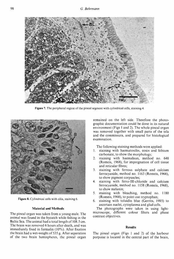
Figure 7. The peripheral region of the pineal segment with cylindrical cells, staining 4.
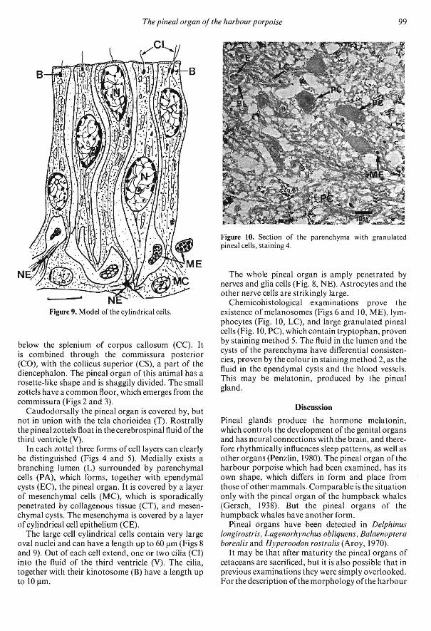
Figure 8. Cylindrical cells with cilia, staining 6.
Material and Methods
The pineal organ was taken from a young male. The animal was found in the bycatch while fishing in the Baltic Sea. Theanimal had a total length of 108.5 cm. The brain was removed 6 hours after death, and was immediately fixed in formalin (10%). After fixation the brain had a wet-weight of 535 g. After separation of the two brain hemispheres, the pineal organ
remained on the left side. Therefore the photographic documentation could be done in its natural environment (Figs I and 2). The whole pineal organ was removed together with small parts of the tela and the commissura, and prepared for histological examination.
The following staining methods were applied: I. staining with haematoxilin, eosin and lithium
carbonate, to show the morphology; 2. staining with haemalaun, method no. 648
(Romeis, 1968), for impregnation of cell tissue and reticular fibres;
3. staining with ferrous sulphate and calcium ferrocyanide, method no. 1163 (Romeis, 1968), to show pigment corpuscles;
4. staining with ferro-III-chloride and calcium ferrocyanide, method no. 1158 (Romeis, 1968), to show melanin;
5. staining with bleaching, method no. 1180 (Romeis, 1968), to point out tryptophan;
6. staining with toluidin blue (Gerrits, 1985) to ascertain nuclei, cytoplasma and glial cells.
The photographs were taken in using lightmicroscope, different colour filters and phase contrast objectives.
Results
The pineal organ (Figs I and 2) of the harbour porpoise is located in the central part of the brain,
99 The pineal organ of the harbour porpoise
Figure 9. Model of the cylindrical cells.
below the splenium of corpus callosum (CC). It is combined through the commissura posterior (CO), with the collicus superior (CS), a part of the diencephalon. The pineal organ of this animal has a rosette-like shape and is shaggily divided. The small lOttels have a common floor, which emerges from the commissura (Figs 2 and 3).
Caudodorsally the pineal organ is covered by, but not in union with the tela chorioidea (T). Rostrally the pineal zottels float in the cerebrospinal fluid of the third ven tricle (V).
In each lOttel three forms of cell layers can clearly be distinguished (Figs 4 and 5). Medially exists a branching lumen (L) surrounded by parenchymal cells (PA), which forms, together with ependymal cysts (EC), the pineal organ. It is covered by a layer of mesenchymal cells (MC), which is sporadically penetrated by collagenous tissue (CT), and mesenchymal cysts. The mesenchyma is covered by a layer of cylindrical cell epithelium (CE).
The large cell cylindrical cells contain very large oval nuclei and can have a length up to 60 Ilm (Figs 8 and 9). Out of each cell extend, one'or two cilia (CI) into the fluid of the third ventricle (V). The cilia, together with their kinotosome (B) have a length up to 10 Ilm.
Figure 10. Section of the parenchyma with granulated pineal cells, staining 4.
The whole pineal organ is amply penetrated by nerves and glia cells (Fig. 8, NE). Astrocytes and the other nerve cells are strikingly large.
Chemicohistological examinations prove the existence of melanosomes (Figs 6 and 10, ME), lymphocytes (Fig. 10, LC), and large granulated pineal cells (Fig. 10, PC), which contain tryptophan, proven by staining method 5. The fluid in the lumen and the cysts of the parenchyma have differential consistencies, proven by the colour in staining method 2, as the fluid in the ependymal cysts and the blood vessels. This may be melatonin, produced by the pineal gland.
Discussion
Pineal glands produce the hormone melatonin, which controls the development of the genital organs and has neural connections with the brain, and therefore rhythmically influences sleep patterns, as well as other organs (Penzlin, 1980). The pineal organ of the harbour porpoise which had been examined, has its own shape, which differs in form and place from those ofother mammals. Comparable is the situation only with the pineal organ of the humpback whales (Gersch, 1938). But the pineal organs of the humpback whales have another form.
Pineal organs have been detected in Delphinus longirostris, Lagenorhynchus obliquens. Balaenoptera borealis and Hyperoodon rostralis (Aroy, 1970).
It may be that after maturity the pineal organs of cetaceans are sacrificed, but it is also possible that in previous examinations they were simply overlooked. For the description of the morphology of the harbour

100 G. Behrmann
porpoise pineal organ, I finally used the publications about pineal organs in horses (Fassbender, 1962), cattle and sheep (Lang, 1959).
I believe that all cetaceans have pineal organs and the research has to be continued.
References
Aroy, L. (1970) Endocrine glands and hormonal secretion in cetaceans. Investigations on Cetacea, G. Pilleri ed., Vol. I1I/2: 230--251.
Fassbender, E. (1962) Topographie und mikroskopischer Feinbau der Epiphysis cerebri des Pferdes. Gegenbauers morphologisches lahrbuch 103,457-483.
Flanigan, N. J. (1972) The central nervous system, Cetacea. In: S. H. Ridgeway (ed.), Mammals of the sea. Ch.C. Thomas Publisher, Springfield, Illinois, U.S.A. 215-246.
Gerrits, P. O. (1985) Verfahren zur Fiirbung von Gewebe. Sonderdruck fiir Kulzer & Co, Wehrheim, 1-38.
Gersch, I. (1938) Note on the pineal gland of the humpback whale. 1. Mammal. 19,477-488.
Hallerstein, von, V. H. (1934) Zerebrospinales Nervensystem. In: L. Bolk et al. (ed.), Handbuch der vergleichenden Anatomie der Wirbeltiere. Verlag Urban & Schwarzenberg, Berlin/Wien, 1-318.
Klinowska, M. (1979) Discussion. In: J. A. Kappers & P. Pevet (eds), Proceedings of the Colloquim of the European Pineal Study Group. Elsevier/North-Holland Biomedical Press, Amsterdam/New York, 37.
Lang, K. (1959) Anatomische und histologische Untersuchungen der Epiphysis cerebri von Rind und Schaf. Inaugural-Dissertation, Tieranatomisches Institut der Universitiit Miinchen, 1-40.
Leonhardt, H. (1985) Histologie, Zytologie und Mikroanatomie des Menschen. Verlag Thieme, Stuttgart/New York,I-498.
Key
A B Bl C CC CE CI CO CP CS CT E EC GC L LC LF LO LT MC ME N NE PA PC R T V
Penzlin, H. (1980) Lehrbuch der Tierphysiologie. Verlag G. Fischer, Stuttgart, 1-569.
Romeis, B. (1968) Mikroskopische Technik. Verlag R. Oldenburg, Miinchen/Wien, 1-757.
Vollrath, L. (1979) Comparative morphology of the vertebrate pineal complex. Progress in Brain Research 52, 25-38.
Adhaesio interthalamica Basal corpuscle Blood vessel Cerebellum Corpus callosum Cylindrical epithelium Cilia Commissura posterior Corpus pineale Collicus superior Collagenous tissue Erythrocyte Ependymal cyst Gyri cingulii Lumen Lymphocyte Lobus frontalis Lobus occipitalis Lamina tecti Mesenchymal cells Melanosome Nucleus Nerve cell Parenchymal cell, pineal gland Granulated pineal cell Thombencephalon Tela chorioidea Ventriculus tertius