Embed Size (px)
Citation preview

1
Revised 01/11/2017
The Physiology of the Senses Chapter 8 - Muscle Sense
www.tutis.ca/Senses/
ContentsObjectives ....................................................................................................................... 1Introduction ..................................................................................................................... 2Muscle Spindles and Golgi Tendon Organs ................................................................... 3Gamma Drive .................................................................................................................. 5Three Spinal Reflexes ..................................................................................................... 6Tremor ............................................................................................................................. 8Muscle Vibration ............................................................................................................ 9How is actual muscle length sensed? ............................................................................ 10Coordinate Transformations .......................................................................................... 11See problems and answers posted on ............................................................................ 12
Objectives 1. List the qualities that best suit the different proprioception receptor types and specify
how each is best suited for that quality. 2. Specify how one's sensitivity to position and velocity can be selectively altered. 3. Demonstrate how the function of the spinal reflexes is met by the choice of
appropriate afferent and circuit design. 4. Explain the role of corollary discharge in sensing absolute position.

2
Revised 01/11/2017
Introduction The previous chapter examined how receptors in the skin are used to recognize the
shape of objects that we touch, a “what” stream attribute. In this chapter we will explore how receptors beneath the skin, in the muscles and joints, are used to determine the shape of our body, a “where” stream property, for example the position of one’s hand. Close your eyes. Bring your finger up to, but not touching, the tip of your nose. Open your eyes. If the sensors considered here are working, you should see that you are quite accurate. This ability depends on 1) proprioception (your sense of limb position) and 2) kinesthesia (your sense of limb movement). This in turn depends in the following receptors (Figure 8.1):
1) Joint afferents are nerve fibers, located in the joints, and are most sensitive to positions at extreme joint angles.
2) Muscle spindles are located in the muscles and are very important for sensing muscle length and the speed with which muscle length is changing (velocity).
3) Golgi tendon organs are located in the muscle tendon and detect the tension or force that the muscle is exerting.
4) Tactile receptors in the overlying skin were covered in the previous session. Recall that the slowly adapting Ruffini afferents sense how stretched or compressed the skin. This in turn can be used to sense the position of your fingers.
5) Corollary discharge. When you command your hand to move, you can also estimate its position by the effort you put into the movement, the internal sense of effort.
Note 1: All receptors contribute to proprioception.
Each receptor type is best at indicating a particular attribute. Some spindles signal
position and others velocity. Golgi tendon organs signal force. While all receptors can provide some information on position, the brain learns to rely on
those that are most accurate. These change in different body parts. For example the tactile afferents are important for position sense in the hand but less important in the shoulder.
Note 2: What happens if one receptor type is lost?
In this case, the brain learns to rely on other receptors to provide the missing
information. Examples: 1) Patients with artificial joints (e.g. artificial hips) lack joint receptors, but their position
sense becomes almost normal.
Figure 8. 1 These receptors contribute to your sense of body position and body movement.

3
Revised 01/11/2017
2) Position sense in the hand is reasonable after the skin is anaesthetized because the brain learns to rely more on the position signal from the muscle spindle.
3) If one has no afferent input at all, one has a sense of position from the motor commands one sends out (corollary discharge). A patient with complete loss of proprioception was reported to still able to shift the standard transmission of his old manual car. This was because his arm “felt” that it moved to the correct position when he issued the previously practiced movement. This ability was lost when he bought a new car whose transmission was more stiff.
Muscle Spindles and Golgi Tendon Organs
Anatomy The length of each muscle is measured by several hundred spindles distributed
throughout the muscle. Within each spindle one finds several muscle-like fibers each wrapped with sensory afferent fibers (Figure 8.2).
The muscle spindles are located in parallel with the regular muscle fibers. This allows spindles to undergo the same length changes as the rest of the muscle (Figure 8.3).
Golgi tendon organs (1b) are located in the
tendon of the muscle. This puts these receptors in series with the muscle, similar to train cars. In this position any force generated by the muscle pulls on the tendon. Thus the Golgi tendon organs are ideally positioned to sense the force the muscle exerts.
Within spindles there are two types of fibers:
nuclear bag and nuclear chain. Large primary afferents, 1a, originate from both bag and chain fibers (Figure 8.2B). Smaller secondary afferents, #2, originate only from chain fibers. The numbers 1a, 1b, 2, etc. refer to the
Figure 8. 2 The Spindles and Golgi Tendon Organs A: Each muscle contains many spindles and each tendon many Golgi tendon organs (1b). B: Each spindle contains several muscle fibres each with a 1a or 2 receptor afferent.
Figure 8. 3 Schematic diagram showing the spindles are located in parallel (beside) muscle fibers and Golgi tendon organs in series with the muscle.

4
Revised 01/11/2017
size of the axon. 1a is the largest and thus the most rapidly conducting. 1b, from the Golgi tendon organ, is slower and 2 is the slowest. Afferents from nuclear bag fibers signal velocity. Like the Pacinian corpuscles, bag fibers adapt quickly when stretched. They produce a phasic response during stretch and adapt quickly to a constant position. Nuclear chain fibers signal position and contribute both to the 1a and to the 2 afferents.
Passive Stretch Vs Active Contraction
Passive stretch (someone else stretches your relaxed muscle):
1a afferents originate from both bag and chain fibers and are therefore sensitive to
velocity, the rate of change in length (phasic response) and to position (tonic response) (Figure 8.4). They are also very sensitive to vibration.
#2 afferents originate from chain fibers and are therefore sensitive to position. 1b afferents’ activity changes little, because the force acting on the tendon during
passive stretch is small.
Active contraction against a load (you contract your muscle): Muscle contraction causes the tendon to stretch and the muscle and its spindles to
shorten. The afferents from the spindles become silent. 1b afferents are very sensitive to active contraction because the contraction stretches the tendon.
Figure 8. 4 The activity of 1a, 2 and 1b afferents is different to passive stretch (something or someone stretches your muscle) and active contraction (you contract your muscle). From top to bottom: the muscle length, the primary afferent firing rate, the secondary afferents firing rate, and the Golgi tendon organ firing rate. The left column illustrates the activity during passive muscle stretch, the right column during active muscle contraction.

5
Revised 01/11/2017
Gamma Drive Gamma drive, which arises from gamma neurons located in the spinal cord, is the
spindle’s volume controller. This drive causes the contraction of the ends of bag and chain fibers. This, in turn, stretches the central region where the afferents are located; increasing the afferent’s sensitivity. Gamma static drive increases the sensitivity of the two afferents on chain fibers (1a and 2) and gamma dynamic drive the sensitivity of the one afferent on bag fibers (1a) (Figure 8.5).
During Muscle Stretch
When gamma static is activated, the
1a’s position sensitivity is increased (via contraction of the chain fiber) (Figure 8.5 left side).
When the gamma dynamic fiber is activated the 1a’s velocity sensitivity is increased (via contraction of the bag fiber).
When gamma static is activated the 2’s position sensitivity is increased (via contraction of the chain fiber) (Figure 8.5 right side).
When the gamma dynamic fiber is activated no change is seen in the 2 afferent because the gamma static fiber only contracts the chain fiber.
Summary: the key things to remember are 1) What gamma static and dynamic activate, 2) that chain fiber activation increases position sensitivity and bag, velocity or speed sensitivity, and 3) the diagram of where 1a and 2 afferents come from, i.e. 1a from bag and 2 from chain.
Figure 8. 5 The 1a (left column) and 2 (right column) afferents have a different response to muscle stretch. Each row shows the activity with: 1) no gamma drive, 2) gamma static drive, and 3) gamma dynamic activity.
6
Revised 01/11/2017
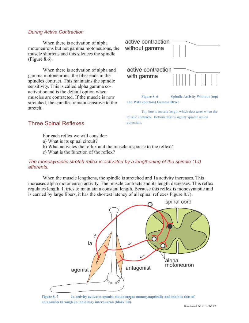
During Active Contraction When there is activation of alpha
motoneurons but not gamma motoneurons, the muscle shortens and this silences the spindle (Figure 8.6).
When there is activation of alpha and
gamma motoneurons, the fiber ends in the spindles contract. This maintains the spindle sensitivity. This is called alpha gamma co-activationand is the default option when muscles are contracted. If the muscle is now stretched, the spindles remain sensitive to the stretch.
Three Spinal Reflexes For each reflex we will consider: a) What is its spinal circuit? b) What activates the reflex and the muscle response to the reflex? c) What is the function of the reflex?
The monosynaptic stretch reflex is activated by a lengthening of the spindle (1a) afferents.
When the muscle lengthens, the spindle is stretched and 1a activity increases. This
increases alpha motoneuron activity. The muscle contracts and its length decreases. This reflex regulates length. It tries to maintain a constant length. Because this reflex is monosynaptic and is carried by large fibers, it has the shortest latency of all spinal reflexes Figure 8.7).
Figure 8. 7 1a activity activates agonist motoneurons monosynaptically and inhibits that of antagonists through an inhibitory interneuron (black fill).
Figure 8. 6 Spindle Activity Without (top) and With (bottom) Gamma Drive
Top line is muscle length which decreases when the muscle contracts. Bottom dashes signify spindle action potentials.

7
Revised 01/11/2017
The Reflex Mediated by the Golgi Tendon Organ If the muscle generates too much force, the Golgi tendon organ is activated. Through an
inhibitory interneuron in the spinal cord this activation reduces the firing rate of motoneurons. This decreases muscle contraction and muscle force (Figure 8.8).
This reflex regulates force i.e. it maintains a constant force. It is used, for example, when
attempting to maintain a constant grip on a paper cup.
Reflexes Mediated by Pain and Cutaneous Receptors
If the foot steps on something
painful, a reflex produces two responses (Figure 8.9):
1) Contraction of flexors on the
same side. This results in a withdraw of the foot from the painful stimulus (red dot).
2) Contraction of extensors on the
opposite side. Because your weight is now on your other leg, this contraction helps you maintain posture and balance.
The spinal cord can, on its own,
produce even more amazing responses using similar circuits, responses such as walking, scratching, and running.
Figure 8. 9 The Withdraw Reflex Stepping on something sharp causes ipsilateral flexion and contralateral extension.
Figure 8. 8 The Golgi Tendon Organ is activated by muscle contraction and through an inhibitory interneuron decreases the activity of the agonist motoneurons.

8
Revised 01/11/2017
Tremor Recall that gamma drive increases the sensitivity of the spindles. Why not keep gamma
activity high (and thus spindle sensitivity) all the time? The reason is that too much of a good thing is bad. In this case it can cause tremor. Do you remember a fine tremor when trying hard to preform something precise (e.g. threading a needle)?
Normally the 1a reflex stops the return movement exactly in the correct position. How
does that occur? When a limb position changes, the 1a reflex is activated. Stretch of the agonist muscle causes a reflex activation of the same muscle (Figure 8.10). This initiates a return movement. The return movement stretches the antagonist spindle, causing reflex activation of the antagonist muscle. If the antagonist muscle’s response is the right size, it will correctly stop the return movement.
If spindle sensitivity is too high, the stretch reflex in the agonist will be too large,
causing a very fast return movement that overshoots the target. This fast return movement and the high spindle sensitivity will produce a large reflex response in the antagonist (Figure 8.11). This will reverse the movement rather than stop it (something like trying to stop your car with the reverse gear rather than the brakes). This process will be repeated again and again, causing tremor.
Figure 8. 10 A Normal Reflex Response A stretch produces a reflexive activation of the agonist muscle (Ag, green response). This initiates a return movement. The movement produces a reflexive response in the antagonist muscle (Ant, orange response) which acts as a brake, stopping the movement.
Figure 8. 11 Tremor can be produced by too much gamma drive. A stretch of the agonist muscle will produce a large reflex response. This will produce a fast return movement which will in turn elicit a large antagonist reflex response. The cycle will be repeated producing a constant tremor.

9
Revised 01/11/2017
The Cortical Response to Muscle Stretch Muscle stretch often
generates two electromyographic (EMG) responses (Figure 8.12):
1) an early spinal monosynaptic stretch reflex.
2) a later long loop reflex, through area 3a and the motor cortex. This response is set, or context dependent, controlled by the cerebellum and what adds learnt motor skills.
Muscle Vibration The following experiment demonstrates that muscle spindle afferents contribute to a
conscious sensation of muscle length.
First apply a vibrator against the tendon of the biceps muscle. The vibration of the tendon
vibrates the whole muscle and activates the 1a spindle afferents. Now ask the subject, whose eyes are closed, to indicate the felt limb position with the other arm. The perceived position, as indicated by the other limb (background Figure 8.13), is longer and more extended than the actual position of the vibrated limb. This is because the vibration activates 1a afferents and this additional activity is interpreted as a longer muscle length.
Figure 8. 12 The EMG response in the muscle has two components: the spinal stretch reflex and the long loop reflex through the cortex.
Figure 8. 13 The right (foreground) and left (background) forelimbs are shown. A vibrator is applied to the tendon of the right arm. The left arm indicates the felt position of the right.

10
Revised 01/11/2017
How is actual muscle length sensed? If you pull a subject's arm while it
is actively contracted, the subject can correctly signal the position of that arm with the other, not contracted, arm even with the eyes closed. The contracted arm has a lot of alpha gamma co-activation, the not contracted arm has little. Presumably, in the contracted arm, the spindles will be more active than in the other, not contracted, arm.
Yet one can sense correctly when
both are at the same length. One is able to do so because of corollary discharge (an internal sense of effort). As first proposed by Helmholtz, motor commands go both to muscles and to the areas of the brain that sense position. The corollary discharge modifies how the afferent signal is interpreted (Figure 8.14)
A) In a relaxed muscle, low alpha
is accompanied by low gamma activity. The low gamma produces a low spindle activity.
B) If one maintains the same
position while actively contracting the muscle, high alpha is accompanied by high gamma activity. High gamma produces a high spindle activity.
In perceiving length, the brain
compares the command to the arm (corollary discharge) to the afferent spindle feedback.
The sensed position in both A and B is the same because during active contraction, a
high spindle signal is cancelled by a high corollary discharge.
Figure 8. 14 A comparison of corollary discharge and spindle activity results in sensed position. A) low alpha-gamma B) high alpha gamma.

11
Revised 01/11/2017
Coordinate Transformations Try the following. Close your eyes. Raise your right
and left hands and touch your two index fingers together. Notice that your brain senses correctly (not perfectly but quite well) where these two fingers are. Seems a simple enough task. Or is it?
How does your brain compute the position of your
two index fingers? The proprioceptive afferents involved project to area 3a of the primary somatosensory cortex (S1) along the bottom of the central sulcus (Figure 8.15). The spindles in the biceps muscle, after an adjustment by corollary discharge (Figure 8.14), signal the length of that muscle. This length is proportional to the elbow angle. In a similar manner angle of the index finger relative to the forearm is coded (the hand angle), as well as the angles of all the body parts relative to each other. Thus the activity in the homunculus represented in area 3a signals all these joint angles.
A sum of all these angles scaled by your limb
lengths is required to compute your finger position relative to some part of your body, for example, your nose or the finger of your other arm (Figure 8.16). This combination is computed in the association areas within the dorsal where stream along the intra-parietal sulcus (IPS) (Figure 8.17). Here all the individual joint angles, passed from area 3a, are combined into a egocentric finger position.
This position is represented in a nested
reference frame (Figure 8.16) because changing the shoulder angle changes the positions of all the more distal joints (e.g. the hand is distal to the elbow). Recall that this is similar to the nested allocentric frame formed by an apple on a table located in a room (discussed in chapter 5). When the table is moved the apple moves as well.
As in the visual pathways, the somatosensory
input flows along two streams (Figure 8.17). The “where” stream codes the egocentric position and movement of the limbs and body parts. This originates primarily from the receptors covered in this chapter, those in the muscles and joints that signal proprioception and kinesthesia. The “where” stream originates primarily from area 3a but also 3b, 1, and 2 and flows to the various areas along the intra-parietal sulcus (IPS) that
Figure 8. 15 Afferents that signal limb position and limb movement enter the cortex through area 3a.
Figure 8. 16 The finger position is estimated by combining the angle of all the joints to the finger, as well as limb lengths.
Figure 8. 17 The somatosensory “where” stream codes the egocentric limb position, possibly in the intra-parietal sulcus (IPS) and the “what” stream projects through the secondary somatosensory cortex (S2) to the inferior temporal lobe.

12
Revised 01/11/2017
were covered in chapter 6. The “what” stream uses touch information, covered in chapter 7, to determine object
shape in allocentric coordinates. Its signal originates from the receptors located in the skin, which are first mapped in area 3b. From there the signal flows to areas 1 and 2 and then to the secondary somatosensory cortex (S2 Figure 8.17) located in the lateral parietal mostly in the Sylvian fissure. Patients with lesions of S2 cannot identify objects by touch, stereognosis.
In summary, touching the two index fingers with your eyes closed is not as simple as it may have seemed. Knowing this complexity, it is even more amazing that a young child learns to do this at a time when the child’s limbs are rapidly changing in length.
See problems and answers posted on
http://www.tutis.ca/Senses/L8Muscle/L8MuscleProb.swf