Embed Size (px)
Citation preview

REVIEW PAPER
The pharmacological bases of the antiangiogenic activityof paclitaxel
Guido Bocci • Antonello Di Paolo • Romano Danesi
Received: 26 June 2012 / Accepted: 15 January 2013 / Published online: 7 February 2013
� The Author(s) 2013. This article is published with open access at Springerlink.com
Abstract In the mid 1990s, researchers began to inves-
tigate the antiangiogenic activity of paclitaxel as a possible
additional mechanism contributing to its antineoplastic
activity in vivo. In the last decade, a number of studies
showed that paclitaxel has antiangiogenic activity that
could be ascribed to the inhibition of either tubule forma-
tion or cell migration, and to an antiproliferative effect
towards activated endothelial cells. Furthermore, paclitaxel
was shown to downregulate VEGF and Ang-1 expression
in tumor cells, and to increase the secretion of TSP-1 in the
tumor microenvironment. Moreover, the new pharmaceu-
tical formulations of paclitaxel (such as liposome-encap-
sulated paclitaxel, ABI-007, and paclitaxel entrapped in
emulsifying wax nanoparticles) enhanced the in vivo
antiangiogenic activity of the drug. Thus, the preclinical
data of paclitaxel may be exploited to implement a novel
and rational therapeutic strategy to control tumor progres-
sion in patients.
Keywords Paclitaxel � Antiangiogenic drugs �Angiogenesis � Preclinical studies
Introduction
Paclitaxel is a cancer chemotherapeutic agent that is com-
monly used as first line therapy for many common malig-
nancies, including lung, breast, ovarian and head and neck
cancer. It also has high antitumor activity against some
uncommon malignancies, such as angiosarcoma and Kaposi
sarcoma. Paclitaxel is a member of the taxane class of
drugs, and it was the first taxane to enter clinical trials and to
receive FDA approval; it is a natural product obtained from
the North American Pacific yew tree (Taxus brevifolia).
Paclitaxel binds to the beta-subunit of polymerized tubulin
and inhibits the dissociation rate of the tubulin subunits
from the tubule. Thus, micromolar paclitaxel administration
to cells results in the formation of microtubule bundles and
asters, arresting cells in mitosis [1].
Besides these known pharmacodynamic properties,
medical researchers have discovered a new and unexpected
additional characteristic of paclitaxel, one that started a
second life for this compound and that created new thera-
peutic approaches using this molecule: that characteristic is
its antiangiogenic activity. Although the anti-vascular
effects of tubulin-binding agents have been previously
reviewed [2–4], the present article focuses solely on the
pharmacological bases of the antiangiogenic activity of
paclitaxel and provides an extensive and updated overview
of the field. Table 1 summarizes the published data on the
antiangiogenic effects of paclitaxel in different in vitro and
in vivo experimental models.
The first ever published evidence about a possible
antiangiogenic activity of paclitaxel was a small report
some 15 years ago. Dordunoo and colleagues prepared
poly(epsilon-caprolactone) (PCL) microspheres containing
paclitaxel, and showed this formulation to inhibit angio-
genesis in the chick chorioallantoic membrane (CAM)
model [5]. A year later, Belotti et al. [6] demonstrated a
strong antiangiogenic activity of paclitaxel, suggesting that
this property might contribute to its antineoplastic activity
in vivo. The aim of that study was to investigate the effect
of paclitaxel on endothelial cell functions, and on angio-
genesis. In vivo, paclitaxel (20–28 mg/kg i.v.) significantly
G. Bocci � A. Di Paolo � R. Danesi (&)
Division of Pharmacology, Department of Clinical and
Experimental Medicine, University of Pisa,
Via Roma 55, 56126 Pisa, Italy
e-mail: [email protected]
123
Angiogenesis (2013) 16:481–492
DOI 10.1007/s10456-013-9334-0
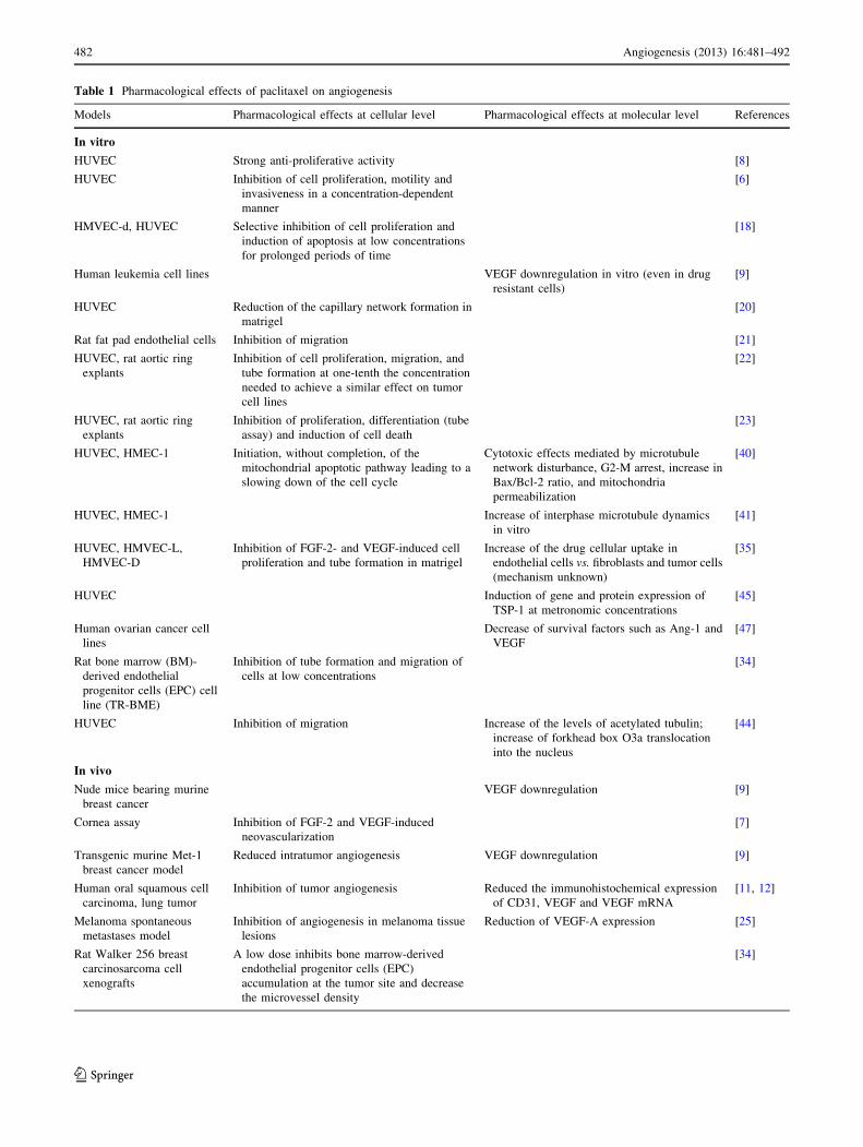
Table 1 Pharmacological effects of paclitaxel on angiogenesis
Models Pharmacological effects at cellular level Pharmacological effects at molecular level References
In vitro
HUVEC Strong anti-proliferative activity [8]
HUVEC Inhibition of cell proliferation, motility and
invasiveness in a concentration-dependent
manner
[6]
HMVEC-d, HUVEC Selective inhibition of cell proliferation and
induction of apoptosis at low concentrations
for prolonged periods of time
[18]
Human leukemia cell lines VEGF downregulation in vitro (even in drug
resistant cells)
[9]
HUVEC Reduction of the capillary network formation in
matrigel
[20]
Rat fat pad endothelial cells Inhibition of migration [21]
HUVEC, rat aortic ring
explants
Inhibition of cell proliferation, migration, and
tube formation at one-tenth the concentration
needed to achieve a similar effect on tumor
cell lines
[22]
HUVEC, rat aortic ring
explants
Inhibition of proliferation, differentiation (tube
assay) and induction of cell death
[23]
HUVEC, HMEC-1 Initiation, without completion, of the
mitochondrial apoptotic pathway leading to a
slowing down of the cell cycle
Cytotoxic effects mediated by microtubule
network disturbance, G2-M arrest, increase in
Bax/Bcl-2 ratio, and mitochondria
permeabilization
[40]
HUVEC, HMEC-1 Increase of interphase microtubule dynamics
in vitro
[41]
HUVEC, HMVEC-L,
HMVEC-D
Inhibition of FGF-2- and VEGF-induced cell
proliferation and tube formation in matrigel
Increase of the drug cellular uptake in
endothelial cells vs. fibroblasts and tumor cells
(mechanism unknown)
[35]
HUVEC Induction of gene and protein expression of
TSP-1 at metronomic concentrations
[45]
Human ovarian cancer cell
lines
Decrease of survival factors such as Ang-1 and
VEGF
[47]
Rat bone marrow (BM)-
derived endothelial
progenitor cells (EPC) cell
line (TR-BME)
Inhibition of tube formation and migration of
cells at low concentrations
[34]
HUVEC Inhibition of migration Increase of the levels of acetylated tubulin;
increase of forkhead box O3a translocation
into the nucleus
[44]
In vivo
Nude mice bearing murine
breast cancer
VEGF downregulation [9]
Cornea assay Inhibition of FGF-2 and VEGF-induced
neovascularization
[7]
Transgenic murine Met-1
breast cancer model
Reduced intratumor angiogenesis VEGF downregulation [9]
Human oral squamous cell
carcinoma, lung tumor
Inhibition of tumor angiogenesis Reduced the immunohistochemical expression
of CD31, VEGF and VEGF mRNA
[11, 12]
Melanoma spontaneous
metastases model
Inhibition of angiogenesis in melanoma tissue
lesions
Reduction of VEGF-A expression [25]
Rat Walker 256 breast
carcinosarcoma cell
xenografts
A low dose inhibits bone marrow-derived
endothelial progenitor cells (EPC)
accumulation at the tumor site and decrease
the microvessel density
[34]
482 Angiogenesis (2013) 16:481–492
123

inhibited the angiogenic process in a pellet of matrigel
containing tumor cell supernatant, that was injected into
mice. In vitro, paclitaxel inhibited endothelial cell prolif-
eration, motility, and invasiveness in a concentration-
dependent manner [6]. Interestingly, the authors stated that
the antiangiogenic activity of paclitaxel was not linked to
its cytotoxicity, since inhibition of endothelial cell che-
motaxis and invasiveness occurred at drug concentrations
which did not affect endothelial cell proliferation [6]. At
the same time, a lower dose of paclitaxel (6 mg/kg i.p.)
inhibited the bFGF and VEGF-induced neovascularization
of the cornea in mice by 45 and 37 %, respectively [7].
Moreover, Iwahana et al. [8] demonstrated a different
chemosensitivity of human umbilical vein endothelial cells
(HUVEC) and of tumor-derived endothelial cells from rat
KMT-17 fibrosarcoma (TEC) when exposed to the same
paclitaxel concentrations. Indeed, paclitaxel had strong
anti-proliferative activity against HUVECs, but only
weakly inhibited the proliferation of TECs, which expres-
sed greater amounts of P-glycoprotein (P-gp), suggesting a
drug resistant phenotype of newly formed capillaries [8].
The first evidence of the antiangiogenic activity of pac-
litaxel via down-regulation of vascular endothelial growth
factor (VEGF) in tumors was obtained in a highly-vascu-
larized transgenic murine Met-1 breast cancer model [9].
Paclitaxel was administered intraperitoneally, at non-
cytotoxic doses of 0–6 mg/kg/day, to nude mice bearing
Met-1 breast tumor. Interestingly, the intratumoral angio-
genesis, measured by microvessel tortuosity and micro-
vessel density, was significantly reduced by paclitaxel
treatment in a dose-dependent manner. Moreover, paclit-
axel also suppressed expression of VEGF in Met-1 tumors
transplanted in nude mice [9]. Interestingly, paclitaxel
monochemotherapy (175 mg/m2 i.v. every 21 days for
three cycles) significantly decreased VEGF plasma levels in
metastatic breast cancer patients with partial response or
stable disease, whereas no decline was observed in patients
with progressive disease [10]. Paclitaxel also showed an
inhibitory effect on tumor angiogenesis in a lung tumor
xenograft [11], and in transplanted human oral squamous
cell carcinoma [12] models, reducing the immunohisto-
chemical expression of CD31 (an endothelial marker),
VEGF, and VEGF mRNA.
However, no unanimous consensus was expressed on
the antiangiogenic activity of paclitaxel in vivo. A paclit-
axel dosage of 6.25 mg/kg s.c., given five times/week,
slowed tumor growth in a CWR22R androgen-independent
xenograft model of prostate cancer, without any antian-
giogenic effects [13]. Similarly, vascular density was not
altered by paclitaxel treatment in an orthotopic pancreatic
tumor model [14].
The metronomic concept and paclitaxel
Kerbel et al. [15], at the beginning of this century, coined
the term ‘‘accidental angiogenesis inhibitors’’ to describe a
number of drugs that were tested in clinical trials as possible
angiogenesis inhibitors, although they were not originally
developed to target tumor angiogenesis. This list of ‘‘acci-
dental’’ angiogenesis inhibitors included also established
agents such as conventional cytotoxic chemotherapeutic
drugs. The antivascular side-effects of chemotherapy were
found to be most evident by administering such drugs
Table 1 continued
Models Pharmacological effects at cellular level Pharmacological effects at molecular level References
Chick chorioallantoic
membrane
Inhibition of neovascularization [5]
Matrigel pellet in mice Inhibition of neovascularization [6]
Chick chorioallantoic
membrane
Inhibition of neovascularization at low
concentrations
[26]
Rat mesentery assay A low dose shortened the length of sprouts in
VEGF-mediated angiogenesis
[27, 28]
4T1 metastatic breast cancer Strong antiangiogenic and anti-
lymphangiogenic activities of low doses
[30]
Rats bearing syngeneic
prostate cancer
(Dunning AT-1) not
expressing TSP-1
Re-induction of TSP-1 expression in tumors [46]
HT-29 colon cancer model;
4T1 metastatic breast
cancer
Upregulation of TSP-1 expression [29, 30]
Ovarian carcinoma xenograft
model
Downregulation of VEGF-B, -D and -A;
upregulation of Tie-1, Tie-2 and VEGFR-2
[49]
Angiogenesis (2013) 16:481–492 483
123

continuously, or on a frequent (e.g. weekly or even daily)
basis, at concentrations well below their maximum tolerated
dose (MTD). This continuous low-dose schedule was
defined as metronomic chemotherapy [16]. Such a frequent
administration in vitro and in vivo of low doses of che-
motherapeutic drugs affects the tumor endothelium and
inhibits tumor angiogenesis, with minimal concomitant side
effects [17–19]. In particular, low concentrations of pac-
litaxel showed only weak effects on in vitro activated
endothelial cells using a short-term exposure (24 h) proto-
col, whereas a striking toxicity toward vascular endothelial
cells was observed by a long-term exposure protocol
(144 h) that used very low paclitaxel concentrations [18].
Moreover, a potent differential effect was found at the same
paclitaxel concentrations if fibroblasts, and drug-sensitive
or multidrug-resistant breast cancer cell lines were used.
Indeed, a growth inhibitory effect, as well as induction of
apoptosis, was observed with IC50 values in the range of
25–143 pM for paclitaxel only in the endothelial cells,
suggesting a selective antiangiogenic therapeutic window
for this taxane [18]. At the same time, Hayot et al. [20]
demonstrated that paclitaxel, vincristine and vindesine, at
non-cytotoxic concentrations, similarly reduced the capil-
lary network formation by HUVEC cells cultured on
Matrigel. Furthermore, paclitaxel was also tested in a che-
mokinetic migration assay using rat fat pad endothelial cells
(RFPECs), showing IC50 values that were approximately
10-9 M [21]. The different sensitivity of activated endo-
thelial cells to paclitaxel was also confirmed by other
studies. Dicker et al. [22] found that paclitaxel inhibited
human endothelial cell proliferation, migration, and tube
formation (differentiation) at one-tenth of the concentration
needed to achieve a similar effect on tumor cell lines
(Fig. 1). Moreover, similar results were obtained on the
proliferation, migration, and differentiation, of cultured
human umbilical vein endothelial cells and in the capillary
sprouting of rat aortic ring explants, demonstrating that
endothelial cells are 10–100-fold more sensitive to paclit-
axel than are tumor cells [22]. Additionally, it was dem-
onstrated that angiogenesis could be blocked by paclitaxel,
and that this effect was primarily due to inhibition of pro-
liferation and differentiation (as measured by Matrigel
assay) and by the induction of cell death [23]. The selec-
tivity of paclitaxel inhibition of cell proliferation is also
species specific. Indeed, the inhibition of proliferation of
human endothelial cells by paclitaxel was observed at ultra
low concentrations (0.1–100 pM), whereas 104 to 105-fold
higher concentrations were necessary to impact human non-
endothelial cells, and nonetheless mouse endothelial cells
were insensitive to such ultra low (pM) concentrations of
paclitaxel [24].
The antiangiogenic activity of paclitaxel at lower doses
was also demonstrated in in vivo models of neovasculari-
zation, such as the chick embryo chorioallantoic membrane
(CAM) [25], where paclitaxel administered at low con-
centrations (4, 8, and 12 nM) produced a significant dose-
dependent antiangiogenic activity [26]. Likewise, using the
rat mesentery assay, the effect of paclitaxel at a low, atoxic
dose, significantly shortened the length of sprouts at the
edge of the expanding network in VEGF165-mediated
angiogenesis [27]. Moreover, metronomic paclitaxel sig-
nificantly suppressed VEGF-A-mediated angiogenesis in
the rat mesentery to an extent that closely mirrored the
significant increase in prostate AT1 tumor necrosis, and the
Antiangiogenic activity ofpaclitaxel
Endothelial cells:
↓ cell proliferation
↓ cell motility
↓ cell migration
↓ interphasemicrotubule function
↑ apoptosis
↑ microtubule dynamics
Tumor site:
↓ EPCs accumulation in tumor tissue
↑ TSP-1 in tumor microenvironment
↓ capillary tube formation and microvessel density
Tumor cells:
↓ VEGF-A,B and C
↓ Ang-1
↑ TSP-1
Fig. 1 Antiangiogenic activity
of paclitaxel. EPC endothelial
progenitor cells, TSP-1thrombospondin-1, Ang-1angiopoetin-1, VEGF vascular
endothelial growth factor
484 Angiogenesis (2013) 16:481–492
123

concomitant decrease in tumor growth rate [28]. Paclitaxel
metronomic chemotherapy, in colon cancer xenografts,
showed stronger antiangiogenic activity than did paclitaxel
administered at the maximum tolerable dose (MTD). In
contrast, the MTD chemotherapy induced more apoptotic
cells than did the metronomic dosage [29]. More interest-
ingly, Jiang et al. [30] investigated the use of low-dose
metronomic (LDM) paclitaxel, as a single agent, in a
highly metastatic mouse model of 4T1 breast cancers, and
compared it with the maximum tolerable dose (MTD)
therapy. LDM therapy displayed a stronger anti-tumor
activity in suppressing primary and metastatic breast
tumors, with less side effects and stronger anti-angiogenic
and anti-lymphangiogenic activities, than MTD therapy.
On the other hand, MTD therapy showed stronger pro-
apoptotic and anti-proliferative activities in situ. Based on
these results, the authors suggested the use of metronomic
paclitaxel therapy to treat advanced breast cancer [30].
Metronomic paclitaxel has been successfully applied in
preclinical models of metastatic tumors. In one protocol
paclitaxel was injected i.p. at 5 mg/kg per day for 3 weeks
to treat preclinical spontaneous melanoma metastasis. This
protocol resulted in a relative decrease in spontaneous
metastases to the lungs, inhibiting angiogenesis in mela-
noma tissue lesions and reducing the expression of VEGF-
A [31]. Moreover, the weekly paclitaxel administration
was also effective in inhibiting angiogenesis, tumor
growth, and bone marrow hematopoiesis in a lung cancer
model [32]. Indeed, the tumor growth rate was reduced by
50 % and the intratumoral microvasculature was inhibited
by 70 %, with a concomitant low degree of leukopenia
[32]. This important inhibition of tumor neovascularization
by paclitaxel was also confirmed in a model of human
breast cancer specimens grown in a fibrin clot. Interest-
ingly, paclitaxel significantly reduced the angiogenic
sprouting and the formation of new microvessels if com-
pared to vehicle-treated controls [33]. Furthermore, low
dose (non-cytotoxic) paclitaxel inhibited bone marrow-
derived endothelial progenitor cells accumulation at the
tumor site in tumor-bearing rats, and caused a decrease in
microvessel density [34] (Fig. 1).
A possible explanation for preclinical reports describing
the endothelial cell’s in vitro sensitivity to paclitaxel, the
in vivo antiangiogenic effects of the drug, and clinical
reports of the relative efficacy of low weekly doses of
paclitaxel in patients refractory to standard (every 21 days)
doses of this agent came from the study by Merchan et al.
[35]. The authors observed that human endothelial cells can
accumulate paclitaxel at more than 5 times greater levels
than do normal human fibroblasts and several human
cancer cell lines. However, the mechanism by which the
endothelial cells accumulate paclitaxel remains to be elu-
cidated. Two possibilities have been suggested: (1) the
increased transport of the drug into the cells or, (2) the
decrease in the efflux of the drug [35].
The effects of paclitaxel on the inflammatory process (that
is a characteristic of cancer progression) and in the tumor
immune escape process are still quite controversial. The
induction of pro-inflammatory genes and proteins by paclit-
axel, especially through the lipopolysaccharide (LPS) sig-
naling pathway, have been reviewed by Fitzpatrick and
Wheeler [36]. Moreover, Olsen [37] have described that tax-
anes (including paclitaxel) promote transcription of the cyclo-
oxygenase (COX)-2 gene and the stabilization of the COX-2
messenger RNA. This pharmacodynamic property provided
the rationale for the use of COX-2 inhibitors in combination
with taxanes, and preclinical studies have shown enhanced
anticancer activity from the addition of COX-2 inhibitors to
taxane treatment [37]. However, more recent studies suggest
that paclitaxel could modulate tissue factor (TF), as is
expressed by tumour-associated endothelial and inflamma-
tory cells (as well as by cancer cells themselves), in human
mononuclear cells, in HUVECs, and in the metastatic breast
carcinoma cell line MDA-MB-231 [38]. Indeed, paclitaxel
significantly reduced the strong TF activity expressed by
MDA-MB-231, and the TF levels in mononuclear cells and
HUVECs even after the induction by LPS, TNF-a and IL-1b.
Interestingly, Napoleone et al. [38] also tested whether pac-
litaxel could influence IL-6 and IL-1b release from the MDA-
MB-231, HUVEC and mononuclear cells, since paclitaxel
was shown to induce the expression of inflammatory genes in
monocytes and in tumour cells. The authors demonstrated that
neither the constitutive expression of IL-6 and IL-1b in MDA-
MB-231, nor the basal and LPS-induced release from mono-
nuclear cells or from HUVECs, was affected by paclitaxel
administration. Recently, another important finding from
Chen et al. [39] underlines the importance of the effects of
paclitaxel on immunity. These authors showed that the anti-
tumor activity of metronomic low doses of paclitaxel depends
on both its capacity to deplete regulatory T cells, and on its
inhibition of tumor angiogenesis. Moreover, the combination
of metronomic paclitaxel plus an antigen-specific DNA
vaccine induced a more potent antigen-specific immune
responses and antitumor effects compared to the vaccine alone
[39]. In conclusion, the effect of paclitaxel on inflammation
and immunity seems to be more complex than previously
hypothesized and surely merits further investigations for its
relevant role in the antitumor effect of this taxane.
Molecular mechanisms of paclitaxel antiangiogenic
activity
The main molecular mechanisms for the antiangiogenic
activity of paclitaxel are graphically summarized in Fig. 2.
Pasquier and colleagues characterized two distinct effects
Angiogenesis (2013) 16:481–492 485
123

of paclitaxel on human endothelial cell proliferation: a
cytostatic effect at low paclitaxel concentrations, and a
cytotoxic effect at higher concentrations [40]. The cyto-
toxic effect involved primarily those signaling networks
that are reported to be impaired in tumor cells (i.e.,
microtubule network disturbance, G2-M arrest, increase in
the Bax/Bcl-2 ratio, and mitochondria permeabilization)
that result in apoptosis. Conversely, the cytostatic effect of
paclitaxel involves the inhibition of endothelial cell pro-
liferation without the induction apoptosis and without any
structural modification to the microtubule network. In
addition, low paclitaxel concentrations just initiate the
apoptotic signaling pathway that is stopped upstream of
mitochondria permeabilization and it does not lead to
endothelial cell death [40]. Moreover, paclitaxel inhibits
angiogenesis by an increase in microtubule dynamics in
endothelial cells and by the impairment of interphase
microtubule functions [41]. Other studies have uncovered
additional mechanisms involved in the antiangiogenic
effects of taxanes, including the degradation of heat shock
protein 90 [42] and the inhibition of Rac1 and of Cdc42
activity [43]. It should be noted that these data have been
obtained with docetaxel, but similar or analogous effects
have yet to be reported with paclitaxel. Although the two
drugs are very similar and belong to the same antineo-
plastic class of compounds, no data are available to show
that such mechanisms could be responsible for the anti-
angiogenic activity of paclitaxel.
Recently, Bonezzi et al. [44] found that paclitaxel
increases the levels of acetylated tubulin in HUVECs. The
induction of tubulin hyperacetylation was concomitant
with the inhibition of cell motility but not of cell prolif-
eration, which only occurred at concentrations much higher
than those that inhibited cell motility. In fact, the overex-
pression of a tubulin deacetylase such as the NAD-
dependent histone deacetylase sirtuin-2 (SIRT2), increases
EPCs accumulationin tumor mass
TSP-1 in tumor microenvironment
capillary tube formation and microvessel density
P a c l i t a x e lEPC
apoptosis
cell proliferation
pericyte
cell motility
endothelial cell
cell migration
interphase microtubule dynamics
Bax/Bcl-2 andmitochondria
permeabilization
levels of acetylated tubulin
endothelial cell
paclitaxel
uptake vs. tumor cell
VEGF-AB and C
TSP-1 Ang-1VEGF-AB and C
TSP-1 Ang-1
sign
alling
sign
alling
sign
alling
sign
alling
sign
alling
sign
alling
TSP-1
Fig. 2 Molecular and cellular mechanisms of antiangiogenic activity of paclitaxel
486 Angiogenesis (2013) 16:481–492
123

cell motility and reduces cell response to the anti-motility
activity of paclitaxel [44]. In the same article, Bonezzi
et al. [44] found another possible target to explain the anti-
motility activity of paclitaxel: forkhead box O3a
(FOXO3a), a member of the family of the forkhead tran-
scription factor FOXO, that negatively regulate cell
motility. Indeed, the authors found that paclitaxel caused
the translocation to the nucleus of the FOXO3a, which is a
pre-requisite for its transcriptional activity.
In 2003 we [45] reported that protracted exposure of
endothelial cells in vitro (144 h) to low concentrations of
several different anticancer agents, including paclitaxel,
caused overexpression of thrombospondin-1 (TSP-1), a potent
and endothelial-specific inhibitor of angiogenesis. Elevated
circulating TSP-1 levels were also detected in the plasma of
mice treated with metronomic chemotherapy and, above all,
the antiangiogenic and antitumor effects of low-dose contin-
uous cyclophosphamide were absent in TSP-1-null C57BL/6
mice. These results suggested that TSP-1 acts as a mediator of
the antiangiogenic effects of low-dose metronomic chemo-
therapy [45]. These data were later confirmed in other in vivo
models. Indeed, systemic low-dose continuous treatment of a
rat malignant prostate cancer model (Dunning AT-1) with
paclitaxel induced the expression of TSP-1 in the tumor tissue,
and inhibited tumor growth [46]. Moreover, metronomic
paclitaxel chemotherapy also caused a dramatic increase in
the expression of TSP-1 (whereas MTD chemotherapy did
not) in a HT-29 colon cancer model [29] and in the highly
metastatic 4T1 mouse model of breast cancer [30]. These
findings support the hypothesis that the anti-tumour effect of
low-dose metronomic paclitaxel is in part mediated by the
induction of TSP-1 (Fig. 2).
Other interesting possible mechanisms include the
downregulation of survival factors for endothelial cells
such as Angiopoietin-1 (Ang-1) and VEGF-A. Ang-1 and
angiopoietin-2 (Ang-2) are major ligands for the endothe-
lium-specific tyrosine kinase receptor Tie-2, and they are
important regulators of endothelial cell survival [46].
Long-term exposure (168 h) to low concentrations (2 nM)
of paclitaxel significantly decreased Ang-1 and VEGF-A
gene and protein expression in human ovarian cancer cell
lines [47]. The downregulation of VEGF-A after paclitaxel
treatment was also observed in tissue samples from 39
patients (diagnosed with invasive carcinoma) that were
cultured with paclitaxel for 24 h. The mean VEGF-A levels
were significantly lower in the treated samples compared to
controls [48]. Moreover, the downregulation of VEGF-B
and -D was also reported in response to paclitaxel treat-
ment in ovarian carcinoma xenografts, whereas the
expressions of Tie-1 and Tie-2 were significantly upregu-
lated. In the same study, paclitaxel treatment significantly
decreased the expression of VEGF-A, and it increased the
expression of VEGFR-2 [49] (Fig. 2).
New pharmaceutical formulations of paclitaxel enhance
its antiangiogenic activity
In the last decade numerous efforts have focused on
removing the use of Cremophor EL to decrease the
hypersensitive side effects, or to improve the pharmaco-
kinetic behavior, of paclitaxel. There are a variety of
nanoparticle carrier systems being explored for chemo-
therapeutic drugs, including paclitaxel, such as liposomes,
pegylated liposomes, protein nanoparticles, and polymeric
nanoparticles [50].
Liposomes represent an established drug delivery sys-
tem for lipophilic substances, and they act by improving
the pharmacokinetics and the therapeutic index of the
anticancer drugs [51]. Indeed, liposomal paclitaxel showed
improved solubility and similar in vitro cytotoxicity against
a variety of tumor cell lines compared to that of the
Cremophor EL paclitaxel preparation [52]. However, a
major limit in the clinical use of conventional liposome is
its rapid clearance by the reticuloendothelial system after
systemic administration, and is low bioavailability [53].
In contrast to the conventional liposomes, positively
charged cationic liposomes have been developed to be
preferentially bound to, and internalized by angiogenic and
negatively charged, endothelial cells that are found in
tumours and in areas of chronic inflammation [54]. Indeed,
Thurston et al. [54] have shown that actively growing
angiogenic endothelial cells exhibit a preferential uptake of
cationic liposomes by endocytosis.
In 2003, Schmitt-Sody et al. [55] evaluated a novel
neovascular targeting therapy, consisting of paclitaxel
encapsulated in cationic liposomes, on tumor selectivity
and antitumoral efficacy in comparison with the paclitaxel
free drug in vivo (Table 2). The endothelial deposition of
paclitaxel in tumor vessels significantly increased during
the infusion time (compared to Cremophor EL paclitaxel
preparation) and both the subcutaneous tumor growth and
the appearance of regional lymph node metastases were
significantly delayed in a amelanotic hamster melanoma
model [55]. Around the same time, also Kunstfeld et al.
[56] demonstrated that paclitaxel encapsulated in cationic
liposomes, which have been shown to target blood vessels,
prevented melanoma growth and invasive behavior, which
overall resulted in an increase in the survival of the tumor-
bearing mice. Moreover, liposome-encapsulated paclitaxel
reduced vessel density at the interface between the tumor
and the dermis, and reduced endothelial cell mitosis. In
contrast, equimolar concentrations of paclitaxel solubilized
in Cremophor EL produced only weak effects on tumor
growth and did not reduce the mitotic index of the endo-
thelium in vivo [56]. These data were also confirmed in an
animal model of prostate cancer. Thus, paclitaxel encap-
sulated in cationic liposomes (EndoTAG-1) caused a
Angiogenesis (2013) 16:481–492 487
123

significant decrease of microvessel density in tumors when
compared to paclitaxel alone, confirming the relative sup-
pression of angiogenesis when compared to the conven-
tional treatment [57] (Table 2).
Another promising approach to the synthesis of
liposomes with prolonged half-life is the grafting of the
liposome with the inert and biocompatible polymer poly-
ethylene glycol (PEG). This modification prevents the
recognition of liposome by opsonins and therefore reduces
its clearance by cells of the reticuloendothelial system [58].
The pegylated liposome is therefore often referred to as a
sterically stabilized liposome (SSL). Huang et al. [59]
evaluated the antiangiogenic activity of sterically stabilized
liposomes containing paclitaxel (SSL-PTX). SSL-PTX
effectively inhibited endothelial cell proliferation and
migration in a concentration-dependent manner and its
metronomic administration induced marked tumor growth
inhibition in the MDA-MB-231 xenograft model via an
antiangiogenic mechanism (i.e., reduced microvessel den-
sity), when compared to the injection of paclitaxel for-
mulated in Cremophor EL [59].
In order to improve the activity of low concentrations
of Cremophor EL-free paclitaxel on endothelial cell
proliferation, motility, and tube formation, and to facilitate
the delivery to blood vessels, new nanoparticles have been
synthesized and tested in vitro and in vivo (Table 2). Col-
loidal carriers have been shown to improve tumor therapy
by increased drug delivery to tumors resulting directly from
the enhanced permeability and retention of the drug. Thus,
paclitaxel entrapped in emulsifying wax nanoparticles
(PTX NPs) showed enhanced efficacy against colon cancer
xenograft models, overcoming paclitaxel resistance and
increasing the drug’s antiangiogenic effect [60]. Moreover,
the development of polymeric nanospheres (NPs), that are
able to selectively target the activated vascular endothelium
and to deliver co-encapsulated anti-angiogenic agents, has
also been studied with paclitaxel [61]. Indeed, NPs loaded
with paclitaxel have been successfully tested for their anti-
angiogenic efficacy in HUVECs in vitro, and in rat aorta
rings ex vivo. These effects were more potent than those
observed with free paclitaxel [61].
Albumin is a very attractive drug carrier for antineo-
plastic drugs [62]. It has been demonstrated that albumin
helps endothelial transcytosis of protein-bound and unbound
plasma constituents, mainly via a cell-surface glycoprotein
receptor (gp60). The gp60 protein binds to caveolin-1
Table 2 Antiangiogenic effects of novel paclitaxel formulations in vitro and in vivo
Paclitaxel formulations Models Pharmacological effects References
Paclitaxel encapsulated in cationic liposomes In vivo amelanotic hamster
melanoma A-Mel-3 model
Increased endothelial deposition of paclitaxel
in tumor vessels;
a remarkable retardation of tumor growth and
appearance of regional lymph node
metastases
[55]
Paclitaxel encapsulated in cationic liposomes In vivo metastatic melanoma
model
Inhibition of newly blood vessels, prevention
of melanoma growth and invasiveness,
improvement of mice survival
[56]
Paclitaxel encapsulated in cationic liposomes
(EndoTAG-1)
In vivo prostate tumor
xenograft
Decrease of microvessel density [57]
Sterically stabilized liposomes containing
paclitaxel (SSL-PTX)
HUVEC proliferation and
migration in vitro; MDA-
MB-231 breast cancer
xenograft models
Inhibition of cell proliferation and migration at
low concentrations; decrease of
microvascular density of tumors treated with
low doses of paclitaxel
[59]
Paclitaxel entrapped in emulsifying wax
nanoparticles (PTX NPs)
Colon cancer xenograft models Increase of antiangiogenic effect in the colon
cancer xenograft models
[60]
Polymeric nanospheres loaded with paclitaxel In vitro HUVEC culture and
ex vivo rat aortic rings
Inhibition of proliferation and inhibition of
endothelial sprouts
[61]
ABI-007, a cremophor EL-free, albumin-bound,
130-nm form of paclitaxel
Rat aortic rings, human
endothelial cell proliferation
and tube formation.
Tumor xenografts
Inhibition of rat aortic microvessel outgrowth,
human endothelial cell proliferation, and
tube formation.
Inhibition of tumor growth
[65]
Hyaluronic acid conjugates of paclitaxel (HA-
PTX)
Female nude mice bearing
ovarian cancer cells
Antitumor and antiangiogenic effects with a
marked increase of TSP-1
[67]
PEG-VC-PABC-PTX; paclitaxel (PTX)
conjugated with p-aminobenzylcarbonyl
(PABC), valine-citrulline (VC), and
polyethylene glycol (PEG)
MCF-7 tumor xenografts Antitumor and antiangiogenic effects in vivo.
Decrease of microvessel density
[68]
488 Angiogenesis (2013) 16:481–492
123

(an intracellular protein) which results in the subsequent
formation of transcytotic vesicles. In addition, gp60 also
binds to osteonectin. Both caveolin-1 and osteonectin are
frequently present in certain neoplasms (such as breast,
lung, and prostate cancer), which could account for the
accumulation of albumin in some tumors. Such accumula-
tion of albumin could facilitate intratumoral accumulation
of albumin-bound drugs [62]. In that respect, albumin-
bound paclitaxel ABI-007 is a novel, cremophor EL-free,
130-nm particle formulation of paclitaxel [63]. This for-
mulation is obtained by high-pressure homogenization of
paclitaxel with serum albumin, resulting in a nanoparticle
colloidal suspension [64]. Preclinical studies on human
breast cancer xenografts have demonstrated that ABI-007
has a higher penetration into tumor cells, with increased
antitumor activity, compared to an equal dose of standard
paclitaxel [63]. Interestingly, the antiangiogenic activity of
ABI-007 was described in the pivotal study by Ng et al. [65],
who employed a therapeutic metronomic approach. Indeed,
recent data showed that clinically relevant concentrations of
vehicles such as Cremophor EL and polysorbate 80 nullify
the antiangiogenic activity of taxanes [66]. These results
could hamper the usefulness of paclitaxel metronomic reg-
imens. In order to solve this problem, Ng et al. [65] tested
ABI-007 both in in vitro and in in vivo models (Table 2).
ABI-007 was found to significantly inhibit rat aortic
microvessel outgrowth, human endothelial cell prolifera-
tion, and tube formation. Moreover, metronomic ABI-007
significantly suppressed tumor growth in xenograft models
with a very low toxicity profile [65].
Recently, hyaluronic acid (HA) conjugates of paclitaxel
(HA-PTX) were shown to produce strong anti-tumor
effects when dosed metronomically. They also induced
anti-angiogenic effects of greater magnitude than those
achieved with MTD administration, or with free PTX
administration in female nude mice bearing ovarian cancer
cells [67]. Moreover, in a taxane-resistant model of ovarian
cancer, a significant reduction in tumor weight was noted
in the metronomic HA-PTX treated groups, whereas the
response of the MTD group was not statistically significant.
Metronomic HA-PTX treatment resulted in marked
increases in TSP-1, confirming the anti-angiogenic mech-
anism of action of this novel formulation [67].
Another interesting new paclitaxel formulation was
published by Liang et al. [68]. The novel paclitaxel conjugate
(PEG-VC-PABC-PTX) was designed and synthesized using
p-aminobenzylcarbonyl (PABC), a spacer, and valine-cit-
rulline (VC), a substrate of cathepsin B, to link polyethylene
glycol (PEG) and PTX. The PEG-VC-PABC-PTX showed
significant antitumor and anti-angiogenic effects in vivo,
while the control conjugate was almost ineffective [68].
The development of an oral formulation of paclitaxel is
necessary for the clinical development of metronomic
chemotherapy. Moes et al. [69] have increased the oral
bioavailability of paclitaxel by combining ritonavir with a
new oral solid dispersion formulation of paclitaxel (1/11
w/w paclitaxel, 9/11 w/w polyvinylpyrrolidone K30, and
1/11 w/w sodium lauryl sulfate)—which they termed
ModraPac001. Currently, the ModraPac001 formulation is
being applied in the first clinical trial with oral metronomic
chemotherapy of paclitaxel [69].
Among all the new formulations of paclitaxel with
demonstrated antiangiogenic characteristics, ABI-007 was
the most successful in clinical practice. Indeed, it was ini-
tially approved by the US Food and drug Administration in
January 2005, and indicated for use with pretreated meta-
static breast cancer patients. ABI-007 is currently approved
in 42 countries, including the European Union, Canada,
India and Japan [64]. Thus, in a relatively short period, there
has been an impressive improvement in our understanding
of the antiangiogenic mechanism of paclitaxel. Further
laboratory and clinical research is necessary to maximize
the benefits of the antiangiogenic properties of paclitaxel for
the treatment of patients with various cancers. In this con-
text, the knowledge and investigation of metronomic che-
motherapy based on the new formulations of paclitaxel,
such as ABI-007, HA-PTX or ModraPac001, constitutes
today one of the most exciting strategies for improving the
clinical control of angiogenesis in primary and metastatic
tumors [70]. On the other hand, such treatment strategies
should be based on solid and significant results of ran-
domized phase III clinical trials, and not only on their safety
profiles or on their impact on the quality of life and on
patient preference. Nonetheless, tolerability and compli-
ance will probably become the most important factors in the
future, according to the emerging overall importance of
quality of life in cancer care [71]. The availability of new
drugs for metronomic chemotherapeutic strategies (i.e.
ABI-007, HA-PTX and ModraPac001), alone or in associ-
ation with other targeted molecules, will give the oncologist
many different effective treatment options for metastatic
patients that are already resistant to standard doses of che-
motherapy [70]. Indeed, both preclinical [72] and clinical
studies [73, 74] have suggested that metronomic chemo-
therapy could be effective in stabilizing tumors already
resistant to the same drug MTD treatments. Moreover, the
ability of paclitaxel to inhibit different pathways of the
angiogenic process may be a great advantage in the treat-
ment of tumors already resistant to antiangiogenic drugs
with a specific target (e.g. bevacizumab or sunitinib).
Conclusion
The aim of this review was to summarize the published data
on the molecular and pharmacological bases of the
Angiogenesis (2013) 16:481–492 489
123
antiangiogenic activity of paclitaxel, and to underline this
particular pharmacodynamic characteristic of the drug for
possible future clinical development. In fact, paclitaxel (and
its novel formulations) both at MTD and at low dose
schedules demonstrated significant antiangiogenic activity.
This effect could be ascribed both to a cytotoxic or to a
cytostatic activity towards activated endothelial cells, or to
the inhibition of capillary formation and cell migration.
Moreover, paclitaxel has been shown to inhibit the release
of VEGF and Ang-1 by tumor cells, and to increase the
secretion of TSP-1 to the tumor microenvironment. It is
therefore possible that the different antiangiogenic mecha-
nisms of paclitaxel described in this review may explain the
antiangiogenic activity of this drug in different human
tumors, such as prostate and breast cancer. Indeed, the
angiogenic process in these tumors may depend on the
balance of different proangiogenic factors and endogenous
angiogenic inhibitors. Paclitaxel can definitely affect those
different pathways, resulting in an overall clinical activity,
and one that is independent from the direct cytotoxic effects
of the drug on cancer cells. Thus, based on the available
published preclinical data, paclitaxel (and especially its
novel formulations) may permit novel and rational thera-
peutic strategies to control tumor progression in patients.
Acknowledgments We thank Anna Fioravanti and Paola Orlandi
for figure drawing and Irving Miramontes, Chantal Vidal, Jasmine
Saenz and Prof. Giulio Francia for help with editing the text. The
study has been funded, in part, by the Italian Association for Cancer
Research (AIRC) and the Istituto Toscano Tumori (ITT).
Conflict of interest All authors have no financial and personal
relationships with other people or organizations that could inappro-
priately influence their work.
Open Access This article is distributed under the terms of the
Creative Commons Attribution License which permits any use, dis-
tribution, and reproduction in any medium, provided the original
author(s) and the source are credited.
References
1. Singh S, Dash AK (2009) Paclitaxel in cancer treatment: per-
spectives and prospects of its delivery challenges. Crit Rev Ther
Drug Carrier Syst 26(4):333–372
2. Schwartz EL (2009) Antivascular actions of microtubule-binding
drugs. Clin Cancer Res 15(8):2594–2601. doi:10.1158/1078-
0432.ccr-08-2710
3. Pasquier E, Andre N, Braguer D (2007) Targeting microtubules to
inhibit angiogenesis and disrupt tumour vasculature: implications
for cancer treatment. Curr Cancer Drug Targets 7(6):566–581
4. Pasquier E, Honore S, Braguer D (2006) Microtubule-targeting
agents in angiogenesis: where do we stand? Drug Resist Updat
9(1–2):74–86. doi:10.1016/j.drup.2006.04.003
5. Dordunoo SK, Jackson JK, Arsenault LA, Oktaba AM, Hunter
WL, Burt HM (1995) Taxol encapsulation in poly(epsilon-cap-
rolactone) microspheres. Cancer Chemother Pharmacol 36(4):
279–282
6. Belotti D, Vergani V, Drudis T, Borsotti P, Pitelli MR, Viale G,
Giavazzi R, Taraboletti G (1996) The microtubule-affecting drug
paclitaxel has antiangiogenic activity. Clin Cancer Res 2(11):
1843–1849
7. Klauber N, Parangi S, Flynn E, Hamel E, D’Amato RJ (1997)
Inhibition of angiogenesis and breast cancer in mice by the
microtubule inhibitors 2-methoxyestradiol and taxol. Cancer Res
57(1):81–86
8. Iwahana M, Utoguchi N, Mayumi T, Goryo M, Okada K (1998)
Drug resistance and P-glycoprotein expression in endothelial
cells of newly formed capillaries induced by tumors. Anticancer
Res 18(4C):2977–2980
9. Lau DH, Xue L, Young LJ, Burke PA, Cheung AT (1999)
Paclitaxel (Taxol): an inhibitor of angiogenesis in a highly vas-
cularized transgenic breast cancer. Cancer Biother Radiopharm
14(1):31–36
10. Lissoni P, Fugamalli E, Malugani F, Ardizzoia A, Secondino S,
Tancini G, Gardani GS (2000) Chemotherapy and angiogenesis in
advanced cancer: vascular endothelial growth factor (VEGF)
decline as predictor of disease control during taxol therapy in
metastatic breast cancer. Int J Biol Markers 15(4):308–311
11. Guo L, Burke P, Lo SH, Gandour-Edwards R, Lau D (2001)
Quantitative analysis of angiogenesis using confocal laser scan-
ning microscopy. Angiogenesis 4(3):187–191
12. Myoung H, Hong SD, Kim YY, Hong SP, Kim MJ (2001)
Evaluation of the anti-tumor and anti-angiogenic effect of pac-
litaxel and thalidomide on the xenotransplanted oral squamous
cell carcinoma. Cancer Lett 163(2):191–200
13. Fox WD, Higgins B, Maiese KM, Drobnjak M, Cordon-Cardo C,
Scher HI, Agus DB (2002) Antibody to vascular endothelial
growth factor slows growth of an androgen-independent xeno-
graft model of prostate cancer. Clin Cancer Res 8(10):3226–3231
14. Ryschich E, Werner J, Gebhard MM, Klar E, Schmidt J (2003)
Angiogenesis inhibition with TNP-470, 2-methoxyestradiol, and pac-
litaxel in experimental pancreatic carcinoma. Pancreas 26(2):166–172
15. Kerbel RS, Viloria-Petit A, Klement G, Rak J (2000) ‘Acciden-
tal’ anti-angiogenic drugs. Anti-oncogene directed signal trans-
duction inhibitors and conventional chemotherapeutic agents as
examples. Eur J Cancer 36(10):1248–1257
16. Kerbel RS, Kamen BA (2004) The anti-angiogenic basis of
metronomic chemotherapy. Nat Rev Cancer 4(6):423–436. doi:
10.1038/nrc1369
17. Klement G, Baruchel S, Rak J, Man S, Clark K, Hicklin DJ,
Bohlen P, Kerbel RS (2000) Continuous low-dose therapy with
vinblastine and VEGF receptor-2 antibody induces sustained
tumor regression without overt toxicity. J Clin Invest
105(8):R15–R24. doi:10.1172/JCI8829
18. Bocci G, Nicolaou KC, Kerbel RS (2002) Protracted low-dose
effects on human endothelial cell proliferation and survival
in vitro reveal a selective antiangiogenic window for various
chemotherapeutic drugs. Cancer Res 62(23):6938–6943
19. Browder T, Butterfield CE, Kraling BM, Shi B, Marshall B,
O’Reilly MS, Folkman J (2000) Antiangiogenic scheduling of
chemotherapy improves efficacy against experimental drug-
resistant cancer. Cancer Res 60(7):1878–1886
20. Hayot C, Farinelle S, De Decker R, Decaestecker C, Darro F,
Kiss R, Van Damme M (2002) In vitro pharmacological char-
acterizations of the anti-angiogenic and anti-tumor cell migration
properties mediated by microtubule-affecting drugs, with special
emphasis on the organization of the actin cytoskeleton. Int J
Oncol 21(2):417–425
21. Hotchkiss KA, Ashton AW, Mahmood R, Russell RG, Sparano
JA, Schwartz EL (2002) Inhibition of endothelial cell function
in vitro and angiogenesis in vivo by docetaxel (Taxotere): asso-
ciation with impaired repositioning of the microtubule organizing
center. Mol Cancer Ther 1(13):1191–1200
490 Angiogenesis (2013) 16:481–492
123
22. Dicker AP, Williams TL, Iliakis G, Grant DS (2003) Targeting
angiogenic processes by combination low-dose paclitaxel and
radiation therapy. Am J Clin Oncol 26(3):e45–e53. doi:10.1097/
01.COC.0000072504.22544.3C
23. Grant DS, Williams TL, Zahaczewsky M, Dicker AP (2003)
Comparison of antiangiogenic activities using paclitaxel (taxol)
and docetaxel (taxotere). Int J Cancer 104(1):121–129. doi:
10.1002/ijc.10907
24. Wang J, Lou P, Lesniewski R, Henkin J (2003) Paclitaxel at ultra
low concentrations inhibits angiogenesis without affecting cel-
lular microtubule assembly. Anticancer Drugs 14(1):13–19
25. Wang F, Cao Y, Liu HY, Xu SF, Han R (2003) Anti-invasion and
anti-angiogenesis effect of taxol and camptothecin on melanoma
cells. J Asian Nat Prod Res 5(2):121–129
26. Vacca A, Ribatti D, Iurlaro M, Merchionne F, Nico B, Ria R,
Dammacco F (2002) Docetaxel versus paclitaxel for antiangio-
genesis. J Hematother Stem Cell Res 11(1):103–118. doi:10.
1089/152581602753448577
27. Albertsson P, Lennernas B, Norrby K (2003) Chemotherapy and
antiangiogenesis: drug-specific effects on microvessel sprouting.
APMIS 111(11):995–1003
28. Lennernas B, Albertsson P, Damber JE, Norrby K (2004) Anti-
angiogenic effect of metronomic paclitaxel treatment in prostate
cancer and non-tumor tissue in the same animals: a quantitative
study. APMIS 112(3):201–209. doi:10.1111/j.1600-0463.2004.
apm1120306.x
29. Zhang M, Tao W, Pan S, Sun X, Jiang H (2009) Low-dose
metronomic chemotherapy of paclitaxel synergizes with cetux-
imab to suppress human colon cancer xenografts. Anticancer
Drugs 20(5):355–363. doi:10.1097/CAD.0b013e3283299f36
30. Jiang H, Tao W, Zhang M, Pan S, Kanwar JR, Sun X (2010)
Low-dose metronomic paclitaxel chemotherapy suppresses breast
tumors and metastases in mice. Cancer Invest 28(1):74–84. doi:
10.3109/07357900902744510
31. Wang F, Cao Y, Zhao W, Liu H, Fu Z, Han R (2003) Taxol
inhibits melanoma metastases through apoptosis induction,
angiogenesis inhibition, and restoration of E-cadherin and nm23
expression. J Pharmacol Sci 93(2):197–203
32. Lau D, Guo L, Gandara D, Young LJ, Xue L (2004) Is inhibition
of cancer angiogenesis and growth by paclitaxel schedule
dependent? Anticancer Drugs 15(9):871–875
33. Lyons JM 3rd, Anthony CT, Thomson JL, Woltering EA (2008)
A novel assay to assess the effectiveness of antiangiogenic drugs
in human breast cancer. Ann Surg Oncol 15(12):3407–3414. doi:
10.1245/s10434-008-0145-2
34. Muta M, Yanagawa T, Sai Y, Saji S, Suzuki E, Aruga T, Kuroi K,
Matsumoto G, Toi M, Nakashima E (2009) Effect of low-dose
paclitaxel and docetaxel on endothelial progenitor cells. Oncol-
ogy 77(3–4):182–191. doi:10.1159/000236016
35. Merchan JR, Jayaram DR, Supko JG, He X, Bubley GJ,
Sukhatme VP (2005) Increased endothelial uptake of paclitaxel as
a potential mechanism for its antiangiogenic effects: potentiation
by Cox-2 inhibition. Int J Cancer 113(3):490–498. doi:10.1002/
ijc.20595
36. Fitzpatrick FA, Wheeler R (2003) The immunopharmacology of
paclitaxel (taxol), docetaxel (taxotere), and related agents. Int
Immunopharmacol 3(13–14):1699–1714
37. Olsen SR (2005) Taxanes and COX-2 inhibitors: from molecular
pathways to clinical practice. Biomed Pharmacother 59(Suppl 2):
S306–S310
38. Napoleone E, Zurlo F, Latella MC, Amore C, Di Santo A,
Iacoviello L, Donati MB, Lorenzet R (2009) Paclitaxel down-
regulates tissue factor in cancer and host tumour-associated cells.
Eur J Cancer 45(3):470–477. doi:10.1016/j.ejca.2008.10.014
39. Chen CA, Ho CM, Chang MC, Sun WZ, Chen YL, Chiang YC,
Syu MH, Hsieh CY, Cheng WF (2010) Metronomic
chemotherapy enhances antitumor effects of cancer vaccine by
depleting regulatory T lymphocytes and inhibiting tumor angio-
genesis. Mol Ther 18(6):1233–1243. doi:10.1038/mt.2010.34
40. Pasquier E, Carre M, Pourroy B, Camoin L, Rebai O, Briand C,
Braguer D (2004) Antiangiogenic activity of paclitaxel is asso-
ciated with its cytostatic effect, mediated by the initiation but not
completion of a mitochondrial apoptotic signaling pathway. Mol
Cancer Ther 3(10):1301–1310
41. Pasquier E, Honore S, Pourroy B, Jordan MA, Lehmann M,
Briand C, Braguer D (2005) Antiangiogenic concentrations of
paclitaxel induce an increase in microtubule dynamics in endo-
thelial cells but not in cancer cells. Cancer Res 65(6):2433–2440.
doi:10.1158/0008-5472.CAN-04-2624
42. Murtagh J, Lu H, Schwartz EL (2006) Taxotere-induced inhibi-
tion of human endothelial cell migration is a result of heat shock
protein 90 degradation. Cancer Res 66(16):8192–8199. doi:
10.1158/0008-5472.CAN-06-0748
43. Bijman MN, van Nieuw Amerongen GP, Laurens N,
van Hinsbergh VW, Boven E (2006) Microtubule-targeting
agents inhibit angiogenesis at subtoxic concentrations, a process
associated with inhibition of Rac1 and Cdc42 activity and
changes in the endothelial cytoskeleton. Mol Cancer Ther
5(9):2348–2357. doi:10.1158/1535-7163.MCT-06-0242
44. Bonezzi K, Belotti D, North BJ, Ghilardi C, Borsotti P, Resovi A,
Ubezio P, Riva A, Giavazzi R, Verdin E, Taraboletti G (2012)
Inhibition of SIRT2 potentiates the anti-motility activity of tax-
anes: implications for antineoplastic combination therapies.
Neoplasia 14(9):846–854. doi:10.1593/neo.12728
45. Bocci G, Francia G, Man S, Lawler J, Kerbel RS (2003)
Thrombospondin 1, a mediator of the antiangiogenic effects of
low-dose metronomic chemotherapy. Proc Natl Acad Sci USA
100(22):12917–12922. doi:10.1073/pnas.2135406100
46. Damber JE, Vallbo C, Albertsson P, Lennernas B, Norrby K
(2006) The anti-tumour effect of low-dose continuous chemo-
therapy may partly be mediated by thrombospondin. Cancer
Chemother Pharmacol 58(3):354–360. doi:10.1007/s00280-005-
0163-8
47. Hata K, Osaki M, Dhar DK, Nakayama K, Fujiwaki R, Ito H,
Nagasue N, Miyazaki K (2004) Evaluation of the antiangiogenic
effect of taxol in a human epithelial ovarian carcinoma cell line.
Cancer Chemother Pharmacol 53(1):68–74. doi:10.1007/s00280-
003-0693-x
48. Loo WT, Fong JH, Cheung MN, Chow LW (2005) The efficacy
of paclitaxel on solid tumour analysed by ATP bioluminescence
assay and VEGF expression: a translational research study.
Biomed Pharmacother 59(Suppl 2):S337–S339
49. Thijssen VL, Brandwijk RJ, Dings RP, Griffioen AW (2004)
Angiogenesis gene expression profiling in xenograft models to
study cellular interactions. Exp Cell Res 299(2):286–293. doi:
10.1016/j.yexcr.2004.06.014
50. Byrne JD, Betancourt T, Brannon-Peppas L (2008) Active tar-
geting schemes for nanoparticle systems in cancer therapeutics.
Adv Drug Deliv Rev 60(15):1615–1626. doi:10.1016/j.addr.2008.
08.005
51. Straubinger RM, Arnold RD, Zhou R, Mazurchuk R, Slack JE
(2004) Antivascular and antitumor activities of liposome-asso-
ciated drugs. Anticancer Res 24(2A):397–404
52. Sharma A, Sharma US, Straubinger RM (1996) Paclitaxel-lipo-
somes for intracavitary therapy of intraperitoneal P388 leukemia.
Cancer Lett 107(2):265–272
53. Poste G, Bucana C, Raz A, Bugelski P, Kirsh R, Fidler IJ (1982)
Analysis of the fate of systemically administered liposomes and
implications for their use in drug delivery. Cancer Res 42(4):
1412–1422
54. Thurston G, McLean JW, Rizen M, Baluk P, Haskell A, Murphy
TJ, Hanahan D, McDonald DM (1998) Cationic liposomes target
Angiogenesis (2013) 16:481–492 491
123

angiogenic endothelial cells in tumors and chronic inflammation
in mice. J Clin Invest 101(7):1401–1413. doi:10.1172/jci965
55. Schmitt-Sody M, Strieth S, Krasnici S, Sauer B, Schulze B, Teifel
M, Michaelis U, Naujoks K, Dellian M (2003) Neovascular tar-
geting therapy: paclitaxel encapsulated in cationic liposomes
improves antitumoral efficacy. Clin Cancer Res 9(6):2335–2341
56. Kunstfeld R, Wickenhauser G, Michaelis U, Teifel M, Umek W,
Naujoks K, Wolff K, Petzelbauer P (2003) Paclitaxel encapsulated
in cationic liposomes diminishes tumor angiogenesis and mela-
noma growth in a ‘‘humanized’’ SCID mouse model. J Invest
Dermatol 120(3):476–482. doi:10.1046/j.1523-1747.2003.12057.x
57. Bode C, Trojan L, Weiss C, Kraenzlin B, Michaelis U, Teifel M,
Alken P, Michel MS (2009) Paclitaxel encapsulated in cationic
liposomes: a new option for neovascular targeting for the treat-
ment of prostate cancer. Oncol Rep 22(2):321–326
58. Moghimi SM, Patel HM (1992) Opsonophagocytosis of lipo-
somes by peritoneal macrophages and bone marrow reticuloen-
dothelial cells. Biochim Biophys Acta 1135(3):269–274
59. Huang Y, Chen XM, Zhao BX, Ke XY, Zhao BJ, Zhao X, Wang
Y, Zhang X, Zhang Q (2010) Antiangiogenic activity of sterically
stabilized liposomes containing paclitaxel (SSL-PTX): in vitro
and in vivo. AAPS PharmSciTech 11(2):752–759. doi:10.1208/
s12249-010-9430-z
60. Koziara JM, Whisman TR, Tseng MT, Mumper RJ (2006)
In-vivo efficacy of novel paclitaxel nanoparticles in paclitaxel-
resistant human colorectal tumors. J Control Release 112(3):312–
319. doi:10.1016/j.jconrel.2006.03.001
61. Hammady T, Rabanel JM, Dhanikula RS, Leclair G, Hildgen P
(2009) Functionalized nanospheres loaded with anti-angiogenic
drugs: cellular uptake and angiosuppressive efficacy. Eur J Pharm
Biopharm 72(2):418–427
62. Fu Q, Sun J, Zhang W, Sui X, Yan Z, He Z (2009) Nanoparticle
albumin-bound (NAB) technology is a promising method for
anti-cancer drug delivery. Recent Pat Anticancer Drug Discov
4(3):262–272
63. Miele E, Spinelli GP, Tomao F, Tomao S (2009) Albumin-bound
formulation of paclitaxel (Abraxane ABI-007) in the treatment of
breast cancer. Int J Nanomedicine 4:99–105
64. Yamamoto Y, Kawano I, Iwase H (2011) Nab-paclitaxel for the
treatment of breast cancer: efficacy, safety, and approval. Onco
Targets Ther 4:123–136. doi:10.2147/ott.s13836
65. Ng SS, Sparreboom A, Shaked Y, Lee C, Man S, Desai N, Soon-
Shiong P, Figg WD, Kerbel RS (2006) Influence of formulation
vehicle on metronomic taxane chemotherapy: albumin-bound
versus cremophor EL-based paclitaxel. Clin Cancer Res
12(14 Pt 1):4331–4338. doi:10.1158/1078-0432.CCR-05-2762
66. Ng SS, Figg WD, Sparreboom A (2004) Taxane-mediated
antiangiogenesis in vitro: influence of formulation vehicles and
binding proteins. Cancer Res 64(3):821–824
67. Lee SJ, Ghosh SC, Han HD, Stone RL, Bottsford-Miller J, Shen
DY, Auzenne E, Lopez-Araujo A, Lu C, Nishimura M, Pecot CV,
Zand B, Thanapprapa D Sr, Jennings NB, Kang Y, Huang J, Hu
W, Klostergaard J, Sood AK (2012) Metronomic activity of
CD44-targeted hyaluronic acid-paclitaxel in ovarian carcinoma.
Clin Cancer Res 18:4114–4121. doi:10.1158/1078-0432.CCR-
11-3250
68. Liang L, Lin SW, Dai W, Lu JK, Yang TY, Xiang Y, Zhang Y, Li
RT, Zhang Q (2012) Novel cathepsin B-sensitive paclitaxel
conjugate: higher water solubility, better efficacy and lower
toxicity. J Control Release 160(3):618–629. doi:10.1016/j.
jconrel.2012.02.020
69. Moes J, Koolen S, Huitema A, Schellens J, Beijnen J, Nuijen B
(2012) Development of an oral solid dispersion formulation for
use in low-dose metronomic chemotherapy of paclitaxel. Eur J
Pharm Biopharm. doi:10.1016/j.ejpb.2012.09.016
70. Kerbel RS (2012) Strategies for improving the clinical benefit of
antiangiogenic drug based therapies for breast cancer. J
Mammary Gland Biol Neoplasia. doi:10.1007/s10911-012-
9266-0
71. Rodrigues G, Sanatani M (2012) Age and comorbidity consid-
erations related to radiotherapy and chemotherapy administration.
Semin Radiat Oncol 22(4):277–283. doi:10.1016/j.semradonc.
2012.05.004
72. Emmenegger U, Francia G, Chow A, Shaked Y, Kouri A, Man S,
Kerbel RS (2011) Tumors that acquire resistance to low-dose
metronomic cyclophosphamide retain sensitivity to maximum
tolerated dose cyclophosphamide. Neoplasia 13(1):40–48
73. Allegrini G, Di Desidero T, Barletta MT, Fioravanti A, Orlandi P,
Canu B, Chericoni S, Loupakis F, Di Paolo A, Masi G, Fontana
A, Lucchesi S, Arrighi G, Giusiani M, Ciarlo A, Brandi G, Danesi
R, Kerbel RS, Falcone A, Bocci G (2012) Clinical, pharmaco-
kinetic and pharmacodynamic evaluations of metronomic UFT
and cyclophosphamide plus celecoxib in patients with advanced
refractory gastrointestinal cancers. Angiogenesis 15(2):275–286.
doi:10.1007/s10456-012-9260-6
74. Allegrini G, Falcone A, Fioravanti A, Barletta MT, Orlandi P,
Loupakis F, Cerri E, Masi G, Di Paolo A, Kerbel RS, Danesi R,
Del Tacca M, Bocci G (2008) A pharmacokinetic and pharma-
codynamic study on metronomic irinotecan in metastatic colo-
rectal cancer patients. Br J Cancer 98(8):1312–1319. doi:10.
1038/sj.bjc.6604311
492 Angiogenesis (2013) 16:481–492
123


![Cardiologie francophone - franco 2005.ppt [Lecture seule] · 2007-05-14 · AMG Pico Elite Paclitaxel Artax Paclitaxel Aachen Resonance EuroCor Taxcor Paclitaxel Biolimus A9 Biomatrix](https://img.dokumen.tips/doc/110x75/5e42b3f5800daf02232992fa/cardiologie-francophone-franco-2005ppt-lecture-seule-2007-05-14-amg-pico.jpg)
![Antiangiogenic Scheduling of Chemotherapy Improves ...[CANCER RESEARCH 60, 1878–1886, April 1, 2000] Antiangiogenic Scheduling of Chemotherapy Improves Efficacy against Experimental](https://img.dokumen.tips/doc/110x75/5f0d25fa7e708231d438e9b7/antiangiogenic-scheduling-of-chemotherapy-improves-cancer-research-60-1878a1886.jpg)