Embed Size (px)
Citation preview
Prostagladin D2 is a mast cell-derived antiangiogenicfactor in lung carcinomaTakahisa Murataa,1, Kosuke Aritakeb, Shigeko Matsumotoa, Shinya Kamauchib, Takayuki Nakagawac, Masatoshi Horia,Eiichi Momotanid, Yoshihiro Uradeb, and Hiroshi Ozakia
aDepartment of Veterinary Pharmacology and cVeterinary Surgery, Graduate School of Agriculture and Life Sciences, University of Tokyo, Tokyo 113-8657,Japan; bDepartment of Molecular Behavioral Biology, Osaka Bioscience Institute, Osaka 565-0874, Japan; and dResearch Team for Paratuberculosis,National Institute of Animal Health, Tsukuba, Ibaraki 305-0856, Japan
Edited by Gregg L. Semenza, The Johns Hopkins University School of Medicine, Baltimore, MD, and approved October 27, 2011 (received for reviewJune 23, 2011)
It is well established that prostaglandins (PGs) are involved intumor angiogenesis and growth, yet the role of prostaglandin D2
(PGD2) remains virtually unknown. Here, we show that host he-matopoietic PGD2 synthase (H-PGDS) deficiency enhances Lewislung carcinoma (LLC) progression, accompanied by increased vas-cular leakage, angiogenesis, and monocyte/mast cell infiltration.This deficiency can be rescued by hematopoietic reconstitutionwith bone marrow from H-PGDS–naive (WT) mice. In tumors onWT mice, c-kit+ mast cells highly express H-PGDS. Host H-PGDSdeficiency markedly up-regulated the expression of proangiogenicfactors, including TNF-α in the tumor. In mast cell-null KitW-sh/W-sh
mice, adoptive transfer of H-PGDS–deficient mast cells causesstronger acceleration in tumor angiogenesis and growth than inWT mast cells. In response to LLC growth, H-PGDS–deficient mastcells produce TNF-α excessively. This response is suppressed by theadministration of a synthetic PGD2 receptor agonist or a degrada-tion product of PGD2, 15-deoxy-Δ12,14-PGJ2. Additional TNF-α defi-ciency partially counteracts the tumorigenic properties seen in H-PGDS–deficient mast cells. These observations identify PGD2 as amast cell-derived antiangiogenic factor in expanding solid tumors.Mast cell-derived PGD2 governs the tumor microenvironment byrestricting excessive responses to vascular permeability and TNF-αproduction.
cancer | inflammation
Angiogenesis is crucial for both tumor progression and metas-tasis. Links between tumor angiogenesis and chronic inflam-
mation have been well documented (1). As well as abnormallyproliferating tumor cells, infiltrating hematopoietic lineage inflam-matory cells, such as macrophages, lymphocytes, and mast cells,induce tumor angiogenesis by furnishing a variety of growth factorsand cytokines (2). Thus, inflammatory cell infiltration has beenwell recognized as a crucial factor in tumor malignancy.Epidemiological studies have provided evidence that non-
steroidal anti-inflammatory drugs that inhibit COX-dependentprostaglandin (PG) synthesis can significantly reduce the risk ofcancer development (1). The main PGs responsible for tumorprogression are currently being investigated. PGE2 has alreadybeen shown to promote tumor growth and angiogenesis. Severalgroups have reported that the PGE2-prostaglandin E receptor(EP) signal facilitates stromal VEGF expression, thus promotingtumor angiogenesis and growth (3, 4).PGD2 is another COX metabolite reported to promote sleep
and to mediate inflammatory responses. There are two distinctisoforms of PGD2 synthase, the hematopoietic-type (H-PGDS)and the lipocalin-type (L-PGDS). H-PGDS is a cytosolic proteinresponsible for the biosynthesis of PGD2 in hematopoietic line-age cells, such as mast cells and Th2 lymphocytes (5). Thus, H-PGDS–derived PGD2 is assumed to act mainly as an inflam-matory mediator. In contrast, L-PGDS is a secretory proteinmainly expressed in the central nervous system and is involved insleep promotion (6).
The biological actions of PGD2 are mediated through the Gprotein-coupled receptor, D prostanoid (DP), and the chemoat-tractant receptor-homologous molecule expressed on Th2 cells(CRTH2). Discrepancies exist regarding the proinflammatoryor anti-inflammatory role of PGD2 in pathophysiological con-ditions (5). Several studies demonstrate that DP signal activationinhibits the migration and activation of inflammatory cells (7, 8).Other groups have demonstrated that DP-deficient mice displaydecreased inflammatory responses to certain allergens (9), andthat DP agonists produce conjunctival inflammation (10). CRTH2is mainly expressed in inflammatory cells, and its activation hasbeen consistently categorized as a proinflammatory signal. Fur-thermore, recent reports suggest that the dehydration product ofPGD2, 15-deoxy-12,14-PGJ2 (15d-PGJ2), inhibits inflammatoryresponses via peroxisome proliferator-activated receptor-γ(PPAR-γ)–dependent or –independent signal activation (11).Thus, PGD2 is likely to have a complex role by being both a pro-and anti-inflammatory mediator, depending on the target celland stimulus.In our previous study, we focused on the physiological function
of PGD2-DP signaling in the neovascular endothelial cells ofexpanding tumors, and found that stimulation of this pathwaystrongly tightens endothelial barriers and suppresses tumor an-giogenesis (12). However, the pathophysiological implication ofPGD2 biosynthesis in tumors is virtually unexplored.In the present study, we attempt to elucidate the contribution
of PGD2, derived from infiltrating cells of hematopoietic lineage,in tumor angiogenesis and growth. We demonstrate that infil-trating mast cells are the primary source of PGD2, and that mastcell-derived PGD2 suppresses vascular leakage and modulatesTNF-α production, thus shaping the tumor microenvironment.
ResultsLoss of Hematopoietic Cell-Derived PGD2 Accelerates Tumor Growth.Lewis lung carcinoma (LLC) or B16 melanoma implanted onto thebacks of H-PGDS–deficient (H-PGDS−/−) mice grew faster thanthose implanted onto WT mice (Fig. 1A and Fig. S1A). Transplan-tation of H-PGDS–naive bone marrow (H-PGDS−/−+WTHemRC)reduced the increased rate of LLC growth on H-PGDS−/− mice tothe same extent as for WT mice. Inversely, transplantation of H-PGDS−/− bone marrow (WT+H-PGDS−/−HemRC) increased tumormalignancy in WT mice. Although abundant PGD2 was detected inthe tumors onWTandL-PGDS−/−mice (Fig. 1B), little was detectedin the tumors on H-PGDS−/− mice. Tetranor-PGDM (11,15-deoxo-
Author contributions: T.M. and Y.U. designed research; T.M., K.A., S.M., S.K., and T.N.performed research; T.M., K.A., M.H., E.M., Y.U., and H.O. contributed new reagents/analytic tools; T.M., K.A., S.M., and S.K. analyzed data; and T.M. wrote the paper.
The authors declare no conflict of interest.
This article is a PNAS Direct Submission.1To whom correspondence should be addressed. E-mail: [email protected].
This article contains supporting information online at www.pnas.org/lookup/suppl/doi:10.1073/pnas.1110011108/-/DCSupplemental.
19802–19807 | PNAS | December 6, 2011 | vol. 108 | no. 49 www.pnas.org/cgi/doi/10.1073/pnas.1110011108
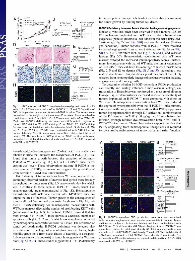
9a-hydroxy-2,3,4,5-tetranorprostan-1,20-dioic acid) is a stable me-tabolite in urine that indicates the biosynthesis of PGD2 (13). Wefound that tumor growth boosted the excretion of tetranor-PGDM in WT mice (Fig. 1C), but in H-PGDS−/− mice its ex-cretion was lower. These observations indicate H-PGDS is themain source of PGD2 in tumors and suggest the possibility ofurine tetranor-PGDM as a tumor marker.H&E staining of tumor sections from WT mice revealed that
commonly observed pockets of necrosis had spread more broadlythroughout the tumor mass (Fig. 1D, arrowheads, day 14), whichwas in contrast to those seen in H-PGDS−/− mice, which hadsmaller necrotic areas (summarized in Fig. 1E). Hematopoieticreconstitution with WT bone marrow into H-PGDS−/− mice en-larged the area of necrosis. Tumor growth is the consequence oftumor-cell proliferation and apoptosis. As shown in Fig. 1F, nei-ther H-PGDS deficiency nor hematopoietic reconstitution withWT bone marrow affected the number of proliferating Ki67+ cells(summarized in Fig. 1G). In contrast, TUNEL showed that tu-mors grown in H-PGDS−/− mice showed a decreased number ofapoptotic cells (Fig. 1 H and I), which was completely correctedby hematopoietic reconstitution from WT mice. The reduction oftumor cell death under H-PGDS–deficiency was detected alsoas a decrease in leakage of a nonhistone nuclear factor, high-mobility group box 1 from nuclei (index of necrosis), or a caspase-3 cleavage (index of apoptosis) in tumor homogenates by Westernblot (Fig. S1D–G). These studies suggest that H-PGDS deficiency
in hematopoietic lineage cells leads to a favorable environmentfor tumor growth by limiting tumor cell death.
H-PGDS Deficiency Increases Tumor Vascular Leakage and Angiogenesis.Similar to what has often been observed in solid tumors, LLC orB16 melanoma implanted into WT mice exhibit substantial an-giogenesis [platelet endothelial cell adhesion molecule (PECAM-1)–staining] (Fig. 2A and Fig. S1B) and vascular leakage (fibrino-gen deposition). Tumor sections from H-PGDS−/− mice revealedincreased angiogenesis (summaries of staining, see Fig. 2B and Fig.S1C; VEGFR-2/Western blot, see Fig. S1 H and I) and vascularleakage (Fig. 2C). Hematopoietic reconstitution with WT bonemarrow restored the increased immunopositivity scores. Further-more, in comparison with that of WT mice, the tumor vasculatureof H-PGDS−/−mice exhibited less coverage of smooth muscle actin(Fig. 2 D and E) or desmin (Fig. S1 J and K), indicating a lessmature vasculature. Thus, our data support the concept that PGD2
secreted from hematopoietic lineage cells reduces vascular leakage,angiogenesis, and tumor growth.To determine whether H-PGD–dependent PGD2 production
can directly and acutely influence tumor vascular leakage, ex-travasation of Evans blue was monitored as a measure of albuminleakage. Fig. 2F demonstrates increased vascular permeability oftumors implanted on H-PGDS−/− mice compared with those onWT mice. Hematopoietic reconstitution from WT mice reducedthe degree of hyperpermeability in the H-PGDS−/− mice tumors.Consistent with our previous observations that PGD2 suppressestumor hyperpermeability through DP activation, administrationof the DP agonist BW245C (100 μg/kg, i.v., 10 min before dyeinfusion) strongly reduced dye extravasation both in WT and H-PGDS−/− mice tumors. These observations support the idea thatPGD2 originating from hematopoietic lineage cells is requiredfor constitutive maintenance of tumor vascular barrier function.
Fig. 1. (A) Tumors on H-PGDS−/− mice have increased growth rates (n = 25each; *†P < 0.05 compared with WT or H-PGDS−/−). (B and C) Detection ofPGD2 in implanted tumors and tetranor-PGDM in urine. The results werenormalized to the weight of the tumor mass (B, n = 4 each) or normalized tocreatinine content (C, n = 4–5; *†P < 0.05 compared with WT or WT+LLC).(D–I) Host H-PGDS deficiency decreases necrotic/apoptotic regions intumors: H&E staining (D), Ki67 staining (F), or TUNEL (H). Ki67 staining(brown) was counterstained with hematoxylin (blue). (Scale bars: D, 200μm; F, 10 μm; H, 50 μm.) TUNEL was counterstained with DAPI (blue) fornuclear labeling. Necrotic areas were quantified relative to total pixeldensity (E ). The numbers of Ki67-positive or TUNEL-positive cells werenormalized to total nuclei number (G and I, n = 6 each; *†P < 0.05 comparedwith WT or H-PGDS−/−).
Fig. 2. H-PGDS–dependent PGD2 production from bone marrow-derivedcells decreases angiogenesis and vascular permeability in tumors. Tumorsections were subjected to immunostaining and then counterstained withDAPI (A and D) (Scale bars: A and D, 50 μm). Angiogenic area (PECAM-1) wasquantified relative to total pixel density (B). Fibrinogen deposition wasnormalized to total PECAM-1+ pixel density (C, n = 6–10). The pixel density ofα-SMA was normalized to total PECAM-1+ pixel density (E, n = 6 each). Dyeextravasations (30 min) into tumor were quantified (F, n = 8 each). *†P < 0.05compared with WT or H-PGDS−/−.
Murata et al. PNAS | December 6, 2011 | vol. 108 | no. 49 | 19803
PHARM
ACO
LOGY

H-PGDS Deficiency Enhances Proangiogenic Properties in Tumors.Wenext examined the angiogenic properties of implanted tumors.As with hydron pellets that contain 100 ng VEGF, tumor piecesdissected from WT mice (day 10) and implanted into recipientmouse corneas showed extended neovasculature from the limbus(4 d after implantation) (Fig. 3 A and B). The tumor piecesdissected from H-PGDS−/− mice exhibited a robust increase(1.82-fold) in angiogenic response compared with that grown onWT. This increase was suppressed by the treatment of BW245C(5 μL of 0.35 ng/mL, twice a day) and 15d-PGJ2 (5 μL of 0.2 ng/mL, twice a day), but not a CRTH2 agonist, 13,14-dihydro-15-keto-PGD2 (DK-PGD2; 5 μL of 0.3 ng/mL, twice a day). Con-sistently, the tumor pieces from H-PGDS−/−+WTHemRC micereduced the increased tumor angiogenic capacity derived fromH-PGDS−/− mice. Quantitative RT-PCR revealed that host H-PGDS deficiency increased proangiogenic gene expression ofTNF-α, IL-6, moncyte chemoattractant protein-1 (MCP-1),VEGF-A, and TGF-β in the tumor (day 10, Fig. 3C). In partic-ular, TNF-α expression was elevated almost fourfold in H-PGDS−/− mice. Hematopoietic reconstitution from WT micelowered all of the elevated gene expressions observed in the tumorson H-PGDS−/− mice. Thus, PGD2 originating from infiltratedhematopoietic lineage cells appears to modulate the productionof proangiogenic factors in the tumor.
Mast Cells Express H-PGDS and TNF-α in the Expanding Tumor.Immunostaining was performed to determine which hemato-poietic lineage cell expressed H-PGDS in the tumor. Althoughthe blood cells labeled with anti-CD4, -8, -11b, -19, and -68antibodies did not merge with the H-PGDS+ signal (CD68, Fig.4C, and CD11b, -4, -8, Fig. S2 A–C, respectively), c-kit+ orFcεR1+ mast cells distinctively expressed H-PGDS (Fig. 4 A andB). Infiltrations of macrophage and mast cells are well recog-nized as being exacerbating factors of tumor growth. Inter-estingly, the number of CD68+ monocytes/macrophages (Fig.4D, Upper) and mast cells (Fig. 4D, Lower), but not CD4− orCD8+ cells (Fig. S2 D and E), were increased in tumors on H-PGDS−/− mice that were ameliorated by hematopoietic recon-stitution from H-PGDS–naive donor mice. These observationssuggest that host PGD2 deficiency accelerates protumorigenicproperties of the tumor microenvironment.Next we attempted to detect the source of angiogenic factors
whose mRNA expression levels were elevated in the tumors onH-PGDS−/− mice. As shown in Fig. 4G, c-kit+ mast cells strongly
express TNF-α as well as PGD2. MCP-1 was mainly detected inCD68+ monocyte/macrophage cells (Fig. 4H). In contrast, IL-6and VEGF were broadly detected throughout the tumor mass(Fig. S2F).
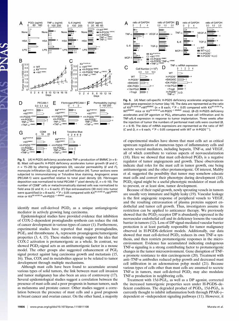
H-PGDS Deficiency Increases TNF-α Productivity of Bone Marrow-Derived Mast Cells. Isolated bone marrow cells proliferate/differ-entiate into mast cells by cultivation with IL-3. As shown in Fig.S3A, the cell-growth rates of WT and H-PGDS−/− bone marrowcells were identical. Five-week cultured H-PGDS−/− cells nor-mally expressed both mast cell surface marker c-kit and FcεR1(Fig. S3 B and C). As previously described (14), gene deficiencyof TNF-α reduced the proliferation and c-kithighFcεRIhigh pop-ulation of bone marrow cells (TNF-α−/− and H-PGDS−/−TNF-α−/−), that were rescued by a supplement with 1 ng/mL TNF-α.Thus, PGD2 does not influence IL-3–mediated mast cell growthand differentiation in vitro, but TNF-α is required for it to occur.We next measured the level of cytokine production for bone
marrow-derived mast cells (BMMCs) (Fig. 5A). For WTBMMCs, antigenic stimulation [10 ng/mL dinitrophenyl-humanserum albumin (DNP-HSA) for 24 h] elevated PGD2 pro-duction, and LPS-stimulation (10 ng/mL for 24 h) elevated theproduction of PGD2, TNF-α, and IL-6, but not VEGF. H-PGDS−/− BMMCs showed no PGD2 production, but did secreteincreased amounts of TNF-α and IL-6, regardless of the kind ofstimulation. It is particularly notable that for TNF-α production,H-PGDS−/− BMMCs showed a more than twofold increasecompared with WT BMMCs for both stimulatory regimens.Additional gene disruption of TNF-α (H-PGDS−/−TNF-α−/−)reduced the increased level of IL-6 production seen for H-PGDS−/− BMMCs. Both H-PGDS−/−TNF-α−/− and TNF-α−/−BMMCs produced significantly less IL-6 than WT BMMCs inresponse to LPS-stimulation, suggesting a TNF-α–dependencyfor IL-6 production. These in vitro observations suggest that anH-PGDS deficiency enhances the protumorigenic properties ofmast cells by elevating the level of TNF-α production.
Mast Cell-Derived PGD2 Attenuates Tumor Expansion. KitW-sh/W-sh
mice lack mature mast cells because of an inversion mutation in theKit gene promoter. In KitW-sh/W-sh mice, implanted tumors grewslower than that in WT mice (Fig. 5B). In tumors on KitW-sh/W-sh
mice, a significantly lower amount of PGD2 was detected com-pared with that on WTmice (WT: 1,311.5 ± 23.1 pg/g; KitW-sh/W-sh:289.6 ± 21.3 pg/g, day 14, P < 0.05, n = 5 each), thus providingevidence that mast cells are the primary source of PGD2 in theimplanted tumor. Adoptive transfer of syngeneic WT BMMCs(KitW-sh/W-sh+WTBMMC) successfully redistributed the mast cellsinto the implanted tumors (Fig. S4C) and increased the tumorgrowth rate on KitW-sh/W-sh mice to that seen on mast cell naiveWT mice (Fig. 6A). Reconstitution of H-PGDS−/− mast cells(KitW-sh/W-sh+H-PGDS−/−BMMC) exhibited a further accelera-tion of tumor expansion even greater than that seen on WT mastcells (Fig. 5C).We next examined the role of TNF-α in the tumorigenic
properties of H-PGDS−/− mast cells. As shown in Fig. 5C, ad-ditional gene disruption of TNF-α (KitW-sh/W-sh+H-PGDS−/−
TNF-α−/−BMMC) reduced the increased rate of tumorigenesisobserved in KitW-sh/W-sh+H-PGDS−/−BMMC mice. However, thetumor growth rate for KitW-sh/W-sh+H-PGDS−/−TNF-α−/−BMMC
mice was still higher than that for KitW-sh/W-sh+TNF-α−/−BMMC
mice. These data provide direct evidence of a critical role formast cell-derived PGD2 and TNF-α on tumor expansion in vivo.Furthermore, the data suggest that TNF-α somehow mediatesthe accelerated tumorigenic properties of H-PGDS−/− mast cells.
Mast Cell-Derived PGD2 Attenuates Tumor Angiogenesis. Consistentwith blunted tumor growth, tumors (day 14) on KitW-sh/W-sh miceexhibited a reduced number of endothelial cells (Fig. 5D),
Fig. 3. (A and B) Host H-PGDS deficiency accelerates angiogenic propertiesof implanted tumors. Hydron pellets containing VEGF (100 ng) or tumorfractions (1.5 mm × 1.5 mm) were implanted into mice corneas. The cornealneovasculature was photographed (A), and quantified (B, n = 4–6). (C) HostH-PGDS deficiency accelerates mRNA expression of angiogenesis-relatedgenes in implanted tumors. The data are represented as the ratio of WT (n =5 each). *†P < 0.05 compared with WT or H-PGDS−/−.
19804 | www.pnas.org/cgi/doi/10.1073/pnas.1110011108 Murata et al.

decreased levels of fibrinogen deposition (Fig. 5E), andmonocyte/macrophage infiltration (Fig. 5G) compared with theWT controls(Figs. 2 B and C, and Fig. 4E). Adoptive transfer of syngeneic WTmast cells (KitW-sh/W-sh+WTBMMC) boosted these histologicalscores, identifying the role of mast cells in exacerbating tumorgrowth. Tumors on mast cell-specific H-PGDS−/− mice showed afurther increase in histological scores similar to those seen withsystemic H-PGDS−/− mice (Fig. 5 D–H). An additional gene de-ficiency of TNF-α (KitW-sh/W-sh+H-PGDS−/−TNF-α−/−BMMC)partially reduced the enhanced scores seen in KitW-sh/W-sh+H-PGDS−/−BMMC mice, excepting that for tumor permeability.Tumors with TNF-α−/− mast cells (KitW-sh/W-sh+TNF-α−/−BMMC)showed lower scores than those for WT mast cells.As shown in Fig. 6A, gene analysis revealed that a mast cell
deficiency (KitW-sh/W-sh) diminished the expression of proangio-genic factors in implanted tumors and that expression was re-stored by reconstitution with WT mast cells. As expected, H-PGDS−/− mast cells (KitW-sh/W-sh+H-PGDS−/−BMMC) enhancedthe level of mRNA expression of four proangiogenic factors,TNF-α, IL-6, MCP-1, and VEGF-A, in the tumor. In particular,TNF-α expression in H-PGDS−/− mast cells showed a 1.7-foldincrease compared with WT mast cells. Additional gene disrup-tion of TNF-α (KitW-sh/W-sh+H-PGDS−/−TNF-α−/−BMMC) par-tially counteracted the elevated expression of TNF-α and IL-6, butnot of MCP-1 or VEGF-A observed in tumors on KitW-sh/W-sh+H-PGDS−/−BMMC mice. The mRNA expression levels of these fourgenes were reduced in tumors on KitW-sh/W-sh+TNF-α−/−BMMC
mice. These observations suggest that mast cell-derived PGD2exhibits antiangiogenic properties at least partially by inhibitingthe production of TNF-α in mast cell itself.
PGD2 Modulates Angiogenic Properties in Tumor-Educated Mast Cells.When LLC cells are injected intraperitoneally, they attach atmultiple locations to form sites for tumor development with thesubsequent development of ascites. Differentiation/infiltrationof mast cells into the peritoneal cavity was significantly higher in
H-PGDS−/− mice than in WT mice (Fig. 6C). Whereas non-stimulated mast cells isolated from the abdominal cavity of WTmice distinctively express TNF-α, peritoneal inoculation of LLCcells (14 d) further boosted TNF-α expression (Fig. 6B). Thus,this procedure allows us to identify the characteristic changesin tumor-educated mast cells. As expected, gene expression ofTNF-α and IL-6 was enhanced in H-PGDS−/− peritoneal mastcells in response to LLC tumor growth. This enhancement waseliminated when the gene expressing TNF-α was disrupted. In-terestingly, H-PGDS deficiency did not influence the expressionof other proangiogenic genes (MCP-1, VEGF-A, and TGF-β) inmast cells, which were increased in the tumors on H-PGDS−/−
mice (Fig. 3C).We next investigated the mechanisms underlying the excessive
activation of H-PGDS−/− mast cells against tumor growth. Asshown in Fig. 6 C and D, concomitant administration of the DPreceptor agonist BW245C (100 μg/kg, twice a day), 15d-PGJ2 (30μg/kg, twice a day), and troglitazone, a synthetic PPAR-γ acti-vator (Trogli: 300 μg/kg, twice a day) significantly reduced ele-vated infiltration, TNF-α expression, and IL-6 expression seen inH-PGDS−/− mast cells. However, the administration of a CRTH2agonist, DK-PGD2 (100 μg/kg, twice a day), did not show anyeffects. Similar results were obtained in 10 ng/mL LPS-inducedTNF-α production in BMMCs (Fig. S4D). These findings suggestthat the mast cell automodulates its own angiogenic/inflammatoryproperties via H-PGDS–dependent PGD2 production into theexpanding tumor. DP receptor activation and PGJ2-PPAR-γ–mediated anti-inflammatory reactions are involved in this auto-regulatory system.
DiscussionIn this study, we investigate the role of H-PGDS–derived PGD2on implanted LLC growth. Although it is well established thatmast cells abundantly produce PGD2, there is no direct evidenceshowing the participation of PGD2 in tumorigenesis. Here, we
Fig. 4. Mast cells express H-PGDS and pro-duce TNF-α (A–C, G), and monocyte/macro-phages produce MCP-1 (H). Sections werestained for H-PGDS and c-kit (A), H-PGDSand FcεR1 (B), H-PGDS and CD68 (C), c-kitand TNF-α (G), or MCP-1 and CD68 (H) withcounterstaining with DAPI. (D–F) H-PGDSdeficiency accelerates monocyte- and mastcell-infiltration into the tumor. Sections werelabeled for CD68 (D, Upper) or Toluidine blue(D, Lower) (Scale bars, 50 μm). The numberof stained cells was normalized to field area(E and F, n = 6 each). *†P < 0.05 comparedwith WT or H-PGDS−/−.
Murata et al. PNAS | December 6, 2011 | vol. 108 | no. 49 | 19805
PHARM
ACO
LOGY
identify mast cell-derived PGD2 as a unique antiangiogenicmediator in actively growing lung carcinoma.Epidemiological studies have provided evidence that inhibition
of COX-2–dependent prostaglandin synthesis can reduce the riskof cancer development in various types of cancer (1). Furthermore,experimental studies have reported that major prostaglandins,PGE2 and thromboxane A2 represents proangiogenic/tumorigenicproperties (3, 4, 15). These studies strongly support the idea thatCOX-2 activation is protumorigenic as a whole. In contrast, weshowed PGD2-signal acts as an antitumorigenic factor in a mousemodel. The other groups also suggested enhancement of PGI2signal protect against lung carcinoma growth and metastasis (15,16). Thus, COX and its metabolites appear to be related to tumordevelopment through multiple mechanisms.Although mast cells have been found to accumulate around
various types of solid tumors, the link between mast cell invasionand tumor malignancy has also been an area of controversy (17).Several epidemiological studies suggest a correlation between thepresence of mast cells and a poor prognosis in human tumors, suchas melanoma and prostate cancer. Other studies suggest a corre-lation between the presence of mast cells and a good prognosisin breast cancer and ovarian cancer. On the other hand, a majority
of experimental studies have shown that mast cells act as criticalupstream regulators of numerous types of inflammatory cells andsecrete several mediators, including heparin, TNF-α, and VEGF,all of which contribute to various aspects of neovascularization(18). Here we showed that mast cell-derived PGD2 is a negativeregulator of tumor angiogenesis and growth. These observationsindicate dual roles for the mast cell in tumor growth, one beingantitumorigenic and the other protumorigenic. Of interest, Maltbyet al. suggested the possibility that tumor may somehow educatemast cells and convert their phenotype during development (18).PGD2 signal might be a useful phenotypic modulator of mast cellsto prevent, or at least slow, tumor development.Because of their rapid growth, newly sprouting vessels in tumors
are usually very immature and highly leaky (19). Vascular leakageis the first angiogenic response of peripheral vessels to VEGF,and the resulting extravasation of plasma proteins support en-dothelial and tumor cell growth. Thus, investigators assume itsrestriction can be applied to anticancer therapy. We previouslyshowed that the PGD2 receptor DP is abundantly expressed in theneovascular endothelial cell and its deficiency loosens the vascularbarrier in tumors (12). Loss of DP signal-mediated vascular barrierprotection is at least partially responsible for tumor malignancyobserved in H-PGDS–deficient models. Additionally, our datashowed that mast cell-derived PGD2 reduces its own TNF-α syn-thesis, and then restricts protumorigenic responses in the micro-environment. Evidence has accumulated indicating endogenousTNF-α signaling is a strong contributing factor to protumorigenicchanges in the tumor microenvironment. Gene disruption of TNF-α promote resistance to skin carcinogenesis (20). Treatment withanti–TNF-α antibodies reduced polyp growth and decreased mastcell infiltration in an adenomatous polyp model (21). Becausevarious types of cells other than mast cells are assumed to secreteTNF-α in tumors, mast cell-derived PGD2 may also modulateTNF-α production in neighboring cells.Treatment with 15d-PGJ2, as well as a DP agonist, corrected
the increased tumorigenic properties seen under H-PGDS–de-ficient conditions. The degraded product of PGD2, 15d-PGJ2, iscurrently thought to inhibit inflammation both through PPAR-γ–dependent or –independent signaling pathways (11). However, it
Fig. 5. (A) H-PGDS deficiency accelerates TNF-α production of BMMC (n = 6–8). Mast cell-specific H-PGDS deficiency accelerates tumor growth (B and C,n = 15–20) by altering angiogenesis (D), vascular permeability (E and F),monocyte infiltration (G), and mast cell infiltration (H). Tumor sections weresubjected to immunostaining or Toluidine blue staining. Angiogenic areas(PECAM-1) were quantified relative to total pixel density (D). Fibrinogendeposition was normalized to total PECAM-1+ pixel density (E, n = 6–10). Thenumber of CD68+ cells or metachromatically stained cells was normalized tofield area (G and H, n = 6 each). (F) Dye extravasations (30 min) into tumorwere quantified (n = 8 each). *†P < 0.05 compared with KitW-sh/W-sh+WTBMMC
mice or KitW-sh/W-sh+H-PGDS−/−BMMC mice.
Fig. 6. (A) Mast cell-specific H-PGDS deficiency accelerates angiogenesis-re-lated gene expression in tumor (day 14). The data are represented as the ratioof KitW-sh/W-sh+WTBMMC (n = 8 each; *†P < 0.05 compared with KitW-sh/W-sh+WTBMMC mice or KitW-sh/W-sh+H-PGDS−/−BMMC mice). (B–D) H-PGDS deficiencyaccelerates and DP agonism or PGJ2 attenuates mast cell infiltration and itsTNF-α/IL-6 expression in response to tumor implantation. Three weeks afterthe injection of tumor the numbers of peritoneal mast cells were counted (B,n = 6–9). The data of mRNA expressions are represented as the ratio of WT(C and D, n = 6 each; *†P < 0.05 compared with WT or H-PGDS−/−).
19806 | www.pnas.org/cgi/doi/10.1073/pnas.1110011108 Murata et al.

is still uncertain whether enough degraded 15d-PGJ2 is presentto produce the observed pathology in vivo. Further investigationis needed to explore the roles of PGJ2 in tumorigenesis.In this study, we identified PGD2 as a mast cell-derived anti-
angiogenic factor in the growing tumor. Mast cell-derived PGD2restricts its own abnormal production of TNF-α and vascularhyperpermeability, thus governing inflammatory response in thetumor microenvironment. Mast cells are often observed in theskin, airway, and perivascular area of tissues in human body, andits activation is involved in the progression of tissue inflamma-tion and cardiovascular events, through the production of a va-riety of cytokines (22). Inhibition of mast cell activity by PGD2-signal enhancement might be useful for future management ofthese diseases as well as tumor malignancy.
Materials and MethodsLLC Transplantation. All experiments were approved by the institutionalanimal care and use committees of The University of Tokyo. H-PGDS−/− orTNF-α−/− mice (C57BL/6J-background) were generated and bred as previouslydescribed (23, 24). Mast cell-deficient KitW-sh/W-sh mice were kindly providedby RIKEN BRC, Japan. For bone marrow reconstitution, 5-wk-old male micereceived 9 Gy of irradiation for bone marrow ablation. Bone marrow cells(2 × 10⁶) freshly isolated from donor mice were injected into the tail vein ofthe recipient. Eight weeks after transplantation, the mice were used for theexperiments. LLC cells (1 × 10⁶ cells) were injected subcutaneously on thebacks of mice. Tumor size was determined by applying the formula to ap-proximate the volume of a spheroid [0.52 × (width)2 × (length)].
Preparation of BMMCs and Adoptive Transfer to KitW-sh/W-sh Mice. To prepareBMMCs, bone marrow cells were cultured with 10% (vol/vol) FBS, 10 ng/mLIL-3, and 50 μM 2-mercaptoethanol for 5–7 wk. BMMCs were transferredvia intradermal injection (5 × 10⁶ cells in 6 × 50-μL aliquots in three rowsdown the length of the back of mouse) and via tail-vein injection (5 × 10⁶
cells) into a 5-wk-old KitW-sh/W-sh mouse. Eight weeks postinjection, mast cellswere redistributed to the back skin and lung of KitW-sh/W-sh mice (Fig. S4 Aand B, respectively), then mice were applied for LLC injection. Additionally,BMMCs (1 × 10⁶) were transferred via tail-vein injection every 5 d afterLLC transplantation.
Tumor-Associated Mast Cell Isolation. For tumor-associated mast cell isolation,5 × 106 LLCs were injected into the mouse peritoneal cavity. Three weeksafter that, mast cells were isolated from a suspension of mouse peritonealcells using a 40% Percoll density gradient. More than 96% of the isolated cellswere metachromatically stained with Toluidine blue. Intraperitoneal treat-ments with BW245C, 13,14-dihydro-15-keto-PGD2, 15d-PGJ2 (Cayman Chem-icals), or Troglitazone (Sigma-Aldrich) were started 2 wk after the LLC injection.
PGD2 or Tetranor-PGDM Measurements. For PGD2 measurement, tumors werehomogenized in ethanol containing 0.02% HCl, and the samples were sep-arated by HPLC. Quantification was performed using a PGD2-MOX EIA kit(Cayman Chemicals). For tetranor-PGDM measurement, collected mouseurines were separated by HPLC. A API3200 triple-quadruple tandem massspectrometer (Applied Biosystems) was used.
Stainings. Paraffin-embedded sections were used for H&E, Toluidine blue,and Ki67 staining. Cryosections were used for all other staining. The primaryantibodies used were anti-Ki67 (Santa Cruz), H-PGDS (Cayman Chemicals),α-SMA, CD4, CD8, CD11b, CD19, CD31 (BD Biosciences), fibrinogen (DAKO),CD68 (Serotec), TNF-α (R&D Systems), and c-kit (Merck Chemical) antibodies.For apoptotic cell labeling, a TUNEL assay kit (Roche) was used.
Tumor Permeability. Tumors were grown to ∼800–1,000 mm3. Evans blue (30mg/kg) was injected through the tail vein and circulated for 30 min. Evansblue extravasated into tumor mass was extracted in formamide and itscontent was quantified by reading at 610 nm in a spectrophotometer.
Corneal Micropocket Assay. Hydron pellets containing VEGF (Sigma-Aldrich)or tumor fractions (∼1.5 mm × 1.5 mm, day 10) were implanted in murinecorneas. Four or 6 d after implantation, corneal vessels were photographedand areas of neovascularization were analyzed using ImageJ 1.37.
Quantitative RT-PCR. Total RNA was reverse-transcribed into cDNA. Sub-sequent real-time PCR using platinum SYBR Green qPCR SuperMix-UDG(Invitrogen) and specific primers (Table S1) was performed.
Flow Cytometric Analysis. Flow cytometric analysis was performed with ananti-c-kit and anti-FcεRI antibodies (Biolegend).
Cytokine Measurements for BMMCs. After overnight incubation with 50 ng/mLanti-DNP IgE (Sigma-Aldrich), BMMCs (1 × 10⁶ cells) were stimulated with 10ng/mL DNP-HSA or 10 ng/mL LPS for 24 h. The supernatants were analyzedusing EIAs for TNF-α, IL-1b, IL-6 (R&D Systems) and PGD2 (Cayman).
Data Representation. All data are shown as mean ± SEM. The statistical dif-ference was determined by Student t test for two-group comparison, byone-way ANOVA with Dunnett’s test for multiple group comparison, or two-way ANOVA with Bonferroni test for comparison of tumor growth rates.
ACKNOWLEDGMENTS. This work was supported by Grant-in-Aid for YoungScientists (A) from the Japan Society for the Promotion of Science, theYasuda Medical Foundation, the Foundation for Promotion of Cancer Re-search, and the Sagawa Foundation.
1. Coussens LM, Werb Z (2002) Inflammation and cancer. Nature 420:860–867.2. Murdoch C, Muthana M, Coffelt SB, Lewis CE (2008) The role of myeloid cells in the
promotion of tumour angiogenesis. Nat Rev Cancer 8:618–631.3. Amano H, et al. (2003) Host prostaglandin E(2)-EP3 signaling regulates tumor-asso-
ciated angiogenesis and tumor growth. J Exp Med 197:221–232.4. Shao J, Sheng GG, Mifflin RC, Powell DW, Sheng H (2006) Roles of myofibroblasts in
prostaglandin E2-stimulated intestinal epithelial proliferation and angiogenesis.
Cancer Res 66:846–855.5. Kostenis E, Ulven T (2006) Emerging roles of DP and CRTH2 in allergic inflammation.
Trends Mol Med 12:148–158.6. Qu WM, et al. (2006) Lipocalin-type prostaglandin D synthase produces prostaglandin
D2 involved in regulation of physiological sleep. Proc Natl Acad Sci USA 103:
17949–17954.7. Monneret G, Gravel S, Diamond M, Rokach J, Powell WS (2001) Prostaglandin D2 is
a potent chemoattractant for human eosinophils that acts via a novel DP receptor.
Blood 98:1942–1948.8. Spik I, et al. (2005) Activation of the prostaglandin D2 receptor DP2/CRTH2 increases
allergic inflammation in mouse. J Immunol 174:3703–3708.9. Matsuoka T, et al. (2000) Prostaglandin D2 as a mediator of allergic asthma. Science
287:2013–2017.10. Hellberg MR, et al. (2002) 3-Oxa-15-cyclohexyl prostaglandin DP receptor agonists as
topical antiglaucoma agents. Bioorg Med Chem 10:2031–2049.11. Scher JU, Pillinger MH (2005) 15d-PGJ2: The anti-inflammatory prostaglandin? Clin
Immunol 114:100–109.12. Murata T, et al. (2008) Role of prostaglandin D2 receptor DP as a suppressor of tumor
hyperpermeability and angiogenesis in vivo. Proc Natl Acad Sci USA 105:20009–20014.
13. Song WL, et al. (2008) Tetranor PGDM, an abundant urinary metabolite reflectsbiosynthesis of prostaglandin D2 in mice and humans. J Biol Chem 283:1179–1188.
14. Wright HV, et al. (2006) IL-3-mediated TNF production is necessary for mast cell de-velopment. J Immunol 176:2114–2121.
15. Pradono P, et al. (2002) Gene transfer of thromboxane A(2) synthase and prosta-glandin I(2) synthase antithetically altered tumor angiogenesis and tumor growth.Cancer Res 62:63–66.
16. Cuneo KC, Fu A, Osusky KL, Geng L (2007) Effects of vascular endothelial growthfactor receptor inhibitor SU5416 and prostacyclin on murine lung metastasis. Anti-cancer Drugs 18:349–355.
17. Ribatti D, Crivellato E (2009) The controversial role of mast cells in tumor growth. IntRev Cell Mol Biol 275:89–131.
18. Maltby S, Khazaie K, McNagny KM (2009) Mast cells in tumor growth: Angiogenesis,tissue remodelling and immune-modulation. Biochim Biophys Acta 1796:19–26.
19. McDonald DM, Baluk P (2002) Significance of blood vessel leakiness in cancer. CancerRes 62:5381–5385.
20. Moore RJ, et al. (1999) Mice deficient in tumor necrosis factor-alpha are resistant toskin carcinogenesis. Nat Med 5:828–831.
21. Gounaris E, et al. (2007) Mast cells are an essential hematopoietic component forpolyp development. Proc Natl Acad Sci USA 104:19977–19982.
22. Abraham SN, St John AL (2010) Mast cell-orchestrated immunity to pathogens. NatRev Immunol 10:440–452.
23. Mohri I, et al. (2006) Prostaglandin D2-mediated microglia/astrocyte interaction en-hances astrogliosis and demyelination in twitcher. J Neurosci 26:4383–4393.
24. Kaneko H, et al. (1999) Role of tumor necrosis factor-alpha in Mycobacterium-inducedgranuloma formation in tumor necrosis factor-alpha-deficient mice. Lab Invest 79:379–386.
Murata et al. PNAS | December 6, 2011 | vol. 108 | no. 49 | 19807
PHARM
ACO
LOGY


![Antiangiogenic Scheduling of Chemotherapy Improves ...[CANCER RESEARCH 60, 1878–1886, April 1, 2000] Antiangiogenic Scheduling of Chemotherapy Improves Efficacy against Experimental](https://img.dokumen.tips/doc/110x75/5f0d25fa7e708231d438e9b7/antiangiogenic-scheduling-of-chemotherapy-improves-cancer-research-60-1878a1886.jpg)