Embed Size (px)
Citation preview
The Perirhinal Cortex Modulates V2 Activity in Response to theAgreement Between Part Familiarity and Configuration Familiarity
Mary A. Peterson,1,2* Laura Cacciamani,1 Morgan D. Barense,3,4 and Paige E. Scalf1,2
ABSTRACT: Research has demonstrated that the perirhinal cortex(PRC) represents complex object-level feature configurations, and partici-pates in familiarity versus novelty discrimination. Barense et al. [(in press)Cerebral Cortex, 22:11, doi:10.1093/cercor/bhr347] postulated that, inaddition, the PRC modulates part familiarity responses in lower-level vis-ual areas. We used fMRI to measure activation in the PRC and V2 inresponse to silhouettes presented peripherally while participants main-tained central fixation and performed an object recognition task. Therewere three types of silhouettes: Familiar Configurations portrayed real-world objects; Part-Rearranged Novel Configurations created by spatiallyrearranging the parts of the familiar configurations; and Control NovelConfigurations in which both the configuration and the ensemble of partscomprising it were novel. For right visual field (RVF) presentation, BOLDresponses revealed a significant linear trend in bilateral BA 35 of the PRC(highest activation for Familiar Configurations, lowest for Part-RearrangedNovel Configurations, with Control Novel Configurations in between).For left visual field (LVF) presentation, a significant linear trend wasfound in a different area (bilateral BA 38, temporal pole) in the oppositedirection (Part-Rearranged Novel Configurations highest, Familiar Config-urations lowest). These data confirm that the PRC is sensitive to theagreement in familiarity between the configuration level and the partlevel. As predicted, V2 activation mimicked that of the PRC: for RVF pre-sentation, activity in V2 was significantly higher in the left hemispherefor Familiar Configurations than for Part-Rearranged Novel Configura-tions, and for LVF presentation, the opposite effect was found in righthemisphere V2. We attribute these patterns in V2 to feedback from thePRC because receptive fields in V2 encompass parts but not configura-tions. These results reveal two new aspects of PRC function: (1) it is sen-sitive to the congruency between the familiarity of object configurationsand the parts comprising those configurations and (2) it likely modulatesfamiliarity responses in visual area V2. VVC 2012 Wiley Periodicals, Inc.
KEY WORDS: medial temporal lobe; visual perception; feedback;figure-ground; laterality
INTRODUCTION
Traditionally, the perirhinal cortex (Brodmann’s cytoarchitectonic areas35 and 36) of the medial temporal lobe is believed to be necessary for
declarative memory, but unnecessary for perception(e.g., Squire and Zola-Morgan, 1991; Suzuki, 2009;Clark et al., 2011; Kim et al., 2011; Squire andWixted, 2011). In recent years, however, an alternativeview has emerged. This view holds that the perirhinalcortex (PRC) represents complex object-level configu-rations assembled from features represented at lowerlevels of the visual hierarchy, and that these complexconfigurations subserve both perception and memory(e.g., Murray et al., 2007; Baxter, 2009; Cowell et al.,2010a; Graham et al., 2010; Lee et al., 2012; Murrayand Wise, 2012). Accumulating evidence supports thisalternative view; however, many of the tasks used totest perception have a substantial working memorycomponent and thus, may support the less transfor-mational view that the PRCs role extends to workingmemory but not to perception.
Recently, Barense et al. (in press) showed that thePRC plays a role in a quintessentially perceptual func-tion: figure-ground perception. Figure-ground percep-tion entails differentiating visual scenes into separateobjects, and in particular, assigning a border shared bytwo regions to only one of them; that region is the‘‘figure’’ whereas the abutting region appears to besimply the figure’s local background. Barense et al.assessed figure-ground perception using a task thatmeasures effects of ‘‘configuration familiarity’’ on fig-ure assignment (Peterson et al., 1998, 2000; Gibsonand Peterson, 1994; Peterson and Gibson, 1994a,b).In this task, participants report whether they perceivethe figure on the left or the right side of a central bor-der (see Fig. 1). There are two types of displays. Inone type, a critical region on one side of the borderportrays a Familiar Configuration (i.e., a portion of awell-known real-world object, Figs. 1A–C) in its typi-cal upright orientation. In the other type, the criticalregion is a matched, equal-area region that portrays anovel object created by spatially rearranging the(familiar) parts of the upright familiar configuration(Part-Rearranged Novel Configurations; Figs. 1D–F).Critical regions in the Familiar Configurations condi-tion are typically perceived as figures substantially andsignificantly more often than critical regions in thePart-Rearranged Novel Configurations condition, eventhough both of these conditions are comprised of thesame ensemble of parts. Consequently, Peterson et al.(1991) concluded that configuration familiarity, butnot part familiarity, serves as a cue to figural status.Because configuration familiarity operates implicitly in
1Department of Psychology, University of Arizona, Tucson, Arizona;2Cognitive Science Program, University of Arizona, Tucson, Arizona;3Department of Psychology, University of Toronto, Toronto; 4RotmanInstitute, TorontoGrant sponsor: NSF; Grant number: BCS 0960529 (to M.A.P.); Grantsponsor: CIHR; Grant number: MOP-115148 (to M.D.B.); Grant sponsor:McKnight Brain Research Foundation Grant sponsor: NSERC DiscoveryGrant (to M.D.B.)*Correspondence to: Mary A. Peterson, Department of Psychology, 1503E. University Blvd., University of Arizona, Tucson, AZ 85721, USA.E-mail: [email protected] for publication 27 July 2012DOI 10.1002/hipo.22065Published online in Wiley Online Library (wileyonlinelibrary.com).
HIPPOCAMPUS 22:1965–1977 (2012)
VVC 2012 WILEY PERIODICALS, INC.

figure-ground perception (Peterson et al., 2000; Peterson andSkow, 2008) and because the effects occur at the configurationlevel (rather than the part level), the figure-ground task is anideal probe of whether the PRC plays a role in perception(Barense et al., in press).
Barense et al. (in press) tested two patients with medial tem-poral lobe (MTL) damage that included the PRC; thesepatients failed to show an effect of configuration familiarity onfigure assignment. Both age-matched control participants andtwo patients with damage limited to the hippocampus only(and not other MTL areas) showed typical effects of configura-tion familiarity on figure assignment. Interestingly, the perform-ance of MTL-damaged patients deviated from control perform-ance in two ways: first, they reported seeing the FamiliarConfigurations as figure somewhat less often than controls, andsecond, they reported seeing the Part-Rearranged NovelConfigurations as figure more often than controls. Taken alone,the first finding is consistent with the theoretical view that thePRC of the MTL contains representations of complex configu-rations (Bussey et al., 2002; Lee et al., 2005; Barense et al.,2005, 2007, 2010a; Bartko et al., 2007; Lee and Rudebeck,2010; Burke et al., 2011), and damage to these configuralrepresentations removes effects of configuration familiarity onfigure assignment. Taken together, however, the two findingssuggest that either output from the PRC is privileged over thatfrom lower-level visual regions during figure-ground assignment
(a feedforward explanation for the data) or that the intact PRCplays a role in modulating processing in lower-level visual areas(a feedback explanation).
On the feedforward view, non-brain-damaged individuals donot show effects of familiar parts when they are arranged in anovel configuration because the ‘‘novelty’’ response to the con-figuration in the PRC would outweigh the ‘‘familiarity’’response to the parts in lower-level visual cortex. If the PRCwere damaged (and thus, silenced) the weights of the ‘‘familiar-ity’’ responses in lower-level visual cortex would be increased,allowing them to influence figure-ground assignment. Thisfeedforward view is consistent with computational models ofPRC function (e.g., Bussey and Saksida, 2002; Cowell et al.,2006, 2010b; for review, see Cowell, 2012); to date, thesemodels do not posit feedback to lower-level visual areas.
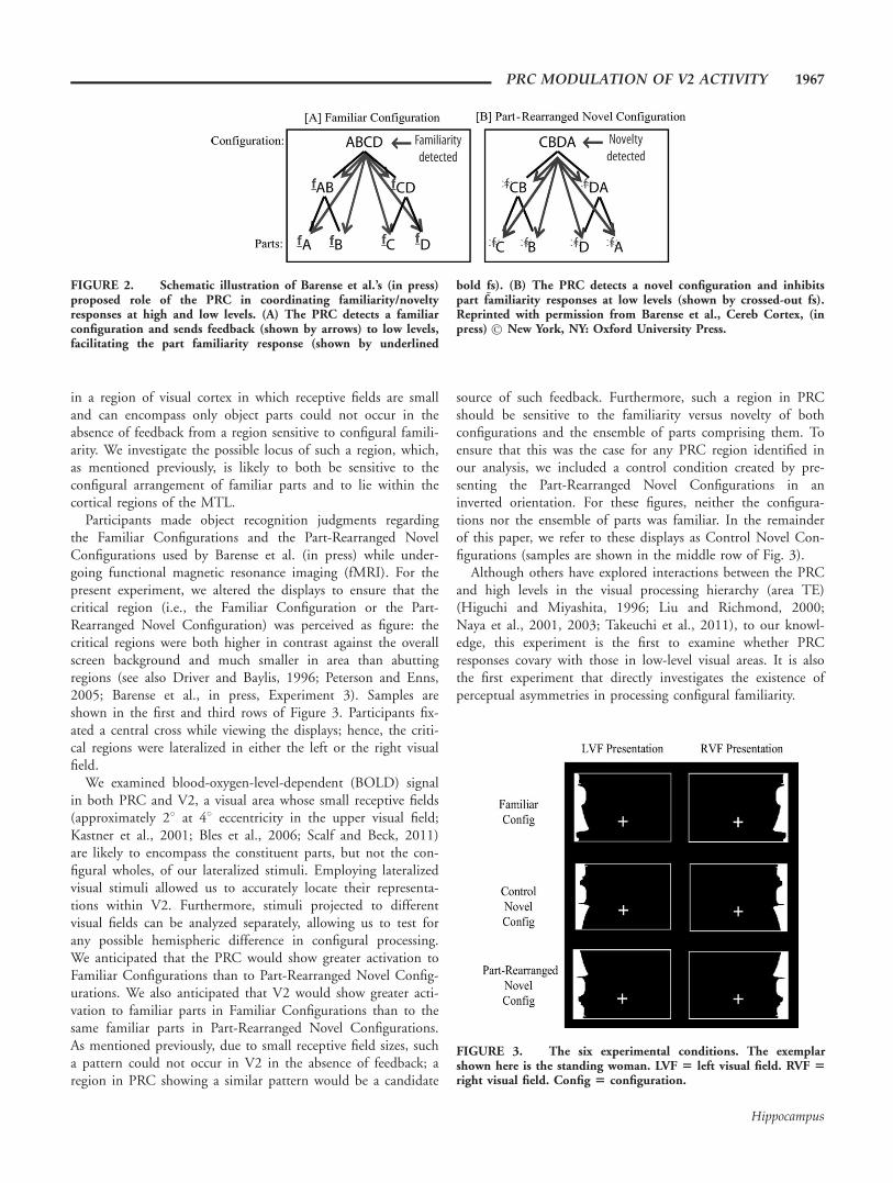
On the feedback view, non-brain-damaged individuals donot show effects of familiar parts when those parts are arrangedin a novel configuration because the intact PRC sends differen-tial feedback to lower-level visual areas sensitive to part famili-arity when it detects a novel configuration composed of famil-iar parts versus a familiar configuration. Specifically, Barenseet al. (in press) proposed that when the PRC detects a configu-ration composed of familiar parts in a novel spatial arrange-ment, it suppresses activation in lower-level visual areas sensi-tive to part familiarity, whereas when it detects a familiar con-figuration (by definition composed of familiar parts), itfacilitates activation of lower-level visual areas sensitive to partfamiliarity (see Fig. 2). As a result of this interaction familiar-ity/novelty responses in lower-level visual regions and higher-level MTL regions are coordinated to drive a coherent behav-ioral response. This feedback interpretation builds on evidencethat the PRC discriminates between novel and familiar objects(Xiang and Brown, 1998; Henson et al., 2003; Kohler et al.,2005; Albasser et al., 2010; Burke et al., 2010; McTighe et al.,2010). The feedback framework explains why effects of part fa-miliarity cannot be observed independently of effects of config-uration familiarity in behavioral tasks conducted with non-brain-damaged participants: when the PRC is intact, part famil-iarity responses at lower-levels are suppressed when familiarparts appear in Part-Rearranged Novel Configurations. On thisview, effects of familiar parts were evident in tests of PRC-dam-aged participants because their PRC damage removed the sup-pression of part familiarity responses at lower levels in the vis-ual hierarchy.
Discriminating between the feedforward and feedback mod-els is important not only for understanding the role of thePRC in perception but also for current debates regardingwhether visual perception requires only feedforward processes(Kirchner and Thorpe, 2006; Serre et al., 2007) or requiresboth feedforward and feedback processes (e.g., Lamme andRoelfsema, 2000; Bullier, 2001; Peterson and Skow, 2008). Inthe present experiment, we investigated whether responses tofamiliar parts in a lower-level visual area are modulated as afunction of their configural arrangement (arranged in a Famil-iar Configuration or in a Part-Rearranged Novel Configura-tion), as predicted by Barense et al. (in press). Such a finding
FIGURE 1. Stimuli Barense et al. (in press) used to test effectsof configuration familiarity on figure assignment. The criticalregions are shown in black on the left. (A–C) The critical regionsdepict the intact configuration of familiar, real-world, objects(from left to right: a woman, a lamp, and a guitar). (D–F) Thecritical regions are formed by spatially rearranging the parts of thefamiliar configurations in (A–C), respectively, such that the config-uration is novel but the parts are the same. Participants’ task wasto indicate whether they saw the black or white region as figure.Black/white color and left/right location of critical regions werebalanced in the experiment. Reprinted with permission from Bare-nse et al., Cereb Cortex, (in press) New York, NY: Oxford Univer-sity Press.
1966 PETERSON ET AL.
Hippocampus
in a region of visual cortex in which receptive fields are smalland can encompass only object parts could not occur in theabsence of feedback from a region sensitive to configural famili-arity. We investigate the possible locus of such a region, which,as mentioned previously, is likely to both be sensitive to theconfigural arrangement of familiar parts and to lie within thecortical regions of the MTL.
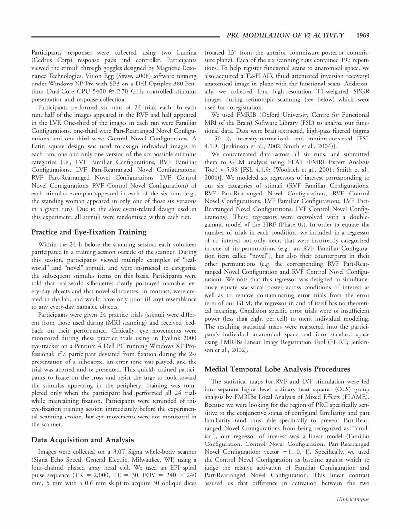
Participants made object recognition judgments regardingthe Familiar Configurations and the Part-Rearranged NovelConfigurations used by Barense et al. (in press) while under-going functional magnetic resonance imaging (fMRI). For thepresent experiment, we altered the displays to ensure that thecritical region (i.e., the Familiar Configuration or the Part-Rearranged Novel Configuration) was perceived as figure: thecritical regions were both higher in contrast against the overallscreen background and much smaller in area than abuttingregions (see also Driver and Baylis, 1996; Peterson and Enns,2005; Barense et al., in press, Experiment 3). Samples areshown in the first and third rows of Figure 3. Participants fix-ated a central cross while viewing the displays; hence, the criti-cal regions were lateralized in either the left or the right visualfield.
We examined blood-oxygen-level-dependent (BOLD) signalin both PRC and V2, a visual area whose small receptive fields(approximately 28 at 48 eccentricity in the upper visual field;Kastner et al., 2001; Bles et al., 2006; Scalf and Beck, 2011)are likely to encompass the constituent parts, but not the con-figural wholes, of our lateralized stimuli. Employing lateralizedvisual stimuli allowed us to accurately locate their representa-tions within V2. Furthermore, stimuli projected to differentvisual fields can be analyzed separately, allowing us to test forany possible hemispheric difference in configural processing.We anticipated that the PRC would show greater activation toFamiliar Configurations than to Part-Rearranged Novel Config-urations. We also anticipated that V2 would show greater acti-vation to familiar parts in Familiar Configurations than to thesame familiar parts in Part-Rearranged Novel Configurations.As mentioned previously, due to small receptive field sizes, sucha pattern could not occur in V2 in the absence of feedback; aregion in PRC showing a similar pattern would be a candidate
source of such feedback. Furthermore, such a region in PRCshould be sensitive to the familiarity versus novelty of bothconfigurations and the ensemble of parts comprising them. Toensure that this was the case for any PRC region identified inour analysis, we included a control condition created by pre-senting the Part-Rearranged Novel Configurations in aninverted orientation. For these figures, neither the configura-tions nor the ensemble of parts was familiar. In the remainderof this paper, we refer to these displays as Control Novel Con-figurations (samples are shown in the middle row of Fig. 3).
Although others have explored interactions between the PRCand high levels in the visual processing hierarchy (area TE)(Higuchi and Miyashita, 1996; Liu and Richmond, 2000;Naya et al., 2001, 2003; Takeuchi et al., 2011), to our knowl-edge, this experiment is the first to examine whether PRCresponses covary with those in low-level visual areas. It is alsothe first experiment that directly investigates the existence ofperceptual asymmetries in processing configural familiarity.
FIGURE 2. Schematic illustration of Barense et al.’s (in press)proposed role of the PRC in coordinating familiarity/noveltyresponses at high and low levels. (A) The PRC detects a familiarconfiguration and sends feedback (shown by arrows) to low levels,facilitating the part familiarity response (shown by underlined
bold fs). (B) The PRC detects a novel configuration and inhibitspart familiarity responses at low levels (shown by crossed-out fs).Reprinted with permission from Barense et al., Cereb Cortex, (inpress) � New York, NY: Oxford University Press.
FIGURE 3. The six experimental conditions. The exemplarshown here is the standing woman. LVF 5 left visual field. RVF 5right visual field. Config 5 configuration.
PRC MODULATION OF V2 ACTIVITY 1967
Hippocampus

MATERIALS AND METHODS
Participants
We tested eight volunteers (5 males; ages 19–38), all withnormal or corrected-to-normal visual acuity. Participants gavewritten informed consent to participate in this study, whichwas approved by the Institutional Review Board of the Univer-sity of Arizona. Two participants were eliminated due to higherror (>33% incorrect, which is greater than two standard devi-ations from the mean error score of the usable participants);thus, data from six participants (three males) were subjected tothe analysis described below.
Stimuli
The stimuli were contrast-reversed versions of those by Bare-nse et al. (in press) used in Experiment 3 (see Fig. 3). All stim-uli were white silhouettes presented on a black screen with athin white rectangular border. A white fixation cross was dis-played in the center of the screen during stimulus presentation,and the white silhouette and its border were shifted up 1.358from this central fixation in order to better target the ventralvisual stream. Silhouettes subtended 6.58 in height and an aver-age of 2.958 in width, and the center-most edge of each figurewas always located 3.58 from fixation. These elongated, lateral-ized displays both caused the critical region to be assigned fig-ural status and allowed retinotopic assignment of striate andextrastriate visual representations.
We used stimuli of three configuration types (24 uniquestimuli per configuration type): Familiar Configurations, Part-Rearranged Novel Configurations, and Control Novel Configu-rations (see Fig. 3). The Familiar Configuration silhouettes por-trayed portions of objects whose configurations and parts werelikely to be known to participants (i.e., a standing woman,
lamp, guitar; see Peterson et al., 2000). The Part-RearrangedNovel Configuration silhouettes were created by dividing theFamiliar Configurations into parts at minima of curvature andspatially rearranging the parts to form a novel configuration;for these stimuli, the ensemble of parts had been seen togetherfrequently, but in a different configuration. The Control NovelConfiguration silhouettes were created by inverting the Part-Rearranged Novel Configuration silhouettes; consequently boththe ensemble of parts and their configuration were novel. Eachstimulus was presented twice; once in the right visual field(RVF) and once in the left visual field (LVF). There weretherefore six stimulus categories in total, with 24 stimuli ineach category: LVF Familiar Configurations, RVF FamiliarConfigurations, LVF Part-Rearranged Novel Configurations,RVF Part-Rearranged Novel Configurations, LVF ControlNovel Configurations, and RVF Control Novel Configurations.
Experimental Design and Equipment
Because our number of stimuli was small, we employed aslow event-related trial design (see Fig. 4). Each trial beganwith a 10-s fixation period; one second before stimulus onset,the fixation cross brightened from gray to white to alert theparticipant to the upcoming stimulus. Participants wereinstructed to maintain fixation on this white cross during the2-s presentation of the stimulus item. The query ‘‘Real World?’’then appeared for 2 s at fixation, prompting the participant toindicate whether the silhouette depicted a real-world or novelobject using a button-box held in the their right hand, withone button assigned to ‘‘yes/real-world object’’ and the otherassigned to ‘‘no/novel object.’’ The query ‘‘Confidence?’’ thenappeared at fixation for 2 s, prompting participants to reporttheir confidence in their ‘‘real world’’ judgment using a button-box held in their left hand, with one button assigned to‘‘high confidence’’ and the other assigned to ‘‘low confidence.’’
FIGURE 4. Trial structure. A sample trial in the LVF Familiar Configuration condition is shown here.
1968 PETERSON ET AL.
Hippocampus
Participants’ responses were collected using two Lumina(Cedrus Corp) response pads and controller. Participantsviewed the stimuli through goggles designed by Magnetic Reso-nance Technologies. Vision Egg (Straw, 2008) software runningunder Windows XP Pro with SP3 on a Dell Optiplex 380 Pen-tium Dual-Core CPU 5400 @ 2.70 GHz controlled stimuluspresentation and response collection.
Participants performed six runs of 24 trials each. In eachrun, half of the images appeared in the RVF and half appearedin the LVF. One-third of the images in each run were FamiliarConfigurations, one-third were Part-Rearranged Novel Configu-rations and one-third were Control Novel Configurations. ALatin square design was used to assign individual images toeach run; one and only one version of the six possible stimuluscategories (i.e., LVF Familiar Configurations, RVF FamiliarConfigurations, LVF Part-Rearranged Novel Configurations,RVF Part-Rearranged Novel Configurations, LVF ControlNovel Configurations, RVF Control Novel Configurations) ofeach stimulus exemplar appeared in each of the six runs (e.g.,the standing woman appeared in only one of those six versionsin a given run). Due to the slow event-related design used inthis experiment, all stimuli were randomized within each run.
Practice and Eye-Fixation Training
Within the 24 h before the scanning session, each volunteerparticipated in a training session outside of the scanner. Duringthis session, participants viewed multiple examples of ‘‘real-world’’ and ‘‘novel’’ stimuli, and were instructed to categorizethe subsequent stimulus items on this basis. Participants weretold that real-world silhouettes clearly portrayed namable, ev-ery-day objects and that novel silhouettes, in contrast, were cre-ated in the lab, and would have only poor (if any) resemblanceto any every-day namable objects.
Participants were given 24 practice trials (stimuli were differ-ent from those used during fMRI scanning) and received feed-back on their performance. Critically, eye movements weremonitored during these practice trials using an Eyelink 2000eye-tracker on a Pentium 4 Dell PC running Windows XP Pro-fessional; if a participant deviated from fixation during the 2-spresentation of a silhouette, an error tone was played, and thetrial was aborted and re-presented. This quickly trained partici-pants to fixate on the cross and resist the urge to look towardthe stimulus appearing in the periphery. Training was com-pleted only when the participant had performed all 24 trialswhile maintaining fixation. Participants were reminded of thiseye-fixation training session immediately before the experimen-tal scanning session, but eye movements were not monitored inthe scanner.
Data Acquisition and Analysis
Images were collected on a 3.0T Signa whole-body scanner(Signa Echo Speed; General Electric, Milwaukee, WI) using afour-channel phased array head coil. We used an EPI spiralpulse sequence (TR 5 2,000, TE 5 30, FOV 5 240 3 240mm, 5 mm with a 0.6 mm skip) to acquire 30 oblique slices
(rotated 138 from the anterior commissure-posterior commis-sure plane). Each of the six scanning runs contained 197 repeti-tions. To help register functional scans to anatomical space, wealso acquired a T2-FLAIR (fluid attenuated inversion recovery)anatomical image in plane with the functional scans. Addition-ally, we collected four high-resolution T1-weighted SPGRimages during retinotopic scanning (see below) which wereused for coregistration.
We used FMRIB (Oxford University Centre for FunctionalMRI of the Brain) Software Library (FSL) to analyze our func-tional data. Data were brain-extracted, high-pass filtered (sigma5 50 s), intensity-normalized, and motion-corrected [FSL4.1.9, (Jenkinson et al., 2002; Smith et al., 2004)].
We concatenated data across all six runs, and submittedthem to GLM analysis using FEAT (FMRI Expert AnalysisTool) v 5.98 [FSL 4.1.9; (Woolrich et al., 2001; Smith et al.,2004)]. We modeled six regressors of interest corresponding toour six categories of stimuli (RVF Familiar Configurations,RVF Part-Rearranged Novel Configurations, RVF ControlNovel Configurations, LVF Familiar Configurations, LVF Part-Rearranged Novel Configurations, LVF Control Novel Config-urations). These regressors were convolved with a double-gamma model of the HRF (Phase 0s). In order to equate thenumber of trials in each condition, we included in a regressorof no interest not only items that were incorrectly categorizedin one of its permutations (e.g., an RVF Familiar Configura-tion item called ‘‘novel’’), but also their counterparts in theirother permutations (e.g. the corresponding RVF Part-Rear-ranged Novel Configuration and RVF Control Novel Configu-ration). We note that this regressor was designed to simultane-ously equate statistical power across conditions of interest aswell as to remove contaminating error trials from the errorterm of our GLM; the regressor in and of itself has no theoreti-cal meaning. Condition specific error trials were of insufficientpower (less than eight per cell) to merit individual modeling.The resulting statistical maps were registered into the partici-pant’s individual anatomical space and into standard spaceusing FMRIBs Linear Image Registration Tool (FLIRT; Jenkin-son et al., 2002).
Medial Temporal Lobe Analysis Procedures
The statistical maps for RVF and LVF stimulation were fedinto separate higher-level ordinary least squares (OLS) groupanalysis by FMRIBs Local Analysis of Mixed Effects (FLAME).Because we were looking for the region of PRC specifically sen-sitive to the conjunctive status of configural familiarity and partfamiliarity (and thus able specifically to prevent Part-Rear-ranged Novel Configurations from being recognized as ‘‘famil-iar’’), our regressor of interest was a linear model (FamiliarConfiguration, Control Novel Configuration, Part-RearrangedNovel Configuration; vector 21, 0, 1). Specifically, we usedthe Control Novel Configuration as baseline against which tojudge the relative activation of Familiar Configuration andPart-Rearranged Novel Configuration. This linear contrastassured us that difference in activation between the two
PRC MODULATION OF V2 ACTIVITY 1969
Hippocampus
conditions reflected differential response to the arrangement offamiliar parts, rather than simply reflecting a novel configura-tion of any kind. We also modeled out each participant’s meanactivation level as a regressor of no interest. We modeled datafrom each visual field in separate analyses because their inclu-sion in a common analysis would have altered the ‘‘baseline’’(meant to be novel configurations from the appropriate visualfield) of the linear trend we intended to test.
We used Brodmann areas (BA20, BA28, BA35, BA36, andBA38) from the Talairach-to-MNI-conversion digital atlas(Lancaster et al., 2000) to identify regions of interest (ROIs) inthe MTL that correspond with the areas that often accompanyperirhinal damage in patients. Perirhinal cortex is typicallydefined as BA 35 and 36, although BA 38 has many of thesame cytoarchitectonic characteristics as PRC (Suzuki and Ama-ral, 1994) and is sometimes included with PRC (see Suzukiand Amaral, 1994 and the ‘‘total perirhinal cortex’’ measure inInsausti et al., 1998). Because the Talairach-to-MNI-conversiondigital atlas strictly limits Brodmann areas to regions that corre-spond to cortical bark in MNI 152 space, these areas are nar-rower than the spatial resolution we could achieve. In order toproduce more usable masks for group analysis, we projectedthe Talairach conversion masks into each participant’s func-tional space, transformed the resulting (larger) probabilisticmap into standard MNI space, and then averaged these trans-formations across all participants. This produced larger ROIsthat were both more appropriate for analyzing data of our spa-tial resolution and directly reflected the individual anatomicalspaces of our subject population. We then used easythresh(FEAT 5.98) to identify clusters of voxels (Z > 1.96) showinga linear trend whose extent was greater than would be predictedby random variation in activation (P < 0.05).
Visual Cortex Analysis Procedures
Each volunteer also participated in a separate retinotopicmapping session. The procedures used during this session arethe same as those described in Scalf and Beck (2010) and arederived from those established by Sereno et al. (1995) andadapted by Kastner et al. (1998). Briefly, each quadrant of vis-ual cortex (upper/lower, left/right) represents 908 of the visualfield in a retinotopic manner. V1 through V4 may be identifiedby locating the regions that represent the boundaries of theseareas (either 08 or 908 on the polar azimuth); once each regionof visual space has been represented in one area, the retinotopicmapping ‘‘reverses’’ and the subsequent visual area representsvisual space in a ‘‘mirror reversed’’ manner. We compared acti-vation between stimulation of the horizontal and vertical meri-dians of the visual field to identify ‘‘reversals.’’ We comparedactivation between upper and lower visual field stimulation tofunctionally segregate ventral and dorsal striate and extrastriatecortex. During this session, EPI spiral pulse sequences (TR 5
2,000, TE 5 30, FOV 5 240 3 240 mm, slice thickness 5 2mm; 30 oblique slices rotated 138 from the anterior commis-sure-posterior commissure plane) were used to collect six runsof 105 repetitions and six runs of 95 repetitions. Four high-re-
solution (1 mm) SPGR T1-weighted images were collected tosubmit to Freesurfer (Dale et al., 1999; Fischl et al., 1999,2001) for averaging, segmentation, and flatmapping. Furtherdetails of retinotopic mapping and analysis are identical tothose reported by Scalf and Beck (2010).
Because a group analysis would average together striate andextrastriate regions with different receptive field sizes, weemployed an ROI analysis to interrogate visual cortex. We usedactivation to the Control Novel Configuration condition in thecontralateral visual field to identify regions in right and left V1through V4 stimulated by our bipartite displays. Striate andextrastriate activation to the Control Novel Configuration con-dition should also have been unmodulated by MTL structures(which were silent during this condition), so ROIs identifiedby this condition should have been relatively unbiased bypotential feedback from the MTL. Our measure of interest invisual cortex was the degree to which each regressor (parameter)predicted change in the BOLD signal; these measures are usu-ally referred to as ‘‘parameter estimates.’’ We note that that per-cent signal change is often the measure of interest in such anal-yses. Although there are a number of ways to calculate percentsignal change, nearly all of them involve scaling the parameterestimates by the inverse of the mean signal; some also use addi-tional scaling factors. Rather than relying on a somewhat arbi-trary method of scaling our parameter estimates, we chose tosimply use the unscaled values as our measure of interest.
Parameter estimates from the Control Novel Configurationconditions for each visual field were projected onto the appro-priate hemisphere in Freesurfer space. Suprathreshold (Z > 2)voxels that were contiguous with the peak activation in eachregion of visual cortex were assigned to that region’s ROI. Themasks for each participant were then projected back into theirindividual high-resolution anatomical space in FSL. We thenused Featquery (Smith et al., 2004) to extract the mean param-eter estimates for the Familiar Configuration and the Part-Rear-ranged Novel Configuration conditions from the ROIs in eachregion of visual cortex. During this process, Featquery appliedthe SPGR to EPI transformation matrix calculated duringimage registration to determine which EPI voxels fell withinthe ROI (selected in MPRAGE space).
RESULTS
Behavioral Results
We subjected the accuracy data to repeated measures analysisof variance (ANOVA) using the factors Visual Field (LVF,RVF) and Configuration Type (Familiar Configuration, Con-trol Novel Configuration, Part-Rearranged Novel Configura-tion). A significant main effect of condition was found, F(10,2)
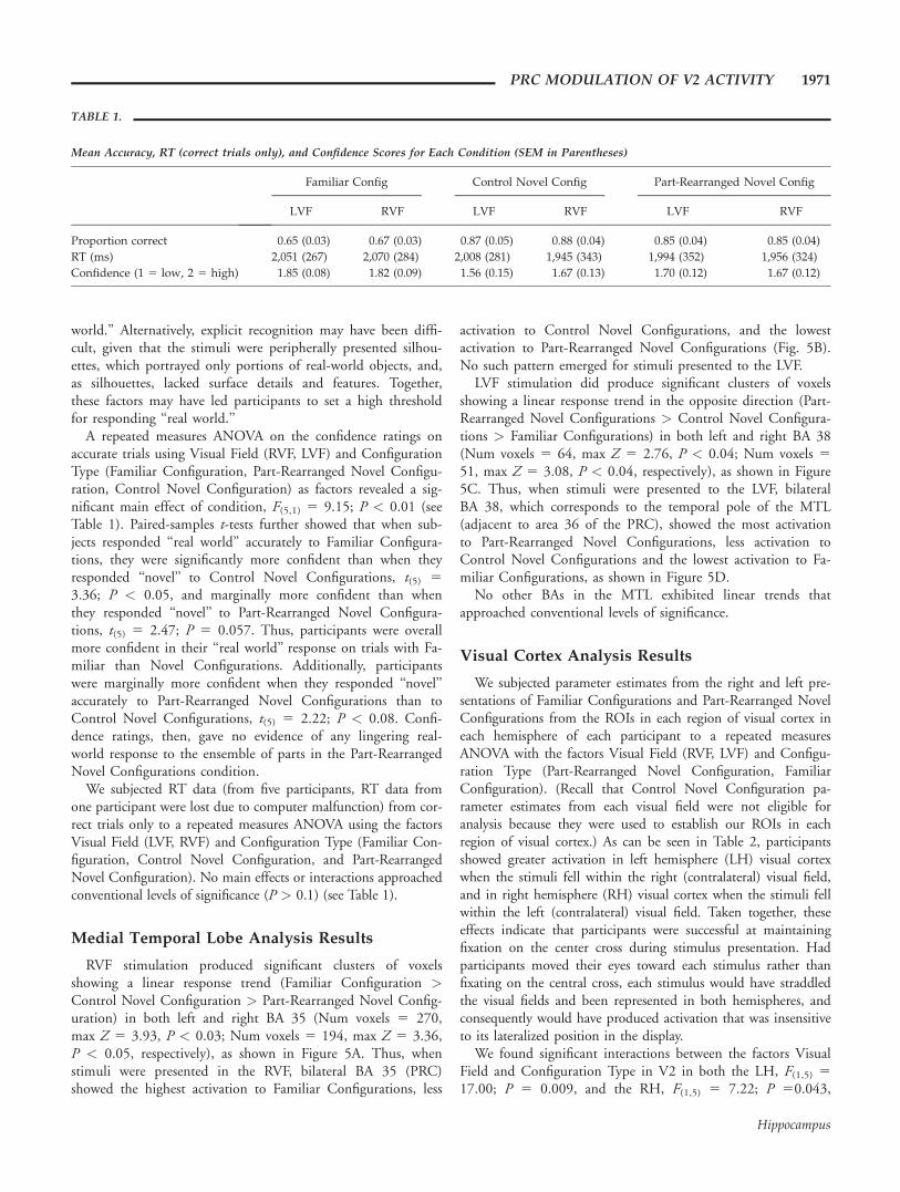
5 24.79; P < 0.001, with participants less accurate in ‘‘realworld’’ than ‘‘novel’’ judgments (see Table 1). This effect maybe due to response bias; 66% of the trials were novel, thusbiasing participants to respond ‘‘novel’’ more often than ‘‘real
1970 PETERSON ET AL.
Hippocampus
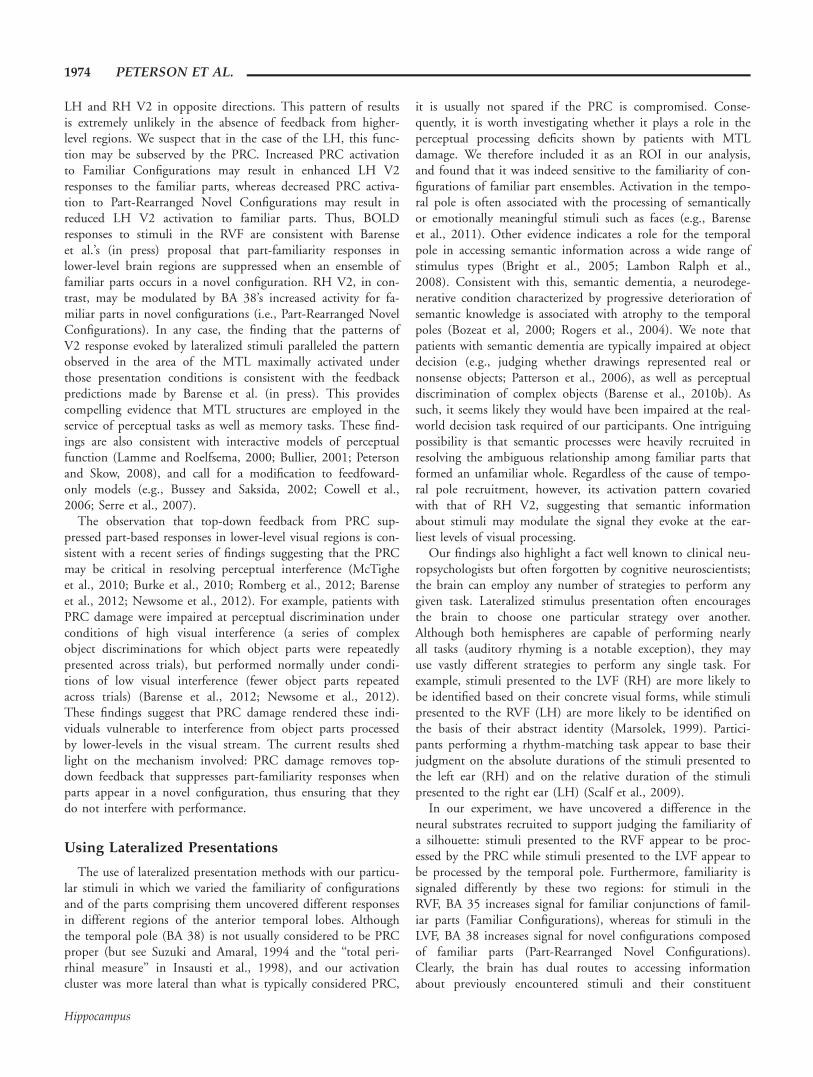
world.’’ Alternatively, explicit recognition may have been diffi-cult, given that the stimuli were peripherally presented silhou-ettes, which portrayed only portions of real-world objects, and,as silhouettes, lacked surface details and features. Together,these factors may have led participants to set a high thresholdfor responding ‘‘real world.’’
A repeated measures ANOVA on the confidence ratings onaccurate trials using Visual Field (RVF, LVF) and ConfigurationType (Familiar Configuration, Part-Rearranged Novel Configu-ration, Control Novel Configuration) as factors revealed a sig-nificant main effect of condition, F(5,1) 5 9.15; P < 0.01 (seeTable 1). Paired-samples t-tests further showed that when sub-jects responded ‘‘real world’’ accurately to Familiar Configura-tions, they were significantly more confident than when theyresponded ‘‘novel’’ to Control Novel Configurations, t(5) 5
3.36; P < 0.05, and marginally more confident than whenthey responded ‘‘novel’’ to Part-Rearranged Novel Configura-tions, t(5) 5 2.47; P 5 0.057. Thus, participants were overallmore confident in their ‘‘real world’’ response on trials with Fa-miliar than Novel Configurations. Additionally, participantswere marginally more confident when they responded ‘‘novel’’accurately to Part-Rearranged Novel Configurations than toControl Novel Configurations, t(5) 5 2.22; P < 0.08. Confi-dence ratings, then, gave no evidence of any lingering real-world response to the ensemble of parts in the Part-RearrangedNovel Configurations condition.
We subjected RT data (from five participants, RT data fromone participant were lost due to computer malfunction) from cor-rect trials only to a repeated measures ANOVA using the factorsVisual Field (LVF, RVF) and Configuration Type (Familiar Con-figuration, Control Novel Configuration, and Part-RearrangedNovel Configuration). No main effects or interactions approachedconventional levels of significance (P > 0.1) (see Table 1).
Medial Temporal Lobe Analysis Results
RVF stimulation produced significant clusters of voxelsshowing a linear response trend (Familiar Configuration >Control Novel Configuration > Part-Rearranged Novel Config-uration) in both left and right BA 35 (Num voxels 5 270,max Z 5 3.93, P < 0.03; Num voxels 5 194, max Z 5 3.36,P < 0.05, respectively), as shown in Figure 5A. Thus, whenstimuli were presented in the RVF, bilateral BA 35 (PRC)showed the highest activation to Familiar Configurations, less
activation to Control Novel Configurations, and the lowestactivation to Part-Rearranged Novel Configurations (Fig. 5B).No such pattern emerged for stimuli presented to the LVF.
LVF stimulation did produce significant clusters of voxelsshowing a linear response trend in the opposite direction (Part-Rearranged Novel Configurations > Control Novel Configura-tions > Familiar Configurations) in both left and right BA 38(Num voxels 5 64, max Z 5 2.76, P < 0.04; Num voxels 5
51, max Z 5 3.08, P < 0.04, respectively), as shown in Figure5C. Thus, when stimuli were presented to the LVF, bilateralBA 38, which corresponds to the temporal pole of the MTL(adjacent to area 36 of the PRC), showed the most activationto Part-Rearranged Novel Configurations, less activation toControl Novel Configurations and the lowest activation to Fa-miliar Configurations, as shown in Figure 5D.
No other BAs in the MTL exhibited linear trends thatapproached conventional levels of significance.
Visual Cortex Analysis Results
We subjected parameter estimates from the right and left pre-sentations of Familiar Configurations and Part-Rearranged NovelConfigurations from the ROIs in each region of visual cortex ineach hemisphere of each participant to a repeated measuresANOVA with the factors Visual Field (RVF, LVF) and Configu-ration Type (Part-Rearranged Novel Configuration, FamiliarConfiguration). (Recall that Control Novel Configuration pa-rameter estimates from each visual field were not eligible foranalysis because they were used to establish our ROIs in eachregion of visual cortex.) As can be seen in Table 2, participantsshowed greater activation in left hemisphere (LH) visual cortexwhen the stimuli fell within the right (contralateral) visual field,and in right hemisphere (RH) visual cortex when the stimuli fellwithin the left (contralateral) visual field. Taken together, theseeffects indicate that participants were successful at maintainingfixation on the center cross during stimulus presentation. Hadparticipants moved their eyes toward each stimulus rather thanfixating on the central cross, each stimulus would have straddledthe visual fields and been represented in both hemispheres, andconsequently would have produced activation that was insensitiveto its lateralized position in the display.
We found significant interactions between the factors VisualField and Configuration Type in V2 in both the LH, F(1,5) 5
17.00; P 5 0.009, and the RH, F(1,5) 5 7.22; P 50.043,
TABLE 1.
Mean Accuracy, RT (correct trials only), and Confidence Scores for Each Condition (SEM in Parentheses)
Familiar Config Control Novel Config Part-Rearranged Novel Config
LVF RVF LVF RVF LVF RVF
Proportion correct 0.65 (0.03) 0.67 (0.03) 0.87 (0.05) 0.88 (0.04) 0.85 (0.04) 0.85 (0.04)
RT (ms) 2,051 (267) 2,070 (284) 2,008 (281) 1,945 (343) 1,994 (352) 1,956 (324)
Confidence (1 5 low, 2 5 high) 1.85 (0.08) 1.82 (0.09) 1.56 (0.15) 1.67 (0.13) 1.70 (0.12) 1.67 (0.12)
PRC MODULATION OF V2 ACTIVITY 1971
Hippocampus

shown in Figure 6. Planned comparisons indicated that, in LHV2, this interaction was driven by greater activation resultingfrom RVF Familiar Configurations than Part-Rearranged NovelConfigurations, P 5 0.027, whereas in RH V2, this interactionwas driven by greater activation resulting from LVF Part-Rear-
ranged Novel Configurations than Familiar Configurations, P5 0.022. Thus, in LH V2, activation mirrors activation seen
TABLE 2.
Mean Parameter Estimates in Visual Cortex for LVF and RVF
Presentation Conditions
LVF RVF F P
LH V1 206 246 4.80 0.08
V2 162 223 9.44 0.03
VP 165 228 31.17 0.00
V4 132 204 19.96 0.01
RH V1 246 179 7.95 0.04
V2 241 157 8.58 0.03
VP 248 154 9.81 0.03
V4 235 159 7.20 0.04
F values and P values for ANOVAs comparing the parameter estimates obtainedwith LVF and RVF presentation for each region in each hemisphere. Highervalues are bolded. The direction of difference in each hemisphere shows thatsubjects were fixating. LH 5 left hemisphere. RH 5 right hemisphere. LVF 5
left visual field. RVF 5 right visual field.
FIGURE 5. Effects of condition in the MTL. (A and B) BA 35 (PRC) activation for RVF presentation. (C and D) BA 38 (temporalpole) activation for LVF presentation. Statistical maps (A and C) were thresholded at P < 0.05 and are shown superimposed on a stand-ard brain in MNI space. The color bar reflects z-scores for the linear trend in the direction of Familiar Configurations > Control NovelConfigurations > Part-Rearranged Novel Configurations; thus, a negative score, as in (C), indicates a significant linear trend in the oppo-site direction. Parameter estimates (B and D) for each condition were extracted from these suprathreshold clusters in both the RH andLH. Error bars represent SEM. RH 5 right hemisphere. LH 5 left hemisphere. LVF 5 left visual field. RVF 5 right visual field.
FIGURE 6. Effects of Visual Field and Configuration Type inV2. Parameter estimates were extracted from each subject’s V2 ROIin each hemisphere, determined based on activation in the ControlNovel Configuration condition for contralateral visual field presen-tation (thus, activation in this control condition was not eligiblefor visual cortex analyses). Error bars represent SEM of the differ-ence between the conditions shown. RH 5 right hemisphere. LH5 left hemisphere. LVF 5 left visual field. RVF 5 right visualfield. *P < 0.05
1972 PETERSON ET AL.
Hippocampus

in PRC BA 35 with RVF presentation, and in RH V2, activa-tion mirrors activation seen in BA 38 with LVF presentation.
We also found a significant crossover interaction in LH V1,F(1,5) 5 12.42; P < 0.02. Planned comparisons showed thatfor LVF presentation, V1 was marginally less active to Famil-iar Configurations than Part-Rearranged Novel Configurations(P 5 0.08), but for RVF presentation, V1 activation did notdiffer for Familiar Configurations versus Part-RearrangedNovel Configurations (P 5 0.16). Because neither plannedcontrast was significant, this interaction was not given anyfurther attention. No other region in visual cortex showed aninteraction between Visual Field and Configuration Type thatmet or approached conventional levels of significance. Themain effect of Configuration Type was not statisticallysignificant.
An additional whole-brain analysis was also performed toassess whether a significant linear trend (in either direction)was evident in any other brain regions; no significant effectswere found (cluster P > 0.05; threshold Z < 2.3).
DISCUSSION
We used fMRI to assess activation in the MTL and V2 whileparticipants made object recognition judgments regarding threetypes of displays: Familiar Configurations (composed of famil-iar parts), Part-Rearranged Novel Configurations (novel config-urations created by spatially rearranging the same familiarparts), and Control Novel Configurations (novel configurationscomposed of novel parts). Others have shown that the PRCresponds more strongly to familiar than to novel configurations(e.g., Barense et al., 2011); we expected to replicate those find-ings. Furthermore, our previous findings (Barense et al., inpress) led us to suspect that the PRC might be particularly im-portant in adjudicating configural familiarity when the configu-ration’s constituent parts are familiar rather than novel. Wetherefore included two novel configuration conditions; one inwhich the ensemble parts of the configuration were familiarand one in which they were novel (Part-Rearranged and Con-trol Novel Configurations, respectively). Each type of configu-ration was presented in each visual field; this manipulation wasnecessary to identify the signal evoked by the items in striateand extrastriate cortex; it also allowed us to test for hemisphericdifferences in configural processing. We found that the hemi-sphere that initially processed the visual stimulus determinedthe region of anterior temporal cortex recruited to support taskperformance (i.e., BA 35 or 38), and determined the direction-ality of the response to configural familiarity. Both BA 35 andBA 38, however, responded differently as a function of thejoint familiarity of the configuration and the ensemble of partscomprising the configuration. This is a new finding, uncoveredbecause we manipulated the familiarity versus novelty of theensemble of parts comprising our novel configurations. To ourknowledge, no previous research has directly investigated thisquestion.
Medial Temporal Lobe Activation
RVF presentations
For RVF presentations, BA 35, which corresponds to thePRC of the MTL, responded bilaterally. Relative to the controlcondition (novel configurations composed of a novel ensembleof parts), activation was increased when an ensemble of familiarparts was presented in its typical spatial arrangement (FamiliarConfiguration) and was decreased when an ensemble of famil-iar parts was presented in a novel spatial arrangement (Part-Rearranged Novel Configuration; Fig. 5). These results indicatethat the PRC detects the familiarity of the ensemble of partscomprising a configuration as well as the familiarity of the con-figuration (i.e., the spatial arrangement of the parts) andresponds to their congruency: responses are facilitated whenboth the parts and the spatial configuration of parts match infamiliarity and are suppressed when the parts and spatial con-figuration of parts mismatch in familiarity (i.e., the ensembleof parts is familiar but the spatial arrangement is novel). Asproposed by Barense et al. (in press), our results indicate thatthe PRC plays a critical role in discriminating familiar configu-rations from novel configurations composed by spatially rear-ranging an ensemble of familiar parts.
LVF presentations
For LVF presentations, BA 38, which corresponds to thetemporal pole of the MTL (adjacent to BA 36 of the PRC),responded bilaterally. BA 38 also responded differentially to theFamiliar Configurations and the Part-Rearranged Novel Config-urations, but the pattern of activity was the opposite of that inBA 35. Relative to the Control Novel Configurations (novelconfigurations composed of a novel ensemble of parts), activa-tion was increased when an ensemble of familiar parts was pre-sented in a novel spatial arrangement and decreased when anensemble of familiar parts was presented in its familiar spatialarrangement. Thus, BA 38 responded maximally to a mismatchin the familiarity of the ensemble of parts and theconfiguration.
Visual Cortex Activation
Responses in area V2 of the LH paralleled those observed inBA 35 with RVF presentation conditions: Responses werelarger for Familiar Configurations than for Part-RearrangedNovel Configurations. Responses in area V2 of the RH, in con-trast, paralleled those observed in BA 38 for LVF presentations:Here, responses were larger for Part-Rearranged Novel Configu-rations than for Familiar Configurations (Fig. 6). Given thesize and eccentricity of our displays (Kastner et al., 2001; Bleset al., 2006; Scalf and Beck, 2011), V2 receptive fields encom-pass parts of our displays, but not configural wholes. Conse-quently, finding differential V2 responses to ensembles of famil-iar parts as a function of their configural familiarity is highlyunlikely in the absence of modulation from other corticalregions. Furthermore, configural familiarity drives responses in
PRC MODULATION OF V2 ACTIVITY 1973
Hippocampus
LH and RH V2 in opposite directions. This pattern of resultsis extremely unlikely in the absence of feedback from higher-level regions. We suspect that in the case of the LH, this func-tion may be subserved by the PRC. Increased PRC activationto Familiar Configurations may result in enhanced LH V2responses to the familiar parts, whereas decreased PRC activa-tion to Part-Rearranged Novel Configurations may result inreduced LH V2 activation to familiar parts. Thus, BOLDresponses to stimuli in the RVF are consistent with Barenseet al.’s (in press) proposal that part-familiarity responses inlower-level brain regions are suppressed when an ensemble offamiliar parts occurs in a novel configuration. RH V2, in con-trast, may be modulated by BA 38’s increased activity for fa-miliar parts in novel configurations (i.e., Part-Rearranged NovelConfigurations). In any case, the finding that the patterns ofV2 response evoked by lateralized stimuli paralleled the patternobserved in the area of the MTL maximally activated underthose presentation conditions is consistent with the feedbackpredictions made by Barense et al. (in press). This providescompelling evidence that MTL structures are employed in theservice of perceptual tasks as well as memory tasks. These find-ings are also consistent with interactive models of perceptualfunction (Lamme and Roelfsema, 2000; Bullier, 2001; Petersonand Skow, 2008), and call for a modification to feedfoward-only models (e.g., Bussey and Saksida, 2002; Cowell et al.,2006; Serre et al., 2007).
The observation that top-down feedback from PRC sup-pressed part-based responses in lower-level visual regions is con-sistent with a recent series of findings suggesting that the PRCmay be critical in resolving perceptual interference (McTigheet al., 2010; Burke et al., 2010; Romberg et al., 2012; Barenseet al., 2012; Newsome et al., 2012). For example, patients withPRC damage were impaired at perceptual discrimination underconditions of high visual interference (a series of complexobject discriminations for which object parts were repeatedlypresented across trials), but performed normally under condi-tions of low visual interference (fewer object parts repeatedacross trials) (Barense et al., 2012; Newsome et al., 2012).These findings suggest that PRC damage rendered these indi-viduals vulnerable to interference from object parts processedby lower-levels in the visual stream. The current results shedlight on the mechanism involved: PRC damage removes top-down feedback that suppresses part-familiarity responses whenparts appear in a novel configuration, thus ensuring that theydo not interfere with performance.
Using Lateralized Presentations
The use of lateralized presentation methods with our particu-lar stimuli in which we varied the familiarity of configurationsand of the parts comprising them uncovered different responsesin different regions of the anterior temporal lobes. Althoughthe temporal pole (BA 38) is not usually considered to be PRCproper (but see Suzuki and Amaral, 1994 and the ‘‘total peri-rhinal measure’’ in Insausti et al., 1998), and our activationcluster was more lateral than what is typically considered PRC,
it is usually not spared if the PRC is compromised. Conse-quently, it is worth investigating whether it plays a role in theperceptual processing deficits shown by patients with MTLdamage. We therefore included it as an ROI in our analysis,and found that it was indeed sensitive to the familiarity of con-figurations of familiar part ensembles. Activation in the tempo-ral pole is often associated with the processing of semanticallyor emotionally meaningful stimuli such as faces (e.g., Barenseet al., 2011). Other evidence indicates a role for the temporalpole in accessing semantic information across a wide range ofstimulus types (Bright et al., 2005; Lambon Ralph et al.,2008). Consistent with this, semantic dementia, a neurodege-nerative condition characterized by progressive deterioration ofsemantic knowledge is associated with atrophy to the temporalpoles (Bozeat et al, 2000; Rogers et al., 2004). We note thatpatients with semantic dementia are typically impaired at objectdecision (e.g., judging whether drawings represented real ornonsense objects; Patterson et al., 2006), as well as perceptualdiscrimination of complex objects (Barense et al., 2010b). Assuch, it seems likely they would have been impaired at the real-world decision task required of our participants. One intriguingpossibility is that semantic processes were heavily recruited inresolving the ambiguous relationship among familiar parts thatformed an unfamiliar whole. Regardless of the cause of tempo-ral pole recruitment, however, its activation pattern covariedwith that of RH V2, suggesting that semantic informationabout stimuli may modulate the signal they evoke at the ear-liest levels of visual processing.
Our findings also highlight a fact well known to clinical neu-ropsychologists but often forgotten by cognitive neuroscientists;the brain can employ any number of strategies to perform anygiven task. Lateralized stimulus presentation often encouragesthe brain to choose one particular strategy over another.Although both hemispheres are capable of performing nearlyall tasks (auditory rhyming is a notable exception), they mayuse vastly different strategies to perform any single task. Forexample, stimuli presented to the LVF (RH) are more likely tobe identified based on their concrete visual forms, while stimulipresented to the RVF (LH) are more likely to be identified onthe basis of their abstract identity (Marsolek, 1999). Partici-pants performing a rhythm-matching task appear to base theirjudgment on the absolute durations of the stimuli presented tothe left ear (RH) and on the relative duration of the stimulipresented to the right ear (LH) (Scalf et al., 2009).
In our experiment, we have uncovered a difference in theneural substrates recruited to support judging the familiarity ofa silhouette: stimuli presented to the RVF appear to be proc-essed by the PRC while stimuli presented to the LVF appear tobe processed by the temporal pole. Furthermore, familiarity issignaled differently by these two regions: for stimuli in theRVF, BA 35 increases signal for familiar conjunctions of famil-iar parts (Familiar Configurations), whereas for stimuli in theLVF, BA 38 increases signal for novel configurations composedof familiar parts (Part-Rearranged Novel Configurations).Clearly, the brain has dual routes to accessing informationabout previously encountered stimuli and their constituent
1974 PETERSON ET AL.
Hippocampus
parts. It behooves researchers to remember this when investigat-ing ‘‘human patients with PRC damage,’’ in whom compro-mised tissue often extends beyond the PRC.
Which hemisphere is in control of adjudicating configural fa-miliarity in the neurologically intact brain? The problem of‘‘metacontrol’’ rarely has a simple solution (Levy et al., 1976).In some cases, performance is dominated by whichever hemi-sphere is superior at the given task (e.g., Hellige et al., 1989).In other cases, task performance during bilateral trials looksnothing like task performance by either hemisphere in isolation(Banich and Karol, 1992). Finally, stimulus order and task con-ditions may influence which hemisphere dominates perform-ance (Scalf et al., 2007). In the present study, we replicatedprevious evidence (Barense, et al., 2011) that PRC (BA 35)activation is higher for familiar than novel objects when weused RVF presentations, but we found the opposite pattern inthe temporal pole (BA 38) when we used LVF presentations. Itremains for future research to investigate the conditions thatdetermine whether BA 35, BA 38, both regions, or some otherregion altogether, is recruited when subjects are asked to makereal-world decisions regarding a centrally presented stimulus.
Acknowledgments
The authors thank Carol Barnes for her support and encour-agement, Lee Ryan for her advice on design, and Lynn Nadeland the members of the University of Arizona Visual Percep-tion and Cognition Lab Group and the Cognition and NeuralSystems Seminar for their input.
REFERENCES
Albasser MM, Poirier GL, Aggleton JP. 2010. Qualitatively differentmodes of perirhinal-hippocampal engagement when rats explorenovel vs. familiar objects as revealed by c-Fos imaging. Eur J Neu-rosci 31:134–147.
Banich MT, Karol DL. 1992. The sum of the parts does not equal thewhole: Evidence from bihemispheric processing. J Exp Psy-chol:HPP 18:763–784.
Barense MD, Bussey TJ, Lee AC, Rogers TT, Davies RR, SaksidaLM, Murray EA, Graham KS. 2005. Functional specialization inthe human medial temporal lobe. J Neurosci 25:10239–10246.
Barense MD, Gaffan D, Graham KS. 2007. The human medial tem-poral lobe processes online representations of complex objects.Neuropsychologia 45:2963–2974.
Barense MD, Henson RN, Lee AC, Graham KS. 2010a. Medial tem-poral lobe activity during complex discrimination of faces, objects,and scenes: Effects of viewpoint. Hippocampus 20:389–401.
Barense MD, Rogers TT, Bussey TJ, Saksida LM, Graham KS. 2010b.Influence of conceptual knowledge on visual object discrimination:Insights from semantic dementia and MTL amnesia. Cereb Cortex20:2568–2582.
Barense MD, Ngo JKW, Hung LHT, Peterson MA. Interactions ofmemory and perception in amnesia: the figure-ground perspective.Cereb Cortex (22:11), doi: 10.1093/cercor/bhr347.
Barense MD, Henson RN, Graham KS. 2011. Perception and concep-tion: Temporal lobe activity during complex discriminations of fa-miliar and novel faces and objects. J Cogn Neurosci 23:3052–3067.
Barense MD, Groen I, Lee AC, Yeung L, Brady S, Gregori M, KapurN, Bussey TJ, Saksida LM, Henson RN. 2012. Intact memory forirrelevant information impairs perception in amnesia. Neuron75:157–167.
Bartko SJ, Winters BD, Cowell RA, Saksida LM, Bussey TJ. 2007.Perirhinal cortex resolves feature ambiguity in configural object rec-ognition and perceptual oddity tasks. Learn Mem 14:821–832.
Baxter MG. 2009. Involvement of medial temporal lobe structures inmemory and perception. Neuron 61:667–677.
Bles M, Schwarzbach J, De Weerd P, Goebel R, Jansma B. 2006.Receptive field size-dependent attention effects in simultaneouslypresented stimulus displays. Neuroimage 30:506–511.
Bozeat S, Lambon Ralph MA, Patterson K, Garrard P, Hodges JR.2000. Non-verbal semantic impairment in semantic dementia.Neuropsychologia 38:1207–1215.
Bright P, Moss HE, Stamatakis EA, Tyler LK. 2005. The anatomy ofobject processing: The role of anteromedial temporal cortex. Q JExp Psychol B 58:361–377.
Bullier J. 2001. Integrated model of visual processing. Brain Res Rev36:96–107.
Burke SN, Wallace JL, Nematollahi S, Uprety AR, Barnes CA. 2010.Pattern separation deficits may contribute to age-associated recogni-tion impairments. Behav Neurosci 124:559–573.
Burke SN, Wallace JL, Hartzell AL, Nematollahi S, Plange K, BarnesCA. 2011. Age-associated deficits in pattern separation functions ofthe perirhinal cortex: A cross-species consensus. Behav Neurosci125:836–847.
Bussey TJ, Saksida LM. 2002. The organization of visual object repre-sentations: A connectionist model of effects of lesions in perirhinalcortex. Eur J Neurosci 15:355–364.
Bussey TJ, Saksida LM, Murray EA. 2002. Perirhinal cortex resolvesfeature ambiguity in complex visual discriminations. Eur J Neuro-sci 15:365–374.
Clark RE, Reinagel P, Broadbent NJ, Flister ED, Squire LR. 2011.Intact performance on feature-ambiguous discriminations in ratswith lesions of the perirhinal cortex. Neuron 70:132–140.
Cowell RA. 2012. Computational models of perirhinal cortical func-tion. Hippocampus 22:1952–1964.
Cowell RA, Bussey TJ, Saksida LM. 2006. Why does brain damageimpair memory? A connectionist model of object recognitionmemory in perirhinal cortex. J Neurosci 26:12186–12197.
Cowell RA, Bussey TJ, Saksida LM. 2010a. Components of recogni-tion memory: Dissociable cognitive processes or just differences inrepresentational complexity? Hippocampus 20:1245–1262.
Cowell RA, Bussey TJ, Saksida LM. 2010b. Functional dissociationswithin the ventral object processing pathway: Cognitive modules ora hierarchical continuum? J Cogn Neurosci 22:2460–2479.
Dale AM, Fischl B, Sereno MI. 1999. Cortical surface-based analysis. I.Segmentation and surface reconstruction. Neuroimage 9:179–194.
Driver J, Baylis GC. 1996. Edge-assignment and figure-ground segmen-tation in short-term visual matching. Cogn Psychol 31:248–306.
Fischl B, Sereno MI, Dale AM. 1999. Cortical surface-based analysis.II. Inflation, flattening, and a surface-based coordinate system.Neuroimage 9:195–207.
Fischl B, Liu A, Dale AM. 2001. Automated manifold surgery: Con-structing geometrically accurate and topologically correct models ofthe human cerebral cortex. IEEE Trans Med Imaging 20:70–80.
Gibson BS, Peterson MA. 1994. Does orientation-independent objectrecognition precede orientation-dependent recognition? Evidencefrom a cueing paradigm. J Exp Psychol:HPP 20:299–316.
Graham KS, Barense MD, Lee AC. 2010. Going beyond LTM in theMTL: A synthesis of neuropsychological and neuroimaging findingson the role of the medial temporal lobe in memory and percep-tion. Neuropsychologia 48:831–853.
Hellige JB, Taylor AK, Eng TL. 1989. Interhemispheric interactionwhen both hemispheres have access to the same stimulus informa-tion. J Exp Psychol:HPP 15:711–722.
PRC MODULATION OF V2 ACTIVITY 1975
Hippocampus

Henson RNA, Cansino S, Herron JE, Robb WG, Rugg MD. 2003. Afamiliarity signal in human anterior medial temporal cortex? Hip-pocampus 13:301–304.
Higuchi S, Miyashita Y. 1996. Formation of mnemonic neuronalresponses to visual paired associates in inferotemporal cortex isimpaired by perirhinal and entorhinal lesions. Proc Natl Acad SciUSA 93:739–743.
Insausti R, Juottonen K, Soininen H, Insausti AM, Partanen K, VainioP, Laakso MP, Pitkanen A. 1998. MR volumentric analysis of thehuman entorhinal, perirhinal, and temporopolar cortices. Am JNeuroradiol 19:659–671.
Jenkinson M, Bannister PR, Brady JM, Smith SM. 2002.Improved optimization for the robust and accurate linear registra-tion and motion correction of brain images. Neuroimage 17:825–841.
Kastner S, De Weerd P, Desimone R, Ungerleider LG. 1998. Mecha-nisms of directed attention in the human extrastriate cortex asrevealed by functional MRI. Science 282:108–111.
Kastner S, de Weerd P, Pinsk MA, Elizondo MI, Desimone R, Unger-leider LG. 2001. Modulation of sensory suppression: implicationsfor receptive field sizes in the human visual cortex. J Neurophysiol86:1398–1411.
Kim S, Jeneson A, van der Horst AS, Frascino JC, Hopkins RO,Squire LR. 2011. Memory, visual discrimination performance, andthe human hippocampus. J Neurosci 31:2624–2629.
Kirchner H, Thorpe SJ. 2006. Ultra-rapid object detection with sacca-dic eye movements: Visual processing speed revisited. Vis Res46:1762–1776.
Kohler S, Danckert S, Gati JS, Menon RS. 2005. Novelty responsesto relational and non-relational information in the hippocampusand the parahippocampal region: A comparison based on event-related fMRI. Hippocampus 15:763–774.
Lambon Ralph MA, Patterson K. 2008. Generalization and differen-tiation in semantic memory: Insights from semantic dementia. AnnNY Acad Sci 1124:61–76.
Lamme VAF, Roelfsema PR. 2000. The distinct modes of visionoffered by feedforward and recurrent processing. Trends Neurosci23:571–579.
Lancaster JL, Woldorff MG, Parsons LM, Liotti M, Freitas CS, RaineyL, Kochunov PV, Nickerson D, Mikiten SA, Fox PT. 2000. Auto-mated Talairach atlas labels for functional brain mapping. HumBrain Mapp 10:120–131.
Lee AC, Bussey TJ, Murray EA, Saksida LM, Epstein RA, Kapur N,Hodges JR, Graham KS. 2005. Perceptual deficits in amnesia:Challenging the medial temporal lobe ‘mnemonic’ view. Neuropsy-chologia 43:1–11.
Lee AC, Rudebeck SR. 2010. Human medial temporal lobe damagecan disrupt the perception of single objects. J Neurosci 30:6588–6594.
Lee AC, Yeung LK, Barense MD. 2012. The hippocampus and visualperception. Front Hum Neurosci 6:91.
Levy J, Trevarthen C. 1976. Metacontrol of hemispheric function inhuman split-brain patients. J Exp Psychol:HPP 2:299–312.
Liu Z, Richmond BJ. 2000. Response differences in monkey TE andperirhinal cortex: Stimulus association related to reward schedules.J Neurophysiol 83:1677–1692.
Marsolek CJ. 1999. Dissociable neural subsystems underlie abstractand specific object recognition. Psychol Sci 10:111–118.
McTighe SM, Cowell RA, Winters BD, Bussey TJ, Saksida LM.2010. Paradoxical false memory for objects after brain damage. Sci-ence 330:1408–1410.
Murray EA, Bussey TJ, Saksida LM. 2007. Visual perception andmemory: A new view of medial temporal lobe function in primatesand rodents. Annu Rev Neurosci 30:99–122.
Murray EA, Wise SP. 2012. Why is there a special issue on perirhinalcortex in a journal called Hippocampus? The perirhinal cortex inhistorical perspective. Hippocampus 22:1941–1951.
Naya Y, Yoshida M, Miyashita Y. 2001. Backward spreading of mem-ory-retrieval signal in the primate temporal cortex. Science291:661–664.
Naya Y, Yoshida M, Miyashita Y. 2003. Forward processing of long-term associative memory in monkey inferotemporal cortex. J Neu-rosci 23:2861–2871.
Newsome RN, Duarte A, Barense MD. 2012. Reducing perceptualinterference improves visual discrimination in mild cognitiveimpairment: Implications for a model of perirhinal cortex function.Hippocampus 22:1990–1999.
Patterson K, Lambon Ralph MA, Jefferies E, Woollams A, Jones R,Hodges JR, Rogers TT. 2006. ‘‘Presemantic’’ cognition in semanticdementia: Six deficits in search of an explanation. J Cogn Neurosci18:169–183.
Peterson MA, Gibson BS. 1994a. Must figure-ground organizationprecede object recognition? An assumption in peril. Psychol Sci5:253–259.
Peterson MA, Gibson BS. 1994b. Object recognition contributions tofigure-ground organization: Operations on outlines and subjectivecontours. Percept Psychophys 56:551–564.
Peterson MA, Gerhardstein PC, Mennemeier M, Rapcsak SZ. 1998.Object-centered attentional biases and object recognition contribu-tions to scene segmentation in left- and right-hemisphere-damagedpatients. Psychobiology 26:557–570.
Peterson MA, de Gelder B, Rapcsak SZ, Gerhardstein PC, Bachoud-Levi A-C. 2000. Object memory effects on figure assignment: Con-scious object recognition is not necessary or sufficient. Vis Res40:1549–1567.
Peterson MA, Harvey EH, Weidenbacher HL. 1991. Shape recogni-tion inputs to figure-ground organization: Which route counts? JExp Psychol 17:1075–1089.
Peterson MA, Enns JT. 2005. The edge complex: Implicit memory forfigure assignment in shape perception. Percept Psychophys 67:727–740.
Peterson MA, Skow E. 2008. Inhibitory competition betweenshapes properties in figure-ground perception. J Exp Psychol:HPP34:251–267.
Rogers TT, Lambon Ralph MA, Garrard P, Bozeat S, McClelland JL,Hodges JR, Patterson K. 2004. Structure and deterioration ofsemantic memory: A neuropsychological and computational investi-gation. Psychol Rev 111:205–235.
Romberg C, McTighe SM, Heath C, Whitcomb D, Cho K, BusseyTJ, Saksida LM. 2012. False recognition in a mouse model of Alz-heimer’s disease: Rescue with sensory restriction and memantine.Brain 135:2103–2114.
Serre T, Oliva A, Poggio TA. 2007. A feedforward architecture accountsfor rapid categorization. Proc Natl Acad Sci 104:6424–6429.
Scalf PE, Banich MT, Kramer AF, Narechania K, Simon CD. 2007.Double take: Parallel processing by the cerebral hemispheres reducedthe attentional blink. J Exp Psychol:HPP 33:298–329.
Scalf PE, Banich MT, Erickson AB. 2009. Interhemispheric interactionfunctionally expands attentional capacity: Evidence from a selectiveattention task in the auditory modality. Exp Brain Res 194:317–322.
Scalf PE, Beck DM. 2010. Competition in visual cortex impedesattention to multiple items. J Neurosci 30:161–169.
Scalf PE, Beck DM. 2011. The effects of dividing attention on targetenhancement and distractor suppression. J Vis 11:104.
Sereno MI, Dale AM, Reppas JB, Kwong KK, Belliveau JW, BradyTJ, Rosen BR, Tootell RBH. 1995. Borders of multiple visual areasin humans revealed by functional magnetic resonance imaging. Sci-ence 268:889–893.
Smith SM, Jenkinson M, Woolrich MW, Beckmann CF, Behrens TEJ,Johansen-Berg H, Bannister PR, De Luca M, Drobnjak I, FlitneyDE, Niazy R, Saunders J, Vickers J, Zhang Y, De Stefano N,Brady JM, Matthews PM. 2004. Advances in functional and struc-tural MR image analysis and implementation as FSL. Neuroimage23:208–219.
1976 PETERSON ET AL.
Hippocampus

Squire LR, Zola-Morgan S. 1991. The medial temporal lobe memorysystem. Science 253:1380–1386.
Squire LR, Wixted JT. 2011. The cognitive neuroscience of humanmemory since H.M. Annu Rev Neurosci 34:259–288.
Straw AD. 2008. Vision egg: An open-source library for realtime vis-ual stimulus generation. Front Neuroinform 2:4, 1–10.
Suzuki WA. 2009. Perception and the medial temporal lobe: Evaluat-ing the current evidence. Neuron 61:657–666.
Suzuki WA, Amaral DG. 1994. Perirhinal and parahippocampal corti-ces of the macaque monkey: Cortical afferents. J Comp Neurol350:497–533.
Takeuchi D, Hirabayashi T, Tamura K, Miyashita Y. 2011.Reversal of interlaminar signal between sensory and memoryprocessing in monkey temporal cortex. Science 331:1443–1447.
Woolrich MW, Ripley BD, Brady JM, Smith SM. 2001. Temporalautocorrelation in univariate linear modeling of FMRI data. Neu-roimage 14:1370–1386.
Xiang JZ, Brown MW. 1998. Differential neuronal encoding of nov-elty, familiarity and recency in regions of the anterior temporallobe. Neuropharmacology 37:657–676.
PRC MODULATION OF V2 ACTIVITY 1977
Hippocampus