Embed Size (px)
Citation preview

THE ONE WORLD/ONE HEALTH CONCEPT APPLIED IN THE
FIELD, LABORATORY, AND HOSPITAL
A Paper Submitted to the Graduate Faculty
of the North Dakota State University
of Agriculture and Applied Science
By
Lee Michael Kiedrowski
In Partial Fulfillment for the Degree of
MASTER OF SCIENCE
Major Program: International Infectious Disease Management & Biosecurity
January 2014
Fargo, North Dakota

North Dakota State University
Graduate School
Title
THE ONE WORLD/ONE HEALTH CONCEPT APPLIED IN THE FIELD, LABORATORY, AND HOSPITAL
By
Lee Michael Kiedrowski
The Supervisory Committee certifies that this disquisition complies with North Dakota State
University’s regulations and meets the accepted standards for the degree of
MASTER OF SCIENCE
SUPERVISORY COMMITTEE: Dr. Jane Schuh, PhD
Chair Dr. Margaret Khaitsa, PhD
Dr. Robert Tweyongyere, PhD
Dr. Eugene Berry, PhD
Dr. Kasey Maddock Carlin, PhD
Approved: 04/11/2014 Dr. Charlene Wolf-Hall, PhD Date Department Chair

iii
ABSTRACT
New infectious disease outbreaks demand new approaches for control and prevention of
disease. The world’s health organizations have adopted the One World/One Health (OWOH)
concept to meet this demand. The previous approach was for the health specialist with expertise
of the organism or system most effected to attempt to solve the outbreak problem. The aim of
OWOH is to go beyond the isolated health specialist approach and open a dialogue to understand
the nature of contemporary infectious disease outbreaks. The premise is that if diverse health
professionals bring their unique perspectives together, the weak areas of previous approaches
would be strengthened, thereby increasing the speed to a solution and reinforcing safeguards
against future outbreaks.

iv
ACKNOWLEDGEMENTS
I am extremely lucky to have received the endless and selfless help from so many great
and inspiring people. I would like to first thank Dr. Jane Schuh for all the help she has given me.
I truly would not have been able to finish without your care and hard work. I would like to thank
Dr. Margaret Khaitsa for her immeasurable efforts to bring this program to fruition and for
introducing me to her country. Special thanks to the professors at North Dakota State University,
Drs. Eugene Berry and Kasey Maddock Carlin, who guided me through this grand journey across
the world and through graduate school. To my graduate committee member from Makerere
University, Dr. Robert Tweyongyere, thank you for offering to take me under your wing. I will
never forget your kindness when you not only allowed me to work in your laboratory but also
took time out of your daily schedule to personally give me a ride to your laboratory. Special
thanks to the United States Department of Agriculture who provided the grant to cover my
Master’s program costs.
Thank you to Dr. Nelva Bryant and the National Center for Emerging and Zoonotic
Infectious Diseases at the CDC for their leadership with the RMSF Rodeo project in Arizona.
My gratitude goes to Dr. Dubert Guerrero for connecting me with his mentor in Cleveland, for
his invaluable guidance and motivation on becoming a better physician-researcher and person.
Thank you to Dr. Robert Bonomo for offering bench space and continued guidance.
Finally, I would like to thank my family and friends who have given me more support
than I could ever repay.

v
TABLE OF CONTENTS
ABSTRACT……………………………………………………………………………………...iii
ACKNOWLEDGEMENTS………………………………………………………………………iv
LIST OF TABLES……………………………………………………………………………….vii
LIST OF FIGURES……………………………………………………………………………..viii
LIST OF ABBREVEATIONS………………………………………………………………...…ix
LIST OF APPENDIX TABLES……………………………………………………………….....xi
INTRODUCTION………………………………………………………………………………...1
CHAPTER 1. PLASMA LEVELS OF IL-5 AMONG SCHISOTSOMA MANSONI INFECTED INDIVIDUALS AND THE EFFECTS OF PRAZIQUANTEL TREATMENT………………….4 Introduction…………………………………………………………………......................4
Materials & Methods…………………………………………………………...................5
Results…………………………………………………………………………………….7
Discussion………………………………………………………………………………..10
CHAPTER 2. ROCKY MOUNTAIN SPOTTED FEVER RODEO PROJECT ON A NATIVE AMERICAN RESERVATION IN ARIZONA………………………………….........12 Introduction……………………………………………………………………………....12
Materials & Methods………………………………………………………………….....14
Results……………………………………………………………………………………15
Discussion………………………………………………………………………………..18
CHAPTER 3. DISINFECTION OF IPAD TO REDUCE CONTAMINATION WITH CLOSTRIDUM DIFFICILE AND METHICILLIN-RESISTANT STAPHYLOCCOUS AUREUS........................................................................................................................................20 Introduction………………………………………………………………………………20
Materials & Methods……………………………………………………….....................20

vi
Results……………………………………………………………………………………22
Discussion………………………………………………………………………………..23
CHAPTER 4. OUTBREAK OF CARBAPENEM-RESISTANT ENTEROBACTER CLOACEA IN A NORTH DAKOTA HEALTHCARE FACILITY: LINK TO LONG TERM ACUTE CARE (LTAC) FACILITIES…………………………………………….…….25 Introduction………………………………………………………………………………25
Materials & Methods…………………………………………………………………….26
Results……………………………………………………………………………………27
Discussion………………………………………………………………………………..30
CONCLUSION…………………………………………………………………………………..31
REFERENCES…………………………………………………………………………………..34
APPENDIX A. CONSENT FORM……………………………………………………………...38
APPENDIX B. INFECTION RESULTS………………………………………………………...42

vii
LIST OF TABLES
Table Page
1. Chart review characteristics of the patients with CRE…………………………………..29
2. Minimum inhibitory concentrations of the 11 CRE isolates to 4 major carbapenems…..29

viii
LIST OF FIGURES
Figure Page
1. The average infection burden of participants before treatment (-1), 3 weeks post-treatment (21), and 3 months post-treatment (180).………………………………………7
2. The IL-5 levels of participants before treatment (-1), 3 weeks post-treatment (21), and 3 months post-treatment (180)………………………………………………..............8
3. The eosinophil count of participant before treatment (-1), 3 weeks post-treatment (21), and 3 months post-treatment (180)…………………………………………………..8
4. The average infection burden of participants compared to IL-5 levels of participants before treatment (-1), 3 weeks post-treatment (21), and 3 months post-treatment (180)…………………………………………………………………………………….....9
5. The eosinophil count of participants compared to IL-5 levels of participants before
treatment (-1), 3 weeks post-treatment (21), and 3 months post-treatment (180)..….........9
6. Comparison of the IL-5, IL-4, IL-10, IL-13, TNF, and INF-γ measured levels and eosinophil counts of participants before treatment (-1), 3 weeks post-treatment (21), and 3 months post-treatment (180)………………………………………………………10
7. Comparison of the number of dogs provided tick collars and the number of those dogs
who still had original collars..……………………………………………………………17
8. Comparison of the tick burden of dogs with or without collars..……………………......17
9. Comparison of the tick removal of dogs with our without collars in June..……………..18
10. Tablet digitizer. The droplets contained MRSA or C. difficile and are located on the digitizer…………………………………………………………………………………..21
11. Becton, Dickinson and Company (BD) GasPak ™ EZ Large Incubation Container........21
12. Remaining organisms on iPad digitizer after disinfection..……………………………...23
13. All isolates from 2010-2012 (11) containing blaKPC. 1kb ladder. 1% agarose gel ran for 40 min at 100V.………………………………………………………........................27
14. All isolates from 2010-2012 (11) appear to contain Tn4401d transposon..……………...28
15. Dendrogram with the relationship between all 2010-2012 isolates..…………………….28

ix
LIST OF ABBREVIATIONS
BD………………….. Becton, Dickinson & Company
CDC………………....Centers for Disease Control and Prevention
CDSA………………. Clostridium Difficle Selective Agar
CFU………………… Colony Forming Units
CRE………………… Carbapenem Resistant Enterobacter
EEHS………………..National Center for Environmental Health/Division of Emergency and Environmental Health Services at the CDC
EID…………………. Emerging Infectious Disease
ELISA……………….Enzyme-Linked Immunosorbant Assay
FAO…………………Food and Agriculture Organization
HPAI………………...Highly Pathogenic Avian Influenza
IHS…………………. Indian Health Service
INFγ…………………Interferon gamma
IRB…………………. Internal Review Board
IL-4………………….Interleukin 4
IL-5………………….Interleukin 5
IL-10………………...Interleukin 10
IL-13………………...Interleukin 13
KPC………………… Klebsiella pneumoniae Carbapenemase-producing organism
LTAC………………..Long-Term Acute Care facility
MDRO………………Multi-Drug Resistant Organism
MIC………………… Minimum Inhibitory Concentration
MRSA……………… Methicillin-Resistant Staphylococcus aureus

x
NDSU………………. North Dakota State University
OIE…………………. World Organization for Animal Health
OWOH……………... One World/One Health
PCR…………………Polymerase Chain Reaction
PI…………………… Principle Investigator
RMSF………………. Rocky Mountain Spotted Fever
TNF………………… Tumor Necrosis Factor
UNICEF……………. United Nations Children’s Fund
USDA………………. United States Department of Agriculture
UVRI……………….. Uganda Viral Research Institute
VAMDRCUM………Virginia-Maryland Regional College of Veterinary Medicine
WHO……………….. World Health Organization

xi
LIST OF APPENDIX TABLES
Table Page
B1. Correlation between hematology parameters and IL-5 levels with S. mansoni infection at enrollment. The CORR Procedure………………………………………...42
B2. Correlation between IL-5 levels with eosinophil counts at enrollment. The CORR
Procedure. Variable: eos & pls_il5…………………………………………………….43 B3. Correlation between IL-5 levels with eosinophil counts at enrollment. The CORR
Procedure. Scatter Plot Matrix: eos & pls_il5…………………………………………44 B4. ANOVA to compare important parameters changing with cure rate and time period.
The GLM Procedure…………………………………………………………………...45 B5. ANOVA to compare important parameters changing with cure rate and time period.
The GLM Procedure. Dependent variable: smepg…………………………………….46 B6. ANOVA to compare important parameters changing with cure rate and time period.
The GLM Procedure. Dependent variable: wbc……………………………………….47 B7. ANOVA to compare important parameters changing with cure rate and time period.
The GLM Procedure. Dependent variable: eos………………………………………...48 B8. ANOVA to compare important parameters changing with cure rate and time period.
The GLM Procedure. Dependent variable: pls_il5…………………………………….49 B9. ANOVA to compare important parameters changing with cure rate and time period.
The GLM Procedure. Dependent variable: pls_il4…………………………………….50 B10. ANOVA to compare important parameters changing with cure rate and time period.
The GLM Procedure. Dependent variable: pls_il10…………………………………..51 B11. ANOVA to compare important parameters changing with cure rate and time period.
The GLM Procedure. Dependent variable: pls_il13…………………………………..52 B12. ANOVA to compare important parameters changing with cure rate and time period.
The GLM Procedure. Dependent variable: plsm_tnf………………………………….53 B13. ANOVA to compare important parameters changing with cure rate and time period.
The GLM Procedure. Dependent variable: plsm_infg………………………………...54 B14. ANOVA to compare important parameters changing with cure rate and time period.
The GLM Procedure. Least Squares Means effect for timept: smepg………………...55

xii
B15. ANOVA to compare important parameters changing with cure rate and time period. The GLM Procedure. Least Squares Means effect for timept: wbc & eos…………….56
B16. ANOVA to compare important parameters changing with cure rate and time period.
The GLM Procedure. Least Squares Means effect for timept: eos & pls_il5…………57 B17. ANOVA to compare important parameters changing with cure rate and time period.
The GLM Procedure. Least Squares Means effect for timept: pls_il4 & plsm_il10………………………………………………………………………………58
B18. ANOVA to compare important parameters changing with cure rate and time period.
The GLM Procedure. Least Squares Means effect for timept: plsm_il10 & plsm_il13………………………………………………………………………………59
B19. ANOVA to compare important parameters changing with cure rate and time period.
The GLM Procedure. Least Squares Means effect for timept: plsm_il13 & plsm_tnf………………………………………………………………………………..60
B20. ANOVA to compare important parameters changing with cure rate and time period.
The GLM Procedure. Least Squares Means effect for timept: plsm_ifng…………….61 B21. ANOVA to compare important parameters changing with cure rate and time period.
The GLM Procedure. Least Squares Means effect for smcat: smepg & wbc…………62 B22. ANOVA to compare important parameters changing with cure rate and time period.
The GLM Procedure. Least Squares Means effect for smcat: wbc & eos…………….63 B23. ANOVA to compare important parameters changing with cure rate and time period.
The GLM Procedure. Least Squares Means effect for smcat: eos & pls_il5………….64 B24. ANOVA to compare important parameters changing with cure rate and time period.
The GLM Procedure. Least Squares Means effect for smcat: pls_il4 & plsm_il10………………………………………………………………………………65
B25. ANOVA to compare important parameters changing with cure rate and time period.
The GLM Procedure. Least Squares Means effect for smcat: plsm_il10 & plsm_il13………………………………………………………………………………66
B26. ANOVA to compare important parameters changing with cure rate and time period.
The GLM Procedure. Least Squares Means effect for smcat: plsm_il13 & plsm_tnf………………………………………………………………………………..67
B27. ANOVA to compare important parameters changing with cure rate and time period.
The GLM Procedure. Least Squares Means effect for smcat: plsm_ifng……………..68

1
INTRODUCTION
One of the largest problems that humanity faces today is the unbridled spread of
infectious disease. Our ‘shrinking’ world is much more connected today than it has ever been 1–3.
Ecosystem destruction 4, insufficient biosecurity in developing countries 5,6, and the
overwhelming percentage—75 %—of new and re-emerging diseases from zoonotic sources7
present a complex scenario that requires a global solution. Recently, the major world health
organizations have headed in a new direction. In order to address the overwhelming issues of
human, domestic animal, wildlife, crop, and environmental health and biosecurity, international
agencies such as the Centers for Disease Control and Prevention (CDC), World Health
Organization (WHO), Food and Agriculture Organization (FAO), World Organization for
Animal Health (OIE), World Bank, United Nations Children’s Fund (UNICEF), and others 8,
have offered solutions in the form of an international and interdisciplinary idea called One
World, One Health (OWOH).
The mission to address disease by encompassing all variables is not new but rather
ancient. Bashford, in her 2012 article in the History of Medicine, states that modern scientists
have been looking for ways humans impact climate, while, physicians have been asking the
opposite question, “What impact does climate have on humans?”. She continues to argue that the
conversation about food, water, and weather and their connection to public health began in 400
B.C. with a book titled, “Airs, Waters, and Places” by the Greek physician Hippocrates.
Hippocrates made the first attempt to elucidate a link between humans and the environment they
inhabit and the unique disease that affect the inhabitants because of the environment 9,10.
A 2004 symposium organized by the Wildlife Conservation Society and hosted by The
Rockefeller University brought together health experts from around the world to discuss the

2
recent and concerning cases of emerging infectious diseases (EID). As a result, the panel
developed a specific 12-step plan that involved international and interdisciplinary agencies to
combat threats to, not only humans, but to all living beings on Earth, called the “Manhattan
Principles”. These ideas call for reorganization of the status quo to integrate human and animal
health systems; enhanced biosecurity systems on local, government, private, and public levels;
restriction of the selling of unscreened and untested forms of food (i.e. bushmeat); and raising
awareness among the world’s people; and policy development 11.
The OWOH concept was reiterated at a 2007 international avian flu conference in New
Delhi. The conference proceedings reported that the participants found the OWOH concept to be
a “contribution to pandemic preparedness and human security” and used the concept as a road
map to prepare for and to prevent another outbreak of Highly Pathogenic Avian Influenza
(HPAI) and other influenza pandemics 12.
The OWOH concept has already borne fruit in northern, western, and sub-Saharan
Africa. The comprehensive report on vaccinations in the northern African country of Chad from
Zinsstag, et al. showed many examples of complete absence of healthcare for humans while
animal disease had been addressed. An assessment of the vaccination rates of nomadic
pastoralists showed “no fully immunized children or women” while their cattle were “largely
vaccinated” because of free veterinary services. The aim of the public health services in the area
was to provide vaccinations to the nomadic people but the services did not have the means to
reach all groups of people. In 2002, a collaborative effort of the national authorities and local
populations piloted a joint human and animal vaccination program in two Chadian provinces.
The results showed a successful campaign of the technical and organizational efforts and a 15%
reduction in cost when the two services were separated 13.

3
Zinsstag, et al. also shared a case where an outbreak of Rift Valley Fever in the western
African country of Mauritania was misdiagnosed and treated as yellow fever. Only after the
veterinary services were called to investigate a rash of spontaneous abortions in cattle was the
correct diagnosis made due to Rift Valley fever being found in the cattle. The report goes on to
show that in Mali, physicians only suspected zoonotic transmission as the source of diseases
after veterinarians showed possible risk factors for their spread.
As Bashford said, considering our relationship with the environment, we have moved
from a state of dependence to interdependence 9. It is true that currently, humans have an
unprecedented ability to change their environment with the largest cities the earth have ever
seen, technology to redirect massive rivers or remove forests, grow more crops, and raise larger
livestock; however, the ability to evade the threat of disease throughout the course of human
history has not proven constant. Frequent misdiagnoses by physicians that were corrected only
after investigation by veterinarians provides more evidence that a dialog between healthcare
services could be very beneficial to the people they serve. Further compounding the problem is
75% of new and re-emerging infectious human diseases are defined as zoonotic in nature 7. Thus,
we must study the effects we have on the environment in order to understand and hopefully
predict the changes the environment will have on all humans. If human and animal healthcare
professionals work together with public health officials and ecologist, we should be better
equipped to fight the current global healthcare problems and prevent future outbreaks.
In my paper I will explain the three research projects and community outreach program I
participated in while completing the requirements for the Masters of Science degree,
International Infectious Disease Management & Biosecurity. The goal is show how my work was
inspired, conceived, and executed while working within the OWOH concept.

4
CHAPTER 1. PLASMA LEVELS OF IL-5 AMONG SCHISTOSOMA MANSONI INFECTED INDIVIDUALS AND THE
EFFECTS OF PRAZIQUANTEL TREAMENT
Introduction
Schistosomiasis is a waterborne helminth infection with acute and chronic implications
for over 200 million people worldwide 14. Childhood infections impact both physical and
cognitive abilities that may have serious and long-term consequences for future generations 15.
Schistosoma mansoni is a trematode parasite that uses a fresh water snail as an intermediate host
for part of its life cycle, with humans acting as the definitive host where the adult parasites
undergo sexual reproduction. Eggs are shed into the feces of the host. Upon contact with fresh
water, the egg hatches and the resulting miracidium emerge to penetrate a snail where a single
miracidium can multiply to produce thousands of cercaria, the stage that infects humans. The
waterborne, motile parasite enters the human host by burrowing into the skin and undergoes
morphological changes after which it finds its way into the blood stream. The female can lay up
to 300 eggs a day, which are deposited in the endothelium surrounding the blood vessels. Some
of the eggs are passed out of the host with the feces to carry on the life cycle. In intestinal
schistosomiasis, the eggs trapped in the tissues trigger an intense immune response, but eggs can
also travel to the brain, liver, skin, muscle, and eyes causing immunopathologic responses in any
of these organs as well. These immune reactions are dominated by a huge granulocyte (largely
eosinophil) infiltration and inflammation. In early stages, this inflammatory response is
reversible, but in chronic conditions fibrosis around the granuloma may cause irreversible tissue
damage. Schistosoma mansoni, S. japonicum and S. haematobium are typical infections in
humans but other species may infect humans, as well 16. The symptoms of acute schistosomiasis
usually resolve within a few weeks, but left untreated, the syndrome can be fatal.

5
Praziquantel is the drug which has shown excellent therapeutic effects against all forms
of human schistosomiasis 17. It is the principle drug used in the control and elimination of
schistosomiasis and its associated morbidity 15,18. Praziquantel acts by disrupting the tegument
of the schistosome, thereby exposing antigens that target a productive immune system response
and attack 19. Praziquantel treatment has been shown to boost schistosome-specific immune
responses in humans 20. Other studies have shown a transient boost in IL-5 levels, usually seen
24 h post treatment 21. There is a need to understand the association of levels of cytokines that
activate eosinophils, such as IL5, with infection intensity and possible impact on reinfection.
The reason for this study is to develop an understanding of the correlation with
hematological parameters and IL-5 levels to the cure rate of Praziquantel and re-infection in S.
mansoni infected people. In this study we hypothesized that the IL-5 levels post-praziquantel
treatment would be slightly higher at 6 weeks when compared to the IL-5 during the pre-
treatment period. Twelve-week levels would be similar to pre-treatment levels.
Materials & Methods
The study utilized data from plasma samples archived at the Uganda Virus Research
Institute, Entebbe-Uganda (UVRI) that were previously collected in an ongoing study examining
immune regulation in schistosomiasis. The samples were collected from S. mansoni-infected
individuals and analyzed by enzyme-linked immunosorbent assay (ELISA). The proposed work
was conducted within and in accordance with the approved UVRI science and Ethics Committee;
The Uganda National Council for Science and Technology Internal Review Board (IRB) protocol of
the ongoing study. The data was provided after all direct or indirect identifiers were removed by
the Ugandan project Principal Investigator (PI), Dr. Robert Tweyongyere.

6
Healthy adult males and females were recruited for the study. Three different sets of
blood samples and stool samples were collected from the study participants to determine
eggs/gram of stool (infection burden), hematology and cytokine levels. The dose of praziquantel
was 40mg/kg given once orally. The first sample was taken before the dose of praziquantel (time
point, -1), the second sample was taken when the study participants returned 21 days later (time
point, 21) and the third and final sample was taken 6 months after the dose of praziquantel (time
point, 180).
An aliquot of the blood sample was used to determine eosinophil counts. After staining
the blood samples, eosinophils were counted using a light microscope.
The blood samples were centrifuged. Plasma was removed and stored at -80°C. IL-5, IL-
4, IL-10, IL-13, TNF, and INF-γ levels were measured with ELISA following the manufacturer’s
recommended protocol.
Infection burden was determined from the number of eggs counted per gram of stool. The
samples were assigned a number on a scale of 0-3. The ranges were as follows: 0 = no eggs
present, 1 = 0-99 eggs/gram of stool, 2 = 100-139 eggs/gram of stool, and 3 = ≥140 eggs/gram of
stool.
The data was analyzed using SAS with instruction from SAS technicians at North Dakota
State University (NDSU).
Ethical Considerations
The consent form provided to study participants is found in Appendix Forms, page 38,
titled “Consent Form”.

7
Results
A combined total of 28 men and women were enrolled in the study. Of the different
aspects measured, IL-5, eosinophil count, and infection burden showed a significant change. The
infection burden (Figure 1) dropped by 72% from day -1 (1.9) to 21 (0.5). IL-5 (Figure 2) and
eosinophil (Figure 3) levels also changed significantly over the same time period, increasing
38% (23.5-37.7) and 32% (67.0-97.9) respectively. As expected, the infection burden and IL-5
levels showed opposing trends (Figure 4), while eosinophil and IL-5 numbers showed similar
patterns to each other (Figure 5). IL-5, IL-4, IL-10, IL-13, TNF, and INF-γ measured levels and
eosinophil counts were compared (Figure 6). Also notable, the infection burden increased by
69.4% (0.5-1.7) from day 21 to 180. Over the same time period, IL-5 decreased by only 18.6%
(37.7-30.7) and eosinophil levels by only 26.2% (97.9-72.2).
A detailed SAS data analysis is found in Appendix B, page 42, titled Infection Results.
Figure 1. The average infection burden of participants before treatment (-1), 3 weeks post-treatment (21), and 3 months post-treatment (180).
0
0.5
1
1.5
2
-1 21 180
Infe
ctio
n B
urde
n
Aver
age
(Sca
le: 0
-3)
Time (Days after Praziquantel Treatment)

8
Figure 2. The IL-5 levels of participants before treatment (-1), 3 weeks post-treatment (21), and 3 months post-treatment (180).
Figure 3. The eosinophil count of participant before treatment (-1), 3 weeks post-treatment (21), and 3 months post-treatment (180).
0
5
10
15
20
25
30
35
40
-1 21 180
IL-5
A
bsor
banc
e (4
50 n
m)
Time (Days Post Treatment)
0
0.2
0.4
0.6
0.8
1
1.2
-1 21 180
Eos
inop
hil C
ount
Time (Days Post Treatment)

9
Figure 4. The average infection burden of participants compared to IL-5 levels of participants before treatment (-1), 3 weeks post-treatment (21), and 3 months post-treatment (180).
10
Figure 5. The eosinophil count of participants compared to IL-5 levels of participants before treatment (-1), 3 weeks post-treatment (21), and 3 months post-treatment (180).
0
5
10
15
20
25
30
35
40
0 0.2 0.4 0.6 0.8
1 1.2 1.4 1.6 1.8
2
-1 21 180
IL-5
Abs
orba
nce
(4
50 n
m)
Infe
ctio
n B
urde
n
(Sca
le: 0
-3) A
vera
ge
Time (Days Post Treatment)
Infection Burden IL-5
0 5 10 15 20 25 30 35 40
0
0.2
0.4
0.6
0.8
1
1.2
-1 21 180
IL-5
Abs
orba
nce
(4
50 n
m)
Eos
inop
hil C
ount
Time (Days Post Treatment)
EosinophilIL-5

10
Figure 6. Comparison of the IL-5, IL-4, IL-10, IL-13, TNF, and INF-γ measured levels and eosinophil counts of participants before treatment (-1), 3 weeks post-treatment (21), and 3 months post-treatment (180).
Discussion
From day -1 to 21, there was a decrease in the infection burden levels that corresponded
to an increase in IL-5 and eosinophil levels. At 21 days, this is expected in a typical response to
praziquantel treatment and antigen-specific cytokine response from the dying worm exposure of
antigen to the immune system. IL-5 is an important cytokine in the recruitment of eosinophils
and corresponds appropriately with an increased recruitment of these immune cells.
A possible reason for the increase of the infection burden levels from day 21 to 180 could
be a reinfection with S. mansoni. Another explanation for the increased infection burden could be
0
0.2
0.4
0.6
0.8
1
1.2
0
20
40
60
80
100
120
-1 21 180
Eos
inop
hil C
ount
Abs
orba
nce
(450
nm
)
Time (Days Post Treatment)
IL-5
IL-4
IL-10
IL-13
TNF
INFg
Eosinophils

11
due to the effective target of praziquantel being adult worms. This would allow the immature
worms in the body to survive the treatment and eventually develop into egg-producing adult
worms.
Future research could include: gathering study participant history, e.g. past infections of
schistosomiasis and daily activities, follow up to determine if the study participants developed
clinical disease, discovering if the study participants had an incompetent immune response
because of some other infection or genetic reason, and elucidating the differences between
immune profiles of individuals who are more likely to become heavily reinfected and those who
resist reinfection, which would be an important lead into possible vaccine development.

12
CHAPTER 2. ROCKY MOUNTAIN SPOTTED FEVER RODEO PROJECT ON A NATIVE AMERICAN RESERVATION IN
ARIZONA
Introduction
Rocky Mountain spotted fever (RMSF) is a life threatening, tickborne disease caused by
the intracellular, Gram-negative bacterium Rickettsia rickettsii 22,23. It is also registered as a
notifiable disease by the Centers for Disease Control and Prevention (CDC) 24. Typical early-
stage symptoms associated with the infection include chills, fever, headache, malaise, and
myalgia 25. A few days after onset of fever, a rash forms as small, red spots in the extremities and
later can spread over the whole body 25. Doxycycline is the first-line treatment for RMSF and is
recommended to be administered immediately after diagnosis 25. Throughout North and South
America, R. rickettsia is transmitted to humans through the bite of any of several species of ticks.
Specifically, the brown dog tick (Rhipicephalus sanguineus) was recently shown to be a vector
of this disease in eastern Arizona 26.
Studies have shown that the case-fatality ratio of RMSF patients in the United States has
been decreasing, but the annual reported incidence rate has been increasing 27,28. From 1993-
2007, the incidence rate of RMSF increased by 5.2 cases per million; from 1.8 cases in 1993 to 7
per million in 2007 27,28. However, the Native American population has shown the highest
incidence rates when compared to other groups 27,28. In a retrospective study by the CDC, health
records were obtained from the Indian Health Service (IHS) National Patient Information
Reporting System from 2001-2008. The southwest region of the United States, which includes
Arizona, held the second highest percentage of reported RMSF cases (20%) and the largest
increasing trend of annual cases with 0 in 2001 to 117 cases in 2008 29.

13
With the increase in RMSF cases the Division of Emergency and Environmental Health
Service in the National Center for Environmental Health (EEHS) at the CDC created a pilot
project with an OWOH approach for decreasing the prevalence of RMSF on the reservation. The
One World, One Health approach calls for a large emphasis on bringing in many different health
professionals, both public and private, to solve a problem. This pilot project brought together 11
different groups. Six public partners; Apache Tribal RMSF Task Force, IHS Office of
Environmental Health, San Carlos Animal Control Office, San Carlos Community Health
Department, Arizona Department of Health Services, and the US Department of Agriculture
(USDA) and five private partners; Bayer Corporation, Animal Health and Environmental
Science Divisions, PetSmart Charities, Virginia-Maryland Regional College of Veterinary
Medicine (VAMDRCVM), and North Dakota State University came together in Arizona to help
in the fight against RMSF.
The problem with RMSF reflects the many facets of the situation and disease. The dogs
on the reservation are not restrained and do not stay in the yard of their owner. These pets roam
around the area and provide transport of the ticks into their owner’s yard and home. Around a
home, the number of ticks may range in the thousands within a 3-foot by 3-foot area. Many dog
owners have too many dogs to provide preventative or even adequate health care to all of their
animals. Many pets, which are actively carrying ticks, sleep in the homes and with the family
members. This provides and easy transfer from of the ticks from the pet to the owner.
The ‘RMSF Rodeo’ project was designed to exterminate the ticks in the area. The goal of
the project was to improve public health by improving the health of the pets, environmental
projects to decrease tick populations, and education about tick borne disease.

14
Materials & Methods
The pilot project used four different approaches to the RMSF problem: educating the
public, environmental action, indirect dog population control, and direct tick population control.
Residents were first introduced to the project and the benefits that it could offer to them
and the community. They were then asked if they or any of their family members had
encountered RMSF and if they understand the symptoms of RMSF. An explanation was given of
the services that would be provided for spaying or neutering dogs and spraying for ticks both
inside and outside their home, services provided by VAMDRCVM and North Dakota State
University.
Residents were given the option of having a professional company spray acaricide inside
and around their house. The acaricide for the treatment of the yards was donated by Bayer
Corporations’ Animal Health Division and applied four times from May to August. The USDA
loaned 4 all-terrain vehicles that were set up with water tanks with acaricide and sprayers for
more efficient application.
Residents with dogs were registered, and their animals were scheduled for surgery. The
dogs were either delivered to the temporary surgery theater by the owner or, if the resident was
unable to bring the dog in for surgery, a team from the RMSF Rodeo would drive to the dog
owner’s home, collect the pet for surgery, and return the pet to the owner the same day. If the pet
needed time to recover, the dog would stay the night in a secure building and be delivered to the
owner the next day.
The dogs were fitted with new, 8-month tick collars that were also donated by Bayer
Corporation’s Animal Health Division. The pet owners were also given tethers to keep the dogs

15
from moving around the area and potentially picking up ticks and bringing them back to the
house.
Results
The project revealed a strong affection between tribal members and their dogs, which
have a special place in the hearts of their owners. Care for the health and happiness of the pet
was very important to the owners, and in some cases the pet was considered to be a member of
the family. There seemed to be a cultural component to the relationship of dogs and community.
Pet owners stated that they knew their dogs roamed around the reservation but purposefully did
not restrict the animals’ movements because it would make the pets “sad”. Families who had
dogs claimed to own from 2 to 10, however, these were only the dogs that the owner could claim
as hers/his and did not include the dogs that would roam the reservation and stay at a house as
long as food and water were supplied. The large number of dogs resulted from the lack of
veterinary services for spaying or neutering available to the tribal members. The combination of
large litters, unrestricted movement of the pet, and absence of veterinary care created an
environment of easy blood meals for ticks and spread RMSF to the whole reservation.
During the community engagement portion of the RMSF Rodeo, many tribal members
shared negative previous experiences that they encountered with RMSF that either affected an
immediate family member or a close friend. Community members shared that those who were
infected with RMSF suffered greatly and some had died. The vast majority of the group
welcomed the project but wished it would have began before the problem got to the point of
taking the lives of their loved ones.

16
The environmental projects encompassed in the RMSF Rodeo created an immediate
impact. As a result of the 4 applications of acaricide, several people commented that they noticed
a dramatic decrease in the number of ticks in and around their home. The tick populations in the
yards sharply declined with each application, and by the end of the project, no ticks were found
around the homes.
Four months before the international public health students from NDSU, veterinary
students from VAMDRCVM arrived in Arizona and the team from the CDC went out into the
community and registered dogs to participate in the program. During registration, information
about the dog was taken and a Bayer tick collar was attached around the neck of the dogs. It was
noted at registration that the vast majority of dogs that were given collars were “covered” with
ticks. When the NDSU and VAMDRCVM teams arrived, the dogs that still had tick collars on
from four months ago (~50%) did not have ticks attached (Figure 7). The owners of the dogs that
did not have ticks collars reported that the collars fell off on their own. The other half of the
dogs who did not have collars had too many ticks to count (Figure 8). After the operation, the
dogs without tick collars were given new collars. After follow-up with the dogs that received the
operation, 99% of them were tick-free (Figure 9).
A total of 178 dogs were spayed or neutered by the VAMDRCVM team. The percent of
dogs spayed or neutered was about 45% (178/400), less than what was anticipated. No
complications or deaths resulted from this intervention. The low number of operation was not
because of the lack of surgical resources or time but most likely do to a combination of lower
than expected walk-ins, inability of staff to find the dog owner at home, and a change of attitude
by the owner. The impact of the low turnout was not immediately measured but could have
drastic long-term implications on dog population and even spread of RMSF.

17
Figure 7. Comparison of the number of dogs provided tick collars and the number of those dogs who still had original collars. In March, a few of the RMSF Rodeo team members traveled to the reservation to register dogs for the project. At the time of registration the dogs were given tick repellent collars. However, when the rest of the team arrived in June, some of the registered dogs that were given tick repellent collars did not have them around their neck.
Figure 8. Comparison of the tick burden of dogs with or without collars. The dogs that were registered in March, given tick repellent collars and had them still around their necks in June had a dramatic decrease in the number of ticks found anywhere one their body. The dogs with tick repellent collars still on from March were not “covered” with ticks and the overwhelming majority did not have a single tick.
0 50
100 150 200 250 300 350 400 450
March June
Num
ber
of D
ogs W
eari
ng th
e Pr
ovid
ed T
ick
Rep
elle
nt
Col
lars
0
50
100
150
200
250
With Tick Collars Without Tick Collars
Num
ber
of D
ogs "
Cov
erd"
w
ith T
icks
in J
une

18
Figure 9. Comparison of the tick removal of dogs with our without collars in June. All of the dogs that came to the RMSF Rodeo were returned to their owner with a tick repellent collar. At the end of the project all of the dogs that came through the RMSF Rodeo project and had their tick repellent collars around their neck were tick-free, regardless if they were wearing a tick repellent collar in June.
Discussion
This collaboration of partners to combat the outbreak of RMSF was a result of the CDC,
the OWOH initiative and a large amount of hard work by the tribal, state, federal, institutions of
higher education, private companies and volunteers. The multifaceted theme of this program
contributed to its success. The success brought more opportunities for parents to have their
minds at ease when they found their children playing with the family pet.
Even with the wide-reaching success of the program, there were limitations. The project
was limited to one small city on the reservation. Many people from outside the city wanted to
participate in the project but had to be turned away. In fact, so many from outside the target area
wanted to participate that it lead the project leaders to think participation rates would have
increased if the project would have been moved to a different area of the reservation.
0
50
100
150
200
With tick repellent collars in June
Without tick repellent collars in June
Tick
-Fre
e D
ogs a
t End
of
Proj
ect

19
The cost to run a project like this were very high when considering the
expensive materials, number of hours of hard labor to spray and collect dogs, the high
level of expertise to run a surgery center, and large number of volunteers. The funding
for this project was enough to run from March until August, but more than one session
is needed to contain the spread of RMSF in Eastern Arizona.

20
CHAPTER 3. DISINFECTION OF IPAD TO REDUCE CONTAMINATION WITH CLOSTRIDIUM DIFFICILE AND METHICILLIN-RESISTANT STAPHYLOCOCCUS AUREUS
Introduction
With an increase in electronic medical records, a call for more efficient access to patient
charts has been raised 30,31. Tablet computers and mobile technology, including iPads, have been
increasingly used in medical practice 32. To date, no specific guidelines or literature has been
published regarding the use of these devices or their potential risk as fomites in transmitting
nosocomial infections. They frequently come in contact with the hands of healthcare workers and
are not routinely cleaned between examinations. Apple recommends cleaning the surface with a
damp, lint-free cloth 33.
The objective of the current study was to conduct a point-prevalence survey to assess
contamination rates of iPads used in Sanford Health hospital, Fargo, ND and to test methods to
eliminate or reduce contamination with common hospital-associated pathogens. Clostridium
difficile (C. difficile) and Methicillin-Resistant Staphylococcus aureus (MRSA) were chosen as
representative microorganisms for this study.
Materials & Methods
Twenty hospital-provided iPads of healthcare providers were sampled by swabbing the
digitizer (Figure 10). Samples were plated onto prereduced BD™ Clostridium Difficle Selective
Agar (CDSA) selective agar plates inside a BD™ GasPak ™ EZ Large Incubation Container
(Figure 11), incubated anaerobically at 37 °C. A separate swab was plated on blood agar and
incubated in room air at 37 °C. Colonies were counted after 48 h of incubation. The colonies

21
with unique morphologies were tested inline with Clinical Laboratories Standards Institute
guidelines 34.
Figure 10. Tablet digitizer. The droplets contained MRSA or C. difficile and are located on the digitizer. The digitizer is the part of the tablet that can be manipulated by the user.
Figure 11. Becton, Dickinson and Company (BD) GasPak ™ EZ Large Incubation Container. Used to produce atmospheres suitable for anaerobic, microaerophilic, or capnophilic bacterial growth. Gas generating sachets are placed inside the chamber, the chamber is sealed and the atmosphere is altered. In this study, the atmosphere inside the chamber was altered to allow anaerobic growth.

22
To evaluate disinfection of contaminated iPads, 10 µL aliquots containing approximately
1.5 x10⁴ spores of C. difficile were inoculated onto the surface of the iPad and allowed to air dry
for 45 min. The surface was then wiped for 10 sec with a 25mm x 51mm 70%-isopropyl alcohol
pad and a 51mm x 51mm soft, lint-free microfiber lens cleaning cloth moistened with sterile
water. The digitizer was then swabbed, and the sample was plated on CDSA for C. difficile and
incubated as described previously. Ten replicates were done for each cleaning material. The
numbers of colonies were counted from each replicate. The steps were repeated with initial
inoculations of 1.5 x 10⁴ colony-forming units (CFU) of MRSA eventually plated on selective
agar containing 10µg/mL cefoxitin.
Results
Of the 20 samples taken from iPads that were in use by medical providers, 3 (15%) grew
Staphylococcus aureus (S. aureus). There was no growth of C. difficile or any other Gram
negative organism.
The decontamination tests showed a significant difference in the three cleaning
techniques for C. difficile used in this study. The bleach wipes were able to clean the digitizer so
that no C. difficile spores were detected (P <0.001). The moist microfiber lens cleaning cloth
resulted in a mean of 0.93 log10 CFU. Finally, the alcohol swab performed the worst at
decontaminating the iPad of C. difficile at a mean of 2.3 log10 CFU.
No difference was shown with the three cleaning techniques of the iPad with MRSA. The
bleach wipe, moist towel and the alcohol swab removed 100% of the MRSA from the iPad
(Figure 12).

23
Discussion
Currently, there are no other published studies that examine the implications of the use of
iPads on infection control measures in the hospital setting. In this small study, we have shown
that iPads can act as a fomite for S. aureus and that different cleaning methods to remove C.
difficile and MRSA. From the survey of 20 iPads, we found that only 15% of them harbored S.
aureus. This could be due to diligent hand washing by hospital staff, low incidence rates of
MRSA bacteria in the hospital at this time, poor viability of the organism on the surface of the
tablet, effective cleaning throughout the hospital, or any combination of these.
Figure 12. Remaining organisms on iPad digitizer after disinfection. The three cleaning methods were used to test their ability to remove C. difficile and MRSA from the iPad’s digitizer. The bleach cleaning method to remove C. difficile was best, followed by moist cloth and alcohol swab. All cleaning methods were substantial enough to remove MRSA.
No protocols exist for cleaning of iPads for infection control purposes. Apple, the maker
of iPad, suggested a damp, lint-free cloth without the use of corrosive chemicals. The screen of
the iPad has an oleophobic coating that repels oil or moisture 33. In specific reference to MRSA,

24
a particular concern in hospital-acquired infections, Apple’s recommendation worked, as all
three cleaning techniques effectively removed the organism in this study. In the C. difficile tests,
friction alone was enough to remove some of the spores, although bleach wipes proved to be the
best cleaning technique of the three tested to remove C. difficile from the surface of an iPad.
This was a small study at one health care facility. More testing should be done to find the
prevalence of contamination of iPads and other mobile devices that are accessed in the vicinity
of infectious patients. Also, further testing to build better protocols for infection control of
personal devices used in the hospital is called for.

25
CHAPTER 4. OUTBREAK OF CARBAPENEM-RESISTANT ENTEROBACTER CLOACAE IN A NORTH DAKOTA
HEALTHCARE FACILITY: LINK TO LONG TERM ACUTE CARE (LTAC) FACILITIES
Introduction
The spread of carbapenem-resistant Enterobacteriaceae (CRE) has created problems for
health care facilities across the nation and the globe and with devastating effects on patient
outcomes 35–37. CRE is predominately associated with a strain of Klebsiella pneumoniae (K.
pneumoniae) that makes the carbapenemase enzyme that renders drug resistance 38. K.
pneumoniae carbapenemase (KPC) is a serine β-lactamase and is produced by the blaKPC gene
which is found within a highly conserved and promiscuous transposon family, Tn4401 38–40. Four
different isoforms have been identified (a, b, c, d) 39,40. The Tn4401a-c isoforms differ by 100- to
200-bp 40, while Tn4401d has a 5.3-kb deletion 39.
KPC has been found in many outbreaks on the east coast of the United States, Israel and
Greece 36,41,42. The clinical outcomes for patients with KPC can be discouraging with high
bacteremia (50-86%), pneumonia (50%), urinary tract infection (50%), and mortality (50%) 43.
Furthermore, KPC-producing Enterobacteriaceae has the potential to spread quickly in long-
term acute care facilities (LTACs) 44.
Of the healthcare facilities in the United States, LTACs could be a unique player in the
spread of KPC-producing Enterobacteriaceae 45. As defined by the Centers for Medicare and
Medicaid Services, LTACs are acute care hospitals with a mean length stay of 25 days 45. LTAC
patient populations are unique in that they have complex, acute medical needs and multiple
comorbidities; weaning of mechanical ventilator, skin ulcers, septicemia, renal failure,
pneumonia, etc 46. Furthermore, LTACs have been shown to have high nosocomial infections

26
with results presenting 84% of Staphylococcus aureus isolate resistant to methicillin and 60% of
Pseudomonas aeruginosa isolates resistant to fluoroquinolone 45,47.
The combination of the absence of clear infection control guidelines for KPC-producing
Entrobacteriaceae species 48, rapid spread of blaKPC, horizontal spread of blaKPC to other
Enterobacter species 43,48, extended hospital stays and exposure to large populations of patients
harboring multidrug resistant organisms (MDRO), LTACs are in need of testing for the presence
and prevalence of MDROs.
This study examined a clonal outbreak of KPC-producing Enterobacter cloacae (E.
cloacae) in a LTAC facility in North Dakota from 2010-2012.
Materials & Methods
Blood, abdomen, urine, axilla, bone sacrum and perineum samples, which all had a
positive modified Hodges test, from 2012 (n=8) were analyzed and compared to samples from
2010 (n=3), the first documented cases in North Dakota. Presence or absence of the blaKPC gene
and the Tn4401 transposon were determined with polymerase chain reaction (PCR). KPC
primers: Forward 5’-ATGTCACTGTATCGCCGTCTAG-3’ Reverse 5’-
TCAGAGCCTTACTGCCCGTTGAC-3’. Tn4401 primers: Forward 5’-
ATGCCCATATCCTGACCCTGAGC-3’ Reverse 5’-CGGCCATGAGAGACAAGACAGC-
3’PCR-based replicon typing was used to survey the isolates for 18 different (FIA, FIB, FIC,
HI1, HI2, I1-Iγ, L/M, N, P, W, T, A/C, K, B/O, X, Y, F, and FIIA) plasmid incompatibility
groups among Enterobacteriaceae 49. Genomic similarities among all isolates were investigated
with repetitive extragenic palindromic PCR (rep-PCR). The minimum inhibitory concentrations

27
(MICs) of the four carbapenems—imipenem, meropenem, doripenem, and ertapenem—were
recorded for all isolates.
Results
PCR testing showed all 11 isolates (3 from 2010 and 8 from 2012) contained blaKPC
(Figure 13) and Tn4401d transposon (Figure 14). Genetic typing with rep-PCR showed all
strains were identical (>97%) (Figure 15). From the PCR-based replicon typing, we did not
detect the presence of the emerging resistance plasmids of Enterobacteriaceae of human and
animal origin 49. From a brief patient chart review, the mean age of patients was 50 years (Range
30 days to 80 years). A total of 13 (75%) had a recent admission to an LTAC. 7 (35%) of the
patients died with 3 (15%) deaths directly due to the infection (Table 1). The MICs showed that
all isolates were resistant at all carbapenems. Elevated resistance was shown to ertapenem with a
mean MIC of 10mg/L. Notably, one isolate from 2010 and one from 2012 did not show any
susceptibility to any carbapenems while the majority of isolates had some susceptibility around
2-10mg/L even though the isolates were >97% genetically similar (Table 2).
Figure 13. All isolates from 2010-2012 (11) containing blaKPC. 1kb ladder. 1% agarose gel ran for 40 min at 100V.

28
Figure 14. All isolates from 2010-2012 (11) appear to contain Tn4401d transposon. The controls were for Tn4401a, Tn4401b, Tn4401d, and no DNA. 100kb ladder. 2% agarose gel ran for 1 hour at 100V.
Figure 15. Dendrogram with the relationship between all 2010-2012 isolates. Isolates are genetically identical with 95% similarity. Pearson Correlation method used.

29
Table 1. Chart review characteristics of the patients with CRE. Characteristics Value Mean Age 50.4 (30d -80yr) Male sex 12 (60) Recent isolation or co-colonization of multidrug-resistant pathogen
16 (80)
Cocolonization with ESBL organism 4 (20) Cocolonization with another CRE organism 2(10) Median imipenem MIC, µg/dL 2 Median meropenem MIC, µg/dL 4 Frequency of meropenem MIC >32 6 (30) LTAC exposure last 12 months 13 (75) ICU stay in the last 3 months 13 (75) Hospitalization from another state in last 3 months 3 (15) Nursing home residence 6 (30) Chronic Hemodialysis 1 (5) Hospitalized in past 3 months 20 (100) Diabetes mellitus 8 (40) Cancer 6 (30) Chronic lung disease 6 (30) Neurologic disease 4(20) Tracheostomy 8 (40) Overall mortality 7 (35) Attributable mortality 3 (15) Table 2. Minimum inhibitory concentrations of the 11 CRE isolates to 4 major carbapenems.
Meropenem Doripenem Ertapenem Imipenem
SR 6 4 8 2 MM >32 >32 >32 >32 GP 8 4 6 3 ND 1 >32 >32 >32 32 ND 4 4 4 16 2 ND 5 8 3 16 2 ND 7 4 3 4 2 ND 8 8 4 24 2 ND 9 8 6 >32 4 ND 10 4 3 2 1.5 ND 11 8 4 4 3 Mean 6.4 3.9 10.0 5.4 Range >32 - 4 >32 - 3 >32 - 2 >32 - 2 freq. of >32 2 2 3 2
Current CLCI
Breakpoints ≤1 ≤0.5 ≤1 ≤1

30
Discussion
North Dakota is far removed from KPC endemic regions 36,41,42. This study
showed, previously unknown, molecular and epidemiological evidence that E. cloacae
CRE pathogens are spreading and causing a burden on patients in a North Dakota
LTAC. Also, the isolates from the first recorded cases of CRE in North Dakota in
2010 were >97% genetically similar to the 2012 isolates from the same LTAC.
Furthermore, the rep-PCR results show no new KPC producing E. cloacae were
introduced to this LTAC. The infection from the 2010 index case remained in the
hospital and spread to all the subsequent patients who contracted CRE. Combined with
the elevated MICs, this creates concerns with infection control protocols at this
facility.
Future studies should include more isolates from this facility and other
facilities that involve patients from the index facility. Other sources of resistance
should be tested with all isolates, e.g. porin mutations, other β-lactamases present, etc.

31
CONCLUSION
Presently, the health professionals who study the infected animal or population also
manage the disease. Human diseases are studied and treated by physicians, animal diseases by
veterinarians, outbreaks by epidemiologists, etc. The OWOH concept joins these silos
(physicians, veterinarians, public health officials, and ecologists) of global healthcare to help
bring about an open dialogue with a task of finding the link between human diseases, animal
diseases, food supply, and the environment, with the ultimate goal of finding a solution.
Physicians, veterinarians, public health officials, and ecologists are coming to the table, and
collaborative efforts such as those seen in Chad and Mali 13 show that the OWOH model can
make an impact.
The aim for my Master’s degree was to develop and work on projects that incorporated
the OWOH concept. Through working with the Veterinary & Microbiological Sciences
professors at North Dakota State University, College of Veterinary Medicine, Animal Resources
& Bio-security professors at Makerere University, UVRI researchers, and EEHS, I was able to
produce three research projects and participate in a community outreach program that all
incorporated the OWOH concept.
My work highlighted three of the four aspects of the OWOH concept; humans, animals,
and environment. In Uganda, I worked with a parasitic worm that requires a snail and stagnant
water to infect humans and complete its lifecycle. Quickly, I realized my previous thought of
solving the parasitic infections with chemical agents was insufficient. We found the study
participants had frequent exposure to the contaminated water and would continually be re-
infected. Thus, the solution was not simply treatment of the infection but included the habitat of
the people and organisms responsible for the infection. The solution needed to go beyond the

32
capabilities of a physician and include specialists such as ecologists and parasitologists to
manage the stagnant water and snail population.
The Rocky Mountain Spotted Fever (RMSF) Rodeo project in Arizona was the highlight
OWOH project of my graduate career. The EEHS of the CDC, who sponsored the RMSF Rodeo
project, incorporates the OWOH concept because of the microbe-environment link and the value
of a comprehensive collaboration of different scientific disiplines50. The RMSF Rodeo project
incorporated the human, animal and environmental parts of the OWOH concept. Concurrently,
the project brought together the scientific disciplines of veterinarians, public health officials,
animal control specialist, and environmental specialists to control the outbreak of RMSF. This
multilateral approach had an immediate impact with the tick-burdens on the pet dogs. The long-
term impact has not been measured.
Lastly, the iPad contamination and outbreak of Carbapenem-resistant Enterobacter
cloacae (CRE) in a North Dakota Healthcare Facility targeted the human and environment
aspects of the OWOH concept. The iPad project assessed the contamination rates and test
different methods to remove Clostridium difficile and Methicillin-Resistant Staphylococcus
aureus (MRSA) on tablets of a rural hospital. The contamination rates were low, with 15% of the
tablet computers growing Staphylococcus aureus. The cleaning methods of isopropyl alcohol,
bleach, and lint-free microfiber lens cleaning cloth with sterile water were promising. All three
techniques removed MRSA, while beach was the best method for removing C. difficile.
Through working with various OWOH themed projects I believe in its ability to solve the
new and reemerging infectious diseases. However, the concept does not come without its faults.
For example, the organization required to bring together different scientific disciplines to believe
in a project takes great leadership and foresight. Also, to sell the idea of a novel and heavy

33
collaborative project requires a refined skill set. Finally, the cost of involving the personal,
collaboration meetings, and equipment is high.
In closing, the OWOH concept does require plenty of organization, strong leadership, and
a deep wallet. It’s also a new idea that has not had enough time to be thoroughly tested so as to
provide more certain outcomes. Even with the problems and uncertainties, I believe the concept
has a great potential to solve future global outbreaks and, at the least, the ability to show the
value of collaborations. I will use this concept and the experiences from my Master’s program in
my practice as a physician.

34
REFERENCES
1. Nations U. World Urbanization Prospects The 2001 Revision Data Tables and Highlights. 2002.
2. Nations U. World Urbanization Prospects The 2007 Revision Highlights. 2007.
3. Nations U. World Urbanization Prospects The 2009 Revision Highlights. 2009.
4. Hoffmann M, Hilton-Taylor C, Angulo A, et al. The impact of conservation on the status of the world’s vertebrates. Science. 2010;330(6010):1503–9.
5. Contributing to One World , One Health * A Strategic Framework for Reducing Risks of Infectious Diseases at the Animal – Human – Ecosystems Interface. 2008.
6. One Health : A New Professional Imperative. 2008.
7. Taylor LH, Latham SM, Woolhouse ME. Risk factors for human disease emergence. Philos. Trans. R. Soc. Lond. B. Biol. Sci. 2001;356(1411):983–9.
8. Contributing to One World, One Health. Available at: http://www.un-influenza.org/?q=content/contributing-one-world-one-health-0. Accessed July 12, 2013.
9. Bashford A, Tracy SW. Introduction: modern airs, waters, and places. Bull. Hist. Med. 2012;86(4):495–514.
10. Rosenberg CE. Epilogue: Airs, Waters, Places. A status report. Bull. Hist. Med. 2012;86(4):661–70.
11. One World, One Health. Conf. Summ. One World, One Heal. Build. Interdiscip. Bridg. to Heal. a Glob. World. 2004. Available at: http://www.oneworldonehealth.org/sept2004/owoh_sept04.html. Accessed September 12, 2013.
12. VISION AND ROAD MAP’ RELEASED AT THE CONCLUSION OF THE THREE-DAY INTERNATIONAL MINISTERIAL CONFERENCE ON AVIAN AND PANDEMIC INFLUENZA. Press Inf. Bur. Gov. India. 2007.
13. Zinsstag J, Schelling E, Wyss K, Mahamat MB. Potential of cooperation between human and animal health to strengthen health systems. Lancet. 2005;366(9503):2142–5.
14. Schistosomiasis. World Heal. Organ. 2013:1–5. Available at: http://www.who.int/mediacentre/factsheets/fs115/en/.
15. Weekly epidemiological record. 2006;81(16):145–164.

35
16. Parasites - Schistosomiasis. Centers Dis. Control Prev. 2012:5–6. Available at: http://www.cdc.gov/parasites/schistosomiasis/.
17. Report of the WHO Informal Consultation on monitoring of drug efficacy in the control of schistosomiasis and intestinal nematodes. 1998:50.
18. Frenzel K, Grigull L, Odongo-Aginya E, et al. Evidence for a long-term effect of a single dose of praziquantel on Schistosoma mansoni-induced hepatosplenic lesions in northern Uganda. Am. J. Trop. Med. Hyg. 1999;60(6):927–31.
19. Fallon, P.G. Cooper, R.O. Probert, A.J. Doenhoff MJ. Immune-dependent chemotherapy of schistosomiasis. Parasitology. 1992;105:S41–S48.
20. Tweyongyere R, Mawa P a, Ngom-Wegi S, et al. Effect of praziquantel treatment during pregnancy on cytokine responses to schistosome antigens: results of a randomized, placebo-controlled trial. J. Infect. Dis. 2008;198(12):1870–9.
21. Fitzsimmons CM, Joseph S, Jones FM, et al. Chemotherapy for Schistosomiasis in Ugandan Fishermen : Treatment Can Cause a Rapid Increase in Interleukin-5 Levels in Plasma but Decreased Levels of Eosinophilia and Worm-Specific Immunoglobulin E. Infect. Immun. 2004;72(7):4023–4030.
22. Dantas-Torres F. Rocky Mountain spotted fever. Lancet Infect. Dis. 2007;7:724–32.
23. Usatine RP, Sandy N. Dermatologic emergencies. Am. Fam. Physician. 2010;82(7):773–80.
24. Rocky Mountain Spotted Fever: Statistics and Epidemiology. Available at: http://www.cdc.gov/rmsf/stats/.
25. Chapman AS, Bakken JS, Folk SM, et al. Diagnosis and Management of Tickborne Rickettsial Diseases : Rocky Mountain Spotted Fever, Ehrlichioses, and Anaplasmosis - United States. A Practical Guide for Physicians and Other Health-Care and Public Health Professionals. MMWR. 2006;55(RR04):1–27.
26. Demma LJ, Traeger MS, Nicholson WL, et al. Rocky Mountain spotted fever from an unexpected tick vector in Arizona. N. Engl. J. Med. 2005;353(6):587–94.
27. Treadwell T a, Holman RC, Clarke MJ, Krebs JW, Paddock CD, Childs JE. Rocky Mountain spotted fever in the United States, 1993-1996. Am. J. Trop. Med. Hyg. 2000;63(1-2):21–6.
28. Openshaw JJ, Swerdlow DL, Krebs JW, et al. Rocky mountain spotted fever in the United States, 2000-2007: interpreting contemporary increases in incidence. Am. J. Trop. Med. Hyg. 2010;83(1):174–82.
36
29. Folkema AM, Holman RC, McQuiston JH, Cheek JE. Trends in clinical diagnoses of Rocky Mountain spotted fever among American Indians, 2001-2008. Am. J. Trop. Med. Hyg. 2012;86(1):152–8.
30. Delpierre C, Cuzin L, Fillaux J, Alvarez M, Massip P, Lang T. A systematic review of computer-based patient record systems and quality of care: more randomized clinical trials or a broader approach? Int. J. Qual. Heal. care. 2004;16(5):407–16.
31. Erstad T. Analyzing computer based patient records: a review of literature. Jounral Healthc. Inf. Manag. 2003;17(4):51–57.
32. Patel B. Impact of mobile tablet computers on internal medicine resident efficiency. Arch. Intern. Med. 2012;172(5):436–438.
33. How to clean Apple products. Available at: http://support.apple.com/kb/HT3226?viewlocale=en_US&locale=en_US.
34. NCCLS. Methods dilution Antimicrob. susceptibility tests Bact. that grow Aerob. 1993;PA:M7–A3.
35. Rhomberg PR, Deshpande LM, Kirby JT, Jones RN. Activity of meropenem as serine carbapenemases evolve in US Medical Centers: monitoring report from the MYSTIC Program (2006). Diagn. Microbiol. Infect. Dis. 2007;59:425–32.
36. Endimiani A, Hujer AM, Perez F, et al. Characterization of blaKPC-containing Klebsiella pneumoniae isolates detected in different institutions in the Eastern USA. J. Antimicrob. Chemother. 2009;63(3):427–37.
37. Schwaber MJ, Carmeli Y. Carbapenem-Resistant Enterobacteriaceae. A Potential Threat. JAMA. 2008;300(24):2911–3.
38. Yigit H, Queenan AM, Anderson GJ, et al. Novel Carbapenem-Hydrolyzing beta -Lactamase , KPC-1 , from a Carbapenem-Resistant Strain of Klebsiella pneumoniae. Antimicrob. Agents Chemother. 2001;45(4):1151–1161.
39. Chen L, Chavda KD, Mediavilla JR, et al. Partial excision of blaKPC from Tn4401 in carbapenem-resistant Klebsiella pneumoniae. Antimicrob. Agents Chemother. 2012;56(3):1635–8.
40. Naas T, Cuzon G, Villegas M-V, Lartigue M-F, Quinn JP, Nordmann P. Genetic structures at the origin of acquisition of the beta-lactamase bla KPC gene. Antimicrob. Agents Chemother. 2008;52(4):1257–63.
41. Nordmann P, Cuzon G, Naas T. The real threat of Klebsiella pneumoniae carbapenemase-producing bacteria. Lancet Infect. Dis. 2009;9(4):228–36.

37
42. Maltezou HC, Giakkoupi P, Maragos A, et al. Outbreak of infections due to KPC-2-producing Klebsiella pneumoniae in a hospital in Crete (Greece). J. Infect. 2009;58(3):213–9.
43. Mathers AJ, Cox HL, Kitchel B, et al. Molecular Dissection of an Outbreak of Carbapenem-Resistant Enterobacteriaceae Reveals Intergenus KPC Carbapenemase Transmission through a Promiscuous Plasmid. Am. Soc. Microbiol. mBio. 2011;2(6):1–7.
44. Won SY, Munoz-Price LS, Lolans K, Hota B, Weinstein R a, Hayden MK. Emergence and rapid regional spread of Klebsiella pneumoniae carbapenemase-producing Enterobacteriaceae. Clin. Infect. Dis. 2011;53(6):532–40.
45. Munoz-Price LS. Long-term acute care hospitals. Clin. Infect. Dis. 2009;49:438–43.
46. Eskildsen M a. Long-term acute care: a review of the literature. J. Am. Geriatr. Soc. 2007;55(5):775–9.
47. Gould C V, Rothenberg R, Steinberg JP. Antibiotic resistance in long-term acute care hospitals: the perfect storm. Infect. Control Hosp. Epidemiol. 2006;27(9):920–5.
48. Vital signs: carbapenem-resistant Enterobacteriaceae. MMWR. Morb. Mortal. Wkly. Rep. 2013;62(9):165–70.
49. Carattoli A. Plasmids in Gram negatives: molecular typing of resistance plasmids. Int. J. Med. Microbiol. 2011;301(8):654–8.
50. National Center for Environmental Health. Natl. Cent. Environ. Heal. Emerg. Environ. Heal. Serv. 2012. Available at: http://www.cdc.gov/nceh/eehs/. Accessed July 14, 2013.

38
APPENDIX A. CONSENT FORM
“Plasma Levels of IL-5 Among Schistosoma mansoni Infected Individuals and the Effect of Praziquantel Treatment”
Consent Form Provided to Study Participants
The aim of this study is to find out the functions of the immune system (the body’s
defense system) in worm infections, using bilharzia as a model.
Worm infections seem to alter the immune system, and it is possible that this may have both
good and bad effects. For example, although worm infections can be harmful, it is also possible
that they may protect against allergic diseases like asthma. The aim of this study is to find out
how this happens. The findings of this study may help in designing better interventions against
such diseases.
Thus you are asked to voluntarily join this study and contribute to the understanding of
how the body defense system is affected by worm infections. If you agree to join the study, this
is what will happen.
• You will be asked questions about your health and about previous treatment against
worms and in particular bilharzia.
• You will be asked to provide a stool sample every day for 3 consecutive days.
• You may be asked to provide a blood sample of up to 10mL.
• You will be given treatment against the worms that will be discovered in your stool

39
• If you are found to have bilharzia you will be treated with praziquantel and you may be
asked at a future date to provide additional stool and blood samples at 3 weeks after the
treatment and six months after the treatment.
• If you are found to have eggs of other worms in your stool you will be treated with
albendazole
The stool samples will be examined for worm eggs. The blood will be used for tests of
immunity and some may be stored for tests to be done at a later date. Some of the blood samples
will be used for tests for malaria and other infections including mansonella and HIV/AIDS. All
the information you give, and the results of the tests, will be completely confidential.
Taking part in this study is not expected to cause major problems for you. However, there
will be some minor discomfort from having blood samples taken. You name will not be used in
any publication that might come out of this research and all efforts will be made to protect your
confidentiality.
If you have any questions regarding this research, please contact:
Dr. Robert Tweyongyere (PI-Uganda, Makerere University/Uganda Virus Research Institute):
Tel +256-71-2-817-220; e-mail: [email protected]
Note that as a study participant, you have a right not to participate in the study or
withdraw from the research study at any time
Participant’s Names ……………………………………………………………….
Participant ID # |___|___|___|___|

40
I have read and/or been fully explained the information sheet concerning my participation
in this study and I understand what will be required of me if I take part in the study.
My participation is voluntary and I understand that I will be required to provide
specimens including blood for laboratory testing, including HIV/AIDS testing. I understand that
part of the specimen may also be stored for other tests in future.
My questions concerning this study have been answered by……………………………….
…………………………………
I understand that at any time I may withdraw from this study without giving a reason and
without affecting my entitlement to routine government health care and management.
“My signature / thumb print below indicates that I voluntarily agree to take part in this study”.
Signature………………………………………………………
Or right thumb print
“My signature / thumb print below indicates that I agree for part of my specimen to be stored for
future studies”.
Signature ………………………………………………………
Or right thumb print Witness*:
Name……………………………………………Signature ……………………………………….

41
*for those using a thumb print, this witness must not be a member of the research staff or a study
participant
Investigator:
Name ………………………………………… Signature ……………………………………….
Date ……………………………………
THE ABOVE INFORMATION WAS TRANSLATED INTO LUGANDA, THE LOCAL
LANGUAGE, AND DELIVERED BY A TRAINED COUNSELOR.

42
APPENDIX B. INFECTION RESULTS
Table B1. Correlation between hematology parameters and IL-5 levels with S. mansoni infection at enrollment. The CORR Procedure.

43
Table B2. Correlation between IL-5 levels with eosinophil counts at enrollment. The CORR Procedure. Variable: eos & pls_il5.
44
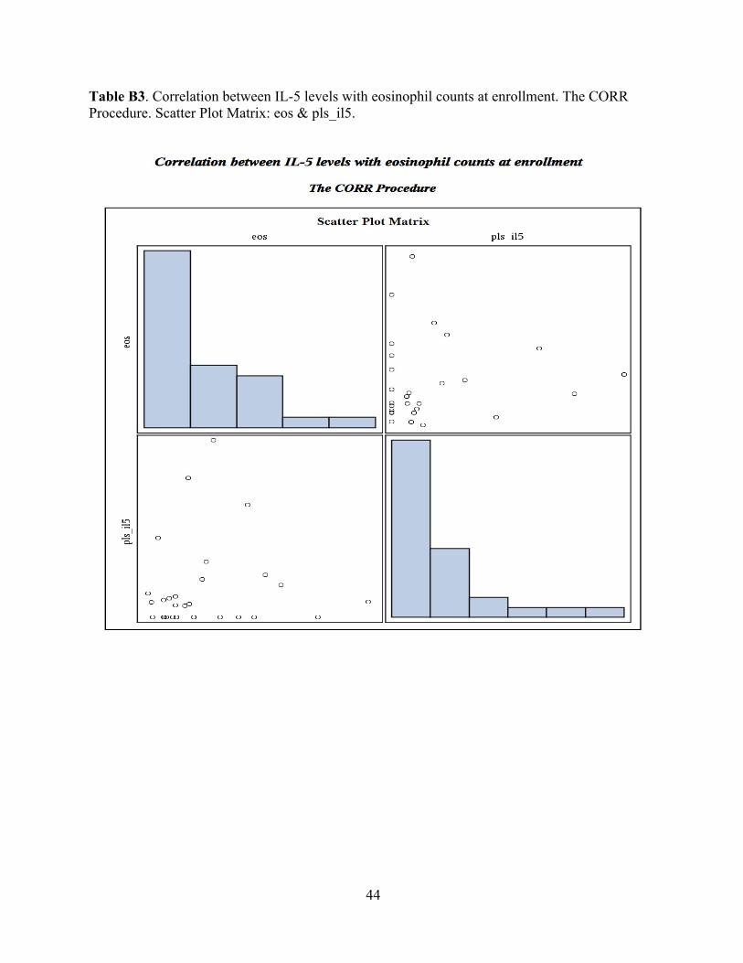
Table B3. Correlation between IL-5 levels with eosinophil counts at enrollment. The CORR Procedure. Scatter Plot Matrix: eos & pls_il5.

45
Table B4. ANOVA to compare important parameters changing with cure rate and time period. The GLM Procedure.

46
Table B5. ANOVA to compare important parameters changing with cure rate and time period. The GLM Procedure. Dependent variable: smepg.

47
Table B6. ANOVA to compare important parameters changing with cure rate and time period. The GLM Procedure. Dependent variable: wbc.

48
Table B7. ANOVA to compare important parameters changing with cure rate and time period. The GLM Procedure. Dependent variable: eos.

49
Table B8. ANOVA to compare important parameters changing with cure rate and time period. The GLM Procedure. Dependent variable: pls_il5.

50
Table B9. ANOVA to compare important parameters changing with cure rate and time period. The GLM Procedure. Dependent variable: pls_il4.

51
Table B10. ANOVA to compare important parameters changing with cure rate and time period. The GLM Procedure. Dependent variable: pls_il10.

52
Table B11. ANOVA to compare important parameters changing with cure rate and time period. The GLM Procedure. Dependent variable: pls_il13.

53
Table B12. ANOVA to compare important parameters changing with cure rate and time period. The GLM Procedure. Dependent variable: plsm_tnf.

54
Table B13. ANOVA to compare important parameters changing with cure rate and time period. The GLM Procedure. Dependent variable: plsm_infg.

55
Table B14. ANOVA to compare important parameters changing with cure rate and time period. The GLM Procedure. Least Squares Means effect for timept: smepg.

56
Table B15. ANOVA to compare important parameters changing with cure rate and time period. The GLM Procedure. Least Squares Means effect for timept: wbc & eos.

57
Table B16. ANOVA to compare important parameters changing with cure rate and time period. The GLM Procedure. Least Squares Means effect for timept: eos & pls_il5.

58
Table B17. ANOVA to compare important parameters changing with cure rate and time period. The GLM Procedure. Least Squares Means effect for timept: pls_il4 & plsm_il10.

59
Table B18. ANOVA to compare important parameters changing with cure rate and time period. The GLM Procedure. Least Squares Means effect for timept: plsm_il10 & plsm_il13.

60
Table B19. ANOVA to compare important parameters changing with cure rate and time period. The GLM Procedure. Least Squares Means effect for timept: plsm_il13 & plsm_tnf.

61
Table B20. ANOVA to compare important parameters changing with cure rate and time period. The GLM Procedure. Least Squares Means effect for timept: plsm_ifng.

62
Table B21. ANOVA to compare important parameters changing with cure rate and time period. The GLM Procedure. Least Squares Means effect for smcat: smepg & wbc.

63
Table B22. ANOVA to compare important parameters changing with cure rate and time period. The GLM Procedure. Least Squares Means effect for smcat: wbc & eos.

64
Table B23. ANOVA to compare important parameters changing with cure rate and time period. The GLM Procedure. Least Squares Means effect for smcat: eos & pls_il5.

65
Table B24. ANOVA to compare important parameters changing with cure rate and time period. The GLM Procedure. Least Squares Means effect for smcat: pls_il4 & plsm_il10.

66
Table B25. ANOVA to compare important parameters changing with cure rate and time period. The GLM Procedure. Least Squares Means effect for smcat: plsm_il10 & plsm_il13.

67
Table B26. ANOVA to compare important parameters changing with cure rate and time period. The GLM Procedure. Least Squares Means effect for smcat: plsm_il13 & plsm_tnf.

68
Table B27. ANOVA to compare important parameters changing with cure rate and time period. The GLM Procedure. Least Squares Means effect for smcat: plsm_ifng.