Embed Size (px)
Citation preview

THE JOURNAL OF BIOLOGICAL CHEMISTRY
Printed m U.S.A. Vol. 257, No. 17, Issue of September 10, pp. 10392-10399, 1982
The Structure of Insulin Receptor and Its Subunits EVIDENCE FOR MULTIPLE NONREDUCED FORMS AND A 210,000 POSSIBLE PRORECEPTOR*
(Received for publication, February 10, 1982)
Masato Kasuga$§f, Jose A. Hedo$, Kenneth M. Yamadall, and C. Ronald Kahn8 From the +Diabetes Branch, National Institute of Arthritis, Diabetes, Digestive and Kidney Diseases and IlLaboratory of Molecular Biology, National Cancer Institute, National Institutes of Health, Bethesda, Maryland 20205 and the SE. P. Joslin Research Laboratory, Joslin Diabetes Center and Dezartment of Medicine, Brigham and Women's Hospital, Harvard Medical School, Boston, Massachusetts 02215
We have identified the subunits of the insulin recep- tor using immunoprecipitation by antibodies to the insulin receptor after either biosynthetic or surface labeling of cultured human lymphocytes (I"9). With this approach, we have found there are two major, M, = 135,000 (a), M, = 95,000 @) and one minor, M, = 210,000 ( y ) subunit. Peptide mapping clearly demon- strates that the major peptides of the a and p subunits are different, whereas similarities exist in the peptide fragments of the y subunit and the a and /3 subunits after limited proteolysis. The y subunit, however, is not simply a disulfide heterodimer of a and p subunits, since this subunit was not reduced by 100 m~ dithio- threitol plus 5% 2-mercaptoethanol, or even under more potent denaturing conditions, such as 8 M guanidine- HC1 and mercaptoethanol at pH 10.5.
In nonreduced gels, free insulin receptor subunits are observed, as well as two higher molecular weight bands of M, = 520,000 and 350,000. On reduction, the 520,000 band was composed primarily of M, = 210,000 and 95,000 subunits, whereas the 350,000 band was com- posed primarily of M, = 135,000 and 95,000 subunits.
These data suggest that the two major subunits of the insulin receptor (a and 8) are distinct. In addition, there is a third component of the receptor identifiable of 210,000 which may be a proreceptor or some closely associated effector protein. Furthermore, it appears that in the native state several kinds of disulfide oli- gomers of these subunits exist. These findings suggest a complex model for insulin receptor synthesis and insertion into the membrane.
During the past several years, significant progress has been made toward elucidating the subunit structure of the insulin receptor using a variety of techniques (1-7). From these studies it appears that the insulin receptor is composed of two major subunits of approximate M, = 135,000 and 95,000. These have been termed the a and p subunits, respectively (8), and appear to be disulfide-linked in the native receptor to form one or more types of complexes of M, > 300,000. Unfortu- nately, affinity techniques label predominantly the a subunit,
* This work was supported in part by a grant from the Juvenile Diabetes Foundation. The costs of publication of this article were defrayed in part by the payment of page charges. This article must therefore be hereby marked "advertisement" in accordance with 18 U.S.C. Section 1734 solely to indicate this fact.
1 Recipient of a Fogarty International Fellowship and a Capps Diabetes Scholarship and is on leave from the Third Department of Internal Medicine, University of Tokyo.
and thus, most of the information regarding the p subunit using this approach has been inferential, based on changes in the electrophoretic mobility of the receptor under nonreduced or only partially reduced conditions.
Recently, we have developed a technique to identify the insulin receptor subunits using immunoprecipitation by anti- bodies to the insulin receptor after either biosynthetic or surface labeling of intact cells (9-11). With this approach both the a and /3 subunits are well labeled, as well as a third protein of M, about 210,000. In this report, we have characterized these three subunits of the insulin receptor by two-dimen- sional gel electrophoresis, peptide mapping, and studies under nonreduced and reduced conditions. These studies suggest that a and p subunits are distinct proteins, that there is an additional 210,000 subunit ( y ) which may be related to the a and/or p subunits, and that the native receptor may exist in forms with various combinations of these three components.
MATERIALS AND METHODS
Chemicals-Chemicals were purchased from the following sources: porcine insulin (lot 1JM95AN) from Elanco Company; Na"'1, ["'SI methionine (translation grade, specific activity > lo00 Ci/mmol), [3H] leucine, ~-[1,6-~H]ghcosamine hydrochloride (39 Ci/mmol), Triton X-100 and EN3HANCE from New England Nuclear; aprotinin, phen- ylmethylsulfonyl fluoride, glucose oxidase and N-acetyl-D-ghcosa- mine from Sigma; wheat germ agglutinin coupled to agarose from Miles Research Laboratories, Elkhart, IN; trypsin and trypsin inhib- itor from Worthington; lactoperoxidase from Boehringer Mannheim; and staphylococcal protein-A (Pansorbin) from Calbiochem. All re- agents for NaDodS04-polyacrylamide gel electrophoresis were from Bio-Rad Laboratories. The sera from patients with autoantibodies against the insulin receptor (12) and from normal volunteers were obtained after an overnight fast, heated at 50 "C for 30 min, and then stored at -20 "C until used. Except where indicated, other chemicals used were reagent grade from commercial suppliers.
Cell Labeling-Human lymphocytes (IM-9 line) were grown in continuous suspension culture a t 37 "C in RPMI-1640 containing 10% fetal calf serum and 25 mM Hepes.' Slightly before reaching saturation density (1-2 X lO'/ml), cells were resuspended in RPMI-1640 medium at 0.5-1 X 10' cells/ml and incubated with [35S]methionine (50 pCi/ ml), ["Hlleucine (50 pCi/ml) or ~-[1,6-~H]glucosamine hydrochloride (50 pCi/ml) at 37 "C for 6-14 h as previously described (9-11). For the [%]methionine, a methionine-free medium was used.
In other experiments, the cells were washed 3 times in Dulbecco's phosphate-buffered saline and surface iodinated as previously de- scribed (11). The reaction was carried out in 100 ml of PBS, which contained 1-5 X lo6 cells/ml, 5 X 10" M NaI, 1 mCi of Na"'1, and 2 mg of lactoperoxidase. One hundred microliters of M Hz02 was added every minute for 15 min, or glucose oxidase (200 milliunits/ml) was added into the PBS containing 20 m~ glucose for 30 min and the suspension was gently mixed at room temperature.
After biosynthetic or surface labeling, IM-9 lymphocytes were
' The abbreviations used are: Hepes, 4-(2-hydroxyethy)-l-pipera- zineethanesulfonic acid; PBS, phosphate-buffered saline.
10392

Insulin Receptor Structure 10393
washed 3 times with cold PBS and solubilized for 60 min at 24 "C in 50 mM Hepes buffer containing 1% Triton X-100, phenylmethylsul- fonyl fluoride (2 mM) and aprotinin (1000 trypsin inhibitor units/ml). Insoluble material was removed by centrifugatlon at 100,000 X g for 90 min at 4 "C. The supernatant was applied to a wheat germ agglutinin agarose column, the column was extensively washed, and desorption of bound glycoproteins performed using 0.3 M N-acetylglu- cosamine. This immobilized lectin allows a 20-fold purification with nearly 100% recovery of the insulin receptor as determined by '"I- insulin binding (13). Typically, 5 ml of N-acetylglucosamine were used and 5 X 1 ml fractions were collected. The 2 fractions with the highest counts were pooled and phenylmethylsulfonyl fluoride (1 mM) and aprotinin (1000 units/ml) were again added.
Immunoprecipitation of the Insulin Receptor-Immunoprecipita- tion of solubilized insulin receptors was performed according to the method previously described (9-11, 14). The solubilized receptors were incubated with normal serum or serum containing autoantibod- ies against the insulin receptor (1:400 dilution) for 6 h a t 4 "C. Immunoprecipitation was then effected by addition of a slight excess of second antibody (sheep anti-human IgG serum; titer of 2 mg/ml) or protein-A (Pansorbin; Calbiochem). After 6 h a t 4 "C or 45 min at room temperature, the suspension was centrifuged for 5 min at 10,OOO x g, and the pellet was washed twice with 50 mM Hepes buffer (pH 7.4) containing 1% Triton X-100 and 0.1% NaDodSO,. The immuno- precipitates were solubilized and boiled (3 min) in 10 m sodium phosphate buffer containing 2% NaDodSO, and 100 m dithiothreitol and 5% (v/v) 2-mercaptoethanol, and 0.01% (v/v) bromophenol blue.
Denaturation by Guanidine Hydrochloride Followed by Alkyla- tion-In some experiments this procedure was performed as described by Weber et al. (15). The solubilized receptors were immunoprecipi- tated by anti-insulin receptor antibody and slight excess of second antibody (sheep anti-human IgG serum). The immunoprecipitates were washed twice with 50 m Hepes buffer (pH 7.4) containing 1% Triton X-100. This pellet was dissolved in 1 ml of 8 M guanidine hydrochloride in 0.1 M Tris-HC1, pH 8.5, a t 100 "C. This sample was immediately transferred to a boiling water bath and 15 1-11 of 2- mercaptoethanol were added. After 3-5 min, the tube was transferred to 37 "C water bath and incubated for 2 h. Then, 0.25 ml of iodoacetic acid (260 mg/ml in 1 M NaOH) was added to 1 ml of the protein solution. The pH was raised to 8-9 by dropwise addition of 2 M NaOH. When no further positive reaction was seen with the nitroprusside test, several more drops of the iodoacetate solution were added to bring the pH to 10.5. After 10 min, excess mercaptoethanol (50 pl) was added and pH was readjusted to about 7.0. The sample for electrophoresis was prepared by prolonged dialysis fust against 9 M urea in 0.1 M Tris-HC1, pH 8, and then against 1 mM sodium phos- phate, pH 7.0, containing 0.1% NaDodSO,. After concentration, the sample was lyophilized and dissolved in the 10 mM sodium phosphate buffer containing 2% NaDodSO, and 100 m dithiothreitol and 5% (v/v) 2-mercaptoethanol.
Electrophoresis-One-dimensional discontinuous acrylamide slab gel electrophoresis in NaDodS04 was performed according to the procedure of Laemmli (16). Depending on the experiment, the acryl- amide concentration of the resolving gel was 5-15%, while that of the stacking gel was 4%.
Peptide mapping by proteolysis in NaDodSO, containing polyacryl- amide gel was performed as described by Bordier et al. (17). Following one-dimensional electrophoresis in 7.5% polyacrylamide under reduc- ing condition with dithiothreitol, an entire lane was cut out and rinsed in 0.125 M Tris-HCI, pH 6.8, 0.1% NaDodSOn for 40 min at room temperature. The gels were digested by placing the lance atop a second sodium dodecyl sulfate gel. The lane was fixed in place with 1% agarose and overlaid with either Staphylococcus aureus V8 pro- tease (Miles) (5 or 30 pg/ml) or papain (Sigma No. P-4762) (40 pg/ ml). Digestion proceeded directly in the stacking gel during electro- phoresis (4 mA, overnight). The resolving gel had an acrylamide concentration of 15%.
Two-dimensional electrophoresis was performed as described by O'Farrell (18) with some modifications. Isoelectric focusing was per- formed in the fust dimension in gels composed of 9.2 M urea, 4% acrylamide, 0.1% N,N-methylene-bis-acrylamide, 2% NP-40, and 2% arnpholines (0.8% pH 2.5-5, 0.8% pH 5-8, 0.4% pH 3-10). Samples were allowed to run for a total of 8000-9000 volt hours. After the run, the gels were incubated with equilibrating buffer (2.5% NaDodSOr, 10% glycerol, 0.0625 M Tris-HCl, pH 6.8, 5% 2mercaptoethanol) for 60 min with continuous gentle shaking. The pH gradient was deter- mined following electrophoresis by slicing a parallel gel into 5-mm pieces and incubating each slice in 1 ml of degassed 0.1% NaCl. For
analysis in the second dimension the equilibrated isoelectric focusing gels were placed on an NaDodS0,-acrylamide slab gel as described above.
After electrophoresis, the slab gel was stained with 0.25% Coomas- sie blue R-250 in 50% trichloroacetic acid, destained in 7% acetic acid, treated with EN"HANCE, dried, and autoradiographed with Kodak X-Omat fdm. For the iodine labeled sample the EN'HANCE was omitted and a Dupont Cronex Lightning Plus enhancing screen was used. In some cases, quantitative estimates of the radioactivity in the labeled bands were obtained by excising the bands from the gels and measuring radioactivity in an Autogamma counter or eluting the slices in 3% Protosol in Econofluor (New England Nuclear) overnight at 37 "C, and measuring radioactivity in a scintillation counter. The films were exposed for 5-20 days and developed. Subunit molecular weights were calculated by using as standard: fibronectin ( M , = 440,000), fdamin (M, = 250,000), myosin (M, = 200,000), phosphoryl- ase B (M, = 94,000), bovine serum albumin (M, = 68,000), ovalbumin (M, = 43,000), and carbonic anhydrase M, = 31,000).
Effects of Trypsin Digestion on Insulin Receptor Subunits-After either labeling with ["S]methionine for 5 h in methionine-free RPMI 1640 or surface labeling with NalZ5I and lactoperoxidase as mentioned above, the labeled cells were washed 2 times in PBS and finally resuspended in 100 mM Hepes buffer, pH 7.4, containing 120 mM NaCI, 1.2 m MgSO,, 2.5 mM KCI, 1 mM EDTA, 10 mM glucose, 1.5 mM sodium acetate, and 1% bovine serum albumin (10" cells/ml). These cells were incubated with 200 pg/ml of trypsin at 37 OC. After 10 min, 400 pg/ml of trypsin inhibitor was added. By this treatment, more than 99% of specific binding of '2sI-insulin was abolished. After trypsin treatment, cells were washed twice with PBS, solubilized by Triton X-100 and immunoprecipitated by anti-receptor antibody with or without purification by wheat germ agglutinin coupled agarose. The gel electrophoresis and autography were performed as mentioned above.
RESULTS
Subunit Structure of Insulin Receptor with Reduction by
x 10-~ Mr
210- ' @d m 6 8
M r
x 10- '
210- n
135-
95-
FIG. 1. Autoradiogram showing 35S-labeled insulin receptor subunits. Cultured human lymphocytes (IM-9) were labeled with ["S]methionine and solubilized with Triton X-100. The insulin recep- tors were purified on wheat germ agglutinin-agarose and then im- munoprecipitated by several sera containing antireceptor antibodies. The immunoprecipitates were analyzed in NaDodS04-polyacrylamide (7.5%) gel electrophoresis after reduction of disulfide bond by dithio- threitol. The gels were dried and subjected to autoradiography. A, the lunes correspond to immunoprecipitation by control pooled serum (C) or sera from the patients B-2, B-8, B-9. All sera were used at a 1:200 dilution. B, the lanes correspond to immunoprecipitation by nonimmune rabbit serum or rabbit serum containing anti-insulin receptor antibody which had been raised against purified insulin receptor of rat liver (provided by Dr. Steve Jacobs at Wellcome Research Laboratory, NC). Both sera were diluted 1:400.

10394 Insulin Receptor Structure Dithiothreitol-Cultured human lymphocytes (IM-9) were biosynthetically labeled with ["S]methionine for 6 h and the insulin receptor was immunoprecipitated by sera containing autoantibodies against the insulin receptor after enrichment on wheat germ agglutinin agarose as described under "Materials and Methods." NaDOSO.,-polyacrylamide gel elec- trophoresis with 100 m~ dithiothreitol and 5% 2-mercaptoeth- anol and autoradiography of immunoprecipitates produced by three different antireceptor sera revealed two major bands of M, = 135,000 and 95,000 and one minor band of M , = 210,000, similar to those previously reported (10, 11) (Fig. M). The 135,000 and 95,000 band correspond to the a and /3 subunits reported by other methods, whereas the 210,000 band does not appear on fully reduced gels using affinity labeling tech- niques (1-8). Although it seemed unlikely that the 210,000 band was an unrelated protein, all three of these sera were derived from patients with an autoimmune disease and, thus, other antibodies are present which might be directed against some other membrane proteins. Thus, for comparison, we also immunoprecipitated the "S-labeled semi-purified fractions using anti-insulin receptor antibody which had been raised against purified insulin receptor of rat liver by immunization
[35S] methionine t3H] leucine Na [1''1]
Mr X 10-3
210-
135-
95-
.
ANTIBODY c 8-2 c 8-2 c 8-2 FIG. 2. Comparison of labeling of insulin receptor subunits
by different methods. IM-9 lymphocytes were labeled either bio- synthetically with ["'S]methionine or with ["Hlleucine, or externally with Na'251 and lactoperoxidase. Cells were solubilized by Triton X- 100, purified on wheat germ agglutinin-agarose, and the immunopre- cipitated by control pooled serum or serum containing anti-insulin receptor antibody B-2 (1:400 dilution). The immunoprecipitates were analyzed in NaDodS0,-acrylamide (7.5%) electrophoresis after reduc- ing with dithiothreitol. The gels were dried and subjected to autora- diography.
TABLE I Comparison of radioactivity incorporated into insulin receptor
subunits The data were obtained by cutting the gels, eluting the slices, and
measuring radioactivity in a scintillation counter (see "Materials and Methods"). The data were expressed as the radioactivity in each band relative to that in the 95.000 band in the same exDeriment.
[%]Methionine 0.3 0.4 1.0 [3H]Leucine 0.2 1.2 1.0 NalZ5I (cell surface iodination) 0.3 1.8 1.0 ["H]Glucosamine" 0.2 1.8 1 .o
" From Ref. 10.
M r x 10 -3
- 210
135
* 95
ANTIBODY C 8-2 FIG. 3. Effect of denaturation with guanidine hydrochloride
and 2-mercaptoethanol on the insulin receptor subunits. ""S- labeled solubilized receptor of IM-9 lymphocytes were immunopre- cipitated by control pooled serum (C) or serum containing anti-insulin receptor antibody (B-2) and slight excess of second antibody. The immunoprecipitates were dissolved in 8 M guanidine hydrochloride and 1.5% of 2-mercaptoethanol in a boiling water bath. After alkyla- tion by iodoacetic acid, the samples were dialyzed against 9 M urea and then 1 mM sodium phosphate, pH 7.0, containing 0.1% NaDodSOr. After lyophilization, the samples were dissolved in the 10 mM dithio- threitol and 5% 2-mercaptoethanol. These samples were analyzed in NaDodS04-polyacrylamide (7.5%) gel and autoradiography.
0 ' J
- 210K
- 135K
- 95K
FIG. 4. Autoradiogram of two-dimensional gels of insulin receptor subunit labeled with Na'''I and lactoperoxidase. IM- 9 lymphocytes were externally labeled with Na""1 and lactoperoxi- dase, solubilized, and immunoprecipitated by anti-receptor serum B- 2. The immunoprecipitates were analyzed by two-dimensional poly- acrylamide gel electrophoresis as described by OFarrell (18) with some modification (see "Materials and Methods").
of rabbits (kindly provided by Dr. Steve Jacobs, Wellcome Research Laboratories, NC). This antibody also immunopre- cipitated the 135,000 and 95,000 proteins, as well as the 210,000 protein, suggesting that indeed all three were components of the insulin receptor complex (Fig. 1B).
For comparison, the IM-9 lymphocytes were biosyntheti- cally labeled with ['Hlleucine by incubating for 12 h at 37 "C or externally labeled with lactoperoxidase and Na'*'I. After solubilization by Triton X-100 and enrichment by a wheat germ agglutinin-agarose column, the labeled insulin receptor

Insulin Receptor Structure 10395
FIG. 5. Insulin receptor structure under nonreduced condition. A, au- toradiogram showing "'S-labeled insulin receptor without reduction of disulfide bonds. IM-9 lymphocytes were labeled with [%]methionine, solubilized, puri- fied on wheat germ agglutinin-agarose and then immunoprecipitated by control pooled serum or anti-receptor sera B-2. The immunoprecipitates were analyzed in NaDodSO4-polyacrylamide (5%) gel electrophoresis without disulfide reduc- tion. B, two-dimensional NaDodSOd- polyacrylamide gel electrophoresis of "S-labeled insulin receptor. Electropho- resis in the fmt dimension was per- formed without reduction as shown in A. Following one-dimensional electropho- resis in 5% polyacrylamide, an entire lane was cut out and rinsed in 0.125 M Tris- HCI, pH 6.8, 0.1% NaDodSOd. The lane was placed atop a second sodium dodecyl sulfate gel, set with 1% agarose, and ov- erlaid with 100 m~ dithiothreitol. Disul- tide bond reduction was carried out dur- ing electrophoresis. The resolving gel in second dimension had an acrylamide concentration of 5%.
M r x 10-3
- C 520
350 440 -
250 -
200 -
116 -
94 -
68 -
C 230
* 190
* 90 210 - 135-
95 -c
520 350 230 1 9 0 t t t t
120 90 t ?
ANTI BODY . , . . ,
was immunoprecipitated by autoantibody B-2 and applied to reduced-NaDodS0,-polyacrylamide gels. As with the methi- onine labeling, two major subunits and one minor subunit were apparent (Fig. 2). However, interestingly, the ratio of activity in the three bands differed with the three labeling methods (Table I). With [3H]leucine labeling, an additional band of molecular weight approximately 30,000 was specifi- cally immunoprecipitated. The nature of this band has not yet been characterized.
Czech and co-workers have previously observed a band of about 210,000 molecular weight when the affinity labeled receptor is run on gels under only partial reducing conditions (8). Under more severe reduction (as above), this band breaks down to yield a and /3 subunits, suggesting that it is a disulfide heterodimer. The 210,000 subunit observed here does not appear to be such a disulfide heterodimer, since under these reducing conditions in parallel experiments with both pho- toaffinity labeling2 and affinity cross-linking with disuccinini- dyl suberate (6), no a-/3 heterodimer was observed.
To clarify further whether the 210,000 subunit was an aggregate of the smaller subunits which were not dissociated by the high concentration of reducing agents (100 m~ dithi- othreitol and 5% 2-mercaptoethanol) or another receptor sub- unit, it was desirable to attempt an even more vigorous method for denaturation and the reduction of disulfide bonds. For this purpose, the "S-labeled insulin receptor was immu- noprecipitated by anti-insulin receptor antibody and the im- munoprecipitates were dissolved in 8 M guanidine hydrochlo- ride, pH 8.5, boiled with 2-mercaptoethanol in a water bath, alkylated, and dialyzed against 9 M urea and then 0.01 M sodium phosphate (see "Materials and Methods"). This pro- cedure nearly always guarantees complete denaturation of the protein and a rapid inactivation of any protease present (15). Using this method of denaturation, again all of the three subunits (210,000, 135,000 and 95,000) were demonstrated (Fig. 3). This result argues strongly that the 210,000 subunit
C.-C. Wang, J.-A. Hedo, C. R. Kahn, D. T. Saunders, P. Thamm, and D. Brandenburg, manuscript in preparation.
observed in these studies is not a simple disulfide-linked heterodimer of a and p subunits of insulin receptor.
Two-dimensional Electrophoresis-Two dimensional anal- ysis (isoelectric focusing and NaDodS04-gel electrophoresis) revealed that the insulin receptor subunits had somewhat different PI values (Fig. 4). The average PI for each of the subunits was: 210,000 subunit, 5.7 f 0.2; 135,000 subunit, 6.0 f 0.2; 95,000 subunit, 5.4 2 0.3 (mean & S.D., n = 4). In each case, but with the 135,000 subunit in particular, the spot was broad, suggesting microheterogeneity with respect to charge. This is not surprising since all three subunits are glycoproteins which may exist in states of varying degrees of glycosylation. The 95,000 subunit was a more acidic protein than 135,000 subunit, consistent with our previous data that the 95,000 subunit is more sialated than 135,000 subunit (10).
Subunit Structure of Insulin Receptor without Reduction of Disulfide Bonds-To characterize these three subunits more carefully, we analyzed the structure of the insulin recep- tor under both nonreducing and reducing conditions, as well as performing peptide maps. The effect of reducing conditions was studied using a two-dimensional gel technique. In the first dimension, the immunoprecipitates were analyzed in NaDodS04-polyacrylamide gel electrophoresis without reduc- tion. Then, the entire lane of interest was cut out, placed horizontally on a second NaDodS04-polyacrylamide gel, and was analyzed with disulfide bond reduction using several concentrations of dithiothreitol.
Under nonreducing conditions, six major bands of specific immunoprecipitation were observed (Fig. 5A). These corre- spond to apparent molecular weights of 520,000, 350,000 230,000, 190,000, 120,000,90,000:? Under reducing conditions
Because of the pitfalls of NaDodSOd-acrylamide electrophoresis in the absence of reduction and because of lack of appropriate markers in higher molecular weight range, the molecular size of those com- ponets are very rough approximations and therefore stoichiometry is very speculative. It also should be noted that all of a, p, y subunits migrate a t a slightly lower molecular weight position in 5% gels without reduction by dithiothreitol, probably due to the existence of intramolecular disulfide bonds and their glycoprotein nature.

10396 Insulin Receptor Structure
210K 135K 95K 1 1 1
C
S. aureus protease (5 pg/rnl)
210k 135k 95k 4 4 1
S. aureus
(3 pg/rnl) protease
D
Na1'2511 13H1-Glucosamine
FIG. 6. Peptide maps of insulin receptor subunits. Human cultured lymphocytes were labeled either biosynthetically with [%SI methionine or [3H]glucosamine, or externally with Na'251 and lacto- peroxidase. Labeled cells were solubilized, purified on wheat germ agglutinin-agarose, and then immunoprecipitated by anti-receptor serum B-2 (1:400 dilution). The immunoprecipitates were analyzed by NaDodS04-polyacrylamide gel electrophoresis after reduction of disulfide bonds by dithiothreitol. Peptide mapping by proteolysis in NaDodS04 containing polyacrylamide gel was performed as described by Bordier and Crettol-Jarvinen (17). Following one-dimensional
TABLE I1 Effect of trypsin treatment on the insulin receptor subunits
trypsin treatment (%I of control) Radioactivity in the band after
210,000 135,000 95,000
NaI29 (cell surface iodination) 2 3 29 ["SIMethionine 24 22 57
(Fig. 5B), the highest molecular weight band (M, = 520,000, fur left) was separated into three spots of M, = 210,000, 135,000, and 95,000, with the relative intensity of label being 210,000 > 95,000 > 135,000. The molecular weight band of 350,000 gave predominantly two spots of M, = 135,000 and 95,000, although a small spot was apparent with an M, = 210,000. The 230,000 band on reduction yielded only the 135,000 protein, suggesting that this is an a-a disulfide dimer. The 190,OOO, 120,000, and 90,000 spots migrated as the y, a, and p subunits, respectively?
Peptide Mapping of Insulin Receptor Subunits-Peptide mapping by proteolysis in NaDodS04-polyacrylamide gel was performed as described by Bordier et ul. (17). Following one- dimensional electrophoresis in 7.5% polyacrylamide under reducing conditions, the entire lane of interest was cut out and placed horizontally atop a second NaDodS04 gel (15%). The first dimension gel was digested by proteases and the peptides
S. aureus S. aureus Papain protease protease (200 pg/ml) (5 pglrnl) (30 pg/rnl)
electrophoresis in 7.5% polyacrylamide, an entire lane was cut out and rinsed in 0.125 M Tris-HC1, pH 6.8,0.1% NaDodS04 for 40 min. The gels were digested by placing the lane atop a second sodium dodecyl sulfate gel during electrophoresis (4 mA, overnight). The resolving gel had an acrylamide concentration of 15%. A, cells were labeled with either NaI2'I (A, B ) or [3H]glucosamine (C, D) , and peptide map were performed by S. aureus V8 protease (A, C, 5 pg/ml, B, D , 30 pg/ml). B, cells were labeled with ["SS]methionine and peptide map were performed by S. aureus V8 protease (A, 5 pg/ml/ B, 30 pg/ml) or papain (200 pg/ml).
resolved by electrophoresis in the second dimension. When the insulin receptor was labeled by NaIz5I or [3H]
glucosamine and digested by S. aureus protease V8 at 5 or 30 pg/ml, the peptide maps of the 135,000 and 95,000 subunits were clearly different (Fig. 6A). The peptide maps of the 210,000 subunit labeled by these methods yielded a pattern which had some peptides of similar mobility to those of the 135,000 subunit, although under most conditions, there was incomplete digestion of the 210,000 band.
When the insulin receptor was labeled by [35S]methionine and digested by S. aureus protease or papain (Fig. 6B), it was again clear that the peptide maps of the 135,000 and 95,000 subunits were different. On the other hand, as in the experi- ment above, the peptide map of the 210,000 protein was similar to that of the 135,000 and 95,000 subunits. Digestion, however, was incomplete and very few of the higher molecular weight peptide fragments observed with the 95,000 subunit were found with the 210,000 subunit (see in particular Fig. 6B).
Effect of Trypsin Digestion on Insulin Receptor Subunits- To characterize the topography of these three subunits in cultured lymphocytes, the labeled cells were treated with trypsin under the conditions which destroy more than 99% of specific '251-insulin binding to these cells. First, the cells were either surface-labeled by iodination or biosynthetically labeled by culture in ["S]methionine. Half of the cells were treated
10397
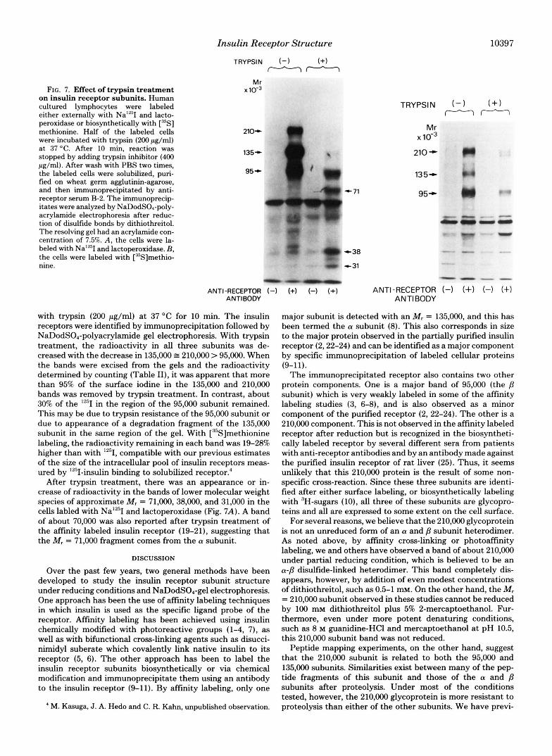
FIG. 7. Effect of trypsin treatment on insulin receptor subunits. Human cultured lymphocytes were labeled either externally with NalZ5I and lacto- peroxidase or biosynthetically with ["'S] methionine. Half of the labeled cells were incubated with trypsin (200 pg/ml) at 37 "C. After 10 min, reaction was stopped by adding trypsin inhibitor (400 pg/ml). After wash with PBS two times, the labeled cells were solubilized, puri- fied on wheat germ agglutinin-agarose, and then immunoprecipitated by anti- receptor serum B-2. The immunoprecip- itates were analvzed bv NaDodSOA-Dolv-
Insulin Receptor Structure
TRYPSIN (-1 (+I mTY'7
Mr x 10-
210+
135-
95-
acrylamide electrophoresis after reduc- tion of disulfide bonds by dithiothreitol. The resolving gel had an acrylamide con- centration of 7.5%. A, the cells were la- beled with Na'*'I and lactoperoxidase. B, the cells were labeled with [%]methio- nine.
.. "
. ANTI-RECEPTOR (-1
ANTIBODY
with trypsin (200 pg/ml) at 37 "C for 10 min. The insulin receptors were identified by immunoprecipitation followed by NaDodS0,-polyacrylamide gel electrophoresis. With trypsin treatment, the radioactivity in all three subunits was de- creased with the decrease in 135,000 = 210,000 > 95,000. When the bands were excised from the gels and the radioactivity determined by counting (Table 11), it was apparent that more than 95% of the surface iodine in the 135,000 and 210,000 bands was removed by trypsin treatment. In contrast, about 30% of the '''1 in the region of the 95,000 subunit remained. This may be due to trypsin resistance of the 95,000 subunit or due to appearance of a degradation fragment of the 135,000 subunit in the same region of the gel. With ["'Slmethionine labeling, the radioactivity remaining in each band was 19-28% higher than with '*'I, compatible with our previous estimates of the size of the intracellular pool of insulin receptors meas- ured by "'I-insulin binding to solubilized receptor:
After trypsin treatment, there was an appearance or in- crease of radioactivity in the bands of lower molecular weight species of approximate M , = 71,000,38,000, and 31,000 in the cells labled with Na'*'I and lactoperoxidase (Fig. 7A). A band of about 70,000 was also reported after trypsin treatment of the affinity labeled insulin receptor (19-21), suggesting that the M , = 71,000 fragment comes from the a subunit.
DISCUSSION
Over the past few years, two general methods have been developed to study the insulin receptor subunit structure under reducing conditions and NaDodS04-gel electrophoresis. One approach has been the use of affinity labeling techniques in which insulin is used as the specific ligand probe of the receptor. Affinity labeling has been achieved using insulin chemically modified with photoreactive groups (1-4, 7), as well as with bifunctional cross-linking agents such as disucci- nimidyl suberate which covalently link native insulin to its receptor (5, 6). The other approach has been to label the insulin receptor subunits biosynthetically or via chemical modification and immunoprecipitate them using an antibody to the insulin receptor (9-11). By affinity labeling, only one
' M. Kasuga, J. A. Hedo and C. R. Kahn, unpublished observation.
Mr x 1 0 - ~
210 - 135-
95*
"" - ""
major subunit is detected with an M , = 135,000, and this has been termed the a subunit (8). This also corresponds in size to the major protein observed in the partially purified insulin receptor (2,22-24) and can be identified as a major component by specific immunoprecipitation of labeled cellular proteins (9-11).
The immunoprecipitated receptor also contains two other protein components. One is a major band of 95,000 (the /3 subunit) which is very weakly labeled in some of the affinity labeling studies (3, 6-8), and is also observed as a minor component of the purified receptor (2, 22-24). The other is a 210,000 component. This is not observed in the affinity labeled receptor after reduction but is recognized in the biosyntheti- cally labeled receptor by several different sera from patients with anti-receptor antibodies and by an antibody made against the purified insulin receptor of rat liver (25). Thus, it seems unlikely that this 210,000 protein is the result of some non- specific cross-reaction. Since these three subunits are identi- fied after either surface labeling, or biosynthetically labeling with 'H-sugars (lo), all three of these subunits are glycopro- teins and all are expressed to some extent on the cell surface.
For several reasons, we believe that the 210,000 glycoprotein is not an unreduced form of an a and /3 subunit heterodimer. As noted above, by affinity cross-linking or photoaffinity labeling, we and others have observed a band of about 210,000 under partial reducing condition, which is believed to be an a-/3 disulfide-linked heterodimer. This band completely dis- appears, however, by addition of even modest concentrations of dithiothreitol, such as 0.5-1 m ~ . On the other hand, the M, = 210,000 subunit observed in these studies cannot be reduced by 100 mM dithiothreitol plus 5% 2-mercaptoethanol. Fur- thermore, even under more potent denaturing conditions, such as 8 M guanidine-HC1 and mercaptoethanol at pH 10.5, this 210,000 subunit band was not reduced.
Peptide mapping experiments, on the other hand, suggest that the 210,000 subunit is related to both the 95,000 and 135,000 subunits. Similarities exist between many of the pep- tide fragments of this subunit and those of the a and /3 subunits after proteolysis. Under most of the conditions tested, however, the 210,000 glycoprotein is more resistant to proteolysis than either of the other subunits. We have previ-

10398 Insulin Receptor Structure
ously shown that the rate of turnover of the 210,000 subunit is faster than that of the two major subunits and also that its turnover is less influenced by insulin than that of other subunits (11). Taken together, these data suggest that the 210,000 subunit could be a precursor of one or both of the insulin receptor subunits (ie. a pr~receptor).~ Alternatively, it may be a third component of the receptor or a closely associ- ated effector protein. Interestingly, the 210,000 subunit ap- pears to be expressed on the surface of the cell (at least in IM- 9 lymphocytes) by surface labeling techniques. This subunit is probably inactive in terms of insulin binding, however, since it is not detected by the affinity labeling techniques (1-8), nor is it observed in the insulin receptor obtained by affinity purification on insulin-agarose columns (2, 22, 24). Alterna- tively, the 210,000 subunit could be the result of a covalent cross-linking by an agent such as transglutaminase (26). This seems unlikely, however, since the 210,000 band is seen in similar amounts after 2 or 24 h of labeling and also the activity of this enzyme has been reported to be very low in IM-9 lymphocytes (27).
The results of the peptide mapping experiments clearly demonstrate that the major peptides of the a and ,8 subunits are different, indicating that the M, = 95,000 component is not derived from the M, = 135,000 component, although both are surface glycoproteins. Several other interesting differences between these two major subunits were found. First, these experiments, as well as our previous studies (10, ll), have shown that the intensity of labeling of these two subunits by various labeling methods is different. The results suggest the M, = 95,000 (8) subunit is less exposed to the cell surface (by iodination) and contains more methionine than the a subunit. The results of two-dimensional electrophoresis showed that the B subunit is a more acidic protein than the a subunit, consistent with our previous observation that subunit ap- pears to possess more terminal sialic acid residues than the a subunit (10). We have also found that the p subunit of the insulin receptor is phosphorylated and this phosphorylation can be stimulated by insulin in cultured human lymphocytes (IM-9) (28). On the other hand, the a subunit is preferentially labeled by several kinds of photoreactive insulins and also using affinity cross-linking methods (1-8).’ Furthermore, tryp- sin treatment of lymphocytes preferentially decreases the labeling of this subunit, consistent with the notion that the a subunit is more exposed in the external surface of the cells. From all of the above data, we propose that the a subunit of insulin receptor is the binding subunit, whereas the p subunit may be the effector subunit for signal transmission.
Using other techniques, it has been suggested that a 45,000 subunit may exist and that this may derive by proteolysis of the 95,000 subunit (2 , 23, 29). If such a subunit exists, it does not appear to be immunoprecipitated by our anti-receptor antibodies, since it is not observed in these studies regardless of the method of labeling or the anti-receptor serum used for precipitation.
When the immunoprecipitates from both biosynthetic la- beling (present study) and surface labeling6 were analyzed in NaDodS04-polyacrylamide gel electrophoresis without reduc- tion of disulfide bonds, we found five or six specific bands. Three bands were observed which correspond to “free” sub- units of M, = 210,000, 135,000, and 95,000. Although the
‘Our antibody also recognized another protein of Mr = 2rn,o@l, which appears to be the earliest biosynthetically labeled component and possesses a high mannose type content (Figs. 2 and 7B) . The relationship between the 200,000 and 210,OOO proteins remains to be determined.
J. A. Hedo, M. Kasuga, K. M. Yamada, and C. R. Kahn, manu- script in preparatiion.
amounts of these free subunits differ from experiment to experiment, they are observed in almost alI experiments. Several possible causes for artifactual reduction were elimi- nated in control experiments. These results were not influ- enced by omitting the protease inhibitors, wheat germ agglu- tinin column step, using second antibody or staphylococcal protein-A or adding N-ethyl-maleimide in the beginning of solubilization. Massague and Czech (30) reported that a par- tially reduced form of insulin receptor is found in rat liver membranes but not in other cells. However, in our hands, these free subunits were found in intact lymphocytes, adipo- cytes, and hepatoma cells and also after affhity labeling or surface labeling with immunoprecipitation, suggesting they are present, at least in part, on the surface of the cell, as well as internally.
In addition to the free subunits, we find two other bands labeled of M, = 520,000 and 350,000.3 The latter appears to correspond to the major band observed in unreduced gel after affinity labeling in adipocytes.2 At present, no data are avail- able on unreduced gels after affinity labeling of lymphocytes. Our results revealed that this 520,000 band was mainly com- posed of M, = 210,000 and 95,000 subunits, and the 350,000 band composed mainly of M, = 135,000 and 95,000 subunits. This would suggest that the 350,000 receptor consists mainly of disulfide-linked aggregates of a and p subunits (possibly a&) with some y ~ubunits .~ This would account for the relative intensity of labeling with a = > y. The 520,000 receptor is more complex since it appears to contain more B and y subunits than a subunits. Possible configurations might be y$ or yap^.^ The percentage of each of these types of complexes on the surface of the cell uersus in intracellular pools remains to be determined.
Trypsin experiments suggest that the intracellular “pool” of insulin receptors, at least glycosylated insulin receptors, in cultured human lymphocytes is low. AU three subunits are trypsin-sensitive, especially the a subunit. These data are compatible to our data that the intracellular pool of insulin receptor is low by measuring the ‘251-insulin binding activity in solubilized fractions from cells with or without trypsin treatment: and with the observation that the turnover rate of 35S-labeled insulin receptor and surface iodinated receptor are very similar (11). If the 210,000 subunit is indeed a prorecep- tor, then one might expect to find it preferentially intracellu- larly. Fractionation of biosynthetically labeled cells to answer this question is underway.
In summary, we have shown that the two major subunits of the insulin receptor (a and 8) are distinct by peptide mapping. In addition, there is a third component (y) which may be a precursor form or independent protein. If this third compo- nent or y subunit is a proreceptor, it does not appear to bind insulin and, thus, like other proteins (31, 32), it may require some form of activation, perhaps by proteolytic cleavage. The CY subunit appears to be the insulin binding subunit, whereas the /3 subunit may be the effector subunit. Free subunits, the Y subunit and two major types of disulfide aggregates of subunits of the receptor, appear to exist in the cell. In one, there appear to be equal amounts of a and subunit, whereas in the other there is an excess of subunit and y subunit. These findings suggest a complex model for insulin receptor synthesis and insertion into the membrane. Further studies in these and other cells should help clarify the exact precursor- product relationship of the subunits and their function in the cell.
Acknowledgments-We wish to thank Dr. Jesse Roth for his generous support and encouragement and Dr. Steve Jacobs for the slft of anti-insulin receptor antibody.
Insulin Receptor Structure 10399
Note Added in Proof-Since submission of this manuscript, we have completed tryptic peptide “fingerprinting” of the a, B, and 210,000 component. These data further substantiate the concept that the a and B subunit are distinct and that the peptides of the 210,000 component (? proreceptor) are present in the a and p subunits.
REFERENCES 1. Yip, C. C., Yeung, C. W. T., and Moule, M. L. (1978) J. Biol.
2. Jacobs, S., Hazum, E, Schechter, Y., and Cuatrecasas, P. (1979)
3. Yip, C. C., Yeung, C. W. T., and Moule, M. L. (1980) Biochemistry.
4. Wisher, M. H., Baron, M. D., Jones, R. H., Sonksen, P. H., Saunders, D. J., Thamm, P., and Brandenburg, D. (1980) Bio- chem. Biophys. Res. Comm. 92,492-498
5. Pilch, P. F., and Czech, M. P. (1980) J. Biol. Chem. 255,1722-1731 6. Kasuga, M., Van Obberghen, E., Yamada, K. M., and Harrison,
L. C. (1981) Diabetes. 30,354-357 7. Hofman, C., Ji, T. H., Miller, B., and Steiner, D. F. (1981) J.
Supramol. Struct. 15, 1-13 8. Massague, J., Pilch, P. F., and Czech, M. P. (1981) Proc. Natl.
Acad. Sci. U. S. A. 77, 7137-7141 9. Van Obberghen, E., Kasuga, M., Le Cam, A., Hedo, J. A., Itin, A.,
and Harrison, L. C. (1981) Proc. Natl. Acad. Sci. U. S. A . 78, 1052-1056
10. Hedo, J. A,, Kasuga, M., Van Obberghen, E., Roth, J., and Kahn, C. R. (1981) Proc. Natl. Acad. Sci. U. S. A. 78,4791-4795
11. Kasuga, M., Kahn, C. R., Hedo, J. A,, Van Obberghen, E., and Yamada, K. M. (1981) Proc. Natl. Acad. Sci. U. S. A . 78, 6917-6921
12. Flier, J . S., Kahn, C. R., Roth, J., and Bar, R. S. (1975) Science.
13. Hedo, J. A., Harrison, L. C., and Roth, J. (1981) Biochemistry 20,
Chem. 253,1743-1745
Proc. Natl. Acad. Sci. U. S. A . 76,4918-4921
19, 70-76
190,63-65
3385-3393
14. Harrison, L. C., Flier, J. S., Roth, J., Karlsson, F. A., and Kahn,
15. Weber, K., Pringle, J. R., and Osborn, M. (1972) Methods En-
16. Laemmli, U. K. (1970) Nature 227,680-685 17. Bordier, C., and Crettol-Jiirvinen, A. (1979) J. Biol. Chem. 254,
18. OFarrell, P. H., (1975) J. Biol. Chem. 250,4007-4021 19. Jacobs, S., Hazum, E., and Cuatrecasas, P. (1980) Biochem.
Bwphys. Res. Commun. 94, 1066-1073 20. Pilch, P. F., Axelrod, J. D., Colello, J., and Czech, M. P. (1981) J.
Biol. Chem. 256,1570-1575 21. Felman, M., Carpentier, J. L., Le Cam, A., Thamm, P., Saunders,
D., Brandenburg, D., Orci, L., and Freychet, P. (1982) J. Cell. Biol. 93,82-87
22. Jacobs, S., Hazum, E., and Cuatrecasas, P. (1980) J. Biol. Chem. 255,6937-6940
23. Harrison, L. C., and Itin, A. (1980) J. Biol. Chem. 255,
24. Siegel, T. W., Ganguly, S., Jacobs, S., Rosen, 0. M., and Rubin,
25. Jacobs, S., Chang, K-J., and Cuatrecasas, P. (1978) Science 200,
26. Keski-Oja, J., Mosher, D. F., and Vaheri, A. (1976) Cell 9, 29-35 27. Baldwin, D., Jr., Prince, M., Marshall, S., Davies, P., and Olefsky,
J. M. (1980) Proc. Natl. Acad. Sci. U. S. A . 77, 5975-5978 28. Kasuga, M., Karlsson, F. A., and Kahn, C. R. (1982) Science 215,
29. Massague, J., Pilch, P. F., and Czech, M. P. (1981) J. Biol. Chem.
30. Massague, J., and Czech, M. P. (1980) Diabetes 29,945-947 31. Davie, E. W., and Fujikawa, K. (1975) Annu. Reu. Biochem. 44,
32. Buchmeier, M. J., and Oldstone, M. B. (1979) Virology 99,
C. R. (1979) J. Clin. Endocrinol. Metab. 48,59-65
zymol. 26,3-27
2565-2567
12066-12072
C. S. (1981) J. Biol. Chem. 256,9266-9273
1283-1284
185-187
256,3182-3190
799-829
111-120