Embed Size (px)
Citation preview
Advanced Review
The nucleolus: structure/functionrelationship in RNA metabolismDaniele Hernandez-Verdun,1∗ Pascal Roussel,1 Marc Thiry,2
Valentina Sirri1 and Denis L. J. Lafontaine3
The nucleolus is the ribosome factory of the cells. This is the nuclear domainwhere ribosomal RNAs are synthesized, processed, and assembled with ribosomalproteins. Here we describe the classical tripartite organization of the nucleolusin mammals, reflecting ribosomal gene transcription and pre-ribosomal RNA(pre-rRNA) processing efficiency: fibrillar center, dense fibrillar component, andgranular component. We review the nucleolar organization across evolution fromthe bipartite organization in yeast to the tripartite organization in humans. Wediscuss the basic principles of nucleolar assembly and nucleolar structure/functionrelationship in RNA metabolism. The control of nucleolar assembly is presentedas well as the role of pre-existing machineries and pre-rRNAs inherited fromthe previous cell cycle. In addition, nucleoli carry many essential extra ribosomalfunctions and are closely linked to cellular homeostasis and human health. Thelast part of this review presents recent advances in nucleolar dysfunctions inhuman pathology such as cancer and virus infections that modify the nucleolarorganization. 2010 John Wiley & Sons, Ltd. WIREs RNA 2010 1 415–431
INTRODUCTION
The nucleoli are specific nuclear domains present inall eukaryotic cells. The nucleolus, a membrane-
less organelle, is the ribosome factory of the cell.1 Incycling cells, nucleoli assemble at the exit from mito-sis, they are functionally active throughout interphase,and they disassemble at the beginning of mitosis. Thenucleolus is the site where different steps of ribosomebiogenesis are grouped together, i.e., transcription ofribosomal genes (rDNAs), maturation/processing ofribosomal RNAs (rRNAs), and assembly of rRNAswith ribosomal proteins.2 It was proposed that thenucleolus is ‘an organelle formed by the act of buildinga ribosome’.3 Indeed, the organization and size of the
∗Correspondence to:[email protected] and cell cycle, Institut Jacques Monod-UMR 7592 CNRS,Universite Paris Diderot, 75205 Paris cedex 13, France2Laboratoire de Biologie Tissulaire, Universite de Liege, Liege,Belgium3Fonds de la Recherche cientifique (FRS-F.N.R.S.), Institut deiologie et de Medecine Moleculaires (IBMM), Universite Librede Bruxelles (ULB), & Center for Microscopy and MolecularImaging (CMMI), Academie Wallonie–Bruxelles, Charleroi-Gosselies, Belgium
DOI: 10.1002/wrna.39
nucleoli are directly related to ribosome production.4
Consequently, the size of the nucleolus is a diagnosticmarker of highly proliferative cancer cells.5 The vari-ability of the nucleolar organization has been inten-sively examined in different biological contexts suchas proliferation, differentiation, development, and dis-ease. The comparison of the nucleolar organizationacross evolution revealed both the conservation inthe basic ‘building blocks’ and a higher complexityin modern eukaryotes.6 The nucleolus constitutes amodel to understand the principles of the organizationof nuclear domains, the dynamics of protein traffick-ing, as well as the interplay between nuclear bodiesdedicated to related functions (Cajal body, promyelo-cytic leukemia body, and nuclear speckles).
Throughout the past 50 years, nucleolar com-plexity was deciphered using multiple approaches.This was possible thanks to technological break-through: specific in situ labeling, three-dimensionalresolution, and improved isolation procedures ofnucleoli for biochemical characterization and pro-teomic analysis. Thus, it was discovered that otherribonucleoproteins (RNPs) in addition to ribosomalsubunits are assembled or processed in the nucleolus.The best example is the nucleolar assembly proposedfor the signal recognition particle.7 In plant cells butnot in animal cells, nucleoli have been implicated as
Volume 1, November/December 2010 2010 John Wiley & Sons, L td. 415

Advanced Review wires.wiley.com/rna
sites of silencing RNA biogenesis.8 In addition, thecomparative proteomics of animal and plant nucleolidemonstrated the nucleolar function adaptation (inhumans9,10 and in plants11).
Today, the nucleolus is considered a multifunc-tional domain. Extra ribosomal functions assigned tothe nucleolus include the involvement in cell cycle andcell proliferation control, stress sensing and tumorsurveillance pathways, apoptosis, telomere forma-tion, transfer RNA modifications, viral life-cycle, etc.Unsurprisingly, nucleolar dysfunction has severe con-sequences for human health.5,12,13 These extra ribo-somal functions of the nucleolus have been reviewedelsewhere and will not be discussed here (reviewed inRefs 14–16).
This review will essentially cover three topics:(1) nucleolar organization depending on ribogenesisactivity and across evolution, (2) the principles ofnucleolar assembly in cycling cells, and (3) thealterations in nucleolar organization in diseases.
NUCLEOLAR ORGANIZATIONREFLECTS rDNA TRANSCRIPTIONAND PRE-rRNA PROCESSINGEFFICIENCY
Nucleoli assemble around the nucleolar organizerregions (NORs), as first proposed by McClintockin Zea mays.17 The NORs are chromosomal regionswhere multiple rDNA copies cluster in arrays. Thenumber of NOR-bearing chromosomes varies depend-ing on the species, ranging from 1 in haploid yeastcells to 10 in human somatic cells (acrocentric chro-mosomes 13, 14, 15, 21, and 22). On mitotic chromo-somes, the active NORs are detected by a specific silverstaining procedure, designated Ag-NOR staining,18
reflecting their continuous association throughoutmitosis with a subset of argyrophilic proteins belong-ing to the rDNA transcription machinery.19
At the end of mitosis, when rDNA transcrip-tion by the RNA polymerase I (pol I) resumes (seebelow), active NORs are directly involved in nucleo-lar reassembly. Within each active NOR, only a subsetof rDNA units are transcribed. In contrast, inactiveNORs are not bound by argyrophilic proteins, they arenot associated with the pol I machinery, and they arenot involved in nucleolar formation. The nucleolus iseither organized around a single NOR or alternativelyseveral active NORs coalesce in a single nucleolus oncerRNA synthesis has initiated.20 For instance, in Xeno-pus laevis or Potorous tridactilys cells, there are twonucleoli per cells each corresponding to a single NOR,whereas in human HeLa cells, there are two to threenucleoli per cell corresponding to six active NORs.
(a)
(b)
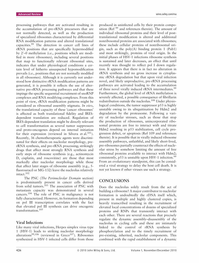
FIGURE 1 | Nucleolar organization of a human HeLa cell preparedby conventional methods for electron microscopy. The cells were fixedby glutaraldehyde and osmic acid. The sections were contrasted byuranyl acetate and lead citrate. (a) Section of one nucleolus and(b) details of the three nucleolar components. In (a) and (b), the threenucleolar components are visible: the fibrillar centers (asterisks), thedense fibrillar component (white arrow), and granular component (GC).Scale bar: (a) = 0.5 µm and (b) = 0.1 µm.
Nucleolar Organization in HigherEukaryotes: A Tripartite OrganizationThe nucleoli observed by electron microscopy (EM)appear to be mainly composed of fibrils andgranules on sections of fixed (formaldehyde andosmium) samples contrasted by uranyl and lead(Figure 1(a) and (b)). A great variability of thenucleolar morphology was described based on thetypes or functions in animal and plant cells.2,21
416 2010 John Wiley & Sons, L td. Volume 1, November/December 2010

WIREs RNA Nucleolus
FIGURE 2 | Perinucleolar heterochromatin inmouse NIH3T3 nuclei observed in lightmicroscopy and EM are shown in left and rightpanels, respectively. (a–c) The heterochromatinis observed after DNA Dapi staining especially atthe nucleolar periphery (arrows); the nucleoli(contrasted structure in phase) appeared asblack holes with Dapi. (d) A protocol topreferentially reveal the nucleic acids in EM wasused. The DNAs and RNAs were contrasted withuranyl after methylation and acetylation of theamino and carboxyl groups. Around thenucleolus, two large clumps of chromatin (arrowheads) are visible as well as the perinucleolarchromatin. White arrows indicate intranucleolarchromatin localized in the GC. One FC is visiblein the middle of the nucleolus. Scalebar = 0.5 µm. EM, electron microscopy; FC,fibrillar center.
(a)
(b)
(c) (d)
However, this variability resides in the arrangementof three fundamental components defined by theirtexture and contrast by EM and designated fibrillarcenters (FCs), the dense fibrillar component (DFC),and the granular component (GC) (reviewed in Ref22). The FCs are clear fibrillar areas of differentsizes ranging from 0.1 to 1 µm containing fibrils(Figures 1(b) and 2(d)). They are partly surroundedby the highly contrasted DFC of compact texture.The FCs and DFC are embedded in the GC thatmainly consists of granules 15–20 nm in diameter ina loosely organized distribution (Figure 1(a) and (b)).Using complementary approaches, a spatiotemporalmap of ribosome biogenesis in these three nucleolarcomponents was obtained including the localization ofrDNAs, rRNAs, small nucleolar RNAs (snoRNAs), aswell as several proteins belonging to transcription andprocessing machineries and ribosomal proteins. It wasestablished that the sites of active pol I transcriptionare localized at the interface between the FCs andthe DFC, where early processing of the pre-rRNAsoccurs in the DFC and late processing in the GC. Thenontranscribed part of the rDNAs as well as the polI complexes and the transcription machinery such asthe upstream binding factor (UBF) and topoisomeraseI are localized in the FCs.23 What is the role of FCs?The FCs appear to be pivotal elements to understandhow pol I transcription organizes the nucleoli. Itwas first proposed that the FCs are the interphasiccounterparts of the mitotic NORs because the nucleoliare reformed around the FCs at the end of mitosis (seeNucleolar Assembly section). The variability of the
FCs (number and volume) was then correlated withthe transcriptional activity of the rDNAs in definedbiological conditions. In the nucleolus of peripheralmature human lymphocytes with low activity, a singlelarge (diameter = 0.2–0.4 µm) FC is visible.24 Uponstimulation, the lymphocytes enter the cell cycle,ribosome biogenesis is stimulated, and the nucleolusbecomes enlarged while the numerous small FCsare formed. It was proposed that when ribosomeproduction is activated, the FCs unfold because afraction of the rDNA copies present in the singleFC are transcribed and the DFC is generated.25 Thisconclusion is also supported by the observation ofnucleoli corresponding to the activity of a single NOR-bearing chromosome as in Potorous tridactylis cells;the clusters of transcribed rDNAs are intercalatedwith repressed genes in the same rDNA tandem[repeat].26 Consequently, we suggest that it wouldbe interesting to re-evaluate the variability in size ofFCs as a percentage of the total nucleolar volume andnot only their number and size in EM sections. Theprediction is that in small resting nucleoli (ring-shapednucleoli of lymphocytes or remnant nucleoli of chickerythrocytes),4 the FC volume is high compared tothe total nucleolar volume. We anticipate that thiscriterion could be a useful index of ribosomal geneactivity.
Presently, it is not possible to exclude that FCshave additional uncharacterized functions. In nucleoliof stimulated rat neurons, the volume of only oneFC increases to 10-fold27,28; the causes for this cyclicvariability are still unknown. In the nucleoli of rat
Volume 1, November/December 2010 2010 John Wiley & Sons, L td. 417

Advanced Review wires.wiley.com/rna
(a)
(b)
FIGURE 3 | GFC in neuron nucleolus. (a) The confocal imageillustrates a trigeminal ganglia neuron immunostained for UBF (green)and counterstained with propidium iodide for nucleic acids. Two cells ofdifferent size are visible: the large cell corresponds to the neuron andthe asterisk indicates the nucleus of a satellite glial cell. The nucleolus(arrow head) of the large neuron contains a prominent GFC visible ingreen and several FCs of normal size. In the enlargement (left corner) ofthe nucleolus, the FCs of normal size are indicated by arrows and theGFC is visible in the center. Scale bar = 5 µm. (b) Nucleolus in atrigeminal ganglia neuron observed in EM. A typical GFC is visible in thecenter of the nucleolus. Scale bar = 1 µm. Unpublished data from thegroup of M. Lafarga (I. Casafont and M. T. Berciano). EM, electronmicroscopy; FC, fibrillar center; GFC, giant FC; UBF, upstream bindingfactor.
sensory ganglia neurons, one giant FC (GFC) wasobserved (Figure 3(a) and (b)). The accumulation ofUBF was demonstrated in this GFC (Figure 3(a)) aswell as the absence of nascent RNAs. GFCs alsocontain components of the SUMO-1 conjugationpathway (SUMO-1 and Ubc9), but their role in GFChas not been determined.29
FIGURE 4 | Localization of nucleolar markers in the three nucleolarcomponents in human HeLa cells. In the right panel, the green labelingsshow the distribution of the proteins and in the left panel in the samecells the nucleoli are visible in dark by phase contrast. Antibodiesagainst UBF decorate several foci in the nucleolar interior correspondingto FCs. Nopp140-GFP (Nopp140) fusions exhibit a dotted labelingcharacteristic of the DFC of nucleoli. NPM/B23-GFP fusions (NPM/B23)decorate the GC. Scale bar: 5 µm. DFC, dense fibrillar component; FCs,fibrillar centers; GC, granular component; UBF, upstream binding factor.
The nucleoli are visible in the nucleus usingphase contrast light microscopy (Figures 2, 4, and 5).The three basic nucleolar components can be mappedby immunolabeling or using fluorescent proteinscorresponding to a precise step in the ribosomeassembly pathway. For example, antibodies againstfibrillarin or fibrillarin-reporter constructs identify theDFC and antibodies against nucleophosmin/initiallyB23 nucleolar protein (NPM/B23) and NPM/B23-GFPidentify the GC.25 This renders the three-dimensionalanalysis of the nucleolar organization in fixed or livingcells possible by confocal microscopy. Typically incells in which ribosome synthesis is active, the threenucleolar components are intermingled reflecting thevectorial formation of ribosomes: the FCs and DFCare distributed in the foci in the internal part of thenucleoli surrounded at the nucleolar periphery by theGC (Figure 4).
418 2010 John Wiley & Sons, L td. Volume 1, November/December 2010

WIREs RNA Nucleolus
FIGURE 5 | Organization of the nucleolusafter inhibition of pol I transcription. In lightmicroscopy, the nucleolar segregation in ahuman HeLa cell treated with a lowconcentration of actinomycin D is observed inthe left panel. (a) The nucleolus is visible in thenucleus by phase contrast. (b–d) The DFCvisualized by fibrillarin–GFP fusion and the GCvisualized by NPM/B23-DsRed fusion disengageand form two juxtaposed structures. In the rightpanel, (e) the segregation of the three nucleolarcomponents observed in electron microscopy.This HeLa cell was treated with lowconcentration of actinomycin D and the Ag-NORstaining (black dots) revealed the Ag-NORproteins in FC. DFC, dense fibrillar component;FC, fibrillar center; GC, granular component.Scale bar = 1 µm.
(a)
(b)
(c)
(d) (e)
Nucleolar Organization in LowerEukaryotes: A Bipartite OrganizationDespite the above classical description of atripartite nucleolar organization in mammaliancells, many eukaryotes, including the geneticallytractable yeast Saccharomyces cerevisiae, have onlytwo morphologically distinct nucleolar components(discussed in Ref 6). Several features distinguish yeastfrom human nucleoli. A major difference lies in theinternal organization of the organelle (number ofcomponents) and occurrence of intranucleolar bodies(Subnucleolar Structure in Budding Yeast section). Inaddition, yeast nucleoli lack condensed perinucleolarchromatin and show extensive nuclear membraneattachment (see below). Finally, yeast is characterizedby a closed mitosis implying that its nucleolus doesnot disassemble during mitosis.
In budding yeast, there is a single nucleolusthat occupies one third of the nuclear volume.In haploid yeast cells, depending on the growthconditions about 100–200 rDNAs cluster in a singleNOR that localizes to the left arm of chromosomeXII. There is about a 10-fold range difference insize between yeast and human nucleoli (≈0.5 µmand from ≈0.5 to 9 µm, respectively) and a humannucleolus is about the size of a yeast nucleus. In yeastnucleoli only two nucleolar components are detected:fibrillar strands (F) and granules (G) (Figure 6(a)). Incontrast to the situation in humans where the fibrillarconstituent generates distinctive FC/DFC modules
(Figures 1(a), (b), and 6(c)), in yeast, F is the onlyfibrillar component. A good demonstration of abipartite nucleolar organization in yeast is providedupon nucleolar segregation conditions (Figure 6(b)and section Nucleolar Organization Related to theActivity of Ribosome Biogenesis).
Nucleolar Structure Across Evolution:Seeking the Transition Between Bi-and Tripartite NucleoliThe emergence of tri-compartmentalized nucleoli thatcoincides with the transition between anamnioticand amniotic vertebrates correlates with a strikingexpansion in the size of intergenic rDNA spacersthat separates the pol I transcription units in rDNAarrays and this has been suggested to underlie thespecialization of a single fibrillar component in twodistinct compartments (discussed in Ref 6). Extendedspacers might have allowed the specific exclusion ofone form of chromatin by ‘looping it out’ from adefined nucleolar location into a novel compartment.At this transition lies the reptile group comprisedof turtles, lizards, sphenodons, snakes, birds, andcrocodiles (Figure 7). Recent analyses indicate thatturtle nucleoli are bipartite, whereas lizard, snake,bird, and crocodile nucleoli show three subnucleolarcompartments (MT and DLJL, unpublished andillustrated for turtles and lizards in Figure 7). Theemergence in higher eukaryotes of a third nucleolarcompartment, the fibrillar center, a repository of pol I
Volume 1, November/December 2010 2010 John Wiley & Sons, L td. 419

Advanced Review wires.wiley.com/rna
FIGURE 6 | Nucleolar organization acrossevolution. Lower and higher eukaryotes arecharacterized by a bipartite (F and G) versus a tripartite(FC, DFC, and GC) nucleolar organization, respectively,as illustrated under physiological (a and c) andsegregation (b and d) conditions. (a) A wild-type yeastnucleolus with fibrillar strands (F) and granules (G).(b) A yeast nucleolus from a cell deleted for srp40, thetwo nucleolar components are segregated and adopt a‘Ying-Yang’ configuration. (c) A wild-type humannucleolus with several FC/DFC (asterisks and arrows)modules embedded into a single GC. (d) A humannucleolus following actinomycin D treatment(0.5 µg/mL, 2 h) with all three componentssegregated. All samples were treated by acetylationand inspected by EM. (a, b) Yeast Saccharomycescerevisiae; (c) HEp-2 larynx carcinoma; and (d) Jurkat Tlymphocyte. Scale bars = 0.2 µm.
(a) (b)
(c) (d)
FIGURE 7 | Nucleolar organization at thetransition between bipartite and tripartiteorganization. (a) A nucleolus from Trachemys scripta(red-eared slider) and (b) a nucleolus from Podarcismuralis (common wall lizard). All samples treated byacetylation and inspected by EM. Scale bars = 0.4 µm.EM, electron microscopy. (a) (b)
complexes ready to engage pol I transcription, mightimpart regulatory functions to nucleolar processes.
Subnucleolar Structures in Budding YeastIn addition to nucleolar fibrillar strands and granules,several specialized subnucleolar domains have beendescribed in budding yeast. These include thenucleolar body (NB) and the ‘No-body’, involvedin snoRNA biogenesis and ribosome surveillance,respectively, as well as a nucleolar domain enrichedin poly(A) RNAs. Subnucleolar compartmentalizationmight facilitate specific reactions such as RNAmodification, RNA processing, and RNA degradation.
Nucleolar BodyThe NB is a spherical body, Ag-NOR positive, ofabout 300 nm in diameter that emanates from fibrillar
strands (Ref 30 and M. T. and D. L. J. L., unpublisheddata). There is one NB per nucleolus. The NBhas primarily been involved in snoRNA maturation.SnoRNAs are transcribed in the nucleoplasm asprecursors carrying noncoding extensions, requiringspecific maturation, and targeting the nucleoluswhere they function in RNA processing, in RNAmodification, and, possibly, in RNA folding. Severalbox C + D snoRNAs were shown to initiallyconcentrate in the NB prior to distributing tothe overall nucleolar volume.30 The transientaccumulation of snoRNAs, such as U3, in theNB is thought to allow 5′-cap trimethylation bythe trimethyl guanosine synthase Tgs1 and 3′-endprocessing. Ectopically expressed human survival ofthe motor neuron protein (SMN), a prototypic Cajalbody/gem antigen, accumulates in the NB and led to
420 2010 John Wiley & Sons, L td. Volume 1, November/December 2010

WIREs RNA Nucleolus
the suggestion that NB and Cajal body are orthologousstructures. A visual screen for altered patterns ofU3 nucleolar distribution led to the identificationof mutations that affect NB localization31: threemutations were identified that led to the formation ofa nucleolar ring corresponding to NB exclusion andtwo to snoRNA accumulation in the NB. Strikingly,mutations that led to NB accumulation affected bothbox C + D and box H + ACA snoRNAs indicating acertain level of commonality in snoRNA intranucleartrafficking pathways.
No-bodyEach of the many steps in ribosome synthesis issubjected to an error rate and the possibility ofproducing misassembled ribosomes with potentiallyimpaired translational capacity, and deleteriousconsequences for cell viability are immense (reviewedin Ref 32). To circumvent such problems, cellshave evolved multiple quality control pathwaysthat recognize and target defective RNP particlesfor rapid clearance. One of the best characterizednuclear surveillance pathway is the TRAMP-Exosomepathway where defective pre-RNPs are targeted fordegradation following the addition of short poly(A)tails at the 3′-end of their RNAs by TRAMP. In thissurveillance, polyadenylation acts as a recruitmentand stimulatory signal for the RNA exosome thatturns over the RNA (reviewed in Refs 33,34). TheRNA exosome is a multiprotein complex endowedwith both 3′–5′ exoRNase and endoRNase activitythat operates in RNA synthesis (formation ofmature RNA 3′-end), RNA degradation (physiologicalRNA turnover), and RNA surveillance (clearance ofdefective RNPs).
The ‘No-body’ is a nucleolar focus, distinctfrom the NB, enriched in pre-rRNAs and RNAsurveillance components that was detected in strainsdefective in the HEAT-repeat containing proteinSda1, a ribosome synthesis factor involved in pre-60S synthesis and export of both small and largesubunits.35 The following components have beenlocalized to the ‘No-body’: small and large ribosomalsubunits, TRAMP components, core exosome as wellas nuclear specific exosome subunits.35 Intact TRAMPand exosome complexes are required for ‘No-body’formation consistent with a role of this organelle inthe surveillance of nuclear-restricted pre-ribosomes.
Other Nucleolar RNA Surveillance CentersOther putative nucleolar ‘surveillance centers’, distinctfrom the NB and ‘No-body’, are composed of focienriched for polyadenylated snRNAs and snoRNAs36
and a focus detected upon Rnt1 (yeast RNase III)
mild overexpression that juxtaposed with primaryrRNA transcripts.37 In human, nucleolar-associatedfoci enriched for the export factor Crm1 and thetranslational repressor CPEB1 have been describedand referred to as CNoBs.38 Whether CNoBs arerelated to NBs, No-bodies, or other nucleolarsurveillance centers remain to be determined.
Nucleolar Organization Related to theActivity of Ribosome BiogenesisNucleoli are characterized by a great variability insize, number, and position within the nuclear volumeand this variability depends on cellular metabolicactivity. In cycling cells, the volume of the nucleoliincreases between the G1 and G2 phases and thenumber of FC doubles in G2.39 In quiescent cells atthe terminal stage of differentiation when ribosomebiogenesis is stopped, small ring-shaped nucleoli ornucleolar remnants (diameter = 0.3 µm) are typicallyobserved in lymphocytes or erythrocytes.4 Thesenucleoli are formed by one clear area containingchromatin and dense fibrils at the periphery. Inerythrocyte nucleoli, active pol I transcription wasnot detected,40 but a modified form of UBF was foundas well as fibrillarin, nucleolin, NPM/B23, U3 and U8snoRNAs, and partially processed pre-rRNAs.41 Cellcycle stimulation of erythrocytes fused with cyclingcells induced the reactivation of ribosome biogenesis innucleolar remnants.42 Typical nucleolar organizationwith FCs, DFC, and GC is restored, whereas thereversibility of the repression was not observed inXenopus erythrocytes upon incubation in extractsthat failed to restore the cell cycle.41
A variety of drug treatments leading totranscriptionally arrested cells typically induce aphenotype of segregation in which the fibrillarand granular components of nucleoli disengage andform three juxtaposed structures (Figures 5(a), (e),and 6(d)). Following nucleolar segregation, nucleolarcomponents are reorganized in such a manner thatthe two fibrillar components appear as individualcaps juxtaposed to a central body correspondingto the GC.43,44 Likewise, nucleolar segregation canbe achieved in yeast, for instance with mutationsaffecting ribosome trans-acting factors such asSrp40, the yeast homolog of mammalian Nopp140;here, consistent with a bipartite organization,only two nucleolar components become segregated(Figure 6(b)). Remarkably, there are physiologicaloccurrences of nucleolar segregation that alsocorrespond to the inhibition of rRNA synthesis atdefined periods of differentiation and cell maturation.4
This process is thought to be directly linked to the
Volume 1, November/December 2010 2010 John Wiley & Sons, L td. 421

Advanced Review wires.wiley.com/rna
inhibition of pol I transcription and indeed it isobserved during transcriptional arrest by inhibitorssuch as the intercalating agent actinomycin D thatexhibits high binding affinity for GpC sites inrDNAs and preferentially affects pol I transcriptionat low doses. Interestingly, studies have shown thatnucleolar segregation in cells treated with high dosesof actinomycin D inhibiting both pol I and pol IItranscription not only implicates reorganization ofnucleolar components but also of an energy-dependentrelocalization of molecules from Cajal bodies such asthe p80 coilin, Cajal body-specific RNAs (scaRNAs)and nucleoplasmic proteins.10,45
The adenosine analog 5,6-dichloro-1-ribo-furanosylbenzimidazole (DRB), a casein kinase (CK2)inhibitor, has a repressive effect on pol II transcription,decreases pol I transcription, and impairs pre-rRNAprocessing.46 DRB reversibly induces unraveling ofnucleoli into necklace structures.47 On one hand,EM studies revealed that the nucleolar necklace iscomposed of small FCs partially surrounded by andconnected to each other by the DFC, indicating thateach bead of the necklace most probably correspondsto one functional transcription domain.48 On the otherhand, the nucleolar proteins involved in pre-rRNAprocessing are mislocalized in large bodies derivedfrom the GC. When DRB is removed, reassembly ofthe nucleoli occurs. This process is CK2-driven andATP/GTP-dependent.49
Similar effects are obtained when cells aretreated with the highly selective cyclin-dependentkinase (CDK) inhibitors, roscovitine, olomoucine,purvalanol, or alsterpaullone.50 These CDK inhibitorsmodify both pol I transcription and pre-rRNAprocessing and induce a dramatic but reversibledisorganization of active nucleoli, whatever theinterphase stage of the cells. Because the transcriptionfactor UBF is regulated by CDKs, e.g., CDK2–cyclinE,51 pol I transcription decreases after CDK inhibitortreatment but remains active. Remarkably, in additionto the decrease in pol I transcription, thesetreatments impair pre-rRNA processing.50 Thus, thetypical organization of nucleoli in the three majorcomponents, i.e., FCs, DFC, and GC, is undoubtedlylinked to both pol I transcription and pre-rRNAprocessing.
The link between pol I transcription, pre-rRNAprocessing, and nucleolar structure is highlightedby studies based on the depletion of nucleolarproteins such as the transcription initiation factorTIF-IA, the mammalian homolog of yeast Rrn3p. InTIF-IA−/− cells, the amount of pol I associatedwith rDNAs is severely reduced as well as pre-rRNA synthesis. After TIF-IA depletion, the size of
the nucleoli decreases, nucleolar structures disappear,and nucleolar proteins (observed for the p19Arf
tumor suppressor, the transcription factor UBF, andNPM/B23) are released from the nucleoli and localizedin the nucleoplasm.52 Similarly, depletion of p19Arf,a nucleolar protein reported to inhibit production ofrRNAs by delaying the processing of 47S/45S and32S pre-rRNAs,53 results in morphological nucleolarchanges.54 Ag-NOR staining of Arf−/− cells showedan increased number of Ag-NORs per nucleus and anirregular shape compared to control cells. At the EMlevel in Arf−/− cells, the authors observed multiple,elongated, irregular nucleoli exhibiting larger FCsin comparison with the round nucleoli of wild-typecells. The depletion of the NPM/B23 multifunctionalnucleolar protein was also reported to cause distortionof the nucleolar structure and fragmentation ofnucleoli.55 Recently, the GTP-binding nucleolarprotein nucleostemin (NS) was shown to play arole in pre-rRNA processing as its depletion delaysthe processing of 32S pre-rRNAs to 28S rRNAsand induces the relocalization of proteins involvedin pre-rRNA processing, i.e., DDX21 and EBP2,from nucleoli to nucleoplasm.56 Interestingly, NSdepletion leads to the dissociation of the componentsof snoRNPs and the telomerase complex, and to thedisruption of the DFC and FCs in the nucleolus.57
The Perinucleolar DomainThe Perinucleolar ChromatinIn most animal and plant cells but not in buddingyeast, a heterochromatin layer is observed at thenucleolar periphery by EM4,58 (Figure 2(d)). This het-erochromatin is visible with DNA Dapi staining (apositive ring surrounding a black hole) demonstrat-ing the high DNA content at the periphery comparedto within the nucleolus (Figure 2(a) and (c)). Inci-dentally, the first protocols established to isolatenucleoli from rat hepatocytes for biochemical pur-poses included a DNase treatment to remove thischromatin layer.59 Around the nucleolus in humancells, it was demonstrated that chromatin motion isconstrained in a manner similar to that of perinuclearchromatin.60 It would be important to characterizethe genes or sequences located in the chromatin layeraround the nucleolus to understand the complex-ity of the interactions of the nucleolar domain inthe nucleus. Recently, nucleolus-associated chromatindomains (NADs) were isolated, sequenced, and char-acterized in human cells.61 Different gene families andcertain satellite repeats were identified as being themajor blocks of NADs; altogether they correspond tonot less than 4% of the total genome sequences.61 In
422 2010 John Wiley & Sons, L td. Volume 1, November/December 2010

WIREs RNA Nucleolus
addition at the periphery of the nucleolus, a specificdomain was designated the perinucleolar compart-ment (PNC).62 The PNC is associated with a specificDNA locus and is highly enriched in RNA-bindingproteins and pol III transcripts.63
The Nucleolus and the Nuclear EnvelopeThere is an intimate and evolutionarily conservedrelationship between nucleoli and the nuclearenvelope. This connection has long been known,but its exact significance remains elusive. Why doesthe yeast nucleolus contact so extensively the nuclearmembrane? In fast-growing yeast cells, not less than2000 ribosomes are exported every minute, andone possibility is that under these circumstances afraction of maturing pre-ribosomes directly transitfrom the nucleolus to the cytoplasm throughthis interface. Nuclear membrane attachment mightalso serve a regulatory function under unfavorablegrowth conditions by promoting transfer of thenucleolar material to the vacuole for bulk degradationand recycling by piecemeal microautophagy of thenucleus.64 In higher eukaryotes, nucleoli are alsofrequently located close to the nuclear envelopeand this location might serve a similar function.65
There are also cases in higher eukaryotes wherecentrally located nucleoli are directly connectedto the cytoplasm through invaginations, the so-called ‘nucleolar canal’, of the nuclear membrane(discussed in Ref 66). Strikingly, ‘nucleolar canal’formation strictly depends on the presence of rDNAtranscription in micronuclei containing one activeNOR,67 consistent with a function in ribosome export.Furthermore, dynamic tubular nuclear channelscomprised of invaginations of the nuclear envelope, inessence cytoplasm incursions, have been detected inmany human cell types.68 These channels which arefenestrated by nuclear pores either intersect the nucleicompletely resulting in ‘doughnut-like’ structuresoccasionally associated with nucleoli or terminatingclose to or at nucleoli; in both cases such topologyis consistent with a role in ribosome export. Thenumber of channels and their complexity (branching)vary widely, but remain characteristic of a given celltype. It is not known whether channel occurrence andcomplexity reflect cell proliferation rates or whetherit increases in disease situations.
NUCLEOLAR ASSEMBLY
Nucleolar assembly during the cell cycle in highereukaryotes and nucleologenesis during embryonicdevelopment have been abundantly described duringthe past two centuries and in the last 20 years the
molecular mechanisms regulating these processes wereprogressively unraveled (reviewed in Refs 69,70). Itwas decided to focus this chapter on some principlesthat govern the establishment of the nucleolar functionafter mitosis in cycling cells or during embryogenesis.
Inherited MachineriesThe assembly of nucleoli in higher eukaryotes isdirectly dependent on pre-existing machineries andcomplexes inherited through mitosis from the previousinterphase. The processing machineries derived fromnucleolar disassembly transit through mitosis andbecome the building blocks for the new nucleoli. Atthe onset of mitosis in early prophase, the pre-rRNAprocessing machineries are released from the nucle-oli concomitantly with condensation of chromatininto mitotic chromosomes and before the arrest ofpol I transcription.71,72 The nucleolar processing pro-teins preferentially localize around the chromosomesand remain attached to the surface of isolated chro-mosomes forming a peripheral chromosome layer.73
The colocalization of the different factors involvedin pre-rRNA processing (GC and DFC proteins andsnoRNAs) suggests that processing complexes are atleast partly maintained during mitosis.74
Pol I transcription is repressed at the begin-ning of mitosis and reactivated in telophase. Duringmitosis, the pol I transcription machinery remainsassociated to rDNAs within NORs that were tran-scriptionally active during the previous interphase.19
As demonstrated in HeLa cells, the six active NORsare inherited and will participate in nucleolar assem-bly in the following G1 phase. Recent quantitativekinetic analyses have revealed that some pol I subunits,including RPA39, RPA16, and RPA194, might tran-siently dissociate from the NORs during metaphaseand reappear in anaphase.75,76 A key issue is thecharacterization of ‘active’ versus ‘inactive’ NORs,i.e., NORs not associated with the pol I transcriptionmachinery and not involved in nucleolar formationat the exit from mitosis.19 It was established thatwhen pol I transcription is arrested during mitosis,UBF remains associated with noncondensed rDNAin active NORs.77 By integrating large arrays ofheterologous UBF-binding sequences at ectopic siteson nonacrocentric human chromosomes, McStay andcollaborators78 described the formation of pseudo-NORs. They established that UBF binding and thesubsequent protein–protein interactions are respon-sible for the formation of structures that exhibit thecharacteristics of active NORs, i.e., the Ag-NOR stain-able secondary constriction and the association withthe pol I transcription machinery. As pseudo-NORs
Volume 1, November/December 2010 2010 John Wiley & Sons, L td. 423

Advanced Review wires.wiley.com/rna
are transcriptionally silent, the pol I transcriptionactivity in the previous interphase is not a prerequisitefor the formation of active NORs. Conversely, thepol I transcription inactivity is insufficient by itselfto explain the existence of ‘inactive’ NORs. HowUBF discriminates between transcriptionally activeand silent rDNAs remains to be elucidated to under-stand the existence of both active and inactive NORs.
Inherited ‘Unprocessed’ rRNAsAt the time of nucleolar assembly, in addition to tran-scription and processing machineries inherited fromthe previous cell cycle, we demonstrated that inherited‘unprocessed’ rRNAs are involved in two biologicalsituations. In X. laevis embryos, transcriptions aresuccessively activated, i.e., pol II and pol III transcrip-tions during mid-blastula transition (MBT) and laterpol I transcription. During MBT unprocessed 40Spre-rRNAs containing 5′-external transcribed spacersequences were detected in embryonic nuclei, local-ized with UBF and fibrillarin before activation of pol Itranscription.79 These pre-rRNAs of maternal origin,stored in the cytoplasm, enter the nucleus and partic-ipate in the structural organization of the nucleolusprior to acquiring its transcription competence.79,80 Inparticular, they are localized in foci called prenucleo-lar bodies (PNBs) and are associated with the NORs.On the contrary, these pre-rRNAs were not importedinto erythrocyte nuclei incubated with egg extractscontaining these pre-rRNAs.41 It is still unknownhow these maternal 40S pre-rRNAs are stabilized inthe cytoplasm of embryonic cells and what is the sig-nal allowing their nuclear import prior to nucleolarassembly.
In cycling cells, the arrest of pre-rRNA pro-cessing occurring at the onset of mitosis takes placebefore the arrest of pre-rRNA synthesis.70,77 Con-sequently, partially processed 45S pre-rRNAs aregenerated at the G2/M transition.81 This confirmsprevious observations that 45S and 32S rRNAsare present in metaphase-arrested cells.82 These 45Spre-rRNAs localize around the chromosome dur-ing mitosis.81 In telophase they are associated withprocessing proteins in PNBs (see below) and arerecruited to UBF-associated NORs independently ofpol I transcription.81
PNB Formation During Nucleolar AssemblyThe pre-rRNA processing complexes persist through-out mitosis mostly at the chromosome periphery.During telophase and early G1, when nuclear func-tions are reactivated, these pre-rRNA processing com-plexes are regrouped in PNBs.83,84 PNB formation
is a general process described in all higher eukary-otic cells inspected at this period of the cell cycle(Figure 8). In addition, in some cells containing abun-dant pre-rRNA processing machineries, the formationof nucleolar-derived foci corresponding to nucleolarprocessing complexes are observed in the cytoplasmduring mitosis and these complexes are imported intonuclei in early G1.85,86 Processing proteins (fibrillarin,NPM/B23, nucleolin, Nop52, etc.) from DFC andGC are localized in the PNBs as well as the boxC + D snoRNA U384 and 45S pre-rRNAs.81 Thus thePNBs are transitory structures that gather the buildingblocks of the nucleolus machineries. What could bethe function of this intermediate step in the deliveryof processing machineries during nucleolar assembly?It was proposed that PNBs move to the sites of pol Itranscription to deliver the pre-rRNA processing com-plexes. PNB dynamics in living cells do not reveal suchdirected movement of PNBs toward the NORs.20,87
Analyses by time-lapse fluorescence resonance energytransfer demonstrates that proteins of the same pre-rRNA processing machinery interact with each otherwithin PNBs, but not when they are localized atthe chromosome periphery.88 The timing of theseinteractions suggests that PNBs could be preassem-bly platforms for pre-rRNA processing complexes.88
This notion is compatible with the recent descriptionof autonomous preassembled protein modules com-prised of several individual ribosome synthesis factorsand the recent description of their stochastic recruit-ment to nascent transcripts (discussed in Ref 32,89).Using photoactivation, the flux of proteins betweenNORs and PNBs was measured in living cells at dif-ferent periods of the nucleolar assembly. It appearsthat the recruitment of the processing complexes firstof DFC and then of GC during nucleolar assembly isdue to PNBs.90
Cell Cycle Control of Nucleolar AssemblyThe mechanism that governs the DFC disassembly ofnucleoli in prophase is linked to the repression of polI transcription, induced at least in part by CDK1-cyclin B-directed phosphorylation of components ofthe pol I transcription machinery.91,92 In prophase,the repression of pre-rRNA processing most probablyoccurs before the repression of pol I transcription.These observations raise the possibility that pol Itranscription and pre-rRNA processing are repressedin prophase either by distinct mechanisms or by similarreactions operating with different kinetics. In favor ofthe second possibility is the observation that the RNA-binding affinity of B23/NPM is decreased followingCDK1 phosphorylation and that this is thought totrigger its release from the nucleolus.93
424 2010 John Wiley & Sons, L td. Volume 1, November/December 2010

WIREs RNA Nucleolus
Timing of nucleolar assembly
15 min 75 min 135 min
Telophase Early G1a Early G1b Interphase G1
FIGURE 8 | In cycling cells, nucleolar assembly takes about 2 h. In HeLa cells, transcription by pol I starts in telophase in the six active NORs,whereas the mitotic chromatin is still condensed (illustrated as two oval dark structure). In early G1a, the mitotic chromatin decondenses (illustratedin grey), the nuclear envelope (broken line) is assembled, numerous PNBs (dark foci) are formed, and the active NORs recruit the processing proteinsin DFC (green). In early G1b, the processing proteins are almost completely transferred from PNBs to GC, and NORs regrouped in two to threenucleoli. Interphasic cells are generated, when the cytoplasmic bridge (not shown) between the two daughter cells is broken. GC, granularcomponent; NOR, nucleolar organizer regions; PNBs, prenucleolar bodies.
At exit from mitosis, the formation of nucleoli(Figure 8) is also a regulated process: inactivation ofCDK1-cyclin B occurring at the end of mitosis inducesthe first events of nucleologenesis. This correspondsto release from mitotic silencing of pol I transcrip-tion, PNB formation around mitotic 45S pre-rRNAs,and traffic of early pre-rRNA processing componentsto transcription sites. In addition to inactivation ofCDK1-cyclin B, another CDK activity is indispensablein early G1 to promote the last events of nucleologe-nesis and to form a functional nucleolus. Indeed, cellsexiting from mitosis in the presence of a CDK inhibitorexhibit neither relocalization of the late pre-rRNAprocessing components from PNBs to pol I transcrip-tion sites, resumption of proper rRNA processing,nor formation of functional nucleoli.50 The balancebetween the CDK1 kinase and PP1 phosphatase activ-ities certainly regulates cell cycle dissociation andre-association of the nucleolar component. Moreover,PP1 is also regulated by CDK1 during mitosis.94
Nucleoli assemble at the exit from mitosisconcomitantly with the resumption of pol Itranscription at the level of active NORs.19 However,the formation of functional nucleoli is not governedsolely by the resumption of pol I transcription.Indeed, (1) the reactivation of pol I transcriptionin mitotic cells does not lead to the formation ofnucleoli,95 (2) initiation of nucleolar assembly occursindependently of pol I transcription,81 and (3) at theexit from mitosis nucleologenesis is impaired in thepresence of either a CDK inhibitor or leptomycin Beven if rDNAs are transcribed.50,90
NUCLEOLUS AND DISEASE
As a testimony to its great plasticity, the occurrence,shape, and size of nucleoli are frequently altered indisease situations involving increased cell proliferationrates or viral infections. These morphological differ-ences are often correlated with both quantitative andqualitative differences in ribosome synthesis. In addi-tion, defective ribosome surveillance recently emergedas a possible causal effect for several human diseaseswith the suggestion that the accumulation of chem-ically modified ribosomes, e.g., oxidized particles,might contribute to the progression of neurodegenera-tive diseases such as Alzheimer and Parkinson diseases(reviewed in Ref 32). Ribosome oxidation might alterribosome function and might result from intracellularexposure to reactive oxygen species or environmen-tal exposure to UV or other debilitating treatments.Finally, several nonribosomal functions of the nucleo-lus, for instance in cell cycle regulation or telomerasetrafficking, are directly required for cellular home-ostasis (reviewed in Ref 15,16). Here we have focusedon cancer and viral infections owing to space limita-tion. Other ribosomopathies are described in recentreviews.96–98
CancerIn aggressive human breast cancer cell lines, theaverage number of FC/DFC modules increases fromfour to six and overall rRNA production by≈20%.99,100 It is not only higher amounts of ribo-somes that are produced but alternative pre-rRNA
Volume 1, November/December 2010 2010 John Wiley & Sons, L td. 425
Advanced Review wires.wiley.com/rna
processing pathways that are activated resulting inthe accumulation of pre-rRNA precursors that arenot normally detected, as well as the productionof specialized ribosomes characterized by differentialRNA modification patterns and altered translationalcapacities.99 The detection in cancer cell lines ofrRNA positions that are specifically hypermodifiedby 2′–0 methylation (i.e., positions which are modi-fied in more ribosomes), including several positionsthat map to functionally relevant ribosomal sites,indicates that under physiological conditions a cer-tain level of hitherto unsuspected hypomodificationprevails (i.e., positions that are not normally modifiedin all ribosomes). Although it is currently not under-stood how distinctive rRNA modification patterns aregenerated, it is possible it reflects the use of alter-native pre-rRNA processing pathways and that theseimpinge the specific sequential recruitment of snoRNPcomplexes and RNA modifying complexes. From thispoint of view, rRNA modification patterns might beconsidered as ribosomal assembly stigmata. In vivo,the translational capacity of these ‘cancer ribosomes’is affected as both translational fidelity and IRES-dependent translation are reduced. Regulation ofIRES-dependent translation might be directly relevantto cell transformation as several tumor suppressorsand proto-oncogenes depend on internal initiationfor their expression (reviewed in Silvera et al.101).Recently, 36 chemotherapeutic agents were discrimi-nated for their effects on nucleolar morphology, pre-rRNA synthesis, and pre-rRNA processing; strikinglydrugs that affect most strongly RNA synthesis andearly steps of ribosome synthesis (e.g., actinomycinD, cisplatin, and roscovitine) are those that mostmarkedly alter nucleolar morphology while thosethat affect later stages of ribosome assembly (e.g., 5-fluorouracil or MG-132) leave the nucleolus relativelyintact.102
The PNC (The Perinucleolar Domain section)is predominantly present in cancer cells derivedfrom solid tumors.103 The association of PNC withmetastasis capacity was demonstrated in severalcancers.104 The role of PNC in malignancy is notfully characterized. However, its formation dependingon pol III transcription correlates with the factthat elevated pol III synthesis can drive oncogenictransformation.105
Viral InfectionsLike many viral infections, Herpes simplex virus type1 (HSV-1) leads to striking nucleolar morphologyalterations99,106 (reviewed in Greco107). Ribosomessynthesized in HSV-1 infected cells differ from those
produced in uninfected cells by their protein compo-sition (Ref 99 and references therein). The amount ofindividual ribosomal proteins and their level of post-translational modification is altered and additionalnonribosomal proteins are associated with ribosomes;these include cellular proteins of nonribosomal ori-gin, such as the poly(A) binding protein 1 (Pab1)and most strikingly, proteins of viral origin. In theinitial phases of HSV-1 infections ribosome synthesisis sustained and later decreases, an effect that untilrecently was thought to reflect pol I down regula-tion. It appears that there is in fact no alteration inrRNA synthesis and no gross increase in cytoplas-mic rRNA degradation but that upon viral infectionnovel, and likely unproductive, pre-rRNA processingpathways are activated leading to the accumulationof three novel virally induced rRNA intermediates.99
Furthermore, the global level of rRNA methylation isseverely affected, a possible consequence of fibrillarinredistribution outside the nucleolus.108 Under physio-logical conditions, the tumor suppressor p53 is highlyunstable owing to its ubiquitination by Hdm2 anddegradation by the proteasome. Following a vari-ety of nucleolar stresses, such as those that stopthe production of ribosomes, unincorporated ribo-somal proteins are free to interact with and titrateHdm2 resulting in p53 stabilization, cell cycle pro-gression defect, or apoptosis (Ref 109 and referencestherein). It is possible that in virally induced ribosomeassembly pathways, unfaithful, and likely short-lived,pre-ribosomes partially counteract the effects of nucle-olar stress by somehow limiting the amount of freeribosomal proteins available to interact with Hdm2;consistently, p53 is unstable upon HSV-1 infection.99
From an evolutionary standpoint, this can be consid-ered a viral strategy to delay the host cell death. It isnot yet known if other viruses use such a strategy.
CONCLUSIONS
Does the nucleolus solely result from the act ofbuilding a ribosome? A major contributor to nucleolarformation is undoubtedly the rDNA itself which,present in multiple and highly clustered copies, isheavily transcribed resulting in the recruitment ofelevated local concentrations of dozens of specializedproteins and RNPs that transiently interact witheach other. There are several reactions that preciselyregulate the dynamic assembly–disassembly of thenucleolus in cycling cells and these are intimatelylinked to the control of rRNA synthesis byphosphorylation and to the timely recruitment ofpre-existing, inherited, nucleolar components. Thiscombined with the rapid establishment of a dynamic
426 2010 John Wiley & Sons, L td. Volume 1, November/December 2010
WIREs RNA Nucleolus
flux of RNAs and proteins to the sites of RNA syn-thesis and RNP assembly is likely to result in themorphologically detectable structures that we havereviewed here. Prokaryotes and Archaea have muchfewer rDNA copies, usually dispersed in the genomeas individual units rather than clustered in arrays,they have much fewer trans-acting factors and nodetectable nucleolar structure. The transient natureof the interactions that take place between nucle-olar constituents underlies the dynamics and greatplasticity of the nucleolus as often illustrated in dis-ease situations. Whether in addition some nucleolarconstituents act more directly within an underlyingstructural framework is not yet known and this isa fascinating question for future research. Anotheroutstanding question is to address what intrinsicphysicochemical properties underlie the remarkablyclear-cut boundaries between each subnucleolar com-partment and to establish whether they correspond tospecific steps in the ribosome synthesis pathway. TheDFC–GC transition might, for instance, correspondto the separation between the 40S and 60S subunits.Several nucleolar subdomains have been described inyeast and human, their quality as ‘RNA surveillancecenters’ requires further work.
The nucleolus and its organization have beenselected and highly conserved during evolution, andfurther, nucleolar complexity has increased with theemergence of amniotic vertebrates and the acqui-sition of a third nucleolar compartment. Amongthe evolutionary benefits that one can think of are:(1) an increased overall efficacy in RNA synthesis andribosome assembly owing to increased local concen-trations of specific trans-acting factors, (2) traffickingand assembly of nonribosomal classes of RNPs, and(3) the opportunity to sequester specific trans-actingfactors within the confines of a dynamic nucleardomain whose assembly–disassembly precisely oscil-lates in relation to the cell cycle (e.g., extra ribosomalfunctions in cell cycle control).
Finally, the concept that ribosomes come in dif-ferent ‘flavors’ and that there is no such thing as asingle ‘ribosome make’ is emerging from recent anal-yses in pathological situations. Future research willuncover that this probably pertains to physiologicalconditions as well as to fine-tuning ribosome abilities(e.g., target-specific transcripts, elicit stress responses,etc.). Whether the production of specialized ribosomesinvolves specific subnucleolar structures is an entirelyopen question.
ACKNOWLEDGEMENTS
We thank the group of M. Lafarga (I. Casafont and M. T. Berciano, Santander, Spain) for sharing an unpublishedillustration of neuron nucleoli. We are grateful to A.-L. Haenni for critical reading of the manuscript. D. H. Vis funded by the ‘Centre National de la Recherche Scientifique’ and the ‘Association pour la Recherche sur leCancer’; M. T. is funded by the Fonds de la Recherche Scientifique Medicale (grant no. 3.4551.10); D. J. L. L.is funded by the FRS/F.N.R.S., Universite Libre de Bruxelles, Region Wallonne (Cibles) and CommunauteFrancaise de Belgique (ARC).
REFERENCES1. Brown DD, Gurdon JB. Absence of ribosomal Rna
synthesis in the anucleolate mutant of Xenopus laevis.Proc Natl Acad Sci U S A 1964, 51:139–146.
2. Hadjiolov AA. The Nucleolus and Ribosome Biogen-esis. Wien, NY: Springer Verlag; 1985.
3. Melese T, Xue Z. The nucleolus: an organelle formedby the act of building a ribosome. Curr Opin Cell Biol1995, 7:319–324.
4. Smetana K, Busch H. The Nucleolus and NucleolarDNA. New York: Academic press; 1974.
5. Montanaro L, Trere D, Derenzini M. Nucleolus, ribo-somes, and cancer. Am J Pathol 2008, 173:301–310.
6. Thiry M, Lafontaine DL. Birth of a nucleolus: the evo-lution of nucleolar compartments. Trends Cell Biol2005, 15:194–199.
7. Politz JC, Lewandowski LB, Pederson T. Signal recog-nition particle RNA localization within the nucleolusdiffers from the classical sites of ribosome synthesis.J Cell Biol 2002, 159:411–418.
8. Pontes O, Li CF, Nunes PC, Haag J, Ream T,Vitins A, Jacobsen SE, Pikaard CS. The Arabidop-sis chromatin-modifying nuclear siRNA pathwayinvolves a nucleolar RNA processing center. Cell 2006,126:79–92.
9. Scherl A, Coute Y, Deon C, Calle A, KindbeiterK, Sanchez J-C, Greco A, Hochstrasser D, Diaz J-J.Functional proteomic analysis of human nucleolus.Mol Biol Cell 2002, 13:4100–4109.
10. Andersen JS, Lyon CE, Fox AH, Leung AKL,Lam YW, Steen H, Mann M, Lamond AI. Directed
Volume 1, November/December 2010 2010 John Wiley & Sons, L td. 427

Advanced Review wires.wiley.com/rna
proteomic analysis of the human nucleolus. Curr Biol2002, 12:1–11.
11. Pendle AF, Clark GP, Boon R, Lewandowska D,Lam YW, Andersen J, Mann M, Lamond AI,Brown JW, Shaw PJ. Proteomic analysis of the Ara-bidopsis nucleolus suggests novel nucleolar functions.Mol Biol Cell 2005, 16:260–269.
12. Hiscox JA. The nucleolus—a gateway to viral infec-tion? Arch Virol 2002, 147:1077–1089.
13. Emmott E, Hiscox JA. Nucleolar targeting: the hub ofthe matter. EMBO Rep 2009, 10:231–238.
14. Olson MOJ. Nontraditional roles of the nucleolus. In:Olson MOJ, ed. The Nucleolus. Austin, TX: Landes;2004, 329–342. ISBN: 0-306-47873-0. Available at:Biosciences/eurekah.com.
15. Boisvert FM, van Koningsbruggen S, Navascues J,Lamond AI. The multifunctional nucleolus. Nat RevMol Cell Biol 2007, 8:574–585.
16. Pederson T. The plurifunctional nucleolus. NucleicAcids Res 1998, 26:3871–3876.
17. McClintock B. The relation of a particular chromoso-mal element to the development of the nucleoli in ZeaMays. Z Zellforsch Mikrosk Anat 1934, 21:294–328.
18. Goodpasture C, Bloom SE. Visualization of nucleolarorganizer regions in mammalian chromosomes usingsilver staining. Chromosoma 1975, 53:37–50.
19. Roussel P, Andre C, Comai L, Hernandez-Verdun D.The rDNA transcription machinery is assembled dur-ing mitosis in active NORs and absent in inactiveNORs. J Cell Biol 1996, 133:235–246.
20. Savino TM, Gebrane-Younes J, De Mey J, SibaritaJ-B, Hernandez-Verdun D. Nucleolar assembly of therRNA processing machinery in living cells. J Cell Biol2001, 153:1097–1110.
21. Shaw PJ, Jordan EG. The nucleolus. Annu Rev CellDev Biol 1995, 11:93–121.
22. Gebrane-Younes J, Sirri V, Junera HR, RousselP, Hernandez-Verdun D. Nucleolus: an essentialnuclear domain. In: Hemmerich P, Diekmann D, eds.Visions of the Cell Nucleus. Stevenson Ranch, CA:American Scientific Publishers; 2005, 120–135. ISBN:1-5883-027-6.
23. Goessens G. Nucleolar structure. Int Rev Cytol 1984,87:107–158.
24. Derenzini M, Farabegoli F, Trere D. Localization ofthe DNA in the fibrillar components of the nucleolus: acytochemical and morphometrical study. J HistochemCytochem 1993, 41:829–836.
25. Ochs RL. Methods used to study structure and func-tion of the nucleolus. Methods Cell Biol 1998,53:303–321.
26. Junera HR, Masson C, Geraud G, Suja J, Hernandez-Verdun D. Involvement of in situ conformation ofribosomal genes and selective distribution of UBF inrRNA transcription. Mol Biol Cell 1997, 8:145–156.
27. Pebusque MJ, Vio-Cigna M, Aldebert B, Seite R. Cir-cadian rhythm of nucleoli in rat superior cervicalganglion neurons: the two types of fibrillar centresand their quantitative relationship with the nucleolarorganizing regions. J Cell Sci 1985, 74:65–74.
28. Dupuy-Coin AM, Pebusque MJ, Seite R, Bouteille M.Localization of transcription in nucleoli of rat sympa-thetic neurons. A quantitative ultrastructural autora-diography study. J Submicrosc Cytol 1986, 18:21–27.
29. Casafont I, Bengoechea R, Navascues J, Pena E,Berciano MT, Lafarga M. The giant fibrillar center:a nucleolar structure enriched in upstream bindingfactor (UBF) that appears in transcriptionally moreactive sensory ganglia neurons. J Struct Biol 2007,159:451–461.
30. Verheggen C, Mouaikel J, Thiry M, Blanchard JM,Tollervey D, Bordonne R, Lafontaine DL, Bertrand E.Box C/D small nucleolar RNA trafficking involvessmall nucleolar RNP proteins, nucleolar factors and anovel nuclear domain. EMBO J 2001, 20:5480–5490.
31. Qiu H, Eifert J, Wacheul L, Thiry M, Berger AC,Jakovljevic J, Woolford JL Jr, Corbett AH,Lafontaine DL, Terns RM, Terns MP. Identificationof genes that function in the biogenesis and local-ization of small nucleolar RNAs in Saccharomycescerevisiae. Mol Cell Biol 2008, 28:3686–3699.
32. Lafontaine DL. A ‘garbage can’ for ribosomes: howeukaryotes degrade their ribosomes. Trends BiochemSci 2010, 35:267–277.
33. Andersen KR, Jensen TH, Brodersen DE. Take the ‘A’tail—quality control of ribosomal and transfer RNA.Biochim Biophys Acta 2008, 1779:532–537.
34. Houseley J, Tollervey D. The many pathways of RNAdegradation. Cell 2009, 136:763–776.
35. Dez C, Houseley J, Tollervey D. Surveillance ofnuclear-restricted pre-ribosomes within a subnucleo-lar region of Saccharomyces cerevisiae. EMBO J 2006,25:1534–1546.
36. Carneiro T, Carvalho C, Braga J, Rino J, Milligan L,Tollervey D, Carmo-Fonseca M. Depletion of theyeast nuclear exosome subunit Rrp6 results in accu-mulation of polyadenylated RNAs in a discretedomain within the nucleolus. Mol Cell Biol 2007,27:4157–4165.
37. Henras AK, Bertrand E, Chanfreau G. A cotranscrip-tional model for 3′-end processing of the Saccha-romyces cerevisiae pre-ribosomal RNA precursor.RNA 2004, 10:1572–1585.
38. Ernoult-Lange M, Wilczynska A, Harper M,Aigueperse C, Dautry F, Kress M, Weil D. Nucleocy-toplasmic traffic of CPEB1 and accumulation in Crm1nucleolar bodies. Mol Biol Cell 2009, 20:176–187.
39. Junera HR, Masson C, Geraud G, Hernandez-VerdunD. The three-dimensional organization of ribosomalgenes and the architecture of the nucleoli vary with G1,S and G2 phases. J Cell Sci 1995, 108:3427–3441.
428 2010 John Wiley & Sons, L td. Volume 1, November/December 2010

WIREs RNA Nucleolus
40. Laval M, Hernandez-Verdun D, Bouteille M. Rem-nant nucleolar structures and residual RNA syn-thesis in chick erythrocytes. Exp Cell Res 1981,132:157–167.
41. Verheggen C, Le Panse S, Almouzni G, Hernandez-Verdun D. Maintenance of nucleolar machineries andpre-rRNAs in remnant nucleolus of erythrocyte nucleiand remodeling in xenopus egg extracts. Exp Cell Res2001, 269:23–34.
42. Hernandez-Verdun D, Bouteille M. Nucleologenesisin chick erythrocyte nuclei reactivated by cell fusion.J Ultrastruct Res 1979, 69:164–179.
43. Sirri V, Urcuqui-Inchima S, Roussel P, Hernandez-Verdun D. Nucleolus: the fascinating nuclear body.Histochem Cell Biol 2008, 129:13–31.
44. Thiry M, Goessens G. The Nucleolus During the CellCycle. Heidelberg: Springer Verlag; 1996.
45. Shav-Tal Y, Blechman J, Darzacq X, Montagna C,Dye BT, Patton JG, Singer RH, Zipori D. Dynamicsorting of nuclear components into distinct nucleo-lar caps during transcriptional inhibition. Mol BiolCell 2005, 16:2395–2413.
46. Granick D. Nucleolar necklaces in chick embryofibroblast cells. I. Formation of necklaces bydichlororibobenzimidazole and other adenosine ana-logues that decrease RNA synthesis and degradepreribosomes. J Cell Biol 1975, 65:398–417.
47. Granick D. Nucleolar necklaces in chick embryofibroblast cells. II. Microscope observations of theeffect of adenosine analogues on nucleolar necklaceformation. J Cell Biol 1975, 65:418–427.
48. Le Panse S, Masson C, Heliot L, Chassery J-M, JuneraHR, Hernandez-Verdun D. 3-D Organization of sin-gle ribosomal transcription units after DRB inhibitionof RNA polymerase II transcription. J Cell Sci 1999,112:2145–2154.
49. Louvet E, Junera HR, Berthuy I, Hernandez-Verdun D. Compartmentation of the nucleolarprocessing proteins in the granular componentis a CK2-driven process. Mol Biol Cell 2006,17:2537–2546.
50. Sirri V, Hernandez-Verdun D, Roussel P. Cyclin-dependent kinases govern formation and maintenanceof the nucleolus. J Cell Biol 2002, 156:969–981.
51. Voit R, Hoffmann M, Grummt I. Phosphorylationby G1-specific cdk-cyclin complexes activates thenucleolar transcription factor UBF. EMBO J 1999,18:1891–1899.
52. Yuan X, Zhou Y, Casanova E, Chai M, Kiss E,Grone HJ, Schutz G, Grummt I. Genetic inactivationof the transcription factor TIF-IA leads to nucleolardisruption, cell cycle arrest, and p53-mediated apop-tosis. Mol Cell 2005, 19:77–87.
53. Sugimoto M, Kuo ML, Roussel MF, Sherr CJ. Nucle-olar Arf tumor suppressor inhibits ribosomal RNAprocessing. Mol Cell 2003, 11:415–424.
54. Apicelli AJ, Maggi LB Jr, Hirbe AC, Miceli AP,Olanich ME, Schulte-Winkeler CL, Saporita AJ,Kuchenreuther M, Sanchez J, Weilbaecher K et al.A non-tumor suppressor role for basal p19ARF inmaintaining nucleolar structure and function. Mol CellBiol 2008, 28:1068–1080.
55. Amin MA, Matsunaga S, Uchiyama S, Fukui K.Depletion of nucleophosmin leads to distortion ofnucleolar and nuclear structures in HeLa cells.Biochem J 2008, 415:345–351.
56. Romanova L, Grand A, Zhang L, Rayner S,Katoku-Kikyo N, Kellner S, Kikyo N. Critical roleof nucleostemin in pre-rRNA processing. J Biol Chem2009, 284:4968–4977.
57. Romanova L, Kellner S, Katoku-Kikyo N, Kikyo N.Novel role of nucleostemin in the maintenance ofnucleolar architecture and integrity of small nucleolarribonucleoproteins and the telomerase complex. J BiolChem 2009, 284:26685–26694.
58. Testillano PS, Sanchez-Pina MA, Olmedilla A,Ollacarizqueta MA, Tandler CJ, Risueno MC.A specific ultrastructural method to reveal DNA:the NAMA-Ur. J Histochem Cytochem 1991,39:1427–1438.
59. Zalta J, Zalta J-P, Simard R. Isolation of nucle-oli. A method that combines high yield, structuralintegrity, and biochemical preservation. J Cell Biol1971, 51:563–568.
60. Chubb JR, Boyle S, Perry P, Bickmore WA. Chro-matin motion is constrained by association withnuclear compartments in human cells. Curr Biol 2002,12:439–445.
61. Nemeth A, Conesa A, Santoyo-Lopez J, Medina I,Montaner D, Peterfia B, Solovei I, Cremer T, DopazoJ, Langst G. Initial genomics of the human nucleolus.PLoS Genet 2010, 6:e1000889.
62. Huang S. Review: perinucleolar structures. J StructBiol 2000, 129:233–240.
63. Pollock C, Huang S. The perinucleolar compartment.Cold Spring Harb Perspect Biol 2010, 2:a000679.
64. Kvam E, Goldfarb DS. Nucleus-vacuole junctions inyeast: anatomy of a membrane contact site. BiochemSoc Trans 2006, 34:340–342.
65. Bourgeois CA, Hubert J. Spatial relationship betweenthe nucleolus and the nuclear envelope: structuralaspects and functional significance. Int Rev Cytol1988, 111:1–52.
66. Hernandez-Verdun D. The nucleolus: a model for theorganization of nuclear functions. Histochem Cell Biol2006, 126:135–148.
67. Geraud G, Laquerriere F, MassonN C, Arnoult J,Hernandez-Verdun D. Three-dimensional organiza-tion of micronuclei induced by colchicine in PtK1
cells. Exp Cell Res 1989, 181:27–39.
68. Fricker M, Hollinshead M, White N, Vaux D. Inter-phase nuclei of many mammalian cell types contain
Volume 1, November/December 2010 2010 John Wiley & Sons, L td. 429
Advanced Review wires.wiley.com/rna
deep, dynamic, tubular membrane-bound invagina-tions of the nuclear envelope. J Cell Biol 1997,136:531–544.
69. Dimario PJ. Cell and molecular biology of nucleo-lar assembly and disassembly. Int Rev Cytol 2004,239:99–178.
70. Hernandez-Verdun D. Behavior of the nucleolus dur-ing mitosis. In Olson MOJ, ed. The Nucleolus. Austin,TX: Landes, Kluwer Academic/Plenum Publishing;2004, 41–57. ISBN: 0-306-47873-0. Available at:Eurekah.com.
71. Gautier T, Fomproix N, Masson C, Azum-GeladeMC, Gas N, Hernandez-Verdun D. Fate of specificnucleolar perichromosomal proteins during mitosis:cellular distribution and association with U3 snoRNA.Biol Cell 1994, 82:81–93.
72. Hernandez-Verdun D, Roussel P, Gautier T. Nucle-olar proteins during mitosis. Chromosomes Today1993, 11:79–90.
73. Gautier T, Robert-Nicoud M, Guilly M-N,Hernandez-Verdun D. Relocation of nucleolar pro-teins around chromosomes at mitosis—a study byconfocal laser scanning microscopy. J Cell Sci 1992,102:729–737.
74. Hernandez-Verdun D, Gautier T. The chromo-some periphery during mitosis. Bioessays 1994,16:179–185.
75. Chen D, Dundr M, Wang C, Leung A, Lamond A,Misteli T, Huang S. Condensed mitotic chromatin isaccessible to transcription factors and chromatin struc-tural proteins. J Cell Biol 2005, 168:41–54.
76. Leung AK, Gerlich D, Miller G, Lyon C, Lam YW,Lleres D, Daigle N, Zomerdijk J, Ellenberg J, Lam-ond AI. Quantitative kinetic analysis of nucleolarbreakdown and reassembly during mitosis in livehuman cells. J Cell Biol 2004, 166:787–800.
77. Gebrane-Younes J, Fomproix N, Hernandez-VerdunD. When rDNA transcription is arrested during mito-sis, UBF is still associated with non-condensed rDNA.J Cell Sci 1997, 110:2429–2440.
78. Mais C, Wright JE, Prieto JL, Raggett SL, McStay B.UBF-binding site arrays form pseudo-NORs andsequester the RNA polymerase I transcription machin-ery. Genes Dev 2005, 19:50–64.
79. Verheggen C, Le Panse S, Almouzni G, Hernandez-Verdun D. Presence of pre-rRNAs before activation ofpolymerase I transcription in the building process ofnucleoli during early development of Xenopus laevis.J Cell Biol 1998, 142:1167–1180.
80. Verheggen C, Almouzni G, Hernandez-Verdun D. Theribosomal RNA processing machinery is recruited tothe nucleolar domain before RNA polymerase I dur-ing Xenopus laevis development. J Cell Biol 2000,149:293–305.
81. Dousset T, Wang C, Verheggen C, Chen D,Hernandez-Verdun D, Huang S. Initiation of nucle-olar assembly is independent of RNA polmeraseI transcription. Mol Biol Cell 2000, 11:2705–2717.
82. Fan H, Penman S. Regulation of synthesis and pro-cessing of nucleolar components in metaphase-arrestedcells. J Mol Biol 1971, 59:27–42.
83. Stevens B. The fine structure of the nucleolus duringmitosis in the grasshopper neuroblast cell. J Cell Biol1965, 24:349–368.
84. Jimenez-Garcia LF, Segura-Valdez MdL, OchsRL, Rothblum LI, Hannan R, Spector DL. Nucle-ologenesis: U3 snRNA-containing prenucleolar bodiesmove to sites of active pre-rRNA transcription aftermitosis. Mol Biol Cell 1994, 5:955–966.
85. Dundr M, Meier UT, Lewis N, Rekosh D, Ham-marskjold M-L, Olson MOJ. A class of non-ribosomal nucleolar components is located inchromosome periphery and in nucleolus-derived fociduring anaphase and telophase. Chromosoma 1997,105:407–417.
86. Dundr M, Olson MOJ. Partially processed pre-rRNAis preserved in association with processing componentsin nucleolus derived foci during mitosis. Mol Biol Cell1998, 9:2407–2422.
87. Dundr M, Misteli T, Olson MOJ. The dynamics ofpostmitotic reassembly of the nucleolus. J Cell Biol2000, 150:433–446.
88. Angelier N, Tramier M, Louvet E, Coppey-Moisan M, Savino TM, De Mey JR, Hernandez-Verdun DD. Tracking the interactions of rRNAprocessing proteins during nucleolar assembly inliving cells. Mol Biol Cell 2005, 16:2862–2871.
89. Henras AK, Soudet J, Gerus M, Lebaron S,Caizergues-Ferrer M, Mougin A, Henry Y. The post-transcriptional steps of eukaryotic ribosome biogene-sis. Cell Mol Life Sci 2008, 65:2334–2359.
90. Muro E, Gebrane-Younes J, Jobart-Malfait A, LouvetE, Roussel P, Hernandez-Verdun D. The traffic of pro-teins between nucleolar organizer regions and prenu-cleolar bodies governs the assembly of the nucleolusat exit of mitosis. Nucleus. 2010, 1:202–211.
91. Heix J, Vente A, Voit R, Budde A, Michaelidis TM,Grummt I. Mitotic silencing of human rRNA synthe-sis: inactivation of the promoter selectivity factor SL1by cdc2/cyclin B-mediated phosphorylation. EMBO J1998, 17:7373–7381.
92. Sirri V, Roussel P, Hernandez-Verdun D. The mitoti-cally phosphorylated form of the transcription termi-nation factor TTF-1 is associated with the repressedrDNA transcription machinery. J Cell Sci 1999,112:3259–3268.
93. Okuwaki M, Tsujimoto M, Nagata K. The RNA bind-ing activity of a ribosome biogenesis factor, nucle-ophosmin/B23, is modulated by phosphorylation with
430 2010 John Wiley & Sons, L td. Volume 1, November/December 2010

WIREs RNA Nucleolus
a cell cycle-dependent kinase and by association withits subtype. Mol Biol Cell 2002, 13:2016–2030.
94. Trinkle-Mulcahy L, Lamond AI. Mitotic phos-phatases: no longer silent partners. Curr Opin CellBiol 2006, 18:623–631.
95. Sirri V, Roussel P, Hernandez-Verdun D. In vivorelease of mitotic silencing of ribosomal gene tran-scription does not give rise to precursor ribosomalRNA processing. J Cell Biol 2000, 148:259–270.
96. Freed EF. When ribosomes go bad: diseases of ribo-some biogenesis. Mol Biosyst 2010, 6:481–493.
97. Deisenroth C, Zhang Y. Ribosome biogenesis surveil-lance: probing the ribosomal protein-Mdm2-p53 path-way. Oncogene 2010, 29:4253–4260.
98. Narla A, Ebert BL. Ribosomopathies: human disor-ders of ribosome dysfunction. Blood 2010, 115:3196–3205.
99. Belin S, Beghin A, Solano-Gonzalez E, Bezin L,Brunet-Manquat S, Textoris J, Prats AC, Mertani HC,Dumontet C, Diaz JJ. Dysregulation of ribosome bio-genesis and translational capacity is associated withtumor progression of human breast cancer cells. PLoSOne 2009, 4:e7147.
100. Beghin A, Belin S, Sleiman RH, Brunet Manquat S,Goddard S, Tabone E, Jordheim LP, Treilleux I,Poupon MF, Diaz JJ et al. ADP ribosylation factorlike 2 (Arl2) regulates breast tumor aggressivity inimmunodeficient mice. PLoS One 2009, 4:e7478.
101. Silvera D, Formenti SC, Schneider RJ. Translationalcontrol in cancer. Nat Rev Cancer 2010, 10:254–266.
102. Burger K, Muhl B, Harasim T, Rohrmoser M,Malamoussi A, Orban M, Kellner M, Gruber-Eber A,Kremmer E, Holzel M et al. Chemotherapeutic drugsinhibit ribosome biogenesis at various levels. J BiolChem 2010, 285:12416–12425.
103. Norton JT, Pollock CB, Wang C, Schink JC, Kim JJ,Huang S. Perinucleolar compartment prevalence is aphenotypic pancancer marker of malignancy. Cancer2008, 113:861–869.
104. Norton JT, Wang C, Gjidoda A, Henry RW, Huang S.The perinucleolar compartment is directly associatedwith DNA. J Biol Chem 2009, 284:4090–4101.
105. Marshall L, Kenneth NS, White RJ. ElevatedtRNA(iMet) synthesis can drive cell proliferation andoncogenic transformation. Cell 2008, 133:78–89.
106. Hiscox JA. RNA viruses: hijacking the dynamic nucle-olus. Nat Rev Microbiol 2007, 5:119–127.
107. Greco A. Involvement of the nucleolus in replicationof human viruses. Rev Med Virol 2009, 19:201–214.
108. Calle A, Ugrinova I, Epstein AL, Bouvet P, Diaz JJ,Greco A. Nucleolin is required for an efficient her-pes simplex virus type 1 infection. J Virol 2008,82:4762–4773.
109. Holzel M, Orban M, Hochstatter J, Rohrmoser M,Harasim T, Malamoussi A, Kremmer E, Langst G,Eick D. Defects in 18 S or 28 S rRNA process-ing activate the p53 pathway. J Biol Chem 2010,285:6364–6370.
Volume 1, November/December 2010 2010 John Wiley & Sons, L td. 431