Embed Size (px)
Citation preview

arbete och hälsa | vetenskaplig skriftserie
isbn 91-7045-769-7 issn 0346-7821
nr 2005:13
The Nordic Expert Group for Criteria Documentation of Health Risks from Chemicals
137. AmmoniaJyrki Liesivuori
National Institute for Working life
Nordic council of Ministers

Arbete och hälsAeditor-in-chief: staffan Marklundco-editors: Marita christmansson, birgitta Meding, bo Melin and ewa Wigaeus tornqvist
© National Institut for Working life & authors 2005
National Institute for Working lifes-113 91 stockholmsweden
IsbN 91–7045–769–7IssN 0346–7821 http://www.arbetslivsinstitutet.se/Printed at elanders Gotab, stockholm
Arbete och Hälsa
Arbete och Hälsa (Work and Health) is a scientific report series published by the National Institute for Working Life. The series presents research by the Institute’s own researchers as well as by others, both within and outside of Sweden. The series publishes scientific original works, dissertations, criteria documents and literature surveys.
Arbete och Hälsa has a broad targetgroup and welcomes articles in different areas. The language is most often English, but also Swedish manuscripts are welcome.
Summaries in Swedish and English as well as the complete original text are available at www.arbetslivsinstitutet.se/ as from 1997.

Preface
The Nordic Council of Ministers is an intergovernmental collaborative body forthe five countries, Denmark, Finland, Iceland, Norway, and Sweden. One of thecommittees, the Nordic Senior Executive Committee for Occupational Environ-mental Matters, initiated a project in order to produce criteria documents to beused by the regulatory authorities in the Nordic countries as a scientific basis forthe setting of national occupational exposure limits.
The management of the project is given to an expert group. At present theNordic Expert Group (NEG) consists of the following members:
Gunnar Johanson, chairman Karolinska Institutet and National Institute forWorking Life, Sweden
Vidir Kristjansson Administration of Occupational Safety and Health,Iceland
Kai Savolainen Finnish Institute of Occupational Health, FinlandVidar Skaug National Institute of Occupational Health, NorwayKarin Sørig Hougaard National Institute of Occupational Health, Denmark
For each document an author is appointed by NEG and the national member actsas a referent. The author searches for literature in different data bases such asHSELINE, Medline and NIOSHTIC. Information from other sources such asWHO, NIOSH and the Dutch Expert Committee on Occupational Standards(DECOS) is also used as are handbooks such as Patty’s Industrial Hygiene andToxicology. Evaluation is made of all relevant scientific original literature found.In exceptional cases information from documents difficult to access is used.Whereas NEG adopts the document by consensus procedures, thereby grantingthe quality and conclusions, the author is responsible for the factual content of thedocument.
The document aims at establishing dose-response/dose-effect relationships anddefining a critical effect based only on the scientific literature. The task is not togive a proposal for a numerical occupational exposure limit value.
The evaluation of the literature and the drafting of this document on ammoniawas made by Dr Jyrki Liesivuori, University of Kuopio, and Finnish Instituteof Occupational Health, Finland. The final version was accepted by NEGSeptember 9, 2005. Editorial work and technical editing was performed by NEGsscientific secretaries, Anna-Karin Alexandrie and Jill Järnberg, at the NationalInstitute for Working Life in Sweden.
All criteria documents produced by NEG may be downloaded fromwww.nordicexpertgroup.org.
We acknowledge the Nordic Council of Ministers for its financial support ofthis project.
Anna-Karin Alexandrie, Jill Järnberg Gunnar JohansonScientific Secretaries Chairman

Abbreviations and acronyms
ACGIH American Conference of Governmental Industrial HygienistsATP adenosine triphosphateCA chromosomal aberrationCI confidence intervalDEPC diethyl pyrocarbonateEPA United States Environmental Protection AgencyFEV1 forced expiratory volume in one secondFVC forced vital capacityFIV forced inspiratory volumeIPCS International Programme on Chemical SafetyLC50 lethal concentration for 50% of the exposed animals at single exposureLD50 lethal dose for 50% of the exposed animals at single administrationLOAEL lowest observed adverse effect levelMNNG N-methyl-N’-nitro-N-nitrosoguanidineMRL minimal risk levelNIOSH United States National Institute of Occupational Safety and HealthNMDA N-methyl-D-aspartateNOAEL no observed adverse effect levelOSHA Occupational Safety and Health AssociationPEFR peak expiratory flow rateRADS reactive airways dysfunction syndromeRD50 concentration, which produce a 50% decrease in respiratory rateSCE sister chromatid exchangeSPIN Substances in Preparation in Nordic CountriesSTEL short-term exposure limitVC vital capacity

Contents
Preface
Abbreviations and acronyms
1. Introduction 1
2. Substance identification 1
3. Physical and chemical properties 2
4. Occurrence, production and use 3
5. Measurements and analysis of workplace exposure 4
6. Occupational exposure data 6
7. Toxicokinetics 77.1 Uptake 77.2 Distribution 97.3 Endogenous ammonia 97.4 Biotransformation 127.5 Excretion 14
8. Biological monitoring 15
9. Mechanisms of toxicity 16
10. Effects in animals and in vitro studies 1710.1 Irritation and sensitisation 1710.2 Effects of single exposure 1810.3 Effects of short-term exposure 2010.4 Mutagenicity and genotoxicity 2110.5 Effects of long-term exposure and carcinogenicity 2210.6 Reproductive and developmental studies 23
11. Observations in man 2411.1 Irritation and sensitisation 2411.2 Effects of single and short-term exposure 2511.3 Effects of long-term exposure 2711.4 Genotoxic effects 3111.5 Carcinogenic effects 3111.6 Reproductive and developmental effects 31
12. Dose-effect and dose-response relationships 3112.1 Animal studies 3112.2 Human studies 31
13. Previous evaluations by national and international bodies 36
14. Evaluation of human health risks 3714.1 Assessment of health risks 3714.2 Groups at extra risk 3814.3 Scientific basis for an occupational exposure limit 38
15. Research needs 38
16. Summary 39

17. Summary in Swedish 40
18. References 41
19. Data bases used in the search for literature 51
Appendix 52

1
1. Introduction
Ammonia (NH3) is a colourless gas with a distinctly pungent odour at normalatmospheric temperatures and pressures. Ammonia dissolves readily in water anda pH dependent equilibrium is established between NH3, and ammonium (NH4
+)and hydroxide (OH-) ions. The ionised form predominates in water solutions andat physiological pH. NH3 diffuses more easily than NH4
+ and can readily passacross membranes. Unless otherwise stated, the term ammonia in this documentrefers to the sum of NH3 and NH4
+.Ammonia is an endogenous compound produced in different metabolic
reactions in the human body. The steady-state level of ammonia in the liver isabout 0.7 mM and in blood plasma about ten times lower (151).
The principal source of atmospheric ammonia is animal husbandry and theremainder is largely released from fertilisers (147). A small fraction originatesfrom crops as leaf emissions. Ammonia is one of the most extensively usedindustrial chemicals. Occupational exposures may occur in ammonia plants,fishing industries, fertiliser manufacturing and animal production (poultry, pigsand fur animals).
This document is limited to anhydrous ammonia and aqueous ammoniasolutions although ammonia may also react with other substances to formammonium compounds including salts such as ammonium chloride, ammoniumnitrate, and ammonium sulphate. A criteria document on ammonia was writtenfor the Nordic Expert Group for Criteria Documentation of Health Risks fromChemicals (NEG) in 1986 (110).
2. Substance identification
CAS No.: 7664-41-7
EINECS No.: 2316353
IUPAC name: ammonia
Molecular formula: NH3
Molecular weight: 17.03
Structural formula:N
HH
H
2
3. Physical and chemical properties (12, 62, 87)
Boiling point at 101.3 kPa: -33.4°C
Melting point at 101.3 kPa: -77.7°C
Vapour pressure at 20°C: 857 kPa
Density, gas at 101.3 kPa and 0°C: 0.7714 g/l
Vapour density (air = 1): 0.6
Density, liquid at 101.3 kPa and -33.4°C: 0.6818 g/l
Viscosity at -33°C: 0.254 centipoise
Viscosity at 20°C: 0.00982 centipoise
Flammability limits: Lower explosive limit 16% vol Upper explosive limit 27% vol
Autoignition temperature: 651°C
Critical temperature: 132.45°C
Critical pressure at 25°C: 11379 kPa
Solubility in water at 101 kPa and: 0°C 895 g/l 20°C 529 g/l 40°C 316 g/l 60°C 168 g/l
Partition coefficients: Log Kow (octanol/water) 0.23 Log Koc (octanol/carbon) 1.155
Odour threshold: 5-6 ppm
Conversion factors at 25°C: 1 mg/m3 = 1.4 ppm1 ppm = 0.7 mg/m3
Ammonia (NH3) is a colourless gas with a distinctly pungent odour under normalconditions. It can be compressed and become a liquid under high pressure. NH3
easily dissolves in water. In aqueous solution, NH3 acts as a base, acquiringhydrogen ions from water to yield ammonium (NH4
+) and hydroxide (OH-) ions.The equilibrium between NH3 and NH4
+ is pH-dependent. The pKa for ammoniadepends on the temperature, being 9.1-9.2 at 37°C. Thus, under normal physio-logical conditions, more that 98% of ammonia is present as NH4
+ (56).The given odour threshold is obtained from Devos et al, who based their
estimates on 11 studies (45). In a more recent study by Sundblad et al the subjectsdetected the smell of ammonia at 5 ppm, the lowest concentration tested (173).

3
4. Occurrence, production and use
Ammonia is one of the principal compounds in the natural cycle of nitrogenin the environment. It is present in air both as a gas, as ammonium salts inaerosols, and as ammonium ions dissolved in droplets after reactions with acidicair components, e.g. sulphur dioxide. Ammonia concentrations are higher overcontinents than over oceans and also higher in urban than in rural areas. Reportedlevels vary from 1 to 16 μg/m3 (1.4-23 ppb). The concentrations tend to be higherin summer than in winter (11). Ammonia can be detected in indoor air, as emis-sions from textiles and some floor and wall materials. Ammonia is present inseveral agricultural activities and is formed in animal facilities mainly frommanure (53).
Ammonia is one of the most commonly produced inorganic compounds in thechemical industry. World industrial ammonia production grew from 119 milliontons in 1980 to a peak of 141 million tons in 1989. Since then, the production hasremained relatively stable with increases only in Asia for fertiliser production.Use of ammonia in other industrial processes does not seem to increase further(86). For industrial use, ammonia is synthesised from nitrogen and hydrogen withcatalysts by the Haber-Bosch method. Eighty-five per cent of the total productionis further processed for fertiliser production. Anhydrous ammonia is widely usedas an inexpensive fertiliser (169). Ammonia was used as a coolant for decades,but other compounds have now replaced it as freezing agent in industrial refri-gerators. However, ammonia is still used in a wide range of commercial andconsumer products, including caustic cleaners, chemical synthesis, explosives,plastics, and dyes.
Ammonia gas is commercially available in a number of grades depending on itsintended use with a minimum purity of 99.5%. Ammonia is shipped and stored asa liquefied gas under high pressure. It is also available in water solution with themost common commercial formulation containing 28-30% ammonia. Solutions ofgreater than 25-30% readily give off ammonia gas at normal temperatures. House-hold products typically contain lower levels of ammonia ranging between 5 and10% (32).
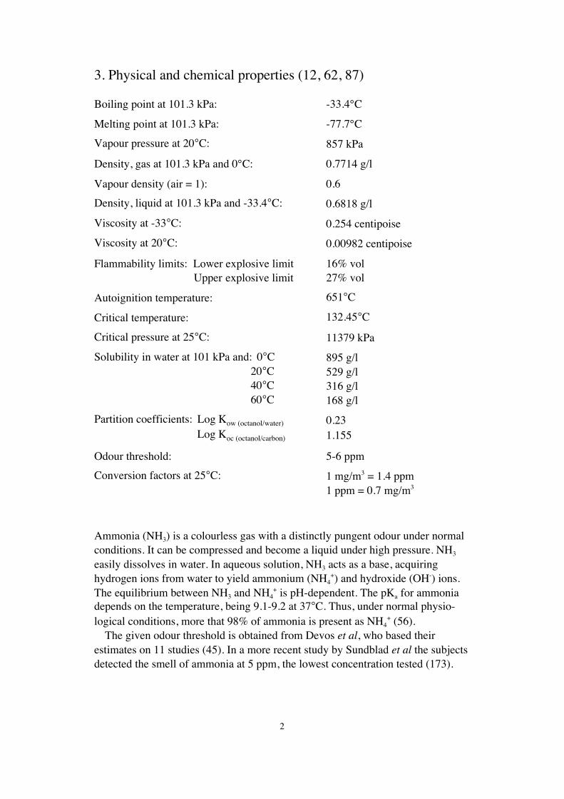
The use of ammonia in the Nordic countries as reported in the chemical productdata base “Substances in Preparation in Nordic Countries” (SPIN), both as totalsand for different purposes, is shown in Table 1.
Ammonia is one of the by-products of protein metabolism, and it is normallyfound in the blood of healthy human subjects at levels below 0.05 mM (41) andin saliva at concentrations of 2.5 mM (84). The steady-state level of ammoniain liver has been reported to be 0.7 mM (151). The concentration of urinaryammonia ranges from 8 to 80 mM in healthy people or approximately 1 000-fold higher than in serum ammonia (95). Muscular activity results in ammoniarelease and blood and urinary levels may increase after exercise (135). Estimatedammonia production from various substrates in the human intestines ranges from10 mg/day in the duodenum to 3 080 mg/day in the colon and faecal contents.
4
Table 1. Annual ammonia consumption in the Nordic countries as reported for 2002 inthe chemical product data base “Substances in Preparation in Nordic Countries” (SPIN)(170).
Country Use category Tonnes
Finland Total 374 120
Sweden Total 209 996Raw materials 114 889pH-regulating agents 862Fertilisers 381Galvano-technical agents 344Metal-staining agents 162
Norway Total 160 141Raw materials and intermediate products 160 105Cleaning and washing agents 29
Denmark Total 11 311
Nearly all the ammonia produced in the intestines is absorbed (172). Somedrugs and chemicals may affect liver and kidney functions and cause an increaseof serum ammonia levels. This is seen, for example, after valproate therapy inpatients with epilepsy (186) or after exposure to ethylene glycol ethers (103).
5. Measurements and analysis of workplace exposure
The most widely used technique for the sampling of ammonia in workshop air isan impinger flask containing diluted sulphuric acid. Samples are then analysedwith a gas selective electrode (137). Another possibility is Nesslerisation inwhich ammonia reacts in dilute sulphuric or boric acid with an alkaline mercuricand potassium iodide solution to form a brown complex. The concentration isobtained by spectrophotometry, with the absorbance value at 440 nm beingcompared with a standard (6). A more sophisticated way to analyse ammonia isby ion chromatography (73). Also direct-reading instruments based on infraredspectroscopy have been shown suitable for measuring ammonia in air. Detectortubes specific for ammonia are available for measurements in the range 1-17 000ppm (119).
Ammonia may be present in air in both the vapour and particulate phaseas ammonia gas and as ammonium salts. In order to avoid interactions fromammonium salts and to separate the particulate phase, the use of filter packs orsampling tubes coated with a selective adsorbent is recommended (98). Thegaseous ammonia is trapped by acids that act as adsorbents on the coated filteror sampling tube. Examples of methods used for determination of ammonia inair are given in Table 2. The recovery is over 95% for most methods.

5
Table 2. Sampling and analytical methods for determining ammonia in air. Adaptedfrom ATSDR (12).
Preparation method Analytical method Detection limit(μg NH3/sample)
Ref.
Passive collection using H2SO4 inliquid sorbent badge
NIOSH method 6701,ion chromatography,conductivity detection
1 (138)
Air samples (stack emissions) collectedthrough an in-stack filter and thenbubbled through H2SO4
EPA method 30,ion chromatography
1 (52)
Prefilter may be used. Ammoniatrapped on H2SO4 - treated silica gel
NIOSH method 6015, colorimetricdetermination of indophenol byvisible light spectrophotometry
0.5 (139)
Prefilter may be used. Ammoniatrapped on H2SO4 - treated silica gel
NIOSH method 6016,ion chromatography
2 (140)
Chromatomembrane cells pre-extractand preconcentrate sample
Ion chromatography withconductivity detection
6 (54)
Collection in H2SO4 - coated activatedcarbon beads in sampling tube
OSHA method ID-188,ion chromatography
2 (19)
Known volume of air drawn throughprefilter and H2SO4 - treated silica gel
NIOSH method S347,ammonia-specific electrode
Notreported
(171)
In biological samples like blood, plasma, and serum, ammonia is present mainlyas ammonium ion. Therefore, the analysis starts with liberation of ammonia bydistillation, aeration, ion-exchange chromatography, microdiffusion, or depro-teinisation. Ammonia in urine has been measured by Nesslerisation, enzymaticassays, and chromatographic methods (83). Direct-reading blood ammoniachecker based on gas specific potentiometry has been used to assay ammonia inbiological samples (119, 155). Some examples of methods used for ammoniadetermination in biological samples are given in Table 3. Sample detection limitswere not reported in any source.
Table 3. Analytical methods for determining ammonia in biological samples. Adaptedfrom ATSDR (12).
Sample matrix Analytical method Reference
Urine Colorimetric (Berthelot reaction) (176)
Urine Indophenol reaction (83)
Urine Glutamate dehydrogenase based auto-analyser method (95)
Saliva Membrane based ammonia-selective electrode (83)
Serum, plasma, whole blood Colorimetric assay based on indophenol production (83)
Serum, plasma, whole blood Titration (83)
Serum, plasma, whole blood Membrane based ammonia-selective electrode (130)

6
6. Occupational exposure data
Occupational exposure to ammonia occurs primarily via inhalation. Dermalexposure may also occur due to splashes and spills during handling of aqueoussolutions. Only in accidental situations may ammonia be swallowed. Ammoniaconcentrations have been measured at different work-sites from agriculture(animal production) to process work in factories as well as in blue-line printingshops (Table 4).
Table 4. Occupational exposure levels of ammonia at different work-sites. Mostmeasurements were personal samples.
Work-site Country Exposure levels Reference
mg/m3 ppm
Hairdressing salon Finland 1.4 – 3.5 2.0 – 5.0 (109)The Netherlands 0.02 – 0.43 0.03 – 0.62 (184)
Blue-line printing shop USA 0.7 – 28 1.0 – 40 (181)
Swine confinement buildings USA 2.3 – 17.5 3.3 – 25.0 (47)United Kingdom 1.0 – 9.2 1.5 – 13.2 (39)Finland 0.7 – 23.8 1.0 – 34.0 (116)Taiwan < 3.5 < 5.0 (31)The Netherlands 0.1 – 17.3 0.2 – 24.7 (74)The Netherlands 0.2 – 2.9 0.3 – 4.2 (152)
Sodium carbonate production USA 6.4 ± 1.0a 9.2 ± 1.4a (81)
Animal facility (mouse rooms) USA 0.07 – 1.0 0.1 – 1.4 (90)
Poultry house Iran 23.2 ± 3.6b 33.2 ± 5.2b (65) Cage-laying house Finland 6.2 – 55.2 8.8 – 78.9 (119) Cage-laying house Finland 2.1 – 27.9 3.0 – 39.8 (121) Floor-laying house Finland 20.1 – 40.3 28.7 – 57.6 (121) Barn systems United Kingdom 7.7 11 (195) Cage systems United Kingdom 4.9 7 (195)
Agricultural slurry stores United Kingdom 0 – 2.7 0 – 3.8 (71)
Municipal sewage plants Finland 0.007 – 3.48 0.01 – 4.97 (92)
Indoor air (during painting) Sweden 0.3 – 2.7 0.4 – 3.9 (141)Finland 0.0007 – 0.052 0.001 – 0.075 (180)
Effluent treatment plant of pulp mill USA 0.07 – 20.3 1.0 – 29.0 (68)
a mean ± standard error of mean.b mean ± standard deviation.

7
The International Programme on Chemical Safety (IPCS) presents the highestoccupational ammonia exposures at mildew-proofing (122.5 ppm), electroplating(53.9 ppm), galvanising (9.8-86.2 ppm), and chemical mixing (58.8-431.2 ppm)(87). In agricultural settings, the ammonia concentrations seem to be higher inpoultry houses (mean 1.6-29.6 ppm, range 1.6-72.9) than in cow houses (mean0.3-7.7 ppm, range 0.1-29.6), and possibly also in swine houses (4.3-20.8 ppm,range 0.23-59.8) (145).
Ammonia seems to be an indoor air pollutant, although it has not been reliablyconfirmed whether the measured compound is in fact ammonia or amine degrada-tion products from protein-containing gluing material. Ammonia concentrationsin indoor air samples vary from 10 to 110 μg/m3 (14-154 ppb) as measured inFinnish residencies (28, 180, 187). In Croatia, in the vicinity of a fertiliser plant,indoor air ammonia concentrations ranged from 32 to 352 μg/m3 (45-493 ppb)while ambient air ammonia concentrations were of the same order of magnitude,from 4 to 420 μg/m3 (6-590 ppb) (66).
Concentrations may be unpredictably high under accidental circumstanceswhere ammonia gas is released into air (131, 174). Because of the sudden natureof accidents only retrospective estimates of exposure levels are available in theliterature. There is one estimate of an exposure level, a report of a fatality at aconcentration of approximately 10 000 ppm (134). In this case, a victim wasfilling a tank wagon with a 25% ammonia solution. Later, it was estimated thatthe ammonia concentrations may have reached 330 000 ppm, at least sporadically(131).
7. Toxicokinetics
7.1 Uptake
Absorption of ammonia is strongly pH dependent. At higher pH, ammonia ispresent as a gaseous, relatively lipophilic molecule (NH3), which readily diffusesthrough cellular and intracellular membranes. At lower pH, ammonia exists as anion (NH4
+), with ionic radius and properties similar to that of the potassium ion(K+). The NH4
+ ion, like K+, can only be transported across membranes by carrier-mediated processes (38). The ionised form predominates (more than 98%) atphysiological pH.
The primary site of absorption is the upper respiratory tract. However, in caseof aerosols in the air and high humidity, ammonia can adsorb onto the aerosolsand be carried deeper into the lungs. Ammonia is probably absorbed percutane-ously, if high concentrations are spilt on intact skin and have caused skin injury(62). Absorption through the eye has been reported. Ammonia diffused withinseconds into cornea, lens, drainage system, and retina. However, the amountsabsorbed were not quantified, and absorption into systemic circulation was notinvestigated (16, 89 cited in 12).
8
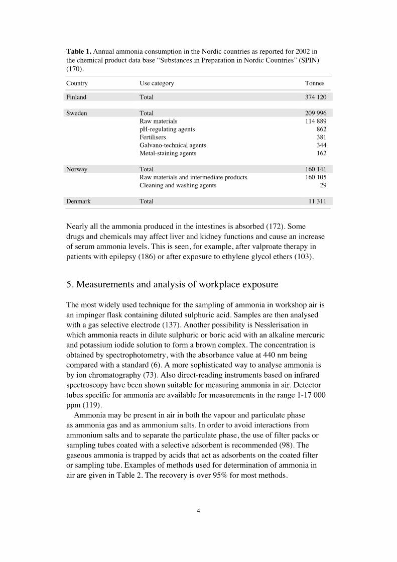
The knowledge of the toxicokinetics of ammonia is limited and informationis primarily available from older human experimental studies. When sevenvolunteers were exposed to an ammonia concentration of 350 mg/m3 (500 ppm)for 30 minutes the retention was around 75%. The ammonia retention decreasedprogressively with time, reaching 23% at steady-state. No effect on blood-nitrogenwas seen (166). In a study by Landahl and Herman two male volunteers wereexposed to ammonia concentrations ranging from 40 to 350 mg/m3 (57-500 ppm)for a maximum of 2 minutes. About 92% retention was reported and the exposurelevel did not affect the retention (104). It is estimated in the IPCS document thatexposure to 25 ppm ammonia would raise the blood ammonia concentration byonly 10% over fasting levels assuming 30% retention (87). This slight increase isevaluated to be well within the normal human capacity to handle ammonia and isunlikely to cause any harm (174).
In healthy subjects, absorbed ammonia is rapidly catabolised by the liver, mainlyto urea. Only relatively small amounts reach the systemic circulation from thegastrointestinal tract as a consequence of this first pass effect (172).
Ammonia absorbed from the intestinal tract arises primarily from bacterialdegradation of amino and nucleic acids in ingested food, endogenous epithelialdebris, and mucosal cell luminal secretions, or from the hydrolysis of ureadiffusing from the systemic circulation into the intestinal tract. Ammonia uptakefrom the human colon, the major site of ammonia production, increases withincreasing pH of the luminal contents. An increase in pH raised the proportionof non-ionised ammonia, showing that most of the ammonia transport relies onpassive diffusion. However, ammonia transport, although greatly diminished, stilloccurred when the luminal pH was reduced to 5, suggesting an active transportmechanism for the ammonium ion (29).
In a study of cerebral uptake of labelled ammonia in Rhesus monkeys it wasfound that after passage of the bolus, a fraction of 40% remained in the brain(148). The brain content of tracer remained almost stable with a half-time of 45minutes. Similar observations were reported in man albeit with a half-time of2.3 hours (149).
In rats exposed to ammonia via inhalation 6 hours/day for 5 days, venousblood ammonia increased linearly from a baseline value of 35 ± 18 mM(mean ± standard deviation) to 44 ± 18 mM at 25 ppm and to 105 ± 14 mM at300 ppm. However, in rats exposed to the same concentrations for 10 or 15 daysthe relationship between dose and blood ammonia level was lost, suggestingmetabolic adaptation (120). Rats were continuously exposed to ammoniaconcentrations of 15, 32, 310, or 1 157 ppm for 24 hours and blood ammoniaconcentration was measured 0, 8, 12 and 24 hours after exposure began. Theblood ammonia level increased significantly in a linear fashion with increasingexposure after 8 hours of exposure. The levels declined over time, indicating anincreased ammonia metabolism (160).

9
7.2 Distribution
The distribution of ammonia between body compartments is strongly influencedby pH. The non-ionised ammonia is freely diffusible, whereas the ammoniumion is less diffusible and relatively confined in compartments (62). Ammoniumions compete with potassium ions for inward transport over the cytoplasmicmembrane, via potassium transport proteins like the Na+/K+-ATPase and theNa+K+2Cl--cotransporter (124).
Ammonia enters the brain from blood by diffusion rather than via a saturabletransport system. It has been estimated that up to 25% of ammonia may enterthe brain as ammonium ion at physiological pH values. The blood transit timethrough brain is in the order of seconds, and the NH4
+ to NH3 conversion rate istoo rapid to limit the rate at which ammonia enters the brain. The lower permea-bility of the blood-brain barrier to NH4
+ implies that transfer of ammonia isdependent upon arterial blood pH and systemic alkalosis exacerbates ammoniatoxicity (146, 192). This is consistent with a higher rate of diffusion of NH3 intobrain at higher blood pH values (56).
Since diffusion of ammonia into the brain is pH dependent, the pH gradientbetween blood and brain may affect brain ammonia concentrations (56).Assuming a blood pH of 7.4 and a brain intracellular pH of 7.1 under normalphysiological conditions, the Hendersson-Hasselbach equation predicts a ratio ofbrain to blood ammonia concentrations of 2 (38). Experimental ratios range from1.5 to 3.0. In hyperammonaemia, the ratio may rise to even 8 (27, 56). In chronicliver failure, prior to the onset of encephalopathy, blood ammonia concentrationsare increased by three-fold in both experimental animals and humans (115) andbrain concentrations are in the 0.3-0.5 mM range (64). The steady-state level ofammonia in the liver of healthy subjects is about 0.7 mM and about ten timeslower in blood plasma (151).
7.3 Endogenous ammonia
In humans and several other species ammonia plays a central role in nitrogenmetabolism (Figure 1).
Ammonia is both a product of protein and nucleic acid catabolism, and a pre-cursor for non-essential amino acids and certain other nitrogenous compounds.The liver is the major site of ammonia metabolism.

10
Figure 1. Exogenous and endogenous sources of ammonia in vertebrates. Adapted fromSeiler (161).
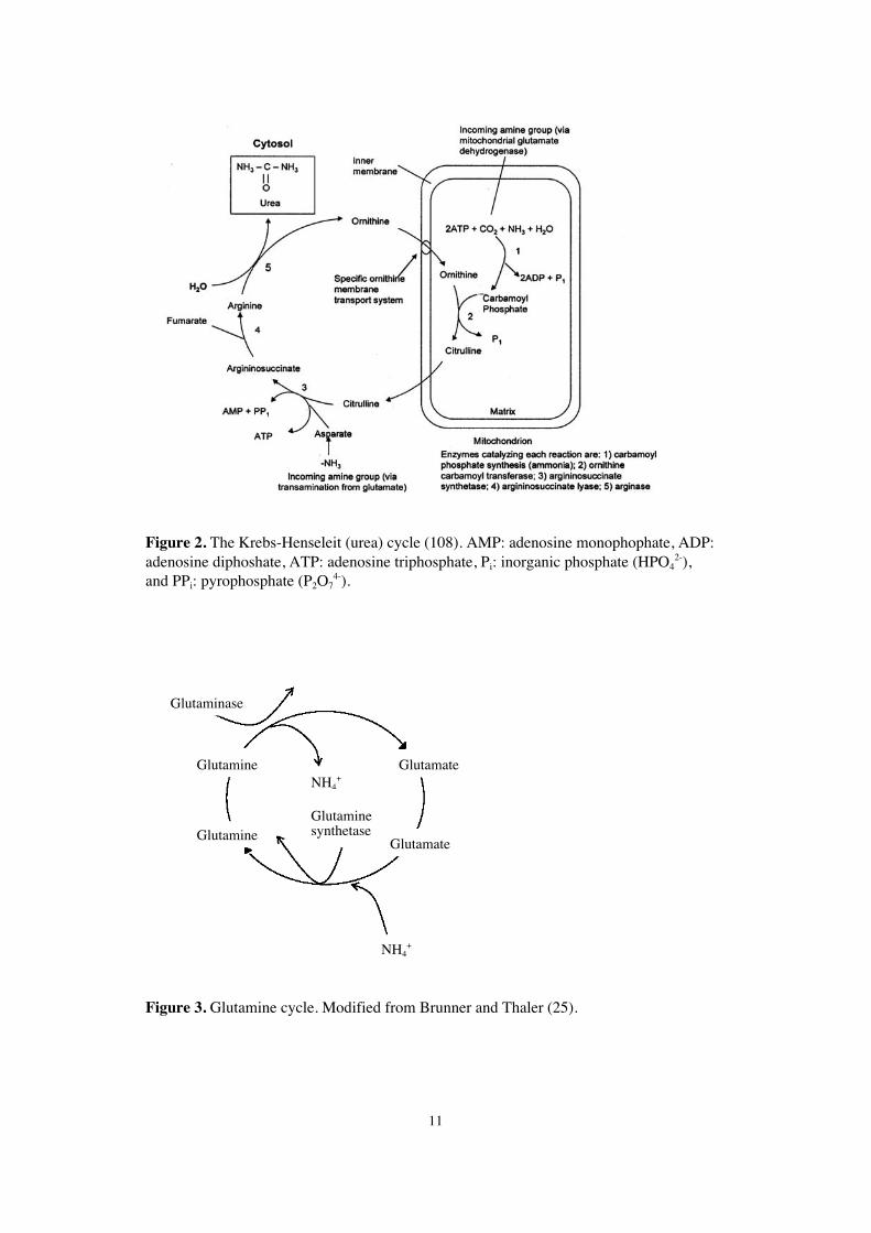
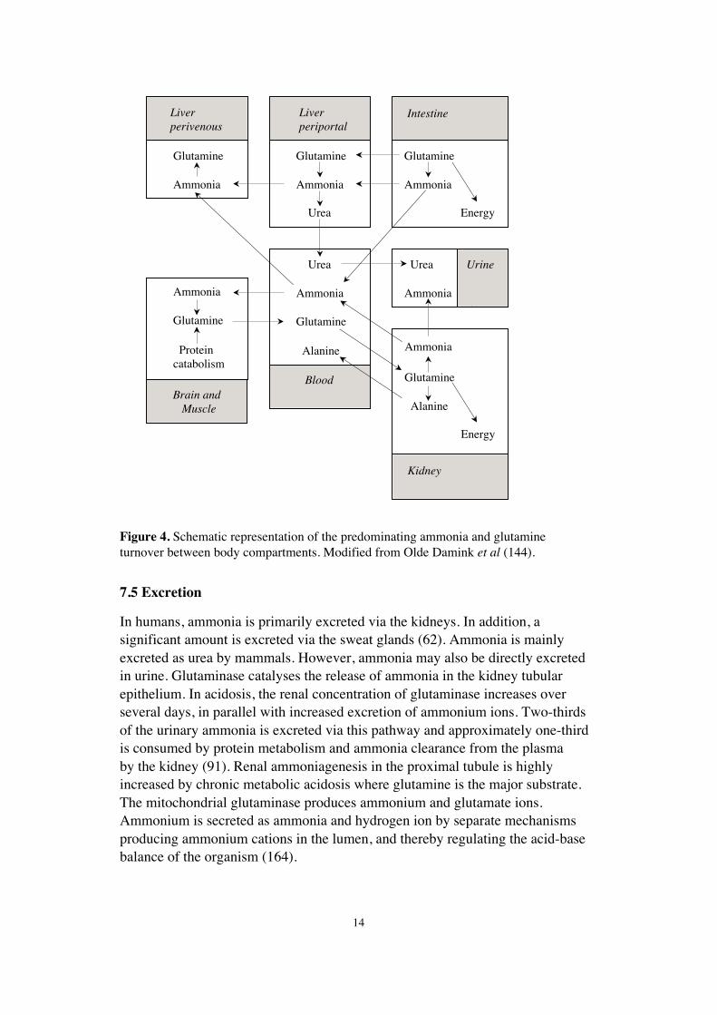
The major features of nitrogen in the body include:– release of nitrogen from amino acids, nucleic acids, and amines,– deamination of glutamate through the action of glutamate dehydrogenase,– conversion of ammonia to urea by the Krebs-Henseleit (urea) cycle (Figure 2),– conversion of urea to ammonia in the gastrointestinal tract by the action of
bacterial urease, and– synthesis of glutamine serving as a short-term storage and transport form of
ammonia in the glutamine cycle (Figure 3).
The Krebs-Henseleit (urea) cycle is tightly controlled to dispose approximately90% of the surplus nitrogen (Figure 2).
Glutamine is quantitatively the second major product of hepatic ammoniametabolism and serves an additional important function in the storage andtransport of ammonia (Figure 3). The synthesis of glutamine also provides adetoxification mechanism for ammonia in the brain through a single enzymaticstep, i.e. via the glutamine synthetase catalysed reaction localised in astrocytes(38). The two nitrogens of glutamine are the major ammonia precursors forammoniagenesis in the kidney tubular cells of most vertebrates. The ammoniaformed in this way enters urine and forms ammonium ions there (151).
Deamination ofaminopurines andaminopyrimidines
Oxidative deaminationof primary amines
Degradation of amino acids(glycine, glutamine, etc.)
Degradation ofhexosamines
Hydrolysis of proteinamido groups
Bacterial infections ofurinary tract (ureas)
Gastrointestinal tract(bacteria, proteins, etc.)
EXOGENOUS SOURCES
ENDOGENOUS SOURCES
AMMONIA
11
Figure 2. The Krebs-Henseleit (urea) cycle (108). AMP: adenosine monophophate, ADP:adenosine diphoshate, ATP: adenosine triphosphate, Pi: inorganic phosphate (HPO4
2-),and PPi: pyrophosphate (P2O7
4-).
Figure 3. Glutamine cycle. Modified from Brunner and Thaler (25).
Glutamate
Glutamate
NH4+
NH4+
Glutaminesynthetase
Glutaminase
Glutamine
Glutamine

12
Table 5. Arterial blood ammonia concentrations in healthy volunteers and in patientswith liver disease (144).
Subjects Arterial ammoniaconcentration (mM)
Reference
Healthy volunteers 0.045 (33)
Patients with chronic liver failure and proven cirrhosis 0.060 (33)
Cirrhotic patients with transjugular intrahepatic portosystemicstent shunta
0.080 (156)
Patients with acute-on-chronic liver disease 0.090-0.120 (33)
Patients with acute liver failure 0.150-0.180 (33)
Patients with end stage acute liver failure 0.340 (88)
aA non-surgical technique for treatment of refractory ascites associated with cirrhosis of the liver.
Mutch and Bannister reviewed the relationship between muscle activity andammonia production. The immediate source of ammonia from muscle appearsto be a result of the deamination of adenosine monophosphate, which is moreapparent in fast twitch fibres than in slow twitch fibres. An increase of bloodammonia levels both in rats after swimming and in humans after manual work,maximal cycle ergometry, and treadmill exercise is observed (135). In a morerecent study, heat stress and exercise increased plasma ammonia from preexerciselevel to 0.06-0.07 mM at the end of submaximal runs, and further to 0.11 mM atthe end of performance runs in hot conditions (123). The accumulation of plasmaammonia following sprint exercise is about 35% lower in women than in men(55).
Arterial blood ammonia concentrations in healthy volunteers and in patientswith liver disease are shown in Table 5.
A variety of xenobiotics (methanol, formic acid, cyanide, 2-ethyl-hexanoicacid, valproate, and acetaminophen) may impair liver function resulting inincreased blood and urine ammonia level (72, 112, 113, 122, 153). Elevatedammonia levels may also be a result from xenobiotics affecting the kidneyfunction. This is seen, for example, after valproate therapy in patients withepilepsy (186) or after exposure to ethylene glycol ethers (103).
Congenital deficiency of enzymes in the urea cycle, such as carbamoyl phos-phate synthetase I and, to a lesser extent, ornithine transcarbamylase, as well asseveral other metabolic disorders like arginosuccinic aciduria may lead tohyperammonaemia and various abnormal urinary constituents (10, 82, 162).
7.4 Biotransformation
Exogenous ammonia, administered intravenously as an ammonium compound,is metabolised to glutamine as the major early product (51). Following admini-stration of 13N-ammonia to rats (via either the carotid artery or cerebrospinalfluid), most of the metabolised label was recovered in glutamine (amide) andminor fractions in glutamate and aspartate. Although ammonia in blood and
13
cerebrospinal fluid are converted largely to glutamine, it is not possible to predictwith certainty the metabolic fate of the bulk of endogenously produced ammonia(36). The ammonia fixed in glutamine may eventually end up in amino acids,purines, pyrimidines, or other nitrogen-containing compounds.
Ingested ammonium chloride and endogenous intestinal ammonia entersthe liver via the portal vein and is converted to urea (59, 67, 150). In humans,absorbed ammonia (both exogenous and endogenous) is converted to theammonium ion as hydroxide and as salts, especially as carbonates. Theammonium salts are then rapidly converted to urea. Protein deamination in thebody yields ammonium ions, which are rapidly converted into urea in the liverand excreted by the kidney or used for synthesis of amino acids. Ammoniumions are produced in the kidney in order to maintain electrolyte balance (62).
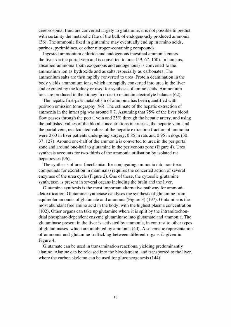
The hepatic first-pass metabolism of ammonia has been quantified withpositron emission tomography (96). The estimate of the hepatic extraction ofammonia in the intact pig was around 0.7. Assuming that 75% of the liver bloodflow passes through the portal vein and 25% through the hepatic artery, and usingthe published values of the blood concentrations in arteries, the hepatic vein, andthe portal vein, recalculated values of the hepatic extraction fraction of ammoniawere 0.60 in liver patients undergoing surgery, 0.85 in rats and 0.95 in dogs (30,37, 127). Around one-half of the ammonia is converted to urea in the periportalzone and around one-half to glutamine in the perivenous zone (Figure 4). Ureasynthesis accounts for two-thirds of the ammonia utilisation by isolated rathepatocytes (96).
The synthesis of urea (mechanism for conjugating ammonia into non-toxiccompounds for excretion in mammals) requires the concerted action of severalenzymes of the urea cycle (Figure 2). One of these, the cytosolic glutaminesynthetase, is present in several organs including the brain and the liver.
Glutamine synthesis is the most important alternative pathway for ammoniadetoxification. Glutamine synthetase catalyses the synthesis of glutamine fromequimolar amounts of glutamate and ammonia (Figure 3) (197). Glutamine is themost abundant free amino acid in the body, with the highest plasma concentration(102). Other organs can take up glutamine where it is split by the intramitochon-drial phosphate-dependent enzyme glutaminase into glutamate and ammonia. Theglutaminase present in the liver is activated by ammonia, in contrast to other typesof glutaminases, which are inhibited by ammonia (40). A schematic representationof ammonia and glutamine trafficking between different organs is given inFigure 4.
Glutamate can be used in transamination reactions, yielding predominantlyalanine. Alanine can be released into the bloodstream, and transported to the liver,where the carbon skeleton can be used for gluconeogenesis (144).
14
Figure 4. Schematic representation of the predominating ammonia and glutamineturnover between body compartments. Modified from Olde Damink et al (144).
7.5 Excretion
In humans, ammonia is primarily excreted via the kidneys. In addition, asignificant amount is excreted via the sweat glands (62). Ammonia is mainlyexcreted as urea by mammals. However, ammonia may also be directly excretedin urine. Glutaminase catalyses the release of ammonia in the kidney tubularepithelium. In acidosis, the renal concentration of glutaminase increases overseveral days, in parallel with increased excretion of ammonium ions. Two-thirdsof the urinary ammonia is excreted via this pathway and approximately one-thirdis consumed by protein metabolism and ammonia clearance from the plasmaby the kidney (91). Renal ammoniagenesis in the proximal tubule is highlyincreased by chronic metabolic acidosis where glutamine is the major substrate.The mitochondrial glutaminase produces ammonium and glutamate ions.Ammonium is secreted as ammonia and hydrogen ion by separate mechanismsproducing ammonium cations in the lumen, and thereby regulating the acid-basebalance of the organism (164).
Liver perivenous
Liver periportal
Glutamine
Ammonia
Urea
Glutamine
Ammonia
Energy
Intestine
Urea
Ammonia
Urine
Glutamine
Ammonia
Urea
Ammonia
Glutamine
Alanine Ammonia
Glutamine
Alanine
Energy
Kidney
Blood
Ammonia
Glutamine
Protein catabolism
Brain and Muscle
15
Ammonia may also be excreted through expired air. Reported levels of ammoniain expired air are 0.1-2.2 mg/m3 (0.15-3.1 ppm) (85) and 0.2-1.2 mg/m3 (0.3-1.7ppm) (105). These values, higher than those expected from equilibrium withplasma- and lung-parenchyma- ammonia levels (0.03-0.05 mg/m3) (0.04-0.07ppm), are most likely due to the synthesis of ammonia from salivary urea by theoral microflora (62).
Less than 1% of the total ammonia produced in the human intestinal tract(4 g/day) is excreted in the faeces (172).
8. Biological monitoring
Ammonia is endogenously produced and is present in all body fluids. Thegenerally accepted reference level used in clinical laboratories for blood ammoniaof healthy subjects is 0.05 mM although lower levels of 0.005-0.007 mM havealso been suggested (41). In unexposed healthy subjects the urinary ammonialevel is reported to be below 1.4 mol/mol creatinine corresponding to about 10mM (112). In clinical chemistry, urinary ammonia levels from 8 to 80 mM areregarded as normal values (95). The mean ammonia concentration in saliva ofhealthy subjects is 2.5 mM (84).
Rats exposed to ammonia vapour showed dose-dependent blood ammonialevels after 5 days of exposure (6 hours per day). However, blood ammoniaconcentrations had returned to baseline levels after 10 and 15 days of continuedexposure (120). In rats continuously exposed to ammonia for 24 hours the level inblood declined over time indicating increased ammonia metabolism during theexposure (160).
There are no reports of increased urinary ammonia excretion after occupationalexposure. Inhalation by volunteers of 500 ppm ammonia for 30 minutes did nothave any effect on blood and urine nitrogen levels (166). It has been estimatedthat exposure to 25 ppm ammonia increases the blood ammonia concentration byonly 10% over fasting levels, assuming 30% retention (87). However, ammoniumconcentrations in blood and urine may be assayed for clinical changes caused byammonium salts and other chemicals affecting the urea cycle like acetaminophen,valproate, 2-ethylhexanoic acid, and formic acid (72, 79, 101, 113).
Due to relatively high endogenous production and adaptive metabolism, bio-monitoring of occupational exposure to ammonia seems of little value.
16
9. Mechanisms of toxicity
Ammonia as a gas, in anhydrous form or in concentrated solutions, possessescorrosive properties, and massive exposure leads to necrosis of skin and mucousmembranes. Ammonia may also cause sensory airway irritation as the trigeminalnerve is affected (131, 174).
The topical damages caused by ammonia are mainly due to its alkaline proper-ties. Because of the high water solubility ammonia dissolves in moisture onthe mucous membranes, skin, and eyes, forming ammonium hydroxide, whichcause a liquefaction necrosis of the tissues (89). Ammonium hydroxide increasessaponification of cell membrane lipids resulting in cell disruption and death.Further, it breaks down cell structural proteins, extracts water from the cells, andinitiates an inflammatory response, which damages the adjoining tissues. Thisreaction is exothermic contributing to tissue damage by cryogenic (thermal) injuryin addition to the alkali burns (7, 9).
The most serious effect of elevated endogenous ammonia is seen in the brainas hepatic encephalopathy due to acute or chronic liver failure. Ammonia ismetabolised by glutamine synthase localised in astrocytes. Astrocytes are alsoinvolved in the uptake of glutamate, an endogenous and most abundant cerebralneurotransmitter that is the primary agonist for N-methyl-D-aspartate (NMDA)receptors (142). Thus, the NMDA receptor is also a primary target of ammoniatoxicity. The ammonium ion is transported into the cell via the binding sites forK+ because of similar ionic radius of hydrated ammonium and K+ (124).
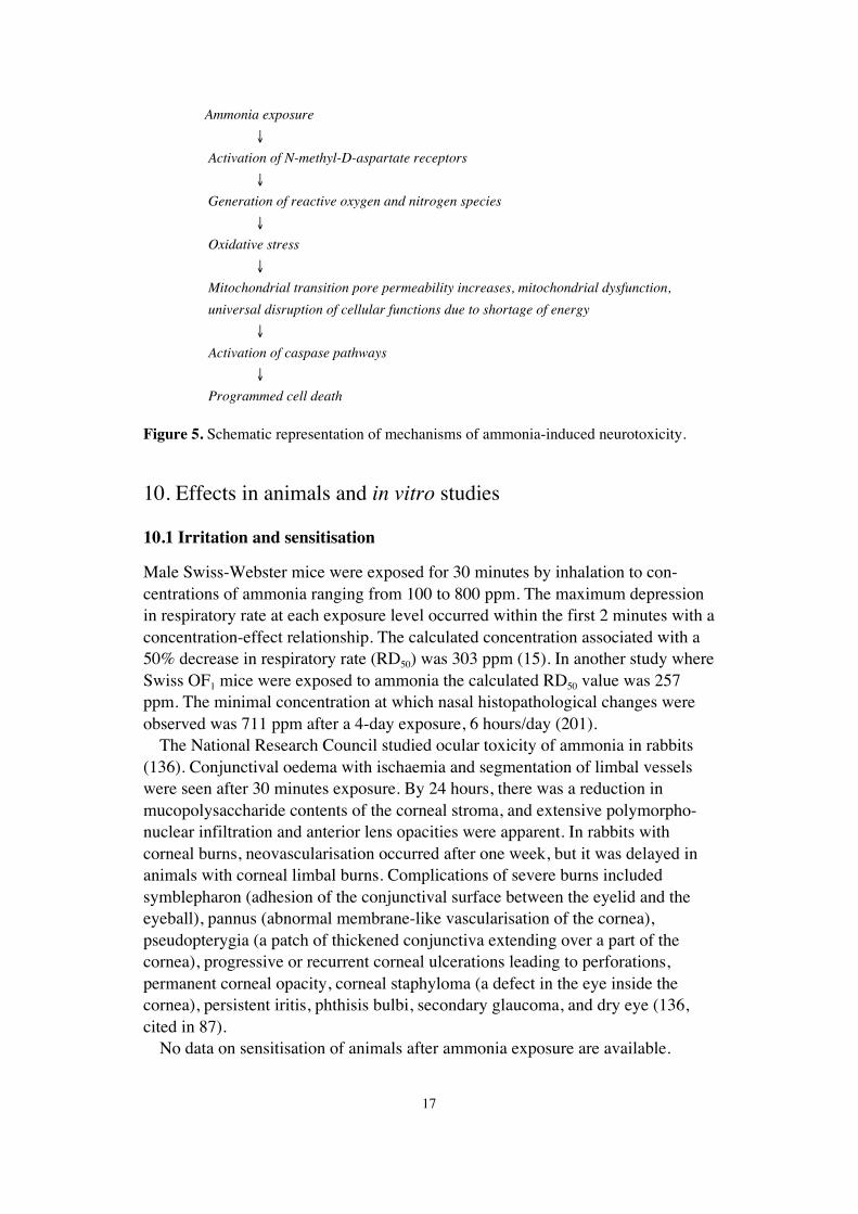
Ammonium neurotoxicity is thus mediated through the activation of NMDAreceptors, increased activity of constitutive neuronal nitric oxide synthase, andsubsequently increased formation of nitric oxide that serves as a neurotransmitterin a number of neuronal cells (56, 125). Nitric oxide in turn activates guanylatecyclase leading to increased formation of cyclic guanosine monophosphate (77).A key effect of ammonia at the cellular level is oxidative stress due to thegeneration of reactive oxygen and nitrogen species subsequent to activation ofNMDA receptors. These events are associated with mitochondrial dysfunctioncharacterised by increased permeability of mitochondrial transition pore. Theopening of the transition pore is associated with cytochrome C release andactivation of a cascade of serinine-threonine proteases, also called caspases.Simultaneous decrease of protein kinase C activity is associated with decreasedphosphorylation and activity, of Na+/K+-ATPase, depletion of ATP (99), andincreased levels of free intracellular calcium, toxic to neuronal cells (100). Theseevents are key-elements in the pathway leading to ammonia-induced programmedcell death, apoptosis (143). Key-events of ammonia-induced effects on neuronalcells are depicted in Figure 5.
17
Ammonia exposure
↓
Activation of N-methyl-D-aspartate receptors
↓
Generation of reactive oxygen and nitrogen species
↓
Oxidative stress
↓
Mitochondrial transition pore permeability increases, mitochondrial dysfunction,
universal disruption of cellular functions due to shortage of energy
↓
Activation of caspase pathways
↓
Programmed cell death
Figure 5. Schematic representation of mechanisms of ammonia-induced neurotoxicity.
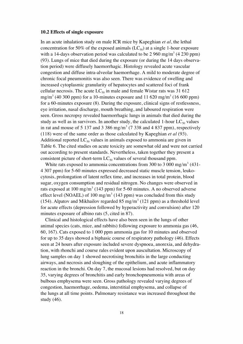
10. Effects in animals and in vitro studies
10.1 Irritation and sensitisation
Male Swiss-Webster mice were exposed for 30 minutes by inhalation to con-centrations of ammonia ranging from 100 to 800 ppm. The maximum depressionin respiratory rate at each exposure level occurred within the first 2 minutes with aconcentration-effect relationship. The calculated concentration associated with a50% decrease in respiratory rate (RD50) was 303 ppm (15). In another study whereSwiss OF1 mice were exposed to ammonia the calculated RD50 value was 257ppm. The minimal concentration at which nasal histopathological changes wereobserved was 711 ppm after a 4-day exposure, 6 hours/day (201).
The National Research Council studied ocular toxicity of ammonia in rabbits(136). Conjunctival oedema with ischaemia and segmentation of limbal vesselswere seen after 30 minutes exposure. By 24 hours, there was a reduction inmucopolysaccharide contents of the corneal stroma, and extensive polymorpho-nuclear infiltration and anterior lens opacities were apparent. In rabbits withcorneal burns, neovascularisation occurred after one week, but it was delayed inanimals with corneal limbal burns. Complications of severe burns includedsymblepharon (adhesion of the conjunctival surface between the eyelid and theeyeball), pannus (abnormal membrane-like vascularisation of the cornea),pseudopterygia (a patch of thickened conjunctiva extending over a part of thecornea), progressive or recurrent corneal ulcerations leading to perforations,permanent corneal opacity, corneal staphyloma (a defect in the eye inside thecornea), persistent iritis, phthisis bulbi, secondary glaucoma, and dry eye (136,cited in 87).
No data on sensitisation of animals after ammonia exposure are available.
18
10.2 Effects of single exposure
In an acute inhalation study on male ICR mice by Kapeghian et al, the lethalconcentration for 50% of the exposed animals (LC50) at a single 1-hour exposurewith a 14-days observation period was calculated to be 2 960 mg/m3 (4 230 ppm)(93). Lungs of mice that died during the exposure (or during the 14 days observa-tion period) were diffusely haemorrhagic. Histology revealed acute vascularcongestion and diffuse intra-alveolar haemorrhage. A mild to moderate degree ofchronic focal pneumonitis was also seen. There was evidence of swelling andincreased cytoplasmic granularity of hepatocytes and scattered foci of frankcellular necrosis. The acute LC50 in male and female Wistar rats was 31 612mg/m3 (40 300 ppm) for a 10-minutes exposure and 11 620 mg/m3 (16 600 ppm)for a 60-minutes exposure (8). During the exposure, clinical signs of restlessness,eye irritation, nasal discharge, mouth breathing, and laboured respiration wereseen. Gross necropsy revealed haemorrhagic lungs in animals that died during thestudy as well as in survivors. In another study, the calculated 1-hour LC50 valuesin rat and mouse of 5 137 and 3 386 mg/m3 (7 338 and 4 837 ppm), respectively(118) were of the same order as those calculated by Kapeghian et al (93).Additional reported LC50 values in animals exposed to ammonia are given inTable 6. The cited studies on acute toxicity are somewhat old and were not carriedout according to present standards. Nevertheless, taken together they present aconsistent picture of short-term LC50 values of several thousand ppm.
White rats exposed to ammonia concentrations from 300 to 3 000 mg/m3 (431-4 307 ppm) for 5-60 minutes expressed decreased static muscle tension, leuko-cytosis, prolongation of latent reflex time, and increases in total protein, bloodsugar, oxygen consumption and residual nitrogen. No changes were observed inrats exposed at 100 mg/m3 (143 ppm) for 5-60 minutes. A no observed adverseeffect level (NOAEL) of 100 mg/m3 (143 ppm) was concluded from this study(154). Alpatov and Mikhailov regarded 85 mg/m3 (121 ppm) as a threshold levelfor acute effects (depression followed by hyperactivity and convulsion) after 120minutes exposure of albino rats (5, cited in 87).
Clinical and histological effects have also been seen in the lungs of otheranimal species (cats, mice, and rabbits) following exposure to ammonia gas (46,60, 167). Cats exposed to 1 000 ppm ammonia gas for 10 minutes and observedfor up to 35 days showed a biphasic course of respiratory pathology (46). Effectsseen at 24 hours after exposure included severe dyspnoea, anorexia, and dehydra-tion, with rhonchi and course rales evident upon auscultation. Microscopy oflung samples on day 1 showed necrotising bronchitis in the large conductingairways, and necrosis and sloughing of the epithelium, and acute inflammatoryreaction in the bronchi. On day 7, the mucosal lesions had resolved, but on day35, varying degrees of bronchitis and early bronchopneumonia with areas ofbulbous emphysema were seen. Gross pathology revealed varying degrees ofcongestion, haemorrhage, oedema, interstitial emphysema, and collapse ofthe lungs at all time points. Pulmonary resistance was increased throughout thestudy (46).

19
Table 6. Reported LC50 values in animals exposed via inhalation.
Species Exposure duration LC50 Reference mg/m3 ppm
White rat 5 min 18 693 26 704 (154)Mouse 10 min 7 060 10 152 (165)Wistar rat 10 min 31 612 40 300 (8)White rat 15 min 12 160 17 372 (154)White rat 30 min 7 035 10 050 (154)ICR mouse 1 h 2 960 4 230 (93)CF1mouse 1 h 3 386 4 837 (118)CFE rat 1 h 5 137 7 338 (118)White rat 1 h 7 939 11 342 (154)Wistar rat 1 h 11 620 16 600 (8)White rat 2 h 7 600 10 860 (4)
LC50: lethal concentration for 50% of the exposed animals at single exposure.
Anaesthetised, mechanically ventilated rabbits exposed to very high levels ofnebulised ammonia (2 ml of 23-27% ammonia solution; estimated by the studyauthors as peak ammonia concentrations of 35 000-39 000 ppm) for 4 minuteshad a decrease in blood oxygen saturation and an increase in airway pressure (ameasure of changes in airway resistance) (167). Arterial oxygen tension decreasedfrom 23.3 ± 3.6 kPa (mean ± standard deviation) to 11.0 ± 3.6 kPa and peakairway pressure increased from 13 ± 2 cm H2O (mean ± standard deviation) to17 ± 2 cm H2O.
Cardiovascular changes have been observed in rabbits exposed to high concen-trations of ammonia for 1 hour (159). Bradycardia was seen at 2 500 ppm, andhypertension and cardiac arrhythmias leading to cardiovascular collapse followedacute exposures to concentrations exceeding 5 000 ppm. Atrophy of pericardial fathas been observed in mice exposed to 4 000 ppm ammonia for 60 minutes (93).
In the LC50 study reported in Table 7, male CFE rats and male CF1 mice wereexposed to different ammonia concentrations for 60 minutes. Immediate nasaland eye irritation was followed by laboured breathing and gasping in all studygroups. In addition, mild changes in the liver were seen at necropsy (118).
Table 7. Dose-effect relationships for 1-hour inhalation exposure in experimental animals(118).
Species Exposure level Effect mg/m3 ppm
CFE rat 6 888 9 840 Liver fatty infiltration in 1/10 survivors5 137 7 338 1-hour LC50
4 347 6 210 No pathological lesions in 10/10 survivorsCF1 mouse 4 004 5 720 Mild congestion in the liver in 1/10 survivors
3 386 4 837 1-hour LC50
2 520 3 600 No pathological lesions in 10/10 survivors
LC50: lethal concentration for 50% of the exposed animals at single exposure.

20
10.3 Effects of short-term exposure
Studies in animals have demonstrated both dose-effect and duration-effectrelationships in changes at the respiratory tract. Acute exposures to lowerammonia concentrations (less than 1 000 ppm) from 1 hour to 1 week causeairway irritation, whereas exposures to high concentrations (4 000 ppm) for3 hours to 2 weeks result in severe damage to the upper and lower respiratorytract and alveolar capillaries (35, 93, 126, 158, 160).
Histopathological changes of the respiratory tract were evaluated in ratscontinuously exposed to a mean ammonia concentration of 200 ppm, range150-250 ppm, for 12 days. Progressive loss of cilia from and stratification of thetracheal epithelial lining was observed. By day 12 a mucilaginous exudate wasapparent in the trachea together with a slight increase in submucosal cellularity(61).
In a 2-month inhalation study on white rats a lowest observed adverse effectlevel (LOAEL) of 100 mg/m3 (143 ppm) was determined based on histologicalchanges in the lungs, including small areas of interstitial pneumonia with signs ofperibronchitis and perivasculitis. No changes were reported in other organs ascompared with the control group. A threshold level for toxic effects of 40 mg/m3
(57 ppm) was reported from this study (5, cited in 87).When rats, rabbits, guinea pigs, dogs and monkeys were continuously exposed
to ammonia at a concentration of 40 mg/m3 (57 ppm) for 114 days, no signs oftoxicity were seen and gross and microscopic examination did not reveal lungabnormalities (35). A NOAEL of 40 mg/m3 (57 ppm) can be concluded from thisstudy.
Coon et al exposed Sprague-Dawley rats continuously by inhalation to 127mg/m3 (181 ppm) and 262 mg/m3 (374 ppm) for 90 days (48 and 49 animals pergroup, respectively) and to 455 mg/m3 (650 ppm) for 65 days (51 animals) (35).The 181 ppm ammonia exposure (NOAEL) did not induce changes in gross ormicroscopic pathology, haematology, or liver histochemistry. The exposure at 374ppm (LOAEL) was without specific effects, but mild nasal discharge was seen in25% of 49 rats. All the 51 rats exposed at 650 ppm showed mild dyspnoea andnasal irritation. There were 32 deaths by day 25, and 50 deaths by day 65 in the650 ppm group. Myocardial fibrosis was seen in rats, guinea pigs, rabbits, dogs,and monkeys after prolonged (90 days) continuous exposure to 470 mg/m3 (671ppm) (35). The contribution of these lesions to the morbidity and mortality ofaffected animals was not determined.
Broderson et al exposed Sherman and Fisher rats to ammonia from naturalsources, at an average concentration of 150 ppm for 75 days, and to purifiedammonia at 250 ppm for 35 days (23). Histological changes in the olfactory andrespiratory epithelia of the nasal cavity were similar in all the exposed rats,showing increased thickness, pyknotic nuclei, and hyperplasia. The submucosawas oedematous with marked dilation of small vessels.
A concentration of 500 ppm was selected in a study of effects of continuousexposure to ammonia, after noting that general toxic effects (particularly on

21
growth rate) were not present at 250-300 ppm (158). Young male specific-pathogen-free rats were age- and weight-matched with controls (27 animals pergroup) and exposed for up to 8 weeks. Nasal irritation began on the 4th day. After3 weeks, exposed rats showed nasal irritation and inflammation of the upperrespiratory tract, but no effects were observed on the bronchioles and alveoli. Thenumber of pulmonary alveolar macrophages was similar to that of controls. After8 weeks, no inflammatory lesions were present.
Swiss mice exposed to 909 ppm ammonia 6 hours/day, 5 days/week for 4-14days expressed histological lesions in the respiratory epithelium in the nasalcavity. These lesions were not seen at 303 ppm. No lesions were observed in thetrachea or lungs at any exposure level (201).
The nasal mucosa was adversely affected in adult male mice exposed to vapoursof a 12% ammonia solution for 15 minutes/day, 6 days/week for 4, 5, 6, 7, or 8weeks (60). Histological changes progressed from weeks 4 to 8 from crowding ofcells forming crypts and irregular arrangements to epithelial hyperplasia, patchesof squamous metaplasia, loss of cilia, and dysplasia of the nasal epithelium.Carcinomas were seen in two animals (see chapter 10.5 for more details).
Animal studies have revealed that ammonia affects the immune system.Exposure of mice to ammonia at a concentration of 500 ppm for one weekfollowed by exposure to Pasteurella multocida at the lethal dose for 50% of theexposed animals (LD50) increased the mortality significantly (158). A significantincrease in the severity of respiratory signs characteristic of murine respiratorymycoplasmosis was observed in rats exposed to ammonia at 25 ppm for 4-6weeks following inoculation with Mycoplasma pulmonis intranasally (23). Guineapigs exposed to 90 ppm for 3 weeks developed a significant decrease in the cell-mediated immune response when challenged with a derivative of tuberculin (175).
Twelve guinea pigs were exposed to an ammonia concentration of about 170ppm, range 140-200 for 6 hours/day, 5 days a week, for up to 18 weeks. Therewere no significant findings at autopsy of animals sacrificed after 6 or 12 weeksof exposure. In animals sacrificed after 18 weeks of exposure, there was conges-tion of the liver, spleen, and kidneys, with early degenerative changes in theadrenal glands. Increased erythrocyte destruction was explained by increasedquantities of haemosiderin in the spleen. In the proximal tubules of the kidneys,there was cloudy swelling of the epithelium and precipitated albumin in the lumenwith some gasts. The cells of the adrenal glands were swollen and the cytoplasmin some areas had lost its normal granular structure (193).
10.4 Mutagenicity and genotoxicity
No studies on mutagenicity and carcinogenicity of ammonia performed accordingto current standards are available. Mutagenicity tests of ammonia have beenperformed in Escherichia coli, chick fibroblast cells, and Drosophila melano-gaster. Positive results were noted in a reverse mutation test in Escherichia coli,but only in treatments using toxic levels of NH4
+ (98% lethality) (43). Slight
22
mutagenic activity was seen also in Drosophila following exposure to ammoniagas, but survival after treatment was less than 2% (114).
Reduced cell division was noted in mouse fibroblasts cultured in media towhich ammonia and ammonia chloride were added (188). The effects were pHindependent. Decreased rate of DNA synthesis in vivo was observed in mousemucosal cells in the ileum and colon when serum NH4
+ levels were significantlyelevated over normal levels. These elevated levels were induced by intraperitonealinjection of urease or infusion of ammonium chloride (200).
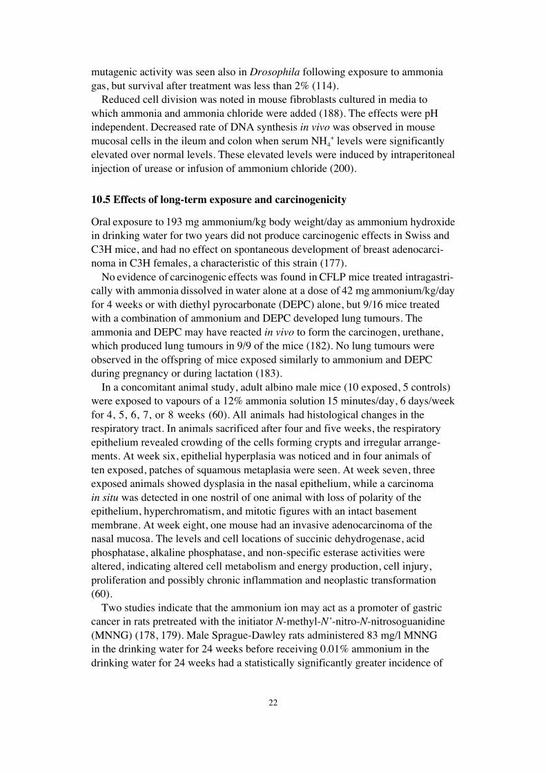
10.5 Effects of long-term exposure and carcinogenicity
Oral exposure to 193 mg ammonium/kg body weight/day as ammonium hydroxidein drinking water for two years did not produce carcinogenic effects in Swiss andC3H mice, and had no effect on spontaneous development of breast adenocarci-noma in C3H females, a characteristic of this strain (177).
No evidence of carcinogenic effects was found in CFLP mice treated intragastri-cally with ammonia dissolved in water alone at a dose of 42 mg ammonium/kg/dayfor 4 weeks or with diethyl pyrocarbonate (DEPC) alone, but 9/16 mice treatedwith a combination of ammonium and DEPC developed lung tumours. Theammonia and DEPC may have reacted in vivo to form the carcinogen, urethane,which produced lung tumours in 9/9 of the mice (182). No lung tumours wereobserved in the offspring of mice exposed similarly to ammonium and DEPCduring pregnancy or during lactation (183).
In a concomitant animal study, adult albino male mice (10 exposed, 5 controls)were exposed to vapours of a 12% ammonia solution 15 minutes/day, 6 days/weekfor 4, 5, 6, 7, or 8 weeks (60). All animals had histological changes in therespiratory tract. In animals sacrificed after four and five weeks, the respiratoryepithelium revealed crowding of the cells forming crypts and irregular arrange-ments. At week six, epithelial hyperplasia was noticed and in four animals often exposed, patches of squamous metaplasia were seen. At week seven, threeexposed animals showed dysplasia in the nasal epithelium, while a carcinomain situ was detected in one nostril of one animal with loss of polarity of theepithelium, hyperchromatism, and mitotic figures with an intact basementmembrane. At week eight, one mouse had an invasive adenocarcinoma of thenasal mucosa. The levels and cell locations of succinic dehydrogenase, acidphosphatase, alkaline phosphatase, and non-specific esterase activities werealtered, indicating altered cell metabolism and energy production, cell injury,proliferation and possibly chronic inflammation and neoplastic transformation(60).
Two studies indicate that the ammonium ion may act as a promoter of gastriccancer in rats pretreated with the initiator N-methyl-N’-nitro-N-nitrosoguanidine(MNNG) (178, 179). Male Sprague-Dawley rats administered 83 mg/l MNNGin the drinking water for 24 weeks before receiving 0.01% ammonium in thedrinking water for 24 weeks had a statistically significantly greater incidence of

23
gastric cancer (70% of rats) and number of tumours per tumour-bearing rat (2.1)than rats receiving only MNNG and tap water (31% and 1.3 tumours/rat) (178).Additionally, the size, depth, and metastasis of the MNNG-initiated tumours wereenhanced by ammonium (179).
These studies suggest that ammonium in the presence of certain other chemicals(i.e. DEPC and MNNG) may contribute to the development of cancer (179, 182).
10.6 Reproductive and developmental studies
No data have been found regarding reproductive and developmental effects inanimals after inhalation exposure to ammonia.
In a study by Miñana et al, Wistar rats were exposed to ammonia from day 1of gestation and through the prenatal and lactation periods via a diet containingammonium acetate (20% by mass) (133). After weaning (at postnatal day 21),the pups were fed a normal diet with no ammonia added. The body weight ofoffspring exposed to ammonia was significantly lower than that of controls, adifference that was still evident one month after cessation of the exposure.Primary cell cultures of cerebellar neurons from 8-day old offspring exhibitedimpairment of the NMDA receptor function, as shown by decreased binding of[3H]MK-801, increased resistance to glutamate and NMDA toxicity, and a lackof increase in aspartate aminotransferase activity when small amounts of NMDAwere added to culture media (133). A study of similar design by Azorin et al, butwith adult male rats, observed significantly lower body weight of the male ratsmaintained on the ammonium diet compared to the controls. Pair feeding showedthat this was due to a combination of lower food intake and lower caloric contentof the ammonium-enriched feed (13). Maternal body weight was not monitoredin the Miñana study, but the results from Azorin and co-workers make it veryprobable, that maternal body weight was reduced during gestation.
Aguilar et al investigated the effect of perinatal hyperammonaemia on activeand passive avoidance behaviour and conditional discrimination learning inmale Wistar rats (2). Pre- and neonatal exposure to ammonia was carried out asdescribed above for Miñana et al (133). However, the exposure to ammonia wascontinued also after weaning, until and during behavioural testing. Animalsexposed to ammonia already during prenatal life exhibited a decreased numberof active avoidances on one of five days of testing, and a decreased step-throughlatency during passive avoidance. These effects were not observed in animals onlyexposed to ammonia during postnatal life, indicating a prenatal component ofammonia related effects. However, it should be noted that exposure to ammoniaduring postnatal life was initiated only two weeks before behavioural testing. Incomparison, exposure of prenatally exposed animals was continued throughoutlactation and some additional weeks until behavioural testing (2).
24
11. Observations in man
11.1 Irritation and sensitisation
Ammonia irritates the upper airways, eyes, and skin in humans. According totwo controlled human exposure studies clear signs of mild respiratory and eyeirritation appear somewhere between 25 and 50 ppm (80, 173) (see chapter 11.2for more details).
Four out of six human subjects described moderate irritation of the nose andeyes when exposed to 50 ppm, but not 30 ppm ammonia gas for 10 minutes (117).When human volunteers were exposed to 100 ppm ammonia in each nostril of thenasal airways for 30 seconds, a significant increase in airway resistance during theexposure period was registered, and 11 out of 23 complained of nasal irritation(128). In another study, human volunteers were exposed to various concentrationsof ammonia vapours for 5 minutes. At the concentration of 134 ppm, severe noseirritation occurred in most subjects. At 72 ppm, several subjects reported the samesymptoms; at 50 ppm, two subjects reported nasal dryness; and at 32 ppm, onesubject reported nasal dryness (97, industrial report).
Ammonia has a greater tendency than other alkalis to penetrate and damagethe iris, and to cause cataract in severe burns. Iritis may be accompanied by hypo-pyon (a layering of white blood cells in the anterior chamber) or haemorrhages,extensive loss of pigment, and severe glaucoma (69, 76, 78). Two cases of ocularinjuries, with a rise in intraocular pressure and cataract formation after ammonia(at unknown concentration) had been squirted into the victims’ eyes during rob-beries, were reported (78). In both cases, the more severely affected eyes showedmarked injection and oedema of the conjunctivae, diffuse corneal damage, semi-dilated, oval, and fixed pupils. A marked persistent increase in the intra-ocularpressure was seen. An open angle glaucoma and cataract formation were seen inboth cases. One drop of a 9% ammonium hydroxide accidentally applied in apatient’s eye resulted in an almost complete loss of the corneal epithelium despiteirrigation with water initiated within 10 seconds. The eye recovered completelyin 3-4 days (70).
Case reports on skin damage following accidental exposure to ammonia arenumerous but quantitative data are lacking. Most reports attribute describedinjuries to the strong alkali effects of ammonia causing chemical burns (7, 196).Direct contact with liquid anhydrous ammonia may cause frostbite in additionto the burns. Anhydrous ammonia in concentrations of 10 000 ppm is sufficientto evoke skin damage (7, 18). However, such high exposure levels are almostcertainly fatal if respiratory equipment is not used.
No data on human sensitisation from exposure to ammonia have been locatedin the current literature.

25
11.2 Effects of single and short-term exposure
There are several reports in the literature of acute human accidental inhalationexposures to ammonia (26, 34, 63, 131, 194, 199). A review of the early literatureon ammonia toxicity cites acute exposure to 5 000-10 000 ppm as being rapidlyfatal in humans (134) and exposure to 2 500-4 500 ppm as being fatal in about30 minutes (76, 132). Elevated levels of metabolites of hydroxylysine have beenobserved in urine from ammonia accident victims. This indicates collagendegradation secondary to inhalation injury (7).
Immediate deaths resulting from acute exposure to ammonia seems to be causedby airway obstruction while infections and other secondary complications are lethalfactors among those who survive for several days or weeks. Chemical burns andoedema of exposed tissues, including the respiratory tract, eyes, and exposed skinare often observed after exposure to lethal levels (12). However, reliable measure-ments of concentrations and durations of exposure are generally lacking (131).
In a liquefied ammonia production plant of the petrochemical industry, a 47-year-old technician was exposed to ammonia for over 45 minutes after burst of apipe carrying liquefied ammonia. The patient manifested cutaneous, respiratory,and ocular damage in addition to a severe cold thermal injury (frostbite), hadflaccid quadriparesis, and episodes of bradycardia (63). The accident had a fataloutcome. The authors estimated the ammonia concentration to be between 2 000and 2 500 ppm.
There is one case report on a 48-year old male who inhaled ammonia fumes asa result of a train spill of urea. The patient developed symptomatology of burningof the eyes and mouth, nausea, throat and mouth irritation. This complicated intoa state that was diagnosed as ammonia-induced sinusitis (20).
A lifelong non-smoker, and a victim of a massive accidental exposure toanhydrous ammonia gas was followed for 10 years. In the acute phase, the patientpresented with severe tracheobronchitis and respiratory failure, caused by verysevere burns of the respiratory mucosa. After some improvement he was left withsevere and fixed airways obstruction. Isotope studies of mucociliary clearance,computed tomography, and bronchography showed mild bronchiectasis. Thus,acute exposure to high concentrations of ammonia leads to acute inhalation injurybut also to long-term impairment of respiratory function (42, 106).
Two painters, (one 41-year old and the other 45-year old with 20 and 25years work experience, respectively), spray painted in a very poorly ventilatedapartment. The paint used was a one-stage vinyl latex primer containing 25%ammonia and 16.6% aluminium chlorohydrate, and a number of other additives.There were no isocyanates or anhydrides. After spray painting for 12 hours, thetwo painters simultaneously noted the onset of generalised weakness, nausea,cough, shortness of breath, paint taste in their mouths, chest tightness, andwheezing. They were hospitalised for about two weeks with provisional diagnosesof “acute chemical bronchitis”. After discharge from hospital, each subjectcontinued to note persistent wheezing, cough, exertional dyspnoea, and eachreported aggravation of symptoms after exposure to non-specific stimuli. At
26
the University of Cincinnati the illness was designated as reactive airways dys-function syndrome (RADS), an asthma-like syndrome with persistent bronchialhyperreactivity but no sensitisation (24).
Six volunteers were exposed to 25, 50, and 100 ppm ammonia by inhalationfor 2-6 hours/day, 5 days/week for 6 weeks in an ammonium bicarbonate plant(two subjects per exposure level). Suitable locations within the plant were selectedto achieve the desired ammonia concentrations. For the 100 ppm exposure level,it was necessary to use a temporary exposure chamber and provide a source ofammonia. Pulse rate, respiration rate, pulmonary functions (forced vital capacity(FVC) and forced expiratory volume in one second (FEV1)), blood pressure,neurological responses, and interferences in task-performance ability wereperiodically examined. A statistical analysis of the results demonstrated that theonly significant change among the vital functions measured was an increase inFEV1 with increasing ammonia concentration. Mild eye, nose, and throat irritationwas noted after examination by a physician, but an acclimatisation was suggestedby the authors as no discomfort was experienced by the subjects after the firstweek. A clear dose-effect relationship was lacking. During occasional excursionsin the range of 150-200 ppm, all subjects experienced some watering of the eyesand a sensation of dryness in the nose and throat (57). The results are difficult toevaluate as the experimental design is unclear, the ammonia levels were not wellcontrolled and only two subjects per exposure level were used.
Sixteen volunteers (eight students with knowledge of the effects of ammoniaand eight students not familiar with ammonia health effects) were exposed four ata time to 50, 80, 110 or 140 ppm ammonia for 2 hours. The respiratory variablesof vital capacity (VC), forced expiratory volume (FEV), and forced inspiratoryvolume (FIV), measured before and after exposure, did not decrease by morethan 10%. During exposure, each participant recorded subjective effect levels forsmell, taste, irritation of eyes, nose, throat or breast, urge to cough, headache andgeneral discomfort every 15 minutes ranking them on a scale ranging from 0 to 5.Subjective responses were ranked higher by those not familiar with the effects.A concentration of 110 ppm was tolerated for 2 hours, but at 140 ppm, all thesubjects not familiar with the effects left the chamber because the exposure wasintolerable. The subjects experienced only mild irritation to eyes, nose, and throatwhen exposed to 50 ppm (185).
Six healthy volunteers and eight subjects with mild asthma were exposedto 16-25 ppm ammonia for 30 minutes. Neither healthy subjects nor asthmaticsshowed any significant changes for pulmonary function (FEV1 and carbonmonoxide diffusing capacity) or bronchial hyperreactivity (metacholine chal-lenge) (163).
Twelve healthy subjects (7 women, 5 men) were randomly exposed to sham orammonia (0, 5 and 25 ppm) for 3 hours in an exposure-chamber. The exposure toammonia did not significantly influence lung function, bronchial responsivenessto metacholine, leucocyte counts and complement factors C3 and C3b in blood,total cell count and interleukins 6 and 8 in nasal lavage, and exhaled nitric oxide

27
levels. The authors reported that these ammonia exposures did not cause detec-table upper-airway inflammation or increased bronchial responsiveness. However,all symptom ratings (discomfort in the eyes, nose, throat and airways, breathingdifficulty, solvent smell, headache, fatigue, nausea, dizziness, and feelingof intoxication) increased significantly during exposure to 25 ppm ammoniacompared to sham exposure. The symptoms remained constant during theexposure, with no signs of adaptation. At 5 ppm, discomfort in the eyes, solventsmell, headache, dizziness, and feeling of intoxication were significantlyincreased. The average ratings of eye discomfort increased from “Not at all”during sham exposure, to “Hardly at all” (approx. 6 mm on the 100-mm visualanalogue scale) at 5 ppm, and “Somewhat” (approx. 20 mm) at 25 ppm ammonia(173). NEG therefore considers 5 ppm as a pragmatic NOAEL and 25 ppm as aLOAEL for irritation.
In another study, 43 healthy males (whereof ten “habituated”, i.e. previouslyexposed to ammonia at work) were exposed 4 hours daily to increasing levelsof ammonia on five consecutive days; day 1: 0 ppm, day 2: 10 ppm, day 3: 20ppm, day 4: 20 ppm and two 30-minutes peaks of 40 ppm, and day 5: 50 ppm.There was no significant trend over time for lacrimation, interleukins 6 and 8 innasal lavage, nasal resistance, lung function, and bronchial responsiveness toacetylcholine. No effects on cognitive functions such as power of concentration,attention, or reaction time were seen. The perceived intensity of symptoms andannoyance (sum of symptom scores using the Swedish Performance EvaluationSystem) increased significantly with increasing ammonia levels. The 33 non-habituated tended to rate symptoms higher than the habituated subjects, althoughthis difference was not statistically significant. The rating of olfactory symptomswas significantly increased already at the lowest level of ammonia (10 ppm)among the non-habituated. Irritative and respiratory symptoms were significantlyincreased in the non-habituated subjects but the exposure levels at which signifi-cance was attained cannot be deduced from the report. At 50 ppm, eye irritation(conjunctival hyperaemia) was seen in 3 of 33 non-habituated (9%). In habituatedsubjects the irritative symptoms increased significantly only at 50 ppm whereasthe respiratory symptoms were not significantly increased at any level (80).
11.3 Effects of long-term exposure
Chronic occupational exposure (about 15 years) to low levels of ammonia(9.2 ± 1.4 ppm) (mean ± standard error of mean) had no effect on respiratory orcutaneous symptoms, pulmonary function, or odour sensitivity in 58 male workersat a soda ash factory compared to 31 non-exposed controls (0.3 ± 0.1 ppm) fromthe same factory (81).
Several studies have been conducted on farmers occupationally exposed toammonia in enclosed livestock buildings. In addition to ammonia total andrespirable dust, carbon dioxide, endotoxins, and microbes were measured (49, 50,74, 75, 157). Of these pollutants ammonia (2.3-20.7 ppm) and dust (0.04-5.64

28
mg/m3) were most frequently associated with respiratory effects, many of whichwere temporary and disappeared with cessation of exposure. Reynolds et al usedregression equations for prediction of cross-shift change in pulmonary functionbased on baseline pulmonary function and environmental exposure to dust,endotoxin, and ammonia (157). The authors calculated the levels of dust (2.5mg/m3) and ammonia (7.5 ppm) associated with a significant decrease in FEV1
over the work-shift among swine production workers. The previous dose-responsestudies with swine workers resulted in exposure limit recommendations of 7 ppm(47) and 7.5 ppm ammonia (50), based on a 3% decline in FEV1. The authorsemphasised that it was difficult to recommend any maximum concentrations forexposure to ammonia, as correlations of ammonia to pulmonary function werenot as consistent as were the correlations of pulmonary functions to total dust(50). Heederik et al examined 27 pig farmers and found a relationship betweenammonia exposure (5.6 mg/m3 ± 1.9) (geometric mean ± geometric standarddeviation) and decrease (5-10%) in lung function, although the change wasgenerally only of borderline significance (75). Donham et al studied a total of257 poultry workers for respiratory symptoms, pulmonary function, and exposureto dust, endotoxin, and ammonia (49). Significant dose-response relationshipsstudied by correlation and multiple regressions were observed between exposuresand pulmonary function decrements over a work shift. Exposure concentrationsassociated with significant pulmonary function decrements were as follows:total dust: 2.4 mg/m3, endotoxin: 614 endotoxin unit/m3 (61.4 ng/m3), andammonia 8.4 mg/m3 (12 ppm). In a later study on the same study population itwas calculated that synergy between ammonia levels and airborn dust explainedup to 43 and 63% of the decline (respectively for FEV1 and forced expiratoryflow rate between 25 to 75% of FVC) in pulmonary function over the work shift(48). Vogelzang et al found a relationship between occupational exposure andbronchial responsiveness in pig farmers and at re-examination three years laterthe same authors demonstrated increased bronchial responsiveness, which wasassociated with the exposure to ammonia and significantly associated with theexposure to inhalable dust (189, 190). Most studies reported an associationbetween exposure to pollutants, including ammonia, in livestock confinementbuildings and an increase in respiratory symptoms (such as bronchial reactivity/hyper-responsiveness, inflammation, cough, wheezing or shortness of breath)and a decrease in pulmonary function. The data indicate that ammonia maycontribute to transient respiratory distress in exposed farmers but it cannot beconcluded to what extent, as the workers were also exposed to other respiratorytoxicants, namely dust and endotoxins (49, 50, 129).
A cross-sectional study investigated the prevalence of respiratory symptoms(cough, phlegm, wheezing and dyspnoea) and diseases among 161 employeeschronically exposed to ammonia in two urea fertiliser producing factories and355 unexposed controls (14). The 84 exposed subjects in the factory (A) withammonia levels ranging between 2 and 130 mg/m3 (3-182 ppm) had significantlyhigher relative risks for all respiratory symptoms. The corresponding relative risks

29
for the 77 subjects in the other factory (B) with considerably lower ammonialevels (0.02-7 mg/m3 or 0.03-10 ppm) were non-significant. The geometric meansfor ammonia were below 18 mg/m3 (25 ppm) except in the urea store and thepacking area of factory A with means of 115 and 18.6 mg/m3 (164 and 26.6 ppm),respectively. Employees in the urea store were not allowed to enter the sectionunless in full protective clothing. The prevalence rate of haemoptysis was alsosignificantly increased in factory A. All haemoptysis cases were reported to workin areas with high ammonia exposures. Stratification by exposure level in thetotal material showed significantly increased relative risks for cough (3.48, 95%confidence interval (CI) 1.84-6.57), phlegm (3.75, 95% CI 1.97-7.11), wheezing(5.01, 95% CI 2.38-10.57), dyspnoea (4.57, 95% CI 2.37-8.81) and bronchialasthma (4.32, 95% CI 2.08-8.98) among subjects exposed to average ammonialevels above 18 mg/m3 (25 ppm). Wheezing was also higher among those exposedat or below 18 mg/m3 (2.26, 95% CI 1.32-3.88). Similarly, stratification bycumulative ammonia levels showed significantly increased relative risks for allfour respiratory symptoms and asthma but also for chronic bronchitis in subjectsexposed above 50 mg/m3-years (70 ppm-years). Wheezing was significantlyhigher in subjects exposed to cumulative ammonia levels at or below 50 mg/m3-years (70 ppm-years). Logistic regression analysis revealed that the ammoniaconcentration was significantly related to cough, phlegm, wheezing with andwithout shortness of breath, and asthma. Furthermore, the ammonia concentrationwas the only significant determinant for wheezing with shortness of breath andasthma. The majority of the asthmatics worked in areas with high ammoniaconcentrations (14). In a later study by the same authors, the effect of ammoniaexposure on pulmonary function (FEV1 and FVC) among 73 workers and 348unexposed control subjects (most likely part of the study population describedabove) was investigated (3). FEV1% predicted and FEV1/FVC% were lower andFVC% predicted was significantly lower in controls than the workers. However,the workers with high cumulative ammonia exposure (above 50 mg/m3-years)had significantly lower FVC% predicted and FEV1% predicted than the workerswith lower cumulative exposure. Furthermore, the symptomatic workers (havingcough, phlegm, wheezing and/or dyspnoea) had significantly lower FEV1%predicted and FEV1/FVC% than the workers without symptoms. After stratifica-tion by smoking habit, the FEV1% predicted remained significantly lower amongthe symptomatic non-smokers (3).
A 39-year old man with no past record of asthma developed symptoms ofcough, breathlessness, and wheezing after five months work with ammonia-containing silver polish in a small poorly ventilated room (107). He could sense astrong ammonia smell during polishing and the ammonia levels in breathing zonewere measured to be approximately 8-15 ppm. In addition to ammonia the silverpolish also contained isopropyl alcohol, clay, fatty acid, and water. The subjecthad no symptoms during brass polishing with ammonia levels below 1 ppm. In acontrolled experiment, the subject developed rhinitis, tearing, and coughing about15 minutes after starting polishing of silverware. Rhonchi were detected in both

30
lungs and the peak expiratory flow rate (PEFR) fell by 42%. Nebulised ventolinwas given and PEFR was normalised but fell again 6 hours later. In anotherexperiment, the same patient was exposed to 12 ppm ammonia. Within 2 minuteshe had an asthmatic attack and rhonchi were detected in both lungs. Non-specificbronchial hyperreactivity as assessed by histamine provocation was observed (107).
One case report describes a 68-year old male who was exposed to ammoniaand developed interstitial pulmonary fibrosis (21). The patient had been employedfor 18 years during which time he made photocopies and operated developingequipment and cameras. He had been exposed frequently to anhydrous ammonialeaks and described the odour of ammonia during these leaks. Throughout thepatient’s years of employment there were documented complaints about smellof ammonia and symptoms during episodes of ammonia leaks at 30-400 ppm.The ammonia concentrations in breathing zone samples taken during cylinderchange and where ammonia was sprayed ranged from 0-2 ppm and 15-20 ppm,respectively (21).
The acute and chronic lung conditions that have been reported in associationwith ammonia exposure are listed in Table 8. According to de la Hoz et al thereare few pathophysiologically well-characterised chronic pulmonary diseases dueto massive exposures to ammonia, and clinical follow-up for at least one year isavailable for only a minority of the cases reported in the literature (42). This is inpart due to the acute mortality: death occurred instantaneously or in hospitalwithin 60 days of the exposure in 20 of the 94 reported cases examined by de laHoz et al (42). Death usually resulted from severe laryngeal oedema and obstruc-tion, non-cardiogenic pulmonary oedema, or extensive pneumonic complications.Inhalation injury has been infrequently associated with chronic interstitial lungdisease, with restrictive ventilatory pattern but case reports and indirect epidemio-logical studies have described interstitial lung disease from ammonia exposure (21).
Table 8. Summary of pulmonary conditions associated with ammonia inhalation injury incase reports. Adapted from de la Hoz et al (42).
Conditions Reference
AcutePulmonary oedema (34, 44, 168)Laryngitis/tracheobronchitis (with or without obstruction) (34, 106)Bronchiolitis (168, 191)Bronchopneumonia (44, 58, 168)Reactive airways dysfunction syndrome (RADS) (24)
ChronicBronchiectasis (34, 94, 106)Bronchospasm/asthma (17, 58)Bronchiolitis (94, 168, 191)Chronic obstructive pulmonary disease (COPD) (22, 111)Interstitial pulmonary fibrosis (21)
31
11.4 Genotoxic effects
Only one study was found in the current literature on the genotoxic effect ofammonia on humans (198). Comparison of blood samples from 22 workersexposed to ammonia in a fertiliser factory and 42 control workers not exposed toammonia showed increased frequency of chromosomal aberrations (CAs) andsister chromatid exchanges (SCEs), increased mitotic index, and increasedfrequency of CAs and SCEs with increasing length of exposure. The length ofworkers’ exposure was 0-5 years (9 persons), 6-10 years (6 persons), and over 11years (7 persons). In addition to ammonia, nitrogen fertiliser production involvesexposure to various nitrate compounds, which may have affected the results.
11.5 Carcinogenic effects
No data on carcinogenic effects of ammonia on humans were found in theliterature.
11.6 Reproductive and developmental effects
No data on reproductive and developmental effects of ammonia on humans werefound in the literature.
12. Dose-effect and dose-response relationships
12.1 Animal studies
Exposure to an ammonia concentration of 57 ppm for 60-114 days did not leadto any sign of toxicity or lung abnormalities in any of the animal species tested(5, 35). Histological changes in the lungs and respiratory tract appeared in ratsafter 4-75 days exposure to 143-200 ppm ammonia (5, 23, 61). The RD50 value inmice used as a measure of respiratory irritation in humans was calculated to be257-303 ppm (15, 201). Dose-effect relationships after exposure to ammonia viainhalation for experimental animals are listed in Table 9.
12.2 Human studies
According to two controlled human exposure studies (3-4 hours) clear signs ofmild respiratory and eye irritation appear somewhere between 25 and 50 ppm(80, 173). No upper-airway inflammation or increased bronchial responsivenesswas seen at these levels. Chamber exposures to a concentration of 110 ppm wastolerated for 2 hours, but at 140 ppm, all subjects not familiar with the effects leftthe chamber because the exposure was intolerable (185).
Chronic occupational exposure to low levels of ammonia (9.2 ± 1.4 ppm)
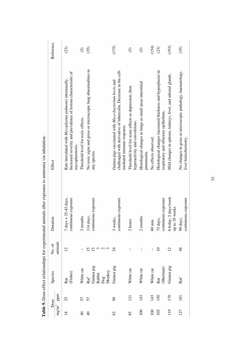
32
Tab
le 9
. Dos
e-ef
fect
rel
atio
nshi
ps f
or e
xper
imen
tal a
nim
als
afte
r ex
posu
re to
am
mon
ia v
ia in
hala
tion.
Dos
eSp
ecie
sD
urat
ion
Eff
ect
Ref
eren
cem
g/m
3pp
mN
o. o
fan
imal
s
1825
Rat
(Fis
her)
127
days
+ 3
5-42
day
s,co
ntin
uous
exp
osur
eR
ats
inoc
ulat
ed w
ith M
ycop
lasm
a pu
lmon
is in
tran
asal
ly.
Incr
ease
d se
veri
ty a
nd p
reva
lenc
e of
lesi
ons
char
acte
rist
ic o
fm
ycop
lasm
osis
.
(23)
4057
Whi
te r
at –
2 m
onth
sT
hres
hold
leve
l for
toxi
c ef
fect
s.(5
)
4057
Rat
a
Gui
nea
pig
Rab
bit
Dog
Mon
key
15 15 3 2 3
114
days
,co
ntin
uous
exp
osur
eN
o to
xic
sign
s an
d gr
oss
or m
icro
scop
ic lu
ng a
bnor
mal
ities
inan
y sp
ecie
s.(3
5)
6390
Gui
nea
pig
243
wee
ks,
cont
inuo
us e
xpos
ure
Gui
nea
pigs
vac
cina
ted
with
Myc
o-ba
cter
ium
bov
is a
ndch
alle
nged
with
der
ivat
ive
of tu
berc
ulin
. Dec
reas
e in
the
cell-
med
iate
d im
mun
e re
spon
se.
(175
)
8512
1W
hite
rat
–2
hour
sT
hres
hold
leve
l for
acu
te e
ffec
ts a
s de
pres
sion
, the
nhy
pera
ctiv
ity a
nd c
onvu
lsio
ns.
(5)
100
143
Whi
te r
at –
2 m
onth
sH
isto
logi
cal c
hang
es in
lung
s as
sm
all a
reas
inte
rstit
ial
pneu
mon
ia.
(5)
100
143
Whi
te r
at –
60 m
inN
o ef
fect
s ob
serv
ed.
(154
)
105
150
Rat
(She
rman
)10
75 d
ays,
cont
inuo
us e
xpos
ure
His
tolo
gica
l cha
nges
(in
crea
sed
thic
knes
s an
d hy
perp
lasi
a) in
resp
irat
ory
and
olfa
ctor
y ep
ithel
ium
.(2
3)
119
170
Gui
nea
pig
126
h/da
y, 5
day
s/w
eek
up to
18
wee
ksM
ild c
hang
es in
spl
eens
, kid
neys
, liv
er, a
nd a
dren
al g
land
s.(1
93)
127
181
Rat
a48
90 d
ays,
cont
inuo
us e
xpos
ure
No
chan
ges
in g
ross
or
mic
rosc
opic
pat
holo
gy, h
aem
atol
ogy,
liver
his
toch
emis
try.
(35)
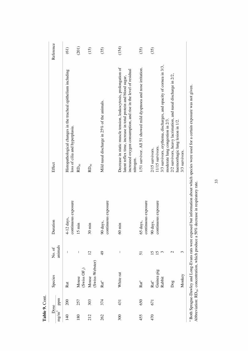
33
Tab
le 9
. Con
t.
Dos
eSp
ecie
sD
urat
ion
Eff
ect
Ref
eren
cem
g/m
3pp
mN
o. o
fan
imal
s
140
200
Rat
–4-
12 d
ays,
cont
inuo
us e
xpos
ure
His
topa
thol
ogic
al c
hang
es in
the
trac
heal
epi
thel
ium
incl
udin
glo
ss o
f ci
lia a
nd h
yper
plas
ia.
(61)
180
257
Mou
se(S
wis
s O
F 1)
–15
min
RD
50(2
01)
212
303
Mou
se(S
wis
s-W
ebst
er)
1230
min
RD
50(1
5)
262
374
Rat
a49
90 d
ays,
cont
inuo
us e
xpos
ure
Mild
nas
al d
isch
arge
in 2
5% o
f th
e an
imal
s.(3
5)
300
431
Whi
te r
at –
60 m
inD
ecre
ase
in s
tatic
mus
cle
tens
ion,
leuk
ocyt
osis
, pro
long
atio
n of
late
nt r
efle
x tim
e, in
crea
se in
tota
l pro
tein
and
blo
od s
ugar
,in
crea
sed
oxyg
en c
onsu
mpt
ion,
and
ris
e in
the
leve
l of
resi
dual
nitr
ogen
.
(154
)
455
650
Rat
a51
65 d
ays,
cont
inuo
us e
xpos
ure
1/51
sur
vivo
r. A
ll 51
sho
wed
mild
dys
pnoe
a an
d no
se ir
rita
tion.
(35)
470
671
Rat
a
Gui
nea
pig
Rab
bit
Dog
Mon
key
15 15 3 2 3
90 d
ays,
cont
inuo
us e
xpos
ure
2/15
sur
vivo
rs.
11/1
5 su
rviv
ors.
3/3
surv
ivor
s, e
ryth
ema,
dis
char
ges,
and
opa
city
of
corn
ea in
3/3
,m
oder
ate
lung
con
gest
ion
in 2
/3.
2/2
surv
ivor
s, h
eavy
lacr
imat
ion,
and
nas
al d
isch
arge
in 2
/2,
haem
orrh
agic
lung
lesi
on in
1/2
.3/
3 su
rviv
ors.
(35)
a Bot
h Sp
ragu
e-D
awle
y an
d L
ong-
Eva
ns r
ats
wer
e ex
pose
d bu
t inf
orm
atio
n ab
out w
hich
spe
cies
wer
e us
ed f
or a
cer
tain
exp
osur
e w
as n
ot g
iven
.A
bbre
viat
ion:
RD
50: c
once
ntra
tion,
whi
ch p
rodu
ce a
50%
dec
reas
e in
res
pira
tory
rat
e.

34
Tab
le 1
0. D
ose-
effe
ct r
elat
ions
hips
in m
an a
fter
exp
osur
e to
am
mon
ia v
ia in
hala
tion.
Con
cent
ratio
nD
urat
ion
Eff
ect
Ref
eren
ce
mg/
m3
ppm
No.
of
expo
sed
3.5
518
0 m
in 1
2N
o up
per-
airw
ay in
flam
mat
ion
or in
crea
sed
bron
chia
l res
pons
iven
ess.
Inc
reas
ed s
ympt
omra
tings
for
dis
com
fort
in th
e ey
es, s
olve
nt s
mel
l, he
adac
he, d
izzi
ness
, and
fee
ling
ofin
toxi
catio
n. R
atin
gs c
orre
spon
d to
“H
ardl
y at
all”
.
(173
)
6.4
9.2a
Chr
onic
exp
osur
e 5
8N
o ef
fect
s on
res
pira
tory
or
cuta
neou
s sy
mpt
oms,
pul
mon
ary
func
tion,
or
odou
r se
nsiti
vity
.(8
1)
0.02
-7.0
0.03
-9.8
Chr
onic
exp
osur
e 7
7N
o ef
fect
s on
res
pira
tory
sym
ptom
s.(1
4)
7-14
10-2
024
0 m
in 4
3In
crea
sed
sym
ptom
rat
ings
in 3
3 no
n-ha
bitu
ated
vol
unte
ers
for:
sum
of
sym
ptom
sco
res,
and
olfa
ctor
y sy
mpt
oms.
(80)
8.4
122
min
1
Ast
hma,
rho
nchi
in b
oth
lung
s.(1
07)
11-1
816
-25
30 m
in 6
+ 8
Nei
ther
hea
lthy
subj
ects
nor
ast
hmat
ics
show
ed s
igni
fica
nt c
hang
e in
pul
mon
ary
func
tion
orbr
onch
ial h
yper
reac
tivity
.(1
63)
1825
180
min
12N
o up
per-
airw
ay in
flam
mat
ion
or in
crea
sed
bron
chia
l res
pons
iven
ess.
Inc
reas
ed r
atin
g fo
r al
lsy
mpt
oms:
dis
com
fort
in th
e ey
es, n
ose,
thro
at a
nd a
irw
ays,
bre
athi
ng d
iffi
culty
, sol
vent
smel
l, he
adac
he, f
atig
ue, n
ause
a, d
izzi
ness
and
fee
ling
of in
toxi
catio
n. I
rrita
tion
ratin
gsco
rres
pond
to “
Som
ewha
t”.
(173
)
≤18
≤25b
Chr
onic
exp
osur
e13
8In
crea
sed
rela
tive
risk
(95
% C
I) f
or w
heez
ing
2.26
(1.
32-3
.88)
.(1
4)
>18
>25
b,c
Chr
onic
exp
osur
e 1
7In
crea
sed
rela
tive
risk
(95
% C
I) f
or c
ough
3.4
8 (1
.84-
6.57
), w
heez
ing
5.01
(2.
38-1
0.57
),ph
legm
3.7
5 (1
.97-
7.11
), d
yspn
oea
4.57
(2.
37-8
.81)
, bro
nchi
al a
sthm
a 4.
32 (
2.08
-8.9
8).
(14)
14 a
nd28
20 a
nd40
240
min
and
2 x
30 m
in 4
3In
crea
sed
sym
ptom
rat
ings
in 3
3 no
n-ha
bitu
ated
vol
unte
ers
for
sum
of
sym
ptom
sco
res,
olfa
ctor
y sy
mpt
oms,
irri
tativ
e sy
mpt
oms.
(80)
2130
10 m
in
5N
o ir
rita
tion
in 3
/5 a
nd “
just
per
cept
ible
” ir
rita
tion
of e
yes
and
nose
in 2
/5.
(117
)

35
Tab
le 1
0. C
ont.
Con
cent
ratio
nD
urat
ion
Eff
ect
Ref
eren
ce
mg/
m3
ppm
No.
of
expo
sed
3550
10 m
in
6“M
oder
ate”
irri
tatio
n of
eye
s an
d no
se in
4/6
.(1
17)
3550
240
min
43
Incr
ease
d sy
mpt
om r
atin
gs f
or s
um o
f sy
mpt
om s
core
s, o
lfac
tory
sym
ptom
s, ir
rita
tive
sym
ptom
s. C
onju
nctiv
al h
yper
aem
ia in
3 o
f 33
(9%
) no
n-ha
bitu
ated
.(8
0)
35-5
650
-80
120
min
16
VC
, FE
V, a
nd F
IV d
id n
ot d
ecre
ase
mor
e th
an 1
0%. M
ild ir
rita
tion
in e
yes
and
thro
at.
(185
)
7010
05-
30 s
23
Incr
ease
d na
sal a
irw
ay r
esis
tanc
e du
ring
the
expo
sure
per
iods
. Nas
al ir
rita
tion
in 1
1/23
.(1
28)
7711
012
0 m
in 1
6V
C, F
EV
, and
FIV
did
not
dec
reas
e m
ore
than
10%
. Irr
itatio
n in
eye
s an
d th
roat
, cou
gh.
(185
)
9814
0≤1
20 m
in 1
6V
C, F
EV
, and
FIV
did
not
dec
reas
e m
ore
than
10%
. Int
oler
able
for
8/1
6.(1
85)
>10
5>
150
6
All
subj
ects
exp
erie
nced
lach
rym
atio
n ac
com
pani
ed b
y dr
ynes
s of
the
nose
and
thro
at d
urin
goc
casi
onal
exc
ursi
ons
abov
e 15
0 pp
m in
sem
i-co
ntro
lled
expo
sure
s to
am
mon
ia in
an
amm
oniu
m b
icar
bona
te p
lant
.
(57)
1 19
0d1
700d
Acc
iden
tC
ough
ing
and
lary
ngos
pasm
alo
ng w
ith o
edem
a of
the
glot
tic r
egio
n.(7
)
1 75
0-3
160d
2 50
0-4
500d
Acc
iden
tFa
tal i
n ap
prox
imat
ely
30 m
in.
(7)
7 00
0d10
000
dA
ccid
ent
Rap
id r
espi
rato
ry a
rres
t. A
nhyd
rous
am
mon
ia in
con
cent
ratio
ns o
f 10
000
ppm
suf
fici
ent t
oev
oke
skin
dam
age.
(18)
a Tim
e-w
eigh
ted
aver
age,
b G
eom
etri
c m
ean,
c Max
imal
exp
osur
e le
vel 1
30 m
g/m
3 (18
5 pp
m),
d R
etro
spec
tive
estim
ates
.A
bbre
viat
ions
: CI:
con
fide
nce
inte
rval
, VC
: vita
l cap
acity
, FE
V: f
orce
d ex
pira
tory
vol
ume,
FIV
: for
ced
insp
irat
ory
volu
me.
36
(mean ± standard error of mean) had no effect on respiratory or cutaneous symp-toms, pulmonary function, or odour sensitivity (81). In another study, no signs ofrespiratory symptoms were seen in workers exposed to ammonia concentrationsbetween 0.03 and 9.8 ppm. However, workers exposed to average ammonia levelsat or below 25 ppm had an increased relative risk for wheezing. Additionally, theworkers exposed to average levels above 25 ppm had higher relative risks forwheezing, cough, phlegm, dyspnoea, and bronchial asthma (14). One case reportdescribes a man who developed asthma-like symptoms after five months exposureto low levels of ammonia (8-15 ppm) from ammonia containing silver polish.Subsequent experimental exposure to 12 ppm produced an immediate asthmaticreaction (107). Dose-effect relationships after exposure to ammonia via inhalationfor humans are shown in Table 10.
13. Previous evaluations by national and international bodies
NEG concluded in 1986 that the significant effect of exposure to ammonia isirritation of the respiratory tract, skin, and eyes. Studies of human inhalation ofammonia at low or moderate concentrations (less than 50 mg/m3 = 70 ppm) hadshown mild eye and upper respiratory tract irritation. Exposure to high concentra-tions of ammonia (by accidental release) may result in skin burns, eye damage,respiratory injury, and death (110).
IPCS concluded that exposure to ammonia in air at a concentration of 390 ppm(280 mg/m3) produced throat irritation; 1 680 ppm (1 200 mg/m3) producedcough; 2 380 ppm (1 700 mg/m3) was life-threatening, and more than 4 900 ppm(3 500 mg/m3) was associated with high mortality. Respiratory symptoms wereusually reversible, but chronic bronchitis had been reported to develop. Irritationoccurred at 50 ppm (35 mg/m3), which was neither discomforting nor painful.However, the report further concluded that ammonia does not present a directthreat to man except as a result of accidental exposure, particularly in industrialsettings (87).
The Health and Safety Executive (HSE), United Kingdom, summarised that theonly clearly established effect arising from exposure to low concentrations (lessthan 200 ppm) of ammonia in humans is irritation of the skin, eyes, and upperrespiratory tract. In unaccustomed subjects, clear indications of irritation werereported with exposure to 50-55 ppm for periods between several or 10 minutesand 6 hours. There is some evidence that repeated exposure to ammonia maylead to some degree of acclimatisation. The HSE report concluded that from thevery limited data available there is no clear evidence that ammonia possessesgenotoxic, carcinogenic, or reprotoxic potential (147).
Ammonia has not been classified for carcinogenic effects by United StatesEnvironmental Protection Agency (EPA), United States Department of Health andHuman Services/National Toxicology Program (DHHS/NTP) or the InternationalAgency for Research on Cancer (IARC).

37
In the criteria document for ammonia by the Commission of the EuropeanCommunities (1993) the current occupational exposure limit of 17 mg/m3 (25ppm) was considered a borderline value. Therefore, an exposure limit of 14mg/m3 (20 ppm) was suggested. If a short-term exposure limit (STEL) value willbe considered necessary, a level of 22 mg/m3 (32 ppm) was suggested (62).
The Agency for Toxic Substances and Disease Registry (ATSDR) has derived aminimal risk level (MRL), of 1.7 ppm for acute-duration inhalation exposure, anda MRL of 0.1 ppm for chronic-duration inhalation exposure to ammonia (12).
The American Conference of Governmental Industrial Hygienists (ACGIH) hasrecommended an 8-hour occupational exposure limit of 25 ppm and a STEL of 35ppm for ammonia. The first value is recommended to minimise the potential eyeand respiratory tract irritation and the second one to provide a greater margin ofsafety against the acute sensory effects from exposure to ammonia (1).
14. Evaluation of human health risks
14.1 Assessment of health risks
Ammonia is an endogenous compound formed and used in various metabolicreactions in the human body. The most serious effects of elevated endogenousammonia are seen in the brain as hepatic encephalopathy after acute or chronicliver failure. At the cellular level, ammonia interacts with various cellular pro-cesses involved in neurotransmission and brain energy metabolism.
The direct local action on mucous membranes results in irritation andinflammation of the skin, eyes, nose, throat, and upper respiratory tract. Acutecontact with high concentrations of ammonia can produce rapid changes in thepulmonary parenchyma due to the affinity of the anhydrous by-products formucous membranes leading to death. Clinically, mild insults produce coughingas well as laryngitis and tracheobronchitis. Ammonia inhalation injury has alsobeen associated with RADS, an asthma-like syndrome with persistent bronchialhyperreactivity but no sensitisation. More severe injuries include in fulminantpulmonary oedema and bronchiectasis. The most frequent injury is desquamationof the epithelial layer of the upper tracheobronchial tree.
Ammonia can penetrate and damage the iris, and cause cataract in severeburns. Iritis may be accompanied by hypopyon (a layering of white blood cells inthe anterior chamber) or haemorrhages, extensive loss of pigment, and severeglaucoma.
Chronic occupational exposure to low levels of ammonia (9.2 ± 1.4 ppm)(mean ± standard error of mean) had no effect on pulmonary function or odoursensitivity in a group of workers. In another study, chronic exposure to averageammonia levels above 25 ppm was associated with increase in respiratorysymptoms and bronchial asthma.
According to two controlled human exposure studies clear signs of mildrespiratory and eye irritation appear somewhere between 25 and 50 ppm. In one

38
of the studies, significant increases in symptom ratings were seen already at5 ppm, however, the ratings corresponded verbally to “Hardly at all”. NEGconsiders 5 ppm as a pragmatic NOAEL and 25 ppm as a LOAEL for irritation.No upper-airway inflammation or increased bronchial responsiveness weredetected at these levels.
RD50 values for ammonia calculated from mouse studies are 260-300 ppm. ANOAEL of 57 ppm and a LOAEL of 143 ppm for toxic effects were reported in a2-month inhalation exposure of white rats. The NOAEL for irritation in humans isoften considered as approximately 3% of the RD50, i.e. 9 ppm ammonia.
14.2 Groups at extra risk
Ammonia is commonly regarded as an irritant. Therefore, persons who are hyper-reactive to other respiratory irritants or who are asthmatic, would also be moresensitive to ammonia. As ammonia is metabolised and excreted by liver andkidney, persons with hepatic or renal impairment might be at risk when exposedto ammonia as well as those with inherited deficiencies of urea cycle enzymes.
14.3 Scientific basis for an occupational exposure limit
Based on short-term (3-4 hours) controlled exposure of volunteers, the criticaleffect of exposure to ammonia is irritation. According to these studies clear signsof mild respiratory and eye irritation appear somewhere between 25 and 50 ppm.In one of the studies, significant increases in symptom ratings were seen alreadyat 5 ppm, however, the ratings corresponded verbally to “Hardly at all”. NEGconsiders 5 ppm as a pragmatic NOAEL and 25 ppm as a LOAEL for irritation.No upper-airway inflammation or increased bronchial responsiveness weredetected at these levels. Chronic occupational exposure to low levels of ammonia(9.2 ± 1.4 ppm) (mean ± standard error of mean) had no effect on pulmonaryfunction or odour sensitivity. In another study, chronic exposure to averageammonia levels above 25 ppm was associated with an increase in respiratorysymptoms and bronchial asthma.
15. Research needs
Long-term animal tests are needed to establish a NOAEL for inhalation exposure.There is a lack of data on health effects and exposure levels from long-term andlow exposure situations where ammonia is the principal agent. More studies onhealth effects are also needed from persons who have been exposed to high levelsof ammonia. In addition, more studies on reproductive toxicity and carcinogeni-city should be undertaken to provide proper data for risk assessment.
39
16. Summary
Liesivuori J. The Nordic Expert Group for Criteria Documentation of HealthRisks from Chemicals. 137. Ammonia. Arbete och Hälsa 2005;13:1-52.
Ammonia (NH3) is a colourless gas with a distinctly pungent odour at normalatmospheric temperatures and pressures. The odour threshold is approximately5 ppm. Ammonia can be liquefied under pressure. In aqueous solution, NH3 actsas a base yielding ammonium (NH4
+) and hydroxide (OH-) ions. Because of thehigh water solubility ammonia dissolves in moisture on the mucous membranes,eyes and skin, forming ammonium hydroxide, which may cause alkali burns andliquefaction necrosis of the tissues.
Ammonia occurs naturally in the environment and is endogenously produced inhumans and other mammalians. It is also man-made in vast quantities. Occupa-tional exposures may occur in ammonia plants, fertiliser manufacturing, andanimal production. The primary route of occupational exposure to ammonia isinhalation, although dermal exposure may also occur during handling of liquidammonia or aqueous ammonia solutions.
The critical effect of exposure to ammonia is irritation. In controlled humanexposure studies (3-4 hours) clear signs of mild respiratory and eye irritationappear somewhere between 25 and 50 ppm. In one of these studies very slightirritation was seen at 5 ppm. NEG considers 5 ppm as a pragmatic NOAEL and25 ppm as a LOAEL for irritation. Chronic exposure to average ammonia levelsabove 25 ppm has been associated with an increase in respiratory symptoms(cough, wheezing, phlegm, and dyspnoea) and bronchial asthma. High acuteexposure levels may result in reactive airways dysfunction syndrome (RADS),an asthma-like syndrome with persistent bronchial hyperreactivity but nosensitisation. Massive exposure to ammonia can cause eye damage, skin burns,severe inflammation of the respiratory tract (laryngitis, tracheobronchitis, andpulmonary oedema), and death.
With very limited data available, there is no evidence that ammonia possessesgenotoxic, carcinogenic or reprotoxic potential.
Keywords: ammonia, asthma, burns, irritation, occupational exposure limit,RADS, review, toxicity

40
17. Summary in Swedish
Liesivuori J. The Nordic Expert Group for Criteria Documentation of HealthRisks from Chemicals. 137. Ammonia. Arbete och Hälsa 2005;13:1-52.
Ammoniak (NH3) är en färglös gas med en påtagligt stickande lukt vid normaltemperatur och tryck. Lukttröskeln har angivits till ungefär 5 ppm. Ammoniakkondenserar till vätska vid högt tryck. Ammoniak är en svag bas som i vattenbildar ammonium- (NH4
+) och hydroxidjoner (OH-). Den höga vattenlöslighetengör att ammoniak löser sig i slemhinnor, ögon och hud och bildar ammonium-hydroxid som i sin tur kan orsaka frätskador och nekros.
Ammoniak förekommer naturligt i vår omgivning och bildas endogent hosmänniskor och andra däggdjur. Ammoniak framställs också industriellt i storakvantiteter. Förutom vid råvaru- och handelsgödselproduktion förekommerammoniakexponering framförallt inom jordbruket i samband med djurhållning.Yrkesmässig exponering för ammoniak sker främst genom inhalation. Hudupptagkan ske vid hantering av flytande ammoniak eller vattenlösningar av ammoniak.
Den kritiska effekten vid exponering för ammoniak är irritation. I kontrolleradeexponeringsstudier på människa (3-4 timmar) uppträder tydliga tecken på mildirritation i övre luftvägar och i ögon någonstans mellan 25 och 50 ppm. I en avdessa studier noterades en mycket svag irritation vid 5 ppm. NEG bedömer 5 ppmsom en pragmatisk icke-effektnivå (NOAEL) och 25 ppm som lägsta observeradeeffektnivån (LOAEL) för irritation. Kronisk exponering för en genomsnittligammoniaknivå över 25 ppm har rapporterats vara förenat med en ökning avluftvägssymptom (hosta, pipande andning, slem och andnöd) och bronkiell astma.Akut exponering för höga halter ammoniak kan ge upphov till ett astma-liknandesjukdomstillstånd, s k RADS (reactive airways dysfunction syndrome) somkännetecknas av kvarstående bronkiell hyperreaktivitet men ingen sensibilisering.Massiv exponering för ammoniak kan orsaka frätskador på hud och ögon,allvarlig inflammation i luftvägarna (laryngit, trakeobronkit och lungödem) ochleda till döden.
Det går inte att bedöma om ammoniak har genotoxisk, carcinogen ellerreproduktiontoxisk potential med tillgängliga data.
Nyckelord: ammoniak, astma, frätskador, irritation, hygieniskt gränsvärde, RADS,toxicitet, översikt

41
18. References
1. ACGIH. Documentation of the threshold limit values. Cincinnati, OH: American Conferenceof Governmental Industrial Hygienists, ISBN 1-882417-43-7, 2001.
2. Aguilar MA, Miñarro J, Felipo V. Chronic moderate hyperammonemia impairs active andpassive avoidance behavior and conditional discrimination learning in rats. Exp Neurol2000;161:704-713.
3. Ali BA, Ahmed HO, Ballal SG, Albar AA. Pulmonary function of workers exposed toammonia: a study in the Eastern Province of Saudi Arabia. Int J Occup Environ Health2001;7:19-22.
4. Alpatov IM. [Study of the toxicity of gaseous ammonia]. Gig Tr Prof Zabol 1964;29:14-18(in Russian).
5. Alpatov IM, Mikhailov VI. [Determination of the toxicity of gas-forming ammonia. I]. GigTr Prof Zabol 1963;35:51-53 (in Russian).
6. American Petroleum Institute (API). The sources, chemistry, fate, and effects of ammonia inaquatic environments. Washington, DC: American Petroleum Institute, 1981:145.
7. Amshel CE, Fealk MH, Phillips BJ, Caruso DM. Anhydrous ammonia burns case report andreview of the literature. Burns 2000;26:493-497.
8. Appelman LM, ten Berge WF, Reuzel PG. Acute inhalation toxicity study of ammonia in ratswith variable exposure periods. Am Ind Hyg Assoc J 1982;43:662-665.
9. Arwood R, Hammond J, Ward GG. Ammonia inhalation. J Trauma 1985;25:444-447.
10. Asai K, Ishii S, Ohta S, Furusho K. Fatal hyperammonaemia in argininosuccinic aciduriafollowing enflurane anaesthesia. Eur J Pediatr 1998;157:169-170.
11. Atkins DHF, Lee DS. Indoor concentrations of ammonia and the potential contribution ofhumans to atmospheric budgets. Atmos Environ 1993;27A:1-7.
12. ATSDR. Toxicological profile for ammonia. Atlanta, GA: Agency for Toxic Substances andDisease Registry. U.S. Department of Health and Human Services, Public Health Service,NTIS PB2004-107331, 2004.
13. Azorin I, Miñana MD, Felipo V, Grisolia S. A simple animal model of hyperammonemia.Hepatology 1989;10:311-314.
14. Ballal SG, Ali BA, Albar AA, Ahmed HO, al-Hasan AY. Bronchial asthma in two chemicalfertilizer producing factories in eastern Saudi Arabia. Int J Tuberc Lung Dis 1998;2:330-335.
15. Barrow CS, Alarie Y, Stock MF. Sensory irritation and incapacitation evoked by thermaldecomposition products of polymers and comparisons with known sensory irritants. ArchEnviron Health 1978;33:79-88.
16. Beare JD, Wilson RS, Marsh RJ. Ammonia burns of the eye: an old weapon in new hands.Br Med J (Clin Res Ed) 1988;296:590.
17. Bernstein IL, Bernstein DI. Reactive airways disease syndrome (RADS) after exposure totoxic ammonia fumes. J Allergy Clin Immunol 1989;83:173.
18. Birken GA, Fabri PJ, Carey LC. Acute ammonia intoxication complicating multiple trauma.J Trauma 1981;21:820-822.
19. Bishop RW, Belkin F, Gaffney R. Evaluation of a new ammonia sampling and analyticalprocedure. Am Ind Hyg Assoc J 1986;47:135-137.
20. Brautbar N. Ammonia exposure: a common cause for sinusitis. A case report and review ofthe literature. Toxicol Ind Health 1998;14:891-895.
21. Brautbar N, Wu MP, Richter ED. Chronic ammonia inhalation and interstitial pulmonaryfibrosis: a case report and review of the literature. Arch Environ Health 2003;58:592-596.
22. Brille D, Hatzfeld C, Laurent R. [Occupational lung diseases except silicosis. IV. Chronicbronchitis, emphysema and occupations: pulmonary emphysema after inhalation of irritantvapors especially ammonia]. Arch Mal Prof 1957;18:320-336 (in French).

42
23. Broderson JR, Lindsey JR, Crawford JE. The role of environmental ammonia in respiratorymycoplasmosis of rats. Am J Pathol 1976;85:115-130.
24. Brooks SM, Weiss MA, Bernstein IL. Reactive airways dysfunction syndrome (RADS).Persistent asthma syndrome after high level irritant exposures. Chest 1985;88:376-384.
25. Brunner H, Thaler H (eds). Hepatology: A festschrift for Hans Popper. New York: RavenPress, 1981:212.
26. Burns TR, Mace ML, Greenberg SD, Jachimczyk JA. Ultrastructure of acute ammoniatoxicity in the human lung. Am J Forensic Med Pathol 1985;6:204-210.
27. Butterworth RF. Pathophysiology of hepatic encephalopathy: a new look at ammonia. MetabBrain Dis 2002;17:221-227.
28. Bäck B, Reijula K, Puhakka E. Measurement of ammonia in indoor air samples. In: Woods J,Grimsrud D, Boschi N, eds. Proceedings of healthy buildings/IAQ'97. Washington, DC:Healthy Buildings/IAQ'97, 1997;3:321-323.
29. Castell DO, Moore EW. Ammonia absorption from the human colon. The role of nonionicdiffusion. Gastroenterology 1971;60:33-42.
30. Cersosimo E, Williams P, Geer R, Lairmore T, Ghishan F, Abumrad NN. Importance ofammonium ions in regulating hepatic glutamine synthesis during fasting. Am J Physiol1989;257:E514-519.
31. Chang CW, Chung H, Huang CF, Su HJ. Exposure assessment to airborne endotoxin, dust,ammonia, hydrogen sulfide and carbon dioxide in open style swine houses. Ann Occup Hyg2001;45:457-465.
32. CHEMINFO database. Ammonia gas. Canadian Centre for Occupational Health and Safety(CCOHS). http://www.worksafesask.ca/files/ccohs/cheminfo/cie48.html, 2005.
33. Clemmesen JO, Kondrup J, Ott P. Splanchnic and leg exchange of amino acids and ammoniain acute liver failure. Gastroenterology 2000;118:1131-1139.
34. Close LG, Catlin FI, Cohn AM. Acute and chronic effects of ammonia burns on therespiratory tract. Arch Otolaryngol 1980;106:151-158.
35. Coon RA, Jones RA, Jenkins LJ, Jr., Siegel J. Animal inhalation studies on ammonia,ethylene glycol, formaldehyde, dimethylamine, and ethanol. Toxicol Appl Pharmacol1970;16:646-655.
36. Cooper AJ, Lai JC. Cerebral ammonia metabolism in normal and hyperammonemic rats.Neurochem Pathol 1987;6:67-95.
37. Cooper AJ, Nieves E, Coleman AE, Filc-DeRicco S, Gelbard AS. Short-term metabolic fateof [13N]ammonia in rat liver in vivo. J Biol Chem 1987;262:1073-1080.
38. Cooper AJ, Plum F. Biochemistry and physiology of brain ammonia. Physiol Rev1987;67:440-519.
39. Crook B, Robertson JF, Glass SA, Botheroyd EM, Lacey J, Topping MD. Airborne dust,ammonia, microorganisms, and antigens in pig confinement houses and the respiratory healthof exposed farm workers. Am Ind Hyg Assoc J 1991;52:271-279.
40. Curthoys NP, Watford M. Regulation of glutaminase activity and glutamine metabolism.Annu Rev Nutr 1995;15:133-159.
41. da Fonseca-Wollheim F. Preanalytical increase of ammonia in blood specimens from healthysubjects. Clin Chem 1990;36:1483-1487.
42. de la Hoz RE, Schlueter DP, Rom WN. Chronic lung disease secondary to ammoniainhalation injury: a report on three cases. Am J Ind Med 1996;29:209-214.
43. Demerec M, Bertani G, Flint J. A survey of chemicals for mutagenic action on E. coli.Am Nat 1951;85:119-136.
44. Derobert L, Proteau J, Caroff J. [Anatomical study of 4 cases of acute poisoning by ammoniagas inhalation]. Ann Med Leg Criminol Police Sci Toxicol 1964;44:362-366 (in French).
43
45. Devos M, Patte F, Rouault J, Laffort P, van Gemert LJ (eds). Standardized human olfactorythresholds. Oxford, UK: IRL Press at Oxford University Press, 1990:26.
46. Dodd KT, Gross DR. Ammonia inhalation toxicity in cats: a study of acute and chronicrespiratory dysfunction. Arch Environ Health 1980;35:6-14.
47. Donham K, Haglind P, Peterson Y, Rylander R, Belin L. Environmental and health studies offarm workers in Swedish swine confinement buildings. Br J Ind Med 1989;46:31-37.
48. Donham KJ, Cumro D, Reynolds S. Synergistic effects of dust and ammonia on theoccupational health effects of poultry production workers. J Agromedicine 2002;8:57-76.
49. Donham KJ, Cumro D, Reynolds SJ, Merchant JA. Dose-response relationships betweenoccupational aerosol exposures and cross-shift declines of lung function in poultry workers:recommendations for exposure limits. J Occup Environ Med 2000;42:260-269.
50. Donham KJ, Reynolds SJ, Whitten P, Merchant JA, Burmeister L, Popendorf WJ.Respiratory dysfunction in swine production facility workers: dose-response relationships ofenvironmental exposures and pulmonary function. Am J Ind Med 1995;27:405-418.
51. Duda GD, Handler P. Kinetics of ammonia metabolism in vivo. J Biol Chem 1958;232:303-314.
52. Eaton WC, Rickman EE, Jayanty RKM. Method 301 evaluation of a technique for collectionand measurement of ammonia in stationary source emissions. Proceedings of the AWMAinternational symposium on measurement of toxic an related air pollutants. Pittsburgh, PA:Air and Waste Management Association, 1996:583-588.
53. ECETOC. Technical report no 62. Brussels: European Centre for Ecotoxicology andToxicology of Chemicals, 1994.
54. Erxleben H, Simon J, Moskvin LN, Vladimirovna LO, Nikitina TG. Automized proceduresfor the determination of ozone and ammonia contents in air by using the chromatomembranemethod for gas-liquid extraction. Fresenius J Anal Chem 2000;366:332-335.
55. Esbjörnsson-Liljedahl M, Jansson E. Sex difference in plasma ammonia but not in muscleinosine monophosphate accumulation following sprint exercise in humans. Eur J ApplPhysiol Occup Physiol 1999;79:404-408.
56. Felipo V, Butterworth RF. Neurobiology of ammonia. Prog Neurobiol 2002;67:259-279.
57. Ferguson WS, Koch WC, Webster LB, Gould JR. Human physiological response andadaption to ammonia. J Occup Med 1977;19:319-326.
58. Flury KE, Dines DE, Rodarte JR, Rodgers R. Airway obstruction due to inhalation ofammonia. Mayo Clin Proc 1983;58:389-393.
59. Furst P, Josephson B, Maschio G, Vinnars E. Nitrogen balance after intravenous and oraladministration of ammonium salts to man. J Appl Physiol 1969;26:13-22.
60. Gaafar H, Girgis R, Hussein M, el-Nemr F. The effect of ammonia on the respiratory nasalmucosa of mice. A histological and histochemical study. Acta Otolaryngol 1992;112:339-342.
61. Gamble MR, Clough G. Ammonia build-up in animal boxes and its effect on rat trachealepithelium. Lab Anim 1976;10:93-104.
62. Garlanda T, Basilico S. Occupational exposure limits : criteria document for ammonia.Health and safety series. Report EUR 14533 EN. Luxembourg: Commission of the EuropeanCommunities. Office for Official Publications of the European Communities, 1993.
63. George A, Bang RL, Lari AR, Gang RK, Kanjoor JR. Liquid ammonia injury. Burns2000;26:409-413.
64. Girard G, Giguere JF, Butterworth RF. Region-selective reductions in activities of glutaminesynthetase in rat brain following portacaval anastomosis. Metab Brain Dis 1993;8:207-215.
65. Golbabaei F, Islami F. Evaluation of workers' exposure to dust, ammonia and endotoxin inpoultry industries at the province of Isfahan, Iran. Ind Health 2000;38:41-46.
44
66. Gomzi M, Saric M. Respiratory impairment among children living in the vicinity of afertilizer plant. Int Arch Occup Environ Health 1997;70:314-320.
67. Goodman LS, Gilman A (eds). The pharmacological basis of therapeutics. 4th ed. New york:The MacMillan Co., 1970:1749.
68. Goyer N, Lavoie J. Emissions of chemical compounds and bioaerosols during the secondarytreatment of paper mill effluents. AIHAJ 2001;62:330-341.
69. Grant WM. Toxicology of the eye. 3rd ed. Springfield, IL: Charles C. Thomas publ., 1986:87.
70. Grant WM, Schuman JS. Toxicology of the eye. 4th ed. Springfield, IL: Charles C. Thomaspubl., 1993:124-131.
71. Groves JA, Ellwood PA. Gases in agricultural slurry stores. Ann Occup Hyg 1991;35:139-151.
72. Gupta S, Rogers LK, Taylor SK, Smith CV. Inhibition of carbamyl phosphate synthetase-Iand glutamine synthetase by hepatotoxic doses of acetaminophen in mice. Toxicol ApplPharmacol 1997;146:317-327.
73. Hallberg BO, Rudling J. A diffuse sampler of gaseous pollutants by collection in liquidmedia. Ann Occup Hyg 1989;33:61-68.
74. Heederik D, Brouwer R, Biersteker K, Boleij JS. Relationship of airborne endotoxin andbacteria levels in pig farms with the lung function and respiratory symptoms of farmers.Int Arch Occup Environ Health 1991;62:595-601.
75. Heederik D, van Zwieten R, Brouwer R. Across-shift lung function changes among pigfarmers. Am J Ind Med 1990;17:57-58.
76. Helmers S, Top FH, Sr., Knapp LW, Jr. Ammonia injuries in agriculture. J Iowa Med Soc1971;61:271-280.
77. Hermenegildo C, Monfort P, Felipo V. Activation of N-methyl-D-aspartate receptors in ratbrain in vivo following acute ammonia intoxication: characterization by in vivo brainmicrodialysis. Hepatology 2000;31:709-715.
78. Highman VN. Early rise in intraocular pressure after ammonia burns. Br Med J 1969;1:359-360.
79. Hjelm M, Oberholzer V, Seakins J, Thomas S, Kay JD. Valproate-induced inhibition of ureasynthesis and hyperammonaemia in healthy subjects. Lancet 1986;2:859.
80. Hoffmann J, Ihrig A, Triebig G. Expositionsstudie zur arbeitsmedizinischen BedeutungAmmoniak-assoziierter gesundheitlicher Effekte. Arbeitsmedizin, Sozialmedizin,Umweltmedizin 2004;39:390-401 (in German).
81. Holness DL, Purdham JT, Nethercott JR. Acute and chronic respiratory effects ofoccupational exposure to ammonia. Am Ind Hyg Assoc J 1989;50:646-650.
82. Honeycutt D, Callahan K, Rutledge L, Evans B. Heterozygote ornithine transcarbamylasedeficiency presenting as symptomatic hyperammonemia during initiation of valproatetherapy. Neurology 1992;42:666-668.
83. Huizenga JR, Tangerman A, Gips CH. Determination of ammonia in biological fluids. AnnClin Biochem 1994;31( Pt 6):529-543.
84. Huizenga JR, Vissink A, Kuipers EJ, Gips CH. Helicobacter pylori and ammoniaconcentrations of whole, parotid and submandibular/sublingual saliva. Clin Oral Investig1999;3:84-87.
85. Hunt RD. Measurement of partial pressure of ammonia in breath and aterial blood: evidencefor secretion of ammonia into expired gas by metabolism of monoamines in the lung. DissAbstr Int B 1977;37:3279.
86. International Fertilizer Industry Association. www.fertilizer.org/ifa/statistics.asp, 2004.
87. IPCS/WHO. Ammonia. Environmental Health Criteria 54. Geneva: International Programmeon Chemical Safety/World Health Organization, 1986:1-210.
45
88. Jalan R, Damink SW, Deutz NE, Lee A, Hayes PC. Moderate hypothermia for uncontrolledintracranial hypertension in acute liver failure. Lancet 1999;354:1164-1168.
89. Jarudi NI, Golden B. Ammonia eye injuries. J Iowa Med Soc 1973;63:260-263.
90. Kacergis JB, Jones RB, Reeb CK, Turner WA, Ohman JL, Ardman MR, Paigen B. Airquality in an animal facility: particulates, ammonia, and volatile organic compounds. Am IndHyg Assoc J 1996;57:634-640.
91. Kamin H, Handler P. Amino acid and protein metabolism. Annu Rev Biochem 1957;26:419-490.
92. Kangas J, Nevalainen A, Manninen A, Savolainen H. Ammonia, hydrogen sulphide andmethyl mercaptides in Finnish municipal sewage plants and pumping stations. Sci TotalEnviron 1986;57:49-55.
93. Kapeghian JC, Mincer HH, Jones AB, Verlangieri AJ, Waters IW. Acute inhalation toxicityof ammonia in mice. Bull Environ Contam Toxicol 1982;29:371-378.
94. Kass I, Zamel N, Dobry CA, Holzer M. Bronchiectasis following ammonia burns of therespiratory tract. A review of two cases. Chest 1972;62:282-285.
95. Katagawa K, Nagashima T, Inase N, Kanayama M, Chida M, Sasaki S, Marumo F. Urinaryammonium measurement by the auto-analyzer method. Kidney Int 1989;36:291-294.
96. Keiding S, Munk OL, Roelsgaard K, Bender D, Bass L. Positron emission tomography ofhepatic first-pass metabolism of ammonia in pig. Eur J Nucl Med 2001;28:1770-1775.
97. Keplinger ML, Schadeberg KJ, Goode JW, Calandra JC. Irritation threshold study withammonia. Report to the International Institute of Ammonia Refrigeration. IBT-663-03161.Northbrook, IL: Industrial Bio-Test Laboratories, Inc., 1973.
98. Kirchner M, Braeutigam S, Ferm M, Haas M, Hangartner M, Hofschreuder P, Kasper-GieblA, Rommelt H, Striedner J, Terzer W, Thoni L, Werner H, Zimmerling R. Field intercom-parison of diffusive samplers for measuring ammonia. J Environ Monit 1999;1:259-265.
99. Kosenko E, Kaminsky Y, Grau E, Miñana MD, Marcaida G, Grisolia S, Felipo V. Brain ATPdepletion induced by acute ammonia intoxication in rats is mediated by activation of theNMDA receptor and Na+,K(+)-ATPase. J Neurochem 1994;63:2172-2178.
100. Kosenko E, Kaminsky Y, Stavroskaya IG, Felipo V. Alteration of mitochondrial calciumhomeostasis by ammonia-induced activation of NMDA receptors in rat brain in vivo. BrainRes 2000;880:139-146.
101. Kröger S, Liesivuori J, Manninen A. Evaluation of workers' exposure to 2-ethylhexanoic acid(2-EHA) in Finnish sawmills. A field study. Int Arch Occup Environ Health 1990;62:213-6.
102. Lacey JM, Wilmore DW. Is glutamine a conditionally essential amino acid? Nutr Rev1990;48:297-309.
103. Laitinen J, Liesivuori J, Savolainen H. Urinary alkoxyacetic acids and renal effects ofexposure to ethylene glycol ethers. Occup Environ Med 1996;53:595-600.
104. Landahl HD, Herrmann RG. Retention of vapors and gases in the human nose and lung. ArchInd Hyg Occup Med 1950;1:36-45.
105. Larson TV, Covert DS, Frank R, Charlson RJ. Ammonia in the human airways:neutralization of inspired acid sulfate aerosols. Science 1977;197:161-163.
106. Leduc D, Gris P, Lheureux P, Gevenois PA, De Vuyst P, Yernault JC. Acute and long termrespiratory damage following inhalation of ammonia. Thorax 1992;47:755-757.
107. Lee HS, Chan CC, Tan KT, Cheong TH, Chee CB, Wang YT. Burnisher's asthma--a case dueto ammonia from silverware polishing. Singapore Med J 1993;34:565-566.
108. Lehninger AL. Biochemistry: The molecular basis of cell structure and function. 2nd ed.New York: Worth Publisher Inc., 1975:579-585.
109. Leino T, Kähkönen E, Saarinen L, Henriks-Eckerman ML, Paakkulainen H. Workingconditions and health in hairdressing salons. Appl Occup Environ Hyg 1999;14:26-33.

46
110. Leira HL, Ophus EM. Ammoniakk. Nordic expert group for documentation of occupationalexposure limits. Solna: Arbetarskyddsverket, Arbete och Hälsa,1986;31:1-33 (in Norwegianwith English summary).
111. Lépine C, Soucy R. [The bronchopneumopathy of toxic origin. Physiopathologicaldevelopment]. Union Med Can 1962;91:7-11 (in French).
112. Liesivuori J, Laitinen J, Savolainen H. Kinetics and renal effects of formic acid inoccupationally exposed farmers. Arch Toxicol 1992;66:522-524.
113. Liesivuori J, Savolainen H. Effect of renal formic acid excretion on urinary calcium andammonia concentrations. Klin Wochenschr 1987;65:860-863.
114. Lobasov M, Smirnov F. On the nature of the action of chemical agents on mutational processin drosophila melanogaster. II. The effect of ammonia on the occurence of the lethaltransgenations. C R Seances Acad Sci Roum, 1934;3:177-178.
115. Lockwood AH, Yap EW, Wong WH. Cerebral ammonia metabolism in patients with severeliver disease and minimal hepatic encephalopathy. J Cereb Blood Flow Metab 1991;11:337-341.
116. Louhelainen K, Kangas J, Veijanen A, Viilos P. Effect of in situ composting on reducingoffensive odors and volatile organic compounds in swineries. AIHAJ 2001;62:159-167.
117. MacEwen JD, Theodore J, Vernot EH. Human exposure to EEL concentrations of mono-methylhydrazine. In: Proceedings 1st annual conference on environmental toxicology.Report No. AMRL-TR-70-102. Wright-Patterson Air Force Base, OH: Aerospace MedicalResearch Laboratory, 1970:355-363.
118. MacEwen JD, Vernot, EH. Toxic Hazards Research Unit - annual technical report. AMRL-TR-72-62. Wright-Patterson Air Force Base, OH: Aerospace Medical Research Laboratory,NTIS AD 755-358, 1972:70-72.
119. Manninen A. Analysis of airborne ammonia: comparison of field methods. Ann Occup Hyg1988;32:399-404.
120. Manninen A, Anttila S, Savolainen H. Rat metabolic adaptation to ammonia inhalation. ProcSoc Exp Biol Med 1988;187:278-281.
121. Manninen A, Kangas J, Linnainmaa M, Savolainen H. Ammonia in Finnish poultry houses:Effects of litter on ammonia levels and their reduction by technical binding agents. Am IndHyg Assoc J 1989;50:210-215.
122. Manninen A, Kröger S, Liesivuori J, Savolainen H. 2-Ethylhexanoic acid inhibits ureasynthesis and stimulates carnitine acetyltransferase activity in rat liver mitochondria. ArchToxicol 1989;63:160-161.
123. Marino FE, Mbambo Z, Kortekaas E, Wilson G, Lambert MI, Noakes TD, Dennis SC.Influence of ambient temperature on plasma ammonia and lactate accumulation duringprolonged submaximal and self-paced running. Eur J Appl Physiol 2001;86:71-78.
124. Martinelle K, Häggström L. Mechanisms of ammonia and ammonium ion toxicity in animalcells: transport across cell membranes. J Biotechnol 1993;30:339-350.
125. Master S, Gottstein J, Blei AT. Cerebral blood flow and the development of ammonia-induced brain edema in rats after portacaval anastomosis. Hepatology 1999;30:876-880.
126. Mayan MH, Merilan CP. Effects of ammonia inhalation on respiration rate of rabbits. J AnimSci 1972;34:448-452.
127. McDermott WJ, Adams RD, Riddell AG. Ammonia metabolism in man. Ann Surg1954;140:539-556.
128. McLean JA, Mathews KP, Solomon WR, Brayton PR, Bayne NK. Effect of ammonia onnasal resistance in atopic and nonatopic subjects. Ann Otol Rhinol Laryngol 1979;88:228-234.
129. Melbostad E, Eduard W. Organic dust-related respiratory and eye irritation in Norwegianfarmers. Am J Ind Med 2001;39:209-217.

47
130. Meyerhoff ME, Robins RH. Disposable potentiometric ammonia gas sensors for estimationof ammonia in blood. Anal Chem 1980;52:2383-2387.
131. Michaels RA. Emergency planning and the acute toxic potency of inhaled ammonia. EnvironHealth Perspect 1999;107:617-627.
132. Millea TP, Kucan JO, Smoot EC, 3rd. Anhydrous ammonia injuries. J Burn Care Rehabil1989;10:448-453.
133. Miñana MD, Marcaida G, Grisolia S, Felipo V. Prenatal exposure of rats to ammonia impairsNMDA receptor function and affords delayed protection against ammonia toxicity andglutamate neurotoxicity. J Neuropathol Exp Neurol 1995;54:644-650.
134. Mulder JS, Van Der Zalm HO. [Fatal case of ammonia poisoning]. Tijdschr SocialeGeneeskunde 1967;45:458-460 (in Dutch).
135. Mutch BJ, Banister EW. Ammonia metabolism in exercise and fatigue: a review. Med SciSports Exerc 1983;15:41-50.
136. National Research Council (NRC). Ammonia. Subcommittee on ammonia. Committee onmedical and biological effects of environmental pollutants. National Research Council.Baltimore, MD: University Park Press, NTIS No. PB-278-027, 1979.
137. NIOSH. NIOSH manual of analytical methods. 2nd ed. Vol 5, S347. Cincinnati, OH:National Institute of Occupational Safety and Health, Publication No. 79-141, 1979.
138. NIOSH. Health hazard evaluation report. Cincinnati, OH: National Institute of OccupationalSafety and Health, PB 87-232-948, 1987.
139. NIOSH. Ammonia. NIOSH manual of analytical methods. 4th ed. Cincinnati, OH: NationalInstitute of Occupational Safety and Health, Publication No. 94-143, August 15, 1994.
140. NIOSH. Ammonia by IC. NIOSH manual of analytical methods. 4th ed. Cincinnati, OH:National Institute of Occupational Safety and Health, May 15, 1996.
141. Norbäck D, Wieslander G, Edling C. Occupational exposure to volatile organic compounds(VOCs), and other air pollutants from the indoor application of water-based paints. AnnOccup Hyg 1995;39:783-794.
142. Norenberg MD. Astrocytic-ammonia interactions in hepatic encephalopathy. Semin Liver Dis1996;16:245-253.
143. Norenberg MD. Oxidative and nitrosative stress in ammonia neurotoxicity. Hepatology2003;37:245-248.
144. Olde Damink SW, Deutz NE, Dejong CH, Soeters PB, Jalan R. Interorgan ammoniametabolism in liver failure. Neurochem Int 2002;41:177-188.
145. Omland O. Exposure and respiratory health in farming in temperate zones-a review of theliterature. Ann Agric Environ Med 2002;9:119-136.
146. Ott P, Larsen FS. Blood-brain barrier permeability to ammonia in liver failure: a criticalreappraisal. Neurochem Int 2004;44:185-198.
147. Payne MP, Delic JI. Ammonia. In: HSE Toxicity review 24. Health and Safety Executive.Suffolk, UK: HSE Books, 1991:1-12.
148. Phelps ME, Hoffman EJ, Raybaud C. Factors which affect cerebral uptake and retention of13NH3. Stroke 1977;8:694-702.
149. Phelps ME, Huang SC, Hoffman EJ, Selin C, Kuhl DE. Cerebral extraction of N-13ammonia: its dependence on cerebral blood flow and capillary permeability - surface areaproduct. Stroke 1981;12:607-619.
150. Pitts RF. The role of ammonia production and excretion in regulation of acid-base balance.N Engl J Med 1971;284:32-38.
151. Powers-Lee SG, Meister A. Ureasynthesis and ammonia metabolism. In: Arias IM, JakobyWB, Popper H, Schachter D, Shafritz DA, eds. The Liver: Biology and Pathobiology. 2nd ed.New York: Raven Press Ltd., 1982:317-329.

48
152. Preller L, Heederik D, Boleij JS, Vogelzang PF, Tielen MJ. Lung function and chronicrespiratory symptoms of pig farmers: focus on exposure to endotoxins and ammonia and useof disinfectants. Occup Environ Med 1995;52:654-660.
153. Preuss HG, Baird K, Goldin H. Oxygen consumption and ammoniagenesis in isolated dogrenal tubules. J Lab Clin Med 1974;83:937-946.
154. Prokop'eva AS, Iushkov GG, Ubasheev IO. [Toxicological characteristics of a one-timeaction produced by ammonium on the animal body under short-term exposure]. Gig Tr ProfZabol 1973;17:56-57 (in Russian).
155. Ratnaike RN, Buttery JE, Hoffmann S. Blood ammonia measurement using a simplereflectometer. J Clin Chem Clin Biochem 1984;22:105-108.
156. Rees CJ, Oppong K, Al Mardini H, Hudson M, Record CO. Effect of L-ornithine-L-aspartateon patients with and without TIPS undergoing glutamine challenge: a double blind, placebocontrolled trial. Gut 2000;47:571-574.
157. Reynolds SJ, Donham KJ, Whitten P, Merchant JA, Burmeister LF, Popendorf WJ.Longitudinal evaluation of dose-response relationships for environmental exposures andpulmonary function in swine production workers. Am J Ind Med 1996;29:33-40.
158. Richard D, Bouley G, Boudene C. [Effects of ammonia gas continuously inhaled by rats andmice (author's transl)]. Bull Eur Physiopathol Respir 1978;14:573-582 (in French).
159. Richard D, Jouany JM, Boudene C. [Acute inhalation toxicity of ammonia in rabbits]. C RAcad Sci Hebd Seances Acad Sci D 1978;287:375-378 (in French).
160. Schaerdel AD, White WJ, Lang CM, Dvorchik BH, Bohner K. Localized and systemiceffects of environmental ammonia in rats. Lab Anim Sci 1983;33:40-45.
161. Seiler N. Ammonia and Alzheimer's disease. Neurochem Int 2002;41:189-207.
162. Sewell AC, Bohles HJ, Herwig J, Demirkol M. Neurological deterioration in patients withurea cycle disorders under valproate therapy--a cause for concern. Eur J Pediatr1995;154:593-594.
163. Sigurdarson ST, O'Shaughnessy PT, Watt JA, Kline JN. Experimental human exposure toinhaled grain dust and ammonia: towards a model of concentrated animal feeding operations.Am J Ind Med 2004;46:345-348.
164. Silbernagl S, Scheller D. Formation and excretion of NH3----NH4+. New aspects of an old
problem. Klin Wochenschr 1986;64:862-870.
165. Silver SD, McGrath FP. A comparison of acute toxicities of ethylene imine and ammonia tomice. J Ind Hyg Toxicol 1948;30:7-9.
166. Silverman L, Whittenberger JL, Muller J. Physiological response of man to ammonia in lowconcentrations. J Ind Hyg Toxicol 1949;31:74-78.
167. Sjöblom E, Höjer J, Kulling PE, Stauffer K, Suneson A, Ludwigs U. A placebo-controlledexperimental study of steroid inhalation therapy in ammonia-induced lung injury. J ToxicolClin Toxicol 1999;37:59-67.
168. Sobonya R. Fatal anhydrous ammonia inhalation. Hum Pathol 1977;8:293-299.
169. Spencer AB, Gressel MG. A hazard and operability study of anhydrous ammonia applicationin agriculture. Am Ind Hyg Assoc J 1993;54:671-677.
170. SPIN database. Ammonia. Substances in Preparations in Nordic Countries.http://www.spin2000.net/spin.html, 2005.
171. SRI International. Directory of chemical producers USA. Menlo Park, CA: SRI International,1988:457-465.
172. Summerskill WH, Wolpert E. Ammonia metabolism in the gut. Am J Clin Nutr 1970;23:633-639.
173. Sundblad BM, Larsson BM, Acevedo F, Ernstgård L, Johanson G, Larsson K, Palmberg L.Acute respiratory effects of exposure to ammonia on healthy persons. Scand J Work EnvironHealth 2004;30:313-321.

49
174. Swotinsky RB, Chase KH. Health effects of exposure to ammonia: scant information. Am JInd Med 1990;17:515-521.
175. Targowski SP, Klucinski W, Babiker S, Nonnecke BJ. Effect of ammonia on in vivo and invitro immune responses. Infect Immun 1984;43:289-293.
176. Tietz NW (ed). Fundamentals of clinical chemistry. Philadelphia, PA: W. B. SaundersCompany, 1970.
177. Toth B. Hydrazine, methylhydrazine and methylhydrazine sulfate carcinogenesis in Swissmice. Failure of ammonium hydroxide to interfere in the development of tumors. Int JCancer 1972;9:109-118.
178. Tsujii M, Kawano S, Tsuji S, Fusamoto H, Kamada T, Sato N. Mechanism of gastricmucosal damage induced by ammonia. Gastroenterology 1992;102:1881-1888.
179. Tsujii M, Kawano S, Tsuji S, Takei Y, Tamura K, Fusamoto H, Kamada T. Mechanism forammonia-induced promotion of gastric carcinogenesis in rats. Carcinogenesis 1995;16:563-566.
180. Tuomainen M, Pasanen AL, Tuomainen A, Liesivuori J, Juvonen P. Usefullness of theFinnish classification of indoor climate, construction and finishing materials: comparison ofindoor climate between two new blocks of flats in Finland. Atmos Environ 2001;35:305-313.
181. Tuskes PM, Tilton MA, Greff RM. Ammonia exposure of blue-line printers in Houston,Texas. Appl Ind Hyg 1988;3:155-157.
182. Uzvolgyi E, Bojan F. Possible in vivo formation of a carcinogenic substance from diethylpyrocarbonate and ammonia. J Cancer Res Clin Oncol 1980;97:205-207.
183. Uzvolgyi E, Bojan F. In vivo formation of a carcinogenic substance from diethyl pyro-carbonate in the presence of ammonia. Arch Toxicol Suppl 1985;8:490-493.
184. Van der Wal JF, Hoogeveen AW, Moons AMM, Wouda P. Investigation on the exposure ofhairdressers to chemical agents. Environ Int 1997;23:433-439.
185. Verberk MM. Effects of ammonia in volunteers. Int Arch Occup Environ Health 1977;39:73-81.
186. Verrotti A, Greco R, Morgese G, Chiarelli F. Carnitine deficiency and hyperammonemia inchildren receiving valproic acid with and without other anticonvulsant drugs. Int J Clin LabRes 1999;29:36-40.
187. Villberg K, Saarinen A, Saarela K, Lukkarinen T, Mussola-Rauhamaa H, Pasanen AL (eds).Project: Indoor air quality control. Progress report. Finnish research programme onenvironmental health SYTTY. Espoo: Technical Research Center of Finland, 1999.
188. Visek WJ, Kolodny GM, Gross PR. Ammonia effects in cultures of normal and transformed3T3 cells. J Cell Physiol 1972;80:373-81.
189. Vogelzang PF, van der Gulden JW, Folgering H, Heederik D, Tielen MJ, van Schayck CP.Longitudinal changes in bronchial responsiveness associated with swine confinement dustexposure. Chest 2000;117:1488-1495.
190. Vogelzang PF, van der Gulden JW, Preller L, Tielen MJ, van Schayck CP, Folgering H.Bronchial hyperresponsiveness and exposure in pig farmers. Int Arch Occup Environ Health1997;70:327-333.
191. Walton M. Industrial ammonia gassing. Br J Ind Med 1973;30:78-86.
192. Warren KS. The differential toxicity of ammonium salts. J Clin Invest 1958;37:497-501.
193. Weatherby JH. Chronic toxicity of ammonia fumes by inhalation. Proc Soc Exp Biol Med1952;81:300-301.
194. Weiser JR, Mackenroth T. Acute inhalatory mass ammonia intoxication with fatal course.Exp Pathol 1989;37:291-295.
195. Whyte RT. Occupational exposure of poultry stockmen in current barn systems for eggproduction in the United Kingdom. Br Poult Sci 2002;43:364-373.

50
196. Wibbenmeyer LA, Morgan LJ, Robinson BK, Smith SK, Lewis RW, 2nd, Kealey GP. Ourchemical burn experience: exposing the dangers of anhydrous ammonia. J Burn Care Rehabil1999;20:226-31.
197. Windmueller HG, Spaeth AE. Uptake and metabolism of plasma glutamine by the smallintestine. J Biol Chem 1974;249:5070-5079.
198. Yadav JS, Kaushik VK. Genotoxic effect of ammonia exposure on workers in a fertilizerfactory. Indian J Exp Biol 1997;35:487-492.
199. Yang G, Tominack RL, Deng JF. An industrial mass ammonia exposure. Vet Hum Toxicol1987;29:476-477.
200. Zimber A, Visek WJ. Effect of urease injections on DNA synthesis in mice. Am J Physiol1972;223:1004-1008.
201. Zissu D. Histopathological changes in the respiratory tract of mice exposed to ten families ofairborne chemicals. J Appl Toxicol 1995;15:207-213.

51
19. Data bases used in the search for literature
In the search for literature the following data bases were used:
ArblineChemical AbstractsHSELINEMedlineNIOSHTICToxlinePubMed (National Library of Medicine)
Last search was performed in April 2005.
Submitted for publication October 27, 2005.
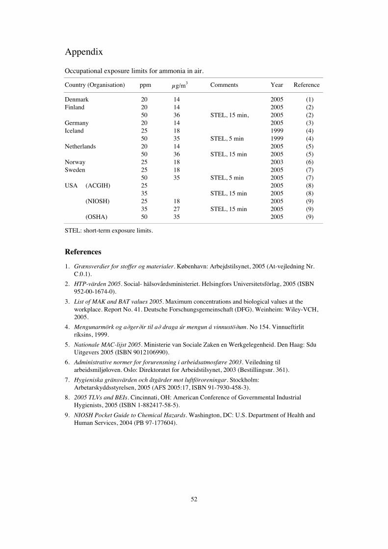
52
Appendix
Occupational exposure limits for ammonia in air.
Country (Organisation) ppm μg/m3 Comments Year Reference
Denmark 20 14 2005 (1)Finland 20 14 2005 (2)
50 36 STEL, 15 min, 2005 (2)Germany 20 14 2005 (3)Iceland 25 18 1999 (4)
50 35 STEL, 5 min 1999 (4)Netherlands 20 14 2005 (5)
50 36 STEL, 15 min 2005 (5)Norway 25 18 2003 (6)Sweden 25 18 2005 (7)
50 35 STEL, 5 min 2005 (7)USA (ACGIH) 25 2005 (8)
35 STEL, 15 min 2005 (8)(NIOSH) 25 18 2005 (9)
35 27 STEL, 15 min 2005 (9)(OSHA) 50 35 2005 (9)
STEL: short-term exposure limits.
References
1. Grænsverdier for stoffer og materialer. København: Arbejdstilsynet, 2005 (At-vejledning Nr.C.0.1).
2. HTP-värden 2005. Social- hälsovårdsministeriet. Helsingfors Universitetsförlag, 2005 (ISBN952-00-1674-0).
3. List of MAK and BAT values 2005. Maximum concentrations and biological values at theworkplace. Report No. 41. Deutsche Forschungsgemeinschaft (DFG). Weinheim: Wiley-VCH,2005.
4. Mengunarmörk og a∂ger∂ir til a∂ draga úr mengun á vinnustö∂um. No 154. Vinnueftirlitríksins, 1999.
5. Nationale MAC-lijst 2005. Ministerie van Sociale Zaken en Werkgelegenheid. Den Haag: SduUitgevers 2005 (ISBN 9012106990).
6. Administrative normer for forurensning i arbeidsatmosfære 2003. Veiledning tilarbeidsmiljøloven. Oslo: Direktoratet for Arbeidstilsynet, 2003 (Bestillingsnr. 361).
7. Hygieniska gränsvärden och åtgärder mot luftföroreningar. Stockholm:Arbetarskyddsstyrelsen, 2005 (AFS 2005:17, ISBN 91-7930-458-3).
8. 2005 TLVs and BEIs. Cincinnati, OH: American Conference of Governmental IndustrialHygienists, 2005 (ISBN 1-882417-58-5).
9. NIOSH Pocket Guide to Chemical Hazards. Washington, DC: U.S. Department of Health andHuman Services, 2004 (PB 97-177604).