Embed Size (px)
Citation preview

The nicotinic acetylcholine receptor: the founding father of the pentameric ligand-gated ion channel superfamily
by Jean-Pierre Changeux
Collège de France & Institut Pasteur, Paris, France
Abstract: a critical event in the history
of biological chemistry was the
chemical identification of the first
neurotransmitter receptor, the nicotinic
acetylcholine receptor. Disciplines as
diverse as electrophysiology,
pharmacology and biochemistry, joined
together in a unified and rational
manner with the common goal of
successfully identifying the molecular
device that converts a chemical signal
into an electrical one in the nervous
system. The nicotinic receptor has
become the founding father of a broad
family of pentameric membrane
receptors, paving the way for their
identification, including that of the
GABAA receptors.
It has been 42 years since the
isolation of the nicotinic acetylcholine
receptor from fish electric organ, the
first ligand–gated ion channel, and first
ion channel, ever identified ; 25 years
since the first GABA-A and glycine
receptor subunits were cloned and
sequenced and concomitantly their
homology with the nicotinic
acetylcholine receptors recognized ;
and 5 years since the discovery that
closely homologous ligand–gated ion
channels are present in prokaryotes
(1). In this review, I briefly retrace the
main steps in the discovery of the
nicotinic acetylcholine receptor, the
titular head of this receptor
superfamily.
The concept of receptor and the chemical identification of the acetylcholine receptor The English physiologist John
Newport Langley, working with
neuromuscular preparations proposed
in 1905 that muscle tissue possesses
«a substance that combines with
nicotine and curare…receives the
stimulus and transmits it». He called
the muscle entity the «receptive
substance». In the subsequent 50
years, the concept of pharmacological
receptors, inspired three main lines of
research. Firstly, the pharmacological
approach aimed at characterizing the
1
http://www.jbc.org/cgi/doi/10.1074/jbc.R112.407668The latest version is at JBC Papers in Press. Published on October 4, 2012 as Manuscript R112.407668
Copyright 2012 by The American Society for Biochemistry and Molecular Biology, Inc.
by guest on April 13, 2019
http://ww
w.jbc.org/
Dow
nloaded from

specificity of the receptor site by using
novel chemical ligands (eg the
distinction between nicotinic and
muscarinic acetylcholine receptors
(AChR) by sir Henry Dale) ; secondly,
the electro-physiological approach
exemplified by Bernard Katz and John
Eccles aimed at understanding the
ionic responses to endogenous
neurotransmitter signals; and thirdly,
the chemical tradition aimed at the
chemical identification of the receptor
molecule(s).
In the late 60’s, lipids,
polysaccharides, proteins, even nucleic
acids, were considered as potential
receptors. The early independant
efforts of Carlos Chagas, Eduardo de
Robertis, and David Nachmansohn to
identify the receptor for acetylcholine in
the electric organ of the fish
Electrophorus electricus were
abandoned because their tissue
extracts lacked specificity (2).
However, in the course of these
studies, Nachmansohn recognized the
extraordinarily rich content of nicotinic
synapses in the electric organ (2). He,
with Ernest Schoffeniels, devised a
method for preparing individual cells,
or electroplaques, from the electric
organ. This offered the opportunity to
investigate, simultaneously, the
electrophysiological, pharmacological
and altogether with the biochemical
characteristics of the response to ACh
within the same biological system (2).
At this time there were also
speculations that the enzyme
acetylcholinesterase (AChE) and the
physiological receptor site for ACh
could reside on the same protein
complex.
The introduction of new
biochemical methods radically
changed the field of receptor
identification. One such method is
affinity labeling which relies on the use
of compounds that are structural
homologs of the neurotransmitter and
also possess a highly reactive group.
This combination allows for specific
binding to the receptor site, and once
bound the probe covalently links to the
protein. For instance, the molecule, p-
trimethylammonium benzenediazonium
fluoroborate (TDF), carries a
trimethylammonium group (as does
ACh) as weIl as a reactive diazonium
group (3). As anticipated TDF
interacted covalently with E. electricus
electroplaque as an irreversible
competitive antagonist, and curare
protected against this covalent
attachment (4). The method was
subsequently improved upon with the
synthesis of 4-(N-maleido)-phenyl-
trimethylammonium iodide (MPTA)
2
by guest on April 13, 2019
http://ww
w.jbc.org/
Dow
nloaded from

whereby the diazonium is substituted
for by a maleimide group (5). The latter
selectively reacts with –SH groups
exposed by treating the electroplaque
membrane with dithiothreitol. However,
at this stage, both the method of tissue
preparation and the specificity of the
compounds used were insufficient to
allow for isolation of the receptor in its
active form from the electric organ.
A second method, that
significantly advanced the field was the
marked improvement of procedures
for fractionation and purification of
membrane fragments rich in AChE
from E. electricus electric organs.
Electron microscopic sections of these
membrane fragments revealed they
formed closed vesicles (6). Inspired by
the technique used with bacterial
permeases (7), it became possible to
measure radioactive Na+ (or K+) ion
fluxes with these microsacs by using a
simple filtration method (8). The
microsacs responded to nicotine
agonists with specificities closely
resembling those recorded by
electrophysiological methods
employing intact electroplaques. The
signal transduction by the
neurotransmitter could be reproduced
in a totally acellular system in the
absence of energy supply and in a
chemically defined environment. Thus,
it became possible to study in vitro the
chemistry of the physiological
response to ACh and of the signal
transduction mechanism involved
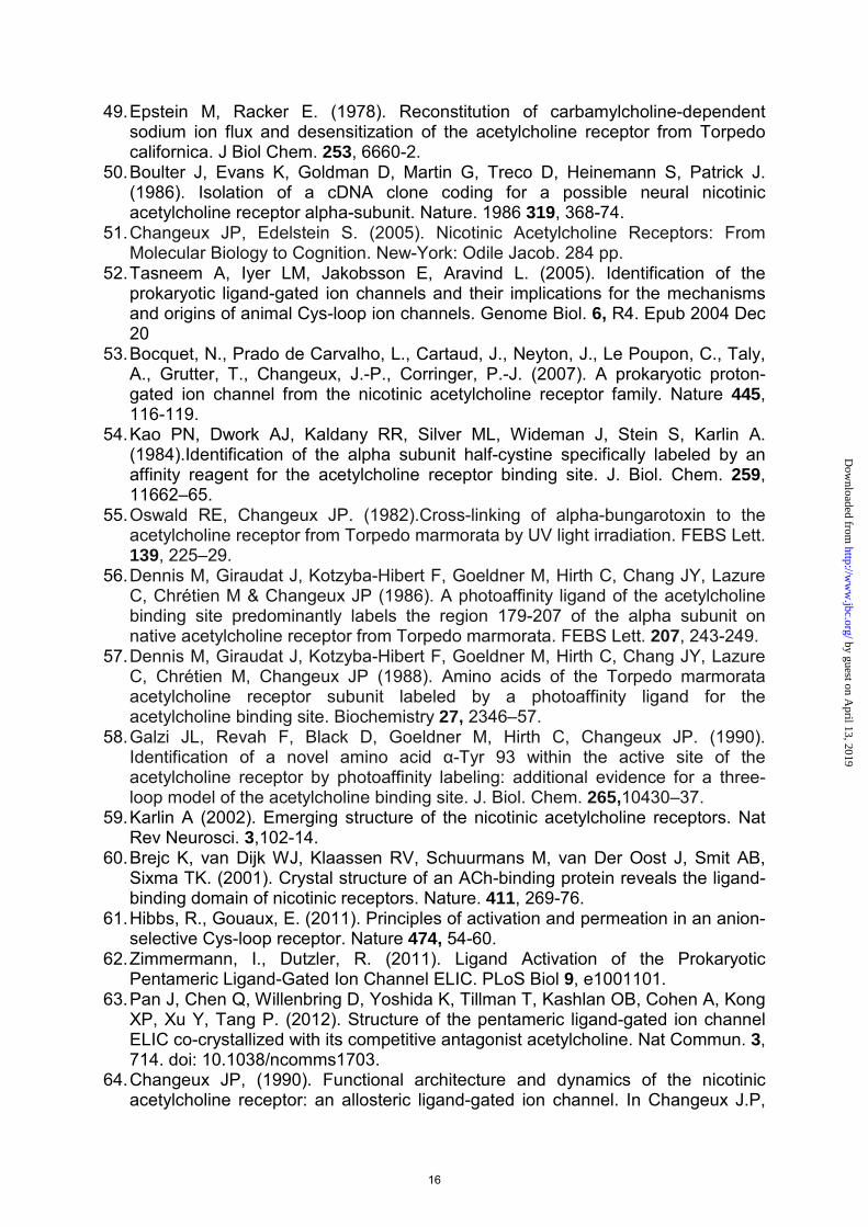
(8).The receptor molecule was
evidently present in the purified
membranes in a functional state. It was
possible now to follow reversible
binding to these purified membranes
using the nicotinic agonist
decamethonium as the radioactive
ligand (by the method of equilibrium
dialysis that Gilbert & Muller-Hill (9)
used to identify the lac-repressor) (Fig 1). The detergent deoxycholate gently
extracted the binding protein without
denaturing it and bound
decamethonium was displaced by
various nicotinic agonists and
antagonists including curare and
flaxedil in the order of their
physiological effects (10). Since then,
similar receptor-binding assays have
been used extensively to characterize
the GABAA and glycine receptors (this
volume).
Third Chen-Yuan Lee, a
Taiwanese pharmacologist, had found
that a snake venom toxin, α-
bungarotoxin (αBGT), specifically
blocks in vivo neuromuscular
transmission in high vertebrates at the
postsynaptic level without interacting
with AChE (11). Aware of Claude
3
by guest on April 13, 2019
http://ww
w.jbc.org/
Dow
nloaded from

Bernard’s lesson to use toxic
compounds as chemical lancets, I
asked Chen-Yuan Lee, who
unexpectedly visited me at the Pasteur
Institute, for a sample of the toxin. A
few days later, I received it and
immediatiely tried it in the three
systems just mentioned. The result
was remarkable : α-BGT blocked the
electroplaque’s electrical response in
vivo and the microsac’s ion-flux
response to nicotinic agonists in vitro ;
α-BGT blocked as well the binding of
radioactive decamethonium to the
detergent extract (Fig 1). This extract
contained a protein, sensitive to
pronase digestion, that bound nicotinic
agonists and the snake venom toxin in
a mutually exclusive manner. This
nicotinic receptor (nAChR) molecule
was shown to be a high molecular
weight hydrophobic protein that could
be physically separated from AChE
(12).
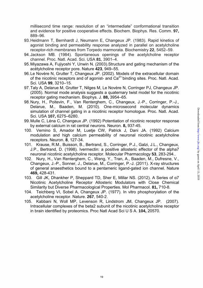
Then, an α-toxin from Naja
nigricollis, closely homologous to
α−BGT, was covalently coupled to
sepharose beads without losing its
binding activity. Mixing the toxin beads
with the membrane extract revealed
that 75–100% of the nAChR protein
bound to the toxin beads, whereas 85–
100% of AChE remained in the
supernatant. The data (13), confirmed
that AChE and the nAChR molecule
were distinct protein entities. These
studies also introduced Cuatrecasas’s
technique of affinity chromatography to
the nAChR field. Many groups then
became aware of these distinct
methods (14-16). We (17,18) and
others used alternative affinity columns
with immobilized quaternary
ammonium agonists or antagonists
(Fig 2) extending Miledi et al. (20) use
of radioactive I131–labeled α−BGT
(which, according to them, selectively
binds to the receptor in its resting
state).
Another rather simple
technological development, that,
retrospectively, had an important
impact on nAChR research was the
isolation of a novel generation of
excitable microsacs exceptionally rich
in nAChR (20–40% of total protein)
prepared from homogenates of T.
marmorata electric organ (21), a
finding was readily confirmed by other
groups. The nAChR-rich membranes
made the structural and functional
properties of the membrane-bound
nAChR accessible to a variety of
biochemical and biophysical methods,
such as purification in large quantities
(22), fluorescence spectroscopy (23),
electron spin resonance (24), and X-
4
by guest on April 13, 2019
http://ww
w.jbc.org/
Dow
nloaded from

ray diffraction (25).
Finally, the nAChR protein
purified from E. electricus and the
purified nAChR-rich membranes from
T. marmorata were examined by
electron microscopy and revealed ring-
like particles (8–9 nm in diameter) with
a hydrophilic core linked to a compact
bundle (26) (Fig 3). Made up of several
(5–6) subunits, they formed closely
packed two-dimensional assemblies in
T. marmorata postsynaptic membranes
(approximately 8–12 000 μm2) (26,27)
(Fig 3). These nAChR images were
the first ever of the structure of a
neurotransmitter receptor. They were
subsequently described in greater
details by Nigel Unwin (rev 28) and
others. Similar pictures later became
available for the GABAA and glycine
receptors (this volume).
The pentameric organization of the nicotinic receptor and the complete sequence of the subunits.
The amount of purified nAChR
was sufficient to identify the subunit
organisation of the protein. A first study
using partial cross-linking of the
purified E. electricus nAChR, revealed
5 well defined bands suggesting a
pentameric organization (29). The
pentameric organization was rapidly
confirmed by the teams of Karlin and
Raftery, who, in addition, discovered
that the nAChR molecule is composed
of four distinct types of subunits with
slight differences of molecular mass,
that assemble into an 2α.1β.1γ.1δ
heteropentamer (30-33).
Nothing was known about the
chemistry of the subunits. However
with the recently developed new
technology of high-resolution
microsequencing, aminoacid
sequences could now be determined
from small quantities of protein. The
sequence of 20-amino-acid comprising
the N-terminal domain of the α−subunit
of T. marmorata receptor was then
established in my laboratory (34). A
chemical identity card of the receptor
was made available, the first ever
established for a neurotransmitter
receptor. It was confirmed in the
Raftery laboratory with the α−subunit
of T. californica (35) and extended to
the N-terminal sequence of the four
subunits revealing a number of
sequence identities among the
subunits (36). Consistent with the
Monod-Wyman–Changeux (1965)
(MWC) model (37), the nAChR protein
was an authentic oligomer, but
pseudosymmetrical, with a fivefold axis
of rotation perpendicular to the plane of
the post synaptic membrane.
5
by guest on April 13, 2019
http://ww
w.jbc.org/
Dow
nloaded from

Knowledge of the initial
sequence data opened the nAChR
field to DNA-recombinant technologies.
The teams of Shosaku Numa (38-40),
Stephen Heinemann (41-42), Eric
Barnard (43), as weIl as by Anne
Devillers-Thiéry and Jérôme Giraudat
(44-45) in my laboratory, struggled to
clone the complementary DNAs of the
different subunits from electric organ
and muscle and to establish their
complete sequence. Experiments by
Eric Barnard and Riccardo Miledi had
demonstrated that messenger RNA
extracted from the electric organ of
Torpedo injected into Xenopus oocytes
led to the synthesis and incorporation
of functional acetylcholine receptors
into the membrane of the oocyte (46).
Injection of the 4 mRNAs transcribed
from the cloned cDNAs yielded
functional nAChRs (47) confirming
earlier biochemical experiments (48-
49) that assembly of the 4 types of
subunits suffice to recover a fully
operational nAChR.
Examination of the complete
cDNA sequences revealed several
common structural domains along the
sequences of the subunits that led to
the first model of transmembrane
organization of nAChR subunits (39,
40, 42, 45). It was proposed that the
long hydrophilic N-terminal segment,
four hydrophobic stretches, and short
hydrophilic segment, were organized
into an extracellular (synaptic) domain,
four transmembrane α-helices, and an
intracellular (cytoplasmic) domain. In
1987 closely homologous sequences
and the organisation of the subunits –
including a cys-loop- were found in
GABAA, glycine, 5HT3, GluCl and
neuronal nAChRs uncluding α7 and
α4β2 nAChRs (50, rev 51) thus
creating the superfamily of pentameric
receptors that is the subject of the
present volume. The recent discovery
of cationic orthologs in prokaryotes
(52-53) has extended recently the
superfamily plunging its evolutionary
origins back 3 billion years (1).
Identification of the acetylcholine-binding sites
The actual tridimensional
topology of the AChR protein and of
the various sites it carries still could not
be directly inferred from DNA-
recombinant technologies.
Identification of the amino-acids
composing the ACh-binding site and
the ion channel relied upon different
technologies. The previously
mentioned method of affinity labeling
proved to be useful at this stage. A first
result was obtained by Karlin’s group,
6
by guest on April 13, 2019
http://ww
w.jbc.org/
Dow
nloaded from

using MPTA (7) which labels the
sulfhydryl groups of the ACh-binding
site (see above). This led to the
identification of a pair of adjacent
cysteines (192–193), located in the N-
terminal domain of the α−subunit (54).
Despite these results, the
pharmacological specificity of the ACh
binding site remained unknown.
Our group demonstrated that
the snake (3H) α-toxin itself, without
additional modification, could be used
as a photolabel. UV irradiation of the
(3H) α-toxin-Torpedo receptor complex
not only resulted in the incorporation of
covalently bound radioactivity into the
α-subunit but also into the γ− and
δ−subunits (55). From this observation
it was concluded that the ACh-binding
sites were located at the interface
between subunits (55) and were
therefore non-equivalent. This was
confirmed in subsequent functional
studies.
The use of p-N,N-
dimethylammonium benzene
diazonium difluoroborate (DDF), an
affinity probe similar to TDF (5),
provided additional important
information (56). The dimethyl
ammonium group of DDF created a
resonant molecule that could be
photoactivated by energy transfer from
the protein. Indeed, eight amino acids
were found labeled by DDF, six of
them with an aromatic side chain, and
all of them located in the long
hydrophilic NH2 terminal domain of the
α-subunit. These amino acids were
distributed into three main loops,
forming a sort of electronegative
aromatic pocket in which the
quaternary ammonium group of
acetylcholine is lodged (56-58) thus
pointing to an analogy with the AChE
binding site where pi bonding is
exhibited as well. These three loops
located on the α-subunit side of the
binding site, are referred to as the
«principal component» were named A,
B, and C (58), a nomenclature that has
been adopted by the receptor
community. In agreement with the
snake (3H) α-toxin photolabeling data,
the affinity probe DDF labeled the γ-
and δ−subunits in addition to the
α−subunit (51, 59). The various groups
working on the receptor, including
those of Arthur Karlin, Jonathan Cohen
and ourselves, further documented this
notion and identified additional loops
D, E, and F on the non-α side of the
subunit interface (ref in 66, 67). These
loops form a «complementary»
component of the acetylcholine-binding
site on the γ− and δ−subunits. These
7
by guest on April 13, 2019
http://ww
w.jbc.org/
Dow
nloaded from

biochemical data were supported by
site-mutagenesis studies of the labeled
amino acids identified in these studies
(ref in 51, 59).
Confirmation of the binding site
organisation has come from the crystal
structure of a soluble snail protein that
binds ACh, the acetylcholine-binding
protein (AChBP), a close homolog of
the nAChR extracellular domain (60),
and of the full-length eukaryotic GluCl
receptor (61) and prokaryotic Erwinia
chrysanthemi receptor (ELIC) bound
with GABA (62) and ACh (as an
antagonist) (63) (rev 1).
Identification of the ion channel By the early 1980s, the
biochemical structure of any ion
channel was not known. The question
was how to chemically identify the
amino-acids that line the pore through
which ions flow. The quest (1974–
1999) proved to be long and difficult
(see 51,64). Pharmacological agents,
such as local anesthetics, known for
decades to block ion currents elicited
by nicotinic agonists, in an indirect,
noncompetitive, manner, proved to be
essential tools for chemical labeling the
channel. The first experiments,
performed with both E. electricus and
T. marmorata receptor-rich
membranes, demonstrated in vitro that,
at pharmacologicaly active
concentrations, the local anesthetics
do not directly displace nicotinic
ligands from the ACh-binding site but
reversibly bind to a different allosteric
site (65,66). One of these compounds,
chlorpromazine, displayed, in addition,
the remarkable property of covalently
linking to the receptor protein by simple
UV irradiation. In receptor-rich
membranes from T. marmorata
chlorpromazine labeled the four types
of subunits of the nAChR (67), and
precise quantitative measurements
demonstrated that it binds to just one
high-affinity site per 2α1.β1.γ1.δ1
oligomer (68). The kinetics of access of
chlorpromazine to this site increased
100-fold when rapidly mixed with ACh
under conditions expected to generate
functional ion channels (69-70). We
proposed that chlorpromazine binds to
a site located within the ion channel
along the pseudosymmetry axis that
becomes accessible to chlorpromazine
when the ion channel opens. The
conditions under which the channel
could be specifically labeled were thus
established.
It took more than a year to
demonstrate that chlorpromazine
labels serine 262, within the second
transmembrane segment (TM2) of the
δ−subunit (71) ; a finding rapidly
8
by guest on April 13, 2019
http://ww
w.jbc.org/
Dow
nloaded from

confirmed by another group using the
same protocol, but with a different
probe (72). Further identification of the
chlorpromazine-labeled amino acids on
the other subunits showed that the
serines not only form a ring (81), but
also revealed the adduct of other
amino acids (leucines and threonines)
located at a distance of three to four
amino acids on both sides of the ring of
serines (73). It was concluded that : (a)
the TM2 segments contribute to the
channel walls ; (b) these segments are
folded into an α-helix ; (c) the
chlorpromazine-binding site is located
at a near-equatorial position in the
channel’s pseudosymmetry axis ; and
(d) there exists a positive reciprocal
allosteric interaction between ACh and
the chlorpromazine-binding sites.
In parallel site-directed
mutagenesis experiments in which
single channel recordings were carried
out after reconstitution in Xenopus
oocytes, a region located in the
δ−subunit was shown to be responsible
for a conductance difference between
Torpedo and bovine channels that
comprises the putative transmembrane
segment TM2 and the adjacent bend
portion between segments TM2 and
TM3 (75). Subsequent analysis (76)
identified rings of negatively charged
glutamine residues, which were
classified as external, intermediate,
and cytoplasmic, that beautifully frame
the amino acid clusters labeled by
chlorpromazine, thus confirming their
proposed location within the ion path
(68-70). The teams of Henry Lester
and Norman Davidson reached a
similar conclusion (77).
Further studies, identified amino
acids which contribute to the ionic
selectivity of the channel (78-80). A
group of three residues was found to
drive the conversion of the cationic
selectivity of the ion channel into one
of anionic selectivity (79-80). For the
first time, an excitatory receptor could
be transformed into an inhibitory one.
This finding, as well as the converse
operation (from anionic to cationic) was
reproduced with other receptors:
GABAA, glycine, GluCl, and 5HT3 (51
and this volume). A functional chimera
was succesfully constructed that joined
the synaptic domain of α7-nAChR and
the transmembrane domain of 5HT3
receptor (81). Even combinations of
prokaryotic and eukaryotic receptor
domains were found functional (82).
This unambiguously demonstrates a
conservation of tertiary organization
between members of the receptor
superfamily. Lastly, the high resolution
X-ray data from prokaryotic ELIC and
GLIC (83-85) are consistent with the
9
by guest on April 13, 2019
http://ww
w.jbc.org/
Dow
nloaded from

biochemical data and EM structure (rev
28, 51) of the nAChR ion channel (1).
They demonstrate further that the
channel domain is topographically
distinct from the neurotransmitter-
binding domain and that the interaction
between the neurotransmitter and the
ion transport mechanism is an
allosteric interaction (1, 64, 86).
Allosteric transitions of the nicotinic receptor : the quaternary twist mechanism
Direct evidence for the
conformational changes that mediate
this interaction was still unavailable.
Early rapid mixing experiments using
snake 3H α−toxin as a probe and
receptor-rich membranes from T.
marmorata revealed changes of
conformation that took seconds to
reach a high-affinity state, possibly
desensitized, from a low-affinity resting
state (87). Consistent findings were
subsequently reported using muscle
cells (88) and Torpedo membranes
(89-90). A refined kinetic analysis of
the binding interaction of the
fluorescent nicotinic agonist, dansyl-
C6-choline with receptor-rich
membranes (91-92), and correlation
with the in vitro measurement of ion
transport through the ion channel (93),
resulted in the demonstration of
allosteric transitions between several
conformational states : a resting
closed-channel state stabilized by
snake α-toxin and nicotinic
antagonists, an active, transient, open-
channel state with low affinity for
acetylcholine and nicotinic agonists ;
and, at least one desensitized, slowly
accessible, refractory state, with a high
affinity for both agonists and
antagonists (Fig 4).
Moreover, under resting conditions a
sizeable fraction (approximately 20%)
of the receptor was found to be present
in the high-affinity, desensitized state,
and spontaneous channel openings of
the muscle nAChR were recorded in
the absence of ACh (94). This ruled
out the induced-fit mechanism to the
benefit of the conformational selection
(MWC) scheme (see 86). Still the
situation appeared more complex than
for regulatory enzymes. There exists
not only one but a cascade of discrete
transitions between open and closed
conformational states (see 1, 51) (Fig 4).
Up until recently, little new
information became available to help
explain the structural transitions of the
nAChR, except for in situ electron
microscopy studies of Torpedo
receptor (95). In silico modeling from
the available structural data brought
10
by guest on April 13, 2019
http://ww
w.jbc.org/
Dow
nloaded from

novel insight into the conformational
transitions of the receptor protein (96-
97). Normal mode analysis performed
on a 3D model of the α7 AChR gave a
breakdown of the protein movements
into discrete modes. Among the first 10
lowest frequency modes, the first mode
produced a structural reorganization
that caused a wide opening of the
channel pore resulting from a
concerted and symmetrical transition—
a quaternary twist motion of the
protein—with opposing rotations of the
upper (extracellular) and lower
(transmembrane) domains, and
significant tertiary reorganizations
within each subunit in particular at the
domain interface. The global
quaternary twist motion accounted
reasonably for the available
experimental data on the gating
process (97). Strong evidence
emerged from the comparison studies
of the X-ray structure of the prokayotic
receptors GLIC (from Gloeobacter
violaceus) that showed an open
channel conformation and ELIC which
displayed a closed channel (83-85).
Comparison of the two structures
indicated that, at least 29% of the
quaternary twist transition model
accounts for channel opening. Future
developments include the molecular
dynamics of the transition in the
microsec time scale (98).
Allosteric modulatory sites. The signal transduction process
mediated by nAChR is regulated by at
least three main categories of allosteric
« modulators » which bind to sites
distinct from the neurotranmitter site
and the ion channel. These modulators
are thought to selectively shift the
allosteric equilibrium in favor of either
an active (positive modulators) or a
resting/desensitized conformation
(negative modulators) without
competing with the neurotransmitter
binding to the orthosteric sites (64, 86
ref in 1).
One category of modulator is
Ca2+, which potentiates most neuronal
nAChRs (99-100), binds to the
extracellular domain below the ACh
site at residues contributed from both
sides of the subunit interface (95).
Another is Zn2+.
A second important category of
modulators that includes galantamine
bind at « non-agonist » interfaces
which, in hetero-pentameric nAChRs,
differ from the neurotransmitter binding
site, and appears to be homolog of the
benzodiazepines site on GABAA
receptors (see R Olsen this volume).
Another group of allosteric
modulators interact with the
11
by guest on April 13, 2019
http://ww
w.jbc.org/
Dow
nloaded from

transmembrane domain. The
antihelminthic ivermectin was originally
discovered to behave as a strong
positive modulator of α7 nAChR. Its
action was altered by mutations within
the transmembrane domain TM2 (101).
General anesthetics (both intravenous
and volatile) negatively modulate
excitatory nAChRs but positively
enhance inhibitory GABA receptors.
Photolabelling studies with GABAA
receptors (see R Olsen & J Cohen this
volume) and X–ray structures of GLIC
complexes with propofol or desfurane
reveal a site within the upper part of
the transmembrane domain of each
subunit (102) to which nicotinic
allosteric modulators may also interact
in neuronal nAChRs (103) (AM in Fig 4).
Allosteric modulatory sites have
also been identified in the cytoplasmic
loop that links TM3 and TM4 in all
eukaryotic (but not prokaryotic)
pentameric receptors. Including in
nAChRs several phosphorylation sites
(104) that control desensitization in
muscle and α7 nAChR and contribute
to endplate localization by agrin-
induced tyrosine phosphorylation of the
cytokeletal protein 43K-rapsyn (22, ref
51). The cytoplasmic domain of the
α4 nAChR subunit also binds a variety
of scaffold protein that interacts with
cytoskeletal proteins, and with G
protein systems that are involved in
intracellular signalling pathways (105).
Conclusion Since the isolation of the nAChR and
the discovery that GABA-A and glycine
receptor subunits are close orthologs
of the nAChR thereby founding the
superfamily of pentameric ligand-gated
ion channels, the whole field of
pentameric receptors for
neurotransmitter has blossomed,
including the discovery of homologous
receptors in prokaryotes. Several of
them are the target of most commonly
used drugs such as benzodiazepines,
barbiturates, curare and general
anesthetics. The recent advances in
the X-ray structure of several of these
receptors (1) open new avenues for
the rational design of pharmacological
agents acting on the brain, in parallel
with the abundant studies on the
GPCRs which were initiated later.
Acknowledgements. JPC gratefully thanks the Woods Hole Marine Biogical Laboratory where a significant part of the review was written and Leonard Warren & Albert Grossman for carefully editing the manuscript. JPC wishes to apologize for omitting important papers due to limitation in reference number.
12
by guest on April 13, 2019
http://ww
w.jbc.org/
Dow
nloaded from
References. 1. Corringer PJ, Poitevin F, Prevost MS, Sauguet L, Delarue M, Changeux JP.
(2012) Structure and pharmacology of pentameric receptor channels: from bacteria to brain. Structure. 20, 941-56.
2. Nachmansohn D. (1959) The Chemical and Molecular Basis of Nerve activity. New-York: Academic Press..
3. Fenton JW 2nd, Singer SJ. (1965) Affinity labeling of antibodies to the p-azophenyltrimethylammonium hapten and a structural relationship among antibody active sites of different specificities. Biochem Biophys Res Commun. 20, 315-20.
4. Changeux JP, Podleski TR, Wofsy L. (1967) Affinity labeling of the acetylcholine-receptor. Proc. Natl.Acad. Sci. USA 58, 2063–70.
5. Karlin A, Winnik M. 1968. Reduction and specific alkylation of the receptor for acetylcholine. Proc.Natl. Acad. Sci. USA 60, 668–74.
6. Changeux JP, Gautron J, Israël M, Podleski T. (1969) [Separation of excitable membranes from the electric organ of Electrophorus electricus] C R Acad Sci Hebd Seances Acad Sci D. 269,1788-91.
7. Cohen GN, Monod J. (1957) Bacterial Permeases. Bacteriol Rev. 21,169-94. 8. Kasai M, Changeux JP. (1970) [Demonstration of the excitation by cholinergic
agonists from fractions of purified membranes, in vitro]. C R Acad Sci Hebd Seances Acad Sci D. 270,1400-3; Kasai M, Changeux JP. (1971) In vitro excitation of purified membrane fragments by cholinergic agonists. J. Memb. Biol. 6,1–23; 4–57; 58–80; 81–88.
9. Gilbert W, Müller-Hill B.(1966) Isolation of the lac repressor. Proc. Natl. Acad. Sci. USA 56,1891–98.
10. Changeux JP, Kasai M, Huchet M, Meunier JC. 1970. (Extraction from electric tissue of Electrophorus of a protein presenting several typical properties characteristic of the physiological receptor of acetylcholine). C. R. Acad. Sci. Hebd. Séances Acad. Sci. D 270, 2864–67
11. Chang CC, Lee CY. (1963) Isolation of neurotoxins from the venom of Bungarus multicinctus and their modes of neuromuscular blocking action. Arch. Int. Pharmacodyn. 144, 316-22.
12. Changeux JP, Kasai M, Lee CY. (1970). Use of a snake venom toxin to characterize the cholinergic receptor protein. Proc. Natl. Acad. Sci. USA 67, 1241–47.
13. Meunier JC, Huchet M, Boquet P, Changeux JP. (1971). (Separation of the receptor protein of acetylcholine and acetylcholinesterase). C. R. Acad. Sci. Hebd. Séances Acad. Sci. D 272,117–20
14. Karlsson E, Heilbronn E Widlund L. (1972). Isolation of the nicotinic acetylcholine receptor by biospecific chromatography on insolubilized Naja naja neurotoxin. FEBS Lett. 28,107–11.
15. Klett RP, Fulpius BW, Cooper D, Smith M, Reich E, Possani LD. (1973).The acetylcholine receptor.I. Purification and characterization of a macromolecule isolated from Electrophorus electricus. J. Biol. Chem. 248, 6841–53.
16. Lindstrom J, Patrick J. (1974). Purification of the acetylcholine receptor by affinity chromatography. In Synaptic Transmission and Neuronal Interaction, ed. MVL Bennett, pp. 191–216. New York: Raven Press
13
by guest on April 13, 2019
http://ww
w.jbc.org/
Dow
nloaded from
17. Olsen R, Meunier JC, Changeux JP. (1972). Progress in purification of the cholinergic receptor protein from Electrophorus electricus by affinity chromatography. FEBS Lett. 28, 96–100
18. Meunier JC, Sealock R, Olsen R, Changeux JP. (1974). Purification and properties of the cholinergic receptor protein from Electrophorus electricus electroplax. Eur. J. Biochem. 45, 371–94
19. Schmidt J, Raftery MA. (1972). Use of affinity chromatography for acetylcholine receptor purification. Biochem. Biophys. Res. Commun. 49, 572–78
20. Miledi R, Molinoff P, Potter LT. (1971). Isolation of the cholinergic receptor protein of Torpedo electric tissue. Nature 229, 554–57
21. Cohen JB, Weber M, Huchet M, Changeux JP. (1972). Purification from Torpedo marmorata electric tissue of membrane fragments particularly rich in cholinergic receptor. FEBS. Lett. 26, 43–47
22. Sobel A, Weber M, Changeux JP. (1977). Large-scale purification of the acetylcholine-receptor protein in its membrane-bound and detergent-extracted forms from Torpedo marmorata electric organ. Eur. J. Biochem. 80, 215–24
23. Cohen JB, Changeux JP. (1973). Interaction of a fluorescent ligand with membrane-bound cholinergic receptor from Torpedo marmorata. Biochemistry 12, 4855–64
24. Brisson AD, Scandella CJ, Bienvenue A, Devaux P, Cohen JB, Changeux JP. (1975).Interaction of a spin-labeled long chain acylcholine with the cholinergic receptor protein in its membrane environment. Proc. Natl. Acad. Sci. USA 72,1087–91
25. Dupont Y, Cohen JB, Changeux JP. (1974). X-ray diffraction study of membrane fragments rich in acetyl-choline receptor protein prepared from the electric organ of Torpedo marmorata. FEBS Lett. 40,130–33
26. Cartaud J, Benedetti L, Cohen JB, Meunier JC, Changeux JP. (1973). Presence of a lattice structure in membrane fragments rich in nicotinic receptor protein from the electric organ of Torpedo marmorata. FEBS Lett. 33,109–13.
27. Nickel E, Potter LT. (1973) Ultrastructure of isolated membranes of Torpedo electric tissue. Brain Res. 57, 508-17.
28. Unwin N. (2005) Refined structure of the nicotinic acetylcholine receptor at 4A resolution. J Mol Biol. 346, 967-89.
29. Hucho F, Changeux JP. 1973. Molecular weight and quaternary structure of the cholinergic receptor protein extracted by detergents from Electrophorus electricus electric tissue. FEBS Lett. 38,11–15.
30. Weill CL, McNamee MG, Karlin A. 1974. Affinity-labeling of purified acetylcholine receptor from Torpedo californica. Biochem. Biophys. Res. Commun. 61, 997–1003.
31. Raftery MA, Vandlen R, Michaelson D, Bode J, Moody T, Chao Y, Reed K, Deutsch J, Duguid J. (1974) The biochemistry of an acetylcholine receptor. J Supramol Struct. 2, 582-92.
32. Lindstrom J, Walter B, Einarson B. (1979) Immunochemical similarities between subunits of acetylcholine receptors from Torpedo, Electrophorus, and mammalian muscle. Biochemistry. 18, 4470-80.
33. Saitoh T, Oswald R, Wennogle LP, Changeux JP. (1980) Conditions for the selective labelling of the 66 000 dalton chain of the acetylcholine receptor by the covalent non-competitive blocker 5-azido-[3H]-trimethisoquin. FEBS Lett. 116, 30-36.
14
by guest on April 13, 2019
http://ww
w.jbc.org/
Dow
nloaded from
34. Devillers-Thiery A, Changeux JP, Paroutaud P, Strosberg AD. (1979). The amino-terminal sequence of the 40000 molecular weight subunit of the acetylcholine receptor protein from Torpedo marmorata. FEBS Lett. 104, 99–105
35. Hunkapiller MW, Strader CD, Hood L, Raftery MA.(1979) Amino terminal amino acid sequence of the major polypeptide subunit of Torpedo californica acetylcholine receptor. Biochem Biophys Res Commun. 91,164-9.
36. Raftery MA, Hunkapiller MW, Strader CD, Hood LE. (1980). Acetylcholine receptor: complex of homologous subunits. Science 208,1454–56
37. Monod J, Wyman J, Changeux JP. (1965). On the nature of allosteric transitions: a plausible model. J. Mol. Biol. 12, 88–118
38. Noda M, Takahashi H, Tanabe T, Toyosato M, Furutani Y, Hirose T, Asai M, Inayama S, Miyata T, Numa S. (1982). Primary structure of alpha-subunit precursor of Torpedo californica acetylcholine receptor deduced from cDNA sequence. Nature 299, 793–97
39. Noda M, Takahashi H, Tanabe T, Toyosato M, Kikyotani S, Hirose T, Asai M, Takashima H, Inayama S, Miyata T, Numa S. (1983a). Primary structures of beta- and delta-subunit precursors of Torpedo californica acetylcholine receptor deduced from cDNA sequences. Nature. 301, 251-5.
40. Noda M, Takahashi H, Tanabe T, Toyosato M, Kikyotani S, Furutani Y, Hirose T, Takashima H, Inayama Inayama S, Miyata T, Numa S. (1983b). Structural homology of Torpedo californica acetylcholine receptor subunits.Nature. 302, 528-32.
41. Ballivet M, Patrick J, Lee J, Heinemann S. (1982). Molecular cloning of cDNA coding for the gamma subunit of Torpedo acetylcholine receptor. Proc. Natl. Acad. Sci. USA 79, 4466–70,
42. Claudio T, Ballivet M, Patrick J, Heinemann S. (1983). Nucleotide and deduced amino acid sequences of Torpedo californica acetylcholine receptor gamma subunit.Proc Natl Acad Sci U S 80,1111-5.
43. Sumikawa K, Houghton M, Smith JC, Bell L, Richards BM, Barnard EA. (1982). The molecular cloning and characterisation of cDNA coding for the alpha subunit of the acetylcholine receptor. Nucleic Acids Res. 10, 5809-22.
44. Giraudat J, Devillers-Thiéry A, Auffray C, Rougeon F, Changeux JP. (1982). Identification of a cDNA clone coding for the acetylcholine binding subunit of Torpedo marmorata acetylcholine receptor. EMBO J. 1,713–1751.
45. DevillersThiéry A, Giraudat J, Bentaboulet M, Changeux JP. (1983).Complete mRNA coding sequence of the acetylcholine binding alpha subunit of Torpedo marmorata acetylcholine receptor: a model for the transmembrane organization of the polypeptide chain. Proc. Natl. Acad. Sci. USA 80, 2067–71
46. Barnard EA, Miledi R, Sumikawa K. (1982). Translation of exogenous messenger RNA coding for nicotinic acetylcholine receptors produces functional receptors in Xenopus oocytes. Proc R Soc Lond B Biol Sci. 215, 241-6.
47. Mishina M, Kurosaki T, Tobimatsu T, Morimoto Y, Noda M, Yamamoto T, Terao M, Lindstrom J, Takahashi T, Kuno M, Numa S. (1984). Expression of functional acetylcholine receptor from cloned cDNAs. Nature. 307, 604-8
48. Hazelbauer GL, Changeux JP. (1974). Reconstitution of a chemically excitable membrane. Proc Natl Acad Sci U S A. 71,1479-83.
15
by guest on April 13, 2019
http://ww
w.jbc.org/
Dow
nloaded from
49. Epstein M, Racker E. (1978). Reconstitution of carbamylcholine-dependent sodium ion flux and desensitization of the acetylcholine receptor from Torpedo californica. J Biol Chem. 253, 6660-2.
50. Boulter J, Evans K, Goldman D, Martin G, Treco D, Heinemann S, Patrick J. (1986). Isolation of a cDNA clone coding for a possible neural nicotinic acetylcholine receptor alpha-subunit. Nature. 1986 319, 368-74.
51. Changeux JP, Edelstein S. (2005). Nicotinic Acetylcholine Receptors: From Molecular Biology to Cognition. New-York: Odile Jacob. 284 pp.
52. Tasneem A, Iyer LM, Jakobsson E, Aravind L. (2005). Identification of the prokaryotic ligand-gated ion channels and their implications for the mechanisms and origins of animal Cys-loop ion channels. Genome Biol. 6, R4. Epub 2004 Dec 20
53. Bocquet, N., Prado de Carvalho, L., Cartaud, J., Neyton, J., Le Poupon, C., Taly, A., Grutter, T., Changeux, J.-P., Corringer, P.-J. (2007). A prokaryotic proton-gated ion channel from the nicotinic acetylcholine receptor family. Nature 445, 116-119.
54. Kao PN, Dwork AJ, Kaldany RR, Silver ML, Wideman J, Stein S, Karlin A. (1984).Identification of the alpha subunit half-cystine specifically labeled by an affinity reagent for the acetylcholine receptor binding site. J. Biol. Chem. 259, 11662–65.
55. Oswald RE, Changeux JP. (1982).Cross-linking of alpha-bungarotoxin to the acetylcholine receptor from Torpedo marmorata by UV light irradiation. FEBS Lett. 139, 225–29.
56. Dennis M, Giraudat J, Kotzyba-Hibert F, Goeldner M, Hirth C, Chang JY, Lazure C, Chrétien M & Changeux JP (1986). A photoaffinity ligand of the acetylcholine binding site predominantly labels the region 179-207 of the alpha subunit on native acetylcholine receptor from Torpedo marmorata. FEBS Lett. 207, 243-249.
57. Dennis M, Giraudat J, Kotzyba-Hibert F, Goeldner M, Hirth C, Chang JY, Lazure C, Chrétien M, Changeux JP (1988). Amino acids of the Torpedo marmorata acetylcholine receptor subunit labeled by a photoaffinity ligand for the acetylcholine binding site. Biochemistry 27, 2346–57.
58. Galzi JL, Revah F, Black D, Goeldner M, Hirth C, Changeux JP. (1990). Identification of a novel amino acid α-Tyr 93 within the active site of the acetylcholine receptor by photoaffinity labeling: additional evidence for a three-loop model of the acetylcholine binding site. J. Biol. Chem. 265,10430–37.
59. Karlin A (2002). Emerging structure of the nicotinic acetylcholine receptors. Nat Rev Neurosci. 3,102-14.
60. Brejc K, van Dijk WJ, Klaassen RV, Schuurmans M, van Der Oost J, Smit AB, Sixma TK. (2001). Crystal structure of an ACh-binding protein reveals the ligand-binding domain of nicotinic receptors. Nature. 411, 269-76.
61. Hibbs, R., Gouaux, E. (2011). Principles of activation and permeation in an anion-selective Cys-loop receptor. Nature 474, 54-60.
62. Zimmermann, I., Dutzler, R. (2011). Ligand Activation of the Prokaryotic Pentameric Ligand-Gated Ion Channel ELIC. PLoS Biol 9, e1001101.
63. Pan J, Chen Q, Willenbring D, Yoshida K, Tillman T, Kashlan OB, Cohen A, Kong XP, Xu Y, Tang P. (2012). Structure of the pentameric ligand-gated ion channel ELIC co-crystallized with its competitive antagonist acetylcholine. Nat Commun. 3, 714. doi: 10.1038/ncomms1703.
64. Changeux JP, (1990). Functional architecture and dynamics of the nicotinic acetylcholine receptor: an allosteric ligand-gated ion channel. In Changeux J.P,
16
by guest on April 13, 2019
http://ww
w.jbc.org/
Dow
nloaded from

LLinas RR, D.Purves, Bloom F, eds Fidia Research Foundation Neuroscience Award Lectures. 4, 17-168. New-York: Raven press.
65. Weber M, Changeux JP. (1974).Binding of Naja nigricollis 3H-alpha-toxin to membrane fragments from Electrophorus and Torpedo electric organs. 3. Effect of local anaesthetics on the binding of the tritiated-neurotoxin. Mol. Pharmacol. 10, 35–40.
66. Cohen JB, Weber M, Changeux JP. (1974). Effects of local anesthetics and calcium on the interaction of cholinergic ligands with the nicotinic receptor protein from Torpedo marmorata. Mol. Pharmacol.10, 904–32.
67. Oswald RE, Changeux JP. (1981). Ultraviolet light-induced labeling by non competitive blockers of the acetylcholine receptor from Torpedo marmorata. Proc. Natl. Acad. Sci. USA 78, 3925–29.
68. Heidmann T, Oswald R, Changeux JP. (1982). (The high affinity binding site for chlorpromazine is present only as a single copy per cholinergic receptor molecule and is shared by four polypeptide chains.) C. R. Séances Acad. Sci. III 295, 345–49.
69. Heidmann T, Changeux JP. (1984). Time-resolved photolabeling by the noncompetitive blocker chlor- promazine of the acetylcholine receptor in its transiently open and closed ion channel conformations. Proc. Natl. Acad. Sci. USA 81,1897–901.
70. Heidmann T, Changeux JP. (1986). Characterization of the transient agonist-triggered state of the acetylcholine receptor rapidly labeled by the noncompetitive blocker [3H]-chlorpromazine: additional evidence for the open channel conformation. Biochemistry 25, 6109–13.
71. Giraudat J, Dennis M, Heidmann T, Chang JY, Changeux JP. (1986). Structure of the high affinity site for noncompetitive blockers of the acetylcholine receptor: serine-262 of the delta subunit is labeled by [3H]-chlorpromazine. Proc. Natl. Acad. Sci. USA 83, 2719–23.
72. Oberthür W, Muhn P, Baumann H, Lottspeich F, Wittmann-Liebold B, Hucho F.(1986).The reaction site of a non competitive antagonist in the delta subunit of the nicotinic acetylcholine receptor. EMBO J. 5,1815–19.
73. Hucho F, Oberthür W, Lottspeich F. (1986). The ion channel of the nicotinic acetylcholine receptor is formed by the homologous helices M II of the receptor subunits. FEBS Lett. 205,137-42.
74. Giraudat J, Dennis M, Heidmann T, Haumont PY, Lederer F, Changeux JP. (1987).Structure of the high-affinity binding site for non competitive blockers of the acetylcholine receptor: [3H] chlorpromazine labels homologous residues in the beta and delta chains. Biochemistry 26, 2410–18.
75. Imoto K, Methfessel C, Sakmann B, Mishina M, Mori Y Konno T, Fukuda K, Kurasaki M, Bujo H, Fujita Y, Numa S. (1986).Location of a delta-subunit region determining ion transport through the acetylcholine receptor channel. Nature 324, 670–74.
76. Imoto K, Busch C, Sakmann B, Mishina M, Konno T, Nakai J, Bujo H, Mori Y, Fukuda K, Numa S. (1988). Rings of negatively charged amino acids determine the acetylcholine receptor channel conductance. Nature 335, 645–48.
77. Leonard RJ, Labarca CG, Charnet P, Davidson N, Lester HA. (1988).Evidence that the M2-membrane-spanning region lines the ion channel pore of the nicotinic receptor. Science 242,1578–81.
78. Villarroel, A., Herlitze, S., Koenen, M., Sakmann, B. (1991). Location of a threonine residue in the alpha-subunit M2-transmembrane segment that
17
by guest on April 13, 2019
http://ww
w.jbc.org/
Dow
nloaded from

determines the ion flow through the acetylcholine receptor channel. Proc. Biol. Sci. 243, 69–74.
79. Galzi JL, Devillers-Thiery A, Hussy N, Bertrand S, Changeux JP, Bertrand D. (1992). Mutations in the ion channel domain of a neuronal nicotinic receptor convert ion selectivity from cationic to anionic. Nature 359, 500–5.
80. Corringer PJ, Bertrand S, Galzi JL, Devillers-Thiéry A, Changeux JP, Bertrand D. (1999). Mutational analysis of the charge selectivity filter of the alpha7-nicotinic acetylcholine receptor. Neuron 22, 831–43.
81. Eiselé JL, Bertrand S, Galzi JL, Devillers-Thiéry A, Changeux JP, Bertrand D. (1993). Chimeric nicotinic-serotonergic receptor combines distinct ligand binding and channel specificities. Nature 366, 479–83.
82. Duret, G, Van Renterghem, C, Weng, Y, Prevost, M, Moraga-Cid, G, Huon, C, Sonner, JM, Corringer, P.-J. (2011). Functional prokaryotic-eukaryotic chimera from the pentameric ligand-gated ion channel family. Proc. Natl. Acad. Sci. USA 108, 12143–48.
83. Bocquet, N., Nury, H., Baaden, M., Le Poupon, C., Changeux, J.-P., Delarue, M., and Corringer, P.-J. (2009). X-ray structure of a pentameric ligand-gated ion channel in an apparently open conformation. Nature 457, 111–114.
84. Hilf, R., and Dutzler, R. (2008). X-ray structure of a prokaryotic pentameric ligand-gated ion channel. Nature 452, 375-379.
85. Hilf, R., and Dutzler, R. (2009). Structure of a potentially open state of a proton-activated pentameric ligand-gated ion channel. Nature 457, 115-118.
86. Changeux JP. (2012). Allostery and the Monod-Wyman-Changeux model after 50 years. Annu Rev Biophys. 41, 103-33.
87. Weber M, David-Pfeuty MT, Changeux JP. (1975). Regulation of binding properties of the nicotinic receptor protein by cholinergic ligands in membrane fragments from Torpedo marmorata. Proc. Natl. Acad. Sci. USA 72, 3443–47.
88. Colquhoun D, Rang HP. (1976). Effects of inhibitors of the binding of iodinated alpha-bungarotoxin to acetylcholine receptors in rat muscle. Mol. Pharmacol. 12, 519–35.
89. Grünhagen HH, Changeux JP. (1976). Studies on the electrogenic action of acétylcholine with Torpedo marmorata electric organ. IV. Quinacrine: a fluorescent probe for the conformational transitions of the cholinergic receptor protein in its membrane bound state. J. Mol. Biol. 106, 497–516. V. Qualitative correlation between pharmacological effects and equilibration processes of the cholinergic receptor protein as revealed by the structural probe quinacrine. J. Mol. Biol. 106, 517–35 ; Grünhagen HH, Iwatsubo M, Changeux JP. (1977). Fast kinetic studies on the interaction of cholinergic agonists with the membrane-bound acetylcholine receptor from Torpedo marmorata as revealed by quinacrine fluorescence. Eur. J. Biochem. 80, 225–42.
90. Weiland G, Georgia B, Lappi S, Chignell CF, Taylor P. (1977). Kinetics of agonist-mediated transitions in state of the cholinergic receptor. J. Biol. Chem. 252, 7648–56.
91. Heidmann T, Changeux JP. (1979). Fast kinetic studies on the interaction of a fluorescent agonist with the membrane-bound acetylcholine receptor from Torpedo marmorata. Eur. J. Biochem. 94:255–79; Id. Fast kinetic studies on the allosteric interactions between acetylcholine receptor and local anesthetic binding sites. Eur. J. Biochem. 94, 281–96.
92. Heidmann T, Changeux JP. (1980).Interaction of a fluorescent agonist with the membrane-bound acetyl-choline receptor from Torpedo marmorata in the
18
by guest on April 13, 2019
http://ww
w.jbc.org/
Dow
nloaded from
millisecond time range: resolution of an “intermediate” conformational transition and evidence for positive cooperative effects. Biochem. Biophys. Res. Comm. 97, 889–96.
93. Heidmann T, Bernhardt J, Neumann E, Changeux JP. (1983). Rapid kinetics of agonist binding and permeability response analyzed in parallel on acetylcholine receptor-rich membranes from Torpedo marmorata. Biochemistry 22, 5452–59.
94. Jackson MB. (1984). Spontaneous openings of the acetylcholine receptor channel. Proc. Natl. Acad. Sci. USA 81, 3901–4.
95. Miyazawa A, Fujiyoshi Y, Unwin N. (2003).Structure and gating mechanism of the acétylcholine receptor pore. Nature 423, 949–55.
96. Le Novère N, Grutter T, Changeux JP. (2002). Models of the extracellular domain of the nicotinic receptors and of agonist- and Ca2+-binding sites. Proc. Natl. Acad. Sci. USA 99, 3210–15.
97. Taly A, Delarue M, Grutter T, Nilges M, Le Novère N, Corringer PJ, Changeux JP. (2005). Normal mode analysis suggests a quaternary twist model for the nicotinic receptor gating mechanism. Biophys. J. 88, 3954–65.
98. Nury, H., Poitevin, F., Van Renterghem, C., Changeux, J.-P., Corringer, P.-J., Delarue, M., Baaden, M. (2010). One-microsecond molecular dynamics simulation of channel gating in a nicotinic receptor homologue. Proc. Natl. Acad. Sci. USA 107, 6275–6280.
99. Mulle C, Léna C, Changeux JP. (1992) Potentiation of nicotinic receptor response by external calcium in rat central neurons. Neuron. 8, 937-45.
100. Vernino S, Amador M, Luetje CW, Patrick J, Dani JA. (1992) Calcium modulation and high calcium permeability of neuronal nicotinic acetylcholine receptors. Neuron. 8, 127-34.
101. Krause, R.M., Buisson, B., Bertrand, S., Corringer, P.J., Galzi, J.L., Changeux, J.P., Bertrand, D. (1998). Ivermectin: a positive allosteric effector of the alpha7 neuronal nicotinic acetylcholine receptor. Molecular Pharmacology 53, 283-294..
102. Nury, H., Van Renterghem, C., Weng, Y., Tran, A., Baaden, M., Dufresne, V., Changeux, J.-P., Sonner, J., Delarue, M., Corringer, P.-J. (2011). X-ray structures of general anaesthetics bound to a pentameric ligand-gated ion channel. Nature 469, 428-431.
103. Gill JK, Dhankher P, Sheppard TD, Sher E, Millar NS. (2012). A Series of α7 Nicotinic Acetylcholine Receptor Allosteric Modulators with Close Chemical Similarity but Diverse Pharmacological Properties. Mol Pharmacol. 81, 710-8.
104. Teichberg VI, Sobel A, Changeux JP. (1977). In vitro phosphorylation of the acetylcholine receptor. Nature. 267, 540-2.
105. Kabbani N, Woll MP, Levenson R, Lindstrom JM, Changeux JP. (2007). Intracellular complexes of the beta2 subunit of the nicotinic acetylcholine receptor in brain identified by proteomics. Proc Natl Acad Sci U S A. 104, 20570.
19
by guest on April 13, 2019
http://ww
w.jbc.org/
Dow
nloaded from
Figure legends. Figure 1. Top : Binding method by equilbrium dialysis used for the identification of the nicotinic receptor. Bottom : effect of the snake toxin α-bungarotoxin on the nicotinic agonist 3H decamethonium binding (from Changeux et al 1970 (12)).
by guest on April 13, 2019
http://ww
w.jbc.org/
Dow
nloaded from
Figure 2. Purification of the nicotinic acetylcholine receptor by affinity chromatography (from Olsen et al 1972 (17)).
by guest on April 13, 2019
http://ww
w.jbc.org/
Dow
nloaded from
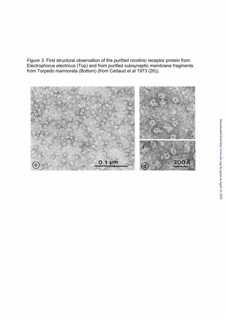
Figure 3. First structural observation of the purified nicotinic receptor protein from Electrophorus electricus (Top) and from purified subsynaptic membrane fragments from Torpedo marmorata (Bottom) (from Cartaud et al 1973 (26)).
by guest on April 13, 2019
http://ww
w.jbc.org/
Dow
nloaded from

Figure 4. Minimal four states model for the allosteric transitions of the nicotinic receptor (from Changeux 1990 (64))
ACh, acetylcholine, CB competitive (orthosteric) blocker, NCB non-competitive (channel) blocker, AM allosteric modulator, P, phosphorylation site.
by guest on April 13, 2019
http://ww
w.jbc.org/
Dow
nloaded from
Jean-Pierre Changeuxligand-gated ion channel superfamily
The nicotinic acetylcholine receptor: the founding father of the pentameric
published online October 4, 2012J. Biol. Chem.
10.1074/jbc.R112.407668Access the most updated version of this article at doi:
Alerts:
When a correction for this article is posted•
When this article is cited•
to choose from all of JBC's e-mail alertsClick here
http://www.jbc.org/content/suppl/2012/11/22/R112.407668.DCAuthor_profileRead an Author Profile for this article at
by guest on April 13, 2019
http://ww
w.jbc.org/
Dow
nloaded from